expression of the proprotein convertases pc1 and pc2 mrnas in thyrotropin releasing hormone neurons...
TRANSCRIPT
Ž .Brain Research 761 1997 77–86
Research report
Expression of the proprotein convertases PC1 and PC2 mRNAs inthyrotropin releasing hormone neurons of the rat paraventricular nucleus of
hypothalamus
Edith Sanchez a, Jean-Louis Charli a,), Claudia Morales a, Gabriel Corkidi b, Nabil G. Seidah c,´Patricia Joseph-Bravo a, Rosa Maria Uribe a
a Departamento de Genetica y Fisiologıa Molecular, Instituto de Biotecnologıa, UniÕersidad Nacional Autonoma de Mexico, CuernaÕaca, Mor., A.P.´ ´ ´ ´ ´510-3, Mexico 62271
b Centro de Instrumentos, UniÕersidad Nacional Autonoma de Mexico, Mexico D.F., Mexico´ ´c J.A. DeSeÕe Laboratories of Biochemical Neuroendocrinology, Clinical Research Institute of Montreal, Montreal, Que., Canada
Accepted 25 February 1997
Abstract
Ž . Ž .PC1 and PC2 are subtilisin-like processing enzymes capable of cleaving thyrotropin releasing hormone TRH precursor pro-TRH atŽ .paired basic residues in vitro. In the paraventricular nucleus of the hypothalamus PVN , pro-TRH is synthesized to control
adenohypophysial thyrotropin and prolactin release. Biochemical and immunological approaches have shown that in the hypothalamus,Ž .pro-TRH is extensively cleaved at pairs of basic amino acids. We quantified, by two different approaches, in situ hybridization ISH on
consecutive cryostat sections or double label ISH, the proportion of PVN TRH neurons containing either PC1 or PC2 mRNAs. Bothtechniques gave similar results: PC2 mRNA was present in 60–70% of TRH neurons, and PC1 mRNA in 37–46%. Values were similarin the anterior and medial parts of the parvocellular PVN. TRH neurons containing either PC1 or PC2 mRNA were found throughout theareas containing TRH cells without any evidence of anatomical segregation. These results suggest a biochemical heterogeneity in PVNTRH biosynthetic machinery. q 1997 Elsevier Science B.V.
Ž .Keywords: Thyrotropin releasing hormone TRH ; Biosynthesis; Processing; Convertase; PC1; PC2; Hybridization, in situ; Hypothalamus; Paraventricularnucleus
1. Introduction
ŽThyrotropin releasing hormone TRH, pGlu-His-.ProNH neurons localized in the paraventricular nucleus2
Ž .PVN of the hypothalamus control the biosynthesis andrelease of thyrotropin and prolactin. Rat TRH precursorŽ .pro-TRH contains 5 Gln-His-Pro-Gly sequences flanked
w xby pairs of basic amino acids and cryptic peptides 24 .ŽSeveral mammalian subtilisin-like endoproteases pro-
.protein convertases, PCs , cleaving at dibasic sites, havebeen characterized by their sequence homology to Kex2, a
w xyeast endoprotease 26 . They include furin, PC1rPC3,w x w xPC2, PC4, PC5rPC6, PACE4 33 and PC7 35 . Some of
these, such as furin, mainly cleave precursors released by
) Ž .Corresponding author. Fax: q52 7 317-0805; E-mail:[email protected]
w xthe constitutive pathway 5 . PC1 and PC2 mRNAs arew xenriched in neural and endocrine tissues 3,20,30,34 . Their
w xbrain distribution is not homogeneous 30 . In vitro studieshave demonstrated that either PC1 or PC2 can efficientlycleave pro-TRH albeit with different kinetics and optimal
w xpH 18,27 .Endocrine cell lines expressing either PC1 or PC2
w xprocess pro-TRH efficiently 36 . Processing of TRH pre-cursor in vivo has been analyzed with biochemical andimmunological techniques. Pro-TRH processing at pairedbasic amino acids is extensive in the hypothalamusw x8,41,42 . With the aid of antibodies directed at elongatedforms of TRH, differential maturation was observed withinrat brain regions; the hypothalamus, for example, presents
Ž .low proportions of prepro-TRH 154–169 and prepro-TRHŽ .172–199 , while the olfactory bulb contains more of theseand other partially processed forms having the Arg–Arg
w xpair 6,12,13 .
0006-8993r97r$17.00 q 1997 Elsevier Science B.V. All rights reserved.Ž .PII S0006-8993 97 00280-1
( )E. Sanchez et al.rBrain Research 761 1997 77–86´78
We have investigated the mode of regulation of TRHbiosynthesis and provided evidence for a fast and transientincrease of TRH mRNA in the PVN upon neuronal stimu-lation, as occurs during exposure to the cold or sucklingw x40 . Coordinated regulation of mRNA for processing en-zymes and their substrates has been observed in some
w xcases 3,17 . As a prerequisite to determine whether thisoccurs for pro-TRH, we determined the percentage of PC1or PC2 mRNA localization in TRH neurons of the PVN.
Ž .We followed two strategies: in situ hybridization ISHusing consecutive sections labelled with distinct probes or,double label ISH. With both techniques we detected ahigher proportion of TRH neurons containing PC2 mRNAthan those that had PC1 mRNA. This suggests that PVNTRH neurons are heterogeneous with regard to their com-plement convertases.
2. Materials and methods
2.1. Animals
Ž .Adult male Wistar rats 250–350 g were maintainedŽunder standard conditions 12 h lightrdark cycle; water
.and food ad libitum and killed by decapitation between10.00 and 13.00 h. The brain was removed and frozenimmediately to be stored at y708C.
2.2. Oligonucleotide probe preparation and labelling
Ž48-mer oligonucleotides for rat PC1 bases 1234–1281. Ž Ž .and 1744–1791 and PC2 1366–1413 A and 1998–2045
Ž .. w x Ž .B 20 , for rat corticotrophin releasing factor CRFŽ . w x Ž . w x496–543 44 and a 50-mer for TRH 317–367 23were synthesized in our Institute on an Applied Biosystem318A synthesizer. Oligonucleotides were labelled withw35 x Ž .S dATP Amersham, 1000 Cirmmol using deoxynu-
Ž .cleotidyl terminal transferase Boehringer ; specific activ-ity: 6–8=108 cpmrmg.
2.3. cRNA probe synthesis and labelling
cRNA probes were generated from rat cDNAs sub-Ž . Ž .cloned in Bluescript q for PC1 and PC2 or pGEM2
Ž . 33 35 Ž .for TRH . P- or S-PC1 cDNA segment 715–1206Ž .and PC2 878–1326 cRNA probes were synthesized using
Ž . 33 35T7 RNA polymerase Boehringer and P- or S-aUTPŽ .Amersham, 1000–2200 Cirmmol . Specific activity ofcRNAs was 1–2=109 cpmrmg. Digoxigenin-cRNA probefor TRH was transcribed from the complete cDNA se-
w xquence 24 using T7 RNA polymerase and digoxigenin-Ž .11-UTP Boehringer .
( )2.4. Single label in situ hybridization histochemistry ISH
Ž .Frozen coronal brain sections 10 mm of the anteriorŽ . Ž .parvocellular APa and medial parvocellular MPa PVN
and, sections from the pituitary, were cut on a cryostat,Ž .thaw-mounted onto slides SuperfrostrPlus, Fisher and
stored at y708C. Sections were fixed in 4% paraformal-Ž .dehyde in phosphate-saline buffer PBS for 15 min, rinsed
in PBS and treated with 0.25% acetic anhydride in 0.1 MŽ .triethanolamine, 4=SSC pH 8.0 for 10 min. After dehy-
dration in increasing concentrations of ethanol they weredelipidated in chloroform for 10 min, rinsed in ethanol,and air-dried.
For oligonucleotide probes, sections were incubated for15 h at 428C in hybridization buffer: 4=SSC, 50% deion-
Žized formamide, 1= Denhard’s 100=s0.5% Ficoll,.0.5% polyvinyl pyrolidone, 0.5% bovine albumin , 500
mgrml sheared salmon sperm DNA, 10% dextran sulfate,50 mM DTT, 250 mgrml poly A, 100 mM Na phosphate
Ž .buffer pH 7.4 , 1% sarcosyl, 250 mgrml tRNA and35S-labelled oligonucleotide probe at a final concentrationof 1 pmolrml. This probe concentration gave the bestsignal to noise ratio. Following incubation, sections were
Žwashed for TRH with: 2=30 min 1=SSC–50% for-mamide at 458C, 4=30 min 0.1=SSC–0.02% SDS at458C; PC1: 2=30 min 2=SSC–50% formamide at 428C,2=30 min 0.1=SSC–0.02% SDS at 428C, 2=30 min0.1=SSC–0.02% SDS at 508C; PC2: 2=30 min 1=
SSC–50% formamide at 458C, 2=30 min 0.1=SSC–0.02% SDS at 458C, 2=30 min 0.1=SSC–0.02% SDS at508C; CRF: 2=10 min 1=SSC at RT, 4=15 min 2=
SSC–50% formamide at 408C, 2=30 min 1=SSC at.RT , dehydrated with ethanol and exposed to hyperfilm
Ž .b-max autoradiography film Amersham at RT; they werelater dipped in Amersham emulsion to visualize the mRNAcellular signal; silver grains were developed with D19Ž .Kodak . Sections were counterstained with hematoxylin–
Ž .eosin and mounted with SrP Accu-mount 60 Baxter .For cRNA probes, 8 ng 33 P- or 35S-labelled probes were
Žadded in 25 ml hybridization buffer 50% formamide,2=SSC, 100 mgrml tRNA, 1= Denhardt’s, 50 mMDTT, 500 mgrml single-stranded salmon sperm DNA,
.10% dextran sulfate and the hybridization carried out at528C during 6–12 h. The sections were washed for 2=15min with 1=SSC at RT, 5 min and 20 min with 2=
SSC–50% formamide at 528C, 2=5 min 2=SSC at RT,Ž .30 min with RNAse A Boehringer; 50 mgrml –2=
SSC–1 mM EDTA pH 8.0 at 378C, 2=3 min 2=SSC atRT, 15 min 2=SSC–50% formamide at 528C. Finallysections were dehydrated with ethanol and exposed asabove.
2.5. Specificity controls
ŽSpecificity was corroborated by various criteria not. Ž .shown : 1 total displacement of oligonucleotide signal
with 10 pmolrml of cold probe and absence of displace-ment by 10 pmolrml of a non-related oligonucleotide in
Ž .the hybridization mixture; 2 the brain or pituitary macro-scopic hybridization patterns were similar with either probe
( )E. Sanchez et al.rBrain Research 761 1997 77–86´ 79
Ž .of the same message; 3 these patterns were identical toŽ .those previously reported; and 4 microscopic analysis
demonstrated a cell distribution in PVN in agreement withw xthese reports 30,31,43 .
2.6. Double-label in situ hybridization
Ž .Coronal cryostat sections 12 mm throughout the APaand MPa PVN were fixed and defatted as described above.For each section, 100 ng of digoxigenin-labelled TRH and8 ng 35S- or 33 P-labelled PC1 or PC2 cRNA probes wereadded in 25 ml of RNA probe hybridization buffer. Sec-tions were then hybridized and washed as above. Tovisualize the digoxigenin probe, they were incubated at RT
Ž1 h with gentle mixing in 5% blocking reagent Boeh-.ringer in PBS, 30 min in 100 mM Tris–HCl, pH 7.6, 100
Ž .mM NaCl buffer A –0.3% Triton X-100–2% BSA,washed twice in buffer A for 10 min and incubated in
w x Žantidigoxigenin–peroxidase Boehringer 1:100 in buffer.A–0.3% Triton X-100–1% BSA for 36–48 h at 48C.
Sections were washed twice in buffer A for 10 min andincubated 15–30 min with 0.02% 3,3X-diaminobenzidine–
Ž .0.02% vrv hydrogen peroxide 30% in 0.1 M Tris–HCl,pH 7.6. Reaction was stopped by incubation for 3 min in0.1 M Tris–HCl, pH 7.6, 300 mM ammonium acetate.Finally sections were rinsed in water, dehydrated in ethanol,dipped in Amersham emulsion and processed as for singlelabel ISH except there was no final counterstaining.
2.7. Digital image analysis
Neuron counting was performed using a HISTOSCAN.SŽ . w ximage analyser Biocom, France 14,21 . This system
allowed us the combination of different magnifications ofthe microscope by tracing the contours of macro-structures
Ž .such as PVN and third ventricule IIIV at low magnifica-Ž .tion 10= , and pointing manually to each neuron at high
Ž .magnification 100= without the risk of counting anyelement twice. Cells labelled with radioactive probes were
Žtaken as positive when a minimum of 8 grains over 3=
.the background was detected; a mark was made in thecenter of their nucleus. All positive cells in the PVN zonewere registered in the image analyzer and the resultingmap and data stored.
Ž .a Single label ISH on consecutive sections: The analy-sis was performed either manually or automatically. Forthe manual analysis, once the section was processed, a
wlaser high resolution cartography was printed out IIIVxsize: 15 cm . IIIV contours were used as the main refer-
ence for juxtaposition of consecutive maps. Pairs of mapswith IIIV contours insufficiently similar were not analysed.The printed laser maps were manually overlapped for avisual marking and counting of the total number of super-
Ž .posing cells less than 0.5 mm diference on the map .We have developed an additional software for overlay-
ing cartographies and colocalization counting. A first coarse
overlapping of consecutive maps was performed automati-cally with the program, taking the IIIV Feret’s biggestˆdiameter and its respective orientation as criteria reference;as a second step, fine adjustment for the best visualoverlay was possible by introducing manually extra nu-meric values of x, y displacements and rotations. Positivecells were automatically identified by determining a circu-
Ž .lar tolerance area radius of 7 microns around the mark ofeach labelled cell; coincident cells were those for whichthese areas touch.
More than 80% of the colocalized cells identified in oneway were also detected the other way around. In each casewe determined the percentage of coincidental events, i.e.the percentage of coinciding cells detected in 2 adjacentsmaps compared to the number of TRH positive cells in oneof the two sections. Both methods generated very similarpercentages of coincidence values; furthermore, a visualanalysis of some printed video images of coinciding cellsdetected through the maps overlapping confirmed positive
Ž .identifications not shown .Ž .b Double label ISH: cells were considered TRH-posi-
tive whenever they presented a strong brown color. Mapswith cells positive for TRH mRNA and either PC mRNAwere generated and reproduced on a laser high resolutionprinter.
3. Results
3.1. Distribution of TRH, PC1 and PC2 mRNA cells in thePVN
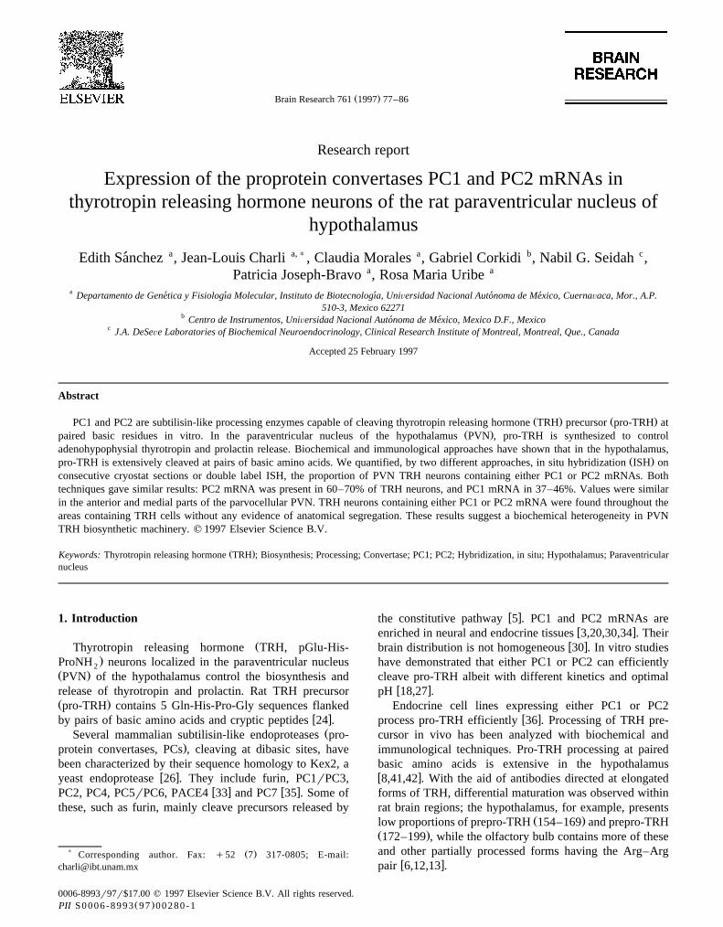
TRH mRNA was detected in regions including theŽ .hypothalamic PVN Fig. 1 , the thalamic reticularis nu-
cleus and the hippocampus. TRH neurons were presentthroughout the rostro-caudal extension of PVN mainly inthe parvocellular zone; number of cells per section was
Ž .similar in the APa and MPa subdivisions: 128"7 ns13Ž . Ž .and 116"6 ns24 respectively means"S.E.M. . They
Žwere dispersed in the anterior parvocellular part not.shown and their position varied along the rostro-caudal
axis in the medial parvocellular part. In the anterior MPa,Žthey were more concentrated along the IIIV Figs. 1 and
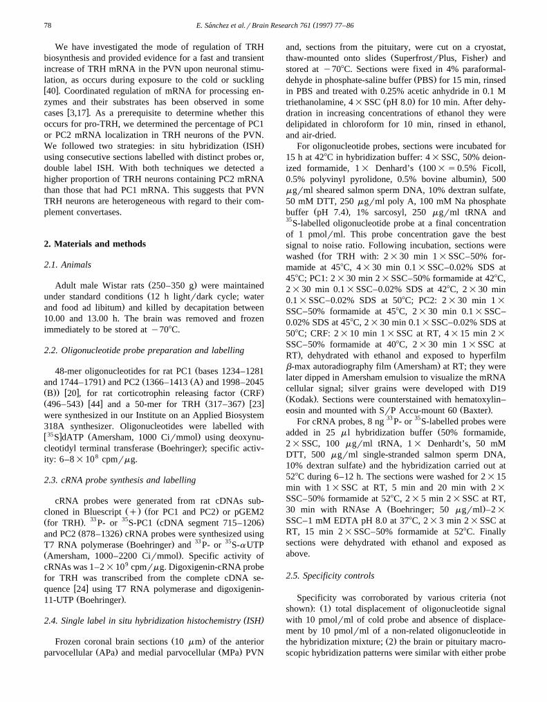
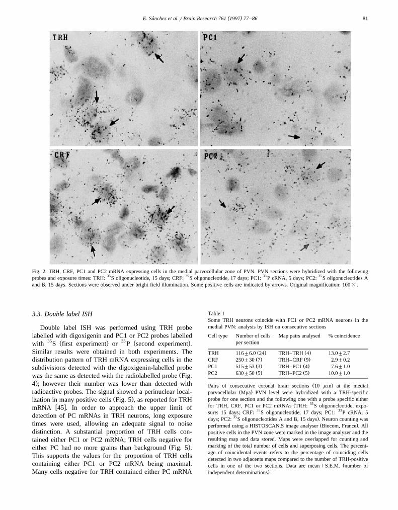
.3 ; in the medial, more dispersed with a tendency to begrouped dorsally and in the caudal zone, concentrated inthe dorsal lateral part. Autoradiographic signal for TRHmRNA was stronger in neurons from the MPa zone, in
Ž .particular in the anterior MPa Fig. 2 .PC1 mRNA was detected in adenohypophysis, interme-
Ž .diate lobe IL of the hypophysis, some layers of cortex,Žhippocampus, supraoptic nucleus of hypothalamus not
. Ž .shown and PVN Fig. 1 . PC1 neurons were presentthroughout the rostro-caudal extension of PVN, in theparvo- and magnocellular subdivisions, with a higher den-
Ž .sity in the magnocellular part not shown . Their numberŽ .was 4-fold higher than that of TRH neurons Table 1 and
( )E. Sanchez et al.rBrain Research 761 1997 77–86´80
Ž .similar in the anterior and medial parts not shown . Cellscontaining PC1 mRNA showed in general, few silver
Ž .grains compared to TRH neurons Fig. 2 except in themagnocellular zone, in particular in the anterior MPa,
Ž .where PC1 signal was intense Fig. 1 .PC2 mRNA signal was intense in IL and low in adeno-
hypophysis. In the brain sections studied, PC2 mRNA wasmainly found in high concentrations in thalamic nuclei,
Ž .some layers of cortex, hippocampus not shown and PVNŽ .Fig. 1 . PC2 neurons were present throughout the rostro-caudal extension of parvocellular and magnocellular subdi-visions of PVN with a slight enhancement of their density
Ž .in the magnocellular part Figs. 1 and 3 . Their numberŽ .was 5-fold higher than that of TRH neurons Table 1 and
Ž .similar in the anterior and medial parts not shown . PC2mRNA positive cells were more intensely labelled than
Ž .PC1 cells throughout most of the PVN area Fig. 2 ;intensity per PC2 cell increased in the rostrocaudal direc-
Ž .tion not shown .
3.2. Single label ISH on consecutiÕe sections
In order to study the localization of converting enzymemRNA in TRHergic cells, we first used ISH in consecutivesections. To determine the maximum number of colocal-ization events which could be detected with this approach,we hybridized some pairs of consecutive sections of themedial zone with TRH probe. We found that 13% of TRH
cells detected in one section coincided in the other oneŽ .Table 1 . To quantify the maximum percentage of coinci-dental events due to imprecise overlapping, we took advan-tage of the fact that CRF cells are numerous in the PVNbut do not significantly colocalize with parvocellular TRH
w xneurons 10 . In coronal sections at PVN level, CRFmRNA positive cells were only detected in the PVN. CRFcells were detected in PVN magno- and parvocellular parts
Ž .throughout the rostro-caudal axis not shown ; in the me-dial part their number was twice that of TRH neuronsŽ .Table 1 . CRF mRNA positive cells were as intensely
Ž .labelled as TRH cells Fig. 2 . We detected 2.9% of medialPVN TRH neurons coinciding with CRF mRNA cellsŽ .Table 1 . This set the upper value for overestimation dueto imprecise overlapping, although some of these coinci-
w xdental events might be real 10 .To determine the percentage of TRH cells containing
either PC1 or PC2 mRNA, we used ISH on consecutivesections from the medial PVN region. One section waslabelled with pro-TRH probe and the next one, with eitherPC1 or PC2 probes. PC1 probe coincided with 7.6% of
Ž .TRH cells while PC2 mRNA, with 10% Table 1 . If wetake into account a maximum coincidence error of 3%Ž . Ž .CRF–TRH and a maximum signal of 13% TRH–TRH ,the data suggest that approximately 70% of TRH cellsexpress PC2 mRNA while 46% PC1 mRNA. There was no
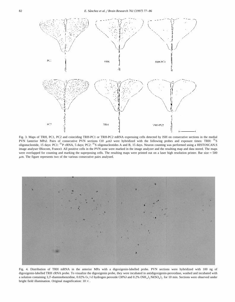
Ž .preferential distribution of colocalization events Fig. 3 .
Fig. 1. Distribution of TRH, CRF, PC1 or PC2 mRNA in the anterior MPa. PVN sections were hybridized with oligonucleotide or cRNA probes, dipped inemulsion and developed. Photomicrographs were obtained under semi-dark field illumination so that white dots correspond to silver grains accumulatingon positive cells. Probes and exposure times: TRH: 35S oligonucleotide, 15 days; CRF:35S oligonucleotide, 17 days; PC1: 33 P cRNA, 5 days; PC2: 35Soligonucleotides A and B, 15 days. Original magnification: 10= .
( )E. Sanchez et al.rBrain Research 761 1997 77–86´ 81
Fig. 2. TRH, CRF, PC1 and PC2 mRNA expressing cells in the medial parvocellular zone of PVN. PVN sections were hybridized with the followingprobes and exposure times: TRH: 35S oligonucleotide, 15 days; CRF: 35S oligonucleotide, 17 days; PC1: 33 P cRNA, 5 days; PC2: 35S oligonucleotides Aand B, 15 days. Sections were observed under bright field illumination. Some positive cells are indicated by arrows. Original magnification: 100= .
3.3. Double label ISH
Double label ISH was performed using TRH probelabelled with digoxigenin and PC1 or PC2 probes labelled
35 Ž . 33 Ž .with S first experiment or P second experiment .Similar results were obtained in both experiments. Thedistribution pattern of TRH mRNA expressing cells in thesubdivisions detected with the digoxigenin-labelled probe
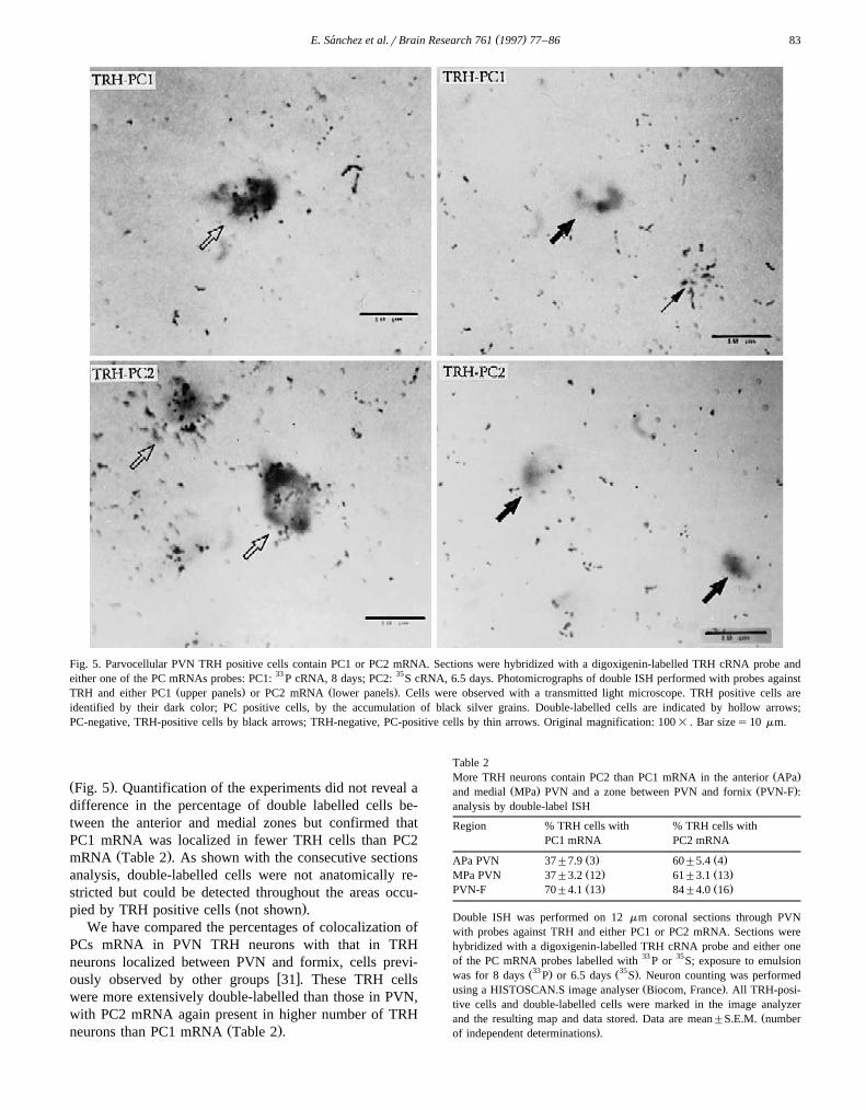
Žwas the same as detected with the radiolabelled probe Fig..4 ; however their number was lower than detected with
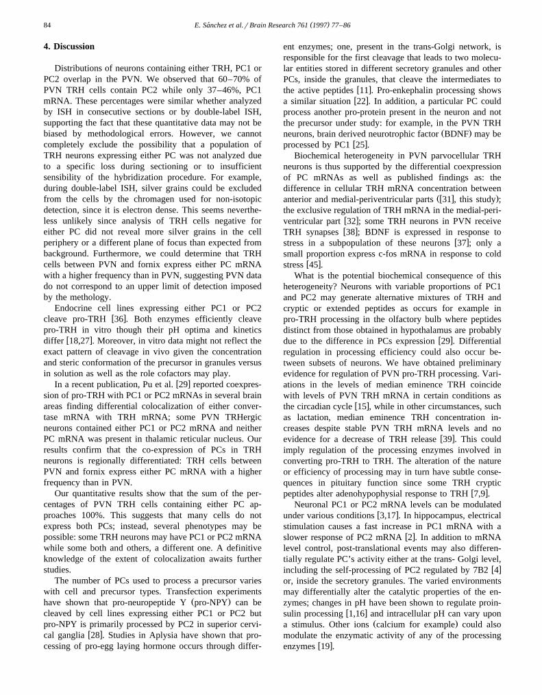
radioactive probes. The signal showed a perinuclear local-Ž .ization in many positive cells Fig. 5 , as reported for TRH
w xmRNA 45 . In order to approach the upper limit ofdetection of PC mRNAs in TRH neurons, long exposuretimes were used, allowing an adequate signal to noisedistinction. A substantial proportion of TRH cells con-tained either PC1 or PC2 mRNA; TRH cells negative for
Ž .either PC had no more grains than background Fig. 5 .This supports the values for the proportion of TRH cellscontaining either PC1 or PC2 mRNA being maximal.Many cells negative for TRH contained either PC mRNA
Table 1Some TRH neurons coincide with PC1 or PC2 mRNA neurons in themedial PVN: analysis by ISH on consecutive sections
Cell type Number of cells Map pairs analysed % coincidenceper section
Ž . Ž .TRH 116"6.0 24 TRH–TRH 4 13.0"2.7Ž . Ž .CRF 250"30 7 TRH–CRF 9 2.9"0.2Ž . Ž .PC1 515"53 3 TRH–PC1 4 7.6"1.0Ž . Ž .PC2 630"50 5 TRH–PC2 5 10.0"1.0
Ž .Pairs of consecutive coronal brain sections 10 mm at the medialŽ .parvocellular Mpa PVN level were hybridized with a TRH-specific
probe for one section and the following one with a probe specific eitherŽ 35for TRH, CRF, PC1 or PC2 mRNAs TRH: S oligonucleotide, expo-
sure: 15 days; CRF: 35S oligonucleotide, 17 days; PC1: 33 P cRNA, 535 .days; PC2: S oligonucleotides A and B, 15 days . Neuron counting was
Ž .performed using a HISTOSCAN.S image analyser Biocom, France . Allpositive cells in the PVN zone were marked in the image analyzer and theresulting map and data stored. Maps were overlapped for counting andmarking of the total number of cells and superposing cells. The percent-age of coincidental events refers to the percentage of coinciding cellsdetected in two adjacents maps compared to the number of TRH-positive
Žcells in one of the two sections. Data are mean"S.E.M. number of.independent determinations .
( )E. Sanchez et al.rBrain Research 761 1997 77–86´82
Fig. 3. Maps of TRH, PC1, PC2 and coinciding TRH-PC1 or TRH-PC2 mRNA expressing cells detected by ISH on consecutive sections in the medialŽ . Ž . 35PVN anterior MPa . Pairs of consecutive PVN sections 10 mm were hybridized with the following probes and exposure times: TRH: S
oligonucleotide, 15 days: PC1: 33 P cRNA, 5 days; PC2: 35S oligonucleotides A and B, 15 days. Neuron counting was performed using a HISTOSCAN.SŽ .image analyser Biocom, France . All positive cells in the PVN zone were marked in the image analyzer and the resulting map and data stored. The maps
were overlapped for counting and marking the superposing cells. The resulting maps were printed out on a laser high resolution printer. Bar sizes500mm. The figure represents two of the various consecutive pairs analysed.
Fig. 4. Distribution of TRH mRNA in the anterior MPa with a digoxigenin-labelled probe. PVN sections were hybridized with 100 ng ofdigoxigenin-labelled TRH cRNA probe. To visualize the digoxigenin probe, they were incubated in antidigoxigenin-peroxidase, washed and incubated with
X Ž . Ž . Ž . Ž .a solution containing 3,3 -diaminobenzidine, 0.02% vrv hydrogen peroxide 30% and 0.2% NH Ni SO for 10 min. Sections were observed under4 2 4 2
bright field illumination. Original magnification: 10= .
( )E. Sanchez et al.rBrain Research 761 1997 77–86´ 83
Fig. 5. Parvocellular PVN TRH positive cells contain PC1 or PC2 mRNA. Sections were hybridized with a digoxigenin-labelled TRH cRNA probe andeither one of the PC mRNAs probes: PC1: 33 P cRNA, 8 days; PC2: 35S cRNA, 6.5 days. Photomicrographs of double ISH performed with probes against
Ž . Ž .TRH and either PC1 upper panels or PC2 mRNA lower panels . Cells were observed with a transmitted light microscope. TRH positive cells areidentified by their dark color; PC positive cells, by the accumulation of black silver grains. Double-labelled cells are indicated by hollow arrows;PC-negative, TRH-positive cells by black arrows; TRH-negative, PC-positive cells by thin arrows. Original magnification: 100= . Bar sizes10 mm.
Ž .Fig. 5 . Quantification of the experiments did not reveal adifference in the percentage of double labelled cells be-tween the anterior and medial zones but confirmed thatPC1 mRNA was localized in fewer TRH cells than PC2
Ž .mRNA Table 2 . As shown with the consecutive sectionsanalysis, double-labelled cells were not anatomically re-stricted but could be detected throughout the areas occu-
Ž .pied by TRH positive cells not shown .We have compared the percentages of colocalization of
PCs mRNA in PVN TRH neurons with that in TRHneurons localized between PVN and formix, cells previ-
w xously observed by other groups 31 . These TRH cellswere more extensively double-labelled than those in PVN,with PC2 mRNA again present in higher number of TRH
Ž .neurons than PC1 mRNA Table 2 .
Table 2Ž .More TRH neurons contain PC2 than PC1 mRNA in the anterior APa
Ž . Ž .and medial MPa PVN and a zone between PVN and fornix PVN-F :analysis by double-label ISH
Region % TRH cells with % TRH cells withPC1 mRNA PC2 mRNA
Ž . Ž .APa PVN 37"7.9 3 60"5.4 4Ž . Ž .MPa PVN 37"3.2 12 61"3.1 13Ž . Ž .PVN-F 70"4.1 13 84"4.0 16
Double ISH was performed on 12 mm coronal sections through PVNwith probes against TRH and either PC1 or PC2 mRNA. Sections werehybridized with a digoxigenin-labelled TRH cRNA probe and either oneof the PC mRNA probes labelled with 33 P or 35S; exposure to emulsion
Ž33 . Ž35 .was for 8 days P or 6.5 days S . Neuron counting was performedŽ .using a HISTOSCAN.S image analyser Biocom, France . All TRH-posi-
tive cells and double-labelled cells were marked in the image analyzerŽand the resulting map and data stored. Data are mean"S.E.M. number
.of independent determinations .
( )E. Sanchez et al.rBrain Research 761 1997 77–86´84
4. Discussion
Distributions of neurons containing either TRH, PC1 orPC2 overlap in the PVN. We observed that 60–70% ofPVN TRH cells contain PC2 while only 37–46%, PC1mRNA. These percentages were similar whether analyzedby ISH in consecutive sections or by double-label ISH,supporting the fact that these quantitative data may not bebiased by methodological errors. However, we cannotcompletely exclude the possibility that a population ofTRH neurons expressing either PC was not analyzed dueto a specific loss during sectioning or to insufficientsensibility of the hybridization procedure. For example,during double-label ISH, silver grains could be excludedfrom the cells by the chromagen used for non-isotopicdetection, since it is electron dense. This seems neverthe-less unlikely since analysis of TRH cells negative foreither PC did not reveal more silver grains in the cellperiphery or a different plane of focus than expected frombackground. Furthermore, we could determine that TRHcells between PVN and fornix express either PC mRNAwith a higher frequency than in PVN, suggesting PVN datado not correspond to an upper limit of detection imposedby the methology.
Endocrine cell lines expressing either PC1 or PC2w xcleave pro-TRH 36 . Both enzymes efficiently cleave
pro-TRH in vitro though their pH optima and kineticsw xdiffer 18,27 . Moreover, in vitro data might not reflect the
exact pattern of cleavage in vivo given the concentrationand steric conformation of the precursor in granules versusin solution as well as the role cofactors may play.
w xIn a recent publication, Pu et al. 29 reported coexpres-sion of pro-TRH with PC1 or PC2 mRNAs in several brainareas finding differential colocalization of either conver-tase mRNA with TRH mRNA; some PVN TRHergicneurons contained either PC1 or PC2 mRNA and neitherPC mRNA was present in thalamic reticular nucleus. Ourresults confirm that the co-expression of PCs in TRHneurons is regionally differentiated: TRH cells betweenPVN and fornix express either PC mRNA with a higherfrequency than in PVN.
Our quantitative results show that the sum of the per-centages of PVN TRH cells containing either PC ap-proaches 100%. This suggests that many cells do notexpress both PCs; instead, several phenotypes may bepossible: some TRH neurons may have PC1 or PC2 mRNAwhile some both and others, a different one. A definitiveknowledge of the extent of colocalization awaits furtherstudies.
The number of PCs used to process a precursor varieswith cell and precursor types. Transfection experiments
Ž .have shown that pro-neuropeptide Y pro-NPY can becleaved by cell lines expressing either PC1 or PC2 butpro-NPY is primarily processed by PC2 in superior cervi-
w xcal ganglia 28 . Studies in Aplysia have shown that pro-cessing of pro-egg laying hormone occurs through differ-
ent enzymes; one, present in the trans-Golgi network, isresponsible for the first cleavage that leads to two molecu-lar entities stored in different secretory granules and otherPCs, inside the granules, that cleave the intermediates to
w xthe active peptides 11 . Pro-enkephalin processing showsw xa similar situation 22 . In addition, a particular PC could
process another pro-protein present in the neuron and notthe precursor under study: for example, in the PVN TRH
Ž .neurons, brain derived neurotrophic factor BDNF may bew xprocessed by PC1 25 .
Biochemical heterogeneity in PVN parvocellular TRHneurons is thus supported by the differential coexpressionof PC mRNAs as well as published findings as: thedifference in cellular TRH mRNA concentration between
Žw x .anterior and medial-periventricular parts 31 , this study ;the exclusive regulation of TRH mRNA in the medial-peri-
w xventricular part 32 ; some TRH neurons in PVN receivew xTRH synapses 38 ; BDNF is expressed in response to
w xstress in a subpopulation of these neurons 37 ; only asmall proportion express c-fos mRNA in response to cold
w xstress 45 .What is the potential biochemical consequence of this
heterogeneity? Neurons with variable proportions of PC1and PC2 may generate alternative mixtures of TRH andcryptic or extended peptides as occurs for example inpro-TRH processing in the olfactory bulb where peptidesdistinct from those obtained in hypothalamus are probably
w xdue to the difference in PCs expression 29 . Differentialregulation in processing efficiency could also occur be-tween subsets of neurons. We have obtained preliminaryevidence for regulation of PVN pro-TRH processing. Vari-ations in the levels of median eminence TRH coincidewith levels of PVN TRH mRNA in certain conditions as
w xthe circadian cycle 15 , while in other circumstances, suchas lactation, median eminence TRH concentration in-creases despite stable PVN TRH mRNA levels and no
w xevidence for a decrease of TRH release 39 . This couldimply regulation of the processing enzymes involved inconverting pro-TRH to TRH. The alteration of the natureor efficiency of processing may in turn have subtle conse-quences in pituitary function since some TRH cryptic
w xpeptides alter adenohypophysial response to TRH 7,9 .Neuronal PC1 or PC2 mRNA levels can be modulated
w xunder various conditions 3,17 . In hippocampus, electricalstimulation causes a fast increase in PC1 mRNA with a
w xslower response of PC2 mRNA 2 . In addition to mRNAlevel control, post-translational events may also differen-tially regulate PC’s activity either at the trans- Golgi level,
w xincluding the self-processing of PC2 regulated by 7B2 4or, inside the secretory granules. The varied environmentsmay differentially alter the catalytic properties of the en-zymes; changes in pH have been shown to regulate proin-
w xsulin processing 1,16 and intracellular pH can vary uponŽ .a stimulus. Other ions calcium for example could also
modulate the enzymatic activity of any of the processingw xenzymes 19 .
( )E. Sanchez et al.rBrain Research 761 1997 77–86´ 85
In conclusion, we suggest that more PVN parvocellularTRH neurons express PC2 mRNA than PC1 mRNA. Abiochemical heterogeneity of the TRH biosynthetic ma-chinery within PVN neurons may exist.
Acknowledgements
The technical assistance of M. Cisneros, X. Alvarado,E. Mata, M. Villa and A. Pichardo is recognized. Weacknowledge the synthesis of oligonucleotides by P. Gay-tan and E. Bustos and the computing support from R.Ciria, A. Ocadiz and A. Linares. This work was partiallysupported by grants from DGAPA-UNAM IN206094,CONACYT 4003344-5-4735N and MRC CanadaPG11474.
References
w x1 R.G.W. Anderson, L. Orci, A view of acidic intracellular compart-Ž .ments, J. Cell Biol. 106 1988 539–543.
w x2 R.V. Bhat, F.A. Tausk, J.M. Baraban, R.E. Mains, B.A. Eipper,Rapid increases in peptide processing enzyme expression in hip-
Ž .pocampal neurons, J. Neurochem. 61 1993 1315–1322.w x3 B.T. Bloomquist, B.A. Eipper, R.E. Mains, Prohormone converting
enzymes: Regulation and evaluation of function using antisenseŽ .RNA, Mol. Endocrinol. 5 1991 2014–2024.
w x4 J.A.M. Braks, G.J.M. Martens, 7B2 is a neuroendocrine chaperonethat transiently interacts with prohormone convertase PC2 in the
Ž .secretory pathway, Cell 78 1994 263–273.w x5 P.A. Bresnahan, R. Leduc, L. Thomas, J. Thorner, H.L. Gibson, A.J.
Brake, P.J. Barr, G. Thomas, Human fur gene encodes a yeastKEX2-like endoprotease that cleaves pro-b-NGF in vivo, J. Cell
Ž .Biol. 111 1990 2851–2859.w x6 M. Bulant, J.C. Beauvillain, A. Delfour, H. Vaudry, P. Nicolas,
Ž .Processing of thyrotropin-releasing hormone TRH prohormone inthe rat olfactory bulb generates novel TRH-related peptides, En-
Ž .docrinology 127 1990 1978–1985.w x7 M. Bulant, J.P. Roussel, H. Astier, P. Nicolas, H. Vaudry, Process-
Ž .ing of thyrotropin-releasing hormone prohormone pro-TRH gener-Ž .ates a biologically active peptide, prepro-TRH- 160–169 , which
regulates TRH-induced thyrotropin secretion, Proc. Natl. Acad. Sci.Ž .USA 87 1990 4439–4443.
w x8 M. Bulant, A. Delfour, H. Vaudry, P. Nicolas, Processing of thy-rotropin-releasing hormone prohormone generates pro-TRH connect-
Ž .ing peptides, J. Biol. Chem. 263 1988 17189–17196.w x9 F.E. Carr, A.H. Reid, M.W. Wessendorf, A cryptic peptide from the
preprothyrotropin-releasing hormone precursor stimulates thy-Ž .rotropin gene expression, Endocrinology 133 1993 809–814.
w x10 S. Ceccatelli, M. Ericksson, T. Hokfelt, Distribution and coexistence¨of corticotropin-releasing factor-, neurotensin-, enkephalin-, chole-cystokinin-, galanin- and vasoactive intestinal polypeptiderpeptidehistidine isoleucine-like peptides in the parvocellular part of the
Ž .paraventricular nucleus, Neuroendocrinology 49 1989 309–323.w x11 J.Y. Chun, J. Korner, T. Kreiner, R.H. Scheller, R. Axel, The
function and differential sorting of a family of Aplysia prohormoneŽ .processing enzymes, Neuron 12 1994 831–844.
w x12 S.M. Cockle, TRH-extended peptides in the olfactory lobe areformed by incomplete cleavage at pairs of arginine residues in the
Ž .TRH prohormone, FEBS Lett. 264 1990 253–256.w x13 S.M. Cockle, D.G. Smith, Specific processing of the thyrotropin-re-
leasing prohormone in rat brain and spinal cord, Eur. J. Biochem.Ž .165 1987 693–698.
w x14 G. Corkidi, Systeme d’analyse de preparations histologiques par` ´imagerie numerique: HISTO 200. Application a l’ etude phys-´ ´iopathologique de la maladie de Parkinson, These de Doctorat,´Universite Paris Val de Marne, 1989.´
w x15 L. Covarrubias, R.M. Uribe, M. Mendez, J.L. Charli, P. Joseph-´Bravo, Neuronal TRH synthesis: Developmental and circadian TRH
Ž .mRNA levels, Biochem. Biophys. Res. Commun. 151 1988 615–622.
w x16 H.W. Davidson, C.J. Rhodes, J.C. Hutton, Intraorganellar calciumand ph control proinsulin cleavage in the pancreatic b cell via two
Ž .distinct site-specific endopeptidases, Nature 333 1988 93–96.w x17 R. Day, M.K.-H. Schafer, S.J. Watson, M. Chretien, N.G. Seidah,¨ ´
Distribution and regulation of the prohormone convertases PC1 andŽ .PC2 in the rat pituitary, Mol. Endocrinol. 6 1992 485–497.
w x18 T.C. Friedman, Y.P. Loh, N.X. Cawley, N.P. Birch, S.S. Huang,I.M.D. Jackson, E.A. Nillni, Processing of prothyrotropin-releasing
Ž .hormone Pro-TRH by bovine intermediate lobe secretory vesicleŽ .membrane PC1 and PC2 enzymes, Endocrinology 136 1995 4462–
4472.w x19 R.S. Fuller, A. Brake, J. Thorner, Yeast prohormone processing
Ž . 2qenzyme KEX2 gene product is a Ca -dependent serine protease,Ž .Proc. Natl. Acad. Sci. USA 86 1989 1434–1438.
w x20 D.J. Hakes, N.P. Birch, A. Mezey, J.E. Dixon, Isolation of twocomplementary deoxyribonucleic acid clones from a rat insulinomacell line based on similarities to Kex2 and furin sequences and thespecific localization of each transcript to endocrine and neuroen-
Ž .docrine tissues in rats, Endocrinology 129 1991 3053–3060.w x21 E.C. Hirsch, O. Lejeune, G. Colliot, G. Corkidi and M. Tajani,
Computer methods in nuclei cartography, in: Methods in Neuro-sciences, vol. 10, Academic Press, New York, 1992, pp. 62–79.
w x22 K. Johanning, J.P. Mathis, I. Lindberg, Role of PC2 in proenkephalinprocessing: antisense and overexpression studies, J. Neurochem. 66Ž .1996 898–907.
w x23 K.J. Koller, R.S. Wolff, M.K. Warden, R.T. Zoeller, Thyroid hor-mones regulate levels of thyrotropin-releasing hormone mRNA in
Ž .the paraventricular nucleus, Proc. Natl. Acad. Sci. USA 84 19877329–7333.
w x24 R.M. Lechan, P. Wu, I.M.D. Jackson, H. Wolf, S. Cooperman, G.Mandel, R.H. Goodman, Thyrotropin releasing hormone precursor:
Ž .Characterization in rat brain, Science 231 1986 159–161.w x25 M. Marcinkiewicz, T. Nagao, R. Day, N.G. Seidah, M. Chretien, M.´
Avoli, Pilocarpine-induced seizures are accompanied by a transientelevation in the mRNA expression of the prohormone convertasePC1 in rat hippocampus: comparison with NGF and BDNF expres-
Ž .sion, J. Neurosci. 76 1997 425–439.w x26 K. Mizuno, T. Nakamura, T. Ohshima, S. Tanaka, H. Matsuo, Yeast
Kex2 gene encodes an endopeptidase homologous to subtilisin-likeŽ .serine proteases, Biochem. Biophys. Res. Commun. 156 1988
246–254.w x27 E.A. Nillni, T.C. Friedman, R.B. Todd, N.P. Birch, Y.P. Loh,
I.M.D. Jackson, Pro-thyrotropin-releasing hormone processing byŽ .recombinant PC1, J. Neurochem. 65 1995 2462–2472.
w x28 L. Paquet, B. Massie, R.E. Mains, Proneuropeptide Y processing inlarge dense-core vesicles: Manipulation of prohormone convertaseexpression in sympathetic neurons using adenoviruses, J. Neurosci.
Ž .16 1996 964–973.w x29 L.P. Pu, W. Ma, J.L. Barker, Y.P. Loh, Differential coexpression of
Ž .genes encoding prothyrotropin-releasing hormone Pro-TRH andŽ .prohormone convertases PC1 and PC2 in rat brain neurons: Impli-
cations for differential processing of pro-TRH, Endocrinology 137Ž .1996 1233–1241.
w x30 M.K.-H. Schafer, R. Day, W.E. Cullinan, M. Chretien, N.G. Seidah,¨ ´S.J. Watson, Gene expression of prohormone and proprotein conver-tases in the rat CNS: A comparative in situ hybridization analysis, J.
Ž .Neurosci. 13 1993 1258–1279.
( )E. Sanchez et al.rBrain Research 761 1997 77–86´86
w x31 T.P. Segerson, H. Hoefler, H. Childers, H.J. Wolfe, P. Wu, I.M.D.Jackson, R.M. Lechan, Localization of thyrotropin-releasing hor-mone prohormone messenger ribonucleic acid in rat brain by in situ
Ž .hybridization, Endocrinology 121 1987 98–107.w x32 T.P. Segerson, J. Kauer, H.C. Wolfe, H. Mobtaker, P. Wu, I.M.D.
Jackson, R.M. Lechan, Thyroid hormone regulates TRH biosynthe-sis in the paraventricular nucleus of the rat hypothalamus, Science
Ž .238 1987 78–80.w x33 N.G. Seidah, M. Chretien, R. Day, The family of subtilisinrkexin
like proprotein and prohormone convertases: Divergent or sharedŽ .functions, Biochimie 76 1994 197–209.
w x34 N.G. Seidah, L. Gaspar, P. Mion, M. Marcinkiewicz, M. Mbikay,M. Chretien, cDNA sequence of two distinct pituitary proteins´homologous to Kex2 and furin gene products: Tissue-specific mR-NAs encoding candidates for prohormone processing proteinases,
Ž .DNA Cell Biol. 9 1990 415–424.w x35 N.G. Seidah, J. Hamelin, M. Mamarbachi, W. Dong, H. Tadros, M.
Mbikay, M. Chretien, R. Day, cDNA structure, tissue distribution´and chromosomal localization of rat PC7, a novel mammalianproprotein convertase closest to yeast kexin-like proteinases, Proc.
Ž .Natl. Acad. Sci. USA 93 1996 3388–3393.w x36 K.A. Sevarino, R.H. Goodman, J. Spiess, I.M.D. Jackson, P. Wu,
Ž .Thyrotropin-releasing hormone TRH precursor processing. Charac-terization of mature TRH and non-TRH peptides synthesized by
Ž .transfected mammalian cells, J. Biol. Chem. 264 1989 21529–21535.
w x37 M.A. Smith, S. Makino, S.Y. Kim, R. Kvetnansky, Stress increasesbrain-derived neurotropic factor messenger ribonucleic acid in the
Ž .hypothalamus and pituitary, Endocrinology 136 1995 3743–3750.
w x38 R. Toni, I.M.D. Jackson, R.M. Lechan, Thyrotropin-releasing hor-mone-immunoreactive innervation of thyrotropin-releasinghormone-tuberoinfundibular neurons in rat hypothalamus: Anatomi-cal basis to suggest ultrashort feedback regulation, Neuroendocrinol-
Ž .ogy 52 1990 422–428.w x39 R.M. Uribe, P. Joseph-Bravo, J. Pasten, G. Ponce, M. Mendez, L.´
Covarrubias, J.L. Charli, Some events of thyrotropin-releasing hor-mone metabolism are regulated in lactating and cycling rats, Neu-
Ž .roendocrinology 54 1991 493–498.w x40 R.M. Uribe, J.L. Redondo, J.L. Charli, P. Joseph-Bravo, Suckling
and cold stress rapidly and transiently increase TRH mRNA in theŽ .paraventricular nucleus, Neuroendocrinology 58 1993 140–145.
w x41 P. Wu, I.M.D. Jackson, Post-translational processing of thyrotropin-releasing hormone precursor in rat brain: Identification of 3 novel
Ž .peptides derived from pro-TRH, Brain Res. 456 1988 22–28.w x42 P. Wu, R.M. Lechan, I.M.D. Jackson, Identification and characteri-
zation of thyrotropin-releasing hormone precursor peptides in ratŽ .brain, Endocrinology 121 1987 108–115.
w x43 W.S. Young, III, Corticotropin-releasing factor mRNA in the hypo-thalamus is differently affected by drinking saline and by dehydra-
Ž .tion, FEBS Lett. 208 1986 158–162.w x44 R.T. Zoeller, P.K. Rudeen, Ethanol blocks the cold-induced increase
in thyrotropin-releasing hormone mRNA in paraventricular nucleibut not the cold-induced increase in thyrotropin, Mol. Brain Res. 13Ž .1992 321–330.
w x45 R.T. Zoeller, A. Simonyi, O. Butnariu, D.L. Fletcher, P.K. Rudeen,S. McCrone, S.L. Petersen, Effect of acute ethanol administrationand cold exposure on the hypothalamic–pituitary thyroid axis, En-
Ž .docrine 3 1995 39–47.