expression of multiple genes regulating cell cycle and apoptosis in differentiating hematopoietic...
TRANSCRIPT

Experimental Hematology 29 (2001) 1060–1069
0301-472X/01 $–see front matter. Copyright © 2001 International Society for Experimental Hematology. Published by Elsevier Science Inc.PII S0301-472X(01)00683-X
Expression of multiple genes regulating cell cycleand apoptosis in differentiating hematopoietic cells is dependent on iron
Orlando Alcantara, Mamta Kalidas, Ioannis Baltathakis, and David H. Boldt
Medicine/Hematology, University of Texas Health Science Center at San Antonio,and South Texas Veterans Health Care System, Audie L. Murphy Division, San Antonio, Texas, USA
(Received 13 December 2000; revised 24 April 2001; accepted 18 May 2001)
Objective.
Iron plays critical roles in many biological processes including hematopoietic cellgrowth and differentiation. Iron is essential for the differentiation of HL-60 promonocytes. HL-60cells stimulated with phorbol myristate acetate (PMA) undergo G1/S phase cell-cycle arrest anddifferentiate to monocyte/macrophages. With iron deprivation, PMA-induced HL-60 cells by-pass differentiation and undergo apoptosis. To investigate the molecular basis underlying thisobservation, we used commercially available gene microarrays to evaluate expression of multiplegenes involved in the regulation of cell cycling and apoptosis.
Methods.
We treated HL-60 cells with PMA
�
desferrioxamine (DF), a potent iron chelator, toproduce iron deprivation. Cells were cultured for 48 hours, and cDNA was prepared and radi-
olabeled with
�
-
32
P dCTP, then hybridized to gene arrays containing specific cDNA fragments.
Results.
Expression of 11 of 43 genes was inhibited greater than 50% by iron deprivation.These genes were Rb; p21 (WAF1/CIP1); bad; cdk2; cyclins A, D3, E1; c-myc; egr-1; iNOS;and FasL. For each gene the microarray results were confirmed by RT-PCR and/or Northernor Western blotting. Nuclear transcription assays indicated that the role of iron in Rb expres-sion was to support gene transcription. Addition of ferrioxamine (iron saturated DF) insteadof DF to PMA-induced cells did not affect gene expression, indicating that diminished expres-sion was due to iron deprivation, not nonspecific toxicity.
Conclusion.
Iron supports expression of multiple cell cycle–regulatory and apoptosis-relatedgenes during HL-60 cell differentiation, and, in this way, is involved in regulation of a critical celldecision point—the decision to pursue a differentiation-related or apoptotic pathway. © 2001
International Society for Experimental Hematology. Published by Elsevier Science Inc.
Iron is a critical nutritional element, essential for a great va-riety of important biological processes including cellgrowth and differentiation, electron transfer reactions, andoxygen transport, activation, and detoxification [1]. Iron isrequired for maintenance of cell viability in vitro. The well-documented iron dependency of the enzyme ribonucleotidereductase (RR) is one key factor that accounts for this re-quirement [2]. RR, an enzyme responsible for synthesis ofdeoxyribonucleotides, is composed of two heterodimericsubunits, one of which contains an essential binuclear non-
heme iron center [2]. Iron also is required for normal cellcycle progression through its relationships with regulatoryfactors including cyclins, cyclin-dependent kinases, and theretinoblastoma susceptibility protein, pRb [3–6]. The mech-anisms by which iron supports the activities of certain cellcycle–regulatory proteins have not been fully defined.
Recently, we observed that iron deprivation blockedphorbol myristate acetate (PMA)-induced differentiation ofHL-60 cells by inhibiting induction of the cyclin-dependentkinase inhibitor, p21(WAF1/CIP1), and releasing cells fromthe G1/S cell cycle checkpoint [7]. In this way iron depriva-tion forced S-phase accumulation of cells and apoptosis asopposed to differentiation. Although many of the effects ofiron deprivation on altering the differentiation program ofthe HL-60 cells could be explained by inhibition of p21 ex-
Offprint requests to: David H. Boldt, M.D., Medicine/Hematology, MailCode 7880, University of Texas Health Science Center, 7703 Floyd CurlDrive, San Antonio, TX 78229-3900; E-mail: [email protected]

O. Alcantara et al./Experimental Hematology 29 (2001) 1060–1069
1061
pression, the substantial deviations consistently observed inthis system suggested that effects on other important cellcycle and/or apoptosis regulatory genes also might be in-volved. Therefore, we embarked on experiments with genearrays targeting components in cell cycling and apoptosispathways to determine if expression of other key proteins inthese pathways might be dependent on cellular iron status.Results indicate that differentiation-associated expressionof multiple genes in these pathways is altered substantiallyduring conditions of iron deprivation.
Materials and methods
Cell cultures
Human HL-60 cells obtained from American Type Culture Collec-tion (Rockville, MD, USA) are maintained in our laboratory [8].HL-60 cells were cultured at a concentration of 2
�
10
5
cells/mL inRPMI-1640 medium (GIBCO-BRL, Gaithersburg, MD, USA) con-taining 10% heat-inactivated fetal bovine serum. Differentiation tomonocyte/macrophages was induced by adding 1–10 nM PMA(Sigma, St. Louis, MO, USA) for 24–72 hours. Cells became ad-herent to flasks after 24 hours and more than 50% of cells ex-pressed the differentiation marker,
�
-naphthol butyrate esterase(NSE) (Sigma), after 48 hours. To induce iron deprivation, the cell-permeable iron chelator, desferrioxamine (DF) (Sigma), was addedat initiation of cultures at a concentration of 20 uM. Ferrioxamine(iron saturated DF), 20 uM, was used in parallel cultures as control.
Microarray experiments
For these experiments we purchased GEArray gene expression arraysystems (SuperArray, Bethesda, MD, USA) Each array consists of56 coordinates containing specific cDNA fragments spotted in du-plicate as well as control sequences (pUC18 as negative control;
�
-actin and GAPDH for loading). Using different arrays we comparedexpression of 43 genes in PMA-induced HL-60 cells under iron-re-plete or -deplete conditions. A list of genes examined is given in Ta-ble 1. For these experiments total cellular RNA was isolated by amodification of the guanidinium isothiocyanate technique [9](RNAzol B, Tel Test, Inc., Friendswood, TX, USA). Integrity ofRNA was assessed by visualization of ethidium bromide–stainedgels. For some experiments, custom arrays produced by SuperArraywere used. Custom arrays contained 11 cDNAs affected by ironavailability (see below and Table 1 for a list of these genes), pUC18,
�
-actin, and GAPDH. All microarrays were employed according tothe manufacturer’s instructions. In brief, using reagents provided,cDNA was prepared from total RNA by reverse transcription withMMLV reverse transcriptase, radiolabeled using [
�
-
32
P] dCTP(3000 Ci per mMol) (ICN Biomedicals, Inc., Costa Mesa, CA,USA), then hybridized under precisely specified conditions to a pos-itively charged nylon membrane containing the arrayed DNA. Afterwashing, the arrays were visualized by autoradiography. Loadingwas adjusted based on intensity of hybridization signals to thehousekeeping genes,
�
-actin and GAPDH, then gene expressionwas quantitated by scanning densitometry. Each experiment wasperformed at least twice to ensure reproducibility of results.
RT-PCR
RT-PCR was performed to confirm results of expression array ex-periments. After extraction of total cellular RNA, reverse tran-
scription was initiated using the GeneAmp RNA PCR kit (PerkinElmer, Branchburg, NJ, USA). Reverse-transcribed cDNA wasamplified in the linear range of amplification in presence of spe-cific primer sets (Table 2) using the following conditions: denatur-ation at 94
�
C (60 seconds); [92
�
C (30 seconds); 60
�
C (30 seconds);75
�
C (90 seconds)] 30 times using an MJ Research PTC-100 ther-mocycler. PCR products were separated on 1.6% agarose gelsstained with ethidium bromide to view the amplified bands.
Northern hybridization
Total cellular RNA was fractionated by electrophoresis through a1% (w/v) agarose gel containing formaldehyde and then transferredto nitrocellulose filters presoaked in 20
�
SSC (SSC
�
0.15 mol/LNaCl, 15 mmol/L sodium citrate dihydrate). The filters were hy-bridized with
32
P-oligolabeled cDNA probes. After washing, thefilters were exposed to Kodak XAR film (Eastman Kodak, Roches-ter, NY, USA) at
�
70
�
C with Dupont Cronex intensifying screens(Dupont, Wilmington, DE, USA). Relative intensities of blots onautoradiographs were quantitated by scanning densitometry.
Immunoblotting
Western immunoblotting was performed as previously described[10]. Briefly, cell lysates were prepared by boiling for 5 minutes in20 mM tris HCl, pH 6.8, 10 mM EDTA, 2% sodium dodecyl sul-fate, 10% glycerol, 0.3%
�
mercaptoethanol, and 0.03% bromphe-nol blue. Lysates, 40 to 50 ug per lane, were resolved by 7.5%SDS-PAGE. Primary antibodies were used at dilutions of 1:100 to1:2500. Secondary antibody conjugated to horseradish peroxidase(Pierce Chemical Co., Rockport, IL, USA) was used at a dilutionof 1:10,000 followed by treatment with chemiluminescence solu-tion (Pierce) and visualization by autoradiography.
Nuclear transcription assay
Active nuclei were isolated as described by Marzluff and Huang[11] and nuclear run-on assays were performed as described previ-ously [12].
Molecular and immunological reagents
The primer sets listed in Table 2 were synthesized in the UTH-SCSA Center for Advanced DNA Technologies. All sequenceswere confirmed by direct nucleotide sequencing and comparedagainst the gene sequences published in GenBank. The iNOScDNA probe, hep-NOS, was a kind gift from Dr. Charles Lowen-stein [13]. Goat anti-human Rb IgG was purchased from SantaCruz Biotechnology (Santa Cruz, CA, USA). Rabbit anti-actin an-
Table 1.
Genes tested in microarray experiments*
bad
(56) cdk1 E2F p53bax
cdk2
(57)
egr-1
(56) p57(KIP2)bcl-2 cdk4 Fas PCNAbcl-w cdk6
Fas ligand
(68) pig7bcl-x
c-myc
(56) gadd45 pig8caspase 1
cyclin A
(80) gadd45B
RB
(85)caspase 3 cyclin B
iNOS
(72) skp1caspase 5 cyclin C mdm2 skp2caspase 6 cyclin D2 NF
B TNFR2caspase 7
cyclin D3
(58) p19(INK4D) TRAILcaspase 10
cyclin E1
(62)
p21(WAF1/CIP1)
(58)
*Genes significantly inhibited by iron deprivation are indicated in boldwith percentage inhibition in parentheses.

1062
O. Alcantara et al./Experimental Hematology 29 (2001) 1060–1069
tibody was from Sigma. The following cDNA probes were used:pCMV-RB, a full-length Rb cDNA expression plasmid kindly pro-vided by Dr. Wen-Hwa Lee;
�
-actin, a 3.8 kb Eco R1-Hind IIIfragment from the genomic clone, HA4 [14].
Results
Expression of multiple genes during HL-60 cell differentiation is influenced by iron availability
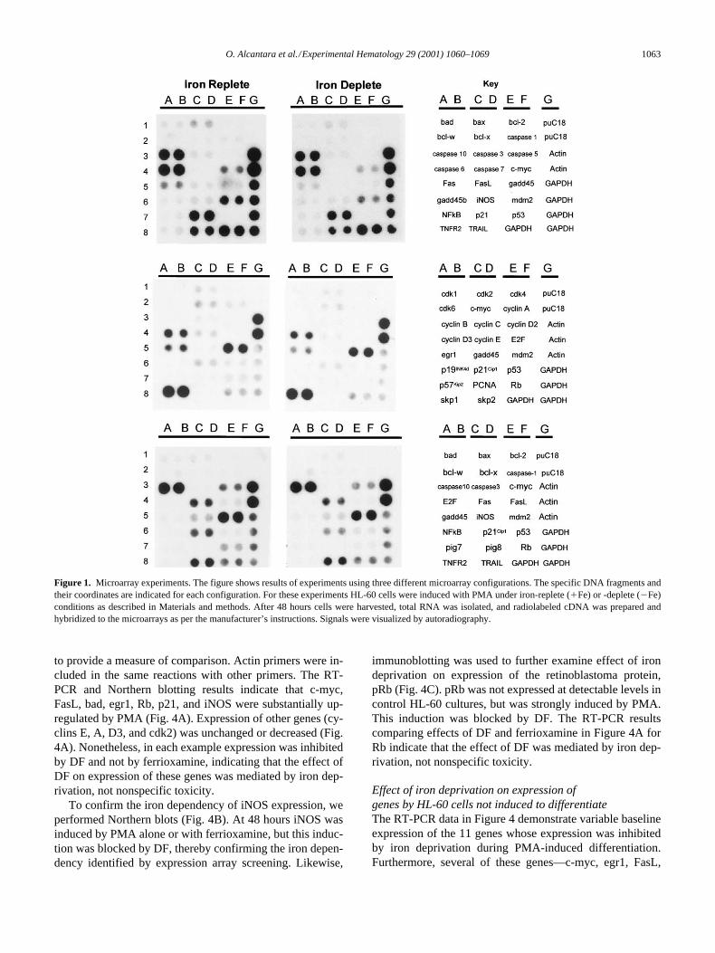
Figure 1 shows representative microarrays comparing geneexpression by HL-60 cells after 48 hours of PMA inductionunder iron-replete or -deplete conditions. Results are sum-marized graphically in Figure 2. Expression of 14 genes waschanged minimally or not at all by iron availability. Thesegenes are caspases 5 and 7, cdks 1, 4, and 6; cyclins B, C,and D2; E2F; p53; p57; pig7; pig8; and TNFR2. Expressionof only four genes was enhanced by iron deprivation—cas-pase 3 (1.65
�
), gadd 45b (1.55
�
), PCNA (1.35
�
), andbcl-x (1.2
�
)—but this enhancement did not exceed twofoldin any instance. Expression of 14 other genes was reduced5–40% by iron deprivation: bax; bcl-2; bcl-w; caspases 1, 6,and 10; fas; gadd 45; mdm2; NF-
B; p19; skp1; skp2; andTRAIL. By contrast, expression of 11 genes was reduced by50% or more under conditions of iron deprivation (Table 1).These genes are the following: bad; cdk2; c-myc; cyclins A,D3, and E1; egr-1; Fas ligand; iNOS; p21(WAF1/CIP1);and Rb. For example, expression of Rb and cyclin A was in-hibited greater than or equal to 80% and that of fas ligandand iNOS approximately 70% by iron deprivation.
As noted earlier, these observations were reproducibleon at least two independent determinations. In addition, sev-eral genes are included in more than one of the microarrayformats that we used and results were consistent betweenthe two formats. For example, Fas L, iNOS, c-myc, Rb, andp21 were identified as genes whose expression was substan-tially inhibited by iron deprivation in each of two or threedifferent formats (Fig. 1). It is also noteworthy that we havepreviously identified p21 as an iron-dependent gene [7] andexpression of iNOS has been shown in other studies to vary
by iron status [15–17]. These considerations provide addedstrength to the microarray findings.
Time course of effects of iron deprivation ongene expression by HL-60 cells induced by PMA
To screen for iron dependence, we chose to examine gene ex-pression after 48 hours of PMA treatment because in this sys-tem commitment to differentiation (PMA alone) or to apopto-sis (PMA
DF) is made within the first 24 to 48 hours. Forexample, with PMA alone, cell adherence occurs by 24 hoursand more than 50% of cells are differentiated by 48 hours, in-dicating that commitment to differentiation occurs within thefirst 24 hours [7]. Furthermore, when addition of DF to cul-tures is delayed beyond 24 hours, there is no increase ofapoptosis measured at 72 or 96 hours (unpublished observa-tions). Time courses of the effect of iron deprivation on geneexpression are depicted in Figure 3. For genes inhibited byiron deprivation, two basic patterns were observed: 1) inhibi-tion at each time point examined, 24, 48, and 72 hours (bad,cdk2, cyclin D3, egr-1, Fas ligand, Rb); 2) inhibition devel-oping only after 24 hours (cyclins A and E, p21, cmyc,iNOS). In most cases inhibition was greater at 72 comparedto 48 hours. Figure 3B depicts time courses for 10 representa-tive genes not affected by iron deprivation. In no case did weobserve inhibition develop later than 48 hours.
Confirmation of microarray data by RT-PCR, Northern hybridization, or immunoblotting
To obtain additional confirmation of the microarray results,we sought independent demonstration that iron was neces-sary for full expression of the 11 genes inhibited at least50% by iron deprivation after 48 hours. For 9 of these genesthis confirmation was obtained by RT-PCR, for 1 gene(iNOS) by Northern hybridization, and for 1 by RT-PCRand Western blotting (Rb) (Fig. 4). For all of these experi-ments cells were treated for 48 hours without additions (C),with PMA alone (P), with PMA plus desferrioxamine (PD),or with PMA plus ferrioxamine (PF). For the RT-PCR reac-tions simultaneous determinations of actin were performed
Table 2.
Primers for RT-PCR
GenBankAccession Number Gene Primer/Forward Primer/Reverse
RTPCRProduct (bp)
U66879 Bad 5
�
GAGTGAGCAGGAAGACTCCAGC3
�
5
�
TCCACAAACTCGTCACTCATCC3
�
342X61622 Cdk2 5
�
CCTGGACACTGAGACTGAGGGT3
�
5
�
CTCAGAATCTCCAGGGAACAGG3
�
517J00120 C-myc 5
�
CTACTGCGACGAGGAGGAGAAC3
�
5
�
CGCAGATGAAACTCTGGTTCAC3
�
284X51688 Cyclin A 5
�
CAGGAGAATATCAACCCGGAAA3
�
5
�
CCAGTCCACGAGGATAGCTCTC3
�
564M90814 Cyclin D3 5
�
AGATGCTGGCTTACTGGATGCT3
�
5
�
AGGGCTACAGGTGTATGGCTGT3
�
711M73812 Cyclin E 5
�
ATCCCCACACCTGACAAAGAAG3
�
5
�
CCTGAACAAGCTCCATCTGTCA3
�
446X52541 Egr1 5
�
CCCTAAGCTGGAGGAGATGATG3
�
5
�
TCATGCTCACTAGGCCACTGAC3
�
398U08137 Fas L 5
�
GCCCTTCAATTACCCATATCCC3
�
5
�
GAGTTCTGCCAGCTCCTTCTGT3
�
334L47233 P21 5
�
GAACTTCGACTTTGTCACCGAG3
�
5
�
CGTTTTCGACCCTGAGAGTCTC3
�
279M15400 Rb 5
�
CTGTGGATGGAGTATTGGGAGG3
�
5
�
GAGCAACATGGGAGGTGAGAGT3
�
458X00351 Actin 5
�
GAACTTCGACTTTGTCACCGAG3
�
5
�
CGTTTTCGACCCTGAGAGTCTC3
�
235X00351 Actin 5
�
CACTGTGTTGGCGTACAGGTCT3
�
5
�
GCGAGAAGATGACCCAGATCAT3
�
548
O. Alcantara et al./Experimental Hematology 29 (2001) 1060–1069
1063
to provide a measure of comparison. Actin primers were in-cluded in the same reactions with other primers. The RT-PCR and Northern blotting results indicate that c-myc,FasL, bad, egr1, Rb, p21, and iNOS were substantially up-regulated by PMA (Fig. 4A). Expression of other genes (cy-clins E, A, D3, and cdk2) was unchanged or decreased (Fig.4A). Nonetheless, in each example expression was inhibitedby DF and not by ferrioxamine, indicating that the effect ofDF on expression of these genes was mediated by iron dep-rivation, not nonspecific toxicity.
To confirm the iron dependency of iNOS expression, weperformed Northern blots (Fig. 4B). At 48 hours iNOS wasinduced by PMA alone or with ferrioxamine, but this induc-tion was blocked by DF, thereby confirming the iron depen-dency identified by expression array screening. Likewise,
immunoblotting was used to further examine effect of irondeprivation on expression of the retinoblastoma protein,pRb (Fig. 4C). pRb was not expressed at detectable levels incontrol HL-60 cultures, but was strongly induced by PMA.This induction was blocked by DF. The RT-PCR resultscomparing effects of DF and ferrioxamine in Figure 4A forRb indicate that the effect of DF was mediated by iron dep-rivation, not nonspecific toxicity.
Effect of iron deprivation on expression of genes by HL-60 cells not induced to differentiate
The RT-PCR data in Figure 4 demonstrate variable baselineexpression of the 11 genes whose expression was inhibitedby iron deprivation during PMA-induced differentiation.Furthermore, several of these genes—c-myc, egr1, FasL,
Figure 1. Microarray experiments. The figure shows results of experiments using three different microarray configurations. The specific DNA fragments andtheir coordinates are indicated for each configuration. For these experiments HL-60 cells were induced with PMA under iron-replete (Fe) or -deplete (�Fe)conditions as described in Materials and methods. After 48 hours cells were harvested, total RNA was isolated, and radiolabeled cDNA was prepared andhybridized to the microarrays as per the manufacturer’s instructions. Signals were visualized by autoradiography.

1064
O. Alcantara et al./Experimental Hematology 29 (2001) 1060–1069
p21, Rb, and iNOS—clearly are upregulated after PMAtreatment (Fig. 4). Cyclins A and D3 appear to be downreg-ulated and little change in expression is apparent for bad,cdk2, or cyclin E. We used custom cDNA arrays containingthese genes to examine the effect of iron deprivation ontheir expression by HL-60 cells cultured for 48 hours butnot induced to differentiate (Fig. 5).These experimentsdemonstrated little effect of iron deprivation in the absenceof the differentiation stimulus provided by PMA.
Iron is required for Rb gene transcription
We performed nuclear run-on transcription assays to deter-mine if iron was required for Rb gene transcription (Fig. 6).Iron deprivation induced by DF inhibited elongation of nascentmRNA transcripts from the Rb- but not
�
-actin gene, consis-tent with a requirement for iron for Rb gene transcription.
Discussion
We have used gene expression profiling with cDNA mi-croarrays to investigate the spectrum of cell cycle–regula-tory and apoptosis-related genes dependent on cellular ironstatus during PMA-induced HL-60 cell differentiation. Weinduced iron deprivation with the highly selective iron che-lator, DF. Because the binding affinity of DF for Fe
3
ismany logs higher than for other metals (Ka, 10
31
vs 10
2
–10
14
M
�
1
) [18], the gene expression changes observed pre-dominantly reflect iron status. Approximately 25% (11/43)of genes examined were inhibited substantially (
�
50%) un-der conditions of iron deprivation. As these 11 genes are im-
portantly involved in critical cell decision points such as thedecision to pursue a differentiation or cell death pathway,this observation suggests that the role of iron in supportinghematopoiesis is likely to be considerably more complexthan heretofore appreciated. We have not performed geneexpression profiling with other cell lines or hematopoieticlineages. Therefore the extent to which the findings may berelevant to differentiation of other cell types is not known.However, we have studied cytokine-induced differentiationof dendritic cells (DCs) and macrophages from peripheralblood monocyte precursors and demonstrated that this dif-ferentiation required iron [19]. DC and macrophage differ-entiation also required induction of p21(WAF1/CIP1),which was impaired under conditions of iron deprivation.Because p21 is one of the 11 genes whose expression inPMA-induced HL-60 cells was inhibited by iron depriva-tion, the data with normal monocyte differentiation providea measure of confidence that the microarray experimentswith HL-60 cells are relevant to normal hematopoiesis.
Ye and Connor have examined effects of iron availabilityon gene expression in human astrocytoma cells [20,21].These investigators reported that expression of a number ofgenes previously not known to be iron responsive increasedor decreased based on iron availability. In addition, somenovel genes also were identified. Of 15 iron-responsive genesconfirmed by Northern blotting, 10 were known genes and 5were novel transcripts. There is no overlap between the genesidentified by Ye and Connor and the 11 genes we have identi-fied. Differences may reflect different tissue specificities ordifferences in the methodologies used to discover iron-re-
Figure 2. Graphical representation of expression of 43 genes under iron-replete or -deplete conditions in HL-60 cells induced with PMA. The figure summa-rizes data from multiple microarray experiments. Cells were cultured as described in the Figure 1 legend and in Materials and methods. Gene expression wasquantitated by scanning densitometry of autoradiographs adjusted for loading based on intensity of hybridization signals for �-actin and GAPDH. Results areexpressed as the ratios of arbitrary density units from iron-deplete to iron-replete cultures. For example, for Rb a result of 0.2 indicates that expression of Rbin iron-deprived cultures averaged only 20% of the comparable expression level in iron-replete cultures. Results represent means of at least two separatedeterminations. Solid bars designate genes inhibited by greater than or equal to 50% by iron deprivation.

O. Alcantara et al./Experimental Hematology 29 (2001) 1060–1069
1065
Figure 3. Time course of effect of iron deprivation on gene expression. Microarray experiments with HL-60 cells were performed as described in the Figure1 legend after 24, 48, or 72 hours with PMA � DF. Hybridization signals were visualized by autoradiography and quantitated by scanning densitometry.Graphs plot percentage decrease or stimulation of expression in cultures with DF compared to those without. (A) Genes inhibited by iron deprivation; con-trols: actin and GAPDH. (B) Genes not inhibited by iron deprivation.

1066
O. Alcantara et al./Experimental Hematology 29 (2001) 1060–1069
sponsive genes. Barisani et al. used differential display to ex-amine effects of iron overload on gene expression in HepG2cells [22]. They showed that several transcripts were modu-lated by cellular iron status, including apolipoprotein B100,semaphorin cd100, and aldose reductase. Neither Ye andConnor nor Barisani et al. determined if the alterations of
mRNA transcripts induced by iron availability were mediatedby transcriptional or posttranscriptional mechanisms.
The mechanisms by which iron supports expression ofthese genes are largely unknown. Iron regulation of gene ex-pression has been documented in bacteria, plants, yeast, andmammals, including humans. In bacteria and yeast, expression
Figure 3. Continued.

O. Alcantara et al./Experimental Hematology 29 (2001) 1060–1069 1067
of iron uptake proteins is regulated at the level of gene tran-scription by DNA-interacting proteins whose binding to DNAis dependent on iron availability [23,24]. To date, similar iron-dependent transcription factors have not been described inmammals. Iron regulation of gene expression in mammaliancells at the posttranscriptional level is well described [25]. Bycontrast, iron-responsive gene transcription has been less wellstudied. We have reported that transcription of both the pro-tein kinase C-� isoform [26,27] and also acid phosphatasetype 5 (Acp5) [also known as tartrate resistant acid phos-phatase (TRAP)] is iron dependent [12]. More recently wehave found that inhibition of p21(WAF1/CIP1) expression inPMA-induced HL-60 or U937 cells by iron deprivation alsowas due to inhibition of gene transcription implicated by nu-clear run-on transcription assays and experiments with p21-CAT reporter gene constructs [7]. In addition, p21 mRNA t1/2was not influenced by iron availability in HL-60 cells [7]. Our
data indicate that Rb should now be added to the list of mam-malian genes whose transcription is dependent on adequatecellular iron. Nuclear run-on assays (Fig. 6) showed inhibitionof Rb gene transcription under conditions of iron deprivation.Although inhibition of Rb transcription under these conditionsdid not appear to be complete, it did account for a substantialportion of the observed decrease in Rb expression. Whereasthis observation may imply that posttranscriptional mecha-nisms may play a role, we have not yet examined the ef-fect of iron deprivation on Rb mRNA t1/2 or performed re-porter assays with Rb-reporter constructs. The molecularmechanism(s) by which iron supports transcription of theRb, PkC-�, Acp5, or p21 genes is not known.
Iron also is known to regulate expression of the iNOSgene by a transcriptional mechanism [15–17]. However, inthis case we are presented with a paradox because in previ-ous studies, DF or other iron chelators such as picolinic acid
Figure 4. Confirmation of microarray results. HL-60 cells were cultured under conditions described in Materials and methods. (A) RT-PCR experimentsusing the primer sets indicated in Table 2. For all reactions the size of the actin band is 235 bp, except for the p21 reaction, in which case the actin band is 548 bp.(B) Northern blot determination for iNOS mRNA. The lower panel shows the parallel Northern blot for actin. (C) Immunoblotting for Rb protein. Abbrevia-tions in A and B: C, control cultures (no additions); P, cultures with PMA; PD, PMA 20 M DF; PF, PMA 20 M ferrioxamine.

1068 O. Alcantara et al./Experimental Hematology 29 (2001) 1060–1069
have been shown to upregulate iNOS expression in murinemacrophage cell lines by transactivation of NF-IL-6 and hy-poxia-inducible factor 1–responsive elements [15,17]. Fur-thermore, in those studies iron addback abolished DF induc-tion of iNOS. However, those studies employed iNOSreporter constructs in murine macrophage cell lines andmay not be comparable to regulation of endogenous iNOSin differentiating human HL-60 cells. In fact, iron was in-volved in induction of iNOS in human A549 lung epithelialcells exposed to crocidolite [16]. The relationship of ironstatus to iNOS induction is clearly complex and situation-
specific. Additional studies will be needed to resolve thispoint as it relates to HL-60 cell differentiation.
The role of iron in supporting expression of mRNA forthe other eight genes significantly affected by iron depriva-tion has not been studied. For example, it is not known if therole of iron in enhancing mRNA expression of these genesis exerted at a transcriptional or posttranscriptional level.And, it is not known if the role of iron is direct or indirect.For example, it is possible that the inhibitory effect of DFon those genes that do not increase (cdk2, cyclin A, D3, andE) during PMA-induced differentiation may reflect a sec-ondary effect of S-phase cell cycle arrest induced by thecombination of PMA and iron deprivation [7]. The observa-tion that iron does not appear to be involved in baseline ex-pression of any of these genes (Fig. 5) indicates that the roleof iron may be confined to specific pathways of gene regu-lation engaged by differentiation stimuli such as PMA.
AcknowledgmentsThe authors thank Cheryl Muzzi Adams for excellent secretarialassistance.
Figure 5. Effect of iron deprivation on expression of genes by HL-60 cells not induced to differentiate. HL-60 cells were cultured under standard conditionsfor 48 hours � DF, 20 M. Cells then were harvested for isolation of total RNA and preparation of radiolabeled cDNA. Hybridization to custom microarrayswas performed as per the manufacturer’s instructions. Signals were visualized by autoradiography. The upper panel depicts actual array hybridizations. Thelower panel is a graphical depiction of densitometry results.
Figure 6. Nuclear run-on transcription assays. HL-60 cells were cultured for48 hours, nuclei were isolated, and nuclear run-on assays were performed asdescribed in Materials and methods. C, control cultures (no additions); P, cul-tures with PMA; PD, PMA 20 M DF; PF, PMA 20 M ferrioxamine.

O. Alcantara et al./Experimental Hematology 29 (2001) 1060–1069 1069
This research was supported by NIH award RO1 DK5042.MK was supported by an NIH K12 Physician Training Award in Aca-demic Medical Oncology/Hematology from the National Cancer In-stitute. IB was supported by a special fellowship from the SouthTexas Veterans Health Care System, Audie L. Murphy Division.
References1. Crichton RR, Ward RJ (1992) Iron metabolism—New perspectives in
view. Biochemistry 31:11,2552. Jordan A, Reichard P (1998) Ribonucleotide reductases. Ann Rev Bio-
chem 67:713. Kulp KS, Green SL, Vulliet R (1996) Iron deprivation inhibits cyclin-depen-
dent kinase activity and decreases cyclin D/cdk4 protein levels in asyn-chronous MDA-MB-453 human breast cancer cells. Exp Cell Res 229:60
4. Lucas JJ, Szepesi A, Domenica J, et al. (1995) Effects of iron depletionon cell cycle progression in normal human T lymphocytes: selectiveinhibition of the appearance of the cyclin A–associated component ofthe p33cdk2 kinase. Blood 86:2268
5. Terada N, Lucas JJ, Gelfand EW (1991) Differential regulation of thetumor suppressor molecules, retinoblastoma susceptibility gene prod-uct (Rb) and p53, during cell cycle progression of normal human Tcells. J Immunol 147:698
6. Terada N, Or R, Szepesi A, Lucas JJ, Gelfand EW (1993) Definition ofthe roles for iron and essential fatty acids in cell cycle progression ofnormal human T lymphoctes. Exp Cell Res 204:260
7. Gazitt Y, Reddy SV, Alcantara O, Yang J, Boldt DH (2001) A newmolecular role for iron in regulation of cell cycling and differentiationof HL-60 human leukemia cells. Iron is required for transcription ofp21(WAF1/CIP1) in cells induced by phorbol myristate acetate(PMA). J Cell Physiol 187:124
8. Collins SJ, Gallo RC, Gallagher RE (1977) Continuous growth anddifferentiation of human myeloid leukaemic cells in suspension cul-ture. Nature 270:347
9. Chirgwin JM, Przybyla AE, MacDonald RJ, Rutter WJ (1979) Isola-tion of biologically active ribonucleic acid from sources enriched in ri-bonuclease. Biochemistry 18:5294
10. Tsai T, Davalath S, Rankin C, et al. (1996) Tumor suppressor gene al-teration in adult acute lymphoblastic leukemia (ALL). Analysis of ret-inoblastoma (Rb) and p53 gene expression in lymphoblasts of patientswith de novo, relapsed, or refractory ALL treated in Southwest Oncol-ogy Group studies. Leukemia 10:1901
11. Marzluff WF, Huang RCC (1984) Transcription of RNA in isolatednuclei. In BD Hames, SJ Higgins (eds): Transcription and translation,a practical approach. Washington, DC: IRL Press, p. 89
12. Alcantara O, Reddy SV, Roodman GD, Boldt DH (1994) Transcrip-
tional regulation of the tartrate resistant acid phosphatase (TRAP) geneby iron. Biochem J 298:421
13. Geller DA, Lowenstein CJ, Shapiro RA, et al. (1993) Molecular clon-ing and expression of inducible nitric oxide synthase from humanhepatocytes. Proc Natl Acad Sci U S A 90:3491
14. Khalili K, Salas C, Weinmann R (1983) Isolation and characterizationof human actin gene cloned in phage lambda vectors. Gene 21:9
15. Melillo G, Taylor LS, Brooks A, Musso T, Cox CW, Varesio L (1997)Functional requirement of the hypoxia-responsive element in the acti-vation of the inducible nitric oxide synthase promoter by the iron che-lator desferrioxamine. J Biol Chem 272:12236
16. Park SH, Aust AE (1998) Regulation of nitric oxide synthase inductionby iron and glutathione in asbestos-treated human lung epithelial cells.Arch Biochem Biophys 360:47
17. Dlaska M, Weiss G (1999) Central role of transcription factor NF-IL6for cytokine and iron-mediated regulation of murine inducible nitricoxide synthase. J Immunol 162:6171
18. Keberle H (1964) The biochemistry of desferrioxamine and its relationto iron metabolism. Ann NY Acad Sci 119:758
19. Kramer J, Alcantara O, Boldt DH (1999) Iron is required for genera-tion of functional dendritic cells (DCs) and macrophages from periph-eral blood monocytes. Blood 94:208a
20. Ye Z, Connor JR (1999) Screening of transcriptionally regulated genesfollowing iron chelation in human astrocytoma cells. Biochem Bio-phys Res Comm 264:709
21. Ye Z, Connor JR (2000) Identification of iron responsive genes byscreening cDNA libraries from suppression subtractive hybridizationwith antisense probes from three iron conditions. Nucleic Acids Res28:1802
22. Barisani D, Meneveri R, Ginelli E, Cassani C, Conte D (2000) Ironoverload and gene expression in HepG2 cells: analysis by differentialdisplay. FEBS Lett 469:208
23. Neilands JB (1995) Siderophores structure and function of microbialiron transport compounds. J Biol Chem 270:26,723
24. Dancis A, Roman DG, Anderson GJ, Hinnebusch AG, Klausner RD(1992) Ferric reductase of Saccharomyces cerevisiae: molecular char-acterization, role in iron uptake, and transcriptional control by iron.Proc Natl Acad Sci U S A 89:3869
25. Haile D (1999) Regulation of genes of iron metabolism by the iron-re-sponse proteins. Am J Med Sci 318:230
26. Alcantara O, Javors M, Boldt DH (1991) Induction of protein kinase CmRNA in cultured lymphoblastoid T cells by iron-transferrin but notby soluble iron. Blood 77:1290
27. Alcantara O, Obeid L, Hannun Y, Ponka P, Boldt DH (1994) Regula-tion of protein kinase C (PKC) expression by iron: effect of differentiron compounds on PKC-� and PKC-� gene expression and role of the5�-flanking region of the PKC-� gene in the response to ferric transfer-rin. Blood 84:3510