expression of androgen receptor in breast cancer & its ... · pdf fileexpression of...
TRANSCRIPT
Expression of androgen receptor in breast cancer & its correlation with other steroid receptors & growth factors
Ashwani K. Mishra, Usha Agrawal, Shivani Negi, Anju Bansal, R. Mohil*, Chintamani Chintamani*, Amar Bhatnagar**, Dinesh Bhatnagar** & Sunita Saxena
National Institute of Pathology (ICMR) & Departments of *Surgery & **Cancer Surgery, Safdarjung Hospital, New Delhi, India
Received February 8, 2010
Background & objectives: Breast cancer is the second most common malignancy in Indian women. Among the members of the steroid receptor superfamily the role of estrogen and progesterone receptors (ER and PR) is well established in breast cancer in predicting the prognosis and management of therapy, however, little is known about the clinical significance of androgen receptor (AR) in breast carcinogenesis. The present study was aimed to evaluate the expression of AR in breast cancer and to elucidate its clinical significance by correlating it with clinicopathological parameters, other steroid receptors (ER and PR) and growth factors receptors (EGFR and CD105).
Methods: Expression of AR, ER, PR, epidermal growth factor receptor (EGFR) and endoglin (CD105) was studied in 100 cases of breast cancer by immunohistochemistry (IHC). Risk ratio (RR) along with 95% confidence interval (CI) was estimated to assess the strength of association between the markers and clinicopathological characteristics. Categorical principal component analysis (CATPCA) was applied to obtain new sets of linearly combined expression, for their further evaluation with clinicopathological characteristics (n=100).
Results: In 31 cases presenting with locally advanced breast cancer (LABC), the expression of AR, ER, PR, EGFR and CD105 was associated with response to neoadjuvant chemotherapy (NACT). The results indicated the association of AR+ (P=0.001) and AR+/EGFR- (P=0.001) with the therapeutic response to NACT in LABC patients. The AR expression exhibited maximum sensitivity, specificity and likelihood ratio of positive and negative test. The present results showed the benefit of adding AR, EGFR and CD105 to the existing panel of markers to be able to predict response to therapy.
Interpretation & conclusions: More studies on the expression profiles of AR+, AR+/CD105+ and AR+/EGFR- in larger set of breast cancer patients may possibly help in confirming their predictive role for therapeutic response in LABC patients.
Key words Androgen receptor - breast cancer - categorical principal component analysis - estrogen receptor - immunohistochemistry - locally advanced breast cancer - progesterone receptor
Indian J Med Res 135, June 2012, pp 843-852
843
844 INDIAN J MED RES, JUNE 2012
Breast carcinoma is the most common malignancy among females globally. Approximately, 1.15 million new cases of breast cancer accounting for nearly one fourth of all malignancies are diagnosed among women worldwide1. Earlier, the reported incidence of breast cancer in Asian and African countries was on the lower side, but the recent estimates exhibit an upward trend in the incidence2. The population based cancer registry programme in India, reveals breast cancer as the commonest cancer among women in Mumbai and Delhi, whereas in Chennai and Bangalore, it is listed as the second most leading site of cancer3. In India, about 80,000 new cases of breast cancer were diagnosed during the year 20014,5. Locally advanced breast cancer (LABC) approximately constitutes more than half of all the breast cancer cases6 and are managed by neoadjuvant chemotherapy (NACT) in addition to surgery for both local and systemic control.
Due to hormonal changes at puberty, the ductal epithelial cells transform and develop the potential for proliferation and differentiation. Proliferation of these cells is triggered by the steroid hormones released from ovaries or by exogenously administered hormones. Steroid hormones stimulate breast cell proliferation by binding to their respective receptors, resulting in the clonal propagation of normal as well as tumour cells, with nucleotide sequence error or spontaneous errors in DNA replication. While such signals may directly affect steroid hormone receptor-positive cells, these also induce release of growth factors that act indirectly upon receptor-negative cells7. The nuclear superfamily of steroid receptors includes estrogen receptor (ER), progesterone receptor (PR), androgen receptor (AR) and vitamin D receptor, and of these, the role of ER and PR in human breast cancer has been extensively studied. AR is known to have a role in normal prostate development and progression of prostate cancer, but it has also been reported to be involved in differentiation, development and regulation of breast cell growth8,9. Androgens may influence breast cancer risk indirectly through their conversion to estradiol or by competing for steroid binding proteins, or directly by binding to the AR10. Among post-menopausal women, circulating androgen levels appear to be positively associated with breast cancer risk, but it is not known whether these effects are mediated through AR.
AR positive breast cancer patients have been reported to have prolonged survival and a better response to hormonal treatment than AR negative patients10. It has been shown that AR expression
correlates well with ER expression, but more so with PR expression11. Hence the co-expression status of receptors may identify more accurately those patients with breast cancer who are most likely to respond to hormonal treatment11.
Androgens (testosterone/DHEA) are known to exert their action by increasing the expression of EGFR (epidermal growth factor receptors) and are regarded as a marker of poor prognosis. In addition, CD105 (endoglin) is a hypoxia-inducible protein acting as a receptor for the transforming growth factor beta (TGFβ) family of growth factors and also associated with angiogenesis and proliferation12. The relationship of the neo-angiogenic marker, endoglin with response to NACT has been reported13. Hence CD105 was also considered in the present study.
Owing to the fact that the effects of steroid receptors are mediated through certain growth factors and their inhibitors are used as chemotherapeutic agents, studies are needed to evaluate the expression of steroid hormone receptors and growth factors, independently as well as in combination. This study was undertaken to assess the expression profile of AR in breast cancer cases and its interaction with other clinicopathological parameters, steroid receptors and growth factors to evaluate its clinical significance. Evaluation of the predictive ability of AR for the therapeutic response among LABC cases was also attempted.
Material & Methods
The present cross-sectional study included 100 consecutive histologically confirmed breast cancer cases, referred from the Departments of Cancer Surgery and General Surgery, Safdarjung Hospital, New Delhi, during the period January 2005-March 2007, to the National Institute of Pathology, Indian Council of Medical Research (ICMR), New Delhi. The institutional ethical clearance and the informed consent from patients were obtained for the study. The clinical parameters evaluated included age, menopausal status, family history, lump size; lymph node involvement; local tumour extension and tumour stage and grade. All patients underwent lumpectomy/mastectomy in Safdarjung Hospital. Among 100 patients, 31 cases presented with LABC, characterized by varying clinical presentations (T3N1M0, T4N1M0, T4N2M0). LABC patients were prescribed NACT [Cyclophosphamide (500 mg/m2); Adriamycin (50 mg/m2) and 5-Flourouracil (500 mg/m2)], in 3 cycles at 3 weekly intervals before surgery and followed up for
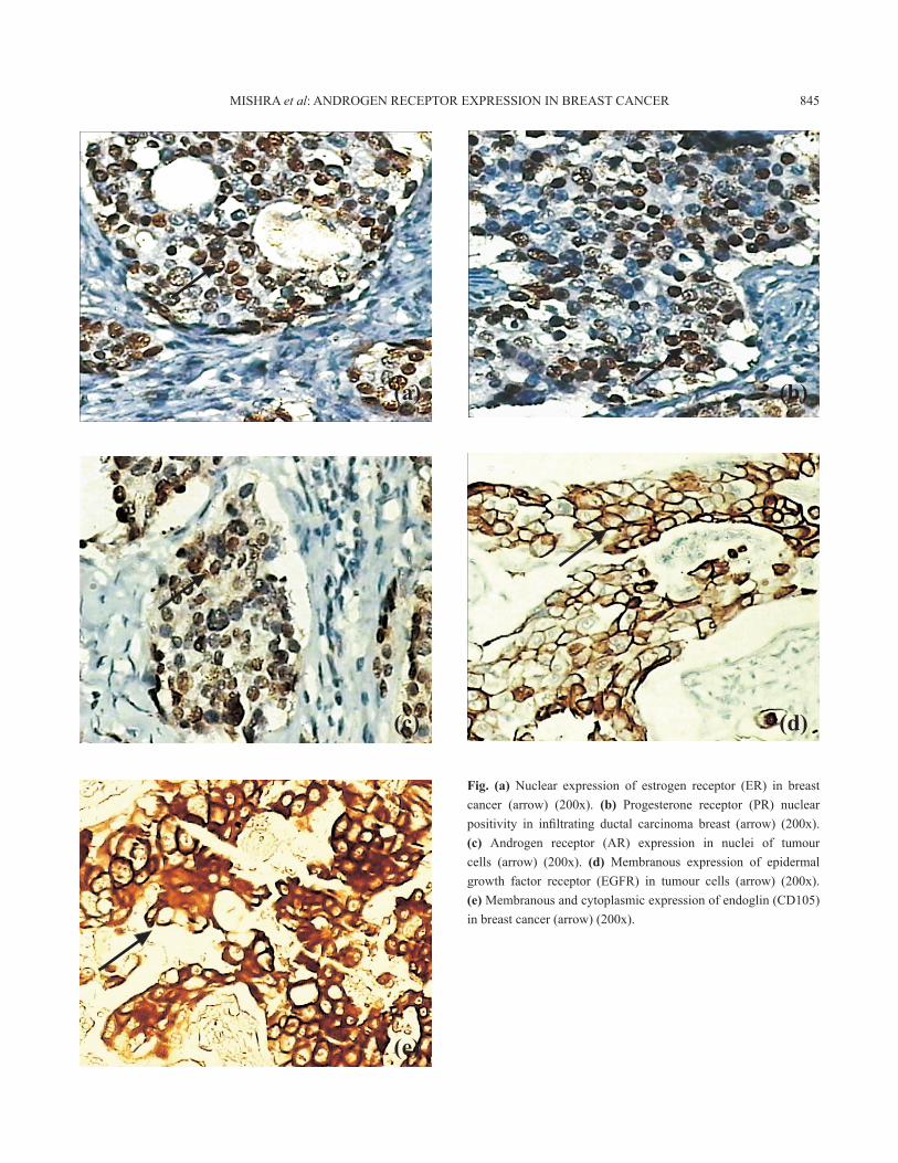
Fig. (a) Nuclear expression of estrogen receptor (ER) in breast cancer (arrow) (200x). (b) Progesterone receptor (PR) nuclear positivity in infiltrating ductal carcinoma breast (arrow) (200x). (c) Androgen receptor (AR) expression in nuclei of tumour cells (arrow) (200x). (d) Membranous expression of epidermal growth factor receptor (EGFR) in tumour cells (arrow) (200x). (e) Membranous and cytoplasmic expression of endoglin (CD105) in breast cancer (arrow) (200x).
MISHRA et al: ANDROGEN RECEPTOR EXPRESSION IN BREAST CANCER 845
(a) (b)
(c) (d)
(e)
of component loading factor(clf) of 0.5 or more for various components under the considered dimensions and correlation pattern/s in CATPCA are considered relevant for further investigation. The sensitivity, specificity, likelihood ratio of a positive and negative test for clinical response to NACT (non responders:<50%; responders: >50% regression in maximum diameter of initial tumour)14,15 along with their 95% CI were estimated for each individual marker as well as for the relevant profiles as obtained in CATPCA.
Results
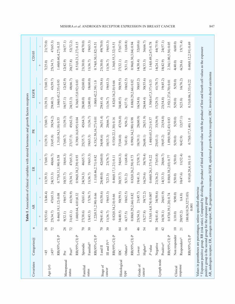
Association of clinical co-variates with markers: The distribution of various clinical parameters among study group (Table I) showed a high percentage of cases above 45 yr (72%) of age, and post-menopausal (72%). Family history of cancer in the siblings and close relatives was seen in 30 per cent cases. Majority of the cases (84%) were of infiltrating ductal of breast, 54.0 per cent were of higher grade and 42 per cent cases showed lymph metastasis (Table I). Among 31 LABC cases, 21 (67.7%) responded to NACT.
Expression of the steroid receptors, AR, ER and PR was observed in 40, 35 and 44 per cent cases, respectively, while that of EGFR and CD105 was seen in 40 and 32 per cent cases, respectively. The expression of ER was not found associated with any of the clinicopathological features, while PR was found to be significantly associated with pre-menopausal status (P=0.04) and presence of family history (P=0.01). EGFR was also seen to be significantly associated with pre-menopausal status (P=0.03) (Table I). The expression of AR was found to be significantly associated with lower grade (p=0.007). Among 31 LABC cases expression of AR, ER, PR, EGFR, CD105 was found in 21 (67.7%), 15 (48.4%), 14 (45.2%), 10 (32.3%) and 10 (32.3%) cases, respectively. Significant association of theraputic response among LABC cases was found with the expression of AR (P=0.001). The expression of both growth factor receptors, EGFR and CD105, was significantly associated with lymph node metastasis (P=0.01, P=0.05, respectively).
Results of CATPCA: As androgen receptor was the only marker associated with clinical response, the contribution of other markers to the clinical behaviour of the tumour was evaluated by CATPCA which depicted four correlation patterns (with a component loading factor of at least 0.50 taken as relevant). These four patterns were identified as positive correlation of AR with CD105 (AR+/CD105+; under 1st component
assessing the therapeutic response in terms of reduction in tumour size.
Histopathological examination and subsequent immunohistochemical (IHC) studies were performed for the biomarkers- AR (Neomarkers; Fremont, CA, 1:50); ER (DAKO; Denmark, 1:50); PR (DAKO; Denmark, 1:50); EGFR (DAKO; Denmark, 1:50) and CD105 (Neomarkers; Fremont, CA, 1:50). Cases were labelled as positive when >10 per cent tumour cells expressed the marker. IHC was performed on 4 µm paraffin sections, with antigen retrieval in citrate buffer at pH 6.0 in the microwave oven; endogenous peroxidase blocking with 3 per cent hydrogen peroxide; incubating with primary mouse monoclonal antibody overnight at 40oC at room temperature; with DAB (diaminobenzidine) as chromogen. Tumour cells were considered positive for the nuclear expression of ER, PR and AR and for the cytoplasmic expression of EGFR and CD105 (Fig.).
The association of various factors viz., age (<45/>45 yr); menopause (pre/post); familial status (sporadic/familial); stage [low (I and II), high (III and IV)]; histological type (IDC/Others); grade (well and moderately differentiated carcinomas categorized as low and poorly differentiated carcinomas as high); lymph node metastases (-/+); clinical response (non responders/responder)11,12 with immunohistochemical expression of three steroid receptors AR, ER and PR (-/+) and two growth factors EGFR and CD105 (+/-) in tumour cells was assessed by the χ2 and Fisher’s exact test. To measure the strength of association, risk ratio (RR) along with 95% confidence interval (CI) was also estimated. The two sided P<0.05 was considered statistically significant.
Co-expression pattern between steroid receptors (SRs) and growth factors (GFs), were analyzed by categorical principal component analysis (CATPCA). The CATPCA is considered as an exploratory analysis. PCA reveals linear combinations of the analyzing variables (in present case SRs and GFs) with large variance. The goal of PCA is to reduce an original set of variables into a smaller set of uncorrelated components that represent most of the information found in the original variables. The CATPCA yields new sets of linearly combined expression, but of these, the biologically relevant profile was investigated for its association with clinicopathological characteristics. Although exploratory, CATPCA helps in providing important information that can be further examined by implementing conformational analysis. The value
846 INDIAN J MED RES, JUNE 2012
Tabl
e I.
Ass
ocia
tion
of c
linic
al v
aria
bles
with
ster
oid
horm
ones
and
gro
wth
fact
ors r
ecep
tors
Cov
aria
tes
Cat
egor
ies@
n
AR
ERPR
EGFR
CD
105
+-
+-
+-
+-
+-
Age
(yr)
<45
2815
(53.
6)13
(46.
4)11
(39.
3)17
(60.
7)11
(39.
3)17
(60.
7)11
(39.
3)17
(60.
7)7(
25.0
)21
(75.
0)
≥45@
72
25
(34.
7)47
(65.
3)24
(33.
3)48
(66.
7)33
(45.
8)39
(54.
2)29
(40.
3)43
(59.
7)25
(34.
7)47
(65.
3)
RR
(95%
CI)
P0.
46(0
.19,
1.12
) 0.0
80.
77(0
.31,
1.90
) 0.5
81.
31(0
.54,
3.18
) 0.5
51.
04(0
.43,
2.55
) 0.9
31.
60(0
.60,
4.27
) 0.3
5
Men
opau
sal
stat
usPr
e 28
9(32
.1)
19(6
7.9)
10(3
5.7)
18(6
4.3)
17(6
0.7)
11(3
9.3)
16(5
7.1)
12(4
2.9)
12(4
2.9)
16(5
7.1)
Post
@
72
31(4
3.1)
41(5
6.9)
25(3
4.7)
47(6
5.3)
27(3
7.5)
45(6
2.5)
24(3
3.3)
48(6
6.7)
20(2
7.8)
52(7
2.2)
RR
(95%
CI)
P1.
60(0
.64,
4.0
1)0.
320.
96(0
.38,
2.39
) 0.9
30.
39(0
.16,
0.95
) 0.0
40.
36(0
.15,
0.92
) 0.0
30.
51(0
.21,
1.27
) 0.1
5
Fam
ilial
st
atus
Spor
adic
70
27(3
8.6)
43(6
1.4)
24(3
4.3)
46(6
5.7)
25(3
5.7)
45(6
4.3)
28(4
0.0)
42(6
0.0)
21(3
0.0)
49(7
0.0)
Fam
ilial
@
3013
(43.
3)17
(56.
7)11
(36.
7)19
(63.
3)19
(63.
3)11
(36.
7)12
(40.
0)18
(60.
0)11
(36.
7)19
(63.
3)
RR
(95%
CI)
P1.
22(0
.51,
2.90
) 0.6
61.
11(0
.46,
2.71
) 0.8
26.
33(2
.50,
16.2
7) 0
.01
1.00
(0.4
2,2.
39) 1
.00.
74(0
.30,
1.82
) 0.5
1
Stag
e of
ca
ncer
I and
II
7029
(41.
4)41
(58.
6)28
(40.
0)42
(60.
0)34
(48.
6)36
(51.
4)29
(41.
4)41
(58.
6)21
(30.
0)49
(70.
0)
III a
nd IV
@
3011
(36.
7)19
(63.
3)7(
23.3
)23
(76.
7)10
(33.
3)20
(66.
7)11
(36.
7)19
(63.
3)11
(36.
7)19
(63.
3)
RR
(95%
CI)
P0.
82(0
.34,
2.0)
0.6
60.
46(0
.17,
1.21
) 0.1
10.
53(0
.22,
1.30
) 0.1
60.
82(0
.34,
2.0)
0.6
61.
36(0
.55,
3.32
) 0.5
1
His
tolo
gica
l ty
peID
C
8434
(40.
5)50
(59.
5)30
(35.
7)54
(64.
3)37
(44.
0)47
(56.
0)34
(40.
5)50
(59.
5)27
(32.
1)57
(67.
9)
Oth
ers@
16
6(37
.5)
10(6
2.5)
5(31
.2)
11(6
8.8)
7(43
.8)
9(56
.2)
6(37
.5)
10(6
2.5)
5(31
.3)
11(6
8.8)
RR
(95%
CI)
P0.
88(0
.29,
2.66
) 0.8
20.
82(0
.26,
2.58
) 0.7
30.
99(0
.34,
2.90
) 0.9
80.
88(0
.29,
2.66
) 0.8
20.
96(0
.30,
3.04
) 0.9
4
Gra
de o
f ca
ncer
Low
er
4625
(54.
3)21
(45.
7)19
(41.
3)27
(58.
7)18
(39.
1)28
(60.
9)16
(34.
8)30
(65.
2)14
(30.
4)32
(69.
6)
Hig
her@
54
15(2
7.8)
39(7
2.2)
16(2
9.6)
38(7
0.4)
26(4
8.1)
28(5
1.9)
24(4
4.4)
30(5
5.6)
18(3
3.3)
36(6
6.7)
P va
lue
0.33
(0.1
4,0.
74) 0
.007
0.60
(0.2
6,1.
37) 0
.22
1.44
(0.6
5,3.
21) 0
.37
1.50
(0.6
7,3.
37) 0
.33
1.14
(0.4
9,2.
67) 0
.78
Lym
ph n
ode
Neg
ativ
e 58
24(4
1.4)
34(5
8.6)
21(3
6.2)
37(6
3.8)
25(4
3.1)
33(5
6.9)
17(2
9.3)
41(7
0.7)
14(2
4.1)
44(7
5.9)
Posi
tive@
42
16(3
8.1)
26(6
1.9)
14(3
3.3)
28(6
6.7)
19(4
5.2)
23(5
4.8)
23(5
4.8)
19(4
5.2)
18(4
2.9)
24(5
7.1)
RR
(95%
CI)
P0.
87(0
.39,
1.20
) 0.7
40.
88(0
.38,
2.03
) 0.7
71.
09(0
.50,
2.43
) 0.8
32.
92(1
.27,
6.70
) 0.0
12.
36(1
.00,
5.56
) 0.0
5
Clin
ical
re
spon
seN
on re
spon
der
101(
10.0
)9(
90.0
)5(
50.0
)5(
50.0
)5(
50.0
)5(
50.0
)5(
50.0
)5(
50.0
)4(
40.0
)6(
60.0
)
Res
pond
ers@
21
20(9
5.2)
1(4.
8)10
(47.
6)11
(52.
4)9(
42.9
)12
(57.
1)5(
23.8
)16
(76.
2)6(
28.6
)15
(71.
4)
RR
(95%
CI)
P18
0.0(
10.2
9,32
73.0
3)
0.00
10.
91(0
.20,
4.10
) 1.0
0.75
(0.1
7,3.
40) 1
.00.
31(0
.06,
1.53
) 0.2
20.
60(0
.12,
2.91
) 0.6
9
Valu
es in
par
enth
eses
are
per
cent
ages
, unl
ess s
peci
fied
othe
rwis
e@
Exp
osur
e po
sitiv
e gr
oup.
RR
(ris
k ra
tio).
Com
pute
d by
div
idin
g th
e pr
oduc
t of t
hird
and
seco
nd v
alue
with
the
prod
uct o
f firs
t and
four
th c
ell v
alue
s as t
he e
xpos
ure
posi
tive
grou
p is
the
seco
nd g
roup
for t
he e
xpos
ure
grou
p
AR
, and
roge
n re
cept
or; E
R, e
stro
gen
rece
ptor
; PR
, pro
gest
eron
e re
cept
or; E
GFR
, epi
derm
al g
row
th fa
ctor
rece
ptor
; ID
C, i
nvas
ive
duct
al c
arci
nom
a
MISHRA et al: ANDROGEN RECEPTOR EXPRESSION IN BREAST CANCER 847
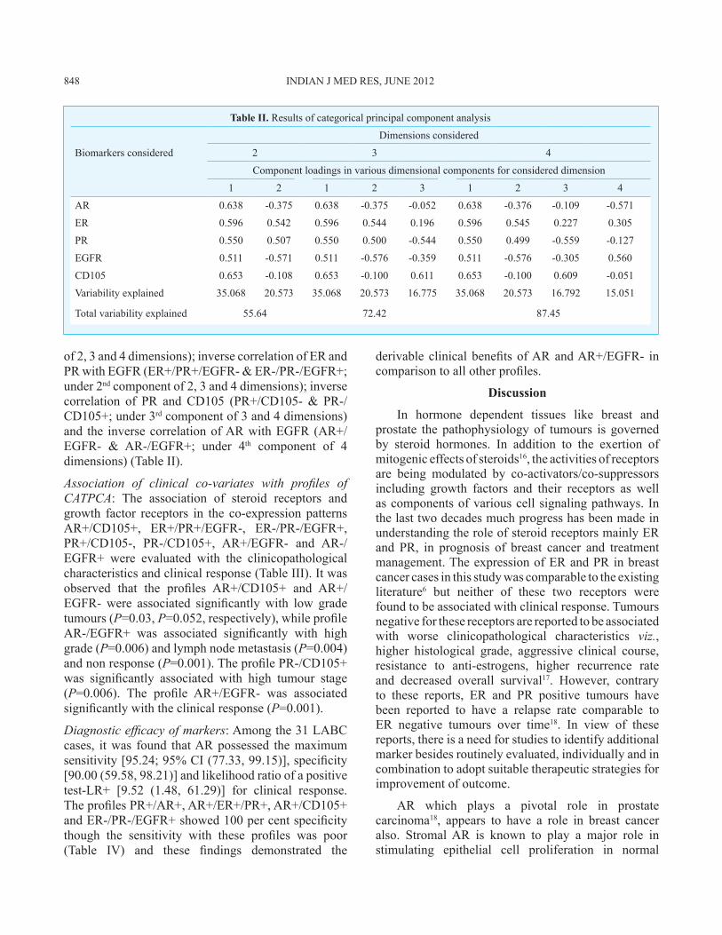
of 2, 3 and 4 dimensions); inverse correlation of ER and PR with EGFR (ER+/PR+/EGFR- & ER-/PR-/EGFR+; under 2nd component of 2, 3 and 4 dimensions); inverse correlation of PR and CD105 (PR+/CD105- & PR-/CD105+; under 3rd component of 3 and 4 dimensions) and the inverse correlation of AR with EGFR (AR+/EGFR- & AR-/EGFR+; under 4th component of 4 dimensions) (Table II).
Association of clinical co-variates with profiles of CATPCA: The association of steroid receptors and growth factor receptors in the co-expression patterns AR+/CD105+, ER+/PR+/EGFR-, ER-/PR-/EGFR+, PR+/CD105-, PR-/CD105+, AR+/EGFR- and AR-/EGFR+ were evaluated with the clinicopathological characteristics and clinical response (Table III). It was observed that the profiles AR+/CD105+ and AR+/EGFR- were associated significantly with low grade tumours (P=0.03, P=0.052, respectively), while profile AR-/EGFR+ was associated significantly with high grade (P=0.006) and lymph node metastasis (P=0.004) and non response (P=0.001). The profile PR-/CD105+ was significantly associated with high tumour stage (P=0.006). The profile AR+/EGFR- was associated significantly with the clinical response (P=0.001).
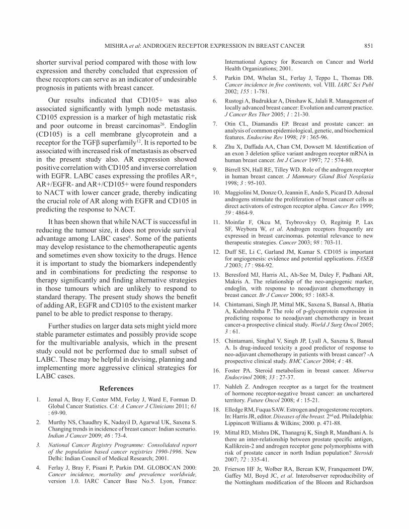
Diagnostic efficacy of markers: Among the 31 LABC cases, it was found that AR possessed the maximum sensitivity [95.24; 95% CI (77.33, 99.15)], specificity [90.00 (59.58, 98.21)] and likelihood ratio of a positive test-LR+ [9.52 (1.48, 61.29)] for clinical response. The profiles PR+/AR+, AR+/ER+/PR+, AR+/CD105+ and ER-/PR-/EGFR+ showed 100 per cent specificity though the sensitivity with these profiles was poor (Table IV) and these findings demonstrated the
derivable clinical benefits of AR and AR+/EGFR- in comparison to all other profiles.
Discussion
In hormone dependent tissues like breast and prostate the pathophysiology of tumours is governed by steroid hormones. In addition to the exertion of mitogenic effects of steroids16, the activities of receptors are being modulated by co-activators/co-suppressors including growth factors and their receptors as well as components of various cell signaling pathways. In the last two decades much progress has been made in understanding the role of steroid receptors mainly ER and PR, in prognosis of breast cancer and treatment management. The expression of ER and PR in breast cancer cases in this study was comparable to the existing literature6 but neither of these two receptors were found to be associated with clinical response. Tumours negative for these receptors are reported to be associated with worse clinicopathological characteristics viz., higher histological grade, aggressive clinical course, resistance to anti-estrogens, higher recurrence rate and decreased overall survival17. However, contrary to these reports, ER and PR positive tumours have been reported to have a relapse rate comparable to ER negative tumours over time18. In view of these reports, there is a need for studies to identify additional marker besides routinely evaluated, individually and in combination to adopt suitable therapeutic strategies for improvement of outcome.
AR which plays a pivotal role in prostate carcinoma18, appears to have a role in breast cancer also. Stromal AR is known to play a major role in stimulating epithelial cell proliferation in normal
Table II. Results of categorical principal component analysis
Dimensions considered
Biomarkers considered 2 3 4
Component loadings in various dimensional components for considered dimension
1 2 1 2 3 1 2 3 4
AR 0.638 -0.375 0.638 -0.375 -0.052 0.638 -0.376 -0.109 -0.571
ER 0.596 0.542 0.596 0.544 0.196 0.596 0.545 0.227 0.305
PR 0.550 0.507 0.550 0.500 -0.544 0.550 0.499 -0.559 -0.127
EGFR 0.511 -0.571 0.511 -0.576 -0.359 0.511 -0.576 -0.305 0.560
CD105 0.653 -0.108 0.653 -0.100 0.611 0.653 -0.100 0.609 -0.051
Variability explained 35.068 20.573 35.068 20.573 16.775 35.068 20.573 16.792 15.051
Total variability explained 55.64 72.42 87.45
848 INDIAN J MED RES, JUNE 2012

Tabl
e II
I. A
ssoc
iatio
n of
clin
ical
var
iabl
es a
nd re
spon
ders
with
rele
vant
pro
file
unde
r CAT
PCA
N=10
0n=
31
Stag
eH
isto
logi
cal t
ype
Gra
deLy
mph
nod
eR
espo
nder
s
I and
IIII
I and
IVID
CO
ther
sLo
wer
Hig
her
-+
R+
R-
AR
/CD
105
+/+
914
(73.
7)5(
26.3
)14
(73.
7)5(
26.3
)13
(68.
4)6(
31.6
)10
(52.
6)9(
47.4
)6(
100.
0)0(
0.0)
Oth
ers
8156
(69.
1)25
(30.
9)70
(86.
4)11
(13.
6)33
(40.
7)48
(59.
3)48
(59.
3)33
(40.
7)15
(60.
0)10
(40.
0)
RR
(95%
CI)
P
1.25
(0.4
1,3.
85) 0
.70
0.44
(0.1
3,1.
46) 0
.17
3.15
(1.0
9,9.
17) 0
.03
0.76
(0.2
8,2.
08) 0
.60
NE,
0.0
6
ER/P
R/E
GFR
+/+/
- 13
11(8
4.6)
2(15
.4)
11(8
4.6)
2(15
.4)
8(61
.5)
5(38
.5)
10(7
6.9)
3(23
.1)
4(80
.0)
1(20
.0)
Oth
ers
8759
(67.
8)28
(32.
2)73
(83.
9)14
(16.
1)38
(43.
7)49
(56.
3)48
(55.
2)39
(44.
8)17
(65.
4)9(
34.6
)
RR
(95%
CI)
P
2.61
(0.5
4,12
.62)
0.2
21.
05(0
.21,
5.29
) 0.9
52.
06(0
.63,
6.83
) 0.2
32.
71(0
.70,
10.5
7) 0
.14
2.12
(0.2
1,21
.95)
0.5
2
-/-/+
13
8(61
.5)
5(38
.5)
11(8
4.6)
2(15
.4)
5(38
.5)
8(61
.5)
6(46
.2)
7(53
.8)
2(10
0.0)
0(0.
0)
Oth
ers
8762
(71.
3)25
(28.
7)73
(83.
9)14
(16.
1)41
(47.
1)46
(52.
9)52
(59.
8)35
(40.
2)19
(65.
5)10
(34.
5)
RR
(95%
CI)
P
0.65
(0.1
9,2.
16) 0
.48
1.05
(0.2
1,5.
29) 0
.95
0.70
(0.2
1,2.
31) 0
.56
0.58
(0.1
8,1.
86) 0
.35
NE,
0.3
1
PR/C
D10
5-/+
15
6(40
.0)
9(60
)12
(80.
0)3(
20.0
)7(
46.7
)8(
53.3
)6(
40.0
)9(
60.0
)3(
60.0
)2(
40.0
)
Oth
ers
8564
(75.
3)21
(24.
7)72
(84.
7)13
(15.
3)39
(45.
9)46
(54.
1)52
(61.
2)33
(38.
8)18
(69.
2)8(
30.8
)
RR
(95%
CI)
P
0.22
(0.0
7,0.
68) 0
.006
0.72
(0.1
8,2.
91) 0
.65
1.03
(0.3
4,3.
10) 0
.96
0.42
(0.1
4,1.
29) 0
.13
0.67
(0.0
9,4.
79) 0
.69
+/-
2719
(70.
4)8(
29.6
)22
(81.
5)5(
18.5
)11
(40.
7)16
(59.
3)17
(63.
0)10
(37.
0)6(
66.7
)3(
33.3
)
Oth
ers
73
51(6
9.9)
22(3
0.1)
62(8
4.9)
11(1
5.1)
35(4
7.9)
38(5
2.1)
41(5
6.2)
32(4
3.8)
15(6
8.2)
7(31
.8)
RR
(95%
CI)
P
1.02
(0.3
9,2.
69) 0
.97
0.78
(0.2
4,2.
50) 0
.68
0.75
(0.3
0,1.
82) 0
.52
1.33
(0.5
4,3.
29) 0
.54
0.93
(0.1
8,4.
86) 0
.94
AR
/EG
FR-/+
18
10(5
5.6)
8(44
.4)
15(8
3.3)
3(16
.7)
3(16
.7)
15(8
3.3)
5(27
.8)
13(7
2.2)
0(0.
0)4(
100.
0)
Oth
ers
8260
(73.
2)22
(26.
8)69
(84.
1)13
(15.
9)43
(52.
4)39
(47.6
)53
(64.
6)29
(35.
4)21
(77.
8)6(
22.2
)
RR
(95%
CI)
P
0.46
(0.1
6,1.
31) 0
.14
0.94
(0.2
4,3.
72) 0
.93
0.18
(0.0
5,0.
67) 0
.006
0.21
(0.0
7,0.
65) 0
.004
NE,
0.0
1
+/-
1810
(55.
6)8(
44.4
)15
(83.
3)3(
16.7
)12
(66.
7)6(
33.3
)12
(66.
7)6(
33.3
)15
(100
.0)
0(0.
0)
Oth
ers
82
60(7
3.2)
22(2
6.8)
69(8
4.1)
13(1
5.9)
34(4
1.5)
48(5
8.8)
46(5
6.1)
36(4
3.9)
6(37
.5)
10(6
2.5)
RR
(95%
CI)
P
0.46
(0.1
6,0.
14) 0
.14
0.94
(0.2
4,3.
72) 0
.93
2.82
(0.9
7,8.
30) 0
.052
1.57
(0.5
4,4.
58) 0
.41
NE,
0.0
01
NE,
not
est
imat
ed d
ue to
zer
o ce
ll fr
eque
ncy
MISHRA et al: ANDROGEN RECEPTOR EXPRESSION IN BREAST CANCER 849
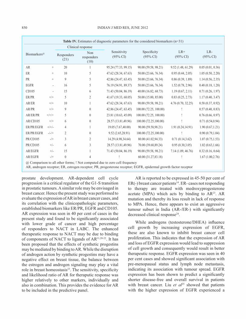
Table IV. Estimates of diagnostic parameters for the considered biomarkers (n=31)
Biomarkers@
Clinical responseSensitivity (95% CI)
Specificity (95% CI)
LR+ (95% CI)
LR- (95% CI)Responders
(21)
Non responders
(10)AR + 20 1 95.24 (77.33, 99.15) 90.00 (59.58, 98.21) 9.52 (1.48, 61.29) 0.05 (0.01, 0.36)
ER + 10 5 47.62 (28.34, 67.63) 50.00 (23.66, 76.34) 0.95 (0.44, 2.05) 1.05 (0.50, 2.20)
PR + 9 5 42.86 (24.47, 63.45) 50.00 (23.66, 76.34) 0.86 (0.39, 1.89) 1.14 (0.56, 2.35)
EGFR - 16 5 76.19 (54.91, 89.37) 50.00 (23.66, 76.34) 1.52 (0.78, 2.96) 0.48 (0.18, 1.28)
CD105 - 15 6 71.43 (50.04, 86.19) 40.00 (16.82, 68.73) 1.19 (0.67, 2.11) 0.71 (0.26, 1.97)
ER/PR +/+ 5 2 41.67 (19.33, 68.05) 50.00 (15.00, 85.00) 0.83 (0.25, 2.73) 1.17 (0.40, 3.47)
AR/ER +/+ 10 1 47.62 (28.34, 67.63) 90.00 (59.58, 98.21) 4.76 (0.70, 32.25) 0.58 (0.37, 0.92)
AR/PR +/+ 9 0 42.86 (24.47, 63.45) 100.00 (72.25, 100.00) ! 0.57 (0.40, 0.83)
AR/ER/PR +/+/+ 5 0 23.81 (10.63, 45.09) 100.00 (72.25, 100.00) ! 0.76 (0.66, 0.97)
AR/CD105 +/+ 6 0 28.57 (13.81,49.96) 100.00 (72.25,100.00) ! 0.71 (0.54,0.94)
ER/PR/EGFR +/+/- 4 1 19.05 (7.67,40.00) 90.00 (59.58,98.21) 1.91 (0.24,14.91) 1.90 (0.67,1.21)
ER/PR/EGFR -/-/+ 2 0 9.52 (2.65,28.91) 100.00 (72.25,100.00) ! 0.90 (0.79,1.04)
PR/CD105 -/+ 3 2 14.29 (4.98,34.64) 80.00 (41.02,94.33) 0.71 (0.14,3.62) 1.07 (0.75,1.53)
PR/CD105 +/- 6 3 28.57 (13.81,49.96) 70.00 (39.68,89.24) 0.95 (0.30,3.05) 1.02 (0.63,1.66)
AR/EGFR +/- 15 1 71.43 (50.04, 86.19) 90.00 (59.58, 98.21) 7.14 (1.09, 46.76) 0.32 (0.16, 0.64)AR/EGFR -/+ 0 4 ! 60.00 (31.27,83.18) ! 1.67 (1.00,2.76)@ Comparison to all other forms; ! Not computed due to zero cell frequencyAR, androgen receptor; ER, estrogen receptor; PR, progesterone receptor; EGFR, epidermal growth factor receptor
prostate development. AR-dependent cell cycle progression is a critical regulator of the G1-S transition in prostatic tumours. A similar role may be envisaged in breast cancer. Hence the present study was performed to evaluate the expression of AR in breast cancer cases, and its correlation with the clinicopathologic parameters, established biomarkers like ER/PR, EGFR and CD105. AR expression was seen in 40 per cent of cases in the present study and found to be significantly associated with lower grade of cancer and high percentage of responders to NACT in LABC. The enhanced therapeutic response to NACT may be due to binding of components of NACT to ligands of AR11,20-22. It has been proposed that the effects of synthetic progestins may be mediated by binding to AR. While the disruption of androgen action by synthetic progestins may have a negative effect on breast tissue, the balance between the estrogen and androgen signaling may play a vital role in breast homeostasis23. The sensitivity, specificity and likelihood ratio of AR for theraputic response was higher relatively to other markers, individually and also in combination. This provides the evidence for AR to be included in the predictive panel.
AR is reported to be expressed in 45-50 per cent of ER(-) breast cancer patients14. ER- cases not responding to therapy are treated with medroxyprogesterone acetate (MPA) which acts by binding to AR23. AR mutation and thereby its loss result in lack of response to MPA. Hence, there appears to exist an aggressive tumour subset in India (AR-/ER-) with significantly decreased clinical response24.
While androgens (testosterone/DHEA) influence cell growth by increasing expression of EGFR, these are also known to inhibit breast cancer cell proliferation. This indicates that the expression of AR and loss of EGFR expression would lead to suppression of cell growth and consequently would result in better therapeutic response. EGFR expression was seen in 40 per cent cases and showed significant association with pre-menopausal status and lymph node metastasis, indicating its association with tumour spread. EGFR expression has been shown to predict a significantly shorter disease-free and overall survival in patients with breast cancer. Liu et al25 showed that patients with the higher expression of EGFR experienced a
850 INDIAN J MED RES, JUNE 2012
shorter survival period compared with those with low expression and thereby concluded that expression of these receptors can serve as an indicator of undesirable prognosis in patients with breast cancer.
Our results indicated that CD105+ was also associated significantly with lymph node metastasis. CD105 expression is a marker of high metastatic risk and poor outcome in breast carcinomas26. Endoglin (CD105) is a cell membrane glycoprotein and a receptor for the TGFβ superfamily12. It is reported to be associated with increased risk of metastasis as observed in the present study also. AR expression showed positive correlation with CD105 and inverse correlation with EGFR. LABC cases expressing the profiles AR+, AR+/EGFR- and AR+/CD105+ were found responders to NACT with lower cancer grade, thereby indicating the crucial role of AR along with EGFR and CD105 in predicting the response to NACT.
It has been shown that while NACT is successful in reducing the tumour size, it does not provide survival advantage among LABC cases6. Some of the patients may develop resistance to the chemotherapeutic agents and sometimes even show toxicity to the drugs. Hence it is important to study the biomarkers independently and in combinations for predicting the response to therapy significantly and finding alternative strategies in those tumours which are unlikely to respond to standard therapy. The present study shows the benefit of adding AR, EGFR and CD105 to the existent marker panel to be able to predict response to therapy.
Further studies on larger data sets might yield more stable parameter estimates and possibly provide scope for the multivariable analysis, which in the present study could not be performed due to small subset of LABC. These may be helpful in devising, planning and implementing more aggressive clinical strategies for LABC cases.
ReferencesJemal A, Bray F, Center MM, Ferlay J, Ward E, Forman D. 1. Global Cancer Statistics. CA: A Cancer J Clinicians 2011; 61 : 69-90. Murthy NS, Chaudhry K, Nadayil D, Agarwal UK, Saxena S. 2. Changing trends in incidence of breast cancer: Indian scenario. Indian J Cancer 2009; 46 : 73-4.National Cancer Registry Programme: Consolidated report 3. of the population based cancer registries 1990-1996. New Delhi: Indian Council of Medical Research; 2001.Ferlay J, Bray F, Pisani P, Parkin DM. GLOBOCAN 2000: 4. Cancer incidence, mortality and prevalence worldwide, version 1.0. IARC Cancer Base No.5. Lyon, France:
International Agency for Research on Cancer and World Health Organizations; 2001.Parkin DM, Whelan SL, Ferlay J, Teppo L, Thomas DB. 5. Cancer incidence in five continents, vol. VIII. IARC Sci Publ 2002; 155 : 1-781.Rustogi A, Budrukkar A, Dinshaw K, Jalali R. Management of 6. locally advanced breast cancer: Evolution and current practice. J Cancer Res Ther 2005; 1 : 21-30.Otin CL, Diamandis EP. Breast and prostate cancer: an 7. analysis of common epidemiological, genetic, and biochemical features. Endocrine Rev 1998; 19 : 365-96.Zhu X, Daffada AA, Chan CM, Dowsett M. Identification of 8. an exon 3 deletion splice variant androgen receptor mRNA in human breast cancer. Int J Cancer 1997; 72 : 574-80.Birrell SN, Hall RE, Tilley WD. Role of the androgen receptor 9. in human breast cancer. J Mammary Gland Biol Neoplasia 1998; 3 : 95-103.Maggiolini M, Donze O, Jeannin E, Ando S, Picard D. Adrenal 10. androgens stimulate the proliferation of breast cancer cells as direct activators of estrogen receptor alpha. Cancer Res 1999; 59 : 4864-9.Moinfar 11. F, Okcu M, Tsybrovskyy O, Regitnig P, Lax SF, Weybora W, et al. Androgen receptors frequently are expressed in breast carcinomas. potential relevance to new therapeutic strategies. Cancer 2003; 98 : 703-11.Duff SE, Li C, Garland JM, Kumar S. CD105 is important 12. for angiogenesis: evidence and potential applications. FASEB J 2003; 17 : 984-92.Beresford MJ, Harris AL, Ah-See M, Daley F, Padhani AR, 13. Makris A. The relationship of the neo-angiogenic marker, endoglin, with response to neoadjuvant chemotherapy in breast cancer. Br J Cancer 2006; 95 : 1683-8.Chintamani, Singh JP, Mittal MK, Saxena S, Bansal A, Bhatia 14. A, Kulshreshtha P. The role of p-glycoprotein expression in predicting response to neoadjuvant chemotherapy in breast cancer-a prospective clinical study. World J Surg Oncol 2005; 3 : 61.Chintamani, Singhal V, Singh JP, Lyall A, Saxena S, Bansal 15. A. Is drug-induced toxicity a good predictor of response to neo-adjuvant chemotherapy in patients with breast cancer? -A prospective clinical study. BMC Cancer 2004; 4 : 48.Foster P16. A. Steroid metabolism in breast cancer. Minerva Endocrinol 2008; 33 : 27-37.Nahleh 17. Z. Androgen receptor as a target for the treatment of hormone receptor-negative breast cancer: an unchartered territory. Future Oncol 2008; 4 : 15-21.Elledge RM, Fuqua SAW. Estrogen and progesterone receptors. 18. In: Harris JR, editor. Diseases of the breast. 2nd ed. Philadelphia: Lippincott Williams & Wilkins; 2000. p. 471-88. Mittal RD, Mishra DK, Thanagraj K, Singh R, Mandhani A. Is 19. there an inter-relationship between prostate specific antigen, Kallikrein-2 and androgen receptor gene polymorphisms with risk of prostate cancer in north Indian population? Steroids 2007; 72 : 335-41. Frierson HF J20. r, Wolber RA, Berean KW, Franquemont DW, Gaffey MJ, Boyd JC, et al. Interobserver reproducibility of the Nottingham modification of the Bloom and Richardson
MISHRA et al: ANDROGEN RECEPTOR EXPRESSION IN BREAST CANCER 851
histologic grading scheme for infiltrating ductal carcinoma. Am J Clin Pathol 1995; 103 : 195-8.
Ogawa 21. Y, Moriya T, Kato Y, Oguma M, Ikeda K, Takashima T, et al. Immunohistochemical assessment for estrogen receptor and progesterone receptor status in breast cancer: analysis for a cut-off point as the predictor for endocrine therapy. Breast Cancer 2004; 11 : 267-75.
Riva 22. C, Dainese E, Caprara G, Rocca PC, Massarelli G, Tot T, et al. Immunohistochemical study of androgen receptors in breast carcinoma. Evidence of their frequent expression in lobular carcinoma. Virchows Arch 2005; 447 : 695-700.
Birrell S23. N, Butler LM, Harris JM, Buchanan G, Tilley WD. Disruption of androgen receptor signaling by synthetic
progestins may increase risk of developing breast cancer. FASEB J 2007; 21 : 2285-93.Seshdari R, Shah PN. Steroid receptors in breast cancer in 24. Indian women. Anticancer Res 1984; 6 : 403-7.Liu Y, Ji R, LiJ, Gu Q, Zhao X, Sun T, Li J, 25. et al. Correlation effect of EGFR and CXCR4 and CCR7 chemokine receptors in predicting breast cancer metastasis and prognosis. J Exp Clin Cancer Res 2010; 24 : 16.Dales J, Garcia S, Bonnier P, Duffaud F, Andrac-Meyer 26. L, Ramuz O, et al. CD105 expression is a marker of high metastatic risk and poor outcome in breast carcinomas: correlations between immunohistochemical analysis and long-term follow-up in a series of 929 patients. Am J Clin Pathol 2003; 119 : 374-80.
Reprint requests: Dr Sunita Saxena, Director, National Institute of Pathology (ICMR), Safdarjung Hospital Campus, Post Box No: 4909, New Delhi 110 029, India
e-mail: [email protected]
852 INDIAN J MED RES, JUNE 2012