evolution of supraclavicular brachial plexus...
TRANSCRIPT
Evolution of Supraclavicular Brachial Plexus Block
Teo Wei Wei1, Beh Zhi Yuen2, Shahridan Mohd Fathil3
1Department of Anaesthesia, National University Hospital, Singapore 2Anaesthesia Department, Changi General Hospital, Singapore 3Anaesthesia Department, Ng Teng Fong General Hospital, Singapore Often considered the ‘spinal anaesthesia of
the upper extremity’, the supraclavicular
approach to the brachial plexus provides
excellent anaesthesia of the upper limb with
rapid onset1. Its use in history dates all the
way back to the 1920s, but gradually fell out
of favour due to high incidence of
pneumothorax, improvement in general
anaesthesia safety and safer alternative
approaches. With the advent of ultrasound-
guided techniques allowing real-time
visualisation of anatomy, there has been
renewed interest in the block due to
increased safety and reduced complication
rate.
ANATOMY
The brachial plexus supplies motor and
sensory innervation to the upper limb. It is
formed by the ventral rami of C5 to T1. They
emerge, as roots, between the anterior and
middle scalene muscles, then proceed to
traverse the posterior triangle, forming three
trunks, the upper, middle and lower.
Posterior to the mid clavicle, each trunk then
divides to form an anterior and posterior
division. The divisions then combine to form
the lateral, medial and posterior cords, which
are named according to their relation to the
second part of the axillary artery. Various
peripheral nerves, including the terminal
branches, emerge from these cords.
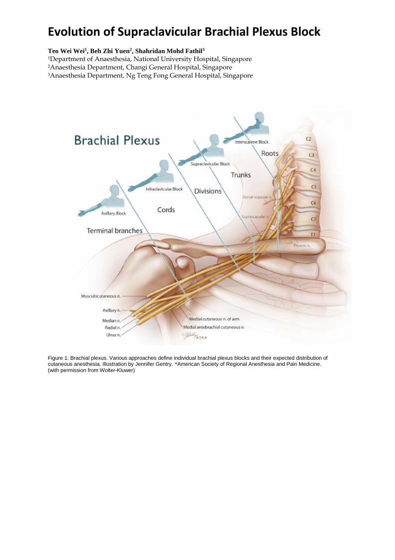
A brachial plexus block can be performed at
multiple sites along its anatomical path.
Common approaches include that at the
interscalene, supraclavicular, infraclavicular
and axillary levels (Figure 1)2. At the level
of the supraclavicular fossa, the plexus is
most compactly arranged, consisting of
distal trunks and origins of divisions. Hence,
the supraclavicular approach of the brachial
plexus has been thought to provide
anaesthesia to the entire upper extremity
with a rapid onset and in the most consistent
manner.
At the supraclavicular fossa, both the
brachial plexus and subclavian artery lie
above the first rib and the pleura. The
brachial plexus is located lateral and
posterior to the subclavian artery, while the
subclavian vein and anterior scalene muscle
are found medial to the subclavian artery.
The pleura is usually situated within 1-2 cm
medial from the brachial plexus.
INDICATIONS AND BENEFITS
The most common indication of the
supraclavicular brachial plexus bock is upper
extremity surgery1. As with all peripheral
nerve blocks (PNBs), supraclavicular
brachial plexus block offers an excellent
anaesthetic alternative for upper limb
surgery. It provides superior, long-lasting
analgesia, and avoids potential side effects of
a general anaesthesia including nausea,
vomiting, dental trauma, sore throat, allergic
reactions and intraoperative haemodynamics
swings. PNBs indeed offer distinct benefits
over general or neuraxial anaesthesia in
certain clinical situations, especially high
risk patients.
HISTORY
The first documented brachial plexus block
was performed by William Steward Halsted
in 1884, who directly exposed the brachial
plexus in the neck with cocaine3. It was only
in 1911 when Kulenkampff performed the
first percutaneous supraclavicular brachial
plexus block4. In collaboration with Persky5,
Kulenkampff’s technique and experience
with 1000 supraclavicular brachial plexus
blocks was published in 1928.
However, Kulenkampff’s technique of
inserting the needle posteriorly, medially
and caudally in the direction of T2 or T3
spinous process, carried an inherent risk of
pneumothorax.5 This, together with
improvements in general anaesthesia safety,
as well as the advent of reportedly safer
alternatives including axillary approach by
Accardo and Adriano (1949)6, Eather and
Burnham (1958)7, later De Jong (1961)8,
supraclavicular approach gradually fell out
of favour in the early 1960s.
Until the last two decades, with the
introduction of real-time ultrasound guided
techniques to reduce risk of inadvertent
pleura puncture, the supraclavicular
approach of the brachial plexus, with its
rapid onset, high success rate and large area
of anaesthesia coverage, has gradually
regained popularity.1, 2, 9, 10
TECHNIQUES
A. Surface landmark with paraesthesia
seeking
Classical approach
(‘Kulenkampff technique’)
The classical Kulenkampff approach
involves the patient to be in the sitting
position, with the arm to be ‘blocked’ lying
in the lap with the shoulder relaxed. If the
sitting position is not possible, the patient
will lie supine, with a pillow under his
scapula and his head rotated opposite from
the side to be blocked4, 5.
The needle is inserted at a point middle of the
clavicle, crossed by a line projected
downward from the external jugular vein. It
is advanced lateral to the subclavian artery
and is directed posteriorly, medially and
caudally to the upper border of the first rib
(i.e. in the direction of the T2 or T3 spinous
process). The classical approach involves
inducing paraesthesia in the finger tips
usually at a depth of 1-2cm. This indicates
the needle’s contact with the plexus. Local
anaesthetic is then slowly injected, with
paraesthesia increasing temporarily until the
local anaesthetic’s action causes the
sensation to disappear.4, 5.
Modified techniques
The medial orientation of the needle in the
classical approach was associated with
increased risk of pleural puncture and
pneumothorax, reported 6% incidence5.
Consequently, attempts to modify the
classical technique were described to reduce
this risk.
Several modified techniques were published
in chronological order:
MacIntosh & Mushin (1942)11
Lamoureux & Bourgeois-Gavardin
(1952)12
Subclavian perivascular technique –
Winnie & Collins (1964) 13
Parascalene technique – Vongvises &
Panijayanond (1972) 14
Dupre & Danel technique (1982)15
Brown’s plump-bob technique (1988)16
There were great diversity of technique with
minimal variations revealed that none of
them was perfect and free from potential
hazard. Below are some techniques worth
mention.
Subclavian perivascular technique –
Winnie & Collins13
This is a surface landmark with paraesthesia
seeking technique. The needle is inserted at
the base of interscalene groove, posterior to
the subclavian artery, in the horizontal plane.
The disadvantages of this technique are
vascular puncture, hematoma, and
pneumothorax (less than 1:1000 in
experienced hand).
Dupre & Danel technique15
This is also a surface landmark with
paraesthesia seeking technique. Surface
landmarks: the external jugular vein, the
sternocleidomastoid muscle, and the
clavicular insertion of the trapezius muscle.
The needle is inserted at the intersection
point between external jugular vein and a
line drawn from the top of supraclavicularis
minor fossa to edge of external clavicular
insertion of trapezius muscle. The advantage
is that it did not require location of
subclavian artery. No pneumothorax
reported in 136 cases.
Brown’s plump-bob technique16
This is initially a surface landmark with
paraesthesia seeking technique which later
incorporate the use of a nerve stimulator.
This is performed with the patient supine on
a horizontal table with the ipsilateral arm at
the side and the head turned opposite the side
to be blocked. The point of needle insertion
is “immediately adjacent and superior to the
clavicle at the lateral-most insertion of the
sternocleidomastoid muscle onto the
clavicle”. The needle direction is
anteroposterior—that is, perpendicular to the
table—as if following the line of a suspended
plumb-bob through the insertion site. Local
anaesthetic is injected at a single site after
adequate paresthesia or motor response by a
nerve stimulator.
B. Surface landmark with nerve
stimulator
Locating nerves by obtaining paraesthesia
could indicate that the needle tip is
intraneural. If local anaesthetic were to be
injected despite the paraesthesia, this could
potentially result in neural damage and
complications. On the contrary, absence of
paresthesia does not reliably exclude the
possibility of needle-to-nerve contact nor
does it prevent post-operative neural injury
(PNI). Nevertheless, severe paresthesia that
occurs with needle advancement or injection
should prompt the cessation of either
maneuver, and repositioning of the needle
should be considered.17.
In 1962, Greenblatt18 was first to describe the
use of a portable solid-state nerve stimulator
with variable current output in nerve
identification and location. Since then,
peripheral nerve stimulation using a low
intensity, short duration electrical stimulus
to obtain a defined response to locate the
nerve/plexus was used in the practice of
PNBs. The goal of nerve stimulation is two-
prong; firstly, to place the needle tip in close
proximity to the target nerve/plexus so as to
inject local anaesthetic in the vicinity of the
nerve; secondly, for identifying intraneural
needle tip placement (i.e. a motor response at
≤0.2 mA is obtained only with intraneural
needle tip location). It has been reported that
flexion of the third and fourth digits
simultaneously, without or without other
digits, is associated with the highest success
rate of a supraclavicular brachial plexus
block.10
The use of nerve stimulation became
commonplace in clinical practice only in the
mid- to late 1990s. Several studies were
published using previously described
modified surface landmark technique with a
nerve stimulator. One of them, Franco et al19
had performed 1001 subclavian perivascular
brachial plexus blocks with a nerve
stimulator with 997 blocks (97.2%) were
completely successful, 16 blocks (1.6%)
were incomplete and needed
supplementation; 12 blocks (1.2%) failed
and required general anaesthesia. Overall
98.8% success rate for regional anaesthesia
in this study and no reported clinical
pneumothorax or major complications.
Surface landmark with paraesthesia seeking
or nerve stimulator are not only associated
with a high incidence of pneumothorax, but
also vascular puncture and unintended
intravascular injection. The latter may lead
to local anaesthetic systemic toxicity with
resultant cardiovascular collapse. A study by
Brown20 in 1995 showed seizures associated
with supraclavicular brachial plexus blocks
to be as high as 79 in 10,000. These
complications are due to the close proximity
of the brachial plexus with the subclavian
artery and pleura. Moreover, success of the
block with the above techniques is largely
dependent on our knowledge and
understanding of the anatomy of the brachial
plexus. However, it has been shown that
there are large anatomical variations in over
50% of the population10.
C. Ultrasound-guided technique
The use of ultrasound guidance in the
practice of regional anaesthesia arguably
began in the late 1980s21, although
ultrasound Doppler technology was used by
La Grange22 in 1978 to locate the subclavian
artery, to indirectly facilitate needle
positioning in a supraclavicular plexus
block. This case series reported a high block
success rate, with the absence of
intravascular injections. Moorthy et. al.23 in
1991 used Doppler technology to identify
and mark the third part of the subclavian
artery (above clavicle) and the first part of
the axillary artery. A needle connected to the
nerve stimulator is then inserted 2cm
superior and posterior to the clavicle, and
1cm lateral and parallel to the identified
subclavian artery. They named this
technique, lateral paravascular approach
with sixty one of the 82 cases (72%) of
supraclavicular lateral paravascular block
produced a good surgical anaesthesia.
Technology subsequently improved. Kapral
et. al.24 first described direct needle, plexus
and local anaesthetic visualization using B-
mode ultrasound in 1994. And ever since
then, ultrasound-guided nerve blockade has
gradually evolved into our daily practice and
become the gold standard technique for
regional anaesthesia25.
Ultrasound compared to other nerve
localization technique results in
improvement in block quality, meaning
faster onset time, better quality of surgical
block, longer duration of block and high
success rate, which definitely not inferior to
other technique (Level 1B evidence)26. In
fact, ultrasound allows visualization and
identification of neural and adjacent
anatomical structures; detection of
anatomical variation10; visualize the spread
of local anaesthetic and the needle tip, hence
can optimally position the needle and avoid
potential complications.
In 2003, Vincent Chan et. al.27 first described
combined ultrasound with nerve stimulator
for supraclavicular approach in 40 patients.
More publications pertaining to ultrasound
guided supraclavicular brachial plexus block
subsequently ensued.
With the patient lying supine and head
rotated opposite from the side to be block, a
linear high-frequency ultrasound probe is
used to scan the the supraclavicular fossa in
a coronal oblique plane, parallel and
posterior to the clavicle. The neurovascular
structures are identified – the pulsatile
hypoechoic subclavian artery and the
compact group of hypoechoic nerve
structures (often referred to as a ‘bundle of
grapes’) lateral and superficial to it. The
probe is then angled until there is
simultaneous visualization of both first rib
and pleura. Both structures appear
hyperechoic on the ultrasound image, with
the former generating an anechoic shadow
beneath it, while the latter a shimmering
shadow (representing lung tissue) and a
‘sliding’ motion of the pleura with the
patient’s respiration in observed.28
Needle Insertion
With real-time ultrasound guidance, the
needle is inserted in-plane with the beam in
either a medial-to-lateral or lateral-to-medial
direction. But in a sub-analysis of a
prospective review of 510 cases, medial-to-
lateral approach resulted in more incidence
of vascular puncture, neurological deficit
and Horner’s syndrome though the
differences were not statistically significant.
No reported clinically evident pneumothorax
in this study. Overall success rate after 1st
attempt of block using either medial-to-
lateral or lateral-to-medial needling direction
was 94.6% 28.
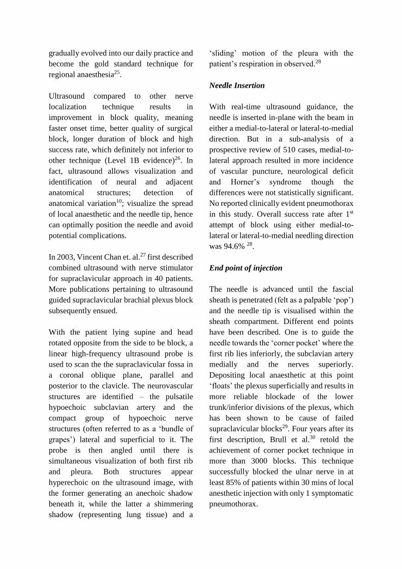
End point of injection
The needle is advanced until the fascial
sheath is penetrated (felt as a palpable ‘pop’)
and the needle tip is visualised within the
sheath compartment. Different end points
have been described. One is to guide the
needle towards the ‘corner pocket’ where the
first rib lies inferiorly, the subclavian artery
medially and the nerves superiorly.
Depositing local anaesthetic at this point
‘floats’ the plexus superficially and results in
more reliable blockade of the lower
trunk/inferior divisions of the plexus, which
has been shown to be cause of failed
supraclavicular blocks29. Four years after its
first description, Brull et al.30 retold the
achievement of corner pocket technique in
more than 3000 blocks. This technique
successfully blocked the ulnar nerve in at
least 85% of patients within 30 mins of local
anesthetic injection with only 1 symptomatic
pneumothorax.
However, due to very close proximity to the
first rib and the risk of pleural puncture,
some authors describe administering two to
three smaller aliquots of local anaesthetic at
different locations within the plexus sheath
as a safer alternative1. Tran et al.31 conducted
a randomized controlled trial in 2009 on 92
patients comparing single versus double
injection. The double-injection ultrasound-
guided supraclavicular block provides no
significant advantages compared with its
single injection counterpart.
Dual Guidance
Concurrent nerve stimulation with
ultrasound guidance is believed to be safer1
but there were case report of permanent
nerve injury on dual guidance32 and study33
showed nerve stimulation as an adjunct to
ultrasound guidance may have a limited role.
For adequately imaged ultrasound guided
supraclavicular nerve blocks, a positive
motor response to nerve stimulation does not
increase the success rate of the block. In
addition, the high false-negative rate of
nerve stimulator suggests that
supraclavicular blocks under ultrasound
guidance are usually effective, even in the
absence of a motor response. However, 21%
of the patients did not have satisfactory nerve
imaging in the same study mentioned
earlier33. Therefore, there is still role of dual
guidance in peripheral nerve blocks
especially in cases involve deep and difficult
blocks whereby the sonoimages of needle
and neural structures are poorly seen.
If dual guidance is used, Bigeleisen et al34 in
his first human study comparing intraneural
versus extraneural stimulation thresholds
during ultrasound-guided supraclavicular
block showed that there was clinical
difference in stimulation thresholds between
outside and inside the nerve. Ultrasound was
able to clearly detect the location of the
needle tip in only 69% of the cases.
Stimulation current of less than or equal to
0.2mA reliable to detect intraneural position
of the needle. Stimulation thresholds greater
than 0.2 and less than or equal to 0.5mA
could not rule out intraneural placement of
the needle. Diabetic patients require higher
stimulation thresholds both outside and
inside the nerve to elicit a motor response.
Based on current evidence, the expert panels
advise against purposefully seek needle to
nerve contact or intentional intraneural
injection35.
Volume of local anaesthetic
It is believed with ultrasound technique, the
spread of the local anaesthetic can be
visualized hence reduce the volume required.
Several studies reported variable local
anaesthetic dosing and volume required for
ultrasound guided supraclavicular block
(USSCB). The mean required volume is still
much lower if compared to non-ultrasound
technique13, 15, 16, which often used 30 to
40ml. The choice of local anaesthetic
concentration is dependent on the surgical
indication.
As an example, Tsui et al36 described 94.2%
success rate with USSCB in 104 patients
undergoing hand surgery, using 20 to 30ml
mixture of Lidocaine 1.5% and bupivacaine
0.125%. Perlas et al28 reported 94.6%
success among 47 different operators using a
mean volume of 33ml for USSCB in 510
patients with Lidocaine 2% and Bupivacaine
0.5% plus epinephrine 5ug/ml. Bigeleisen et
al34 reported 100% success using 25ml
admixture of Lidocaine 1% and Bupivacaine
0.25% plus epinephrine 3.33 mcg/ml. Brull
et al.30 used 15 – 25ml of local anaesthetic
deposited at the corner pocket area for
reliable surgical anaesthesia.
Current recommendation
Due to wide variety of practice in ultrasound
guided brachial plexus block, a set of
standardized approaches to upper extremity
nerve blocks based on the current literature
has been proposed.37
The current recommended technique for
ultrasound guided supraclavicular block is
needle injection in plane (most common),
lateral to medial. Assess the depth of brachial
plexus, insert needle in shallow angle and
adjust accordingly. The ideal spread of local
anaesthetic will be within brachial plexus
fascial sheath, lateral to the subclavian artery
but superficial to the first rib. Number of
injections would be 2 to 3 follow the
principle of bolus, observe, and reposition.
Recommended volume of local anaesthetic
20 – 25ml. If nerve stimulator is used, look
for motor response of forearm and hand.
COMPLICATIONS AND
CONTRAINDICATIONS
The overall complications associated with
supraclavicular brachial plexus block is low.
These include vascular punctures, local
anesthetic systemic toxicity as a result of fast
absorption or unintended intravascular
injection, neural damage, sympathetic
ganglion blockade with Horner syndrome,
recurrent laryngeal nerve blockade, and
phrenic nerve palsy28, 32. The incidence of
pneumothorax has reduced significantly
since the advent of ultrasound- guided
techniques28. No incident of pneumothorax
in 510 cases received USSCB in study by
Perlas et al28. Only 5 cases (1%) of
symptomatic diaphragmatic paresis, 5 cases
(1%) of Horner’s syndrome, 2 cases (0.4%)
of vascular puncture and 2 cases (0.4%) had
neurological deficit.
Phrenic nerve blockade with resultant
hemidiaphragmatic paresis results in a
reduction in functional residual capacity by
25%. Patients may present with dyspnea or
chest pain, although most affected healthy
individuals remain asymptomatic. Diagnosis
is made with an upright chest radiography, in
which a pneumothorax should be excluded.
Rates of transient hemidiaphragmatic as high
as 50% to 67% have been reported, and is
reportedly reduced when a lower volume is
used38. Patient selection is vital, and should
be contraindicated in patients with
significant respiratory disease or pre-
existing contralateral hemidiaphragmatic
paresis.
Real-time ultrasound techniques have also
markedly reduced the rates of vascular
puncture and unintended intravascular
injections28 .Vessels in the vicinity include
the subclavian artery, dorsal scapular artery
transverse cervical artery and their venous
counterparts. Slight elevation of the head of
the bed allows for better drainage and less
prominence of the neck veins, the use of
colour Doppler before needle placement,
aspirating before injection and real-time
visualization of local anaesthetic spread
during injection are methods used to reduce
vascular puncture and intravascular
injections28.
CONCLUSION
Ever since the introduction of ultrasound
guidance in regional anaesthesia, there has
been a resurgence of interest in the
supraclavicular approach to the brachial
plexus. The ability to image the surrounding
anatomy and needle placement has
significantly reduced the incidence of
pneumothorax as well as vascular puncture.
Moreover, ultrasound guidance has allowed
smaller volumes of local anaesthetic to
produce an equally rapid and dense upper
extremity blockade. Ultrasound guided
supraclavicular brachial plexus block is the
most popular regional technique of choice
for upper extremity surgery.
REFERENCES
1. Hadzic A. Supraclavicular Brachial
Plexus Block. Hadzic’s Peripheral Nerve
Blocks and Anatomy for Ultrasound
Guided Regional Anaesthesia. 2nd
Edition. New York, USA: McGraw Hill
2012; 167 – 174, 361 – 368
2. Neal JM, Gerancher JC, Hebl JR, Iffeld
BM, et al. Upper Extremity Regional
Anesthesia: Essentials of Our Current
Understanding. Reg Anesth Pain Med
2009;34: 134 – 170
3. Russon K, Pickworth T, Harrop Griffith
W. Upper Limb Blocks. Anaesthesia.
2010; 65(1): 48 – 56
4. Kulenkampff D. Die Anästhesierung des
plexus brachialis. Beitr Klin. Chir 1912;
79: 550 – 552.
5. Kulenkampff D. Brachial Plexus
Anaesthesia: Its Indications, Technique,
and Dangers. Ann Surg 1928; 87: 883 –
91
6. Accardo NJ, Adriani J. Brachial Plexus
Block: A simplified technique using the
axillary route. South Med J. 1949 Oct; 42
(10): 920 – 923
7. Burnham PJ, Eather KF. Axillary
brachial plexus block. Anaesthesiology.
1958 Sep-Oct; 19 (5): 683 – 685
8. De Jong RH. Axillary brachial plexus
block. Anesthesiology. 1961 Mar-Apr;
22: 215 – 225
9. Lanz E, Theiss D, Jankovic D. The extent
of blockade following various
techniques of brachial plexus block.
Anesth Analg 1983; 62: 55-8.
10. Vermeylen K, Sermeus L.
Supraclavicular Brachial Plexus Blocks:
Review and Current Practice. Acta
Anaesth. 2011; 62:15-21
11. MacIntosh RR, Mushin WW. Local
anaesthesia: brachial plexus. Oxford:
Blackwell Scientific Publications,
1944:56
12. Lamoureux L, Bourgeois-Gavardin M.
La ThCorie des trois perpendiculaires
dans l’infiltration du plexus brachial.
Union Med Can 1951; 80: 927 – 34
13. Winnie A, Collins V. The Subclavian
Perivascular Technique of Brachial
Plexus Anesthesia. Anesthesiology
1964; 25: 353 – 63
14. Vongvises P, Panijayanond T. A
parascalene technique of brachial plexus
anesthesia. Anesth Analg 1979; 58: 267
– 73
15. Dupre LJ, Danel V, Legrand JJ, Stieglitz
P, Surface landmarks for supraclavicular
block of the brachial plexus. Anesth
Analg 1982; 61:28-31
16. Brown DL, Bridenbaugh LD. Physics
applied to regional anesthesia results in
an improved supraclavicular block the
"plumbbob technique. Anesthesiology
1988; 69: A376.
17. Neal JM, Barrington MJ, Brull R, Hadzic
A, et. al. The Second ASRA Practice
Advisory on Neurologic Complications
Associated with Regional Anaesthesia
and Pain Medicine. Reg Anesth Pain
Med 2015; 40: 401 – 430
18. Greenblatt G, Denson J. Needle Nerve
Stimulator Locator: Nerve Blocks with a
New Instrument for Locating Nerves.
Anesth Analg. 1962; 41:599 – 602
19. Franco C, Vieira Z. 1,001 Subclavian
Perivascular Brachial Plexus Blocks:
Success with a Nerve Stimulator. Reg
Anesth Pain Med. 2000; 25:41 – 46
20. Brown DL, David M, Hall JA, et. al.
Regional Anesthesia and Local
Anesthetic Induced Systemic Toxicity:
Seizure frequency and Accompanying
Cardiovascular Changes. Anesth Analg
1995; 81 (2) 321 – 328
21. Ting PL, Sivagnanaratnam V:
Ultrasonographic study of the spread of
local anaesthetic during axillary brachial
plexus block. Br J Anaesth 1989; 63: 326
– 329
22. La Grange P, Foster PA, Pretorius LK.
Application of the Doppler Ultrasound
Bloodflow Detector in Supraclavicular
Brachial Plexus Block. Br J Anaesth.
1978; 50: 965 – 967.
23. Moorthy SS, Schmidt SL, Dierdorf SF. A
supraclavicular lateral paravascular
approach for brachial plexus regional
anesthesia. Anesth Analg 1991; 72: 241
– 244
24. Kapral S, Krafft P, et al. Ultrasound-
guided Supraclavicular Approach for
Regional Anesthesia of the Brachial
Plexus. Anesthesia and Analgesia 1994;
78:507-13
25. Hopkins PM. Ultrasound Guidance As a
Gold Standard in Regional Anaesthesia.
Br J Anaesth. 2007 Mar; 98 (3): 299 –
301
26. Liu SS. Evidence Basis for Ultrasound
Guided Block Characteristics Onset,
Quality, and Duration. Reg Anesth Pain
Med 2016; 41: 205 – 220
27. Chan VW, Perlas A et al. Ultrasound-
Guided Supraclavicular Brachial Plexus
Block. Anesth Analg. 2003; 97(5): 1514
– 1517
28. Perlas A., Lobo G, et al. Ultrasound-
guided Supraclavicular Block: Outcome
of 510 Consecutive Cases. Reg Anesth
Pain Med 2009; 34(2):171-6
29. Soares L, Brull R, Lai J, Chan VW. Eight
Ball, Corner Pocket: The Optimal
Needle Position for Ultrasound-Guided
Supraclavicular Block. Reg Anesth Pain
Med. 2007; 32: 94 – 5
30. Brull R, Chan VW. The Corner Pocket
Revisited. Regional Anesthesia and Pain
Medicine. 2011; 6: 308
31. Tran DQH, Munoz L, et al. A
Prospective, Randomized Comparison
between Single and Double Injection
Ultrasound-guided Supraclavicular
Brachial Plexus Block. Reg Anesth Pain
Med 2009;34: 420 – 24
32. Reiss W, Kurapati S, Shariat A, Hadzic
A. Nerve Injury Complicating
Ultrasound/Electrostimulation-Guided
Supraclavicular Brachial Plexus Block.
Reg Anesth Pain Med 2010; 35: 400 –
401
33. Beach ML, Sites BD, Gallagher JD. Use
of a nerve stimulator does not improve
the efficacy of ultrasound-guided
supraclavicular nerve blocks. J Clin
Anesth. 2006 Dec; 18 (8): 580 – 4
34. Bigeleisen PE, Moayeri N, Groen GJ.
Extraneural versus intraneural
stimulation thresholds during ultrasound
–guided supraclavicular block.
Anesthesiology. 2009 Jun; 110 (6): 1235
– 43
35. Neal JM, Barrington MJ, Brull R, Hadzic
A, et. al. The Second ASRA Practice
Advisory on Neurologic Complications
Associated with Regional Anaesthesia
and Pain Medicine. Reg Anesth Pain
Med 2015;40: 401–430
36. Tsui BCH, Doyle K, Chu K, Pillay J,
Dillane D. Case series: ultrasound-
guided supraclavicular block using a
curvilinear probe in 104 day-case hand
surgery patients. Can J Anaesth. 2009;
56: 46 – 51
37. Kwofie K, Shastri U, Vandepitte C.
Standard Approaches for upper
extremity nerve blocks with an emphasis
on outpatient surgery. Curr Opin
Anaesthesiol. 2013 Aug; 26 (4): 501 – 8
38. Renes S, Spoormans HH, Gielen MJ, et
al. Hemidiaphragmatic Paresis can be
avoided in Ultrasound-Guided
Supraclavicular Brachial Plexus Block.
Regional Anesthesia and Pain Medicine.
2009; 34: 595 – 9
Evolution of Supraclavicular Brachial Plexus Block
Teo Wei Wei1, Beh Zhi Yuen2, Shahridan Mohd Fathil3
1Department of Anaesthesia, National University Hospital, Singapore 2Anaesthesia Department, Changi General Hospital, Singapore 3Anaesthesia Department, Ng Teng Fong General Hospital, Singapore
Figure 1: Brachial plexus. Various approaches define individual brachial plexus blocks and their expected distribution of cutaneous anesthesia. Illustration by Jennifer Gentry. *American Society of Regional Anesthesia and Pain Medicine. (with permission from Wolter-Kluwer)