evolution of invertebrate deuterostomes and hox/parahox … · ral colinearity between their...
TRANSCRIPT
GENOMICSPROTEOMICS &BIOINFORMATICS
www.sciencedirect.com/science/journal/16720229
Review
Evolution of Invertebrate Deuterostomes and Hox/ParaHox Genes
Tetsuro Ikuta* Marine Genomics Unit, Okinawa Institute of Science and Technology, Uruma, Okinawa 904-2234, Japan.
Genomics Proteomics Bioinformatics 2011 Jun; 9(3): 77-96 DOI: 10.1016/S1672-0229(11)60011-9
Received: Jan 28, 2011; Accepted: Mar 21, 2011
Abstract
Transcription factors encoded by Antennapedia-class homeobox genes play crucial roles in controlling develop-ment of animals, and are often found clustered in animal genomes. The Hox and ParaHox gene clusters have been regarded as evolutionary sisters and evolved from a putative common ancestral gene complex, the ProtoHox clus-ter, prior to the divergence of the Cnidaria and Bilateria (bilaterally symmetrical animals). The Deuterostomia is a monophyletic group of animals that belongs to the Bilateria, and a sister group to the Protostomia. The deu-terostomes include the vertebrates (to which we belong), invertebrate chordates, hemichordates, echinoderms and possibly xenoturbellids, as well as acoelomorphs. The studies of Hox and ParaHox genes provide insights into the origin and subsequent evolution of the bilaterian animals. Recently, it becomes apparent that among the Hox and ParaHox genes, there are significant variations in organization on the chromosome, expression pattern, and func-tion. In this review, focusing on invertebrate deuterostomes, I first summarize recent findings about Hox and ParaHox genes. Next, citing unsolved issues, I try to provide clues that might allow us to reconstruct the common ancestor of deuterostomes, as well as understand the roles of Hox and ParaHox genes in the development and evolution of deuterostomes.
Key words: invertebrate deuterostome, Hox, ParaHox, evolution
Introduction
Animal morphology along the body axes is greatly diverse, requiring both a system that confers posi-tional character and a competence to respond to these positional cues. The Antennapedia-class Hox and ParaHox genes encode transcription factors that play crucial roles in controlling morphological develop-ment of animals. Hox genes have been noted for sev-eral striking properties, including their conserved roles in providing regional identities along the ante-
*Corresponding author. E-mail: [email protected] © 2011 Beijing Institute of Genomics.
rior–posterior (AP) axis and the spatial and/or tempo-ral colinearity between their expression patterns along the AP axis and their positions within clusters on a chromosome (1-3). These factors generate the unique combinations of Hox genes expressed at the different AP axial levels during development; this arrangement is referred to as the “Hox code” (4). These observa-tions lead to the hypothesis that the physical organiza-tion on the chromosome, expression pattern, and functions of the Hox genes are important for proper morphological patterning along the AP axis (5) and, in turn, for evolutionary changes in the animal body plan. The ParaHox cluster was first characterized in the cephalochordate amphioxus, in which three member genes, Gsx, Xlox and Cdx, are linked in a manner
This is an open access article under the CC BY license (http://creativecommons.org/licenses/by/4.0/).
Ikuta / Invertebrate Deuterostome Hox/ParaHox Evolution
Genomics Proteomics Bioinformatics 2011 Jun; 9(3): 77-96 78
reminiscent of the Hox genes (6). The ParaHox clus-ter was regarded as an evolutionary sister of the Hox cluster; and both clusters were evolved from a puta-tive common ancestral gene complex, the ProtoHox cluster, which is prior to the divergence of cnidarians and bilaterians (6-8). Subsequently, the Hox cluster was expanded by tandem duplications of the member genes during evolution; in contrast, the ParaHox clus-ter maintained a constant size of three genes.
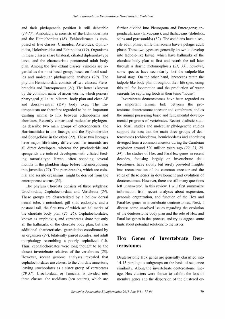
The Deuterostomia comprise one of the major groups within the animal kingdom; it is a mono-phyletic group of animals (9) that belongs to the Bi-
lateria, and a sister group to the Protostomia (Figure 1). Deuterostomes are characterized by having a “second mouth”; i.e., during embryo development, the blastopore becomes the anus, whereas the mouth forms in a secondary anterior location. Deuterostomes can be further subdivided into two major clades: Am-bulacraria and Chordata (to which humans belong) (9-11). Some authorities also include the newly de-scribed and enigmatic group Xenoturbellida (or Xenacoelomorpha even including acoelomorphs) (12, 13); however, I will not discuss this group here, mainly because their morphology is very divergent
Figure 1 Deuterostome phylogeny with schematics of the genomic organization of Hox and ParaHox genes. Colored rectangles and ovals indicate Hox and ParaHox genes, respectively. Anterior Hox and Gsx are indicated in red; group3 and Xlox in yellow; central Hox in green; and posterior Hox and Cdx in blue, according to the nomenclature in a previous study (7). Lines passing under rectan-gles or ovals indicate clustered gene linkage on a chromosome. Slashes on the line represent a large gap between Hox or ParaHox genes, indicating disorganization of the cluster. Unconnected lines also indicate an unlinked situation. Rectangles and ovals without horizontal lines passing under them indicate the genes whose linkage has not been investigated. Information about vertebrate and xenoturbella (or xenacoelomorph) Hox and ParaHox genes is omitted.
Ikuta / Invertebrate Deuterostome Hox/ParaHox Evolution
Genomics Proteomics Bioinformatics 2011 Jun; 9(3): 77-96 79
and their phylogenetic position is still debatable (14-17). Ambulacraria consists of the Echinodermata and the Hemichordata (18). Echinodermata is com-posed of five classes: Crinoidea, Asteroidea, Ophiur-oidea, Holothuroidea and Echinoidea (19). Organisms in these classes share bilateral, ciliated dipleurula-type larva, and the characteristic pentameral adult body plan. Among the five extant classes, crinoids are re-garded as the most basal group, based on fossil stud-ies and molecular phylogenetic analyses (20). The phylum Hemichordata consists of two classes: Ptero-branchia and Enteropneusta (21). The latter is known by the common name of acorn worms, which possess pharyngeal gill slits, bilateral body plan and clear AP and dorsal–ventral (DV) body axes. The En-teropneusta are therefore regarded to be an important existing animal to link between echinoderms and chordates. Recently constructed molecular phylogen-ies describe two main groups of enteropneusts: the Harrimaniidae in one lineage; and the Ptychoderidae and Spengelidae in the other (22). These two lineages have major life-history differences: harrimaniids are all direct developers, whereas the ptychoderids and spengelids are indirect developers with ciliated feed-ing tornaria-type larvae, often spending several months in the plankton stage before metamorphosing into juveniles (22). The pterobranchs, which are colo-nial and sessile organisms, might be derived from the enteropneust worms (23).
The phylum Chordata consists of three subphyla: Urochordata, Cephalochordata and Vertebrata (24). These groups are characterized by a hollow dorsal neural tube, a notochord, gill slits, endostyle, and a postanal tail, the first two of which are hallmarks of the chordate body plan (25, 26). Cephalochordates, known as amphioxus, and vertebrates share not only all the hallmarks of the chordate body plan, but also additional characteristics: gastrulation coordinated by an organizer (27), bilaterally paired somites, and adult morphology resembling a poorly cephalized fish. Thus, cephalochordates were long thought to be the closest invertebrate relatives of the vertebrates (28). However, recent genome analyses revealed that cephalochordates are closest to the chordate ancestors, leaving urochordates as a sister group of vertebrates (29-31). Urochordata, or Tunicata, is divided into three classes: the ascidians (sea squirts), which are
further divided into Pleurogona and Enterogona; ap-pendicularians (larvaceans); and thaliaceans (doliolids, salps and pyrosomids) (32). The ascidians have a ses-sile adult phase, while thaliaceans have a pelagic adult phase. These two types are generally known to develop into tadpole-like larvae, which have hallmarks of the chordate body plan at first and resorb the tail later through a drastic metamorphosis (25, 33); however, some species have secondarily lost the tadpole-like larval stage. On the other hand, larvaceans retain the tadpole-like body plan throughout their life span, using this tail for locomotion and the production of water currents for capturing foods in their tunic “house”.
Invertebrate deuterostomes have been regarded as an important animal link between the pro-tostome–deuterostome ancestor and vertebrates, and as the animal possessing basic and fundamental develop-mental programs of vertebrates. Recent cladistic stud-ies, fossil studies and molecular phylogenetic studies support the idea that the main three groups of deu-terostomes (echinoderms, hemichordates and chordates) diverged from a common ancestor during the Cambrian explosion around 520 million years ago (22, 23, 28, 34). The studies of Hox and ParaHox genes in recent decades, focusing largely on invertebrate deu-terostomes, have slowly but surely provided insights into reconstruction of the common ancestor and the roles of these genes in development and evolution of deuterostomes. However, there are still many questions left unanswered. In this review, I will first summarize information from recent analyses about expression, genomic organization, and function of the Hox and ParaHox genes in invertebrate deuterostomes. Next, I discuss some unsolved issues regarding the evolution of the deuterostome body plan and the role of Hox and ParaHox genes in that process, and try to suggest some hints about potential solutions to the issues.
Hox Genes of Invertebrate Deu-terostomes
Deuterostome Hox genes are generally classified into 14-15 paralogous subgroups on the basis of sequence similarity. Along the invertebrate deuterostome line-age, Hox clusters were shown to exhibit the loss of member genes and the dispersion of the clustered or-
Ikuta / Invertebrate Deuterostome Hox/ParaHox Evolution
Genomics Proteomics Bioinformatics 2011 Jun; 9(3): 77-96 80
ganization (35, 36); however, all members of inverte-brate deuterostomes are regarded to have a single set of Hox genes.
In the sea urchin Stronglyocentruotus purpuratus, an echinoderm, eleven Hox genes have been identified (37,38). The recent sequencing of bacterial artificial chro-mosome (BAC) clones from the genome of S. purpu-ratus revealed that the sea urchin Hox cluster is large, occupying 588 kb, and contains rearrangements both of transcriptional orientation and gene order (38). Whereas only two Hox genes are transcribed during the development of bilateral pluteus larva, ten Hox genes of S. purpuratus are transcribed during formation of the pentameral adult rudiment (39). Among them, Hox7, 8, 9/10, 11/13a and 11/13b exhibit spatially staggered expression in the somatocoels of mesodermal origin during the period of adult rudiment formation (9); however, the details of expression of the anterior member genes remain unknown. We have almost no data on function of sea urchin Hox genes, except for Hox11/13b, which may be involved in cell adhesion as well as in hindgut specification during larval develop-ment (40). In the most ancestral extant echinoderm, eight Hox genes have been identified in the stalked crinoid Metacrinus rotundus. Among these, Hox5, 7, 8 and 9/10 exhibit spatially staggered expression in the mesodermal somatocoels of the bilateral auricularia larva (41); however, their genomic organization and function remain unknown. Although it was thought that echinoderms lacked the ortholog of Hox4, recent stud-ies on asteroid and crinoid Hox genes demonstrated that the absence of Hox4 from echinoids is a derived state (41, 42), and the ancestral echinoderm probably had a Hox gene complements not dissimilar to that of hemichordates.
Among hemichordates, Hox genes were relatively well studied in the harrimaniid species Saccoglossus kowalevskii, a direct-developing enteropneust. In this species, eleven Hox genes have been identified; these genes exhibit spatially staggered expression in the surface ectoderm during embryogenesis (43, 44). On the other hand, in the ptychoderid Ptychodera flava, an indirect-developing enteropneust, eight Hox mem-ber genes have been identified (45). Even so, it was unknown whether the ortholog of Hox8 is absent in hemichordates; consequently, no consensus view about Hox gene complements in hemichordates has
been established. Recently, however, we isolated the presumably full complement of twelve Hox genes, including the ortholog of Hox8 in Balanoglossus si-modensis, suggesting that the ancestral hemichordate had intact complements of ambulacrarian prototypical Hox genes (46). However, as regards to hemichordate Hox genes, there is no report of their genomic ar-rangement, developmental roles or expression pattern in the larval development.
It has been pointed out that the cephalochordate amphioxus retains a relatively intact Hox cluster or-ganization, remarkably similar to that inferred for the direct ancestor of the vertebrates (47). The amphioxus Branchiostoma floridae has a single contiguous Hox cluster, containing an extra 14th and 15th member genes, spanning 470 kb (48, 49). Recent phylogenetic analysis suggested that the amphioxus and vertebrate Hox14 genes were not orthologous, but arose inde-pendently through tandem gene duplications of Hox13 genes (50). Moreover, the posterior Hox genes of ambulacrarians and chordates may result from two independent sets of tandem duplications (38, 45, 46). Thus, the true prototypical complement for deu-terostome Hox gene members is still unclear. An ini-tial expression study of amphioxus Hox genes re-vealed that Hox1, 3 and 4 exhibit spatial and temporal colinearity in the developing neural tube, although Hox2 is exceptional (51). Furthermore, expression of amphioxus Hox6 was described to break colinearity (52). On the other hand, a recent study showed that not only Hox1, 3 and 4, but also Hox2 and 6 join in spatial colinearity in the neural tube from the mid-neurula to larval stage, correcting the previous reports (53). However, expression patterns for more posterior member genes remain unknown. Functional studies were only conducted for Hox1, in which Hox1 mediates retinoic acid (RA) signalling in establishing the posterior limit of the pharynx and regionalization of the hindbrain, as well as in specification of motor neurons (53, 54).
Among the urochordates, the larvacean Oikopleuradioica has two anterior, one central, and six posterior Hox genes. Although the locations of these genes on the chromosomes are unknown, genomic walking suggests that they are dispersed within the O. dioica genome (55), and the situation is the same even after recent draft genome sequencing (56); each Hox gene

Ikuta / Invertebrate Deuterostome Hox/ParaHox Evolution
Genomics Proteomics Bioinformatics 2011 Jun; 9(3): 77-96 81
is located on a different scaffold. Despite that, some Oikopleura Hox genes exhibit spatially staggered ex-pression in the notochord, tail muscle, nerve cord, and epidermis (55). Among ascidians, Hox genes were most extensively studied in Ciona intestinalis. In this species, the draft genome analysis identified nine Hox genes (57). We revealed that some, if not all, Ciona Hox genes exhibit spatially staggered expression within the central nervous system (CNS) during larval development and in the gut of the juvenile. Neverthe-less, nine Ciona Hox genes are dispersed on two chromosomes, with rearrangements in gene orders and transcriptional orientations (58). Among them, seven Hox genes, Hox 1-6 and 10, are distributed with some large gaps between them that contain many non-Hox genes, spanning approximately half the length of a chromosome, which could be roughly cal-culated as 5 Mb; i.e., extraordinarily long in com-parison to that of a typical higher vertebrate Hox cluster (59). Thus, urochordates like Oikopleura and Ciona provide some of the most extreme cases of Hox cluster disintegration reported to date. Significantly, in these species, the temporal staggering of Hox ini-tiation is lost, while the spatial staggering is retained, supporting the hypothesis that the mechanisms pro-ducing temporal colinearity are likely the major con-straining forces on gene cluster maintenance, e.g., a shared regulatory mechanism, such as a shared en-hancer(s) (60, 61). Interestingly, lacZ reporter con-structs connected to the genomic fragments, including the flanking sequence of Hox genes and/or Hox gene sequence with introns, roughly mimic endogenous expression of each Hox gene, suggesting the loss of cis-regulatory element sharing in Ciona (62). Fur-thermore, surprisingly, our recent analysis suggested that the contribution of Hox genes to the larval de-velopment of C. intestinalis might be very limited, despite the fact that two Hox genes, Hox10 and 12, play important roles in neuronal and tail development, respectively (63).
ParaHox Genes of Invertebrate Deu-terostomes
The ParaHox gene cluster was first discovered in the cephalochordate amphioxus, and is composed of
members of three Hox-related homeobox gene fami-lies: Gsx, Xlox and Cdx. Gsx was described to be most similar to the anterior Hox genes, Xlox to group 3 Hox genes, and Cdx to the posterior Hox genes (6,7). All invertebrate deuterostomes are regarded to have a single set of ParaHox genes.
Among echinoderms, ParaHox genes have been well studied in S. purpuratus. The sea urchin has three ParaHox genes, Gsx, Xlox and Cdx, but each ParaHox gene is located on a different genomic scaffold (>300 kb each), suggesting that they are not linked into a single coherent cluster (64). In spite of this, strikingly, the three member genes show both spatially and tem-porally staggered expression; Gsx is expressed first, followed by Xlox and finally Cdx, although Xlox and Cdx exhibit staggered expression in the larval gut, whereas transcripts of Gsx are detected in the ecto-derm and not in the gut (64). A recent functional analysis of S. purpuratus Xlox and Cdx showed that these two genes interact in patterning of the larval hindgut (65).
Information about hemichordate ParaHox genes is limited. In S. kowalevskii, extensive EST screening identified only Cdx (66). On the other hand, in P. flava, four ParaHox member genes have been isolated: one Gsx, two Xlox and one Cdx (45). Furthermore, we recently reported the full complement of three Para-Hox genes from B. simodensis, suggesting that the ancestral hemichordate had intact complements of the ambulacrarian prototypical ParaHox genes (46). However, the genomic arrangement, developmental expression and function of hemichordate ParaHox genes remain unknown.
In the amphioxus B. floridae, the three member genes are linked in a genomic region of 56 kb, with Gsx adjacent to Xlox in the same orientation, followed by Cdx on the opposite strand (6, 67). Of the inverte-brates studied to date, the linkage of the three genes has been demonstrated only in amphioxus. The am-phioxus ParaHox cluster exhibits both spatial and temporal colinearity. Initially Cdx is activated during mid-gastrulation in a ring around the blastopore, and then remains in a continuous domain in the posterior neuroectoderm and hindgut during neurulation and somitogenesis. Xlox expression commences slightly later than Cdx in the posterior endoderm and mesen-doderm, but later becomes restricted to a more central
Ikuta / Invertebrate Deuterostome Hox/ParaHox Evolution
Genomics Proteomics Bioinformatics 2011 Jun; 9(3): 77-96 82
region of the gut in the developing larva, with tran-sient expression in two cells of the neural tube ap-proximately level with the anterior boundary of somite five (where the first pigment spot will form). The last ParaHox gene to be activated is Gsx, which is initially expressed in the neural tube during neurula-tion at the same location level as the transient expres-sion of Xlox, followed by activation in the cerebral vesicle (68). Thus, interestingly, the temporal colin-earity in the amphioxus ParaHox cluster along the AP axis is inverted with respect to the pattern in the Hox cluster (anterior to posterior) as well as with the ob-servation in the sea urchin ParaHox genes (Gsx to Cdx) (54). I speculate that the temporal order observed in amphioxus and mouse (Cdx2, Ipf1 and Gsx1) (69-71) is the conserved pattern, and the observation in sea urchin will be incidental, based on the hypotheses that temporal colinearity is the principal constraining force on cluster organization (60, 61); as long as this colin-earity of only three genes is a subsistent system and has not occurred simply by chance. Developmental roles of amphioxus ParaHox genes are unknown.
In urochordates, the full complement of ParaHox genes, Gsx, Xlox and Cdx, has been identified in the C. intestinalis genome. An extensive mapping of BAC clones revealed that three genes are dispersed on two chromosomes: Gsx on chromosome 2q, and Xlox and Cdx on chromosome 14q (72). The latter two genes are approximately 240 kb apart, with head-to-head transcriptional orientation, and with many intervening genes between them. In Ciona, expression studies showed that Gsx is expressed in the posterior sensory vesicle from the mid-gastrula to tailbud stages (73); Xlox in the larval sensory vesicle, visceral ganglion and mesenchymal cells (74); and Cdx in the neural and posterior muscle lineage cells in mid-gastrula (75), the nerve cord, posterior epidermis and endodermal strand at the tailbud stage (67), and the nerve cord at the larval stage (76). Although expression patterns of Ciona ParaHox genes from metamorphosis onward are unknown, in another ascidian, Herdmania curvata,Cdx is expressed in the intestine of the juvenile as well as the posterior CNS during larval development (77), whereas expression patterns of Gsx and Xlox in this species remain unknown. An initial functional study of ascidian ParaHox genes was conducted for Cdx of third ascidian Halocynthia roretzi; the results
suggested that Halocynthia Cdx is required for larval tail formation, probably by controlling ectodermal cell movement (78). In C. intestinalis, functional inhibi-tion using a dominant negative Cdx under control of a FoxD promoter/enhancer produced a phenotype simi-lar to that observed in the Halocynthia embryo in which Cdx function was suppressed. The authors con-cluded that Ciona Cdx is required for neural tube formation (79). Furthermore, a recent study aimed at establishing the gene regulatory network underlying CNS development of Ciona revealed several down-stream genes under the transcriptional control of Cdx (80). The developmental roles of urochordate Gsx and Xlox have not yet been reported.
Hox Genes and Evolution of the Deu-terostome Nervous System
It has been proposed that in the Bilateria, the ancestral function of Hox genes was in AP patterning and specification of ectodermal and neuroectodermal anlagen (81, 82). Indeed, the expression and function of Hox genes in Drosophila melanogaster and chor-dates are most evident in the ectodermal and neu-roectodermal tissues, and Hox genes are associated with the development of CNS in many different taxa (83-86).
The ancestral character of the deuterostome nerv-ous system is still enigmatic. Considering the similar ciliary band anatomy and apical organ structure in the echinoderm and hemichordate larva, this shared larval type apparently reflects the development of the am-bulacrarian ancestor (87). However, Hox genes probably do not contribute to axial patterning of the neuroectoderm of the ambulacrarian larva, since no staggered expression of Hox genes has been observed in the neuroectoderm of echinoderm larvae (39, 88). The nervous systems of adult echinoderms and hemi-chordates are quite different. The centralized part of the echinoderm nervous system consists of a circu-moral (or circumanal, in the case of crinoids) ring connecting five radial nerve trunks that run out along each arm of the animal (24). Again, staggered expres-sion of Hox genes in the development of the adult nervous system has not been observed in S. purpura-tus. However, it has been hypothesized that the cir-
Ikuta / Invertebrate Deuterostome Hox/ParaHox Evolution
Genomics Proteomics Bioinformatics 2011 Jun; 9(3): 77-96 83
cumoral and radial nervous system of echinoids may represent a small portion of the ancestral bilaterian CNS derived from a region anterior to much or all of the Hox patterning domain (89). As a corollary to this, the circumanal nervous system of crinoids would be expected to express more posterior patterning genes, including Hox genes. Thus, a study of the expression pattern of crinoid Hox genes during the development of the adult nervous system would contribute signifi-cantly to our understanding of the origin of the deu-terostome nervous system and the evolution of the peculiar body plan of echinoderms.
In contrast to echinoderms, the major organiza-tional feature of the hemichordate nervous system is that it is basiepithelial. It has been described that there is no CNS, and cell bodies are scattered throughout the epithelium (24). Lowe et al described the stag-gered expression of over twenty orthologs of neural patterning genes, including Hox genes in S.kowalevskii, in which Hox genes are expressed in circular areas in the ectoderm around the entire ani-mal, with an AP arrangement nearly identical to that found in chordates (43, 44). This prompted the idea of ancient “skin brains”, which proposes that the deu-terostome ancestor had a nervous system that was not central but diffuse; in this model, the CNS evolved independently in the protostome and deuterostome lineages (90, 91). In contrast, in the 19th century, comparative anatomist Anton Dohrn proposed that the chordate CNS is homologous to that of protostome annelids and arthropods (92). Since that time, a growing list of genes has been proposed to play a conserved role in the patterning of CNS of Drosophila and of vertebrates. This led to the hypothesis that the CNS of arthropods and chordates are homologous (93, 94). Furthermore, Denes et al elegantly demonstrated that the mediolateral neural architecture of the devel-oping trunk CNS of the annelid Platynereis dumerilii is quite similar to that of the developing vertebrate neural tube, supporting a common origin for nervous system centralization in the Bilateria (95). In addition to this, Nomaksteinsky et al recently showed that both juvenile and adult of the indirect-developing en-teropneust P. flava in fact have a bona fide CNS, i.e., dense agglomerations of neurons, forming two cords, ventral and dorsal, which merge anterior–dorsally at the level of the collar to form a chordate-like neural
tube. Contrary to previous assumptions, the greater part of the adult enteropneust skin is non-neural, al-though elements of the peripheral nervous system (PNS) are found there. The authors proposed that the previously described “diffuse” nervous system present at earlier developmental stages in Saccoglossus is a transitory feature that may correspond to the larval nervous system of other enteropneusts (96). These findings have reopened the possibility that centraliza-tion of the nervous system predates the chordates; i.e., the deuterostome ancestor had a centralized nervous system. This encourages more intensive comparative CNS research, especially the study of the roles of the axial patterning genes, including Hox genes, in the development of the enteropneust CNS. We have re-vealed that P. flava has intact complements of the twelve prototypical ambulacrarian Hox genes, and that all of them except for one are located on two con-tiguous BAC clones; i.e., the cluster can be estimated to fit within 200-300 kb (unpublished data). As for the one exceptional Hox gene, I have not been able to isolate this gene from the two sets of BAC libraries. I speculate that this gene is dropped out of the BAC libraries over the course of several additional experi-ments, and that the full complement of twelve Hox genes will be clustered in the P. flava genome. If their gene order and transcriptional orientation are intact and the cluster has no intervening genes, the P. flava Hox cluster will be the most compact, clearly organ-ized example in invertebrates studied to date, proba-bly representing the ancestral character of the deu-terostome Hox cluster. On the other hand, Duboule proposed that the Hox cluster was originally disor-ganized in the ancestral bilaterian as well as the an-cestral deuterostome (97). Thus, in order to build a consensus view about the ancestral features of deu-terostome Hox genes, and to cast in a clearer light the origin of the deuterostome body plan including the nervous system, we must conduct a comprehensive study of the genomics, expression, functions and regulation of the hemichordate Hox genes.
A number of hypotheses for the origin of the chor-date nervous system have been proposed, and distin-guishing between them will obviously depend on reaching a conclusion about the ancestral form of the deuterostome nervous system, as described above. Since this complex topic has been reviewed elsewhere

Ikuta / Invertebrate Deuterostome Hox/ParaHox Evolution
Genomics Proteomics Bioinformatics 2011 Jun; 9(3): 77-96 84
(43, 98-100), I do not cover it here. But in any case, one outstanding problem is determining whether Hox genes contributed to AP patterning of CNS of the chordate ancestor. Considering the clear staggered expression of five amphioxus Hox genes (Hox1-4 and 6), and the role of Hox1 in regionalization of the am-phioxus hindbrain as well as specification of motor neurons (101), the answer is probably “yes”. In verte-brate gnathostomes, the specification of rhombomere segmental identities and neurons also depends on the highly organized expression patterns of the Hox genes (85, 102-104). However, Murakami et al proposed that the relationship between Hox gene expression domains and motoneuron identity may be an ancestral feature conserved throughout the AP axis of the chor-date CNS, whereas the neuromeric compartments of the segmentation process are evolutionarily as well as developmentally independent. That is, the AP specifi-cation of branchiomotor neurons was already under control of a Hox code in the chordate ancestor, but the registration of hindbrain segmentation and Hox code regulation appeared in the gnathostome lineage (105). Indeed, the amphioxus hindbrain lacks obvious seg-ments. Thus, in order to more precisely understand the role of Hox genes in CNS of the chordate ancestor, a functional assay for each amphioxus Hox gene will be an urgent task. On the other hand, in the tadpole larva of the ascidian C. intestinalis, the neural circuits of the motor system are simply composed of at most five pairs of cholinergic motor neurons in the visceral ganglion, and two pairs of GABA/glycinergic in-terneurons in the anterior nerve cord (106). However, we demonstrated that knock-down of Ciona Hox1, 3 and 5 does not affect the development of these neu-rons (63), despite the fact that they precisely exhibit staggered expression in the visceral ganglion and an-terior nerve cord, both of which are regarded as ho-mologous with vertebrate rhombospinal region (58, 107, 108). The limited roles of ascidian Hox genes in larval CNS development are probably related to the ascidian’s simple body plan and rapid, determinative embryogenesis, and may also be influenced by exten-sive genomic rearrangement and gene loss, including disintegration of the Hox cluster and loss of some Hox gene members (63, 109). Similarly, Seo et al, using data from larvaceans, correlated disintegration of Hox genes and loss of Hox gene members with size
reduction and determinative development in the uro-chordate lineage (55). To confirm these scenarios, comprehensive studies of the Hox genes in a wider variety of urochordates, including functional study of larvacean Hox genes, will be required.
Basic Roles of Hox Genes in the De-velopment of the Mesodermal Organs
Although the expression and function of Hox genes in Drosophila and vertebrates are most evident in the ectodermal and neuroectodermal tissues (82), Hox genes are also expressed in the mesoderm of Droso-phila (110, 111) and vertebrates (112-114). In inverte-brate deuterostomes, echinoderms are a particularly good example of staggered expression of Hox genes in mesoderm. In sea urchins, expression of five Hox genes in the mesodermal somatocoels at the late plu-teus stages, i.e., during formation of the pentameral adult rudiment, follows the curved gut underneath; thus it points in a curved AP direction (9). Does this mesodermal expression of Hox genes in sea urchins represent the ancestral character of deuterostomes?
In Drosophila, four Hox genes expressed in the visceral mesoderm of the midgut are crucial for gut morphogenesis (110). In vertebrates as well, Hox genes exhibit region specific expression in splanchnic mesoderm (113, 114). Based on these observations, staggered expression of sea urchin Hox genes in the larval somatocoels might reflect the ancestral role of Hox genes in the development of digestive tract. In echinoids, generally the stomach and most of hindgut survive metamorphosis and contribute to adult gut (115). Thus, this idea does not contradict the general view that Hox genes are used in patterning of the adult body plan rather than for development of the larva-specific structures (116). Here, I should note that the word “larva” requires careful handling. The sort of ciliated planktonic larvae of bilaterians termed as “primary larvae” by Jägersten (117), such as the trochophore larva of annelids or the dipleurula larva of echinoderms, are qualitatively different from the secondary larvae found in several taxa, such as ar-thropods, ascidians and vertebrates. Secondary larvae, as recognized by Jägersten, are modified adult (or juvenile) forms comparable to the adults of other bi-

Ikuta / Invertebrate Deuterostome Hox/ParaHox Evolution
Genomics Proteomics Bioinformatics 2011 Jun; 9(3): 77-96 85
laterians (116). Thus in this review, the term “indirect development” means that the organism grows into the primary larva stage before becoming an adult.
Getting back to the role of Hox genes in the echi-noderm somatocoels, an alternative idea is that echi-noderm Hox genes are involved in patterning the somatocoels along the AP axis. For instance, by com-paring Hox gene expression patterns in sea urchin larvae and crinoid larvae, Hara et al proposed that staggered expression of medial and posterior Hox genes in the somatocoels along AP axis reflects an ancestral feature of echinoderms (or possibly of am-bulacrarians), and that Hox5 ortholog expression in the crinoid somatocoels will be associated with the differentiation of the chambered organ and reflects an ancestral trait involved in adult stalk formation (41). However, Omori et al recently showed that Six3, Pax6,Otx and Hox genes exhibit staggered expression in the archenteron and later in the coeloms along the AP axis during crinoid larval development (118). The stag-gered expression of the homeobox genes in the cri-noid endomesoderm resembles strikingly the expres-sion patterns of the gene orthologs of chordate and hemichordate in the neuroectoderm. From the results, Omori et al proposed that the stalked crinoids adopt Six3, Pax6, Otx and Hox genes as a patterning system of the larval endomesoderm, suggesting that a radical alteration of the expression and function of homeobox genes (119) has occurred in basal echinoderms (118). That is, Hox genes may be co-opted (120) to different roles in echinoderm development. Being highly sug-gestive, staggered expression of Hox genes has not been observed in the coeloms of direct-developing hemichordate S. kowalevskii (44), even though the coeloms of echinoderms and hemichordates are re-garded as homologous (121). Therefore, expression study of Hox genes in the larval development of indi-rect-developing hemichordates, as well as functional assay of Hox genes in the echinoderm somatocoels, would give us a better understanding of the basic role of Hox genes in deuterostome mesodermal develop-ment.
On the other hand, what does the information on invertebrate chordate Hox genes tell us? In amphioxus, spatial colinearity is confined to the developing neural tube, and staggered expression has not been observed
in the mesoderm. Based on this, Wada et al proposed that spatial colinearity of Hox genes was confined to the neural tube in the ancestral chordates (51). In urochordates, ascidian Hox genes do not exhibit stag-gered expression in the mesoderm during larval de-velopment or at the juvenile stage (58). In contrast, in the larvacean O. dioica, some, if not all, Hox genes exhibit spatially staggered expression in the noto-chord and tail muscle (55). Although expression of Hox genes in the notochord received less attention, Prince et al reported spatially staggered expression of Hox genes in the zebrafish notochord (122). As the notochord has a well-documented role in patterning adjacent structures (123, 124), the authors proposed that Hox genes may play an important functional role in the notochord, namely induction of localized ex-pression of molecules with a role in patterning adja-cent structures (122). If this expression pattern is the ancestral character in chordates, it is very interesting: the notochord is a direct descendant of the organizer region, which is capable of patterning an entire sec-ondary axis (125). However, I wonder why staggered expression of amphioxus Hox genes has not been ob-served in the notochord. Further expression study of more 5 -Hox genes of amphioxus may enable us to address this issue and move the field forward. We face a similar situation in regard to Hox expression in the larvacean tail muscle. The staggered expression in the muscle reminds us of the well-known role of Hox genes in the paraxial mesoderm in vertebrates (112, 126). Furthermore, it was reported that, in Drosophila, Antp, Ubx, abd-A and Abd-B are involved in muscle pattern diversification cell-autonomously in the so-matic mesoderm (111, 127). Likewise, in order to help us understand whether Hox gene expression in the larvacean muscle represents the ancestral role of Hox genes in chordates (or even in deuterostomes), again, we await expression study of the complete set of am-phioxus Hox genes as well as functional study of lar-vacean Hox genes in development. On the other hand, I suppose that loss of staggered Hox expression in mesoderm of the ascidian tail may reflect a simple function (e.g., only swimming) of the ascidian tail against putative sophisticated functions of the larva-cean tail, which appears to have several distinct beat-ing patterns (109).

Ikuta / Invertebrate Deuterostome Hox/ParaHox Evolution
Genomics Proteomics Bioinformatics 2011 Jun; 9(3): 77-96 86
Basic Roles of Hox Genes in the De-velopment of the Endodermal Organs
Hox genes are also expressed in the endoderm of Drosophila (128) and vertebrates (129). In inverte-brate deuterostomes, staggered expression of Hox genes in the endoderm was first reported in the intes-tine of the ascidian juvenile, in which Hox10, 12 and 13 exhibit clearly staggered expression (58). Also in vertebrates, Hox genes exhibit staggered expression in the developing digestive tract (129). However, it was noted that the anterior expression limits of Hox genes in the endoderm do not always correlate with bounda-ries between organs. Furthermore, there is no evi-dence that Hox mutations cause homeotic transforma-tions in the gut, although malformations have been observed (129). Thus, a systematic understanding of the role of these genes in vertebrate endoderm devel-opment is still lacking. However, in mouse, most Hox genes are expressed from the pharynx to the esopha-gus and intestinal endoderm, suggesting that they may play more essential roles in pharyngeal and intestinal development than in the formation of the other major endodermal organs (69). Intriguingly, a similar pattern was observed in C. intestinalis; Hox1 is expressed in the larval trunk endoderm, which likely contributes to the esophageal region in adult (130), and the remain-ing genes expressed in the endoderm are Hox10, 12 and 13 in the juvenile intestine (58). Furthermore, in amphioxus, Hox1 expressed just posterior to the pharynx mediates the effect of RA signalling in set-ting the posterior limit of the pharynx (54). Therefore, the developmental roles (probably AP patterning) of Hox genes in the anterior and posterior endoderm may reflect the ancestral character, at least in chor-dates. In the hemichordate S. kowalevskii, Hox1 is also expressed in the endoderm in hatched juvenile, and the domain of expression below the gill slit marks the posterior boundary of the pharyngeal endoderm (44). Aronowicz and Lowe pointed out that a similar expression domain is exhibited by labial gene of Drosophila (44). In Drosophila, labial is the only Hoxgene expressed in the endoderm (128), where it speci-fies the most conspicuous cell type in the larval mid-gut, the so-called copper cells (131). Similarly, in the polychaete Chaetopterus, Hox1 is highly expressed at
the foregut–midgut boundary (84). Therefore, the role of Hox1/lab in endodermal patterning may be largely shared among bilaterians. With regards to posterior Hox genes, in the sea urchin S. purpuratus, Hox11/13b is expressed in the anus–hindgut region at the boundary between the ectoderm and the endoderm during embryogenesis, and is involved in cell adhe-sion at the endoderm–ectoderm boundary of the hind-gut, as well as in specification of the hindgut via re-pression of several midgut-specific regulatory genes (40). Also in the development of the hemichordate S.kowalevskii, Hox11/13b and 11/13c are expressed in the posterior-most endoderm (44). However, it is dif-ficult to conclude that the role of Hox genes in pat-terning the posterior endoderm reflects the ancestral character of deuterostomes, because there is no evi-dence that ambulacrarian posterior Hox genes exhibit staggered expression as seen in ascidians and verte-brates, and the orthology of ambulacrarian Hox11/13 to chordate Hox11-13 remains unclear (see above). Clearly we need more information from further gene phylogenetic analysis and functional study of Hox genes in each animal in order to establish the basal role of deuterostome Hox genes in endodermal de-velopment.
Expression of ParaHox Genes in the Gut and Evolution of Deuterostomes
It has been proposed that in the Bilateria, the ancestral role of Hox genes was primarily in AP patterning and specification of ectodermal and neuroectodermal anlagen. Likewise, it was suggested that ParaHox genes might play a parallel role in AP patterning in the endoderm (or the digestive tract) of the develop-ing embryo (81, 82). Based on the original work on ParaHox genes by Brooke et al, Holland hypothesized that the three ParaHox genes originated from the Proto-Hox cluster, and that these genes pattern the anterior, middle and posterior gut regions in a colinear manner in basal animals (6, 81). According to this hypothesis, Gsx played this role in the anterior gut development in basal animals, and lack of Gsx ex-pression in the anterior gut of deuterostomes is then explained by loss of the primary mouth and evolution of a secondary “new mouth” (81). But what does the

Ikuta / Invertebrate Deuterostome Hox/ParaHox Evolution
Genomics Proteomics Bioinformatics 2011 Jun; 9(3): 77-96 87
“new mouth” mean? One must not confuse the advent of the “new mouth” with “mouth relocation”, which likely occurred in the deuterostome lineage (or more precisely, in the chordates). Mouth relocation is cen-tral to the DV inversion hypothesis, which proposes that the ventral region of protostomes is homologous to the dorsal side of the deuterostomes (132, 133), and that in deuterostomes, the mouth is relocated to the former dorsal side. However, the available molecular data now strongly indicate that hemichordates share the DV orientation of protostomes (134). Additionally, the recent finding of right-sided expression of pitx and nodal in sea urchin larvae has been presented as evi-dence that echinoderms are also uninverted (135). Thus, it is chordates alone that seem to be inverted relative to both non-chordate deuterostomes and pro-tostomes; the mouth of non-chordate deuterostomes opens on the ventral side, homologous to that of pro-tostomes (136). Therefore, in deuterostomes, the term “new mouth” exclusively refers to the fate of the blastopore. That is, in deuterostomes, the blastopore becomes the anus and the mouth forms at a secondary anterior location. The data supporting Holland’s hy-pothesis have been reported in the development of two polychaetes and one gastropod, in which Gsx is expressed in the developing mouth, the stomodeum (137-139), although the result from one polychaete species do not support such a model, since Gsx ex-pression is limited to a restricted region of the form-ing brain (140). In the sea urchin S. purpuratus, Gsx is expressed in the ectoderm and not in the foregut (64). The latter data also might seem to support the Holland model, but it should be noted that the mouth of echi-noderms opens on the same side as in protostomes, as described above. Furthermore, it has been suggested that the larval mouth regions of protostomes and deu-terostomes are molecularly homologous, despite the difference in their larval gut ontogeny (141). More-over, Martindale and Hejnol recently proposed that all oral openings are homologous across the Metazoa (except chordates) irrespective of the fate of the blas-topore (142). In that case, why is Gsx not expressed in the S. purpuratus foregut? Alternatively, if the evolu-tion of protostomy and deuterostomy would require reversal of gut polarity, why is Gsx not expressed in the deuterostome hindgut? Clearly, the homology of the mouth across bilaterians is still highly contentious
(142-145), despite the fact that classification of the Bilateria into the protostomes and deuterostomes over the last 100 years was principally based upon the mode of formation of the mouth. Therefore, a wider range of deuterostomes as well as protostomes must be sampled to obtain a consensus on the basic role of Gsx and the other “mouth” genes deduced to be in-volved in early mouth specification and regionaliza-tion. We have demonstrated that in the indi-rect-developing hemichordate P. flava, three ParaHox genes: Gsx, Xlox2 and Cdx are located on only a sin-gle BAC clone (unpublished data). If their gene order is intact and the cluster has no intervening genes, this will be the first example of an intact ParaHox cluster in non-chordate animals, probably representing the prototypical deuterostome (or even bilaterian) Para-Hox cluster. If this is the case, it will be most inter-esting to observe how ParaHox genes are used in the development of P. flava.
The next issue arises from the role of S. purpuratus Xlox and Cdx, both of which interact in patterning of the gut during embryogenesis (65). I agree with the authors that the results represent the shared role of ParaHox genes in deuterostomes (or even in bilateri-ans). As noted above, there is a widely held view that Hox genes are used primarily during the larval stage (with some apparent co-option during embryogenesis) in cells destined to become parts of the adult body plan, rather than for development of the larva-specific structures (116). On the other hand, such a situation has not been described with respect to ParaHox genes. The generality of Hox gene exclusion from embryo-genesis in indirect developers is based on evolution of set-aside cells or the posterior growth zone, which was defined as tissues that do not contribute to larval fates and remain multipotent for use in the generation of adult structures (144, 146). However, for feeding larvae, development of the functional digestive tract must be completed once prior to the unfolding of the set-aside cells, i.e., during embryogenesis. Therefore, the functional exclusion from embryogenesis in indi-rect developers will not be applicable to ParaHox genes. In protostome lophotrochozoans, every species in which expression of ParaHox genes has been in-vestigated has non-feeding larva; and staggered ParaHox expression in the gut can be observed at the late larval stage at the earliest, in parallel with the
Ikuta / Invertebrate Deuterostome Hox/ParaHox Evolution
Genomics Proteomics Bioinformatics 2011 Jun; 9(3): 77-96 88
development of the digestive tract (137-140). Thus, it would be very interesting to investigate expression of ParaHox genes in the developing gut of the feeding trochophore larva, as well as the enteropneust tornaria larva. In addition, I speculate that the evolutionary sister gene clusters (Hox and ParaHox) performed some basic functions almost concurrently in basal direct-developing animals at some time in the prime-val past. The advent of set-aside cells with adult body patterning mechanisms, including the Hox code, would be a key factor in the evolution of indirect de-velopment, and the origin of biphasic development is crucial to our understanding of the ancestral form of deuterostome development. But since this conundrum cannot be resolved exclusively using the information from the Hox and ParaHox genes, and this has been discussed elsewhere (116, 144, 146-148), I shall not go into it further here.
Expression of ParaHox Genes in the Neuroectoderm
As observed in vertebrates (69, 149), amphioxus (6, 68) and sea urchins (64), the role of ParaHox genes in the regionalization of the endoderm (except for Gsx, which is in question) is arguably shared among deu-terostomes. On the other hand, we cannot overlook the expression of ParaHox genes in the neuroecto-derm of these animals. By comparison of Gsx expres-sion in the neuroectoderm in a variety of bilaterians, Hui et al hypothesized that the pattern of Gsx expres-sion in the protostome–deuterostome ancestor was complex, with roles in eyes, neurosecretory cells and regionalization of the neural tube/column, and was secondarily reduced to small patches of expression in the anterior CNS in several lineages (138). Among deuterostomes, the chordates amphioxus (6, 68) and C. intestinalis (73), as well as possibly the sea urchin (64), have only small, restricted patches of expression in the anterior CNS, whereas vertebrates have more extensive and complicated expression patterns that are comparable to the polychaete P. dumerilii (71, 138, 150-155). This may mean that secondary simplifica-tion to narrow anterior CNS expression has occurred independently in the amphioxus, ascidian and possi-bly echinoderm (or ambulacrarian) lineages. Distin-
guishing whether the Platynereis–vertebrate com-parison really does provide a better reflection of the ancestral condition than these simplified lineages re-quires a consensus regarding whether Gsx expression in the CNS is simple and anterior or complex and ex-tended. More species need to be sampled in order to obtain a clearer consensus about the expression of Gsx, e.g., crinoids, hemichordates and larvaceans, as well as expression during development of the adult echino-derm nervous system. Furthermore, it is also important to investigate the regulatory control of Gsx in pro-tostomes and deuterostomes, in order to reveal whether there are or not comparable regulatory networks that would be consistent with conserved, complicated or simplified expression of Gsx in each lineage.
Among deuterostomes, neural expression of Xlox was demonstrated for chordates. Although expression of Xlox in the CNS has been observed in a very lim-ited range of chordates, there seems to be two basic patterns of Xlox expression. Mouse has extensive and complicated expression pattern in the telencephalon, mesencephalon, cerebellar primordium, and area postrema within the medulla (156). In C. intestinalis, signals of immunostaning were localized in the sen-sory vesicle and visceral ganglion, which certainly correspond to the vertebrate fore-midbrain and hind-brain/spinal cord, respectively (74, 107, 108). There-fore, Xlox expression in the fore-midbrain and hind-brain in mouse and Ciona may represent the ancestral condition in chordates. In contrast, amphioxus Xlox exhibits strong but transient expression in two cells of the neural tube opposite anterior boundary of somite five, at a site probably comparable to a part of the hindbrain (6, 157). It is possible that amphioxus has lost Xlox expression in the fore-midbrain region. Al-ternatively, the hindbrain expression might reflect the basal condition of the ancestral chordates; in that case the fore-midbrain expression may represent a novelty that evolved specifically on the higher chordate line-age including urochordates. Just for information, in protostomes, two polychaetes (P. dumerilii and Nereis virens) and one gastropod (Gibbula varia) have Xlox expression in the cerebral ganglion, which shows Otx gene expression, and the ventral neuroectoderm, which shows Hox gene expression (137-139, 158), although in one polychaete species (Capitella teleta), Xlox expression is detected neither in the head nor in

Ikuta / Invertebrate Deuterostome Hox/ParaHox Evolution
Genomics Proteomics Bioinformatics 2011 Jun; 9(3): 77-96 89
the ventral neuroectoderm (140). Also perplexingly, in N. virens, three ParaHox genes exhibit spatially stag-gered expression in the ventral neuroectoderm (137). As in the case of Gsx, comprehensive studies in a broader range of taxa are clearly required in order for us to understand the basic expression patterns of Xlox and the impact of such a plasticity of expression on the evolution of nervous system patterning.
In vertebrates, most of our understanding of the function of Cdx genes is restricted to their role in paraxial mesoderm of mouse, where they have been shown to integrate FGF, RA and Wnt signals into co-herent Hox gene expression (159). However, informa-tion has increasingly accumulated regarding the func-tion of Cdx genes in CNS development. Among in-vertebrate deuterostomes, expression of Cdx in the neuroectoderm was reported only in ascidians and amphioxus. In the ascidian H. roretzi, Cdx expression begins at the mid-gastrula stage in the precursors of the lateral walls of the nerve cord. In mid-tailbud em-bryos, expression of Cdx is evident in the lateral walls of the nerve cord. The anterior border of Cdx expres-sion in the nerve cord is at the junction of the trunk and the tail. To suppress the function of Halocynthia Cdx, two different techniques were employed: sup-pression at the RNA level using phosphorothiolated antisense oligonucleotide (PO), and suppression at the protein level using a dominant negative molecule. The shared suppression phenotypes included reduction of the length of the tail, inhibition of neural tube forma-tion including body bent to the dorsal side unlike normal embryos, and delay or failure to complete gas-trulation (78). In C. intestinalis, Cdx expression is similarly detected in the nerve cord, and functional inhibition using a dominant negative Cdx under con-trol of the FoxD promoter/enhancer produced a phe-notype similar to that observed in the Halocynthia (79). By contrast, loss of function of Ciona Cdx in-duced by antisense morpholino oligonucleotide (MO) seems to produce a much milder phenotype than that described above. Although the length of the trunk and tail appear slightly short, gastrulation and neural tube formation seem to progress normally (80). Further-more, in Ciona, transcription of Cdx is up-regulated in the embryo injected with Cdx MO (80), whereas in Halocynthia, treatment with PO results in significant reduction of the transcription level of Cdx (78). The
causes of these differences are not known, but the weaker phenotype with MO in Ciona might be due to incomplete inhibition of the Cdx function under the experimental conditions.
Nevertheless, the functional study of Ciona Cdx using MO, by Imai et al (80), provided some impor-tant insights into the role of Cdx in ascidian CNS de-velopment. The ascidian CNS comprises four com-partments: the sensory vesicle, neck, visceral ganglion, and nerve cord (108). However, Cole and Meinertz-hagen acknowledged that the posterior boundary of the visceral ganglion is poorly defined (160). Accord-ing to Imai et al, A11.116 and A11.115 cells, which are the progenies of Cdx positive A9.29 cell, contrib-ute to the nerve cord (80); in contrast, Cole and Meinertzhagen interpreted the progenies of A11.116 cell as the visceral ganglion, on the basis of anatomi-cal data (160). In vertebrates, a landmark for the boundary between the spinal cord and hindbrain is the anterior limit of expression of Hox5 (59); however in Ciona, Hox5 exhibits a dynamic expression pattern in the progeny of A11.115 and A11.116 cells (107), and expressional overlapping with Cdx is still unknown. In any event, in Cdx-MO-injected embryos, Engrailed,COE, Lhx3 and Neurogenin, all of which are normally expressed in the progenies of A9.30 cell, i.e., the cells that contribute to the visceral ganglion, exhibit ec-topic expression in A11.116 and A11.115 cells (80). That is, in Ciona, down-regulation of Cdx function results in posterior expansion of the visceral ganglion at the expense of the nerve cord, or at least posterior-ization of the visceral ganglion. This reminds us that loss of Cdx function causes posterior expansion of the hindbrain at the expense of trunk and tail in zebrafish (161, 162). In this case, inhibiting the caudal-related genes cdx1a and cdx4 in zebrafish embryos causes ectopic expression of genes (including Hox genes) that are normally expressed in the posterior hindbrain and anterior spinal cord, and leads to posterior, mir-ror-image duplication of posterior hindbrain and ante-rior spinal cord (161, 162). The authors concluded that Cdx1a and Cdx4 repress posterior hind-brain-specific gene expression, including Hox genes, in the posterior neural tissue by modifying the com-petence of these tissues to respond to the Fgf and RA signals (161). By contrast, in Ciona, the posterior ex-pansion of the visceral ganglion does not reach the

Ikuta / Invertebrate Deuterostome Hox/ParaHox Evolution
Genomics Proteomics Bioinformatics 2011 Jun; 9(3): 77-96 90
posterior end of the nerve cord, and the mirror-image duplication demonstrated in zebrafish has not been observed; however, we should keep in mind the dif-ference between the phenotype induced by MO and that by the dominant negative Cdx. Furthermore, the effect of functional inhibition of Cdx on expression of Hox genes is not known in Ciona [I suspect that in Halocynthia, expression of Hox1 in the neuroecto-derm is not affected by treatment with PO (78)], al-though Cdx proteins are known to directly regulate the expression of the posterior Hox genes through direct binding to the cis-regulatory elements of the Hox genes (163-167). Thus, Ciona Cdx may play a role in inhibiting the posterior expansion of the vis-ceral ganglion in the anterior nerve cord; however, the gene regulatory relationships among Fgf, Wnt, RA, Cdx and Hox in the posterior body seem to be differ-ent from those observed in vertebrates, as we showed previously (63). In fact, in Ciona, Nodal is an activa-tor of Cdx expression (79), whereas in vertebrates expression of Cdx in the posterior region of develop-ing embryos is regulated by extracellular signals, such as RA, Wnt, and FGF (168). Further exploration of the molecular network regulating ascidian tail devel-opment will be very interesting subject for future re-search.
In amphioxus, the neural Cdx expression seems to reach up to the anterior boundary of somite five at the earliest neurula stage. Thereafter, Cdx in neural tube has strong posterior expression, decreasing in a gra-dient toward the anterior. Later on, the anterior end of this gradient becomes definitely more posterior than the somite five level. Finally, after one week of de-velopment, anterior neural expression is down-regu-lated. This expression pattern is very similar to that of Cdx-1 in mouse (169, 170). Meyer and Gruss indi-cated a possible relation between this retracting pat-tern of Cdx and the expression of clustered Hox genes (169), and Gaunt et al obtained the experimental re-sults consistent with a model, in which anterior boundaries of Hox gene expression become posi-tioned along a developing instructional Cdx protein gradient (164). Although it has been revealed that RA-signaling controls expression of five Hox genes in the amphioxus CNS (53), the presence of synergic regulation with Cdx and/or other signaling mecha-nisms cannot be ruled out.
Conclusion
Since discovery of the Hox cluster in Drosophila as the gene complex associated with homeotic transfor-mation (171-173) and following findings of the ho-mologous genes in vertebrates (1, 3, 174, 175), the conserved aspects of these genes have come under the spotlight. This attention has led to the notion that Hox genes play a central role in the AP patterning throughout animal phylogeny (35, 176-179). Subse-quently, we have recognized that changes in Hox gene numbers, sequence, and regulation are responsible for body plan evolution and diversification (36, 180). Recently, it has been pointed out that there are sig-nificant variations in the level of organization of the Hox and ParaHox genes as well as their expression patterns through eumetazoans (8, 97, 138, 140, 181-185). As shown in this review, in invertebrate deuterostomes as well, there are notable differences in the physical organization on the chromosome, expres-sion pattern, and functions of the Hox genes and ParaHox genes. In this review, I described clues that may help in reconstructing the common ancestor of deuterostomes, and in understanding the basic roles of Hox and ParaHox genes in the development of the body plan of the ancestral animal, as well as their roles in the subsequent evolution. Currently, however, information is still too limited to establish a clear view for them. As I have noted at the end of the every section, a number of critical tasks remain. I hope that the research subjects crystallized in this review will inspire future studies of deuterostome evolution and the roles of Hox and ParaHox genes in this process.
Acknowledgements
I would like to thank Dr. Nori Satoh (OIST), Dr. Hi-detoshi Saiga (Tokyo Metropolitan University), Dr. Asao Fujiyama (National Institute of Genetics), Dr. Kunifumi Tagawa (Hiroshima University), Dr. Tom Humphreys (University of Hawaii) and Dr. Takeshi Kawashima (OIST) for allowing us to include unpub-lished data in this review. Thanks are also due to Dr. Shonan Amemiya (The University of Tokyo) and Dr. Masaaki Yamaguchi (Kanazawa University) for pro-viding valuable information about echinoderm devel-
Ikuta / Invertebrate Deuterostome Hox/ParaHox Evolution
Genomics Proteomics Bioinformatics 2011 Jun; 9(3): 77-96 91
opment, and Dr. David Ferrier (University of St An-drews) for providing information about expression of ParaHox genes in amphioxus.
References
1 Duboule, D. and Dolle, P. 1989. The structural and functional-organization of the murine HOX gene family resembles that of Drosophila homeotic genes. EMBO J. 8: 1497-1505.
2 Duboule, D. 1994. Temporal colinearity and the phylotypic progression: a basis for the stability of a vertebrate Bauplan and the evolution of morphologies through heterochrony. Dev. Suppl. 1994: 135-142.
3 Graham, A., et al. 1989. The murine and Drosophila homeobox gene complexes have common features of organization and expression. Cell 57: 367-378.
4 Kessel, M. and Gruss, P. 1991. Homeotic transformations of murine vertebrae and concomitant alteration of Hox codes induced by retinoic acid. Cell 67: 89-104.
5 Duboule, D. and Morata, G. 1994. Colinearity and functional hierarchy among genes of the homeotic complexes. Trends Genet. 10: 358-364.
6 Brooke, N.M., et al. 1998. The ParaHox gene cluster is an evolutionary sister of the Hox gene cluster. Nature 392: 920-922.
7 Garcia-Fernàndez, J. 2005. Hox, ParaHox, ProtoHox: facts and guesses. Heredity 94: 145-152.
8 Hui, J.H., et al. 2008. Do cnidarians have a ParaHox cluster? Analysis of synteny around a Nematostella homeobox gene cluster. Evol. Dev. 10: 725-730.
9 Arenas-Mena, C., et al. 2000. Spatial expression of Hox cluster genes in the ontogeny of a sea urchin. Development 127: 4631-4643.
10 Peterson, K.J. and Eernisse, D.J. 2001. Animal phylogeny and the ancestry of bilaterians: inferences from morphology and 18S rDNA gene sequences. Evol. Dev. 3: 170-205.
11 Blair, J.E. and Hedges, S.B. 2005. Molecular phylogeny and divergence times of deuterostome animals. Mol. Biol. Evol. 22: 2275-2284.
12 Bourlat, S.J., et al. 2003. Xenoturbella is a deuterostome that eats molluscs. Nature 424: 925-928.
13 Telford, M.J. 2008. Xenoturbellida: the fourth deuterostome phylum and the diet of worms. Genesis 46: 580-586.
14 Nielsen, C. 2010. After all: Xenoturbella is an acoelomorph! Evol. Dev. 12: 241-243.
15 Mwinyi, A., et al. 2010. The phylogenetic position of Acoela as revealed by the complete mitochondrial genome of Symsagittifera roscoffensis. BMC Evol. Biol. 10: 309.
16 Philippe, H., et al. 2011. Acoelomorph flatworms are deuterostomes related to Xenoturbella. Nature 470: 255-258.
17 Hejnol, A., et al. 2009. Assessing the root of bilaterian animals with scalable phylogenomic methods. Proc. Biol. Sci. 276: 4261-4270.
18 Smith, A.B., et al. 2004. From bilateral symmetry to pentaradiality: the phylogeny of hemichordates and echinoderms. In Assembling the Tree of Life (eds. Cracraft, J. and Donoghue, M.J.), pp.365-383. Oxford University Press, New York, USA.
19 Mallatt, J. and Winchell, C.J. 2007. Ribosomal RNA genes and deuterostome phylogeny revisited: more cyclostomes, elasmobranchs, reptiles, and a brittle star. Mol. Phylogenet. Evol. 43: 1005-1022.
20 Janes, D. 2001. Phylogenetic relationships of extant echinoderm classes. Can. J. Zool. 79: 1232-1250.
21 Cameron, C.B. 2005. A phylogeny of the hemichordates based on morphological characters. Can. J. Zool. 83: 196-215.
22 Cameron, C.B., et al. 2000. Evolution of the chordate body plan: new insights from phylogenetic analyses of deuterostome phyla. Proc. Natl. Acad. Sci. USA 97: 4469-4474.
23 Swalla, B.J. and Smith, A.B. 2008. Deciphering deuterostome phylogeny: molecular, morphological and palaeontological perspectives. Philos. Trans. R. Soc. Lond. B Biol. Sci. 363: 1557-1568.
24 Brusca, R.C. and Brusca, G.J. 2003. Invertebrates. Sinauer Associates, Sunderland, USA.
25 Satoh, N. 1994. Developmental Biology of Ascidians. Cambridge University Press, New York, USA.
26 Satoh, N. 2008. An aboral-dorsalization hypothesis for chordate origin. Genesis 46: 614-622.
27 Yu, J.K., et al. 2007. Axial patterning in cephalochordates and the evolution of the organizer. Nature 445: 613-617.
28 Wada, H. and Satoh, N. 1994. Details of the evolutionary history from invertebrates to vertebrates, as deduced from the sequences of 18S rDNA. Proc. Natl. Acad. Sci. USA 91: 1801-1804.
29 Bourlat, S.J., et al. 2006. Deuterostome phylogeny reveals monophyletic chordates and the new phylum Xenoturbellida. Nature 444: 85-88.
30 Delsuc, F., et al. 2006. Tunicates and not cephalochordates are the closest living relatives of vertebrates. Nature 439: 965-968.
31 Putnam, N.H., et al. 2008. The amphioxus genome and the evolution of the chordate karyotype. Nature 453: 1064-1071.
32 Swalla, B.J., et al. 2000. Urochordates are monophyletic within the deuterostomes. Syst. Biol. 49: 52-64.
33 Godeaux, J., et al. 2008. Anatomy of Thaliacea. In The Biology of Pelagic Tunicates (ed. Bone, Q.), pp.1-24. Oxford University Press, New York, USA.

Ikuta / Invertebrate Deuterostome Hox/ParaHox Evolution
Genomics Proteomics Bioinformatics 2011 Jun; 9(3): 77-96 92
34 Valentine, J.W., et al. 1996. Developmental evolution of metazoan bodyplans: the fossil evidence. Dev. Biol. 173: 373-381.
35 Balavoine, G., et al. 2002. Hox clusters and bilaterian phylogeny. Mol. Phylogenet. Evol. 24: 366-373.
36 Wagner, G.P., et al. 2003. Hox cluster duplications and the opportunity for evolutionary novelties. Proc. Natl. Acad. Sci. USA 100: 14603-14606.
37 Martinez, P., et al. 1999. Organization of an echinoderm Hox gene cluster. Proc. Natl. Acad. Sci. USA 96: 1469-1474.
38 Cameron, R.A., et al. 2006. Unusual gene order and organization of the sea urchin hox cluster. J. Exp. Zool. B Mol. Dev. Evol. 306: 45-58.
39 Arenas-Mena, C., et al. 1998. Expression of the Hox gene complex in the indirect development of a sea urchin. Proc. Natl. Acad. Sci. USA 95: 13062-13067.
40 Arenas-Mena, C., et al. 2006. Hindgut specification and cell-adhesion functions of Sphox11/13b in the endoderm of the sea urchin embryo. Dev. Growth Differ. 48: 463-472.
41 Hara, Y., et al. 2006. Expression patterns of Hox genes in larvae of the sea lily Metacrinus rotundus. Dev. Genes Evol. 216: 797-809.
42 Long, S., et al. 2003. Evolution of echinoderms may not have required modification of the ancestral deuterostome HOX gene cluster: first report of PG4 and PG5 Hox orthologues in echinoderms. Dev. Genes Evol. 213: 573-576.
43 Lowe, C.J., et al. 2003. Anteroposterior patterning in hemichordates and the origins of the chordate nervous system. Cell 113: 853-865.
44 Aronowicz, J. and Lowe, C.J. 2006. Hox gene expression in the hemichordate Saccoglossus kowalevskii and the evolution of deuterostome nervous systems. Integr. Comp. Biol. 46: 890-901.
45 Peterson, K.J. 2004. Isolation of Hox and Parahox genes in the hemichordate Ptychodera flava and the evolution of deuterostome Hox genes. Mol. Phylogenet. Evol. 31: 1208-1215.
46 Ikuta, T., et al. 2009. Ambulacrarian prototypical Hox and ParaHox gene complements of the indirect-developing hemichordate Balanoglossus simodensis. Dev. Genes Evol. 219: 383-389.
47 Garcia-Fernandez, J. and Holland, P.W. 1994. Archetypal organization of the amphioxus Hox gene cluster. Nature 370: 563-566.
48 Holland, L.Z., et al. 2008. The amphioxus genome illuminates vertebrate origins and cephalochordate biology. Genome Res. 18: 1100-1111.
49 Ferrier, D.E., et al. 2000. The amphioxus Hox cluster: deuterostome posterior flexibility and Hox14. Evol. Dev. 2: 284-293.
50 Kuraku, S., et al. 2008. Noncanonical role of Hox14
revealed by its expression patterns in lamprey and shark. Proc. Natl. Acad. Sci. USA 105: 6679-6683.
51 Wada, H., et al. 1999. Colinear and segmental expression of amphioxus Hox genes. Dev. Biol. 213: 131-141.
52 Cohn, M.J. 2002. Evolutionary biology: lamprey Hox genes and the origin of jaws. Nature 416: 386-387.
53 Schubert, M., et al. 2006. A retinoic acid-Hox hierarchy controls both anterior/posterior patterning and neuronal specification in the developing central nervous system of the cephalochordate amphioxus. Dev. Biol. 296: 190-202.
54 Schubert, M., et al. 2005. Retinoic acid signaling acts via Hox1 to establish the posterior limit of the pharynx in the chordate amphioxus. Development 132: 61-73.
55 Seo, H.C., et al. 2004. Hox cluster disintegration with persistent anteroposterior order of expression in Oikopleura dioica. Nature 431: 67-71.
56 Denoeud, F., et al. 2010. Plasticity of animal genome architecture unmasked by rapid evolution of a pelagic tunicate. Science 330: 1381-1385.
57 Dehal, P., et al. 2002. The draft genome of Cionaintestinalis: insights into chordate and vertebrate origins. Science 298: 2157-2167.
58 Ikuta, T., et al. 2004. Ciona intestinalis Hox gene cluster: its dispersed structure and residual colinear expression in development. Proc. Natl. Acad. Sci. USA 101: 15118-15123.
59 McGinnis, W. and Krumlauf, R. 1992. Homeobox genes and axial patterning. Cell 68: 283-302.
60 Ferrier, D.E. and Holland, P.W. 2002. Ciona intestinalis ParaHox genes: evolution of Hox/ParaHox cluster integrity, developmental mode, and temporal colinearity. Mol. Phylogen. Evol. 24: 412-417.
61 Ferrier, D.E. and Minguillon, C. 2003. Evolution of the Hox/ParaHox gene clusters. Int. J. Dev. Biol. 47: 605-611.
62 Keys, D.N., et al. 2005. A saturation screen for cis-acting regulatory DNA in the Hox genes of Ciona intestinalis. Proc. Natl. Acad. Sci. USA 102: 679-683.
63 Ikuta, T., et al. 2010. Limited functions of Hox genes in the larval development of the ascidian Ciona intestinalis. Development 137: 1505-1513.
64 Arnone, M.I., et al. 2006. Genetic organization and embryonic expression of the ParaHox genes in the sea urchin S. purpuratus: insights into the relationship between clustering and colinearity. Dev. Biol. 300: 63-73.
65 Cole, A.G., et al. 2009. Two ParaHox genes, SpLox and SpCdx, interact to partition the posterior endoderm in the formation of a functional gut. Development 136: 541-549.
66 Freeman, R.M., Jr., et al. 2008. cDNA sequences for transcription factors and signaling proteins of the hemichordate Saccoglossus kowalevskii: efficacy of the expressed sequence tag (EST) approach for evolutionary and developmental studies of a new organism. Biol. Bull. 214: 284-302.
Ikuta / Invertebrate Deuterostome Hox/ParaHox Evolution
Genomics Proteomics Bioinformatics 2011 Jun; 9(3): 77-96 93
67 Ferrier, D.E., et al. 2005. The chordate ParaHox cluster. Curr. Biol. 15: R820-822.
68 Osborne, P.W., et al. 2009. Differential regulation of ParaHox genes by retinoic acid in the invertebrate chordate amphioxus (Branchiostoma floridae). Dev. Biol. 327: 252-262.
69 Sherwood, R.I., et al. 2009. Transcriptional dynamics of endodermal organ formation. Dev. Dyn. 238: 29-42.
70 Li, H., et al. 1999. Selective agenesis of the dorsal pancreas in mice lacking homeobox gene Hlxb9. Nat. Genet. 23: 67-70.
71 Valerius, M.T., et al. 1995. Gsh-1: a novel murine homeobox gene expressed in the central nervous system. Dev. Dyn. 203: 337-351.
72 Shoguchi, E., et al. 2006. Chromosomal mapping of 170 BAC clones in the ascidian Ciona intestinalis. Genome Res. 16: 297-303.
73 Hudson, C. and Lemaire, P. 2001. Induction of anterior neural fates in the ascidian Ciona intestinalis. Mech. Dev. 100: 189-203.
74 Corrado, M., et al. 2001. Ci-IPF1, the pancreatic homeodomain transcription factor, is expressed in neural cells of Ciona intestinalis larva. Mech. Dev. 102: 271-274.
75 Imai, K.S., et al. 2006. Regulatory blueprint for a chordate embryo. Science 312: 1183-1187.
76 Kusakabe, T., et al. 2002. Gene expression profiles in tadpole larvae of Ciona intestinalis. Dev. Biol. 242: 188-203.
77 Hinman, V.F., et al. 2000. Neuroectodermal and endodermal expression of the ascidian Cdx gene is separated by metamorphosis. Dev. Genes Evol. 210: 212-216.
78 Katsuyama, Y., et al. 1999. Ascidian tail formation requires caudal function. Dev. Biol. 213: 257-268.
79 Mita, K. and Fujiwara, S. 2007. Nodal regulates neural tube formation in the Ciona intestinalis embryo. Dev. Genes Evol. 217: 593-601.
80 Imai, K.S., et al. 2009. Gene regulatory networks underlying the compartmentalization of the Ciona central nervous system. Development 136: 285-293.
81 Holland, P.W. 2001. Beyond the Hox: how widespread is homeobox gene clustering? J. Anat. 199: 13-23.
82 Garcia-Fernàndez, J. 2005. The genesis and evolution of homeobox gene clusters. Nat. Rev. Genet. 6: 881-892.
83 Lumsden, A. and Krumlauf, R. 1996. Patterning the vertebrate neuraxis. Science 274: 1109-1115.
84 Irvine, S.Q. and Martindale, M.Q. 2000. Expression patterns of anterior Hox genes in the polychaete Chaetopterus: Correlation with morphological boundaries. Dev. Biol. 217: 333-351.
85 Schilling, T.F. and Knight, R.D. 2001. Origins of anteroposterior patterning and Hox gene regulation during chordate evolution. Philos. Trans. R. Soc. Lond. B
Biol. Sci. 356: 1599-1613. 86 Hinman, V.F., et al. 2003. Expression of anterior Hox
genes during larval development of the gastropod Haliotis asinina. Evol. Dev. 5: 508-521.
87 Byrne, M., et al. 2007. Apical organs in echinoderm larvae: insights into larval evolution in the Ambulacraria. Evol. Dev. 9: 432-445.
88 Angerer, L.M., et al. 1989. Progressively restricted expression of a homeo box gene within the aboral ectoderm of developing sea urchin embryos. Genes Dev. 3: 370-383.
89 Popodi, E. and Raff, R.A. 2001. Hox genes in a pentameral animal. Bioessays 23: 211-214.
90 Holland, N.D. 2003. Early central nervous system evolution: an era of skin brains? Nat. Rev. Neurosci. 4: 617-627.
91 Gerhart, J. 2006. The deuterostome ancestor. J. Cell Physiol. 209: 677-685.
92 Dohrn, A. 1875. Der Ursprung der Wirbelthiere und das Princip des Functionswechsels. Verlag von Wilhelm Engelmann, Leipzig, Germany.
93 Hirth, F., et al. 2003. An urbilaterian origin of the tripartite brain: developmental genetic insights from Drosophila. Development 130: 2365-2373.
94 Lichtneckert, R. and Reichert, H. 2005. Insights into the urbilaterian brain: conserved genetic patterning mechanisms in insect and vertebrate brain development. Heredity 94: 465-477.
95 Denes, A.S., et al. 2007. Molecular architecture of annelid nerve cord supports common origin of nervous system centralization in bilateria. Cell 129: 277-288.
96 Nomaksteinsky, M., et al. 2009. Centralization of the deuterostome nervous system predates chordates. Curr. Biol. 19: 1264-1269.
97 Duboule, D. 2007. The rise and fall of Hox gene clusters. Development 134: 2549-2560.
98 Nielsen, C. 1999. Origin of the chordate central nervous system—and the origin of chordates. Dev. Genes Evol. 209: 198-205.
99 Arendt, D., et al. 2008. The evolution of nervous system centralization. Philos. Trans. R. Soc. Lond. B Biol. Sci. 363: 1523-1528.
100 Brown, F.D., et al. 2008. Man is but a worm: chordate origins. Genesis 46: 605-613.
101 Schubert, M., et al. 2004. Retinoic acid influences anteroposterior positioning of epidermal sensory neurons and their gene expression in a developing chordate (amphioxus). Proc. Natl. Acad. Sci. USA 101: 10320-10325.
102 Hunt, P. and Krumlauf, R. 1991. Deciphering the Hox code: clues to patterning branchial regions of the head. Cell 66: 1075-1078.
103 Hunt, P., et al. 1991. A distinct Hox code for the branchial region of the vertebrate head. Nature 353: 861-864.

Ikuta / Invertebrate Deuterostome Hox/ParaHox Evolution
Genomics Proteomics Bioinformatics 2011 Jun; 9(3): 77-96 94
104 Krumlauf, R. 1993. Hox genes and pattern formation in the branchial region of the vertebrate head. Trends Genet. 9: 106-112.
105 Murakami, Y., et al. 2004. Segmental development of reticulospinal and branchiomotor neurons in lamprey: insights into the evolution of the vertebrate hindbrain. Development 131: 983-995.
106 Horie, T., et al. 2010. Simple motor system of the ascidian larva: neuronal complex comprising putative cholinergic and GABAergic/glycinergic neurons. Zoolog.Sci. 27: 181-190.
107 Ikuta, T. and Saiga, H. 2007. Dynamic change in the expression of developmental genes in the ascidian central nervous system: revisit to the tripartite model and the origin of the midbrain-hindbrain boundary region. Dev. Biol. 312: 631-643.
108 Wada, H., et al. 1998. Tripartite organization of the ancestral chordate brain and the antiquity of placodes: insights from ascidian Pax-2/5/8, Hox and Otx genes. Development 125: 1113-1122.
109 Ikuta, T. and Saiga, H. 2005. Organization of Hox genes in ascidians: present, past, and future. Dev. Dyn. 233: 382-389.
110 Panganiban, G.E., et al. 1990. A Drosophila growth-factor homolog, decapentaplegic, regulates homeotic gene-expression within and across germ layers during midgut morphogenesis. Development 110: 1041-1050.
111 Michelson, A.M. 1994. Muscle pattern diversification in Drosophila is determined by the autonomous function of homeotic genes in the embryonic mesoderm. Development 120: 755-768.
112 Burke, A.C., et al. 1995. Hox genes and the evolution of vertebrate axial morphology. Development 121: 333-346.
113 Kawazoe, Y., et al. 2002. Region-specific gastrointestinal Hox code during murine embryonal gut development. Dev. Growth Differ. 44: 77-84.
114 Sakiyama, J., et al. 2001. HoxA and HoxB cluster genes subdivide the digestive tract into morphological domains during chick development. Mech. Dev. 101: 233-236.
115 Hyman, L.H. 1955. Echinodermata. In The Invertebrates, IV. McGraw-Hill, New York, USA.
116 Peterson, K.J., et al. 2000. Bilaterian origins: significance of new experimental observations. Dev. Biol. 219: 1-17.
117 Jägersten, G. 1972. Evolution of the Metazoan Life Cycle. Academic Press, London, UK.
118 Omori, A., et al. 2011. Gene expression analysis of Six3, Pax6, and Otx in the early development of the stalked crinoid Metacrinus rotundus. Gene Expr. Patterns. 11: 48-56.
119 Lowe, C.J. and Wray, G.A. 1997. Radical alterations in the roles of homeobox genes during echinoderm evolution. Nature 389: 718-721.
120 Davidson, E.H. 2001. Genomic Regulatory Systems:
Development and Evolution. Academic, San Diego, USA. 121 Peterson, K.J., et al. 2000. The A/P axis in echinoderm
ontogeny and evolution: evidence from fossils and molecules. Evol. Dev. 2: 93-101.
122 Prince, V.E., et al. 1998. Hox gene expression reveals regionalization along the anteroposterior axis of the zebrafish notochord. Dev. Genes Evol. 208: 517-522.
123 Placzek, M. 1995. The role of the notochord and floor plate in inductive interactions. Curr. Opin. Genet Dev. 5: 499-506.
124 Halpern, M.E. 1997. Axial mesoderm and patterning of the zebrafish embryo. Amer. Zool. 37: 311-322.
125 Ho, R.K. 1992. Axis formation in the embryo of the zebrafish, Brachydanio rerio. Sem. Dev. Biol. 3: 53-64.
126 Alexander, T., et al. 2009. Hox genes and segmentation of the hindbrain and axial skeleton. Annu. Rev. Cell Dev. Biol. 25: 431-456.
127 Capovilla, M., et al. 2001. Direct regulation of the muscle-identity gene apterous by a Hox protein in the somatic mesoderm. Development 128: 1221-1230.
128 Mlodzik, M., et al. 1988. Molecular structure and spatial expression of a homeobox gene from the labial region of the Antennapedia-complex. EMBO J. 7: 2569-2578.
129 Grapin-Botton, A. and Melton, D.A. 2000. Endoderm development: from patterning to organogenesis. Trends Genet. 16: 124-130.
130 Hirano, T. and Nishida, H. 2000. Developmental fates of larval tissues after metamorphosis in the ascidian, Halocynthia roretzi. II. Origin of endodermal tissues of the juvenile. Dev. Genes Evol. 210: 55-63.
131 Hoppler, S. and Bienz, M. 1994. Specification of a single cell type by a Drosophila homeotic gene. Cell 76: 689-702.
132 Arendt, D. and Nubler-Jung, K. 1994. Inversion of dorsoventral axis? Nature 371: 26.
133 De Robertis, E.M. and Sasai, Y. 1996. A common plan for dorsoventral patterning in Bilateria. Nature 380: 37-40.
134 Lowe, C.J., et al. 2006. Dorsoventral patterning in hemichordates: insights into early chordate evolution. PLoS Biol. 4: e291.
135 Duboc, V., et al. 2005. Left-right asymmetry in the sea urchin embryo is regulated by nodal signaling on the right side. Dev. Cell 9: 147-158.
136 Christiaen, L., et al. 2007. Evolutionary modification of mouth position in deuterostomes. Sem. Cell Dev. Biol. 18: 502-511.
137 Kulakova, M.A., et al. 2008. ParaHox gene expression in larval and postlarval development of the polychaete Nereis virens (Annelida, Lophotrochozoa). BMC Dev. Biol. 8: 61.
138 Hui, J.H., et al. 2009. Features of the ancestral bilaterian inferred from Platynereis dumerilii ParaHox genes. BMCBiol. 7: 43.
139 Samadi, L. and Steiner, G. 2010. Conservation of
Ikuta / Invertebrate Deuterostome Hox/ParaHox Evolution
Genomics Proteomics Bioinformatics 2011 Jun; 9(3): 77-96 95
ParaHox genes’ function in patterning of the digestive tract of the marine gastropod Gibbula varia. BMC Dev. Biol. 10: 74.
140 Fröbius, A.C. and Seaver, E.C. 2006. ParaHox gene expression in the polychaete annelid Capitella sp. I. Dev. Genes Evol. 216: 81-88.
141 Arendt, D., et al. 2001. Evolution of the bilaterian larval foregut. Nature 409: 81-85.
142 Martindale, M.Q. and Hejnol, A. 2009. A developmental perspective: changes in the position of the blastopore during bilaterian evolution. Dev. Cell 17: 162-174.
143 Hejnol, A. and Martindale, M.Q. 2009. The mouth, the anus, and the blastopore- open questions about questionable openings. In Animal Evolution: Genomes, Fossils and Trees (eds. Littlewood, D.T.J. and Telford, M.J.), pp.33-40. Oxford University Press, New York, USA.
144 Arenas-Mena, C. 2010. Indirect development, transdifferentiation and the macroregulatory evolution of metazoans. Philos. Trans. R. Soc. Lond. B Biol. Sci. 365: 653-669.
145 Arendt, D. and Nubler-Jung, K. 1997. Dorsal or ventral: similarities in fate maps and gastrulation patterns in annelids, arthropods and chordates. Mech. Dev. 61: 7-21.
146 Peterson, K.J., et al. 1997. Set-aside cells in maximal indirect development: evolutionary and developmental significance. Bioessays 19: 623-631.
147 Sly, B.J., et al. 2003. Who came first—larvae or adults? Origins of bilaterian metazoan larvae. Int. J. Dev. Biol. 47: 623-632.
148 Nielsen, C. 2009. How did indirect development with planktotrophic larvae evolve? Biol. Bull. 216: 203-215.
149 Kumar, M., et al. 2003. Signals from lateral plate mesoderm instruct endoderm toward a pancreatic fate. Dev. Biol. 259: 109-122.
150 Weiss, J.B., et al. 1998. Dorsoventral patterning in the Drosophila central nervous system: the intermediate neuroblasts defective homeobox gene specifies intermediate column identity. Genes Dev. 12: 3591-3602.
151 Hsieh-Li, H.M., et al. 1995. Gsh-2, a murine homeobox gene expressed in the developing brain. Mech. Dev. 50: 177-186.
152 Szucsik, J.C., et al. 1997. Altered forebrain and hindbrain development in mice mutant for the Gsh-2 homeobox gene. Dev. Biol. 191: 230-242.
153 Li, H., et al. 1996. Gsh-1, an orphan Hox gene, is required for normal pituitary development. EMBO J. 15: 714-724.
154 Cheesman, S.E. and Eisen, J.S. 2004. gsh1 demarcates hypothalamus and intermediate spinal cord in zebrafish. Gene Expr. Patterns 5: 107-112.
155 Deschet, K., et al. 1998. Expression domains of the medaka (Oryzias latipes) Ol-Gsh 1 gene are reminiscent of those of clustered and orphan homeobox genes. Dev.
Genes Evol. 208: 235-244. 156 Schwartz, P.T., et al. 2000. Pancreatic homeodomain
transcription factor IDX1/IPF1 expressed in developing brain regulates somatostatin gene transcription in embryonic neural cells. J. Biol. Chem. 275: 19106-19114.
157 Holland, L.Z. and Holland, N.D. 1998. Developmental gene expression in amphioxus: new insights into the evolutionary origin of vertebrate brain regions, neural crest, and rostrocaudal segmentation. Amer. Zool. 38: 647-658.
158 Nielsen, C. 2005. Larval and adult brains. Evol. Dev. 7: 483-489.
159 Deschamps, J. and van Nes, J. 2005. Developmental regulation of the Hox genes during axial morphogenesis in the mouse. Development 132: 2931-2942.
160 Cole, A.G. and Meinertzhagen, I.A. 2004. The central nervous system of the ascidian larva: mitotic history of cells forming the neural tube in late embryonic Cionaintestinalis. Dev. Biol. 271: 239-262.
161 Shimizu, T., et al. 2006. Cdx-Hox code controls competence for responding to Fgfs and retinoic acid in zebrafish neural tissue. Development 133: 4709-4719.
162 Skromne, I., et al. 2007. Repression of the hindbrain developmental program by Cdx factors is required for the specification of the vertebrate spinal cord. Development 134: 2147-2158.
163 Charite, J., et al. 1998. Transducing positional information to the Hox genes: critical interaction of cdx gene products with position-sensitive regulatory elements. Development 125: 4349-4358.
164 Gaunt, S.J., et al. 2004. Additional enhancer copies, with intact cdx binding sites, anteriorize Hoxa-7/lacZ expression in mouse embryos: evidence in keeping with an instructional cdx gradient. Int. J. Dev. Biol. 48: 613-622.
165 Isaacs, H.V., et al. 1998. Regulation of Hox gene expression and posterior development by the Xenopuscaudal homologue Xcad3. EMBO J. 17: 3413-3427.
166 Pownall, M.E., et al. 1996. eFGF, Xcad3 and Hox genes form a molecular pathway that establishes the anteroposterior axis in Xenopus. Development 122: 3881-3892.
167 Subramanian, V., et al. 1995. Disruption of the murine homeobox gene Cdx1 affects axial skeletal identities by altering the mesodermal expression domains of Hox genes. Cell 83: 641-653.
168 Lohnes, D. 2003. The Cdx1 homeodomain protein: an integrator of posterior signaling in the mouse. Bioessays 25: 971-980.
169 Meyer, B.I. and Gruss, P. 1993. Mouse Cdx-1 expression during gastrulation. Development 117: 191-203.
170 Gaunt, S.J., et al. 2003. Vertebrate caudal gene expression gradients investigated by use of chick cdx-A/lacZ and mouse cdx-1/lacZ reporters in transgenic

Ikuta / Invertebrate Deuterostome Hox/ParaHox Evolution
Genomics Proteomics Bioinformatics 2011 Jun; 9(3): 77-96 96
mouse embryos: evidence for an intron enhancer. Mech. Dev. 120: 573-586.
171 Lewis, E.B. 1978. A gene complex controlling segmentation in Drosophila. Nature 276: 565-570.
172 McGinnis, W., et al. 1984. A homologous protein-coding sequence in Drosophila homeotic genes and its conservation in other metazoans. Cell 37: 403-408.
173 Scott, M.P. and Weiner, A.J. 1984. Structural relationships among genes that control development: sequence homology between the Antennapedia, Ultrabithorax, and fushi tarazu loci of Drosophila. Proc. Natl. Acad. Sci. USA 81: 4115-4119.
174 Hart, C.P., et al. 1985. Homeo box gene complex on mouse chromosome 11: molecular cloning, expression in embryogenesis, and homology to a human homeo box locus. Cell 43: 9-18.
175 Munke, M., et al. 1986. The murine Hox-2 cluster of homeo box containing genes maps distal on chromosome 11 near the tail-short (Ts) locus. Cytogenet. Cell Genet. 42: 236-240.
176 Gehring, W.J. 1993. Exploring the homeobox. Gene 135: 215-221.
177 Sreenath, T.L., et al. 1996. Functional specificity of Hoxa-4 in vertebral patterning lies outside of the
homeodomain. Proc. Natl. Acad. Sci. USA 93: 9636-9640.
178 Carroll, S.B. 1995. Homeotic genes and the evolution of arthropods and chordates. Nature 376: 479-485.
179 de Rosa, R., et al. 1999. Hox genes in brachiopods and priapulids and protostome evolution. Nature 399: 772-776.
180 Gellon, G. and McGinnis, W. 1998. Shaping animal body plans in development and evolution by modulation of Hox expression patterns. Bioessays 20: 116-125.
181 Lemons, D. and McGinnis, W. 2006. Genomic evolution of Hox gene clusters. Science 313: 1918-1922.
182 Monteiro, A.S. and Ferrier, D.E. 2006. Hox genes are not always colinear. Int. J. Biol. Sci. 2: 95-103.
183 Chiori, R., et al. 2009. Are Hox genes ancestrally involved in axial patterning? Evidence from the hydrozoan Clytia hemisphaerica (Cnidaria). PLoS One 4: e4231.
184 Furlong, R.F. and Mulley, J.F. 2008. ParaHox cluster evolution—hagfish and beyond. Zoolog. Sci. 25: 955-959.
185 Prohaska, S.J. and Stadler, P.F. 2006. Evolution of the vertebrate ParaHox clusters. J. Exp. Zool. B. Mol. Dev. Evol. 306: 481-487.