evidence for protein kinase c independent activation of phospholipase d by phorbol esters in...
TRANSCRIPT

Vol. 171, No. 3, 1990
September 28, 1990
BIOCHEMICAL AND BIOPHYSICAL RESEARCH COMMUNICATIONS
Pages 955-962
EVIDENCE FOR PROTEIN KINASE C INDEPENDENT ACTIVATION OF PHOSPHOLIPASE D BY PHORBOL ESTERS IN LYMPHOCYTES
Yu-Zhang Cao, C. Channa Reddy*, and Andrea M. Mastro
Departments of Veterinary Science and Molecular and Cell Biology
The Pennsylvania State University University Park, Pennsylvania 18802
Received August 6, 1990
SUMMARY: Recently it was reported that tumor-promoting phorbol esters stimulate the production of phosphatidylethanol (PEt) in lymphocytes through the activation of phospholipase D (PLD). However, it remains unclear whether this activation is mediated through protein kinase (PKC). The study reported here shows that tumor promoters 12-O-tetradecanoylphorbol-13-acetate (TPA), phorbol dibutyrate (PDBU), 12deoxyphorbol-13-phenylacetate (DOPP), 12deoxyphorbol-13-phenylacetate-20- acetate (DOPPA) and mezerin activated PLD, as measured by the formation of PEt, whereas Concan - valin A
4 ConA) had no effect. Inhibitors of PKC, sphingosine (2x10⁶M - 5x10sM), H-7, HA1004 (5x10 3
- 5x10 M) and K252a (1~10‘~ - lxlOBM) failed to block the PEt synthesis induced by TPA. In fact, sphingosine increased it. Other PKC activators, 1 -oleoyi-2-acetylglycerol (OAG) and dioctanoylglycerol (DiC8) had no effect on lymphocyte PLD activity. Analysis of the phospholipid contents after stimulation by TPA showed that only phosphatidylchdine (PC) was significantly decreased. Interestingly, TPA activated PLD in intact cells but not in lysates or subcellular fractions. These observations suggest that stimulation of PLD-catalyzed PEt synthesis by TPA is not solely mediated through PKC activation. @I990 Academic Press, Inc.
Phospholipase D (EC 3.1.4.4) (PLD) catalyzes the hydrolysis of nondiglyceride esters at the
phosphodiester bond in a variety of phospholipids to produce phosphatidic acid (PA) and an alcohol
(1). PLD also is an active transphosphatidylase with a large variety of alcohol acceptors. For example,
ethanol is exchanged for a polar group in phospholipids to produce PEt (2). Recently, several
researchers observed a significant increase in PEt production in a number of cell types treated with
either phorbol esters or a chemotactic peptkie in the presence of 0.5% ethanol (3-6). Such an increase
has been reliably attributed to an increase of PLD activity (56). The importance of phorbol esters in
tumor promotion and cell activation is well known (7); many studies link phorbol esters to PKC (8-9).
However, the role of PKC in PLD activation by phorbol esters remains to be established (10).
Work from Mueller’s laboratory (3) first showed that TPA induced PLD activity in quiescent
bovine lymph node lymphocytes. ConA, a mitogenicledin, stimulates quiescent lymphocytes to
proliferate. Wn the work reported here, we investigated the effect of ConA on PLD activity and found that
although TPA activated PLD, ConA did not. In addition, PKC seemed to play no role in the activation of
PLD by TPA in lymphocytes.
l To whom correspondence should be addressed.
0O06-291XK’O $1.50
955
Copyright 0 1990 by Academic Press, Inc. All rights of reproduction in any form reserved.

Vol. 171, No. 3, 1990 BIOCHEMICAL AND BIOPHYSICAL RESEARCH COMMUNICATIONS
MATERIALS AND METHODS
Materials: T91
following materials were purchased from the company shown in parentheses. [l- Clarachidonic add (specific activff, 51.7 mCi/mmol) (DuPont NEN, Boston, MA); TPA (Midland Chemical Co., Brewster, NY); Mezerein and other phorbol esters (LC Services, Woburn, MA); 1 -oleoyl-2-acetylglycerol (OAG) and dioctanoylglycerd (DiC6) (Avanti, Birmingham, AL); protein kinase inhibitors H7 and HA1004 (Seikagaku Kogyo Co., Ltd., St. Petersburg, FL); K252a (Kamiya Biomedical Co., Thousand Oaks, CA); plastic backed silica gel plates (Kodak Co., Rochester, NY).
Preoaration of Bovine Lvmph Node Lvmohocvtes: Lymphocytes were prepared from retropharyngeal lymph nodes and cultured as described earlier (11). This preparation normally contains 70 to 65% T- cells, 15 to 30% B-cells, 1 to 15% monocyte-macrophages, and ~10% RBC (12). The viability of cells ranged from 90 to 95%.
Lioid Extraction and Thin-Laver Chromatoctraohy: Fdlowing labeling with (l-14C) arachidonic acid (0.1 u Ci/ml) at 3P C for 60 min and incubation wfth the reagents indicated, the cells were centrifuged at iO0 ‘x g’ for 10 min and washed once wfth phosphate-buffered saline (PBS). The lipids extracted as described (6) were applied to plastic-backed silica gel TLC plates and developed using the organic phase of ethyl acetate-2,2,4-trimethylpentane-acetic acid-water (11:5:2:10, v/v/v/v) (Solvent System I). In experiments requiring an analysis of phosphdipid content, the glass silica gel G plates and the solvent system II consisting of chlorofom\-methanol-ammonium hydroxide (30:15.5:1.5, v/v/v) were used. The lipids were visualized by staining wfth iodine and the radioactfvll in each fraction was determined by liquid scintillation spectrometry.
Preparation of a PEt Standard: A PEt standard was synthesized using cabbage PLD and egg lecithin following a slight modification of published procedures (13).
Preoaration of 11-14C1 Arachidonic Acid-Labeled PC: [l -14C]arachldonic acid-labeled PC was prepared according to a published procedure (14) using incubation of isolated lymphocytes instead of rat hearts perfusion.
Macroohaae Depletion: We depleted lymph node cells of macrophages by adherence of macrophages first to plastic and then to cotton fibers (12). Each passage of cells over the cotton column resulted in approximately 50% loss in cell number with no change in viability or subpopulations (15).
Preoaration of Subcellular Fractions: Cells (6x10⁶) resuspended in 1.5 ml of 20 mM Tris-HCI buffer, pH 7.5, containina 0.25 M sucrose and 1 mM EDTA, were sonicated three times, 30 seconds each at 4” C in a Branson S&ifier (Model 450) fitted with a micro-tip at an output of 25 watts. Sonic&es were centrifuged at 250 -500 g for 5 min at 4O C. The supernatants were collected as the lysates (10) and the microsomal, mftochondrfal, and cytosol fractions of these lysates were separated by differential centrifugation (16).
Phospholioase D assay: The PLD activity in subcellular fractions or lysates of lymphocytes was assayed with [1-14C] arachiionic acid-labeled PC as described (6,16). The reaction mixture (0.5 ml) contained 50 mM Hepes buffer, pH 7.4, 1 mM C&l , (500-600 dpm/nmole) dispersed in the b s
10 mM MgCl2 3% ethanol and 200400 nmde labeled-PC er by sonication. The concentration of ethand (3%) used in
the assay was not toxic to PLD activity (10). The assays were initiated by the addition of enzyme 10.2 ml subcellular fractions or lysates, or 125 rg (45 unit) cabbage PLD] and Incubated at 30” C for 60 min. The PEt in the extracts was separated by TLC with solvent system I. The transphosphatidylation activity
was determined by the radioactlvky associated with the PEt region of each TLC lane.
RESULTS
Time Course and Concentration Dependence of TPA Stimulated PEt Accumulation: Recently, Pai, et al.
(5) attributed the appearance within 30 set of alkyf-PA in HL66 granulocytes double-labeled with
[3H]alkyl-lyso PC and alkyf-[32P]-lyso PC primarily to PLD and not PLC. Additionally, they found that
alkyf-PEt was formed exclusively by PLD. Therefore, in order to investigate the activation of PLD in
response to TPA, we chose the formation of PEt as an indicator. The time course of PEt accumulation in

Vol. 171, No. 3, 1990 BIOCHEMICAL AND BIOPHYSICAL RESEARCH COMMUNICATIONS
TPA-stimulated lymph node lymphocytes indicated that significant PEt accumulated within 15 min. The
rate of increase remained relatively linear for 3 h. The increase in PEt with TPA was approximately 10
times greater than with DMSO alone. The highest stimulation of PEt synthesis was seen at 1 x10-‘M TPA
(data not shown).
The Abilitv of Phorbol Esters and Related Comoounds to Induce PEt Svnthesis: Previous reports
indicate that the biological activities of different phorbd esters correlate well with their ability to bind to
and activate PKC (8,s). Similarly, PEt formation appeared to be specific for tumor-promoting phorbol
esters (Table I). For example, among those tested, TPA, the most potent promoter, was also the best
stimulator of PEt synthesis (Table I). Mezerein, PDBU, DOPP, and DOPPA. also stimulated PEt
synthesis. Meanwhile 4-B phorbol and Q: -TPA, inert in tumor-promotion $ &Q, caused essentially no
PEt stimulation even at a relatively high concentration (5x10-‘M).
We also found that ConA, a T lymphocyte mitogen, did not stimulate PEt accumulation at 5
pg/ml, a concentration at which it activates PKC and causes lymphocyte proliferation (17). This
Table I. Ability of Dhorbol esters and related compounds to
induce PEtsvnthesis
Compounds
DMSO 0.12
TPA 1~10-~M
4a-TPA 1x10-'M
PDBU 1~10-~M
Formation of PEt I Stimulation with (DPM) respect to TPA
3112 60 0
3147~122* 100
413+ 35 3.9
1313*147* 41.4
Mezerein 1~10-~M 5~10-~M
1953*123* 2435i199*
67.0 87.7
DOPP 1x10-'M 2~10-~M
8312 42* 14.3 U391*205* 54.0
DOPPA 1~10-~M a45fl25* 14.9 2~10-~M 2092*201* 67.0
4%Phorbol 5~10-~M
30af 57 0
con4 %dml 305+ 44 0
The values with a superscript * are significantly different from the values obtained for cells incubated with 0.12 DMSO (~0.05). Cultures of lymph node lymphocytes (2x10⁷ cells/ml. lOm1 culture) were incubated with [1-14C] arachidonic acid (0.1 &i/ml) for 1 h at 37OC before 50 ~1 ethanol (0. 51) and indicated compounds were added. After incubation for another 3 h, [l-14C] arachidonic acid- labeled PEt was separated on TLC and the radioactivity determined. Data are expressed as means f S.D. of [ 1-14C ] arachidonic acid -labeled PEt for three individual experiments, each of which was determined in duplicate. The levels of X stimulation with respect to TPA were
corrected for background (0.1% DMSO).
957

Vol. 171, No. 3, 1990 BIOCHEMICAL AND BIOPHYSICAL RESEARCH COMMUNICATIONS
observation suggested that the activation of PLD by TPA might not be mediated through PKC.
Accordingly, we investigated the involvement of PKC during TPA-stimulated PEt synthesis.
The Effect of PKC Inhibitors on the Activation of Phosohoiipase D bv TPA: Because sphingosine is a
potent inhibitor of [3H]-PDBU binding to PKC as well as an inhibitor of enzyme activity both in vitro and
& vjvo (18,19), many investigators have used it as a pharmacologic tool to probe the role of PKC in
signal transduction (20,21). Hence its effect on the incorporation of [l-14C] arachidonic acid into PEt
and other phospholipids induced by TPA was determined. The addition of sphingosine alone at
2xlO8M or 5x10⁸M had no effect on the formation of PEt in lymphocytes (Tabte II). However, when
cells were incubated with sphingosine at PxlO8M or 5xlO8fvl for 30 min before the addition of 10m7 M
TPA, PEt formation significantly increased (PcO.05). To determine the mechanism of this increase, we
analyzed the total amount of [l-14C] arachidonic acid uptake by the cells and found that it had not
significantly changed (data not shown) among the experimental groups. The percentage of arachidonic
acid uptake by the cells was *5%. However, when we examined phosphdipids other than PEt, we
found the incorporation of [I-14C] arachiionic acid into total phosphotipids was significantly enhanced
Table II. The effect of erotein kinase C inhibitors on the
incorporation of ll-14C1 arachidonic acid into PEt and other
phosuholinids induced by TPA
Incubation compounds
PEt (DPM)
Other phospholipids (DPM)
DMSO 0.12 410224
Sphingosine 5x10m6M 485?34
Ii7 5~10-~M 492+39
HA1004 5~10-~M 348f61
K252a 10s6M 32Sf52
TPA 10-7M 2536f318*
Sphingosine 5x10e6M + TPA 10-7M
H7 5~10-~# + TPA 10-7M
3402t297*
2354f212*
HA1004 SX~O-~M + TPA 10-7M 2186f218*
K252a 10m6M + TPA 10-7M 2348?230*
194247+18341
261980+2178a*
170081+14427
104492+15246
195850*22511
214880+26370
331166+20424*
196597+23017
183441+21047
194645+26139
Means with a superscript * are significantly different from the control (0.12 DMSO) value (p<O.OS). The pre-labeling and incubation
conditions were the same as in Table I. Compounds were tested at several concentrations, only the results of the highest concentrations are shown. In the last four cultures. Sphingosine. H7. HA1004 or K252a were added 30 min before TPA. After incubation, the phospholipids were extracted and separated as described previously. The results are expressed as means f S.D. (n=6).
958
Vol. 171, No. 3, 1990 BIOCHEMICAL AND BIOPHYSICAL RESEARCH COMMUNICATIONS
(40~90%) in cells treated with 5~10~ M sphingosine, 2~10~ M sphingosine plus 1x10-‘M TPA, and
5x10⁸M sphlngoslne plus 1x10v7M TPA. These observations suggested that the enhancement of PEt
formation by sphingosine may be attributable to an increase In [l-l4 C] arachldonlc acid Incorporation
into total phosphdipids. These results are similar to that of Lavie et al. (22) who reported that
sphingosine (5x10m5M) activates PLD in NG108-15 neural-derived cells. But more importantly, our study
revealed that TPA-stimulated PEt formation was not inhibited by sphingosine at concentrations as high
as 5x10BM, a concentration that appears to inhibit a number of cellular function in bovine lymphocytes
(23). Two other protein kinase inhibitors of the lsoquinoline-sulfonamide class, H7 and HAl004, were
tested for their ability to inhibit TPA stimulated PEt synthesis. Both inhibit CAMP-dependent and cGMP-
dependent protein kinases with equal affinities (24). In spite of the fact that H7 has been shown by some
workers to inhibit PKC activity in lymphocytes (25), we found that at concentrations as high as 5x10e5M,
H7 failed to inhibit TPA-stimulated PEt synthesis (Table II and data not shown). HA1004 also had no
effect. We also tested a more potent inhibitor, K252a which also has been shown to block PKC activity
in lymphocytes (26). Alone (10-7-10sM) or in combination with TPA, K252a failed to affect either PEt
formation or l-14C-arachidonic acid incorporation into other phospholipids (Table II and data not
shown). It didn’t change the response seen with TPA. Taken together, these results plus the finding in
our laboratory that K252 blocks Co&driven interleukin-2 production and lymphocyte proliferation (27)
strongly suggested that PKC did not mediate activation of PLD by TPA in lymphocytes.
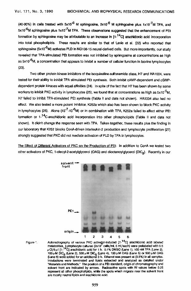
The Effect of Different Activators of PKC on the Production of PEt: In addition to ConA we tested two
other activators of PKC. l-oleoyl-2-acetylglycerol (OAG) and dioctanoylglycerol (DiC8). Recently in our
sfzY t
PEt
origin
-c
-c
- 123456
Figure 1. Autoradiography of various PKC actfv ;Y
or-induced [1-14C] arachiionic acid labeled metabolites. Lymphocyte cultures (2x10 cells/ml, 5 ml/each) were prelabeled with 0.5 JJCI/~~I [l-14C] arachiionlc acid for 1 h. 0.1% DMSO (Lane I), 100 nM TPA (Lane 2). IOO~M DiC6 (Lane 3). 5OOpM DiC6 (Lane 4), IOOpM OAG (Lane 5) or 6OOFtM OAG (Lane 6) were added for an additional 3 h. Ethanol was present at (0.5%) in all samples. Incubations were terminated and llplds. extracted and analyzed as detailed under ‘Materials and Methods.” The position of a PEt standard, origin of chromatography and solvent front are indicated by arrows. Radioactive spots with Rf values below 0.25 represent all other phosphdlpids, while the spots which migrate near the solvent front are mostly neutral lipids and arachidonic acid.
959

Vol. 171, No. 3, 1990 BIOCHEMICAL AND BIOPHYSICAL RESEARCH COMMUNICATIONS
laboratory we have determined that DiCB (15-3OpM) and OAG (SO-2OO~M) mimic TPA in that,
combined with ionomycin they enhance IL-2 production and lymphocyte proliferation (27). OAG and
DiCB did not stimulate the formation of PEt (Fig. 1.). These results obtained with PKC activators
provided more evidence that PKC plays little or no role in the activation of PLD by TPA in lymphocytes.
The Lack of a Need for Macrophaaes on the Production of PEt: TPA stimulated PEt formation in
lymphocytes depleted of macrophages was not significantly different from the cultures that contained
macrophages (data not shown). These results suggest that the act’wation of PLD by TPA was not
accessory cell dependent as Is stimulation of PKC by ConA.
The Effect of TPA on Il-‘4C1 Arachldonic Acid Incorporation into Phospholipid: We analyzed the
phospholipid composition of the cells after labeling with [1-14C] arachkfonic acid and incubation wlth
TPA or DMSO. Phospholipids were separated by TLC and the amount of [l-14C] arachidonic acid
incorporated into each fraction was determined. The results (Table Ill) indicated no significant
differences in most phospholipid classes between the treated and the control groups. But, the levels of
the major phospholipkf, PC, significantly decreased In the lymphocytes treated with lx10W7M TPA during
the period of PLD activation. Because the amount of arachiionic acid and diacyJglycerol were not
significantly changed, this decrease in [l-14C] PC could be attributable to an increase in endogenous
PLD activity.
The Effect of TPA on the PhosDholioase D Activll in Lvmphocvte Lvsates and Subcellular Fractions: To
determine the mechanism of activation of PLD by TPA, we asked lf TPA could activate PLD in cell lysates
or in subcellular fractions. In one case, we tested the microsomal fractions and the lysates from the
Table III. The effect of TPA on the content of lioids in lvmnh node lvmuhocvtes
Lipid content DMSO 2 0.1%
TPA 1x10-'M
Phosphatidic acid
PI + PS
LPC PC
PE
Arachidonic acid DG + TG
PEt
0.26f0.11
0.68f0.02
6.40f3.70 59.0i1.09a
21.Ok2.42
7.61f2.09 4.89f1.81
0.16io.07b
0.25f0.10
0.95f0.25
Ci.50+3.01 53.6f1.0ga
20.4f2.58
10.13f2.25 6.49f2.13
1.68k0.24b
a,b Means sharing a common superscript letter are significantly different (~~0.05).
The pre - labeling and incubation conditions were the same as in Table I. After incubation, the cells were harvested by centrifugation. the lipids extracted with 8 ml chloroform-methanol (l:l, v/v) and 3.2 ml water. The lower lipid-containing phase dried under N2 and applied to a glass silica gel G plate (Whatman). The plate was developed using solvent system II, and visualized by iodine vapor staining. The silica gel areas containing various lipids were quantified by liquid scintillation spectrometry. The results are expressed as means f S.D. (n=6).
960

Vol. 171, No. 3, 1990 BIOCHEMICAL AND BIOPHYSICAL RESEARCH COMMUNICATIONS
intact lymphocytes. The addition of 1x10e7M TPA to the mlcrosomes or the lysates did not increase PLD
activity as it had in the intact cells, even in presence of 5 mM ATP, a condition others used to see the
stimulation of PLD activii by TPA (10) (data not shown). To determine if pretreatment of the intact cells
with TPA was required for the activation of PLD in the isolated fractions, the cells were treated with
l~lO-~fvl TPA before lysis. There was no significant increase of PLD activii in the subcellular fractions
from the cells pretreated with TPA for 4 h, compared to the control cells treated with DMSO. To exclude
the possible effect of other cellular components or experimental conditions, TPA was added directly to
cabbage PLD, but there was no change of PLD activity. When lysates were added to cabbage PLD to
determine if an inhibitor was present, cabbage PLD activity was slightly depressed. However, heat
inactivated bovine serum caused the same results, implying a non-specific effect (data not shown).
DISCUSSION
In summary, we found that TPA stimulated PLD independently of PKC activation whereas ConA
did not affect PLD activiiy. Activators of PKC also did not stimulate PLD. Similarly inhibitors of PKC did
not block PLD. The literature contains several pieces of information in other cell types to suggest that
activation of PKC and PLD by TPA may not be directly related. For example, Billah and his colleagues
reported that PKC inhibitors, H7 and sphingosine, at concentrations up to 100 PM had no detectable
effect on TPA-induced PEt production. K252a (0.5-10 p M) inhibited PEt production by TPA only by 30%,
although it effectively inhibited TPA-induced protein phosphoryiation (4). Rider et al. (28) suggested that
because the PKC inhibitor H7 fails to inhibit TPA-stimulated DG accumulation in human neutrophils,
alternate mechanisms for TPA should be considered. Recently, others used labeled choline release and
labeled PA formation as indicators of PLD activation and repotted that TPA activates PLD via PKC (29).
Although the source of PA generation has been shown to be mostly from PC degradation by PLD in
intact cells treated with TPA or other agonists, labeled PA is transient and its formation is an unreliable
measure of PLD activii (6). For example, it was calculated that at 30 seconds after stimulating HL-60
granulocytes with FMLP, 85% of the alkyi-PA was formed by PLD and the remainder from the DG kinase
pathway. At 5 min, the DG kinase pathway accounted for nearly 50% of the alkyl-PA (5). Furthermore
labeled choline can be released from PC by many sources, e.g. PLC-phosphocholine phosphatase,
PLD, and base exchange enzymes. On the other hand, the assay of PEt formation has many
advantages over the assay of the hydrolysis products of PLD. A double-labeling method demonstrated
that PEt was formed exclusively by the action of PLD and that PEt was stable, up to 12 h after TPA or
FMLP treatment (5,6).
To better our understanding of the mechanism of PLD activation and PEt formation by TPA, we
also studied cell lysates and subcellular fractions. We failed to observe the formation of PEt in these
TPA-exposed lysates or subcellular fractions. Although we saw PEt formation using cabbage PLD &
vitro under the same assay conditions, no significant difference was found between the TPA-treated and
the control enzyme. Furthermore cell lysates did not significantly decrease the activii of the cabbage
enzyme. These results may mean that the activation of PLD by TPA requires a living cell and perhaps
activation involves a receptor-linked mechanism that is disrupted in the preparation of lysates. Our
results agree with those of many others who were unable to detect PLD activii in cell homogenates
using PC as substrate or who found that TPA does not modify PLO in vitrq experiment (29). Recently, others reported that GTPys induced PA formation in rat liver plasma membranes (30) and GTbs plus

Vol. 171, No. 3, 1990 BIOCHEMICAL AND BIOPHYSICAL RESEARCH COMMUNICATIONS
Cat stimulates PLD in homogenates from HL-68 granulocytes (31). One explanation for this
discrepancy is the differences of cell types and agonists; however at this time, the exact reason remains
unknown.
Taken together, our results clearly show that PKC did not solely mediate the activation of PLD
by TPA in bovine lymph node lymphocytes. TPA-induced PEt formation in these lymphocytes seems to
occur via PC catabolism by activation of PLD. The formation of PEt may require a “receptor-linked”
activation of PLD induced by TPA in living cells.
ACKNOWLEDGMENTS
This work was supported by an Intercollegiate Research Project grant from the College of Agriculture, Penn State University to Drs. A. M. Mastro, C. C. Reddy and R. W. Scholz and by grants from the National Institutes of Health, NCI, CA24385 (to A. M. Mastro) and HL31245, CA37979 (to C. C. Reddy).
1.
2. 3.
4.
5. 6. 7. 6.
9. 10. 11. 12. 13. 14.
15. 16. 17. 16.
19.
20. 21.
22. 23. 24. 25. 26.
27.
28. 29. 30.
31.
REFERENCES
Van den Bosch, H. (1982) In Phosoholipids (Hawthorne, J. N.. and Ansell, G. B., eds), pp. 344- 351. Yang, S. F., Freer, S., and Benson, A. A. (1967) J. Biol. Chem. 242,477484. Pai, J.-K., Liehl, E. C., Tettenborn, C. S., Ikegwounu, F. I., and Mueller, G. C. (1987) Carcinogenesis 8, 173-178. Billah, M. M., Pai, J.-K., Mullmann, T. J., Egan, R. W., and Siegel, M. I. (1989) J. Biol. Chem. 264,9069-9876. Pai, J.-K., Siegel, M. I., Egan, R. W.. and Billah, M. M. (1988) J. Bid. Chem. 263, 12472-12477. Tettenbom, C. S., and Mueller, G. C. (1967) Biochim. Biophys. Acta 931, 242-250. Diamond, L, O’Brien, T. G., and Baird, W. M. (1986) Adv. in Cancer Res. 32, l-74. Castagna, M., Takai, Y., Kaibuchi, K., Sano, K., Kikkawa, U., and Nishizuka, Y. (1982) J. Biol. Chem. 257,7847-7851. Niadel, J. E., Kuhn, L. J., and Vandenbark, G. R. (1983) Proc. Natl. Acad. Sci. USA 88,36-46. Tettenbom, C. S., and Mueller, G. C. (1988) B&hem. Biophys. Res. Commun. 155,249-255. Mastro, A. M., and Mueller, G. C. (1974) Exp. Cell Res. 88,46-46. Mastro, A. M., and Pepin, K. G. (1982) Cancer Res. 42, 1630-1635. Kobayashi, M., and Kanfer, J. N. (1987) J. Neurochem. 46, 1597-1663. Cao. Y-Z., Tam, S. W., Arthur, G., Chen, H., and Choy, P. C. (1987) J. Bid. Chem. 262, 16927- 16935. Grier Ill, C. E., and Mastro, A. M. (1988) J. Immunol. 141,2585-2592. Arthur, G., and Choy, P. C. (1984) Biochim. Biophys. Acta 795,221-229. Grove, D. S., and Mastro, A. M. (1987) J. Cell. Physiol. 132,415-427. Merrill, Jr., A. H., Sereni, A. M., Stevens, V. L., Hannun, Y. A., Bell, R. M., and Kinkade, J. M. (1986) J. Bid. Chem. 261, 12610-12615. Hannun, Y. A., Loomis, C. R., Merrill, Jr., A. H., and Bell, R. M. (1986) J. Bid. Chem. 261, 12604-12669. Hannun. Y. A., Greenberg, C. S., and Bell, R. M., (1987) J. Bid. Chem. 262, 1362613626. Wilson, E., Rice, W. G., Kinkade, Jr., J. M., Merrill, Jr., A. H., Arnold, R. R., and lambeth, J. D. (1988) Arch. Biochem. Biophys. 259,204-214. Lavie, Y., and Liscovitch, M. (1996) J. Biol. Chem. 265,38683872. Grove, D. S., and Ma&o, A. M. (1988) Biochem. Biophys. Res. Commun. t51,94-99. Hidaka, H., Inagaki, M., Kawamoto, S., and Sasaki, Y. (1984) Biochemistry 23, 5036-5041. Rush, J. S., Show, E. C., and Weachter, C. J. (1988) Arch. Biochem. Biophys. 267,365316. Gschwendt, M., Leibersperger. H., and Marks, F. (1989) Biochem. Biophys. Res. Commun. 164,974-982. Grove, D. S., and Mastro, A. M. (1996) Differential activation and inhibition of lymphocyte proliferation by Modulators of PKC. (Manuscript in preparation). Rider, L. G., Dougherty, R. W., and Niedel, J. E. (1988) J. Immunol. 146,260-207. Cabot, M. C., Welsh, C. J., Zhang, Z-C., and Cao, H-T. (1989) FEBS Lett. 245,85-96. Bocckino, S. B., Blackmore, P. F., Wilson, P. B., and Exton, J. H. (1987) J. Biol. Chem. 262, 15309-15315. Anthes, J. C., Eckel, S., Siegel, M. I.. Egan, R. W., and Billah, M. M. (1989) Biochem. Biophys.
Res. Commun. 163,657664.
962