evidence for ascending noradrenergic mediation of hypothalamic hyperphagia
TRANSCRIPT

Pharmacology Biochemistry and Behavior, Vol. 1, pp. 81-87. ANKHO International Inc., 1973. Printed in the U.S.A.
Evidence for Ascending Noradrenergic Mediation of Hypothalamic Hyperphagia'
GREGORY KAPATOS AND RICHARD M. GOLD z
State University o f New York at Cortland, Cortland, New York 13045
(Received 13 October 1972)
KAPATOS, G. AND R. M. GOLD. Evidence for ascending noradrenergic mediation of hypothalamic hyperphagia. PHARMAC. BIOCHEM. BEHAV. 1(1) 81-87, 1973.-Rats sustaining unilateral ablation of the ventral ascending noradrenergic bundle, in combination with a contralateral parasagittal knife cut rostrolateral to the ventromedial nucleus of the hypothalamus (VMN), became obese. Ablation of the ventral noradrenergic (NA) bundle was effective at both mammillary and midbrain levels. The weight gains produced by this asymmetrical technique were comparable to those obtained following bilateral parasagittal knife cuts. Rats with unilateral mammillary lesions that spared the ventral NA bundle or unilateral midbrain lesions which only partially destroyed the ventral NA bundle, in combination with the contralateral parasagittal knife cut, did not become obese. The neural substrate damaged in the production of hypothalamic hyperphagia thus appears to be that part of the ascending ventral NA bundle which projects medially through the effective parasagittal knife cut locus rostrolateral to the VMN.
Hyperphagia Hypothalamus Obesity Mammillary Noradrenaline
BILATERAL lesions in the vicinity of the ventromedial nucleus of the hypothalamus (VMN) can produce dramatic hyperphagia and, as a result, obsesity in rats [3,14]. Bilateral parasagittal knife cuts rostrolateral to VMN [ 1,9, 25] also produce hyperphagia and obesity. In unpublished work, Gold has localized the most effective parasagittal knife cut locus to the frontal planes midway between the paraventricular and ventromedial nuclei (Figs. 30--32 in K/Snig and Klippel [ 17] ). In addition, horizontal knife cuts dorsal to VMN [22], and large bilateral lesions at the extreme caudal end of the hypothalamus in the vicinity of the mammillary nuclei, can produce hyperphagia and obesity [ 12,15]. These studies suggest that the critical fibers destroyed in hypothalamic obesity may originate at a mammillary, midbrain, or brainstem site, and project rostrally through the hypothalamus. These fibers may run in the lateral hypothalamus (LH) as a component of the medial forebrain bundle (MFB), or they may pass diffusely through the medial hypothalamus. At the rostral end of the hypothalamus the fibers, it appears, must turn medially (or
laterally) and dorsally through the effective knife cut loci. A projection in the opposite (caudal) direction is also possible.
Recent data, using an asymmetrical lesion technique, have clearly demonstrated that feeding inhibition is indeed mediated by a longitudinal projection [11 ]. A unilateral parasagittal knife cut rostralateral to VMN was combined with a contralateral unilateral electrolytic lesion in the mammillary area. The rats rapidly gained weight at a rate equivalent to that which follows bilateral parasagittal knife cuts.
The only fibers lesioned bilaterally were those that pass through both the knife cut region and the mammillary area lesion. All other fibers were only damaged unilaterally. Thus, a common neural system appears to be interrupted by both the parasagittal rostrolateral to VMN knife cut and the mammillary area lesion.
The mammillary ablation in the previous study unilater- ally destroyed the mammillary nuclei and the medial region of the MFB [11]. In Experiment 1 of the present study
~This study was supported by United States Public Health Service Grant MH-13561 to Richard M. Gold. The assistance of Patricia M. Quackenbush is gratefully acknowledged.
2 Requests for reprints should be sent to Richard M. Gold, Department of Psychology, State University of New York at Cortland, New York 13045.
81

82 KAPATOS AND ( ;OLD
asymmetr ical lesions are used to investigate the possible impor tance to hypotha lamic hyperphagia of fiber systems efferent and afferent to the mammil lary bodies. Experi- ment 2 also uses asymmetr ica l lesions to fur ther trace the critical project ions in the midbrain, and relates hypotha lam- ic hyperphagia to recent his tochemical and pharmacological data regarding the ascending monoamine pathways and their possible role in the control of food intake [ 19,28].
level of the mammil lary bodies (A 4.7, L 1.2, V 8.0). Unilateral control animals received only the unilateral VMN lesion or parasagittal knife cut, while the bilateral controls received bilateral parasagittal hypotha lamic knife cuts. Sham operat ions consisted of ether anesthesia, placement in the s tereotaxic instrument , and exposure of the dura.
Af te r 5 weeks of postoperat ive ad lib feeding on the high fat diet, all lesioned animals were perfused, and frozen frontal plane brain sections were stained with cresyl violet for cells or a Weigert Weil stain for fibers.
Method
E X P E R I M E N T 1 Resu l t s
The mammil la ry nuclei are innervated afferent ly by the fornix, MFB, and mammil la ry peduncle, while efferent project ions are via the principle mammil lo tha lamic tract. The principle mammil lo tha lamic tract ascends f rom the mammil lary bodies and divides into the mammil lo tha lamic and mammil lo tegmenta l tracts [18] . Both the mammil lo- tegmental tract and the mammil la ry peduncle are, respec- tively, major descending and ascending fiber systems linking the hypotha lamus and the midbrain tegmenta l nuclei [6 ,18] . The medial forebrain bundle carries fibers which appear to project f rom the septum to the mammil la ry nuclei [ 16,24]. ,', 5 0 0
In this study lesions were made in the principle E mammil lo tha lamic tract (in order to destroy both mammil- Iothalamic and mammil lo tegmenta l project ions) , the mam- I.- millary peduncle, the mammil lary nuclei, or the adjacent -I- MFB. Unilateral lesions of one o f the four loci were always ,'7;, 400 combined with a unilateral controla tera l e lectrolyt ic VMN lesion or parasagittal rostrolateral to VMN knife cut. >.
O r~
Animals and diets. Animals were individually housed, f e m a l e C a r w o r t h C F E r a t s , 85 95 days old at the t ime of surgery. A high fat diet consisting of 67% (by weight) Purina labora tory meal and 33% Crisco brand hydrogenated vegetable shortening was available ad lib f rom an 8-cm dia. glass cup secured to a corner of each cage. Tap water from inverted bot t les was also available ad lib. Details of diet preparat ion and intake measures were as described previously [10] . Twice each week the rats were weighed, food and water intakes were determined by difference weights, and fresh food and water were provided. Clean cages and fresh l i t ter were provided at f requent intervals, and room tempera ture was maintained at 72 ° --75 °F ( 22 -24°C) .
Surgery and Histology. Parasagittal knife cuts rostrolat- eral to VMN were produced with a 3 mm long razor blade knife [10] . Elec t ro ly t ic lesions were produced via a 0.25 mm dia. stainless steel insulated wire anode with a 0.2--0.5 mm conical tip exposed. The top of the incisor bar was always 3 mm below the center of the ear bars. Anter ior , lateral ,and vertical s tereotaxic reference points were,respec- tively, ear bar center, midsagittal sinus and dura. Asymme- trically lesioned animals received ei ther a unilateral electro- lytic vent rodmedia l hypotha lamic (VMN) lesion (A 6.5, L 0.5, V 8.2), or a unilateral rostrolateral to VMN parasagittal knife cut (A 6.5, L 0.9, V to base). This was combined with a contralateral unilateral lesion placed in one of four loci: the principle mammil lo tha lamic tract (A 5.0, L 0.75, V 7.8); the mammil lary peduncle (A 3.6, L 0.7, V 8.45); the mammil lary nuclei (A 4.7, L 0.6, V 8.2); or the MFB at the
Mean body weights and food intakes for 5 weeks fol lowing recovery f rom postoperat ive weight loss (if any) are shown in Fig. 1. Mean body weight gains are also shown in Table 1.
3 0 0
2 5
. i
'-' 2 0 ,< I - z - 15 a 0 0 10 I . I .
o . . . . o C u t , B i l a t e r a l a m--m H y p X M F B .,/,'" o . . . . o C u t X V N A / t ~ ' / • • ~ = H y p X M a m m / ~ ' •
• n " O z~ . . . . & H y p . , U n , l a t e r a l s / m / / .
• • S h a m / ~ ' o . / , 0 / s SS°
/~ • .0 °S I / s S S
os S
I I I . [ ~ . . . . / I I
/ m ~ m"" m < '0 ~-0
/ f / O . . . . . 0 " ~ 0
L /~1 l / . , • •
I * i , I I I -1 O p 1 2 3 4 5
W E E K S
FIG. 1. Mean body weights and food intakes (high fat diet). Cut=parasagittal knife cut rostrolateral to the ventromedial hypo- thalamus (VMN; Hyp.=either a unilateral knife cut or VMN lesion; X=crossed combination; MFB=medial forebrain bundle; Mature. = mammillary nuclei and fiber systems: VNA=ascending ventral
noradrenergic bundle.
Rats having unilateral VMN lesions or unilateral para- sagittal knife cuts displayed modest weight gains (means 1.4
NA M E D I A T I O N OF H Y P O T H A L A M I C H Y P E R P H A G I A 83
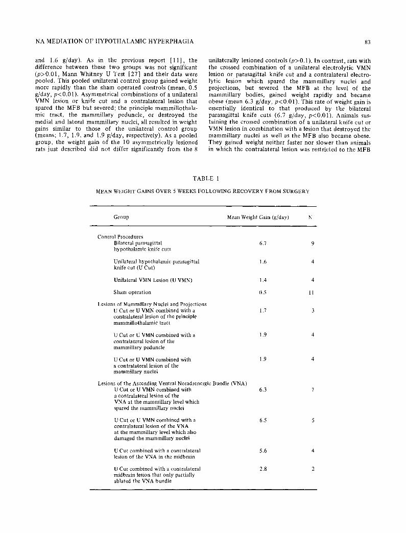
and 1.6 g/day) . As in the previous repor t [ 1 1 ] , t he d i f fe rence b e t w e e n these t w o groups was no t s ignif icant ( p > 0 . 0 1 , M a n n Whi tney U Test [27] and the i r da ta were pooled. This pooled uni la tera l c o n t r o l g roup gained weight more rapidly t h a n the sham ope ra t ed con t ro l s (mean , 0.5 g/day, p < 0 . 0 1 ) . Asym m et r i c a l c o m b i n a t i o n s of a uni la te ra l VMN lesion or kni fe cut and a con t ra la te ra l lesion t ha t spared the MFB bu t severed; the pr inciple mammi l lo tha l a - mic t ract , the m a m m i l l a r y pedunc le , or des t royed t he medial and lateral mammi l l a ry nuclei , all resul ted in weight gains similar to those of the uni la te ra l con t ro l g roup (means ; 1.7, 1.9, and 1.9 g/day, respect ively) . As a poo led group, the weight gain of the 10 a symmet r i ca l ly les ioned rats jus t descr ibed did no t d i f fer s ignif icant ly f rom the 8
uni la te ra l ly les ioned con t ro l s (p> 0.1). In cont ras t , rats w i th the crossed c o m b i n a t i o n of a uni la tera l e lec t ro ly t ic VMN lesion or parasagi t ta l kni fe cut and a con t ra l a t e ra l e lectro- lyt ic lesion wh ich spared the m a m m i l l a r y nuclei and projec t ions , bu t severed the MFB at the level of the mammi l l a ry bodies , gained weight rapidly and b e c a m e obese ( m e a n 6.3 g /day, p < 0 . 0 1 ) . This rate of weight gain is essential ly ident ical to t h a t p roduced by the bi la teral parasagi t ta l kni fe cuts (6.7 g /day, p < 0 . 0 1 ) . Animals sus- ta in ing the crossed c o m b i n a t i o n of a uni la tera l kni fe cut or VMN lesion in c o m b i n a t i o n wi th a lesion t ha t des t royed the mammi l l a ry nuclei as well as the MFB also b e c a m e obese. They gained weight ne i the r fas ter n o r s lower t h a n animals in wh ich the con t ra la te ra l lesion was res t r ic ted to the MFB
T A B L E 1
MEAN WEIGHT GAINS OVER 5 WEEKS FOLLOWING RECOVERY FROM SURGERY
Group Mean Weight Gain (g/day) N
Control Procedures Bilateral parasagittal hypothalamic knife cuts
Unilateral hypothalamic parasagittal knife cut (U Cut)
Unilateral VMN Lesion (U VMN)
Sham operation
Lesions of Mammillary Nuclei and Projections U Cut or U VMN combined with a contralateral lesion of the principle mammillothalamic tract
U Cut or U VMN combined with a contralateral lesion of the mammillary peduncle
U Cut or U VMN combined with a contralateral lesion of the mammillary nuclei
Lesions of the Ascending Ventral Noradrenergic Bundle (VNA) U Cut or U VMN combined with a contralateral lesion of the VNA at the mammillary level which spared the mammiUary nuclei
U Cut or U VMN combined with a contralateral lesion of the VNA at the mammillary level which also damaged the mammillary nuclei
U Cut combined with a contralateral lesion of the VNA in the midbrain
U Cut combined with a contralateral midbrain lesion that only partially ablated the VNA bundle
6.7 9
1.6 4
1.9 4
1.9 4
6.3 7
6.5 5
5.6 4
2.8 2
1.7 3
1.4 4
0.5 11

84 KAPATOS AND GOLD
(means 6.5 and 6.3 g/day, p>0 .1) . Thus, unilateral damage to the mammil la ry bodies nei ther produces, enhances, or impairs obesity.
As noted above, there was no significant weight gain difference be tween unilateral parasagittal knife cuts and unilateral VMN lesions. Similarly, when combined with a contralateral MFB lesion, there was no significant weight gain difference be tween unilateral parasagittal knife cuts (n =3) and unilateral VMN lesions (n = 4, p>0.1) . This is further jus t i f icat ion for pooling unilateral VMN lesion and parasagittal knife cut groups.
Anatomical findings. Recons t ruc t ions of representative lesions are shown in Fig. 2. Ablat ions of the principle mammil lo tha lamic tract (Fig. 2A) were localized to, and in all 3 cases severed, the origins of both the mammil lo tha lam- ic and mammil lo tegmenta l tracts. The MFB, fornix, and the mammil lary nuclei in part icular were consistent ly spared by these lesions.
Lesions of the mammil la ry peduncle (Fig. 2B), poster ior to the mammil lary nuclei, were restricted exlcusively to the peduncle, with the except ion that 2 of the 4 animals also sustained partial damage to the fasciculus retroflexus. Al though the mammil lary nuclei were spared damage, there was degenerat ion in the lateral and medial mammil la ry nuclei ipsilateral to the lesion, as previously described by Cowan, Guillery and Powell [6] .
Dest ruct ion of the mammil lary bodies typically involved both the medial and lateral mammil lary nuclei as well as the mammil lary peduncle (Fig. 2C). These lesions extended rostrally to the premammil lary nuclei and caudally to the in terpeduncular nucleus, which in one case was dest royed unilateral ly along with the fasciculus retroflexus. The MFB was spared.
Lesions destroying the MFB (Fig. 2D) extended rostrally to the level of the premammil la ry nuclei and caudally to the level of the in terpeduncular nucleus. In all 7 cases the MFB was comple te ly severed and the mammil lary nuclei and project ions were spared. In isolated cases there was minimal damage to the medial lemiscus, the internal capsule, the zona compacta of the substantia nigra, or the fields of Forel.
All rostrolateral to VMN parasagittal knife cuts, both unilateral and bilateral, met the anatomical criteria for knife cuts that induce obesi ty as localized by Gold ( [19] and unpublished). Unilateral VMN lesions typical ly des t royed the ventromedial nucleus and immedia te ly adjacent tissue, but wi thout crossing the midline or extending lateral to the fornix. On occasion, however , the unilateral VMN lesion was more dorsal than intended and spared all but the dorsal edge of the VMN, while destroying the dorsomedial nucleus (DMN). Figure 2E shows such a unilateral VMN lesion which, in combina t ion with a contralateral lesion of the MFB, produced marked hyperphagia and rapid weight gains (8.1 g/day).
Discussion
In a previous report [1 1 ], it was shown that the fibers which must be destroyed for the p roduc t ion of hypothal - amic hyperphagia course longitudinal ly through the mam- millary area and the vicinity of the VMN. Exper iment 1 of the present s tudy demonst ra tes conclusively that destruc- tion of nei ther the mammil la ry bodies nor their efferent or afferent project ions are involved in hypotha lamic hyper- phagia and obesity. In contrast , animals with unilateral
'i ,, : )1
ILi.7 gin/day G /1~ ? ~ ' / 7
~ - F MFB ~
i /
/~.ggm/da~'%~.l-.'" / ". )
P C M A
(u, (i - ' " ) ' (
/ / '~ i / i. "\,
,\
( [ , D e N
~8 I gin/day 0 / " ,A 489o~( i] J% '? " - ~ / .IVIN M F B . ~"
FIG. 2. Reconstructions of representative lesions superimposed on frontal sections of the Konig and Klippel atlas [17]. Weight gains are means over 5 weeks. A, principle mamillothalamic tract; B, mammillary peduncle; C, mammillary nuclei; D. medial forebrain bundle. A through D were combined with either a contralateral parasagittal knife cut rostrolateral to the ventromedial hypo- thalamus (VMN) or a contralateral electrolytic VMN lesion. E, an atypical VMN lesion which spared most of the ventromedial hypothalamic nucleus but ablated the dorsomedial hypothalamic nucleus. This lesion combined with a contralateral lesion of the medial forebrain bundle produced rapid weight gains. Abreviations: DNA=ascending dorsal noradrenergic bundle; DMN=dorsomedial hypothalamic nucleus; F=fornix; FR=fasciculus retroflexus; MFB=medial forebrain bundle; MM=mammillary nuclei; PCMA= mammillary peduncle; PMTT=principle mammillothalamic tract: VNA=ascending ventral noradrenergic bundle; VMN=ventromedial
hypothalamic nucleus.
e lectrolyt ic lesions of the poster ior MFB combined with a contralateral unilateral e lectrolyt ic VMN lesion or para- sagittal knife cut did become hyperphagic and obese. These asymmetr ical ly lesioned rats displayed weight gains equiva- lent to those of animals sustaining bilateral parasagittal knife cuts. It can be concluded from these data that the neural system involved in hypotha lamic twperphagia pro- jects longitudinally within the MFB.
If lesions or knife cuts near the VMN and lesions in the caudal MFB produce obesity by severing one and the same longitudinal neural pathway, then frontal plane knife cuts or e lectrolyt ic lesions caudal to VMN but rostral to the mammil lary nuclei should also sever the pathway. At tempts to produce hypotha lamic obesi ty with bilaterally symmetr i - cal cuts or lesions have produced cont radic tory results, perhaps because more than one neural system involved in feeding is interrupted. Large bilateral lesions in the mam- millary area can produce hyperphagia and obesity [1 1, 12, 15]. These lesions typical ly encroach on the medial hypotha lamus and more laterally on the medial por t ion of the MFB. Large bilateral frontal cuts immedia te ly caudal to

NA MEDIATION OF HYPOTHALAMIC HYPERPHAGIA 85
the VMN produce only a modest weight gain and only if the lateral as well as medial areas of the hypothalamus are severed [ 13], while cuts restricted to either the medial or lateral hypothalamus alone produce no effect, aphagia, or hypophagia [2, 13, 23, 26]. The apparent discrepancy between these small or negative weight gains and our data may be due to our use of a bilaterally asymmetrical rather than bilaterally symmetrical surgical procedure. The major advantage of this approach is that bilateral damage only occurs to those neural systems which pass through both lesion loci. This allows a more selective destruction to individual neural pathways. A more discriminative beha- vioral deficit should thus be obtainable, and the unlesioned side of the brain at each lesion locus is available for histological assistance.
One must still account for the conflicting data of previous studies that used bilaterally symmetrical lesions or knife cuts. Some of the studies report hypothalamic hyperphagia [11, 12, 13] while others report aphagia or hypophagia [2,23] or no effect [26]. A tentative hypothesis to account for these differences involves the discrete location within the MFB of the separate monoamine pathways as described by Ungerstedt [28].
At the level of the caudal VMN the most dorsolateral portion of the MFB contains dopaminergic fibers which arise from the substantia nigra and project to the corpus striatum. The destruction of these nigro-striatal dopamin- ergic fibers produces a period of aphagia and adipsia surpassing that produced by lesions of the more medial portion of the medial forebrain bundle [8, 21, 29]. Bilaterally symmetrical surgery caudal to VMN may there- fore produce aphagia if the frontal knife cut or lesion severs the dorsolateral (dopaminergic) portion of the medial forebrain bundle, even if satiety fibers are also severed. If the surgery does not produce damage to the dorsolateral MFB, but does sever satiety fibers located more medially in the MFB, hyperphagia and obesity should occur. Unpub- lished data from our laboratory has shown that bilateral electrolytic lesions of the entire posterior MFB produce an initial period of aphagia and adipsia in aggrement with the data of Albert, Storlien, Wood and Ehman [2]. These same animals later became hyperphagic and obese on a high fat diet. This finding is quite similar to data presented by Carlisle and Stellar [5], in that their animals which sustained damage to both the VMN and LH were at first aphagic and adipsic, but later became hyperphagic if a high fat diet was used.
EXPERIMENT 2
Experiment 1 demonstrated that hypotbalamic obesity results from damage to fibers that course longitudinally within the MFB and bypass the mammillary bodies. Recent histochemical mapping of the monoamine pathways has shown that at the level of the mammillary bodies the MFB contains ascending dopaminergic (DA), noradrenergic (NA), and serotonergic (5-HT) fibers which arise from nuclei in the brainstem and midbrain tegmentum [28]. In Experi- ment 1 the lesions of the MFB which produced hyperphagia and obesity when combined with contralateral parasagittal knife cuts may have damaged all of these ascending monoamine systems. However, of the monoamines only NA has been implicated as possibly playing a role in the inhibition of feeding in the rat [4,19].
Ungerstedt [28] has separated the ascending NA axons into a dorsal and ventral bundle. The dorsal bundle innervates the cortex and hippocampus, while the ventral bundle supplies NA terminals to the pons, medulla, and more importantly to the entire hypothalamus. At the level of the posterior interpeduncular nucleus these two bundles are separate from each other and from the DA and 5-HT systems. In Experiment 2, lesions of the ascending ventral NA bundle were combined with contralateral parasagittal knife cuts rostrolateral to VMN in order to determine what role, if any, these ascending NA fibers play in hypothalamic hyperphagia.
Method
Except as noted below, the methods were as in Experiment 1. Asymmetrically lesioned animals received a unilateral parasagittal knife cut in combination with a contralateral electrolytic lesion of the ascending ventral NA pathway.
In order to produce lesions which conformed to the dimensions of the ventral NA bundle, (see FIG. 4A), two electrode placements were used: A 2.5, L 1.3, V 7.0 and A 2.5, L 1.6, V 6.6.
Control data for rats receiving either unilateral or bilateral parasagittal knife cuts or sham operations were taken from Experiment 1, which was run concurrently.
Results
Mean body weights and food intakes for 5 weeks following recovery from postoperative weight loss (if any) are shown in Fig. 1 (Cut X VNA group). Mean body weight gains are shown in Table 1.
Animals with the asymmetrical combination of a uni- lateral parasagittal knife cut and a contralateral electrolytic lesion of the ascending ventral NA bundle became hyper- phagic and obese (mean weight gain, 5.6 g/day), in comparison to the unilateral or sham operated animals (p's<0.01). The weight gains were similar to but signifi- cantly lower than those found following bilateral knife cuts (mean 6.7 g/day, p<0.05), although one asymmetrically lesioned animal did display a weight gain of 6.2 g/day.
Anatomical findings. The ascending ventral NA bundle as localized by Ungerstedt [28] was ablated in four of six animals (Fig. 3, A-D) with damage also occurring in varying degrees to the lateral portion of the superior cerebellar peduncle and the recticular formation. The lesions typically extended rostrally to the interpeduncular nucleus, and caudally to the level of the trigmental nerve. Minimal damage produced by the lesion or the electrode track may also have occurred in the ascending dorsal NA bundle. The animal sustaining maximum damage to the dorsal NA bundle (Fig. 3D) displayed the lowest rate of weight gain of animals in this asymmetrical group (5.0 g/day). Gold [8] has previously described a critical midbrain area, which when lesioned produced aphagia and adipsia. The ascending dorsal NA bundle is situated within this critical midbrain region. Since all four animals appear to have damage to the dorsal NA bundle, this may account for the failure of these lesions to produce as great a rate of weight gain as some of the other procedures. Alternatively, NA fibers at the periphery of the ventral bundle may have been spared by these small lesions. These possible explanations are sup- ported by Experiment 1, where lesions of the ascending ventral NA bundle at the mammillary level, which did not

86 KAPA'IOS ,\NI) (;OLD
encroach on the dorsal NA bundle (Fig. 2D), produced weight gains which were no different from those produced by bilateral knife cuts (Fig. 1 ).
Figure 3E and F are reconstructions of two midbrain lesions which only partially destroyed the ascending ventral NA bundle. The lesion in Fig. 3E destroyed the ascending ventral NA bundle. Tire lesion in Fig. 3E destroyed only the lateral portion of the ventral NA bundle, while that of Fig. 3F ablated only the medial portion. Both these lesions produced similar modest weight gains, which seems to indicate that no one portion of the ventral bundle is specifically involved in the inhibition of feeding, but rather that the inhibitory fibers project diffusely within the entire bundle.
/ ( < k >
i 6 2 gin/day ~ --VNA '
a ', i ' - ! ~ + + / \ + ~ ~ .:, , . ._ ........ .
/p: .,+~ ; "% ~x
u ::: i )y+:7 ' ..... " 5+6 gm/dav /~ , ' :
( I
,' J
FIG. 3. Reconstructions of individual lesions in the ascending ventral noradrenergic bundle. Each lesioned animal also had a contralateral parasagittal knife cut rostrolateral to the ventromedial hypothalamus (VMN). Rates of weight gain are over a 5 week period. Abbreviations: DNA=ascending dorsal noradrenergic bundle; IP~-interpeduneular nucleus; PCS=superior cerebellar peduncle;
VNA=ascending ventral noradrenergic bundle.
G E N E R A L D I S C U S S I O N
Unilateral ablation of the ascending ventral noradre- nergic (NA) pathway at a midbrain or mammillary level, in combination with a contralateral parasagittal knife cut, resulted in hyperphagia and obesity. These data are in agreement with Ahlskog and Hoebel's [4] d e m o n s t r a t i o n
that bilateral destruction of the ascending ventral NA bundle at the midbrain level produces hyperphagia and obesity. Unilateral lesions in tile midbrain which only partially ablated tile ventral NA bundle produced only modest hyperphagia when combined with contralateral parasagittal knife cuts. Further rostral, unilateral lesions which spared the ventral NA pathway as it ascends within tile MFB, but destroyed the nlammillary nuclei or their fiber systems, did not produce obesity when combined with contralateral parasagittal knife cuts.
The neural substrate which must be lesioned to produce hypothalamic hyperphagia therefore may be tile ascending ventral NA bundle. This bundle projects rostrally from NA cell groups in the medulla and pons [7,281. The NA brainstem nuclei have been shown to possess neural connections with the nuclei of autonomic nerves, such as the nucleus tractus solitarius, which receives oropharangeal afferent taste fibers via the glossopharyngeal, lingual, and vagus nerves I20,281. These brainstem nuclei could thereby receive hunger related information by way of the peripheral nervous system, and conduct this information via the ascending ventral NA bundle to the hypothalamus where an integration of food related sensory cues might occur.
From the brainstem NA nuclei the ventral NA bundle ascends through the recticular formation, along the nredial lemniscus, and continues rostrally within the MFB from which NA fibers then turn medially to innervate tile hypothalamus (28]. Some of these medially projecting fibers appear to be interrupted by the parasagittal hypo- thalamic knife cuts or by tire more traditional electrolytic VMN lesions.
There are no NA terminals within the VMN itself [281. The degree of hyperphagia and obesity following VMN lesions has been shown to correlate positively with the size of the lesion [3], suggesting fibers of passage adjacent to the VMN may be critical. Frontal knife cuts anterior to the VMN can produce hyperphagia, quite possibly by severing the same fibers as rostrolateral to VMN parasagittal knife cuts [ 13,23].
Destruction of the MFB at tire level of the mammillary nuclei results in a depletion of NA terminals ipsilaterally in the entire hypothalamus. Lesions of the MFB just rostral to the mammillary bodies do not produce a depletion of hypothalamic NA terminals, with the exception of the paraventricular nucleus and the preoptic area [281. The NA fibers to the hypothatanrus thus appear to turn medially within the hypothalamus long before they reach the level they innervate. Since parasagittal knife cuts at the caudal end of the hypothalamus which would sever the medially projecting fibers to the hypothalamus, do not produce hyperphagia and obesity 191, the medially projecting NA fibers that are interrupted in hypothalamic hyperphagia may innervate more rostral structures such as the paraven- tricular nucleus. If the fibers that are critically damaged in hypothalamic obesity normally inhibit feeding, then the loci containing the neurons thay they inhibit must be spared by lesions that produce obesity. Thus, if the fibers involved in the inhibition of feeding do indeed ascend from brainstenr NA nuclei, then the VMN and adjacent loci may not be the recipient of these inhibitory NA terminals, but rather the fibers may course near the VMN and project to more rostrally located structure (s).
Finally, the conclusion that destruction of the ventral NA bundle may be responsible for hypothalamic hyper- phagia has been based on the destruction, albeit at several

NA M E D I A T I O N OF H Y P O T H A L A M I C H Y P E R P H A G I A 87
levels and w i th a symmet r i ca l lesions, of loci at w h i c h the maps p repa red b y Unger s t ed t [28] place the vent ra l bundle . Damage to the vent ra l b u n d l e was no t quan t i f i ed di rect ly b y h i s to- f luorescent or b iochemica l techniques . Also, the e lec t ro ly t ic lesions and kni fe cuts used do no t
selectively damage adrenergic fibers. It is en t i re ly possible t ha t some p a t h w a y o t h e r t han the vent ra l bundle , bu t which runs a parallel course, is responsible for hypo- tha lamic hype rphag ia w h e n des t royed .
REFERENCES
1. Albert, D. J. and L. H. Storlien. Hyperphagia in rats with cuts between the ventromedial and lateral hypothalamus. Science 165: 599--600, 1969.
2. Albert, D. J., L. H. Storlien, D. J. Wood and G. K. Ehman. Further evidence for a complex system controlling feeding. Physiol. Behav. 5: 1075--1082, 1970.
3. Anand, B. K. and J. R. Brobeck. Hypoth~amic control of food intake in rats and cats. Yale J. biol. Med. 24: 123-140, 1951.
4. Ahlskog, J. E. and B. G. Hoebel. Hyperphagia resulting from selective destruction of an ascending adrenergic pathway in the rat brain. Fedn Proc. 31: 377, 1972.
5. Carlisle, H. J. and E. Stellar. Caloric regulation and food preference in normal, hyperphagic and aphagic rats. J. eomp. physiol. Psychol. 69: 107--114, 1969.
6. Cowan, W. M., R. W. Guillery and T. P. S. Powell. The orgin of the mammillary peduncle and other connections from the midbrain. J. Anat. 98: 345, 1964.
7. Dahlstr6m, A. and K. Fuxe. Evidence for the existence of monaonine-containing neurons in the central nervous system. I. Demonstration of monoamines in the cell bodies of brainstem neurons. Acta. physiol, seand. 62: Supple. 232, 1--55, 1965.
8. Gold, R. M. Aphagia and adipsia following unilateral and bilaterally asymmetrical lesions in rats. Physiol. Behav. 2: 211--220, 1967.
9. Gold, R. M. Hypothalamic hyperphagia produced by parasagit- tal knife cuts. Physiol. Behav. 5: 23--25, 1970.
10. Gold, R. M. Hypothalamic hyperphagia: Males get just as fat as females. J. comp. physiol. Psychol. 71: 347--356, 1970.
11. Gold, R. M., P. M. Quackenbush and G. Kapatos. Obesity following combination of rostrolateral to VMH cut and controUateral mammillary area lesion. J. comp. physiol. Psychol. 79: 210-218, 1972.
12. Graff, H. and E. Stellar. Hyperphagia, obesity and finickiness. J. comp. physiol. Psychol. 55: 418--424, 1962.
13. Grossman, S. P. Changes in food and water intake associated with an interruption of the anterior or posterior fiber connections of the hypothalamus. J. comp. physiol. Psychol. 75: 23--31, 1971.
14. Hetherington, A. W. and S. W. Ranson. Hypothalamic lesions and adiposity in the rat. Anat. Rec. 78: 149--172, 1940.
15. Hetherington, A. W. and S. W. Ranson. The relationship of various hypothalamic lesions to adiposity in the rat. J. eomp. Neurol. 76: 475-499, 1942.
16. Johnson, T. N. An experimental study of the fornix and hypothalamo-tegmental tracts in the cat. J. comp. Neurol. 125: 29, 1966.
17. K~nig, J. F. R. and R. A. Klippel. The Rat Brain: A Stereotaxic Atlas o f the Forebrain and Lower Parts of the Brain Stem. Baltimore, Maryland: Williams and Wilkins, 1963.
18. Krieckhaus, E. E. The mammillary bodies: Their function and anatomical connections. Proceedings of a Symposium: The Functional Properties o f Hypothalamus. Held in Lubin, September, 1966.
19. Liebowitz, S. F. Central adrenergic receptors and the regula- tion of hunger and thirst. Presented to the association for Research in Nervous and Mental Disease, New York City, December 5, 1970.
20. Makous, W., S. Nord, B. Oakley and C. Pfaffman. The gustatory relay in the medulla. In: Olfaction and Taste, edited by Y. Zotterman. Oxford: Pergamon Press, 1963, pp. 381-393.
21. Oltmans, G. A. and J. A. Harvey. LH Syndrome and brain catecholamine levels after lesions of the nigrostriatal bundle. Physiol. Behav. 8: 69--78, 1972.
22. Palka, Y., R. A. Liebelt and V. Critchlow. Obesity and increased growth following partial or complete isolation of the ventromedial hypothalamus. Physiol. Behav. 7: 187-194, 1971.
23. Paxinos, G. and D. Bindra. Hypothalamic knife cuts: effects on eating, drinking, irritability, aggression and copulation in the male rat. J. comp. physiol. Psychol. 79: 219-230, 1972.
24. Powell, E. W. Septal efferents in the cat. ExplNeurol. 14: 328, 1966.
25. Sclafani, A. and S. P. Grossman. Hyperphagia produced by knife cuts between the medial and lateral hypothalamus in the rat. Physiol. Behav. 4: 533--537, 1969.
26. Sclafani, A. Neural pathways involved in the ventromedial hypothalamic lesion syndrome in the rat. J. comp. physiol. Psychol. 77: 70--96, 1971.
27. Siegel, S. Nonparametric Statistics for the Behavioral Sciences. New York: McGraw-Hill, 1956, pp. 152--156.
28. Ungerstedt, U. Stereotaxic mapping of the monoamine path- ways in the rat.Actaphysiol, scand. Supple. 367, 1--48, 1971.
29. Ungerstedt, U. Adipsia and aphagia after 6-hydroxydopamine induced degeneration of the nigro-striatal dopamine system. Acta physiol, scand. Supple. 367, 95-122, 1971.