evaluation of thyroid function in dogs suffering from recurrent flank
TRANSCRIPT
Evaluation of thyroid function in dogs sufferingfrom recurrent flank alopecia
Sylvie Daminet, Manon Paradis
Abstract - Thyroid function was assessed in euthyroid dogs (n = 20), dogs suffering fromcanine recurrent flank alopecia (CRFA, n = 18), and hypothyroid dogs (n = 21). Blood samplesobtained from all dogs in each group were assayed for total thyroxine (TT4), thyrotropin (TSH), andthyroglobulin autoantibody (TgAA) serum concentrations. Total T4 and TSH serum concentrationswere significantly decreased and increased, respectively, in the hypothyroid group compared withthe other 2 groups. No significant differences in TT4 and TSH serum values were found between theeuthyroid and CRFA groups. Thyroglobulin autoantibodies were detected in 10, 1 1.1, and 61.9% ofeuthyroid dogs, dogs with CRFA, and hypothyroid dogs, respectively. In conclusion, dogs suffer-ing from CRFA have a normal thyroid function, and the determination of TT4 and TSH serum con-centrations allows differentiation of these dogs from dogs with hypothyroidism, in most cases.Occasionally, the 2 diseases can be concomitant.
Resume- Evaluation de la fonction thyroidienne chez des chiens souffrant d'alopeciesrecidivantes des flancs. La fonction thyroidienne a ete evaluee chez des chiens euthyroidiens (n =20), chez des chiens souffrant d'alopecie canine recidivante des flancs (ACRF; n = 18) et chez deschiens hypothyroYdiens (n = 21). Dans les echantillons de sang provenant de chacun des chiens dechaque groupe, on a mesure les concentrations seriques de la thyroxine totale (TT4), de la thyrotropine(TSH) et des auto-anticorps de la thyroglobuline. Les concentrations seriques de la T4 totale et dela TSH etaient significativement diminuees et augmentees respectivement dans le groupe hypothy-roidien par rapport aux 2 autres groupes. Aucune difference significative dans les valeurs seriquesde TT4 et de TSH n'a ete observee entre les groupes d'euthyroidiens et d'ACRF. Les auto-anticorpsde la thyroglobuline ont ete detectes respectivement chez 10, 1 1,1 et 61,9 % des chiens euthyrofdiens,de ceux presentant de I'ACRF et des hypothyroidiens. En conclusion, les chiens souffrant d'ACRFont une fonction thyroidienne normale et la determination des concentrations seriques de TT4 et deTSH permet dans la plupart des cas de differencier ces chiens de ceux souffrant d'hypothyroidie. Detemps en temps, les 2 maladies peuvent etre concomitantes.
(Traduit par docteur Andre Blouin)Can Vet J 2000;41:699-703
IntroductionThe diagnosis of canine hypothyroidism can be chal-Ilenging for 2 main reasons: first, several other diseasescan present with similar clinical signs, and second,there is no ideal test to assess canine thyroid function.Alopecia is a common clinical sign of hypothy-
roidism; 25% of hypothyroid dogs have been reported topresent with symmetrical bilateral endocrine alopecia (1).The symmetrical alopecia described in canine recur-rent flank alopecia (CRFA) can easily mislead one to adiagnosis of hypothyroidism. Canine recurrent flankalopecia is a recently recognized skin disorder ofunknown etiology, characterized by episodes of truncalhair loss that often occur on a recurrent basis. It has beendescribed under several synonyms: seasonal flank alope-
Departement de sciences cliniques, Faculte de medecineve'trinaire, Universite de Montreal, C.P. 5000, Saint-Hyacinthe(Qu6bec) J2S 7C6.
Address correspondence and reprint requests to Dr. SylvieDaminet.
This project was funded in part by the Fonds du Centenaire ofthe Faculte de medecine veterinaire, Universite de Montreal.
cia, seasonal growth hormone deficiency, canine idio-pathic cyclic flank alopecia, cyclic follicular dysplasia,and follicular dysplasia. Typically, this alopecia affectsadult dogs on a yearly, seasonal basis and resolvesspontaneously without treatment within several months.The episodes do not always recur every year. Severalhypotheses, such as photoperiod and melatonin defi-ciency, have been proposed, but the etiology remainsunclear (2-5). Assessment of thyroid function in dogswith CRFA through determination of endogenous thy-rotropin (TSH) concentrations or serum thyroglobulinautoantibodies (TgAA) has not been reported, to ourknowledge.
Overall, the assessment of canine thyroid function isalso difficult because of the lack of one reliable test.Since the unavailability of bovine TSH, previouslyused to perform thyroid stimulation testing, muchemphasis has been placed in recent years on determi-nation of endogenous TSH concentrations. Currently, thecombined use of thyroxine (T4) and TSH serum con-centrations seems to be the easiest and most reliable wayto assess canine thyroid function (6,7). Most cases ofcanine hypothyroidism are primary and the result of alymphocytic thyroiditis. The presence of circulatingTgAA seems to correlate with this thyroiditis (8-1 1).
Can Vet J Volume 41, September 2000 699

The aim of this study was to assess the thyroid func-tion of dogs affected by CRFA. The objectives of ourstudy were to determine serum total thyroxine (TT4),TSH, and TgAA concentrations in euthyroid dogs, dogswith CRFA, and hypothyroid dogs.
Materials and methodsThe control group consisted of 20 dogs aged from 1.5 to14 y (median 2 y). Body weights ranged from 8.5 to54 kg (median 16 kg). Ten different breeds were rep-resented in this group. These dogs were privately owned,or were part of a colony belonging to the Faculte demedecine veterinaire, Universite de Montreal, and wereassessed as healthy, based on an unremarkable historyand findings on physical examination. None of themreceived medication. A TSH stimulation test was not per-formed, as bovine TSH was not available. To be includedin this group, TT4 serum concentration had to be2 15 nmol/L.A second group consisted of 18 dogs with signs com-
patible with CRFA. Ages ranged from 2 y to 12 y(median, 4.5 y) and weights from 20 kg to 51 kg (median,27.5 kg). Clinical diagnosis of CRFA was based onhistory and clinical signs, such as breed predisposi-tion, time of the year of onset of the alopecia, sponta-neous regrowth in the alopecic area, and absence ofother clinical signs suggestive of hypothyroidism orhyperadrenocorticism. Seven different breeds were rep-resented (boxer n = 7, giant schnauzer n = 2, Airedalen = 5, Great Dane n = 1, Dalmatian n = 1, Labradorretriever n = 1, and American pit bull terrier n = 1). Ofthe 18 dogs in this group, 8 were spayed females, 2were intact males, 4 were intact females, and 4 were cas-trated males.A third group consisted of 21 hypothyroid dogs.
Diagnosis was based on clinical history, signs consistentwith hypothyroidism, a TT4 concentration below15 nmol/L, and/or a serum TSH concentration above0.6 ng/mL, and complete resolution of clinical signs afterthyroid supplementation in all dogs. Ages ranged from2 y to 15 y (median, 5 y), and weights from 5.5 kg to68 kg (median, 24 kg). This group was composed ofcocker spaniels (n = 5), golden retrievers (n = 3), Shetlandsheepdogs (n = 3), giant schnauzers (n = 2), and one ofeach of the following breeds: bloodhound, Dalmatian,St. Bernard, boxer, mixed breed dog, Akita, weimaranerand fox terrier. Five of these dogs were spayed females,5 were intact males, 2 were intact females, and 9 wereneutered males. Blood samples were collected by jugu-lar venipuncture and centrifuged; each serum sample wasfrozen in 3 aliquots at -20°C until assayed. Total T4 con-centrations were determined by using a validated solid-phase radioimmunoassay (Coat-a-Count canine T4;Diagnostic Products Corporation, Los Angeles, California,USA) (12). Serum TSH concentrations were deter-mined by using a validated, commercially availablesolid-phase radioimmunoassay (Coat-a-Count canineTSH IRMA; Diagnostic Products Corporation) (12,13).Serum TgAA concentrations were measured at theEndocrinology Animal Health Diagnostic Laboratory ofMichigan State University using a previously validatedtechnique. A positive result was defined as at least
35
30-25
E 20
1 5
1 0
5-T0
Fcontrol hypothyroid CRFA
Figure 1. Mean serum TT4 concentrations in control dogs,hypothyroid dogs, and dogs with CRFA. Bars represent a95% confidence interval.
4.54-
3.5-
2.5-2 -
I, 1.5
.5
I ~~~control hypothyroid CRFA
Figure 2. Mean TSH serum concentrations for the controlgroup, hypothyroid dogs, and dogs with CRFA. Bars representa 95% confidence interval.
twice (200%) the optical density of the negative controlsample (11). Data were analyzed by using a factorialanalysis of variance test and post hoc Bonferoni test,when appropriate. Results are presented as mean ±
standard deviation (mean, s) and were considered sig-nificant at P < 0.05.
ResultsTotal thyroxineMean basal serum TT4 concentrations were 27.5,s = 8.86 nmol/L (range, 15 to 43.32 nmol/L); 25.54,s = 11.95 nmol/L (range, 7.21 to 48.5 nmol/L); and4.16, s = 4.53 nmol/L (range, 0.05 to 14.92 nmol/L) ineuthyroid, CRFA, and hypothyroid dogs, respectively.There was a significant difference in serum TT4 con-centrations between the hypothyroid group and theother 2 groups (P < 0.05). No differences in TT4 con-centrations were found between the CRFA and controlgroups (Figure 1).
ThyrotropinMean values for TSH concentrations in the euthyroid,CRFA, and hypothyroid groups were 0.16, s = 0.1 ng/mL(range, 0.03 to 0.39 ng/ml); 0.34, s = 0.28 ng/mL (range,0.03 to 1.17 ng/mL); and 3.14, s = 2.5 ng/mL (range, 0.46to 8.1 ng/mL); respectively. A significant differencein serum TSH concentrations was found between thehypothyroid group and the other 2 groups (P < 0.05). Nodifferences were found between the concentrations ofTSH in the CRFA and control groups (Figure 2).
Three dogs with CRFA had serum TSH values above0.6 ng/mL at one point in time; the results for theirserum TT4, TSH, and TgAA concentrations are given inTable 1. None of these 3 dogs received any medication.The first dog was a boxer and was presented in May 1996
7ua-e--Vlm 1 epebr20700 Can Vet J Volume 41, September 2000
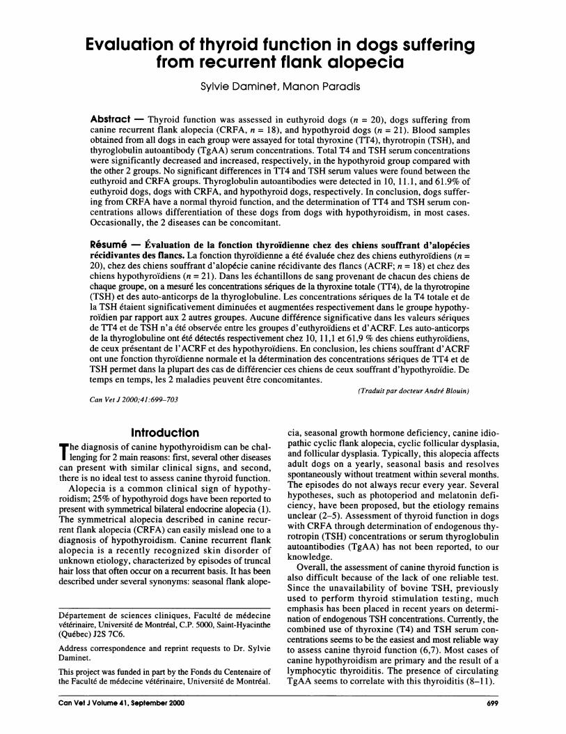
Table 1. Tests results and follow-up of 3 dogs with CRFA and increased TSH serum concentrations. None ofthese dogs received T4 supplementation at any time, they remained clinically normal throughout this study(except for the alopecia), and each episode of alopecia resolved spontaneously
Dates of episodesDate of TT4 TSH TgAA of alopecia
No. Dog testing (nmol/L) (ng/mL) (+ or-) (as of 08/99) Results of skin biopsy
I Boxer 05/98 11.23 1.17 + 05/96 compatible with(MN, 4 y) 05/97 CRFA
2 Giant schnauzer 01/96 10.6 1.67 + 01/96 not done(SF, 2 y) 12/96 11.7 0.01 05/99
08/99 7.21 0.53
3 Dalmatian 05/98 16.6 1.34 - 05/97 compatible with(SF, 3 y) 08/99 30.9 0.49 05/98 CRFA
with bilateral symmetrical flank alopecia. His com-plete blood cell count and biochemical profile (includ-ing cholesterol values) were normal, and the dog had noother clinical signs suggestive of hypothyroidism. Thealopecia resolved completely within a few months. Thedog suffered another episode of flank alopecia 1 y later,which again resolved spontaneously, confirming theCRFA. Despite the author's insistence, it was not pos-sible to get a follow-up of thyroid function in this dog.The second dog, a giant schnauzer, had episodes offlank alopecia in January 1996 and in May 1999, bothepisodes of alopecia resolved spontaneously withoutany treatment. A follow-up in August 1999 revealed anormal physical examination and a healthy coat.According to the owner, the dog remains clinically nor-mal. The third dog, a Dalmatian, suffered 2 episodes ofCRFA; initial results of thyroid function tests sug-gested hypothyroidism, although follow-up tests revealednormal thyroid function.
Thyroglobulin autoantibodiesTwo of the 20 (10%) control dogs were positive forserum TgAA. Two of 18 (11.1%) dogs with CRFAtested positive for TgAA. Thirteen of the 21 (61.9%)hypothyroid dogs tested positive for TgAA.
DiscussionAs expected, TT4 serum concentrations were decreasedand TSH concentrations were increased in most hypothy-roid dogs, as compared with the control group dogs.Nineteen of the 21 hypothyroid dogs (90.5%) had aTSH concentration above 0.6 ng/mL. Two dogs (9.5%),with TSH concentrations of 0.46 and 0.50 ng/mL,respectively, were included in the hypothyroid groupbecause their clinical signs suggested hypothyroidism;their serum TT4 levels were 1.1 and 1.57 nmol/L,respectively, in the absence of any concomitant dis-ease, and all their clinical signs resolved with thyroid sup-plementation. This finding is not surprising, as it has beenreported in several studies that 24% to 38% of hypothy-roid dogs have TSH values within the reference range(6,7).
Results from previous studies have suggested a cor-relation between serum TgAA and the presence of a lym-phocytic thyroiditis (8-11). Until recently, the methodsused to measure serum TgAA and/or the diagnostic
cut-off values used easily yielded false positive results,especially in dogs with systemic disease. This limitedtheir clinical usefulness (8-10). Nachreiner et al (11)recently published the validation of a reliable TgAAassay with a useful diagnostic threshold, which shouldincrease the clinical utility of determining serum TgAAin some dogs. This method for TgAA measurementwas used in our study. We observed that 2 dogs (10%)from the control group tested positive for serum TgAA,whereas Nachreiner et al reported that 3.3% of a randompopulation of dogs had positive results for TgAA. Oneof our positive control dogs had only a mild-positiveresult with an optical density of 208%, when the cut-offvalue for a positive sample is 200% of the negativecontrol result. It is, however, still unclear whether thepresence of circulating TgAA precedes the clinicaldevelopment of hypothyroidism, and further studiesthat follow dogs with circulating TgAA over a longerperiod of time are warranted. Thirteen of 21 (61.9%)hypothyroid dogs tested positive for serum TgAA,which is similar to the prevalence of 42% to 59% forTgAA in hypothyroid dogs reported in the literature(8,10). Therefore, our findings also support a correlationbetween lymphocytic thyroiditis and hypothyroidism.Dogs with CRFA present with dermatological signs
that can be suggestive of hypothyroidism. Dogs ofeither sex and of any reproductive status can be affected.Canine recurrent flank alopecia is characterized by a non-scarring alopecia, most often confined to the thora-columbar region. Lesions are usually bilaterally symmetric,but occasionally, only one side of the body is affected,or one side is more affected than the other (4). Typically,the alopecic lesions are "geographic" in shape withwell-demarcated borders, and the alopecic skin is oftenmarkedly hyperpigmented (2,5,14). Mean age at theonset of the first episode is around 4 y, but it can be quitevariable (range, 8 mo to 11 y). The onset of alopecia isvariable, but in the northern hemisphere, it most com-monly occurs between November and March (4).Spontaneous regrowth of hair occurs within 3 to 8 mo,in most cases, and usually consists of normal pelage den-sity. Although approximately 20% of dogs will only haveone isolated episode of flank alopecia in their life span,most dogs will develop recurrent alopecic episodes foryears (4). Other dogs have an occasional year whenthe alopecia does not recur. The degree of alopecia isvariable, with some dogs developing a virtually identical
Can Vet J Volume 41, September 2000 701

hair loss (size and duration) year after year, and otherdogs developing larger areas and/or longer episodesof hair loss as years go by. Occasionally, after severalconsecutive episodes of alopecia, some affected dogs willnot experience complete hair regrowth before the onsetof the next episode (4).The cause of CRFA remains obscure. The higher
incidence of CRFA at higher latitudes and its seasonalnature of recurrence suggest that photoperiod may beinvolved in the pathogenesis, with melatonin and/orprolactin, 2 photo-dependent hormones, being potentiallyimplicated in the process.
For most cases of CRFA, the diagnosis is based on his-tory, clinical signs, and rule-out of hypothyroidism(evaluation of thyroid function in dogs > 2 y of age).Histopathologic findings observed on skin biopsies,such as atrophic, dilated, and truncated hair follicles withhyperkeratosis extending to secondary hair follicles(hence the colloquial names "witch's feet" and "octopus-like hair follicles"), are suggestive of, but not patho-gnomonic for, CRFA (4,5,14). Moreover, these findingsare not always present in cases of CRFA and they maybe found with low frequency in other alopecic diseases.
Hypothyroidism is an important differential diagno-sis for CRFA. Moreover, hypothyroidism and CRFAcould both occur in the same animal. The clinicianmust, therefore, remain vigilant, as this can represent areal diagnostic and therapeutic challenge. A breed pre-disposition seems to exist for CRFA, with boxers (mayaccount for approximately half of all reported cases),Airedale terriers, schnauzers, and English bulldogsbeing the more frequently cited breeds (2,3,14). However,it has been reported in several other breeds (5).Interestingly, the same breeds are suspected of being pre-disposed to hypothyroidism (1). Furthermore, the recog-nition of CRFA or seasonal flank alopecia as a separatedermatological condition became clear only in the 1990s(3). Therefore, some dogs, previously classified ashypothyroid based on the presence of flank alopeciaresolving with thyroid supplementation, might verywell have been dogs suffering from CRFA. Anotherexplanation is that the breeds cited earlier are predisposedto both hypothyroidism and CRFA. Both diseases aremore frequent in young to middle-aged dogs. As mostdogs with hypothyroidism have a lymphocytic thy-roiditis, believed to be of autoimmune origin, the ques-tion arises whether CRFA could have an immune-mediated origin as well. Results of the TSH responsetest were reported in 5 dogs with CRFA and were normal,but serum TSH values and TgAA were not available (3).
Results of T4 and TSH tests in our study were not sig-nificantly different between euthyroid dogs and dogs withCRFA. However, 3 of 18 dogs with CRFA were foundto have results that could suggest concomitant hypothy-roidism. All episodes of flank alopecia resolved com-pletely without any treatment in all 3 dogs. Their history,clinical presentation, and follow-up confirmed CRFA.The increased TSH and positive TgAA suggested con-comitant hypothyroidism due to a lymphocytic thy-roiditis in dogs 1 and 2. None of these 3 dogs had ahistory or clinical signs suggestive of hypothyroidism,except for the flank alopecia, and they did not developclinical hypothyroidism. The reported specificity of
serum TSH concentrations in the diagnosis of hypothy-roidism ranges from 0.88 to 0.93 (6,7). Total T4 and TSHserum concentrations were repeated a year later in thesecond dog, and the TSH value was within the referencerange. One possible explanation could be that TSH issecreted in a pulsatile fashion, as it is in humans (15,16).One study reported an absence of significant diurnal vari-ation of the TSH concentrations in healthy dogs and dogswith 1311-induced hypothyroidism; however, dogs withnaturally developing hypothyroidism had TSH valuesfluctuating in and out the reference range (17). Thiscould, at least in part, explain the low sensitivity ofdetermination of TSH concentrations in the diagnosis ofhypothyroidism. The TT4 serum values in dogs 1 and 2,with CRFA and increased TSH values, were slightlybelow the reference range but higher than is expected inmost dogs with hypothyroidism. The human medical lit-erature refers to subclinical hypothyroidism, where anincrease in TSH values maintains adequate thyroid hor-mone concentrations and few or no clinical signs ofhypothyroidism are present (18). Progression towardsovert hypothyroidism is slow in humans (18). Whethersubclinical hypothyroidism exists in dogs is not known.Our results emphasize that flank alopecia can be
misleading, and appropriate testing through a combi-nation of T4 and endogenous TSH determinations, orthrough a TSH stimulation test, when available, isimportant to differentiate these conditions in somedogs. In addition, these disorders could be concomitantin some dogs. When a dog is presented with a typical pre-sentation of CRFA, a justifiable option is to wait a fewmonths and see if the condition resolves spontaneously.Results of thyroid testing should be interpreted in lightof the history and clinical findings to avoid treatingeuthyroid dogs with thyroid supplementation. Monitoringof dogs with slightly abnormal or equivocal results ofthyroid function tests is recommended. The etiologyof CRFA is still unclear, but thyroid dysfunction does notseem to be a predisposing factor for CRFA.
In conclusion, dogs suffering from CRFA have anormal thyroid function. In most cases, determination ofTT4 and TSH serum concentrations allows dogs withCRFA to be differentiated from dogs with hypothy-roidism. Hypothyroidism and CRFA could be con-comitant in some dogs. cvJ
References1. Feldman EC, Nelson RW. Canine and Feline Endocrinology and
Reproduction. 2nd ed. Philadelphia: WB Saunders, 1996:68-111.2. Curtis CF, Evans H, Lloyd DH. Investigation of the reproductive
and growth hormone status of dogs affected by idiopathic recur-rent flank alopecia. J Small Anim Pract 1996;37:417-422.
3. Scott DW. Seasonal flank alopecia in ovariohysterectomizeddogs. Cornell Vet 1990;80:187-195.
4. Paradis M. Melatonin therapy for canine alopecia. In: Bonagura JD,ed. Kirk's Current Veterinary Therapy XIII. Philadelphia:WB Saunders, 1999:546-549.
5. Fontaine J, Beco L, Paradis M. Alopecie recidivante des flancs:Etude de douze cas chez le griffon Korthals. Point Vet 1998;29:445-449.
6. Peterson ME, Melidn C, Nichols R. Measurement of serum totalthyroxine, triiodothyronine, free thyroxine, and thyrotropin con-centrations for diagnosis of hypothyroidism in dogs. J Am Vet MedAssoc 1997;211:1396-1402.
7. Scott-Moncrieff JC, Nelson RW, Bruner JM, Williams DA.Comparison of serum concentrations of thyroid-stimulating
702 Can Vot J Volume 41, September 2000

hormone in healthy dogs, hypothyroid dogs, and euthyroid dogswith concurrent disease. J Am Vet Med Assoc 1998;3:387-391.
8. Haines DM, Lording PM, Penhale WJ. Survey of thyroglobulinantibodies in dogs. Am J Vet Res 1984;45:1493-1497.
9. Beale KM, Torres S. Thyroid pathology and serum antithy-roglobulin antibodies in hypothyroid and healthy dogs. J VetIntern Med 1991;5:128.
10. Thacker EL, Refsal KR, Bull RW. Prevalence of autoantibodies tothyroglobulin, thyroxine, or triiodothyronine and relationship ofautoantibodies and serum concentrations of iodothyronines indogs. Am J Vet Res 1992;53(4):449-453.
11. Nachreiner RF, Refsal KR, Graham PA, Hauptman J, Watson GL.Prevalence of autoantibodies to thyroglobulin in dogs with non-thyroidal illness. Am J Vet Res 1998;8:951-954.
12. Daminet S, Paradis M, Refsal KR, Price C. Influence of prednisoneand phenobarbital on canine thyroid function. Can Vet J 1999;40:411-415.
13. Williams DA, Scott-Moncrieff JC, Bruner J, et al. Validation of animmunoassay for canine thyroid-stimulating hormone and changes
in serum concentration following induction of hypothyroidism indogs. J Am Vet Med Assoc 1996;209:1730-1732.
14. Miller MA, Dunstan RW. Seasonal flank alopecia in Boxersand Airedale Terriers: 24 cases (1985-1992). J Am Vet MedAssoc 1993;203:1567-1572.
15. Morley JE. Neuroendocrine control of thyrotropin secretion.Endocr Rev 198 1;2(4):396-435.
16. Brabant G, Prank K, Hoang-Vu C, Hesch D, von zur Mtihlen A.Hypothalamic regulation of pulsatile thyrotropin secretion. J ClinEndocrinol Metab 1991;72:145-150.
17. Bruner JM, Scott-Moncrieff C, Williams DA. Effect of time of sam-ple collection on serum thyroid-stimulating hormone concentra-tions in euthyroid and hypothyroid dogs. J Am Vet Med Assoc1998; 10: 1572-1575.
18. Utiger RD. Disorders of the thyroid gland. In: Kelley WN, ed.Textbook of Internal Medicine. 3rd ed. Philadelphia: Lippincott-Raven, 1997:2204-2218.
September and October Special Deals
Surgery kbles15% DISCOUNT
Davis & Geck Sutue(Dermalon, Surgilene,Novafil, Dezou, Maxon, etc)up to 25%
DISCOUNT
Heat Pumps andaccessore
7.5Z% DISCOUNT
DISCOUNT
* Perry Surgical Gloves ..................XX22%* Surgical Masks ......................259/o
* I.V. Stands 20°h* Mayo Instrument Table. 9 a . . . . o o . o a . a . .20%Revolving Stool ...................200/
Consult our promotional Bulletin or
contact your CDMV representative for moreinformation.
_ Canada-wide DistributorofVeternary Products
J TTo order: 1 800 668-CDMV
estt v By fx: 1 800 363-3134
Um mt~~~~~~~~~~~~~~~~~~~~~fl~~~~~~~~eIN a mU1~~~~~~~~~~~~~~~~~~~~~1__ o_ * _
si ..... . ..- ,,^^1,x,¢.xz
\ tS=as.s, -.. .b.;t s-jr r +.:
*__. . . i. - ||llLL b | _
Can Vet J Volume 41, September 2000 703