eur j anat, 8 (2): 61-65 (2004) the trans-cervical plane...
TRANSCRIPT
SUMMARY
The currently used subdivisions of the neck arenot helpful in neck surgery. In addition, the wideuse of minimally invasive neck surgery has madeit necessary to find reference points that makethese procedures easier and safer. Here, clinical,anatomical and radiological study was underta-ken to determine the relationships between thetrans-cervical plane (TCP) and important neckstructures. One hundred and ninety healthyvolunteers were examined to determine the sur-face anatomy of the TCP together with 17 CTscans on the same plane and, five cadavers weredissected in an attempt to describe the anatomyof the mid-cervical region. The distance betweenthe submental point and the sternal notch wasmeasured, and the important anatomic featuresat this level were recorded.
The anatomical location of the TCP was confir-med. TCP was opposite to the lower border ofthyroid cartilage in 90% of the cases, and in 10%it was at the cricothyroid membrane. The averagedistance from the submental point to the TCP inthe hyperextended neck was (6.5-11.5 cm). Inspite of the wide range of variation of the distan-ce between the submental point and sternal notch(13-23 cm), the middle of this distance is constantand often related to important anatomical structu-res: the junction between the upper 1/3 andlower 2/3 of the thyroid lobes, superior parathy-roid, and the body of the 6th cervical vertebra.
It is concluded that the trans-cervical plane isan important landmark in the neck region that
enables accurate and rapid localization of the cri-cothyroid membrane for emergency cricothyroi-dotomy and the tracheal rings for percutaneousdilatational tracheostomy and provides a refe-rence point to mark skin incisions necessary forminimally invasive neck surgery.
Key words: Cricothyroidotomy – Dilatationaltracheostomy – Invasive neck surgery
INTRODUCTION
The neck extends anteriorly from the uppersurfaces of the clavicles and the manubriumsterni to the lower border of the mandible. Themid-sagittal line divides the neck into symme-trical halves. Each half is subdivided into ante-rior and posterior triangles according to its res-pective relation to the sternocleidomastoidmuscle. The anterior triangle is further subdivi-ded into smaller triangles by the superior bellyof the omohyoid and the bellies of the digastricmuscles; the smaller triangles are the muscular,carotid, submental and digastric triangles. Onthe other hand, the posterior triangle of theneck is subdivided into two triangles by theinferior belly of the omohyoid: the supraclavi-cular and occipital triangles (Hiatt and Gartner,2001; Snell, 2000; Moore and Daley, 1999; Ber-kovitz and Moxham, 1998; Williams et al.,1995). Another system introduced for clinicalassessment of the neck structures divides theneck into two areas; midline area within 2 cm
Eur J Anat, 8 (2): 61-65 (2004)
61
The trans-cervical plane (TCP): A new anatomical landmark for minimally
invasive neck surgery
D.H. Badran1, N.A. Younes2 and A. Al Hadidi3
1- Department of Anatomy and Histology2- Department of Surgery3- Department of RadiologyFaculty of Medicine, University of Jordan, Amman, Jordan
Correspondence to:Dr. Darwish H. Badran. Department of Anatomy and Histology, Faculty of Medicine, Universityof Jordan, Amman, 11942, Jordan. Tel.: +962 79 5641188; Fax: +962 65356746E-mail: [email protected]
Submitted: April 14, 2004Accepted: June 8, 2004
from the midline and lateral neck area outsidethe previous one (Polk et al., 1987). A third sys-tem introduced by Monson et al. (1969) dividesthe neck into three zones; the area below animaginary horizontal line passing just inferior tothe cricoid cartilage is zone I; the area aboveanother imaginary line passing through theangles of the mandible is zone III, while zoneII is the wide area that lies between these twoimaginary lines.
These systems do not provide referencepoints to the surface anatomy of the importantunderlying structures or offer any help in placingsurgical incisions especially in emergency proce-dures or minimally invasive neck surgeries. Inthis paper the relationship of a newly definedanatomical point (TCP) placed at the middle of adistance between the submental point and thesternal notch to gross anatomical structures inhealthy volunteers, cadaveric specimens and CTindividual images was examined.
Regardless of the length of the neck, the loca-tion of the TCP at the middle of a distance bet-ween two fixed points is constant. The goal ofthis study was to describe precisely the locationof the TCP with respect to the important underl-ying natomical structures. The use of such ana-tomical methods should result in an improveddescription of neck structures and a more preci-se approach for neck surgery.
MATERIALS AND METHODS
VolunteersOne hundred and ninety normal healthy
volunteers were included in this study. Theywere 147 males and 43 females; their averageage was 30.7 (range 18-72 years).
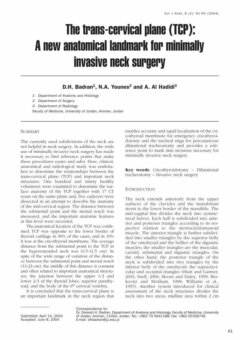
While the neck was in the hyperextensionposition, the inferior border of the mandiblewas palpated at the mid line as a starting point(submental point) and the distance to thesuprasternal notch was measured. An Imagi-nary line was placed at the center of the pre-vious distance and named the TCP (Fig. 1).Then, the lower border of the thyroid cartilagewas palpated and the distances between it andthe submental point and the suprasternal notchwere measured.
Radiological studyEighteen high-resolutions CT scans were
obtained from patients with no neck pathologyand who had never undergone neck surgery. Aline was extended from the midpoint betweenthe submental point and the sternal notch pos-teriorly to the cervical vertebra while the neckwas in the hyperextended position; the anato-mical position and structures at that level wererecorded.
Anatomical studyThe necks of five formaldehyde-embalmed
bodies were dissected to verify the level of thecricothyroid membrane. Dissection proceededsystemically from superficial to deep layers. Allstructures seen at each level were recorded andhand drawn.
RESULTS
In all volunteers it was easy to identify thereference points i.e. submental point (felt atthe lower border of the mandible at the midline), thyroid cartilage, cricoid cartilage andthe suprasternal notch. In the morphometricstudy of our volunteers, the distance betweenthe submental point and the suprasternal notchin the hyperextended neck ranged between13-23 cm, the TCP was placed at the middle ofthis distance (6.5-11.5 cm). The distance bet-ween the submental point and the inferior bor-der of the thyroid cartilage ranged between 7-12 cm. The TCP passed along the inferiorborder of the thyroid cartilage in 90% of thevolunteers and in 10% over the cricothyroidmembrane in the hyperextended neck andover the cricothyroid membrane in the anato-mical position.
D.H. Badran, N.A. Younes and A. Al Hadidi
62
Fig. 1.- Diagram showing reference points and lines; (A)submental point, (B) lower border of thyroid cartilage,(C) sternal notch.

The trans-cervical plane (TCP): A new anatomical landmark for minimally invasive neck surgery
63
Fig. 3.- Diagram showing a needle in the cricothyroid membranein criocthyroidotomy.
Fig. 4.- Proposed skin incisions for minimally invasive superiorparathyroidectomy (PTx), minimally invasive thyroidectomy(Tx) and, percutaneous dilatational tracheostomy (PDT).
Fig. 2.- Lateral CT scan showing the level of the TCP. THe dotted line is passes through the body of C6.
Computerized tomography (CT) of the neckin the anatomical position showed that the cri-cothyroid membrane was at the middle of thedistance between the submental point and thesternal notch. Lateral views revealed that thisplane passes along the body of the 6th cervicalvertebra (Fig. 2).
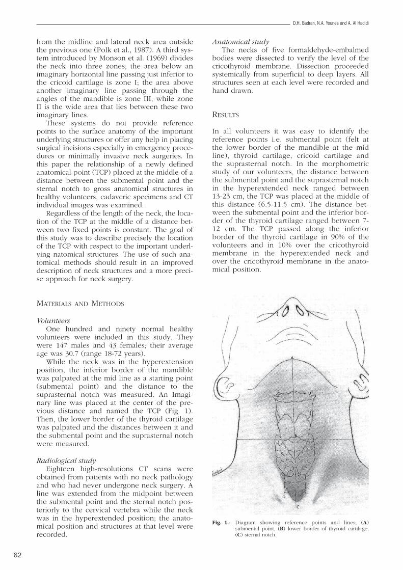
Dissection showed that the TCP passesthrough the junction of the upper 1/3 and lower2/3 of the thyroid lobes. The superior parathy-roid glands were found around this plane. Theintermediate tendon of the omohyoid musclewas seen just below the level of the cricoid car-tilage in two cadavers and below that in theother 3 cases. In all cases, at the level of the 6th
cervical vertebra, the inferior thyroid arteryarched medially before branching; the vertebralartery entered the foramen transversarium, andthe middle cervical ganglion was located on thesides of the vertebral body.
DISCUSSION
Many authors have addressed minimally invasiveneck surgery in the outpatient setting and in thecontext of bedside surgery in the intensive careunit. The technique and outcome of cricothyroi-dotomy (Jones and Roberts, 2002; Mori et al.,2002; Schroeder, 2000; Warmington et al., 2000),percutaneous dilatational tracheostomy (Carrilloet al., 1997; McHenry et al., 1997; Cobean et al.,1996; Hill et al., 1996) minimally invasive thyroi-dectomy (Ohgami et al., 2000; Miccoli et al.,2001a; Miccoli et al., 2001b; Lorenz et al., 2001;Miccoli, 2002), and parathyroidectomy (McGreal,2001; Sackett et al., 2002; Agarwal et al., 2002)are well documented.
One of the most common problems encoun-tered in performing emergency or minimallyinvasive neck surgery is the difficult anatomicalrelationships and crowding of the structures inthe central neck region. Several different anato-mical descriptions of the neck region have beengiven. One description distinguishes two necktriangles, the anterior, and posterior triangle.According to this system, the neck is devided bythe sternocleidomastoid muscle into two trian-gles and the anterior triangle is further subdivi-ded into several triangles by the omohyoid andthe digastric muscles. Another system devidesthe neck into two regions: midline and lateralneck region. A third system divides the neck into3 zones. None of these systems provides a relia-ble antomical landmark that can be used forneck surgery. These systems do not provide refe-rence points to the surface anatomy of theimportant underlying structures or offer any helpin placing surgical incisions, especially in emer-gency procedures or minimally invasive necksurgeries. Thus, it is mandatory to find anotherreference point that can be used for surgicalneck procedures. In the present study we des-cribe the TCP by using fixed anatomical land-marks -the submental point and the suprasternalnotch- as reference points. In humans Both land-marks can be easily identified by palpation. Inthis study, the distance between the submentalpoint and the sternal notch was 13-23 cm andthe TCP was located at the distance (6.5-11.5 cm)in 90% of the volunteers.
Although most of our volunteers were relati-vely young individuals, with rather obvious anato-mical landmarks, the site of the TCP was fairlyconstant along the lower border of the thyroid car-tilage when the distance between submental pointand sternal notch was measured in the hyperex-tended neck. In the anatomical position (midwaybetween extension and flexion), the TCP descendsdownward close to the cricothyroid membranelevel. The site of larynx may vary between indivi-duals (low-lying and high-lying larynx). However,these variations are presumably related to thelength of the neck, which does not influence thesite were the centeral point was placed.
Anatomically, the level of TCP (the body ofC6) represents an important landmark. At thislevel the inferior thyroid artery curves medially,the middle thyroid vein leaves the thyroid gland,the common carotid artery can be pressedagainst the carotid tubercle, and the vertebralartery enters the foramen transversarium (Fig. 3).The TCP also marks the location of the middlecervical ganglion (Cobean et al., 1996; Carrillo etal., 1997; Berkovitz and Moxham, 1998; Hiatt andGartner, 2001; Agarwal et al., 2002). The bloodsupply of the area below the TCP is from bran-ches of the subclavian artery and the only cranialnerves seen in it are the vagus and the accessorynerves. Branches from the external carotid artery
D.H. Badran, N.A. Younes and A. Al Hadidi
64
Fig. 5.- Some of the structures seen at the level of C6.

supply the area above the TCP. Also, the TCPpasses along the junction of the upper and themiddle thirds of the thyroid lobes, where there isthe greatest concentration of C cells (Thompsonand Vinik, 1983) and the where the superiorparathyroid glands are usually located. Therefo-re, the TCP has practical importance for the sur-geon. The intersection between the TCP and thesagittal plane can easily be identified on thepatient’s neck to perform emergency cricothyroi-dotomy just below this point (Fig. 4), and appro-ximately 15-20 mm below it percutaneous dilata-tional tracheostomy can be performed (Fig. 5).
Secondly, the location of the TCP is of a greatimportance for neck surgeons because it can beused as a guide to plan incisions in this region.The TCP line can be drawn on the patient’s neckprior to minimally invasive superior parathyroidsurgery. Depending on the location of a superiorparathyroid adenoma, a 1-2 cm skin incision onthe TCP line can be used to remove the adenoma,and thyroid incisions can also be made at a lineapproximately 1.5 cm below the TCP line (Fig. 5).
We have examined the precise relationshipbetween the location of the TCP and the impor-tant neck structures in humans. Our results con-firm the location of the TCP at the lower borderof thyroid cartilage, just above the cricothyroidmembrane. The TCP can be used as a referencepoint that provides an accurate and rapid locali-zation of the cricothyroid membrane for emer-gency procedures, helps mark the skin incisionsnecessary for minimally invasive neck surgery,and represents the level at which important ana-tomical structures can be located.
ACKNOWLEDGEMENTS
The authors thank Mr. Abdallah Hamdan for histechnical assistance and Miss Sanaa Al Tamimifor the artistic work.
REFERENCES
AGARWAL G, BARRACLOUGH BH, ROBINSON BG, REEVE TS andDELBRIDGE LW (2002). Minimally invasive parathyroidec-tomy using the ‘focused’ lateral approach. I. Results ofthe first 100 consecutive cases. ANZ J Surg, 72: 100-104.
BERKOVITZ BKB and MOXHAM BJ (1998). A Textbook of Headand Neck Anatomy. Wolfe Medical Publications Ltd.,London.
CARRILLO EH, SPAIN DA, BUMPOUS JM, SCHMIEG RE, MILLER FBand RICHARDSON JD (1997). Percutaneous dilatational tra-cheostomy for airway control. Am J Surg, 174: 469-473.
COBEAN R, BEALS M, MOSS C and BREDENBERG CE (1996). Per-cutaneous dilatational tracheostomy: a safe, cost-effecti-ve bedside procedure. Arch Surg, 131: 265-271.
HIATT JL and GARTNER LP (2001). Textbook of Head andNeck Anatomy. 3rd ed. Lippincott Williams and Wilkins,Philadelphia.
HILL BB, ZWENG TN, MALEY RH, CHARASH WE, TOURSARKISSIAN
B and KEARNEY PA (1996). Percutaneous dilatational tra-cheostomy: report of 356 cases. J Trauma, 40: 238-244.
JONES I and ROBERTS K (2002). Towards evidence-basedemergency medicine: best BETs from the ManchesterRoyal Infirmary. To stab or slash: the percutaneous dila-tation or standard surgical approach to cricothyroido-tomy in prehospital care. Emerg Med J, 19: 434.
LORENZ K, MICCOLI P, MONCHIK JM, DUREN M and DRALLE H(2001). Minimally invasive video-assisted parathyroidec-tomy: multi institutional study. World J Surg, 25: 704-707.
MCGREAL G, WINTER DC, SOOKHAI S, EVOY D, RYAN M, O’SU-LLIVAN GC and REDMOND HP (2001). Minimally invasive,radioguided surgery for primary hyperparathyroidism.Ann Surg Oncol, 8: 856-860.
MCHENRY CR, RAEBURN CD, LANGE RL and PRIEBE PP (1997).Percutaneous tracheostomy: a cost-effective alternativeto standard open tracheostomy. Am Surg, 63: 646-652.
MICCOLI P (2002). Minimally invasive surgery for thyroid andparathyroid diseases. Surg Endosc, 16: 3-6.
MICCOLI P, BERTI P, RAFFAELLI M, CONTE M, MATERAZZI G andGALLERI D (2001a). Minimally invasive video-assistedthyroidectomy. Am J Surg, 181: 567-70.
MICCOLI P, BERTI P, RAFFAELLI M, MATERAZZI G, BALDACCI S andROSSI G (2001b). Comparison between minimally invasi-ve video-assisted thyroidectomy and conventional thy-roidectomy: a prospective randomized study. Surgery,130: 1039-1043.
MONSON DO, SALETTA JD and FREEARK RJN (1969). Carotidvertebral trauma. J Trauma, 9: 987-999.
MOORE KL and DALEY AF (1999). Clinically oriented Anatomy.4th ed. Lippincott Williams and Wilkins, Philadelphia.
MORI M, FUJIMOTO J, IWASAKA H and NOGUCHI T (2002). Emer-gency percutaneous dilatational cricothyroidotomy afterfailed intubation. Anaesth Intensive Care, 30: 101-102.
OHGAMI M, ISHII S, OHMPRI T, NOGA K, FRUKAWA T and KITAJI-MA M (2000). Scarless endoscopic thyroidectomy. Breastapproach for better cosmesis. Surg Laparosc EndoscPercutan Tech, 10: 1-4.
POLK HC, STONE HH and GARDNER B (1987). Basic Surgery.3rd ed. Appleton-Century-Crofts, Norwalk, Connecticut.
SACKETT WR, BARRACLOUGH B, REEVE TS and DELBRIDGE LW(2002). Worldwide trends in the surgical treatment ofprimary hyperparathyroidism in the era of minimallyinvasive parathyroidectomy. Arch Surg, 137: 1055-1059.
SCHROEDER AA (2002). Cricothyroidotomy: when, why, andwhy not? Am J Otolaryngol, 21: 195-201.
SHIMIZU K, AKIRA S, JASMI AY, KITAMURA Y, KITAGAWA W andTANAKA S (1999). Video-assisted neck surgery: Endosco-pic resection of thyroid tumors with a very minimalneck wound. J Am Coll Surg, 188: 697-703.
SNELL RS (2000). Clinical Anatomy for Medical Students. 6th
ed. Lippincott Williams and Wilkins, Philadelphia.
THOMPSON NW and VINIK AI (1983). The technique of initialparathyroid exploration and reoperative parathyroidec-tomy. In: Thompson NW, Vinik AI (eds). Endocrine Sur-gery Update. Grune & Stratton, New York, pp 368.
WARMINGTON A, HUGHES T, WALKER J, REID A and PEARSON J(2000). Cricothyroidotomy and transtracheal high-fre-quency jet ventilation for elective laryngeal surgery. Anaudit of 90 cases-reply. Anaesth Intensive Care, 28: 711.
WILLIAMS PL, BANNISTER LH, BERRY MM, COLLIN P, DYSON M,DUSSEK JE and FERGUSON MWJ, editors (1995). Gray’sAnatomy. 38th ed. Churchill Livingstone, London.
The trans-cervical plane (TCP): A new anatomical landmark for minimally invasive neck surgery
65
