essential thioredoxin-dependent peroxiredoxin system from
TRANSCRIPT

JOURNAL OF BACTERIOLOGY,0021-9193/01/$04.0010 DOI: 10.1128/JB.183.6.1961–1973.2001
Mar. 2001, p. 1961–1973 Vol. 183, No. 6
Copyright © 2001, American Society for Microbiology. All Rights Reserved.
Essential Thioredoxin-Dependent Peroxiredoxin System fromHelicobacter pylori: Genetic and Kinetic Characterization
LAURA M. S. BAKER,1 AUSRA RAUDONIKIENE,2† PAUL S. HOFFMAN,2 AND LESLIE B. POOLE1*
Department of Biochemistry, Wake Forest University School of Medicine, Winston-Salem,North Carolina,1 and Department of Microbiology and Immunology,
Dalhousie University, Halifax, Nova Scotia B3H 4H7, Canada2
Received 18 October 2000/Accepted 3 January 2001
Helicobacter pylori, an oxygen-sensitive microaerophile, contains an alkyl hydroperoxide reductase homo-logue (AhpC, HP1563) that is more closely related to 2-Cys peroxiredoxins of higher organisms than to mostother eubacterial AhpC proteins. Allelic replacement mutagenesis revealed ahpC to be essential, suggesting acritical role for AhpC in defending H. pylori against oxygen toxicity. Characterization of the ahpC promoterregion divulged two putative regulatory elements and identified the transcription initiation site, which wasmapped to 96 and 94 bp upstream of the initiation codon. No homologue of ahpF, which encodes the dedicatedAhpC reductase in most eubacteria, was found in the H. pylori genome. Instead, homologues of Escherichia colithioredoxin (Trx) reductase (TrxR, HP0825) and Trx (Trx1, HP0824) formed a reductase system for H. pyloriAhpC. A second Trx homologue (Trx2, HP1458) was identified but was incapable of AhpC reduction, althoughTrx2 exhibited disulfide reductase activity with other substrates [insulin and 5,5*-dithiobis(2-nitrobenzoicacid)]. AhpC interactions with each substrate, Trx1 and hydroperoxide, were bimolecular and nonsaturable(infinite Vmax and Km values) but rapid enough (at 1 3 105 to 2 3 105 M21 s21) to suggest an important rolefor AhpC in cellular peroxide metabolism. AhpC also exhibited a wide specificity for hydroperoxide substrates,which, taken together with the above results, suggests a minimal binding site for hydroperoxides composed oflittle more than the cysteinyl (Cys49) active site. H. pylori AhpC was not reduced by Salmonella typhimuriumAhpF and was slightly more active with E. coli TrxR and Trx1 than was S. typhimurium AhpC, demonstratingthe specialized catalytic properties of this peroxiredoxin.
Infection with Helicobacter pylori, a microaerophilic, gram-negative bacterium, is associated with type B gastritis and pep-tic ulcer disease and is a risk factor for gastric carcinomas inhumans (8, 17, 27). It so prevalently affects the world popula-tion that H. pylori has been described as “the most commonchronic infection” (http://www.cdc.gov/ncidod/dbmd/diseaseinfo/hpylori_t.html), with some developing countries experiencingnearly 100% infection rates. While H. pylori infection is gen-erally controlled with a cocktail of antibiotics and bismuth (60),the specter of emerging antibiotic resistance necessitates thesearch for alternative drug strategies and a clearer understand-ing of bacterial defense systems.
H. pylori colonizes the mucosal layer of the stomach andsecretes immunogenic products that recruit macrophages andpolymorphonuclear leukocytes to the site of infection (59).Here, the resulting oxidative burst by the phagocytic cells pro-duces reactive oxygen species (ROS), such as superoxide anion(O2˙2), hydrogen peroxide (H2O2), and the hydroxyl radical(OH˙2), that damage gastric tissues. Increased ROS levels arepresent in H. pylori patients (19), and it is thought that long-term exposure to ROS contributes to the development of can-cerous gastric cells (16).
To resist oxidative damage from chronic inflammation,H. pylori relies on a variety of protective enzymatic systems,
including catalase (the katA gene product) (47) and superoxidedismutase (the sodB gene product) (63), which eliminate H2O2
and O2˙2, respectively; however, it is unknown how H. pyloricoordinates its oxidative stress response because the bacteriumlacks the oxidatively activated regulatory genes, soxRS andoxyR, common to other eubacteria (67). We report herein thatH. pylori also expresses a thioredoxin (Trx)-dependent alkylhydroperoxide reductase (AhpC) protein, a member of theperoxiredoxin (Prx) family (62), whose activity can detoxifylipid hydroperoxides analogous to those created in membranesexposed to ROS (32). All Prx enzymes are dimers, decamers,or, in some cases, higher-order aggregates (1, 38, 61) with oneessential, N-terminal Cys residue per subunit (Cys46 of Salmo-nella typhimurium AhpC) (21). Some Prxs contain only thissingle, conserved Cys (the 1-Cys Prxs), but most includeanother conserved Cys residue (the 2-Cys Prxs) analogous toCys165 in S. typhimurium AhpC, which links the two subunitsvia an intersubunit disulfide bond with the N-terminal Cys inthe oxidized protein (21, 53). Reduced S. typhimurium AhpCdirectly converts hydroperoxides to alcohols with concomitantformation of a sulfenic acid (Cys-SOH) at Cys46; condensationbetween the two active-site cysteinyl derivatives then regener-ates the stable intersubunit disulfide bond of the oxidized pro-tein (20, 21).
Our search of the H. pylori genomic database for an AhpChomologue revealed a gene (ahpC, HP1563) that had previ-ously been reported to encode a species-specific antigen byO’Toole et al. (48). Although not identified as a peroxidase atthat time, the H. pylori AhpC sequence containing the twoconserved Cys residues was later classified as a 2-Cys Prx (12).
* Corresponding author. Mailing address: Wake Forest UniversitySchool of Medicine, Department of Biochemistry, Medical CenterBlvd., Winston-Salem, NC 27157. Phone: (336) 716-6711. Fax: (336)716-7671. E-mail: [email protected].
† Present address: Aventis Pasteur Ltd., Toronto, Ontario M2R3T4, Canada.
1961

To reduce AhpC, most bacteria, including S. typhimurium, ex-press a specialized flavoprotein reductase, AhpF, which is ho-mologous to Escherichia coli Trx reductase (TrxR), except thatAhpF contains an additional N-terminal region directly in-volved in AhpC reduction (54, 55). Alternatively, some eukary-otic Prx systems utilize TrxR and Trx to reduce the peroxidase(sometimes referred to as Trx-dependent Prxs or TPxs), whereTrxR catalyzes the reduction of Trx, which, in turn, reduces thePrx disulfide bond. In these cases, electron transfer proceedsalong the following path: NADPH 3 TrxR 3 Trx 3 AhpC(TPx) 3 ROOH.
While eukaryotic examples of Trx-dependent Prx proteinsabound (e.g., Entamoeba histolytica AhpC [52], Saccharomycescerevisiae TPx [10], and multiple human Prx homologues [11]),to our knowledge, only one other bacterial Trx-dependent Prx,besides the H. pylori AhpC described herein, has been exper-imentally demonstrated [TPx from the cyanobacterium Syn-echocystis (75)]. A distantly related Prx family member, E. colibacterioferritin comigratory protein, has also been shown toexhibit low levels of Trx-dependent peroxidase activity (34).Inspection of the H. pylori genome yielded no ahpF, but anE. coli trxA homologue (trx1, HP0824 in the annotation ofTomb et al. [67]) encoding Trx1 was identified along with anE. coli trxB homologue (trxR, HP0825) encoding TrxR. Furtherexamination uncovered HP1164, annotated as a TrxR locusdue to its similarity to Plasmodium falciparum trxR (21% iden-tity). HP1164 does not, however, encode a putative catalyticdisulfide motif (CXXC or CXXXXC) indicative of such a re-dox center and was therefore excluded from these studies. Fur-ther genomic searches revealed a second Trx locus (trx2, HP1458)encoding Trx2. Therefore, we considered TrxR (HP0825)-Trx1(HP0824) and TrxR (HP0825)-Trx2 (HP1458) to be good can-didates as AhpC-reducing systems.
Herein, we present the first example of a Trx-dependentalkyl hydroperoxide reductase system from a gastric pathogenand describe the cloning, purification, and kinetic character-ization of AhpC, Trx1, Trx2, and TrxR from H. pylori. Alongwith the genetic characterization of H. pylori ahpC, we havealso shown that AhpC plays a critical role in the defenseagainst oxygen toxicity that is essential for survival and growth,even in microaerophilic environments.
(Abstracts reporting some of this information have beenpublished earlier [5, 6]).
MATERIALS AND METHODS
Materials. Sodium dodecyl sulfate (SDS), ultrapure glycine, ultrapure urea,EDTA, dithiothreitol, Tris base, and other buffer reagents were purchased fromResearch Organics (Cleveland, Ohio). Bacteriological medium components were
from Difco Laboratories (Detroit, Mich.). Ethanol was obtained from WarnerGraham Company (Cockeysville, Md.). Isopropyl-b-D-thiogalactopyranoside(IPTG) and 5-bromo-4-chloro-3-indolyl-b-D-galactopyranoside (X-Gal) stockswere from Inalc (Milan, Italy). Vent DNA polymerase, Moloney murine leuke-mia virus reverse transcriptase, and T4 polynucleotide kinase were purchasedfrom New England Biolabs (Beverly, Mass.). Restriction enzymes, ligase, calf in-testinal phosphatase, and restriction buffers were obtained from Promega (Madi-son, Wis.). Agarose medium EEO (electrophoresis grade) and H2O2 were fromFisher (Fairlawn, N.J.). Acrylamide-Bis (40%) solution was purchased fromBio-Rad (Hercules, Calif.). Ampicillin (AMP) powder, cumene hydroperoxide(CHP), glucose-6-phosphate, glucose-6-phosphate dehydrogenase, linoleic acid,lipoxygenase, and insulin were from Sigma (St. Louis, Mo.). Ethyl hydroperoxide(EtOOH) was from Polysciences, Inc. (Warrington, Pa.), and tert-butyl hydroper-oxide (t-BOOH) was from Aldrich (Milwaukee, Wis.). NADPH and NADH werefrom Roche Molecular Biochemicals (Mannheim, Germany).
RNA isolation and primer extension analysis. For RNA isolation, 50 ml ofH. pylori strain HP 26695 (67) was grown in Brucella broth supplemented with10% fetal calf serum (Sigma) in a microaerobic environment (7% O2, 5% CO2)at 37°C to an optical density at 590 nm of 1.0 and harvested by centrifugation at4°C for 10 min. Cells were lysed in 50 mM Tris-HCl (pH 8.0)–1 mM EDTA–50 mM NaCl buffer supplemented with 1.5% SDS for 5 min at 95°C. Followingphenol extraction and precipitation, the RNA was dissolved in diethylpyrocar-bonate-treated water and the total RNA concentration was determined at 260nm. For the primer extension studies, an oligonucleotide (59-GCCAGAAGAAAAGAATCGCACCGT-39; Gibco-BRL Custom Primers) was 59- end labeled inthe presence of [g-32P]ATP (5,000 Ci/mmol; Amersham) and 20 U of T4 polynu-cleotide kinase. Total H. pylori RNA (100 mg) was incubated with the 32P-labeledoligonucleotide and annealed under the following conditions: 80°C for 5 min,65°C for 5 min, 42°C for 10 min, and 37°C for 20 min. Following annealing, theRNA was precipitated with ethanol, dried, and resuspended in 7 ml of diethylpy-rocarbonate-treated water and 13 ml of reverse transcriptase buffer (50 mMTris-HCl at pH 8.3, 30 mM KCl, 10 mM MgCl2, 1 mg of actinomycin D [Sigma],1 mM dithiothreitol, 2.5 mM each deoxynucleoside triphosphate). One microliterof Moloney murine leukemia virus reverse transcriptase (50 U/ml) was added tothe sample, and reverse transcription was carried out at 42°C for 60 min; 1 ml of0.5 M EDTA and 1 ml of RNase (10 mg/ml) were added, and the reactionmixture was incubated at 37°C for an additional 30 min. The reaction wasstopped by phenol-chloroform extraction, followed by ethanol precipitation. Thesample was then dried and resuspended in 10 ml of sequencing loading buffer. Inparallel, dideoxy DNA sequencing of the cloned ahpC gene and the upstreamregion was performed using the same oligonucleotide primer to determine thetranscriptional start site. After denaturation at 95°C for 2 min, aliquots weresubjected to 6% urea polyacrylamide gel electrophoresis (PAGE) and autora-diographed.
Allelic replacement mutagenesis of ahpC. Genomic DNA isolated from H. py-lori strain 26695 (67) was used to amplify ahpC with forward and reverse oligo-nucleotide primers (Table 1) under typical PCR conditions to generate a product930 bp long. Isolated ahpC was digested at flanking EcoRI and SacI sites andthen inserted into pBluescript-SK (1) plasmid vector (Stratagene, La Jolla,Calif.) digested with the same restriction enzymes. Vector derivatives of pBlue-script were stably maintained in E. coli strains but were not replicated in H. pylorihosts. The ahpC-containing plasmid was then digested with XbaI (located ap-proximately 330 bp from the ahpC translational start site), and a chloramphen-icol resistance (camR) cassette originating from Campylobacter coli (70) digestedwith XbaI was inserted to interrupt the coding sequence of ahpC. Four H. pyloristrains were transformed with the ahpC::camR construct: 26695 (67), SS1 (40),HP1 (28), and HP1061 (25). The same strains were also transformed with aconstruct in which the nitroreductase gene (rdxA) was interrupted with camR
TABLE 1. Primers used to amplify and subclone H. pylori structural genes
Expressionvector and
gene
Primer (59–39)
Forward Reverse
pPROK1ahpC CGCCCGGGAGGAGGAAGAATAGATGTTAGTTACAAAACTTGCC CCCTGCAGCTACAGCTTGATGGAATTTTCTTTAAGATtrx1 CGCCCGGGAGGAGGAAGAATAGATGAGTCACTATATTG CCCTGCAGTTAGCCTAAAAGTTTGTTCAATTGCtrx2 GCGAATTCAGGAGGAAGAATAGATGTCAGAAATGATTAACGGG GCAAGCTTCCCCTTACAATAACGCTTTTAGAGCtrxR CGCCCGGGAGGAGGAAGAATAGATGATAGATTGCGCGATTATTGG CCAAGCTTGATTTAATGGTG
pBluescript-SKahpC TGGAATTCGCCCAATAACGATGAAACAAG GAGAGCTCCGATTAAAGCTTAATGGA
1962 BAKER ET AL. J. BACTERIOL.

(26). H. pylori transformation (electroporation) and subsequent evaluation of anycolonies obtained after up to 7 days of incubation in selective medium werecarried out as described elsewhere (57).
Cloning of ahpC, trx1, trx2, and trxR into expression vectors. The H. pylorigenomic database was searched for genes corresponding to the E. coli sequencesfor ahpC, trxA, trxB, and trxC (www.tigr.org/tdb/mdb/hpdb/hpdb.html). ClonesGHPDQ26, GHPAB86, GHPCL04, and GHPAE26, containing trx1, trx2, trxR,and ahpC, respectively, were ordered from The Institute of Genomic Research-ATCC Microbial Genome Special Collection. Plasmid DNA was purified fromthe E. coli host using the Wizard Miniprep Kit (Promega). The genes of interestwere amplified using PCR primers (listed in Table 1) synthesized in the DNASynthesis Core Laboratory of the Comprehensive Cancer Center of Wake ForestUniversity. PCR mixtures (50 ml) contained 300 mM deoxynucleoside triphos-phates, 1 U of vent polymerase, 10 pmol each of the forward and reverseoligonucleotides, 0.5 mM MgCl2, and 0.5 mg of template DNA. Reactions werecarried out as follows in a Mini Cycler (MJ Research, Waltham, Mass.): 95°C for30 s, 40°C for 45 s, and 72°C for 1.5 min (5 cycles); 98°C for 30 s and 72°C for 1min (35 cycles); and then 72°C for 15 min. The PCR products were purified usingthe QIAquick PCR Cleanup kit (Qiagen, Studio City, Calif.). The PCR products,600 bp for ahpC, 300 bp for trx1 and trx2, and 932 bp for trxR, were eachseparately ligated into the pCR2.1 TA cloning vector (Invitrogen, Carlsbad,Calif.). Plasmids containing each insert were digested with the restriction en-zymes corresponding to the engineered restriction sites (Table 1), and then DNAfragments of interest were excised from agarose gels and purified using the GeneClean II Kit (Bio 101, Inc., Vista, Calif.). These fragments were inserted, usingT4 DNA ligase, into pPROK1 expression vectors (Clontech, Palo Alto, Calif.;expression under control of the tac promoter), appropriately digested, isolatedfrom agarose gels, and pretreated with calf intestinal phosphatase.
Bacterial strains and culture procedures. Ligated DNA was transformed intocompetent E. coli XL-1 Blue cells (Stratagene), except the AhpC-expressingplasmid, which was transformed into an ahpCF deletion strain of E. coli, TA4315(65). Single colonies were selected on Luria-Bertani (LB) plates containing AMPat 50 mg/ml, and those containing the recombinant DNA were evaluated forprotein expression on SDS-polyacrylamide gels after induction with 0.4 mMIPTG. Isolated plasmid DNA for each construct was sequenced throughout thecoding region by automated DNA sequencing at the Comprehensive CancerCenter of Wake Forest University. Bacterial stocks containing each plasmid withthe subcloned gene were prepared from a single colony and stored at 280°C inLB broth containing 15% (vol/vol) glycerol. Culture procedures were generallythe same as reported earlier (53).
Purification of recombinant AhpC. Purification of AhpC was performed asdescribed earlier (53), with some modifications. All procedures were carried outin a standard buffer (pH 7.0) consisting of 25 mM potassium phosphate with 1.0mM EDTA. Briefly, 100 ml of E. coli TA4315 harboring the pPROK1/ahpCplasmid was added to 10 liters of LB medium containing 0.5 g of AMP supple-mented with 0.2% glucose in a BioFlo 2000 fermentor (New Brunswick Scien-tific, Edison, N.J.). IPTG (0.4 mM) was added at A600 5 0.9, and bacteria wereharvested by centrifugation 16 h after induction. Pelleted bacteria were disruptedwith a Bead Beater (BioSpec Products, Bartlesville, Okla.), cell extracts weretreated with streptomycin sulfate to precipitate nucleic acids, and 20 and 60%ammonium sulfate treatments were carried out to precipitate proteins as de-scribed previously (53). The protein resuspended in standard buffer containing20% ammonium sulfate was applied to a Phenyl Sepharose 6 Fast Flow Column(24 by 2.5 cm; Pharmacia LKB Biotechnology Inc.), washed with 20% ammo-nium sulfate buffer, and eluted with a linear gradient of 20 to 0% ammoniumsulfate in standard buffer. Protein fractions were evaluated for purity of overex-pressed AhpC by SDS-PAGE, and the purest fractions were pooled. Afterdialysis against 10 mM potassium phosphate buffer (pH 7.0), the protein wasloaded onto a DEAE-cellulose column (24 by 2.5 cm; Whatman DE52) andeluted with a linear gradient of 10 to 100 mM potassium phosphate (1-liter totalvolume). Again, fractions were analyzed for AhpC by SDS-PAGE and purefractions were pooled, concentrated, and aliquotted for storage at 220°C.
Purification of recombinant Trx1. A 10-liter culture of E. coli XL-1 Blueharboring the pPROK1/trx1 plasmid was grown in the fermentor and induced asdescribed above for AhpC expression. The crude cell extract obtained as de-scribed above was treated with streptomycin sulfate, and following centrifuga-tion, the supernatant was subjected to heat denaturation at 70°C for 4 min toremove contaminating proteins. Denatured proteins were removed via centrifu-gation at 23,000 3 g for 20 min, and the supernatant was dialyzed overnight inthree changes (6 liters each) of 5 mM potassium phosphate buffer (pH 7.0). Thedialyzed protein was applied to a DEAE-cellulose column (24 by 2.5 cm; What-man DE52) pre-equilibrated with 10 mM potassium phosphate (pH 7.0). Trx1eluted upon application of a linear gradient of 10 to 100 mM potassium phos-
phate (1-liter total volume) and was identified by SDS-PAGE. Pooled fractionswere concentrated to 2 ml, and the concentrate was applied to a BioGel A-0.5 magarose column (Bio-Rad) equilibrated in the standard buffer. The fractionswere collected, assessed for purity using SDS-PAGE, pooled, and aliquotted forstorage at 220°C. Further molecular weight and purity analysis required the useof Tris-Tricine gels as described by Ausubel et al. (3).
Purification of recombinant TrxR. A 10-liter culture of XL-1 transformed withthe pPROK1/trxR plasmid was grown, induced, and harvested; the crude extractwas prepared essentially as described for AhpC. After the 60% ammoniumsulfate treatment, the resuspended and dialyzed protein was loaded onto theDEAE-cellulose column and eluted as described for Trx1. Purified fractions, asassessed by the ratio of A280 to A450, were pooled and loaded onto an Affi-GelBlue column (24 by 2.5 cm; Bio-Rad). Fractions were eluted with a lineargradient of 25 mM potassium phosphate buffer to 600 mM potassium phosphateand 0.3 M NaCl (pH 7.0; 700-ml total volume). Protein purity was assessed byA280/A450 ratio determination and SDS-PAGE. Pure fractions were pooled,dialyzed in standard buffer, and aliquotted for storage at 220°C.
Purification of recombinant Trx2. A 10-liter culture of XL-1 transformed withthe pPROK1/trx2 plasmid was grown under the conditions described for Trx1.Bacteria containing Trx2 were treated as described for Trx1, except for thefollowing modifications. Instead of heat denaturation, the supernatant from aninitial treatment with 50% ammonium sulfate was brought to 80%. The pelletedprotein was resuspended in 5 mM potassium phosphate buffer, dialyzed over-night in the same buffer, and applied to the DEAE-cellulose column pre-equil-ibrated with 5 mM potassium phosphate buffer (pH 7.0). The nonbinding fractionwas then applied to a carboxymethyl cellulose column (24 by 2.5 cm; WhatmanCM) pre-equilibrated with 5 mM potassium phosphate buffer (pH 7.0); this wasfollowed by elution with a linear gradient of 5 to 60 mM potassium phosphate(700-ml total volume). SDS-PAGE was again used to assess purity, and concen-trated protein was aliquotted for storage at 220°C in standard buffer.
Other protein purifications. Purification of E. coli TrxR and Trx1 was doneessentially as described by Poole et al. (54). S. typhimurium AhpF and AhpC werepurified as reported previously (53).
Sequence analysis. AhpC homologues were identified through BLAST (2)searches with the H. pylori AhpC sequence. Pairwise comparisons of the H. pyloriAhpC sequence were generated with AhpC sequences from other organismsusing the BLAST Two Sequences program at the National Center for Biotech-nology Information website (http://www.ncbi.nlm.nih.gov/).
Spectroscopic experiments. Most spectral assays were carried out using athermostatted Gilford 220 updated recording spectrophotometer with a Beck-man DU monochromator and a Kipp and Zonen (Delft, The Netherlands) chartrecorder. Extinction coefficient determination using microbiuret assays for pro-teins at 280 nm and for bound flavin at 450 nm, thiol content determination us-ing 5,59-dithiobis(2-nitrobenzoic acid) (DTNB), and disulfide detection using2-nitro-5-thiosulfobenzoate (NTSB) were all performed as described previously(51, 53). Further quantification of H. pylori proteins relied on the followingextinction coefficients experimentally obtained at 280 nm (except where noted):AhpC, 24,800 6 300 M21 cm21; Trx1, 17,200 6 1,000 M21 cm21; Trx2, 23,300 6300 M21 cm21; TrxR, 11,900 6 110 M21 cm21 (450 nm). To quantitate the con-centration of other proteins and reagents by absorbance, the following extinctioncoefficients were used: E. coli TrxR, 11,300 M21 cm21 (454 nm) (73); E. coliTrx1, 13,700 M21 cm21 (280 nm) (31); S. typhimurium AhpF, 13,100 M21 cm21
(450 nm); AhpC, 24,300 M21 cm21 (280 nm) (53); TNB22, 14,150 M21 cm21
(412 nm) (58); NADPH, 6,200 M21cm21 (340 nm); NADH, 6,220 M21 cm21
(340 nm).Determination of H2O2 content using ferrithiocyanate. To monitor the disap-
pearance of hydrogen peroxide in solution, experiments were conducted essen-tially as described by Thurman et al. (66), except that peroxidase reactions (0.5ml of AhpC [20 mM], Trx1 [2.0 mM], and TrxR [0.2 mM]) were initiated with theaddition of H2O2 (1 mM). Reactions mixtures were incubated at 37°C, andaliquots were removed at various time points up to 30 min. Reactions wereterminated by the addition of 0.5 ml of 12.5% trichloroacetic acid, precipitatedproteins were removed by centrifugation, and peroxide content was analyzed byferrithiocyanate complex formation [0.2 ml of 10 mM Fe(NH4)2(SO4)2 and 0.1ml of 2.5 mM KSCN were added to a 50-ml aliquot of supernatant]. Redferrithiocyanate complex was measured at 480 nm, and peroxide concentrationwas determined from a standard curve with H2O2.
AhpC activity assays and reductase system determination. All AhpC activityassays were conducted on an Applied Photophysics DX.17 MV stopped-flowspectrophotometer at 25°C, except where noted otherwise. Aerobic, steady-stateAhpC activity assays were monitored by following the decrease in A340 due toNADPH oxidation. All peroxidase assays were conducted with 50 mM potassiumphosphate buffer (pH 7.0)–0.1 M ammonium sulfate–0.5 mM EDTA. Proteins in
VOL. 183, 2001 ALKYL HYDROPEROXIDE REDUCTASE SYSTEM FROM H. PYLORI 1963

one syringe were mixed with substrates in the other syringe, except where indi-cated otherwise. The rate of change in A340 was assessed by linear regressionanalysis of the first 10% of the linear region of the resulting trace. Preparationof the reaction chamber for anaerobic assays was conducted essentially as de-scribed previously (44).
Steady-state kinetic analysis of TrxR with Trx1. Km and kcat measurements ofTrxR for NADPH and Trx1 were conducted essentially as previously described(41), except that they were carried out on the stopped-flow spectrophotometer.Assays contained 0 to 40 mM NADPH (0.2 mM at the lowest concentration), 0to 100 mM Trx1, and 34 nM TrxR (final concentrations are indicated), andactivity was followed by monitoring the change in NADPH fluorescence overtime (excitation at 340 nm and emission recorded at 90° using a 400-nm filter).The decrease in fluorescence over time was converted to absorbance units perminute using a standard curve. Primary rate data were fitted to a rectangularhyperbolic function obtained using the Marquardt-Levenberg curve-fitting algo-rithm in SigmaPlot (Jandel Scientific, San Rafael, Calif.). The rate data obtainedafter varying each substrate were transformed and displayed in a primary Hanes-Wolf plot and yielded lines that intersected on the y axis, indicating a substitutedmechanism for TrxR. A substituted (ping-pong) mechanism was then assumedfor all equations, and the initial rate in the absence of products was representedby the following equation (15): v 5 Vab/(KmBa 1 KmAb 1 ab), where a is theconcentration of Trx1, b is the concentration of NADPH, V is the maximumvelocity, and Km is the Michaelis constant. Slopes and y intercepts obtained fromthe linear regression lines for each concentration of Trx1 were replotted in asecondary Hanes-Wolf plot to obtain the true Vmax and Km for each substrate.The y intercept of the secondary plot was equivalent to KmB/V, and the slope wasequal to 1/V. To solve for KmA, the following relationship was used: V(app)/Km(app) 5 V/KmA, where the V(app)/Km(app) ratio was obtained from Michae-lis-Menten plots of the initial-rate data.
The apparent Km and Vmax of TrxR for Trx1 and Trx2 were also determinedon a thermostatted Milton Roy Spectronic 3000 diode array spectrophotometerat 340 nm using an insulin reduction assay as described by Holmgren andBjornstedt (30). Reactions (500 ml) were conducted at 25°C in 100 mM potas-sium phosphate buffer (pH 7.5) with EDTA (1.5 mM) containing Trx1 or Trx2 (0to 50 mM), insulin (80 mM), NADPH (150 mM), and TrxR (7.0 nM; added last).Kinetic parameters were obtained using the curve-fitting function in Sigma Plotas described above. Assays using DTNB (200 mM) instead of insulin wereconducted essentially as described above, except that the reaction was monitoredat 412 nm to observe the rate of formation of TNB22.
The oxidase activity of TrxR was measured as described earlier (53). Briefly,H. pylori TrxR (30 to 240 nM), S. typhimurium AhpF (15 to 60 nM), or E. coliTrxR (30 to 120 nM) was added to 500 ml of air-saturated standard buffercontaining NADPH (150 mM) and the change in A340 was monitored. The TrxRactivity of H. pylori TrxR or Trx1 mixed with E. coli TrxR or Trx1 in heterologousmixtures was determined on the Gilford spectrophotometer using the DTNBreduction assay described above. Reaction mixtures contained 0.5 to 100 mMTrx1 (from either H. pylori or E. coli) and were started with the addition of 0.5mM TrxR (from either H. pylori or E. coli).
Bisubstrate steady-state kinetic analysis of AhpC. Aerobic peroxidase assaysmonitoring the A340 change of NADPH were carried out on a stopped-flowspectrophotometer as described above. In one syringe, peroxide substrates (10 to40 mM) were incubated in the peroxidase assay buffer. (All concentrations shownare final concentrations achieved upon mixing.) In the other syringe, AhpC (0 to2 mM), Trx1 (20 mM), and E. coli TrxR (2 mM) were incubated with NADPH(150 mM) for 5 min prior to assay with the peroxide substrate. (Use of E. coliTrxR rather than H. pylori TrxR in this assay did not significantly affect the resultsbut did greatly decrease the problem of AhpC-independent NADPH oxidation.)The kinetic coefficients were obtained as described by Forstrom et al. (24). Datawere analyzed using the integrated Dalziel rate equation for a two-substrateenzymatic system in which enzyme-substrate complexes were not experimentallyobserved: E0(t/[ROH]t) 5 f1 ln([ROOH]t/([ROOH]02[ROH]t)/[ROH]t) 1 f2/[Trx1] 1 f0, where [ROOH]0 is the initial hydroperoxide concentration, [ROH]t
is the product (alkyl alcohol) concentration at time t, and f1, f2, and f0 are theDalziel coefficients. Data were chosen and analyzed as described previously fortryparedoxin peroxidase from Crithidia fasciculata (46). Linoleic acid hydroper-oxide (LOOH) was prepared by enzymatic oxidation with soybean lipoxygenaseas described by Maiorino et al. (43).
RESULTS
Amino acid sequence alignments. In an amino acid sequencecomparison of the putative AhpC from H. pylori with the more
extensively studied S. typhimurium enzyme, Cys49 in H. pyloriAhpC aligns perfectly with the essential, conserved Cys46 fromS. typhimurium AhpC (21). Cys49 in H. pylori AhpC is, there-fore, most likely the site of interaction with peroxides and thesite of sulfenic acid formation. The H. pylori AhpC amino acidsequence was also compared to the deduced amino acid se-quences for other 1- and 2-Cys AhpC homologues from a widerange of organisms (Table 2). Of all the known homologues,H. pylori AhpC shares the most sequence identity with a selectgroup of bacterial Prxs including AhpC from Campylobacterjejuni (67%), TPx from Rickettsia prowazekii (54%), and AhpCfrom Legionella pneumophila (52%). Other than the proteinsfrom these three bacterial sources, however, H. pylori AhpC ismore similar to 2-Cys Prx protein sequences from higher or-ganisms than to other bacterial AhpC proteins (Table 2). Ofthe sequences shown, S. cerevisiae Prx (2-Cys), C. elegans Prx,the human proliferation-associated gene (2-Cys), and Synecho-cystis sp. strain PCC6803 (a cyanobacterium) TPx have all beenshown experimentally to be recycled by a Trx-reducing system(10, 36, 52).
TrxR and Trx1 from H. pylori share a moderate amount ofsequence identity (37 and 51%, respectively) with the corre-sponding E. coli homologues and a high percentage of identity(60 and 68%, respectively) with the homologues from theclosely related species C. jejuni. As in H. pylori, the absence ofahpF in C. jejuni makes TrxR and Trx1 reasonable candidatesas C. jejuni’s AhpC reductase system. H. pylori Trx2 shares thehighest sequence identity with a Trx homologue from Archaeo-globus fulgidus (Trx4, 42%) (39) and is more closely relatedto E. coli Trx2 (41% identity) (45) than to E. coli Trx1 (33%identity) (29). Known redox centers and binding motifs ofother Trx and TrxR proteins are completely conserved in theH. pylori proteins. Unlike the vast majority of TrxR-Trx1 sys-tems from other bacteria, the gene encoding TrxR is posi-tioned just downstream from trx1 in the H. pylori genome. Theclustering of trx1 and trxR in this order has also been observedin Streptomyces clavuligerus (14); however, in mycobacteria (72)and in C. jejuni (49), the opposite orientation, trxR and thentrx1, is observed. In most other species, trxR and trx1 are widelyseparated on the chromosome. Interestingly, H. pylori trx2(HP1458) is not found near trx1 (HP0824) or trxR (HP0825).
Characterization of purified H. pylori proteins. AhpC, Trx1,Trx2, and TrxR expressed from pPROK1 in the respectiveE. coli strains were purified to homogeneity (Fig. 1). All pro-teins were obtained in high yields as soluble proteins afterinduction with IPTG at 37°C. Migration of the H. pylori pro-teins was compared to that of pure S. typhimurium or E. colihomologues during SDS-PAGE, which allowed the detectionof each protein during purification and assessment of purity.Pure, reduced AhpC corresponded to an apparent molecularmass of 26 kDa, which is slightly higher than its expectedmolecular mass of 22,235 Da. When prepared in nonreducingsample buffer and analyzed by SDS-PAGE, AhpC migrated asa 50-kDa protein, indicating that the purified protein is oxi-dized and contains one or more intersubunit disulfide bonds(Fig. 1A, lane 6). TrxR migrated as a 33-kDa protein on SDS-PAGE under both reducing and nonreducing conditions (Fig.1A, lanes 5 and 7; expected mass, 33,538 Da). Previously, anintersubunit disulfide bond between TrxR subunits was sug-gested by Windle et al. (74); our results, however, rule out such
1964 BAKER ET AL. J. BACTERIOL.

a linkage between subunits. Spectral analyses (data not shown)of TrxR revealed a strong A454, which is typical of enzymescontaining a bound flavin cofactor. Trx1 and Trx2 both mi-grated as monomers in the presence or absence of b-mercap-toethanol and gave masses of approximately 12 kDa, close totheir expected masses of 11,854 and 11,744 Da, respectively.
Kinetic characterization of Trx1, Trx2, and TrxR. H. pyloriTrxR and Trx1 were shown in a series of assays to act as ageneral disulfide reductase system analogous to their counter-parts from E. coli. When assayed with small-molecule (DTNB)or protein (insulin) disulfide-containing substrates, both Trx1and Trx2 from H. pylori exhibited TrxR-dependent reductaseactivities that were hyperbolically dependent on the Trx1 andTrx2 concentrations. Insulin reduction in vitro by TrxR-Trx1gave a Vmax(app) of 19.9 6 1.4 mM min21 and a Km(app) forTrx1 of 13.4 6 2.7 mM (Fig. 2), while the TrxR-Trx2 systemgave a Vmax(app) of 10.6 6 1.5 mM min21 and a Km(app) forTrx2 of 11.0 6 1.9 mM (Fig. 2). While Trx2 exhibited abouthalf of the turnover rate of Trx1 in insulin assays, both Trx1and Trx2 reduced the small, non-protein disulfide substrateDTNB with about the same catalytic parameters as the TrxR-Trx1-insulin system (Fig. 2). In TrxR-Trx1-insulin experimentsin which NADPH was replaced with NADH, no decrease inthe NADH A340 was observed upon addition of TrxR to thereaction mixture, demonstrating the high specificity of TrxRfor NADPH. H. pylori TrxR and Trx1 were also capable offorming an efficient reducing system when mixed with theirE. coli counterparts (data not shown). Using the DTNB-linkedTrxR assay, reaction mixtures containing one H. pylori reduc-tase component mixed with a corresponding E. coli partnerprotein gave catalytic efficiencies (kcat/Km for Trx1) which werejust a fewfold lower for the heterologous systems comparedwith the natural systems. The fact that the H. pylori TrxR andTrx1 proteins can interact efficiently with the E. coli proteins
suggests that the H. pylori proteins share a great deal of struc-tural similarity with their E. coli counterparts and also illus-trates the functional homology H. pylori Trx1 and TrxR sharewith other TrxR systems.
To establish the true kcat and Km values for each substrate ofH. pylori TrxR, assays varying NADPH at different fixed con-centrations of Trx1 were conducted in the absence of addi-tional electron acceptors and were monitored on the stopped-flow spectrophotometer by the time-dependent decrease inNADPH fluorescence. A representative primary Hanes-Wolfplot of the initial-rate data over a range of NADPH concen-trations (Fig. 3A) showed that lines representing five differentTrx1 concentrations intersected at the y axis, denoting a sub-stituted (i.e., ping-pong) reaction mechanism for TrxR (15).The replot of slopes with respect to Trx1 concentration (Fig.3B) yielded kcat and Km values, reported in Table 3, which wereonly slightly different from those for the corresponding E. coliproteins. Nonetheless, the catalytic efficiencies (kcat/Km) ofboth systems and both substrates are in a similar range, indi-cating that TrxR and Trx1 act as an effective general proteindisulfide reductase system similar to the homologous E. colisystem.
AhpC cysteine thiol and disulfide quantification. In previouswork, our laboratory has demonstrated that peroxidase activityin S. typhimurium AhpC is reliant on an essential, conservedcysteine residue (Cys46) while a second active-site cysteine(Cys1659) contributed by a different AhpC subunit stabilizesthe oxidized protein through the formation of an intersubunitdisulfide bond (21). Thiol quantification of reduced AhpC re-vealed the presence of two cysteine thiol groups per monomer(2.10 6 0.17). Denaturation of reduced H. pylori AhpC did notchange the thiol titer, indicating a high degree of accessibilityof the cysteine thiol groups in the reduced protein. As isolatedfrom E. coli, overexpressed H. pylori AhpC was in its oxidized
TABLE 2. Pairwise sequence alignments (percent identities) of H. pylori AhpC with AhpC homologues from other organisms
Organisma
Eubacteria
Arc
haeo
n,M
.jan
nasc
hii
(ana
erob
ic)
Eukaryota (aerobic)
H.p
ylor
i(m
icro
aero
phili
c)
Aerobic Anaerobic Yeast
C.e
lega
ns
Human
M.t
uber
culo
sis
Syne
choc
ystis
C.p
aste
uria
num
S.m
utan
s
S.ty
phim
uriu
m
1-C
ysS.
cere
visi
ae
2-C
ysS.
cere
visi
ae
1-C
ysH
.sap
iens
2-C
ysH
.sap
iens
H. pylori 100 41 46 39 34 32 32 30 46 47 27 45M. tuberculosis 100 37 38 32 33 26 32 36 35 32 39Synechocystis 100 51 36 35 34 29 47 55 26 57C. pasteurianum 100 32 34 38 32 50 53 30 52S. mutans 100 62 27 23 34 37 22 36S. typhimurium 100 33 30 31 37 27 37M. jannaschii 100 45 33 36 41 401-Cys S. cerevisiae 100 29 31 48 342-Cys S. cerevisiae 100 66 28 60C. elegans 100 29 741-Cys H. sapiens 100 312-Cys H. sapiens 100
a The accession numbers (National Center for Biotechnology Information database) of the sequences are as follows: AhpC of H. pylori, P21762; open reading frameC of Clostridium pasteurianum, P23161; AhpC of Streptococcus mutans, BAA25695; AhpC of Salmonella typhimurium, P19479; AhpC of Mycobacterium tuberculosis,CAB03768; SLL0755 of Syechocystis sp. strain PCC6803, Q55624; AhpC of Methanococcus jannaschii, AAB98731; Prx1 of Saccharomyces cerevisiae, CAA80784; TSA-I(type Ia TPx) of S. cerevisiae, P34760; AhpC/TSA of Caenorhabditis elegans, AAA79342; Prx (hORF6) of Homo sapiens, BAA03496; proliferation-associated proteinTPx2 of H. sapiens, Q06830.
VOL. 183, 2001 ALKYL HYDROPEROXIDE REDUCTASE SYSTEM FROM H. PYLORI 1965

form and lacked free thiol groups; NTSB assays revealed thatoxidized AhpC contained one disulfide bond per monomer(0.90 6 0.07). Taken together, along with the presence ofintersubunit disulfide bonds in nonreducing SDS-PAGE gelsof AhpC (see above), these results support a head-to-tail ar-rangement of monomers to form two active sites per dimer inH. pylori AhpC, as is the case with S. typhimurium AhpC.
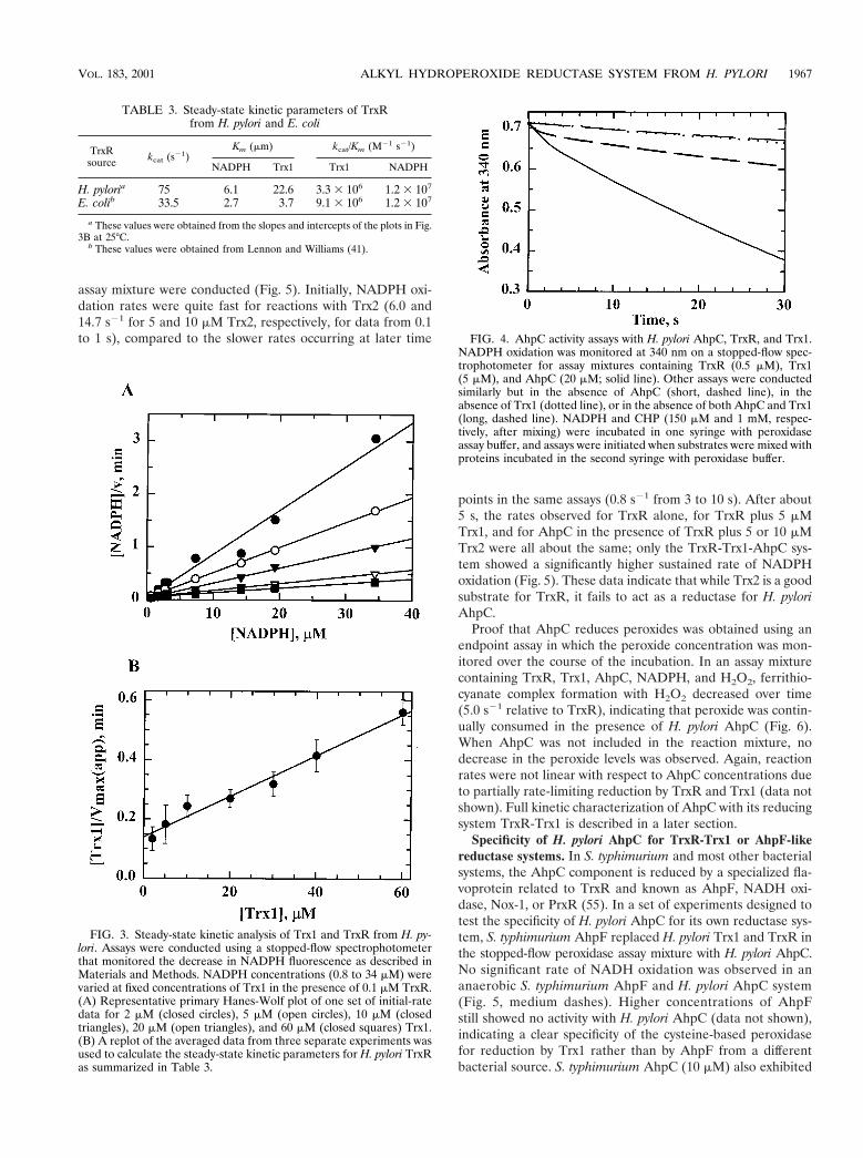
Peroxidase activity of AhpC, Trx1 or Trx2, and TrxR. To testthe ability of H. pylori AhpC to reduce hydroperoxides withH. pylori Trx1 and TrxR acting as the reducing system, theproteins were mixed with NADPH and cumene hydroperoxideand the change in A340 was monitored. The maximal sustainedrate of NADPH oxidation was observed when all three pro-teins, TrxR, Trx1, and AhpC, were included in the assay mix-ture (Fig. 4). Alone, TrxR possessed oxidase activity (Fig. 4,long, dashed lines), which was observed as a steady decrease inA340 at 0.70 s21 in the absence of peroxide; E. coli TrxRexhibited a much slower rate of NADPH oxidation (0.01 s21)under these conditions. In the presence of peroxide, NADPHoxidation by H. pylori TrxR alone increased to 0.80 s21, whichis indicative of weak peroxidatic activity of TrxR. In the ab-sence of AhpC, a small burst in NADPH oxidation was firstobserved (between 0.3 and 1 s) at a rate of 5.1 s21 relative tothe TrxR concentration, which tapers off to a rate similar tothat of TrxR alone (0.9 s21) once all of the Trx1 had beenreduced (reduction of 5 mM Trx1 accounts for a decrease inA340 of 0.03). With all three H. pylori proteins present, an ini-tial burst of NADPH oxidation (;6.6 s21 relative to TrxR), ata rate similar to that of TrxR and Trx1 alone, was observed.The somewhat slower but sustained rate observed between 10and 20 s (2.5 s21) was not significantly changed when AhpCconcentrations were varied, suggesting that both Trx1 reduc-tion by TrxR and AhpC reduction by Trx1 were partially ratelimiting under these conditions.
Investigation of the H. pylori genome yielded two Trx ho-mologues, Trx1 and Trx2, both of which contain the catalyticWCXXC site required for reductase activity. To determine ifTrx2 could act as a reductant for H. pylori AhpC, stopped-flowperoxidase experiments in which Trx2 replaced Trx1 in the
FIG. 1. SDS-PAGE analysis of purified AhpC, Trx1, Trx2, andTrxR from H. pylori. (A) Crude lysates and recombinant purifiedAhpC and TrxR were analyzed on the same 12% polyacrylamide gel inreducing sample buffer (except where noted) as follows: lane 1, mo-lecular mass markers (Broad Range Molecular Weight Standards; Bio-Rad); lane 2, crude extracts of E. coli cells transformed with pPROK1/ahpC and induced with 0.4 mM IPTG for at least 3 h at 37°C; lane 3,pure, recombinant AhpC; lane 4, crude extracts of E. coli cells trans-formed with pPROK1/trxR and induced as described above; lane 5,pure, recombinant TrxR protein; lane 6, pure AhpC in nonreducingsample buffer; lane 7, pure TrxR in nonreducing sample buffer. (B)Crude lysates and recombinant Trx1 and Trx2 were analyzed on a 10%Tris-Tricine gel as follows: lane 1, molecular mass markers; lane 2,crude extracts of E. coli cells transformed with pPROK1/trx1 afterinduction with IPTG; lane 3, pure, recombinant Trx1; lane 4, crudeextracts of E. coli cells transformed with pPROK1/trx2 after inductionwith IPTG; lane 5, recombinant purified Trx2 after purification. Equiv-alent protein masses (10 mg) were loaded in all lanes of both gels.Molecular masses (in kilodaltons) are indicated on the left.
FIG. 2. Comparative kinetic parameters of Trx1 and Trx2 fromH. pylori. Trx activity assays using either 200 mM DTNB (black bars) or80 mM insulin (gray bars) as a substrate for 0 to 50 mM Trx1 or Trx2were conducted in a standard buffer of 100 mM potassium phosphateand 2 mM EDTA, pH 7.4, with 150 mM NADPH. Assays were startedwith the addition of 7.0 nM TrxR and were monitored at 340 nm wheninsulin was used as the substrate or at 412 nm when DTNB was usedas the substrate to observe the release of TNB22. Rates were deter-mined from the first 10% of the reaction and then fitted to a hyperbolato determine the Vmax(app) and Km(app) values for each.
1966 BAKER ET AL. J. BACTERIOL.
assay mixture were conducted (Fig. 5). Initially, NADPH oxi-dation rates were quite fast for reactions with Trx2 (6.0 and14.7 s21 for 5 and 10 mM Trx2, respectively, for data from 0.1to 1 s), compared to the slower rates occurring at later time
points in the same assays (0.8 s21 from 3 to 10 s). After about5 s, the rates observed for TrxR alone, for TrxR plus 5 mMTrx1, and for AhpC in the presence of TrxR plus 5 or 10 mMTrx2 were all about the same; only the TrxR-Trx1-AhpC sys-tem showed a significantly higher sustained rate of NADPHoxidation (Fig. 5). These data indicate that while Trx2 is a goodsubstrate for TrxR, it fails to act as a reductase for H. pyloriAhpC.
Proof that AhpC reduces peroxides was obtained using anendpoint assay in which the peroxide concentration was mon-itored over the course of the incubation. In an assay mixturecontaining TrxR, Trx1, AhpC, NADPH, and H2O2, ferrithio-cyanate complex formation with H2O2 decreased over time(5.0 s21 relative to TrxR), indicating that peroxide was contin-ually consumed in the presence of H. pylori AhpC (Fig. 6).When AhpC was not included in the reaction mixture, nodecrease in the peroxide levels was observed. Again, reactionrates were not linear with respect to AhpC concentrations dueto partially rate-limiting reduction by TrxR and Trx1 (data notshown). Full kinetic characterization of AhpC with its reducingsystem TrxR-Trx1 is described in a later section.
Specificity of H. pylori AhpC for TrxR-Trx1 or AhpF-likereductase systems. In S. typhimurium and most other bacterialsystems, the AhpC component is reduced by a specialized fla-voprotein related to TrxR and known as AhpF, NADH oxi-dase, Nox-1, or PrxR (55). In a set of experiments designed totest the specificity of H. pylori AhpC for its own reductase sys-tem, S. typhimurium AhpF replaced H. pylori Trx1 and TrxR inthe stopped-flow peroxidase assay mixture with H. pylori AhpC.No significant rate of NADH oxidation was observed in ananaerobic S. typhimurium AhpF and H. pylori AhpC system(Fig. 5, medium dashes). Higher concentrations of AhpFstill showed no activity with H. pylori AhpC (data not shown),indicating a clear specificity of the cysteine-based peroxidasefor reduction by Trx1 rather than by AhpF from a differentbacterial source. S. typhimurium AhpC (10 mM) also exhibited
FIG. 3. Steady-state kinetic analysis of Trx1 and TrxR from H. py-lori. Assays were conducted using a stopped-flow spectrophotometerthat monitored the decrease in NADPH fluorescence as described inMaterials and Methods. NADPH concentrations (0.8 to 34 mM) werevaried at fixed concentrations of Trx1 in the presence of 0.1 mM TrxR.(A) Representative primary Hanes-Wolf plot of one set of initial-ratedata for 2 mM (closed circles), 5 mM (open circles), 10 mM (closedtriangles), 20 mM (open triangles), and 60 mM (closed squares) Trx1.(B) A replot of the averaged data from three separate experiments wasused to calculate the steady-state kinetic parameters for H. pylori TrxRas summarized in Table 3.
FIG. 4. AhpC activity assays with H. pylori AhpC, TrxR, and Trx1.NADPH oxidation was monitored at 340 nm on a stopped-flow spec-trophotometer for assay mixtures containing TrxR (0.5 mM), Trx1(5 mM), and AhpC (20 mM; solid line). Other assays were conductedsimilarly but in the absence of AhpC (short, dashed line), in theabsence of Trx1 (dotted line), or in the absence of both AhpC and Trx1(long, dashed line). NADPH and CHP (150 mM and 1 mM, respec-tively, after mixing) were incubated in one syringe with peroxidaseassay buffer, and assays were initiated when substrates were mixed withproteins incubated in the second syringe with peroxidase buffer.
TABLE 3. Steady-state kinetic parameters of TrxRfrom H. pylori and E. coli
TrxRsource kcat (s21)
Km (mm) kcat/Km (M21 s21)
NADPH Trx1 Trx1 NADPH
H. pyloria 75 6.1 22.6 3.3 3 106 1.2 3 107
E. colib 33.5 2.7 3.7 9.1 3 106 1.2 3 107
a These values were obtained from the slopes and intercepts of the plots in Fig.3B at 25°C.
b These values were obtained from Lennon and Williams (41).
VOL. 183, 2001 ALKYL HYDROPEROXIDE REDUCTASE SYSTEM FROM H. PYLORI 1967

considerable specificity for its own reductase, AhpF (0.5 mM),compared with reduction by E. coli TrxR (0.5 mM) plus E. coliTrx1 (5 mM); turnover rates were 42 s21 with AhpF and 1.7 s21
with E. coli TrxR-Trx1 (data not shown). Nonetheless, the rateof turnover of S. typhimurium AhpC with the E. coli TrxR-Trx1system was only about twofold lower than that of H. pyloriAhpC with its own TrxR-Trx1 system (3.2 s21 under the sameconditions) while H. pylori AhpC and S. typhimurium AhpF in-teraction was undetectable. Among the proteins under inves-tigation, H. pylori TrxR and Trx1 and E. coli TrxR and Trx1 werethe most interchangeable in peroxidase assays. H. pylori AhpC(2 mM) assayed with the E. coli proteins TrxR (2 mM) and Trx1(25 mM) exhibited a rate of NADPH oxidation that was aboutthe same as that obtained with H. pylori TrxR-Trx1 under thesame conditions (1.9 versus 3.2 s21).
Steady-state kinetics of AhpC. To further investigate theperoxidase activity of H. pylori AhpC, reaction conditions werefirst established under which initial rates were directly propor-tional to AhpC at a Trx1 concentration (30 mM) that was atleast 10-fold higher than the maximal concentration of AhpC(3 mM). Because the very low intrinsic NADPH oxidase activ-ity of E. coli TrxR allowed the observation of low peroxidaserates above background NADPH turnover, E. coli TrxR re-placed H. pylori TrxR using concentrations that were highenough (2 to 3 mM) to support rapid H. pylori Trx1 recycling(i.e., additional TrxR did not further increase observed rates ofNADPH oxidation). AhpC-dependent rates of NADPH oxida-tion measured over a range of Trx1 concentrations (15 to 30mM) suggested a simple bimolecular interaction between re-duced Trx1 and oxidized AhpC at a rate of 1.0 3 105 M21 s21
(data not shown).Given the putative nonsaturable interaction between Trx1
and AhpC suggested by the studies described above and theneed for information concerning interactions between reducedAhpC and hydroperoxide substrates, kinetic data were gener-
ated and analyzed as described previously by Dalziel (18).Because the secondary Dalziel plots (e.g., Fig. 7B) intersect they axis at the origin (within experimental error), the Km valuesof AhpC for peroxides and Trx1 and Vmax values are infinite.Parallel lines in the primary plot (Fig. 7A) also indicate a sub-stituted (ping-pong) mechanism for H. pylori AhpC by whichAhpC does not form detectable enzyme-substrate complexeswith either substrate and displays nonsaturating kinetics, a ki-netic pattern that has been observed for other nonheme per-oxidases, such as glutathione peroxidase (23) and tryparedoxinperoxidase (46).
With the above observations in mind, the rate data wereplotted using the Dalziel equation for two-substrate reactionsin which enzyme-substrate complexes are not observed: [E]0/v 5 f1/[ROOH] 1 f2/[Trx1], where [E] is the concentration ofAhpC and f1 and f2 are the Dalziel kinetic coefficients. Be-cause enzyme-substrate complexes are not observed experi-mentally, the interactions of AhpC with its two substrates canbe depicted as a sequence of consecutive, bimolecular, non-reversible reactions: AhpCred 1 ROOH3 AhpCox 1 ROH 1H2O and AhpCox 1 TrxRed3 AhpCred 1 Trx1ox. The appar-ent limiting rates for these two reactions are characterized bytheir kinetic rate constants, k19 and k29, respectively, which arethe reciprocals of the Dalziel coefficients, f1 and f2. To obtainthe rate constants, the rate data were evaluated using theintegrated Dalziel equation (24). After elucidating product andreactant amounts at various times, the data were substitutedinto the integrated rate equation (given in Materials and Meth-ods) and plotted (Fig. 7A). The slopes of the lines in theprimary plot are equal to f1: y intercepts from Fig. 7A replot-ted in Fig. 7B versus the reciprocal of the Trx1 concentrationproduced a line with a slope of f2. By taking the reciprocal off2, the second-order rate constant, k29, of 1.0 3 105 M21 s21
was determined for the interaction between AhpC and Trx1,giving the same value as that obtained using the AhpC-depen-dent assays described above, in which the peroxide substrate
FIG. 5. AhpC activity assay with Trx1 or Trx2 as the reductant. Thedecrease in NADPH absorbance was monitored on a stopped-flowspectrophotometer when AhpC (20 mM) and TrxR (0.5 mM) wereassayed with Trx1 (5.0 mM, solid line) or with Trx2 (5.0 mM, dottedline; 10 mM, long dashes) in peroxidase assay buffer as described in thelegend to Fig. 4. Assays of TrxR-Trx1 excluding the AhpC protein(dashed-dotted line) and assays of TrxR alone (small dashes) were alsoconducted. AhpF (0.5 mM) from S. typhimurium was included in placeof TrxR-Trx1 with H. pylori AhpC (medium dashes), and in this case,NADH rather than NADPH was used as the reducing substrate underanaerobic conditions.
FIG. 6. Peroxide consumption by H. pylori AhpC. The disappear-ance of peroxide was monitored by ferrithiocyanate complex formationfollowing incubations of a mixture containing 20 mM AhpC (triangles)or 0 mM AhpC (circles) with TrxR (0.2 mM), Trx1 (2.0 mM), and anNADPH-regenerating system consisting of glucose-6-phosphate (10mM), glucose-6-phosphate dehydrogenase (0.2 U/ml), and NADPH(10 mM) in 500-ml volumes. All reactions were started with the addi-tion of H2O2 (1 mM) as described in Materials and Methods.
1968 BAKER ET AL. J. BACTERIOL.

was in excess. Using a similar strategy, the value for k19 (2.0 3105 M21 s21) was determined for the interaction of AhpC withH2O2 and was of a magnitude similar to that of k29 for theoxidation of Trx1. Therefore, both steps are partially rate lim-iting under these conditions.
AhpC was also tested for the ability to reduce peroxides oth-er than H2O2, including EtOOH, t-BOOH, CHP, and linoleicacid hydroperoxide (LOOH). Using one concentration of Trx1(2.0 mM), the slopes from the primary plot were determined
for each peroxide to yield the following apparent k19 values:H2O2, 2.0 3 105 M21 s21; EtOOH, 1.7 3 105 M21 s21;t-BOOH, 1.6 3 105 M21 s21; CHP, 1.2 3 105 M21 s21; LOOH,1.1 3 105 M21 s21. AhpC was capable of reducing the differentperoxides, including the more structurally complex compoundst-BOOH and LOOH, with similar apparent rate constants. Nochange in k29 reflecting the interaction between Trx1 andAhpC was observed for the different hydroperoxide substrates.
Essentiality of H. pylori ahpC. Allelic replacement mutagen-esis of ahpC was conducted to study the physiological role ofAhpC in H. pylori and to determine the effect of AhpC removalon H. pylori’s responses to oxidative stress. When ahpC inter-rupted with the camR cassette was introduced into H. pyloristrains HP26695, SS1, HP1061, and HP1, there was no growthof colonies on selective medium after 7 days of incubation,whereas a few thousand colonies are normally obtained after 2to 3 days for nonessential-gene knockouts carried out in thismanner (e.g., with the rdxA knockout control). While singlecrossover events have been described in H. pylori using thismethod of transformation (57), no such phenomenon was ob-served even after several repeated transformation attempts. Asimilar construct was successfully generated in which the ni-troreductase (rdxA) gene was knocked out with the same camRcassette (26) and introduced by transformation and homolo-gous recombination into the same H. pylori strains. Both Camr
and Mtzr (metronidazole-resistant) colonies were formed, in-dicating that rdx is not essential for growth. Therefore, the lossof growth under microaerobic conditions with the loss of ahpCstrongly suggests an essential role for AhpC in H. pylori viabil-ity.
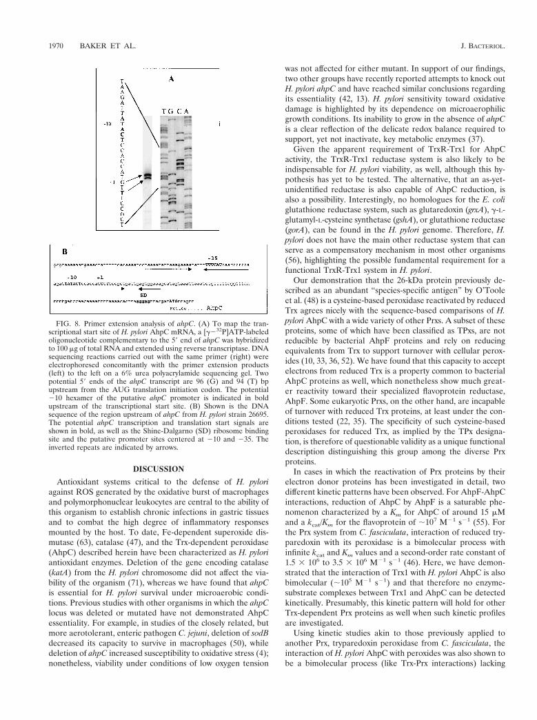
Genetic characterization of ahpC. The gene encoding AhpCis located between ceuE (HP1562, encoding an iron III ABCtransporter) transcribed in the opposite direction and a geneencoding an outer membrane protein (HP1564) in the sameorientation as ahpC and containing endogenous promoter se-quences (71). The location of the transcriptional start site ofahpC was determined by primer extension analysis. Two prom-inent reverse transcript bands migrate parallel to G and Tresidues, 96 and 94 bp upstream, respectively, from the AUGtranslation initiation codon (Fig. 8A). Sequences resemblingthe H. pylori s70 consensus sequence (25) were located at theexpected distance from the transcriptional start site (Fig. 8B,bold). The potential 210 hexamer of the putative ahpC pro-moter, TATACT, displays a high degree of identity (5 of 6 bp)to the 210 consensus sequence for H. pylori, TAtaaT (25).Other putative regulatory sequence elements were identifiedin the promoter region centered around 240 and 110 (un-derlined, Fig. 8B), underscoring the possibility that ahpC tran-scription is regulated. However, no Fur (ferric uptake reg-ulator) binding site (69) was evident in the H. pylori ahpCpromoter region and the H. pylori genome lacks a structuralgene for OxyR, the redox-sensitive transcription factor whichupregulates AhpC production in response to oxidative stress inother eubacteria (65). Although the deduced H. pylori AhpCamino acid sequence has already been published, the proteinwas not identified as an alkyl hydroperoxide reductase and thetranslational start site of ahpC was not correctly positioned byO’Toole et al. (48).
FIG. 7. Kinetic analysis of AhpC from H. pylori by the Dalzielmethod. Assays of AhpC were conducted on a stopped-flow spectro-photometer using AhpC (4.0 mM), Trx1 (2.0 mM), and E. coli TrxR(1.0 mM) with NADPH (150 mM) and limiting amounts of peroxide(10 to 40 mM). Proteins were incubated with NADPH for 5 min priorto mixing with peroxide. The resulting reaction traces were used tocalculate [ROOH] and [ROH] over time. (A) In the Dalziel primaryplot, a least-squares linear regression line was obtained for the calcu-lated data points for five different concentrations of Trx1: 4.0 mM(closed triangles), 2.0 mM (closed circles), 1.6 mM (open triangles),and 1.0 mM (open circles). The resulting lines give slopes of f1 andintercepts of f2/[Trx1]. (B) The apparent maximum velocities (y inter-cepts from the primary plot) were replotted in a Dalziel secondary plotagainst the reciprocal of the [Trx1] at which they were obtained. Theslope of the resulting linear regression line is f2.
VOL. 183, 2001 ALKYL HYDROPEROXIDE REDUCTASE SYSTEM FROM H. PYLORI 1969
DISCUSSION
Antioxidant systems critical to the defense of H. pyloriagainst ROS generated by the oxidative burst of macrophagesand polymorphonuclear leukocytes are central to the ability ofthis organism to establish chronic infections in gastric tissuesand to combat the high degree of inflammatory responsesmounted by the host. To date, Fe-dependent superoxide dis-mutase (63), catalase (47), and the Trx-dependent peroxidase(AhpC) described herein have been characterized as H. pyloriantioxidant enzymes. Deletion of the gene encoding catalase(katA) from the H. pylori chromosome did not affect the via-bility of the organism (71), whereas we have found that ahpCis essential for H. pylori survival under microaerobic condi-tions. Previous studies with other organisms in which the ahpClocus was deleted or mutated have not demonstrated AhpCessentiality. For example, in studies of the closely related, butmore aerotolerant, enteric pathogen C. jejuni, deletion of sodBdecreased its capacity to survive in macrophages (50), whiledeletion of ahpC increased susceptibility to oxidative stress (4);nonetheless, viability under conditions of low oxygen tension
was not affected for either mutant. In support of our findings,two other groups have recently reported attempts to knock outH. pylori ahpC and have reached similar conclusions regardingits essentiality (42, 13). H. pylori sensitivity toward oxidativedamage is highlighted by its dependence on microaerophilicgrowth conditions. Its inability to grow in the absence of ahpCis a clear reflection of the delicate redox balance required tosupport, yet not inactivate, key metabolic enzymes (37).
Given the apparent requirement of TrxR-Trx1 for AhpCactivity, the TrxR-Trx1 reductase system is also likely to beindispensable for H. pylori viability, as well, although this hy-pothesis has yet to be tested. The alternative, that an as-yet-unidentified reductase is also capable of AhpC reduction, isalso a possibility. Interestingly, no homologues for the E. coliglutathione reductase system, such as glutaredoxin (grxA), g-L-glutamyl-L-cysteine synthetase (gshA), or glutathione reductase(gorA), can be found in the H. pylori genome. Therefore, H.pylori does not have the main other reductase system that canserve as a compensatory mechanism in most other organisms(56), highlighting the possible fundamental requirement for afunctional TrxR-Trx1 system in H. pylori.
Our demonstration that the 26-kDa protein previously de-scribed as an abundant “species-specific antigen” by O’Tooleet al. (48) is a cysteine-based peroxidase reactivated by reducedTrx agrees nicely with the sequence-based comparisons of H.pylori AhpC with a wide variety of other Prxs. A subset of theseproteins, some of which have been classified as TPxs, are notreducible by bacterial AhpF proteins and rely on reducingequivalents from Trx to support turnover with cellular perox-ides (10, 33, 36, 52). We have found that this capacity to acceptelectrons from reduced Trx is a property common to bacterialAhpC proteins as well, which nonetheless show much great-er reactivity toward their specialized flavoprotein reductase,AhpF. Some eukaryotic Prxs, on the other hand, are incapableof turnover with reduced Trx proteins, at least under the con-ditions tested (22, 35). The specificity of such cysteine-basedperoxidases for reduced Trx, as implied by the TPx designa-tion, is therefore of questionable validity as a unique functionaldescription distinguishing this group among the diverse Prxproteins.
In cases in which the reactivation of Prx proteins by theirelectron donor proteins has been investigated in detail, twodifferent kinetic patterns have been observed. For AhpF-AhpCinteractions, reduction of AhpC by AhpF is a saturable phe-nomenon characterized by a Km for AhpC of around 15 mMand a kcat/Km for the flavoprotein of ;107 M21 s21 (55). Forthe Prx system from C. fasciculata, interaction of reduced try-paredoxin with its peroxidase is a bimolecular process withinfinite kcat and Km values and a second-order rate constant of1.5 3 106 to 3.5 3 106 M21 s21 (46). Here, we have demon-strated that the interaction of Trx1 with H. pylori AhpC is alsobimolecular (;105 M21 s21) and that therefore no enzyme-substrate complexes between Trx1 and AhpC can be detectedkinetically. Presumably, this kinetic pattern will hold for otherTrx-dependent Prx proteins as well when such kinetic profilesare investigated.
Using kinetic studies akin to those previously applied toanother Prx, tryparedoxin peroxidase from C. fasciculata, theinteraction of H. pylori AhpC with peroxides was also shown tobe a bimolecular process (like Trx-Prx interactions) lacking
FIG. 8. Primer extension analysis of ahpC. (A) To map the tran-scriptional start site of H. pylori AhpC mRNA, a [g232P]ATP-labeledoligonucleotide complementary to the 59 end of ahpC was hybridizedto 100 mg of total RNA and extended using reverse transcriptase. DNAsequencing reactions carried out with the same primer (right) wereelectrophoresed concomitantly with the primer extension products(left) to the left on a 6% urea polyacrylamide sequencing gel. Twopotential 59 ends of the ahpC transcript are 96 (G) and 94 (T) bpupstream from the AUG translation initiation codon. The potential210 hexamer of the putative ahpC promoter is indicated in boldupstream of the transcriptional start site. (B) Shown is the DNAsequence of the region upstream of ahpC from H. pylori strain 26695.The potential ahpC transcription and translation start signals areshown in bold, as well as the Shine-Dalgarno (SD) ribosome bindingsite and the putative promoter sites centered at 210 and 235. Theinverted repeats are indicated by arrows.
1970 BAKER ET AL. J. BACTERIOL.

detectable enzyme-substrate complexes. This ping-pong mech-anism has also been observed for the distantly related gluta-thione peroxidases (23, 68). The higher rate of enzyme-perox-ide interaction for glutathione peroxidase (;108 M21 s21) hasbeen attributed to the unique reactivity of the selenocysteine atthe active site (64), although at 105 to 106 M21 s21, the rate ofperoxide reduction is still quite high and indicative of an im-portant role for H. pylori AhpC in cellular peroxide metabo-lism. Using k19 to characterize the protein-peroxide interac-tion, our experiments demonstrated essentially no specificitywhen AhpC was tested with a wide variety of small, bulky, aro-matic, or lipid hydroperoxide substrates, as was true of theC. fasciculata peroxidase (46). Differential reactivities towardparticular hydroperoxide substrates have been reported forsome other Prx enzymes based on less quantitative analyses(9, 33, 34). In addition, peroxynitrite (OONO2) has recentlybeen shown to be a substrate for H. pylori AhpC, with the rateof decomposition occurring at a second-order rate constant of1.21 3 106 M21 s21 (7). These results are all consistent with aminimal binding site on AhpC for hydroperoxides (and per-oxynitrite) consisting of little more than the catalytic residue(Cys49) at the active site.
On the basis of results reported here and mechanisms out-lined for other 2-Cys AhpC homologues (10, 51), electronsfrom NADPH proceed along the path outlined in Fig. 9 for thereduction of peroxides to alcohols. This scheme is highly anal-ogous to that for electron transfer through the S. typhimuriumAhpC system, except that TrxR and Trxl replace AhpF in theH. pylori system. Nonetheless, one tightly bound flavin andthree disulfide redox centers mediate electron transfer frompyridine nucleotide to peroxide in both cases.
Little information exists on potential redox or iron regula-tion of H. pylori AhpC expression, although in other studies ofTrxR and Trx1 from H. pylori, Windle et al. (74) observed thatTrxl expression dramatically increased under conditions of oxi-dative stress; Trx1 was therefore classified as a stress responseelement in H. pylori. While the proximal location of trxR andtrx1 in the chromosome could provide the bacterium with amechanism for a coordinated response eliciting expression ofboth proteins, an increase in TrxR expression did not accom-pany increased Trx1 expression under oxidative stress condi-tions (74). Interestingly, reductase activity of the TrxR-Trx1reductase system was also reportedly present in the media of
culture supernatants and could be available to support extra-cellular peroxide reduction by AhpC if the latter protein is alsoexported.
In conclusion, this is the first report of a Trx-dependent alkylhydroperoxide system from a gastric pathogen. To our knowl-edge, AhpC from H. pylori is only the second known bacterialAhpC to be demonstrated experimentally to require the Trx-reducing system for reduction of the oxidized AhpC active site.An essential role for AhpC in H. pylori has been establishedwith our ahpC mutagenesis experiments, and this is, to ourknowledge, the only case in which an ahpC locus has beenshown to be required for viability. The presence of this essen-tial Trx-dependent peroxidase in H. pylori suggests importantroles for TrxR, Trx1, and AhpC in the removal of alkyl hydro-peroxides to protect against oxidative stress and in the preser-vation of the microaerobic environment required for H. pyloriviability.
ACKNOWLEDGMENTS
Funding for this work was provided in part by grants from theNational Institutes of Health (NIH R01 GM50389) to L.B.P. and fromthe Canadian Institutes for Health Research (CIHR MT11318 andRP14292) to P.S.H.
We are particularly grateful to C. M. Reynolds for the purifica-tion of E. coli Trx1 and TrxR and to Desnee Wynn, Lois LaPrade,and Louis Bryden for technical assistance. Special thanks to JosephO’Flaherty for his assistance with the fatty acid hydroperoxide prepa-ration and to James Luba for helpful discussions of the kinetic data.
REFERENCES
1. Alphey, M. S., C. S. Bond, E. Tetaud, A. H. Fairlamb, and W. N. Hunter.2000. The structure of reduced tryparedoxin peroxidase reveals a decamerand insight into reactivity of 2-Cys peroxiredoxins. J. Mol. Biol. 300:903–916.
2. Altschul, S. F., W. Gish, W. Miller, E. W. Myers, and D. J. Lipman. 1990.Basic local alignment search tool. J. Mol. Biol. 215:403–410.
3. Ausubel, F. M., R. B. Brent, R. E. Kingston, D. D. Moore, J. G. Seidman,J. A. Smith, and K. Struhl (ed.). 1999. Short protocols in molecular biology.John Wiley & Sons, Inc., New York, N.Y.
4. Baillon, M. L., A. H. van Vliet, J. M. Ketley, C. Constantinidou, and C. W.Penn. 1999. An iron-regulated alkyl hydroperoxide reductase (AhpC) con-fers aerotolerance and oxidative stress resistance to the microaerophilicpathogen Campylobacter jejuni. J. Bacteriol. 181:4798–4804.
5. Baker, L. M. S., and L. B. Poole. 1999. An alkyl hydroperoxide reductasesystem from the gastric pathogen, Helicobacter pylori: cloning, purification,and kinetic studies. FASEB J. 13:A1447.
6. Baker, L. M. S., and L. B. Poole. 2000. Thioredoxin 1, not Thioredoxin 2,selectively reduces the peroxidase component of Helicobacter pylori’s alkylhydroperoxide reductase system. FASEB J. 14:A1525.
7. Bryk, R., P. Griffin, and C. Nathan. 2000. Peroxynitrite reductase activity ofbacterial peroxiredoxins. Nature. 407:211–215.
8. Buck, G. K., W. K. Gourley, W. K. Lee, K. Subramanyam, J. M. Latimer, andA. R. DiNuzzo. 1986. Relation of Campylobacter pyloridis to gastritis andpeptic ulcer. J. Infect. Dis. 153:664–669.
9. Cha, M. K., C. H. Yun, and I. H. Kim. 2000. Interaction of human thiol-specific antioxidant protein 1 with erythrocyte plasma membrane. Biochem-istry 39:6944–6950.
10. Chae, H. Z., S. J. Chung, and S. G. Rhee. 1994. Thioredoxin-dependentperoxide reductase from yeast. J. Biol. Chem. 269:27670–27678.
11. Chae, H. Z., S. W. Kang, and S. G. Rhee. 1999. Isoforms of mammalianperoxiredoxin that reduce peroxides in presence of thioredoxin. MethodsEnzymol. 300:219–226.
12. Chae, H. Z., K. Robinson, L. B. Poole, G. Church, G. Storz, and S. G. Rhee.1994. Cloning and sequencing of thiol-specific antioxidant from mammalianbrain: alkyl hydroperoxide reductase and thiol-specific antioxidant define alarge family of antioxidant enzymes. Proc. Natl. Acad. Sci. USA 91:7017–7021.
13. Chalker, A. F., H. W. Minehart, N. J. Hughes, K. K. Koretke, M. A. Lonetto,K. K. Brinkman, P. V. Warren, A. Lupas, M. J. Stanhope, J. R. Brown, andP. S. Hoffman. 2001. Systematic identification of selective genes in Helico-bacter pylori by genome prioritization and allelic replacement mutagenesis.J. Bacteriol. 183:1259–1268.
14. Cohen, G., M. Yanko, M. Mislovati, A. Argaman, R. Schreiber, Y. Av-Gay,and Y. Aharonowitz. 1993. Thioredoxin-thioredoxin reductase system of Strep-
FIG. 9. Pathway for transfer of reducing equivalents from NADPHto hydroperoxide in the alkyl hydroperoxide reductase system fromH. pylori. Note that the enzyme species shown do not necessarily rep-resent actual catalytic intermediates. FAD, flavin adenine dinucleotide.
VOL. 183, 2001 ALKYL HYDROPEROXIDE REDUCTASE SYSTEM FROM H. PYLORI 1971

tomyces clavuligerus: sequences, expression, and organization of the genes.J. Bacteriol. 175:5159–5167.
15. Cornish-Bowden, A. 1999. Fundamentals of enzyme kinetics. Portland Press,Ltd., London, England.
16. Correa, P. 1995. Helicobacter pylori and gastric carcinogenesis. Am. J. Surg.Pathol. 19:S37–S43.
17. Correa, P. 1988. A human model of gastric carcinogenesis. Cancer Res. 48:3554–3560.
18. Dalziel, K. 1957. Initial steady state velocities in the evaluation of enzyme-coenzyme-substrate reaction mechanisms. Acta Chem. Scand. 11:1706–1723.
19. Davies, G. R., N. J. Simmonds, T. R. Stevens, M. T. Sheaff, N. Banatvala,I. F. Laurenson, D. R. Blake, and D. S. Rampton. 1994. Helicobacter pyloristimulates antral mucosal reactive oxygen metabolite production in vivo. Gut35:179–185.
20. Ellis, H. R., and L. B. Poole. 1997. Novel application of 7-chloro-4-nitro-benzo-2-oxa-1,3-diazole to identify cysteine sulfenic acid in the AhpC com-ponent of alkyl hydroperoxide reductase. Biochemistry 36:15013–15018.
21. Ellis, H. R., and L. B. Poole. 1997. Roles for the two cysteine residues ofAhpC in catalysis of peroxide reduction by alkyl hydroperoxide reductasefrom Salmonella typhimurium. Biochemistry 36:13349–13356.
22. Fisher, A. B., C. Dodia, Y. Manevich, J. W. Chen, and S. I. Feinstein. 1999.Phospholipid hydroperoxides are substrates for non-selenium glutathioneperoxidase. J. Biol. Chem. 274:21326–21334.
23. Flohe, L., G. Loschen, W. A. Gunzler, and E. Eichele. 1972. Glutathioneperoxidase. V. The kinetic mechanism. Hoppe Seyler’s Z. Physiol. Chem.353:987–999.
24. Forstrom, J. W., F. H. Stults, and A. L. Tappel. 1979. Rat liver cytosolicglutathione peroxidase: reactivity with linoleic acid hydroperoxide and cu-mene hydroperoxide. Arch. Biochem. Biophys. 193:51–55.
25. Forsyth, M. H., and T. L. Cover. 1999. Mutational analysis of the vacApromoter provides insight into gene transcription in Helicobacter pylori.J. Bacteriol. 181:2261–2266.
26. Goodwin, A., D. Kersulyte, G. Sisson, S. J. Veldhuyzen van Zanten, D. E.Berg, and P. S. Hoffman. 1998. Metronidazole resistance in Helicobacterpylori is due to null mutations in a gene (rdxA) that encodes an oxygen-insensitive NADPH nitroreductase. Mol. Microbiol. 28:383–393.
27. Graham, D. Y., and P. D. Klein. 1987. Campylobacter pyloridis gastritis: thepast, the present, and speculations about the future. Am. J. Gastroenterol.82:283–286.
28. Guruge, J. L., P. G. Falk, R. G. Lorenz, M. Dans, H. P. Wirth, M. J. Blaser,D. E. Berg, and J. I. Gordon. 1998. Epithelial attachment alters the outcomeof Helicobacter pylori infection. Proc. Natl. Acad. Sci. USA 95:3925–3930.
29. Holmgren, A. 1968. Thioredoxin. 6. The amino acid sequence of the proteinfrom Escherichia coli B. Eur. J. Biochem. 6:475–484.
30. Holmgren, A., and M. Bjornstedt. 1995. Thioredoxin and thioredoxin reduc-tase. Methods Enzymol. 252:199–306.
31. Holmgren, A., and P. Reichard. 1967. Thioredoxin. 2. Cleavage with cyano-gen bromide. Eur. J. Biochem. 2:187–196.
32. Jacobson, F. S., R. W. Morgan, M. F. Christman, and B. N. Ames. 1989. Analkyl hydroperoxide reductase from Salmonella typhimurium involved in thedefense of DNA against oxidative damage. Purification and properties.J. Biol. Chem. 264:1488–1496.
33. Jeong, J. S., S. J. Kwon, S. W. Kang, S. G. Rhee, and K. Kim. 1999. Purifi-cation and characterization of a second type thioredoxin peroxidase (type IITPx) from Saccharomyces cerevisiae. Biochemistry 38:776–783.
34. Jeong, W., M. K. Cha, and I. H. Kim. 2000. Thioredoxin-dependent hydro-peroxide peroxidase activity of bacterioferritin comigratory protein (BCP) asa new member of the thiol-specific antioxidant protein (TSA)/alkyl hydro-peroxide peroxidase C (AhpC) family. J. Biol. Chem. 275:2924–2930.
35. Kang, S. W., I. C. Baines, and S. G. Rhee. 1998. Characterization of amammalian peroxiredoxin that contains one conserved cysteine. J. Biol.Chem. 273:6303–6311.
36. Kang, S. W., H. Z. Chae, M. S. Seo, K. Kim, I. C. Baines, and S. G. Rhee.1998. Mammalian peroxiredoxin isoforms can reduce hydrogen peroxidegenerated in response to growth factors and tumor necrosis factor-alpha.J. Biol. Chem. 273:6297–6302.
37. Kelly, D. J. 1998. The physiology and metabolism of the human gastricpathogen Helicobacter pylori. Adv. Microb. Physiol. 40:137–189.
38. Kitano, K., Y. Niimura, Y. Nishiyama, and K. Miki. 1999. Stimulation ofperoxidase activity by decamerization related to ionic strength: AhpC pro-tein from Amphibacillus xylanus. J. Biochem. (Tokyo) 126:313–319.
39. Klenk, H. P., R. A. Clayton, J. F. Tomb, O. White, K. E. Nelson, K. A.Ketchum, R. J. Dodson, M. Gwinn, E. K. Hickey, J. D. Peterson, D. L.Richardson, A. R. Kerlavage, D. E. Graham, N. C. Kyrpides, R. D. Fleisch-mann, J. Quackenbush, N. H. Lee, G. G. Sutton, S. Gill, E. F. Kirkness, B. A.Dougherty, K. McKenney, M. D. Adams, B. Loftus, J. C. Venter et al. 1997.The complete genome sequence of the hyperthermophilic, sulphate-reducingarchaeon Archaeoglobus fulgidus. Nature 390:364–370.
40. Lee, A., J. O’Rourke, M. C. De Ungria, B. Robertson, G. Daskalopoulos, andM. F. Dixon. 1997. A standardized mouse model of Helicobacter pylori in-fection: introducing the Sydney strain. Gastroenterology 112:1386–1397.
41. Lennon, B. W., and C. H. Williams, Jr. 1997. Reductive half-reaction of
thioredoxin reductase from Escherichia coli. Biochemistry 36:9464–9477.42. Lundstrom, A. M., and I. Bolin. 2000. A 26 kDa protein of Helicobacter pylori
shows alkyl hydroperoxide reductase (AhpC) activity and the mono-cistronictranscription of the gene is affected by pH. Microb. Pathog. 29:257–266.
43. Maiorino, M., C. Gregolin, and F. Ursini. 1990. Phospholipid hydroperoxideglutathione peroxidase. Methods Enzymol. 186:448–457.
44. Mallett, T. C., and A. Claiborne. 1998. Oxygen reactivity of an NADHoxidase C42S mutant: evidence for a C(4a)-peroxyflavin intermediate and arate-limiting conformational change. Biochemistry 37:8790–8802.
45. Miranda-Vizuete, A., A. E. Damdimopoulos, J. Gustafsson, and G. Spyrou.1997. Cloning, expression, and characterization of a novel Escherichia colithioredoxin. J. Biol. Chem. 272:30841–30847.
46. Nogoceke, E., D. U. Gommel, M. Kiess, H. M. Kalisz, and L. Flohe. 1997. Aunique cascade of oxidoreductases catalyses trypanothione-mediated perox-ide metabolism in Crithidia fasciculata. Biol. Chem. 378:827–836.
47. Odenbreit, S., B. Wieland, and R. Haas. 1996. Cloning and genetic char-acterization of Helicobacter pylori catalase and construction of a catalase-deficient mutant strain. J. Bacteriol. 178:6960–6967.
48. O’Toole, P. W., S. M. Logan, M. Kostrzynska, T. Wadstrom, and T. J. Trust.1991. Isolation and biochemical and molecular analyses of a species-specificprotein antigen from the gastric pathogen Helicobacter pylori. J. Bacteriol.173:505–513.
49. Parkhill, J., B. W. Wren, K. Mungall, J. M. Ketley, C. Churcher, D. Basham,T. Chillingworth, R. M. Davies, T. Feltwell, S. Holroyd, K. Jagels, A. V.Karlyshev, S. Moule, M. J. Pallen, C. W. Penn, M. A. Quail, M. A. Rajan-dream, K. M. Rutherford, A. H. van Vliet, S. Whitehead, and B. G. Barrell.2000. The genome sequence of the food-borne pathogen Campylobacterjejuni reveals hypervariable sequences. Nature 403:665–668.
50. Pesci, E. C., D. L. Cottle, and C. L. Pickett. 1994. Genetic, enzymatic, andpathogenic studies of the iron superoxide dismutase of Campylobacter jejuni.Infect. Immun. 62:2687–2694.
51. Poole, L. B. 1996. Flavin-dependent alkyl hydroperoxide reductase fromSalmonella typhimurium. 2. Cystine disulfides involved in catalysis of perox-ide reduction. Biochemistry 35:65–75.
52. Poole, L. B., H. Z. Chae, B. M. Flores, S. L. Reed, S. G. Rhee, and B. E.Torian. 1997. Peroxidase activity of a TSA-like antioxidant protein from apathogenic amoeba. Free Radic. Biol. Med. 23:955–959.
53. Poole, L. B., and H. R. Ellis. 1996. Flavin-dependent alkyl hydroperoxidereductase from Salmonella typhimurium. 1. Purification and enzymatic activ-ities of overexpressed AhpF and AhpC proteins. Biochemistry 35:56–64.
54. Poole, L. B., A. Godzik, A. Nayeem, and J. D. Schmitt. 2000. AhpF can bedissected into two functional units: tandem repeats of two thioredoxin-likefolds in the N-terminus mediate electron transfer from the thioredoxinreductase-like C-terminus to AhpC. Biochemistry 39:6602–6615.
55. Poole, L. B., C. M. Reynolds, Z. A. Wood, P. A. Karplus, H. R. Ellis, andM. Li Calzi. 2000. AhpF and other NADH:peroxiredoxin oxidoreductases,homologues of low Mr thioredoxin reductase. Eur. J. Biochem. 267:6126–6133.
56. Prieto-Alamo, M. J., J. Jurado, R. Gallardo-Madueno, F. Monje-Casas, A.Holmgren, and C. Pueyo. 2000. Transcriptional regulation of glutaredoxinand thioredoxin pathways and related enzymes in response to oxidativestress. J. Biol. Chem. 275:13398–13405.
57. Raudonikiene, A., N. Zakharova, W. W. Su, J. Y. Jeong, L. Bryden, P. S.Hoffman, D. E. Berg, and K. Severinov. 1999. Helicobacter pylori with sepa-rate beta- and beta9-subunits of RNA polymerase is viable and can colonizeconventional mice. Mol. Microbiol. 32:131–138.
58. Riddles, P. W., R. L. Blakeley, and B. Zerner. 1979. Ellman’s reagent:5,59-dithiobis(2-nitrobenzoic acid)—a reexamination. Anal. Biochem. 94:75–81.
59. Rieder, G., R. A. Hatz, A. P. Moran, A. Walz, M. Stolte, and G. Enders. 1997.Role of adherence in interleukin-8 induction in Helicobacter pylori-associatedgastritis. Infect. Immun. 65:3622–3630.
60. Salyers, A. A., and D. D. Whitt. 1994. Bacterial pathogenesis—molecularapproach, p. 273–281. ASM Press, Washington, D.C.
61. Schroder, E., J. A. Littlechild, A. A. Lebedev, N. Errington, A. A. Vagin, andM. N. Isupov. 2000. Crystal structure of decameric 2-Cys peroxiredoxin fromhuman erythrocytes at 1.7 A resolution. Structure 8:605–615.
62. Schroder, E., and C. P. Ponting. 1998. Evidence that peroxiredoxins arenovel members of the thioredoxin fold superfamily. Protein Sci. 7:2465–2468.
63. Spiegelhalder, C., B. Gerstenecker, A. Kersten, E. Schiltz, and M. Kist. 1993.Purification of Helicobacter pylori superoxide dismutase and cloning andsequencing of the gene. Infect. Immun. 61:5315–5325.
64. Stadtman, T.C. 1996. Selenocysteine. Annu. Rev. Biochem. 65:83–100.65. Storz, G., F. S. Jacobson, L. A. Tartaglia, R. W. Morgan, L. A. Silveira, and
B. N. Ames. 1989. An alkyl hydroperoxide reductase induced by oxidativestress in Salmonella typhimurium and Escherichia coli: genetic characteriza-tion and cloning of ahp. J. Bacteriol. 171:2049–2055.
66. Thurman, R. G., H. G. Ley, and R. Scholz. 1972. Hepatic microsomal ethanoloxidation. Hydrogen peroxide formation and the role of catalase. Eur. J. Bio-chem. 25:420–430.
67. Tomb, J. F., O. White, A. R. Kerlavage, R. A. Clayton, G. G. Sutton, R. D.Fleischmann, K. A. Ketchum, H. P. Klenk, S. Gill, B. A. Dougherty, K.
1972 BAKER ET AL. J. BACTERIOL.

Nelson, J. Quackenbush, L. Zhou, E. F. Kirkness, S. Peterson, B. Loftus, D.Richardson, R. Dodson, H. G. Khalak, A. Glodek, K. McKenney, L. M. Fitze-gerald, N. Lee, M. D. Adams, J. C. Venter, et al. 1997. The complete genomesequence of the gastric pathogen Helicobacter pylori. Nature 388:539–547.
68. Ursini, F., M. Maiorino, and C. Gregolin. 1985. The selenoenzyme phos-pholipid hydroperoxide glutathione peroxidase. Biochim. Biophys. Acta 839:62–70.
69. van Vliet, A. H., M. L. Baillon, C. W. Penn, and J. M. Ketley. 1999. Campy-lobacter jejuni contains two fur homologs: characterization of iron-responsiveregulation of peroxide stress defense genes by the PerR repressor. J Bacte-riol. 181:6371–6376.
70. Wang, Y., and D. E. Taylor. 1990. Chloramphenicol resistance in Campylo-bacter coli: nucleotide sequence, expression, and cloning vector construction.Gene 94:23–28.
71. Westblom, T. U., S. Phadnis, W. Langenberg, K. Yoneda, E. Madan, andB. R. Midkiff. 1992. Catalase negative mutants of Helicobacter pylori. Eur.J. Clin. Microbiol. Infect. Dis. 11:522–526.
72. Wieles, B., D. van Soolingen, A. Holmgren, R. Offringa, T. Ottenhoff, andJ. Thole. 1995. Unique gene organization of thioredoxin and thioredoxinreductase in Mycobacterium leprae. Mol. Microbiol. 16:921–929.
73. Williams, C. H., Jr., G. Zanetti, L. D. Arscott, and J. K. McAllister. 1967.Lipoamide dehydrogenase, glutathione reductase, thioredoxin reductase,and thioredoxin. J. Biol. Chem. 242:5226–5231.
74. Windle, H. J., A. Fox, D. Ni Eidhin, and D. Kelleher. 2000. The thioredoxinsystem of Helicobacter pylori. J. Biol. Chem. 275:5081–5089.
75. Yamamoto, H., C. Miyake, K. J. Dietz, K. Tomizawa, N. Murata, andA. Yokota. 1999. Thioredoxin peroxidase in the cyanobacterium Synechocys-tis sp. PCC 6803. FEBS Lett. 447:269–273.
VOL. 183, 2001 ALKYL HYDROPEROXIDE REDUCTASE SYSTEM FROM H. PYLORI 1973