esmo e-learning multidisciplinary treatment of glioblastoma
TRANSCRIPT

MULTIDISCIPLINARY TREATMENT OF GLIOBLASTOMA
Univ.-Prof. Dr. Matthias Preusser
Division of Oncology, Medical University of Vienna, Austria
EPIDEMIOLOGY OF GLIOBLASTOMA
Most common primary brain tumour of adults
Incidence: 4-5/100.000/year
Twice as common in European descendants as compared to African American or Asian descendants
Median age at diagnosis
◆ Primary glioblastoma: 64 years
◆ Secondary glioblastoma: 45 years
Male to female ratio = 1.3:1

RELATIVE FREQUENCIES OF GLIOMAS
Roman numerals denote World Health Organisation (WHO) tumour grades. Redrawn from: Preusser M, et al. Ann Neurol 2011;70(1):9–21.

RISK FACTORS OF GLIOBLASTOMA
Cranial irradiation
Hereditary tumour syndromes (<5% of glioblastomas)
◆ Li Fraumeni syndrome (TP53 mutations)
◆ Turcot syndrome (APC, MLH1, MSH2, MSH6, MPS2 mutations)
◆ Neurofibromatosis 1 (Neurofibromin mutations)
◆ Neurofibromatosis 2 (Merlin mutations)
No clear evidence for occupational factors or cell phone use as risk factor

CLINICAL PRESENTATION
Most cases (>90%) develop de novo with a short clinical history of days to a few months (primary glioblastoma)
Few cases (<10%) develop from lower grade gliomas (secondary glioblastomas), typically with a clinical history of
years
Clinical presentation is highly variable, depends on tumour localisation and size
◆ Focal neurological signs (aphasia, paraesthesia, hemiparesis, visual disturbances, etc.)
◆ Mood and personality changes
◆ Seizures
◆ Symptoms of increased intracranial pressure (nausea, vomiting, headache)

MAGNETIC RESONANCE IMAGING (MRI)
Reproduced from: Preusser M, et al. Current concepts and management of Glioblastoma. Ann Neurol 2011;70(1):9–21, with by permission of John Wiley and Sons, Copyright © 2011 American Neurological Association.
Axial post contrast T1-weighted MRI Axial T2-weighted MRI

HISTOPATHOLOGY: DIAGNOSTIC FEATURES
Reproduced from: Preusser M, et al. Current concepts and management of Glioblastoma. Ann Neurol 2011;70(1):9–21, with by permission of John Wiley and Sons, Copyright © 2011 American Neurological Association.
Neuropathology of glioblastoma
(A) Histopathology of a typical case of glioblastoma
showing cellular glial tumour tissue with central necrosis
(x) with perinecrotic nuclear pseudopalisading and
microvascular proliferates (arrows; hematoxylin and eosin
staining; original magnification, 3100)
(B) Immunostaining for the astroglial marker glial fibrillary
acidic protein showsstrong labeling of the tumour cells
(brown signal; original magnification, 3400)
(C) Immunostaining for the endothelial marker CD34
shows glomeruloid microvascular proliferates (original
magnification, 3200)
(D) Immunostaining for the cell-cycle–related antigen Ki67
shows that many tumour cells undergo mitosis (brown
signal; original magnification, 3400)

WHO CLASSIFICATION
Reprinted by permission from Springer Nature, Acta Neuropathologica, The 2016 World Health Organization Classification of Tumors of the Central Nervous System: a summary, Louis DN, et al. COPYRIGHT 2016.
Histology
IDH status
1p/19q and other
genetic parameters
IDH mutant IDH wild-type
ATRX loss*
TP53 mutation*1p/19q codeletion
Glioblastoma,
IDH mutant
IDH mutant IDH wild-type
Glioblastoma,
IDH wild-type
Diffuse astrocytoma,
IDH mutantOligodendroglioma,
IDH mutant and
1p/19q codeleted
After exclusion
of other entities:
Diffuse astrocytoma,
IDH wild-type
Oligodendroglioma,
NOS
Diffuse astrocytoma, NOS
Oligodendroglioma, NOS
Oligoastrocytoma, NOS
Genetic testing not done
or inconclusive
Astrocytoma Oligoastrocytoma Oligodendroglioma Glioblastoma
*Characteristic but not required for diagnosis.

TOP 20 MUTATED GENES IN GLIOBLASTOMA BASED
ON 712 SAMPLES OF ASTROCYTOMA GRADE IV
Nørøxe DS, et al. ESMO Open 2016;1:e000144. Copyright © European Society for Medical Oncology. All rights reserved.
Top 20 genesGene name (frequency)
TP53 (22%)
EGFR (14%)
PTEN (12%)
CHEK2 (10%)
H3F3A (6%)
PIK3CA (6%)
RB1 (6%)
NF1 (5%)
PIK3R1 (4%)
HIF1A (4%)
ATRX (4%)
IDH1 (4%)
PDGFRA (3%)
KMT2C (3%0
BCOR (2%)
BRCA1 (2%)
ACVR1 (2%)
BRAF (2%)
STAG2 (2%)
ROS1 (2%)

PROGNOSTIC FACTORS
Patient age
◆ Young age favourable
Karnofsky performance status
◆ High Karnofsky index favourable
Extent of resection
◆ Gross total resection more favourable than partial resection or biopsy
Molecular information, especially O6-methylguanine-methyltransferase gene (MGMT) promoter methylation status
and isocitrate dehydrogenase gene (IDH) mutation status
◆ MGMT promoter hypermethylation favourable
◆ Presence of IDH mutation favourable

NEWLY DIAGNOSED GLIOBLASTOMATherapy

NEUROSURGERY
Maximal safe resection is the initial therapy of choice
May rapidly improve symptoms
Due to infiltrative growth residual tumour cells persist even after macroscopically complete resection
Tumour localisation in functionally important CNS regions (e.g. eloquent cortex) may allow only partial debulking
or biopsy
Modern neurosurgery involves multimodal planning of the procedure by advanced neuroimaging

FLUORESCENCE-GUIDED NEUROSURGERY
Reprinted from The Lancet Oncol, 7.5, Stummer W, et al. Fluorescence-guided surgery with 5-aminolevulinic acid for resection of malignant glioma: a randomised controlled multicentre phase III trial, 392–401, Copyright
2006, with permission from Elsevier..
Pro
gres
sion
-fre
e su
rviv
al (
%)
Time (months)
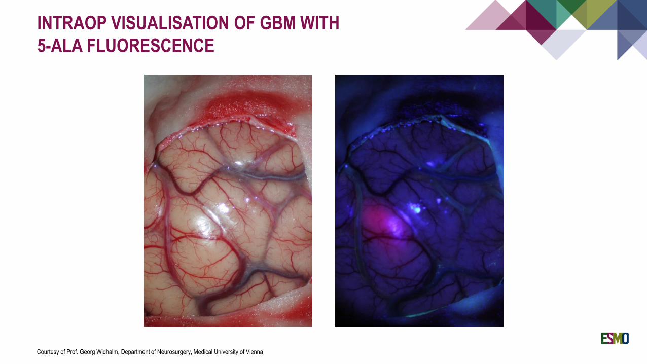
INTRAOP VISUALISATION OF GBM WITH
5-ALA FLUORESCENCE
Courtesy of Prof. Georg Widhalm, Department of Neurosurgery, Medical University of Vienna
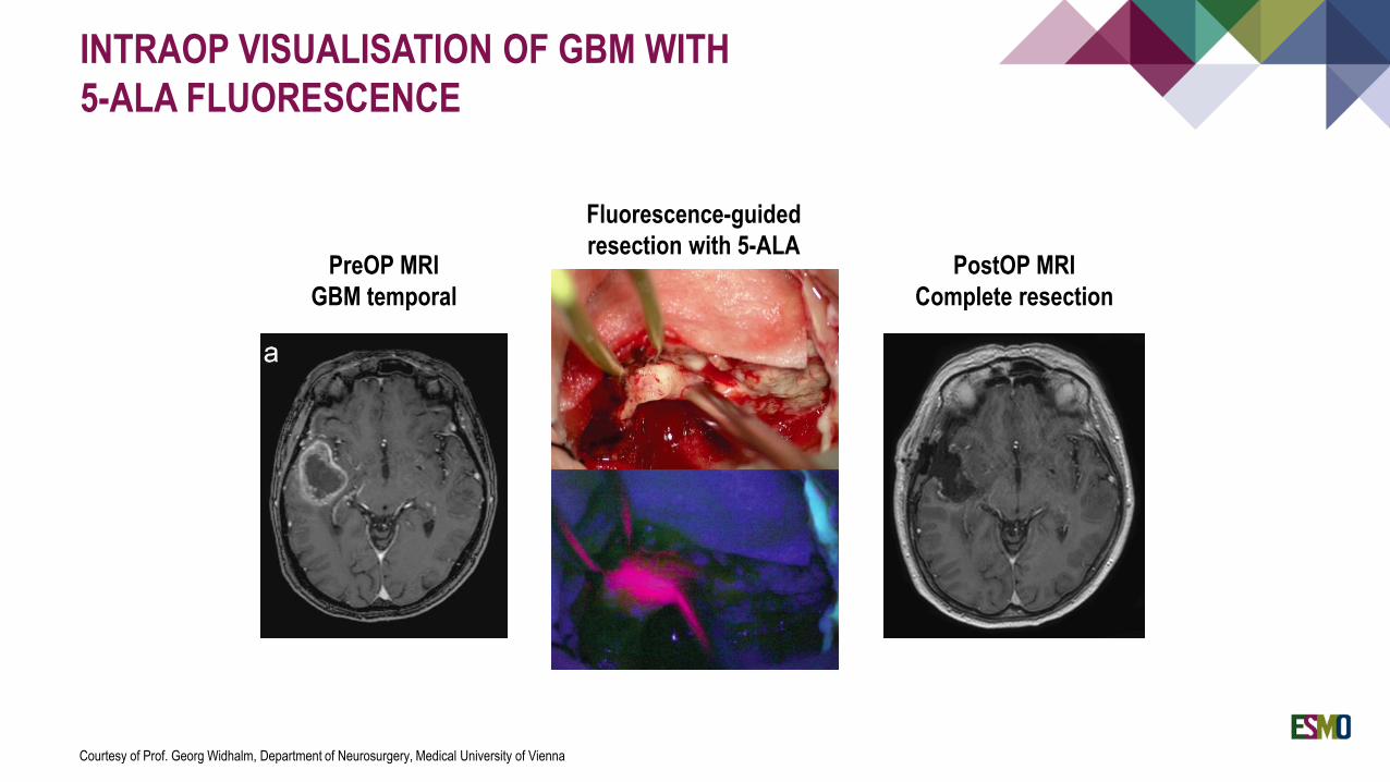
PreOP MRI
GBM temporal
PostOP MRI
Complete resection
Fluorescence-guided
resection with 5-ALA
INTRAOP VISUALISATION OF GBM WITH
5-ALA FLUORESCENCE
Courtesy of Prof. Georg Widhalm, Department of Neurosurgery, Medical University of Vienna

COMBINED CHEMORADIATION
From N Engl J Med, Stupp R, et al. Radiotherapy plus concomitant and adjuvant temozolomide for glioblastoma, 352., 987-996. Copyright © (2005) Massachusetts Medical Society. Reprinted with permission from
Massachusetts Medical Society.
Pro
babi
lity
of o
vera
ll su
rviv
al (
%)
Months
RADIOTHERAPY
Reproduced from: Preusser M, et al. Current concepts and management of Glioblastoma. Ann Neurol 2011;70(1):9–21, with by permission of John Wiley and Sons, Copyright © 2011 American Neurological Association..
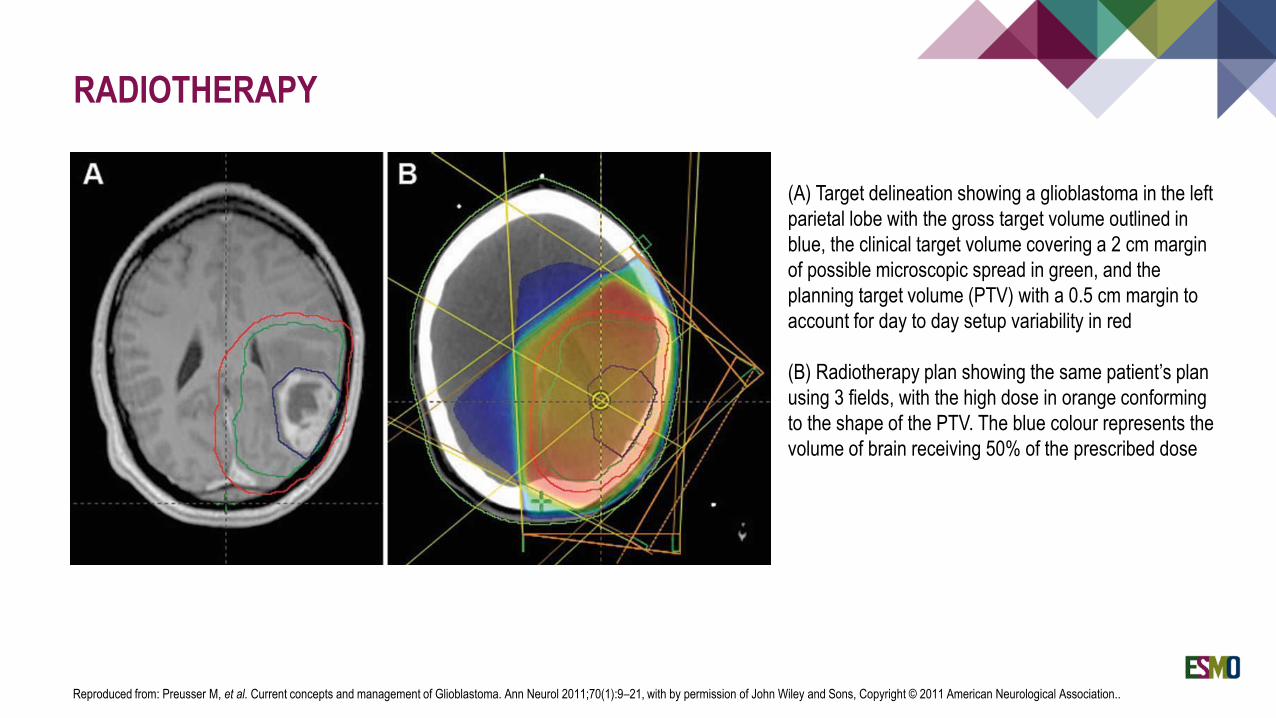
(A) Target delineation showing a glioblastoma in the left
parietal lobe with the gross target volume outlined in
blue, the clinical target volume covering a 2 cm margin
of possible microscopic spread in green, and the
planning target volume (PTV) with a 0.5 cm margin to
account for day to day setup variability in red
(B) Radiotherapy plan showing the same patient’s plan
using 3 fields, with the high dose in orange conforming
to the shape of the PTV. The blue colour represents the
volume of brain receiving 50% of the prescribed dose
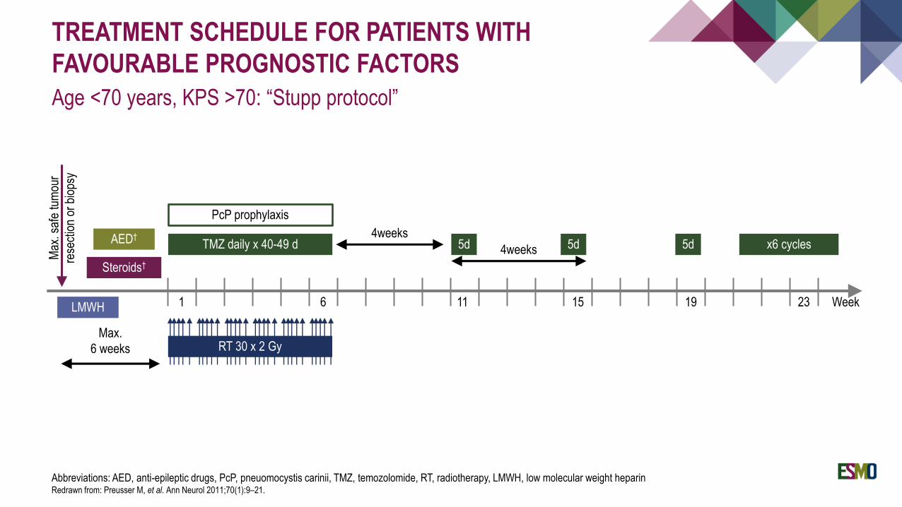
TREATMENT SCHEDULE FOR PATIENTS WITH
FAVOURABLE PROGNOSTIC FACTORS
Age <70 years, KPS >70: “Stupp protocol”
Abbreviations: AED, anti-epileptic drugs, PcP, pneuomocystis carinii, TMZ, temozolomide, RT, radiotherapy, LMWH, low molecular weight heparinRedrawn from: Preusser M, et al. Ann Neurol 2011;70(1):9–21.
Max.
6 weeks
LMWH
Max
. saf
e tu
mou
r
rese
ctio
n or
bio
psy
AED†
Steroids†
1 6 11 15 19 23 Week
4weeks
4weeksTMZ daily x 40-49 d
PcP prophylaxis
5d 5d 5d x6 cycles
RT 30 x 2 Gy

LOMUSTINE-TEMOZOLOMIDE COMBINED WITH
60GY/30 FRACTION RADIATION THERAPY
In patients with newly diagnosed glioblastoma with methylated MGMT promoter
Reprinted from The Lancet, 393, Herrlinger U, et al., Lomustine-temozolomide Combination Therapy Versus Standard Temozolomide Therapy in Patients With Newly Diagnosed Glioblastoma With Methylated MGMT
Promoter (CeTeG/NOA-09): A Randomised, Open-Label, Phase 3 Trial, P678–88, Copyright 2019, with permission from Elsevier..
Caveat: non-definitive data due to limited statistical power

TUMOUR-TREATING FIELDS (TTF) THERAPY IN
PATIENTS WITH NEWLY DIAGNOSED GLIOBLASTOMA
1. Stupp et al, JAMA 2017;318(23):2306–16.
Reproduced with permission from Novocure Inc. © 2020 Novocure - all rights reserved.
TTF device is applied after completion of
concomitant chemo-radiation
Caveat: role of TTF in standard treatment
unclear due to controversies around trial
design and interpretation
Overall survival1
Median survival from randomization:
• 20.9 months for TTF plus temozolomide (n=466) vs.
16.0 months for temozolomide-alone (n=229)
(HR, 0.63; 95% CI: 0.53, 0.76; p<0.001)
• Median follow-up: 44 months (range 25–91 months)
in both groups.

REDUCED CHEMORADIATION REGIMEN
In elderly patients(age >65) with newly diagnosed glioblastoma
From N Engl J Med, Perry JR, et al. Short-Course Radiation plus Temozolomide in Elderly Patients with Glioblastoma, 376:1027–37. Copyright © 2017 Massachusetts Medical Society. Reprinted with permission from
Massachusetts Medical Society..
Overall survival

Time (days)
RADIOTHERAPY VERSUS CHEMOTHERAPY
STRATIFICATION BY MGMT STATUS
In elderly patients (age >60) with newly diagnosed glioblastoma
Reprinted from The Lancet Oncol, 13(7), Wick W, et al. Temozolomide chemotherapy alone versus radiotherapy alone for malignant astrocytoma in the elderly: the NOA-08 randomised, phase 3 trial, 707–15, Copyright 2012,
with permission from Elsevier.
Ove
rall
surv
ival
(%
)

TREATMENT SCHEDULE FOR PATIENTS WITH
FAVOURABLE PROGNOSTIC FACTORS
Age <70 years, KPS >70
Abbreviations: AED, anti-epileptic drugs, PcP, pneuomocystis carinii, TMZ, temozolomide, RT, radiotherapy, LMWH, low molecular weight heparinRedrawn from: Preusser M, et al. Ann Neurol 2011;70(1):9–21.
Max.
6 weeks
LMWH
Max
. saf
e tu
mou
r
rese
ctio
n or
bio
psy
AED†
Steroids†
1 6 11 15 19 23 Week
4weeks
4weeksTMZ daily x 40-49 d
PcP prophylaxis
5d 5d 5d x6 cycles
RT 30 x 2 Gy

RECURRENT GLIOBLASTOMATherapy

PSEUDOPROGRESSION
Wen PY, et al. J Clin Oncol 2010;28(10):1963–972. Reprinted with permission. © 2010 American Society of Clinical Oncology:.
Before surgery After surgery
After radio-chemotherapy After re-surgery, which
showed only necrotic tissue
without tumour

TREATMENT OPTIONS FOR
RECURRENT GLIOBLASTOMA
Neurosurgery
Radiotherapy
Systemic therapy
◆ Nitrosoureas
◆ Temozolomide
◆ Bevacizumab (according to approval status per country)
◆ Other
Clinical trial

PSEUDORESPONSE
Wen PY, et al. J Clin Oncol 2010;28(10):1963–972. Reprinted with permission. © 2010 American Society of Clinical Oncology:.
Before Vascular Endothelial Growth Factor
(VEGF) inhibitor
One day after VEGF inhibitor

TREATMENTSupportive care
ANTI-OEDEMA THERAPY
Results from leakage of plasma into the tissue through disrupted BBB
Detectable on T2-weighted and FLAIR MRI images
Increased intracranial pressure with headache, vertigo, nausea/vomiting
May lead to life-threatening brainstem compression and herniation
Drug of choice: Dexamethasone
◆ Initial daily dose usually 12–16 mg
◆ Steroid dose should be rapidly reduced and tapered to individual need (“as much as needed, as little
as possible”)
Dexamethasone may be combined with osmotic agents such as mannitol or glycerol
Obstructive hydrocephalus may be treated with CSF shunt
Bevacizumab may reduce brain oedema and is associated with decreased corticosteroid need

ANTICONVULSIVE THERAPY
Overview of antiepileptic drugs commonly used in glioblastoma patients
EIAED, enzyme-inducing antiepileptic drugRedrawn from: Preusser M, et al. Ann Neurol 2011;70(1):9–21
EIAED/Non-EIAED Drug Common adverse effects
EIAED Phenobarbital Sedation, rash, impaired cognitive function
Phenytoin Gingival hypertrophy, hirsutism, hepatotoxicity, rash, lymphadenopathy
CarbamazepineDrowsiness, dizziness, diplopia, rash, leukopaenia, hyponatraemia, hepatotoxicity,
nausea/vomiting, cardiac arrhythmia
Oxacarbazepine Drowsiness, dizziness, diplopia, rash, hyponatraemia, hepatotoxicity, nausea/vomiting
Non-EIAED Valproic acid Weight gain, nausea/vomiting, hair loss, thrombocytopenia, hepatotoxicity
Gabapentin Somnolence, dizziness, agitation/anxiety, ataxia
Lamotrigine Somnolence, dizziness, rash, hepatotoxicity
Levetiracetam Drowsiness, fatigue, agitation/anxiety, headache
Pregabalin Somnolence, dizziness, weight gain, ataxia

OUTLOOK: SELECTED ONGOING TRIALS
Proteasome-inhibitor marizomib
CDK-inhibitor TG02
Targeted treatment based on molecular profiling (e.g. tumour mutational burden, BRAF mutations, NTRK fusions,
FGFR fusions, MET amplifications/fusions)

THERAPEUTIC APPROACH TO GLIOBLASTOMA
*Additional treatment with tumour-treating fields (TTFs) may be offered to eligible patients. **Depending on availability and approval status.
GTR, gross total resection; IDH, isocitrate dehydrogenase; KPS, Karnofsky performance score; MGMT, O6-methylguanine DNA methyltransferase; RT, radiation therapy; TMZ, temozolomide.From Weller M, et al. ESMO Open 2019;4(Suppl 2):e000520. Copyright © European Society for Medical Oncology. All rights reserved. Reproduced under the Creative Commons Attribution Non Commercial (CC BY-NC 4.0) license
(http://creativecommons.org/licenses/by-nc/4.0/, accessed March 2020)
Treatment algorithm for glioblastoma
Therapeutic approach
to glioblastoma

TREATMENT SCHEDULE FOR PATIENTS WITH
FAVOURABLE PROGNOSTIC FACTORS
Age <70 years, KPS ≥70
Abbreviations: AED, anti-epileptic drugs, PcP, pneuomocystis carinii, TMZ, temozolomide, RT, radiotherapy, LMWH, low molecular weight heparinRedrawn from: Preusser M, et al. Ann Neurol 2011;70(1):9–21.
Max.
6 weeks
LMWH
Max
. saf
e tu
mou
r
rese
ctio
n or
bio
psy
AED†
Steroids†
1 6 11 15 19 23 Week
4weeks
4weeksTMZ daily x 40-49 d
PcP prophylaxis
5d 5d 5d x6 cycles
RT 30 x 2 Gy

SUMMARY
Glioblastoma is the most common primary brain tumour of adults
High morbidity and mortality
Standard first line therapy: maximal safe resection and combined chemoradiation with temozolomide
To be considered in elderly patients: reduced chemoradiation or stratification by MGMT promoter methylation status
into radiotherapy (MGMT unmethylated or unknown) versus temozolomide (MGMT methylated)
Recurrent glioblastoma commonly treated with CCNU (lomustine)
Supportive therapy with anti-oedema and anticonvulsive therapy of importance in most patients
Trials for newly diagnosed and recurrent glioblastoma ongoing

THANK YOU!