epistaxis 2016: national audit of management
TRANSCRIPT
University of Plymouth
PEARL https://pearl.plymouth.ac.uk
01 University of Plymouth Research Outputs University of Plymouth Research Outputs
2017-12-27
Epistaxis 2016: national audit of
management
INTEGRATE
http://hdl.handle.net/10026.1/10618
10.1017/S002221511700202X
Journal of Laryngology and Otology
Cambridge University Press (CUP)
All content in PEARL is protected by copyright law. Author manuscripts are made available in accordance with
publisher policies. Please cite only the published version using the details provided on the item record or
document. In the absence of an open licence (e.g. Creative Commons), permissions for further reuse of content
should be sought from the publisher or author.
EPISTAXIS 2016: NATIONAL AUDIT OF MANAGEMENT INTEGRATE (The National ENT Trainee Research Network)
Steering Committee: M. Ellis, A. Hall, J. Hardman, N. Mehta, M. E. Smith, R. J. Williams*
Executive Committee: S. Carrie, C. Hopkins
Statisticians: J. Chynoweth, B. G. Jones, K Stevens
Site Leads: Y. Abbas, Southend Hospital; M. Abdelkader, Basildon and Thurrock Hospital; M. Adams, Altnagelvin; A. Addison, James Paget Hospital; R. Advani,
North Manchester General Hospital; T. Ahmed, Dorset County Hospital; V. Alexander, Brighton General Hospital; V. Alexander, Royal Sussex County Hospital; B. Alli, Bradford Royal Infirmary; S. Alvi, Leighton Hospital; N. Amiraraghi, Crosshouse Hospital; A. Ashman, The Great Western Hospital; R. Balakumar,
Gloucestershire Royal Hospital; J. Bewick, Addenbrookes Hospital; D. Bhasker, Pinderfields General Hospital; S. Bola, Wexham Park Hospital; P. Bowles, Worthing Hospital; N. Can Guru Naidu, Barnet Hospital; N. Caton, East Surrey Hospital; J. Chapman, Salisbury District Hospital; G. Chawdhary, Milton Keynes General
Hospital; M. Cherko, John Radcliffe Hospital; M. Coates, Sunderland Royal Hospital; K. Conroy, Wythenshawe Hospital, Macclesfield District General Hospital; P. Coyle, Peterborough City Hospital; O. Cozar, University Hospital of North Staffordshire; M. Cresswell, Derriford Hospital; L. Dalton, Arrowe Park; J. Danino, New
Cross Hospital; C. Daultrey, Worcester Royal Hospital; K. Davies, Morriston Hospital; K. Davies, Royal Liverpool University Hospital; D. Dick, Royal Victoria Hospital; P. Dimitriadis, Royal Hallamshire Hospital; N. Doddi, Princess of Wales Hospital; M. Dowling, Stepping Hill Hospital; R. Easto, Monklands Hospital; r. edmiston, Royal Albert Edward Infirmary; D. Ellul, Western General Hospital; S. Erskine, West Suffolk Hospital; A. Evans, Barnsley District General Hospital; A. Farboud, University Hospital of Wales; C. Forde, King George Hospital; J. Fussey, Walsall Manor Hospital; A. Gaunt, Queens Medical Centre; J. Gilchrist, Royal
Bolton Hospital; R. Gohil, New Royal Infirmary of Edinburgh; E. Gosnell, Royal Blackburn Hospital; D. Grech Marguerat, Royal Cornwall Hospital; R. Green, Ninewells Hospital; R. Grounds, Medway Hospital; A. Hall, Royal National TNE Hospital; J. Hardman, St. Marys Hospital; A. Harris, Royal Gwent Hospital; L. Harrison, Northampton General Hospital; R. Hone, Royal Surrey County Hospital; E. Hoskison, University Hospital of Coventry & Warwickshire; J. Howard,
Wrexham Maelor Hospital; D. Ioannidis, Royal Hampshire County Hospital; I. Iqbal, Freeman Hospital; N. Janjua, Queen Alexandra Hospital; k. jolly, Russells Hall Hospital; S. Kamal, Poole Hospital; T. Kanzara, Countess of Chester; N. Keates, Torbay Hospital; A. Kelly, Antrim Area Hospital; H. Khan, Fairfield General Hospital; T. Korampalli, Rotherham District General Hospital; M. Kuet, Colchester General Hospital; P. Kulloo, Royal London Hospitals; R. Lakhani, St Georges Hospital; A. Lambert, Charing Cross Hospital; C. Leonard, Royal Belfast Hospital for Sick Children; G. Lloyd, Guys hospital; E. Lowe, Southampton General Hospital; J. Mair, Birmingham Heartlands Hospital; E. Maughan, University College Hospital London; T. Mayberry, Queen Elizabeth Hospital - University Hospitals Birmingham
NHSFT; L. McCadden, Craigavon Area Hospital; F. McClenaghan, West Middlesex University Hospital; G. McKenzie, Hull Royal Infirmary, Castle Hill Hospital; R. Mcleod, West Wales Hospital; S. Meghji, Norfolk & Norwich University Hospital; M. Mian, Furness General Hospital; A. Millington, Ipswich Hospital NHS Trust; O. Mirza, Royal Preston Hospital; S. Mistry, Calderdale Royal Hospital; A. Mitchell-Innes, Birmingham Childrens Hospital; E. Molena, Frimley Park Hospital; J. Morris, Royal United Hospital; T. Myuran, Basildon Hospital, Princess Alexandra Hospital; A. Navaratnam, Northwick Park Hospital; E. Noon, Blackpool Victoria Hospital; O. Okonkwo, Salford Royal Hospital; B. Oremule, Royal Lancaster Hospital; L. Pabla, James Cook University Hospital; E. Papesch, Broomfield Hospital; V. Pattni,
Bristol Royal Infirmary; V. Puranik, Ysbyty Gwynedd Hospital; R. Roplekar, Raigmore Hospital; E. Ross, Birmingham City Hospital; E. Schechter, Luton & Dunstable Hospital; A. Senior, Midlands Treatment Centre (Burton); N. Sethi, Leeds General Infirmary; S. Sharma, Central Middlesex Hospital; R. Sharma, St Johns Hospital;
Z. Sherazi, Tameside General Hospital; A. Tahir, Cumberland Infirmary; T. Tikka, Queen Elizabeth University Hospital; O. Tkachuk Hlinicanova, Glan Clwyd Hospital; K. To, Royal Hospital for Sick Children Edin; E. Toll, Royal Devon & Exeter Hospital; K. Ubayasiri, Royal Derby Hospital; S. Unadkat, Whipps Cross
University Hospital; N. Upile, University hospital of Aintree; A. Vijendren, Lister Hospital; H. Walijee, Alder Hey; M. Williams, Darlington Memorial Hospital; G. Wilson, Leicester Royal Infirmary; W. Wong, York District Hospital; G. Wong, North Hampshire Hospital; C. Xie, Princess Royal University Hospital; A. Yao,
Manchester Royal Infirmary; H. Zhang, Queens Hospital;
* = lead author and steering committee chair
Corresponding author: Mr Richard Williams Institute of Naval Medicine, Crescent Rd, Alverstoke, Gosport. PO12 2DL [email protected] Conflicts of interest: None declared Key words: Epistaxis; assessment; cautery; packing; haematology; surgery. Funding: This national audit was funded by ENTUK. The funding body had no influence over content.

ABSTRACT
Background: Epistaxis is a common condition that can be associated with significant morbidity and places a
considerable burden on our healthcare system. This national audit of management seeks to assess current
practice against newly created consensus recommendations and expand our current evidence base.
Method: Patients that met inclusion criteria at 113 registered sites across the UK, had their management
compared with audit standards during a 30-day window. Data was further utilised for explorative analysis.
Results: 1826 cases were uploaded to the database, representing 94% of all cases that met the inclusion
criteria at participating sites. 62% of patients were successfully treated by ENT clinicians within 24 hours
and the 30-day recurrent presentation rate across the dataset was 13.9%. Significant event analysis
revealed an all-cause 30-day mortality rate of 3.4%.
Conclusion: Audit findings demonstrate a varying alignment with consensus guidance with explorative
analysis countering some previously well-established tenets of management.
INTRODUCTION
The Care Quality Commission highlights ‘Good Governance’ as a fundamental standard of care. Central to
this, clinical audit seeks to assess the quality and safety of this care. Whilst local audit is well established in
otorhinolaryngology, there has been a paucity of national centrally-delivered audit within our specialty
since the National Prospective Tonsillectomy Audit(1) and the National Audit of Sino-nasal Surgery(2)
published in 2005 and 2006 respectively. ENT-UK and the British Rhinological Society (BRS) sought to
address this deficiency by challenging INTEGRATE (the National ENT Trainee Research Network) to design
and deliver a national audit of management for the hospital treatment of epistaxis.
Epistaxis is the most common acute disorder managed by ENT services in the UK, with around 25,000 acute
presentations to NHS hospitals each year (http://www.hscic. gov.uk/ accessed 11/07/17) . Despite this high
incidence, prior to this initiative there were no nationally accepted guidelines for its management. A pilot
audit(3) led by INTEGRATE has confirmed significant variation in existing treatment between hospital trusts.
This audit aims to compare current management against agreed consensus management guidelines(4),
identify variation in practice and perform exploratory analysis of the dataset to expand our current

understanding of this common condition. As a result of this audit, we seek to generate a program of
change to deliver improved and standardised evidence-based hospital care of epistaxis across the UK.
METHOD
Organisation and design: This audit was designed and delivered by INTEGRATE following the creation of a
project steering committee comprising of 6 trainees and 2 consultant executive members. One trainee was
nominated to chair the steering committee and co-ordinate the audit.
Audit standards: Following a comprehensive and systematic review of the literature(5-9), national consensus
recommendations for the hospital management of epistaxis were developed(4). These recommendations
were utilised to generate a draft data-collection tool (DCT) for the audit including 30-day outcome data.
The draft DCT was adapted in line with lessons learnt from a multi-centre pilot audit(3) before undergoing
multi-level scrutiny, initially by the audit steering committee and subsequently by the ENT-UK executive
committee. The agreed DCT was then optimised by commissioned statisticians at the Peninsula Clinical
Trials Unit (PenCTU) for ease of subsequent analysis following data collection.
Audit period: A 30-day data audit window was identified from midnight on the 7th of November 2016.
Inclusion/Exclusion: During the audit window, patients ≥16yrs old presenting emergently, as an
unscheduled event with a diagnosis of epistaxis that were subsequently managed by ENT services were
included in the audit. This included patients referred from the Emergency Department (ED), primary care
or from other specialties within participating units. Patients attending pre-arranged appointments for the
management of chronic self-limiting epistaxis were excluded, as well as patients seen and treated by
Emergency Department staff without referral to ENT services. Telephone encounters were not included.
Collaborator Engagement: Site-leads were recruited from ENT departments throughout the UK via a
network of regional trainee representatives, engagement with the Specialist Advisory Committee and
through open advertisement via the Association of Otolaryngologists in Training. Site-leads were given on-
line supporting material to aid in the local delivery of the audit and were invited to attend a launch event
prior to the audit window to further clarify the audit process. Throughout local preparation, site-leads
were requested to complete an on-line task list detailing their progress to allow the steering committee to
maintain a strategic overview of the project.
Data collection: Cases meeting the inclusion and exclusion criteria were identified prospectively at the
point of presentation. Throughout the audit window, ED and ENT practitioners were encouraged to make
detailed clinical notes following all epistaxis related clinical encounters in line with good medical
practice(10) guidance. Audit proformas were not utilised by treating clinicians to prospectively record
clinical information due to concerns regarding their impact on pre-existing standard practice. At the point
of discharge audit data was gathered by collaborators from hospital notes, uploaded via a web-based
portal and stored in the Data Safe Haven on a secure server hosted at University College London. The
server was certificated to the ISO27001 information security standard and conforms to the NHS
Information Governance Toolkit. All connections to the Data Safe Haven server via the web portal were
secured with 256-bit SHA encryption.
Following closure of the 30-day window a clinical coding search was conducted in all participating units to
highlight all epistaxis presentations during the audit period to established whether any cases had been
missed. All cases were examined against inclusion/exclusion criteria and then cross referenced with the
cases uploaded to the database. Where cases were missed, hospital notes were requested and if possible,
added retrospectively to the database. Any cases identified retrospectively were highlighted as such for
the purposes of data analysis. 30-day outcome data was gathered for all uploaded cases via note retrieval
through local audit departments. Units were also asked to provide trust quoted population at risk data. No
identifiable patient data was uploaded to the server, all submissions were anonymised using an audit
specific key for each patient devised and stored locally at each site in line with the clinical governance
policy at each site.
Management of data submission and quality: The audit steering committee utilised a dedicated online
platform (https://www.entintegrate.org) to facilitate communication between collaborators, to rapidly
respond to potential problems and to support timely and high-quality data submission. On completion of
data submission, steering committee members inspected submitted data for errors, duplication or
omissions. Where necessary, site-leads were contacted to remedy identified discrepancies.
Audit and ethical approval: Prior to data collection, an audit proposal was submitted for approval to the
audit departments of all participating units, in line with local policy. Caldicott Guardians were contacted at
all sites and approval sought for the method of data collection. Although formal patient consent was
neither sought nor required, patient information regarding the audit was displayed at all participating sites
and individuals invited to be excluded from the audit if they desired. Many evidence gaps were identified
during the consensus recommendations process for epistaxis management(ref). Therefore, we sought NHS
Research Ethics Committee (NREC) guidance regarding the use of the dataset beyond comparison against
identified audit standards. Completion of the Health Research Authority Guidance Tool confirmed formal
NREC approval was not required for this purpose.
Statistical Analysis Strategy: When presenting our analysis, we define length of stay (LOS) as the time from
hospital presentation to discharge, which reflects the entire treatment process. We define time to ENT as
the time from presentation through to first ENT review. Haemostasis time (HT) is the time from first ENT
review to when final haemostasis is achieved prior to discharge. A limitation of the data set is that it is
unknown whether the patient was actively bleeding on arrival to ENT, however, all patients had been
referred for emergency ENT input. We define discharge time as the time from final haemostasis to
discharge. Each interval was calculated by gathering data on the absolute date and time of each event.
Additionally, we will discuss rates of 30-day recurrent presentation (RP), defined as representations to
hospital with epistaxis in the 30 days following the point of initial presentation. The objective of the
statistical analysis was primarily to assess compliance with national consensus recommendations that
formed our audit standards, and thereafter, to identify which patient factors and treatments (both prior to
ENT and at the first ENT review) were associated with variation in outcome in terms of HT and RP.
During the exploratory analysis, it was determined that HT was positively skewed and multimodal as there
were two modes (i.e. peaks in the data). To compensate for the skewed distribution of the data, a log
transformation was applied to HT. Plots of HT are presented in log hours, but summary statistics are given
in hours unless otherwise stated. Although this provides visual representation to observed differences in
treatment, it was unable to account for all potential confounding factors. A future study will conduct
inferential analysis of the dataset via regression modelling, which will provide us with the tools to account
for associations with multiple treatments and potential confounders.
RESULTS
The audit was conducted simultaneously in 113 hospitals from all regions of the UK serving a combined
population at risk of 51 million people. 1826 cases were uploaded, of which 1358 were gathered
prospectively, in total this represented 94% of all cases presenting to involved units that met inclusion
criteria. The median number of cases uploaded per unit was 14 (range 1-50). Scrutiny of the dataset
discovered 89 cases did not meet inclusion criteria and 157 cases did not report a HT and so were excluded
from analysis. Follow-up data was available for 1469 patients who could be matched with the audit data.
Once excluding those not within the audit period and undertaking relevant data manipulation, we were
left with 1152 patients. 30 patients had a HT of 0 suggesting ED treatment had successfully treated
patients without ENT input. Consequently, these patients were excluded leaving 1122 patients for
analysis. The RP rate for these patients was 24.1%, compared with 13.9% for the larger group of 1122
patients. Whilst the difference appears substantial, any conclusions drawn from only 30 patients should be
treated cautiously.
The median age was 73 years, the inter-quartile range (IQR) was 62-82 years and the range was 16 - 100
years. with larger proportion of males versus females (56% vs 44%). The median LOS was 29.5 hours with
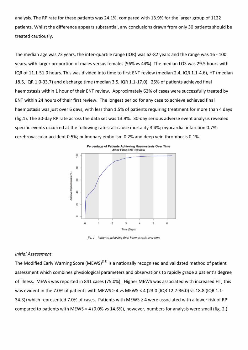
IQR of 11.1-51.0 hours. This was divided into time to first ENT review (median 2.4, IQR 1.1-4.6), HT (median
18.5, IQR 1.0-33.7) and discharge time (median 3.5, IQR 1.1-17.0). 25% of patients achieved final
haemostasis within 1 hour of their ENT review. Approximately 62% of cases were successfully treated by
ENT within 24 hours of their first review. The longest period for any case to achieve achieved final
haemostasis was just over 6 days, with less than 1.5% of patients requiring treatment for more than 4 days
(fig.1). The 30-day RP rate across the data set was 13.9%. 30-day serious adverse event analysis revealed
specific events occurred at the following rates: all-cause mortality 3.4%; myocardial infarction 0.7%;
cerebrovascular accident 0.5%; pulmonary embolism 0.2% and deep vein thrombosis 0.1%.
fig. 1 – Patients achieving final haemostasis over time
Initial Assessment:
The Modified Early Warning Score (MEWS)(11) is a nationally recognised and validated method of patient
assessment which combines physiological parameters and observations to rapidly grade a patient’s degree
of illness. MEWS was reported in 841 cases (75.0%). Higher MEWS was associated with increased HT; this
was evident in the 7.0% of patients with MEWS ≥ 4 vs MEWS < 4 (23.0 (IQR 12.7-36.0) vs 18.8 (IQR 1.1-
34.3)) which represented 7.0% of cases. Patients with MEWS ≥ 4 were associated with a lower risk of RP
compared to patients with MEWS < 4 (0.0% vs 14.6%), however, numbers for analysis were small (fig. 2.).
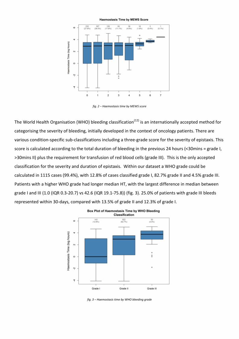
fig. 2 – Haemostasis time by MEWS score
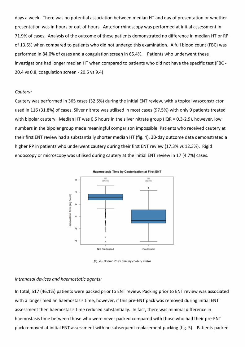
The World Health Organisation (WHO) bleeding classification(12) is an internationally accepted method for
categorising the severity of bleeding, initially developed in the context of oncology patients. There are
various condition-specific sub-classifications including a three-grade score for the severity of epistaxis. This
score is calculated according to the total duration of bleeding in the previous 24 hours (<30mins = grade I,
>30mins II) plus the requirement for transfusion of red blood cells (grade III). This is the only accepted
classification for the severity and duration of epistaxis. Within our dataset a WHO grade could be
calculated in 1115 cases (99.4%), with 12.8% of cases classified grade I, 82.7% grade II and 4.5% grade III.
Patients with a higher WHO grade had longer median HT, with the largest difference in median between
grade I and III (1.0 (IQR 0.3-20.7) vs 42.6 (IQR 19.1-75.8)) (fig. 3). 25.0% of patients with grade III bleeds
represented within 30-days, compared with 13.5% of grade II and 12.3% of grade I.
fig. 3 – Haemostasis time by WHO bleeding grade

The initial ENT assessment was predominantly performed by junior-grade doctors (n=950, 86.6%) followed
by middle-grade doctors (n=107, 9.2%), nurse practitioners (n=38, 3.5%) and consultants (n=8, 0.7%) (tab.
1). For each WHO bleed grade, specialist nurses have the lowest median HT. However, the number of
patients seen by specialist nurses and consultants was very small. Therefore, it is unlikely that the
estimates for median HT are representative or reliable. Neither a nurse specialist nor consultant saw a
patient with a MEWS > 4, 95% of these patients were attended to by junior grade doctors. Although the
evidence suggested the nurses tended to have a lower HT, the 30-day recurrent rates were notably higher.
RP for patients seen by nurse practitioners was 21.6%, compared to junior-grades with 13.6%, middle-
grades with 14.6% and consultants with 14.3%.
Practitioner seniority
WHO I WHO II WHO III
Median (IQR) n Median (IQR) n Median (IQR) n
Consultant 0.3 1 5.5 (1.4 - 25.0) 7 - 0
Junior grade 1.0 (0.3-20.3) 122 19.2 (2.0 - 34.7) 781 42.6 (22.4, 72.1) 42
Middle grade 3.5 (1.5 - 20.3) 9 23.8 (1.4 - 37.2) 84 75.9 (31.6 - 81.2) 6
Nurse specialist 0.7 (0.5 - 5.3) 6 1.0 (0.4 - 26.7) 31 8.0 1 Tab.1 Median homeostasis time in hours (IQR) grouped by severity of bleeding and seniority of practitioner
Regarding co-morbidities, a past medical history of hypertension (formally diagnosed through sustained
ambulatory blood pressure monitoring) was reported in 55% of cases. The median HT in these patients
was 20.3 (IQR 1.5-36.0) compared with 15.2 in those without a history of hypertension (IQR 0.6-29.7) and
RP rates were 14.0% vs 13.8% respectively. 26.0% of cases reported previous epistaxis presentations with
in the preceding year. Median HT was 21.3 (IQR 1.3-37.2) in patients declaring previous bleeds vs 17.7
(IQR 0.9-31.3) who did not, RP was nearly double in these patients (20.2% vs 12.1%). Patients who
subsequently represented within 30-days of discharge had a shorter median HT during their initial
treatment (16.5 vs 18.6). 156 (14.4%) individuals had a past medical history of diabetes mellitus and 333
(30.4%) had ischaemic heart disease. However, neither condition demonstrated a potential association
with varying haemostasis time. The RP was greater in those with a history of diabetes mellitus (21.4% vs
12.3%) and also greater in those with a history of ischaemic heart disease (14.9% vs 13.0%).
A history of previous epistaxis presentation within the preceding year was reported in 26.0% of cases.
Patients declaring previous bleeds had a longer median HT of 21.3 (IQR 1.3-37.2) compared to 17.7 (IQR
0.9-31.3) for those who did not. The RP was also greater in these patients at 20.2% compared to 12.1%. In
the subset of patients who subsequently represented with a second episode of epistaxis within 30-days of
their primary presentation, the subsequent median HT was found to be longer (16.5 vs 18.6 hours).
Data were collected regarding time of presentation to investigate any associations with out of hours
treatment. Working hours were defined as 0800-1700 and out of hours were defined as 1700-0800, seven
days a week. There was no potential association between median HT and day of presentation or whether
presentation was in-hours or out-of-hours. Anterior rhinoscopy was performed at initial assessment in
71.9% of cases. Analysis of the outcome of these patients demonstrated no difference in median HT or RP
of 13.6% when compared to patients who did not undergo this examination. A full blood count (FBC) was
performed in 84.0% of cases and a coagulation screen in 65.4%. Patients who underwent these
investigations had longer median HT when compared to patients who did not have the specific test (FBC -
20.4 vs 0.8, coagulation screen - 20.5 vs 9.4)
Cautery:
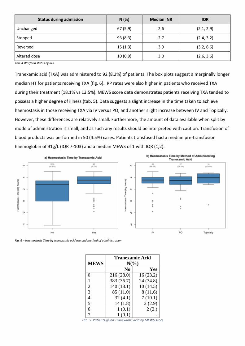
Cautery was performed in 365 cases (32.5%) during the initial ENT review, with a topical vasoconstrictor
used in 116 (31.8%) of cases. Silver nitrate was utilised in most cases (97.5%) with only 9 patients treated
with bipolar cautery. Median HT was 0.5 hours in the silver nitrate group (IQR = 0.3-2.9), however, low
numbers in the bipolar group made meaningful comparison impossible. Patients who received cautery at
their first ENT review had a substantially shorter median HT (fig. 4). 30-day outcome data demonstrated a
higher RP in patients who underwent cautery during their first ENT review (17.3% vs 12.3%). Rigid
endoscopy or microscopy was utilised during cautery at the initial ENT review in 17 (4.7%) cases.
fig. 4 – Haemostasis time by cautery status
Intranasal devices and haemostatic agents:
In total, 517 (46.1%) patients were packed prior to ENT review. Packing prior to ENT review was associated
with a longer median haemostasis time, however, if this pre-ENT pack was removed during initial ENT
assessment then haemostasis time reduced substantially. In fact, there was minimal difference in
haemostasis time between those who were never packed compared with those who had their pre-ENT
pack removed at initial ENT assessment with no subsequent replacement packing (fig. 5). Patients packed

prior to ENT review and patients packed at initial ENT review had a lower RP when compared with patients
who were never packed (packed prior to ENT = 12.8% vs 14.9%, packed by ENT = 9.4% vs. 15.5%). 49 (6.4%)
of patients who were packed received antibiotics upon discharge.
fig. 5 – Haemostasis time by packing status
The type of packing used was classified into one of five categories: inflatable (e.g. Rapid rhino), non-
dissolvable (e.g. Merocel), dissolvable (e.g. Nasopore), urinary catheter or haemostatic agent (e.g.
Kaltostat). 75.4% of the patients packed prior to ENT received an inflatable packing (tab. 2). The median
and inter quartile range for HT by pack type shows no difference between inflatable and non-dissolvable
packs used prior to and by ENT. The number of patients packed with dissolvable, catheter and haemostatic
packs was too small to gain useful insight.
Consultation Pack type n (%) Median HT (IQR)
Prior to first ENT review
Inflatable 390 (74.7) 23.4 (150, 36.9) 14.1%
Non dissolvable 128 (24.5) 23.3 (15.7, 37.9) 9.1%
At first ENT review
Inflatable 250 (83.3) 30.6 (20.0, 46.1) 8.2%
Non-dissolvable 32 (10.7) 28.8 (19.5, 47.5) 6.9%
Dissolvable 14(4.7) 11.3 (1.1, 29.4) 33.3%
Catheter 4 (1.3) 41.3 (16.4, 75.5) 25.0%
Tab. 2 Type of packs used and HT. Prior to the first ENT review there were also cases with dissolvable packs (n=2), catheter (n=1) and haemostatic agent (n=1).
Of those packed with inflatable or non-dissolvable packs, the median number of packs placed per patient
was 1 (IQR 1-2, range 1-6). Patients requiring multiple packs were associated with an HT of 32.3 (IQR 20.3-
48.5) versus those with a single pack of 21.5 (IQR 12.5-34.5) and a RP of 9.7% vs 13.8%. Cautery was
performed in 295 (38.8%) of packed patients following removal of their final pack. The RP was lower in this

cautery group at 10.9% versus 12.9% amongst those packed but not cauterized following removal. The
median time to discharge from removal of final pack was 4.1 hours (IQR 1.5-16.0 hours).
Haematological factors:
In total 572 (51.0%) of the patients were on some form of antithrombotic medication (tab.3). Patients not
on antithrombotic medication had a shorter median HT than those on anti-thrombotic medication (17.0
(IQR 0.9-32.9) vs 19.3 (IQR 1.3-34.7)). Patients whose medication was stopped or reversed had a prolonged
median HT compared to those who continued some form of treatment, this was the case across all
medication types. The cessation of antiplatelet agents was associated with lower RP as was the cessation
or reversal of warfarin, however, this was not seen when DOACs were withheld. The International
Normalised Ratio (INR) of patients who had their warfarin reversed was higher than any other group of
patients (tab. 4). Median HT for patients taking warfarin was longer than patients taking DOACs. The
median HT for patients on warfarin was 23.3 (IQR 4.9-39.0) vs oral anticoagulants 18.4 (IQR 1.6-29.0). The
RP for all patients taking warfarin compared with oral anticoagulants was similar at 14.9% vs. 14.3%.
Medicaton Status during admission
n (%) Median HT (IQR) RP (%)
Aspirin Unchanged 157 (14.0) 12.5 (0.4, 26.0) 11.7
Stopped or 53 (4.7) 25.1 (17.5, 36.5) 6.5
Altered 1 (0.1)
Clopidogrel Unchanged 67 (6.0) 17.5 (0.4, 38.0) 14.5
Stopped 33 (3.0) 25.2 (16.5, 38.3) 9.7
Altered 2(0. 1)
Heparins Unchanged 18 (1.6) 2.8 (0.5, 26.6) 18.8
Stopped 6 (0.5) 44.1 (20.0, 50.6) 0.0
Altered 2 (0.2)
DOAC Unchanged 65 (5.8) 2.9 (0.5, 21.3) 12.3
Stopped 74 (6.6) 23.4 (13.7, 37.9) 14.5
Altered 3 (0.3) 20.8 (10.6, 35.1) 33.3
Warfarin Unchanged 85 (7.1) 11.0 (0.4, 27.3) 24.3
Stopped 105 (9.4) 28.8 (18.0, 42.4) 9.8
Reversed 16 (1.4) 36.9 (23.8, 49.4) 0.0
Altered 11 (1.0) 19.0 (0.9, 33.6) 12.5
Tab. 3 Table showing modifications to antithrombotic medications against HT and RP
Status during admission N (%) Median INR IQR
Unchanged 67 (5.9) 2.6 (2.1, 2.9)
Stopped 93 (8.3) 2.7 (2.4, 3.2)
Reversed 15 (1.3) 3.9 (3.2, 6.6)
Altered dose 10 (0.9) 3.0 (2.6, 3.6)
Tab. 4 Warfarin status by INR
Tranexamic acid (TXA) was administered to 92 (8.2%) of patients. The box plots suggest a marginally longer
median HT for patients receiving TXA (fig. 6). RP rates were also higher in patients who received TXA
during their treatment (18.1% vs 13.5%). MEWS score data demonstrates patients receiving TXA tended to
possess a higher degree of illness (tab. 5). Data suggests a slight increase in the time taken to achieve
haemostasis in those receiving TXA via IV versus PO, and another slight increase between IV and Topically.
However, these differences are relatively small. Furthermore, the amount of data available when split by
mode of administration is small, and as such any results should be interpreted with caution. Transfusion of
blood products was performed in 50 (4.5%) cases. Patients transfused had a median pre-transfusion
haemoglobin of 91g/L (IQR 7-103) and a median MEWS of 1 with IQR (1,2).
Fig. 6 – Haemostasis Time by tranexamic acid use and method of administration
MEWS
Tranexamic Acid
N(%)
No Yes
0 216 (28.0) 16 (23.2)
1 383 (36.7) 24 (34.8)
2 140 (18.1) 10 (14.5)
3 85 (11.0) 8 (11.6)
4 32 (4.1) 7 (10.1)
5 14 (1.8) 2 (2.9)
6 1 (0.1) 2 (2.)
7 1 (0.1) - Tab. 5. Patients given Tranexamic acid by MEWS score
Surgery and Radiological Intervention:
36 (3.2%) of included patients underwent surgery for epistaxis during the audit period. The median time
between first ENT review and surgery was 24.7 and IQR (8.9, 54.2). Prior to surgery, these patients had a
median number of pre-op non-dissolvable pack (inflatable + non-dissolvable) placements of 1 (IQR 1-2,
range 0-6). Patients undergoing surgery had a median MEWS of 1 and WHO grade of 2, this was the same
as patients in the non-surgery group. Data were available on the grade of principal surgeon grade for 34 of
the 36 patients with consultants operating most commonly (52.9%) followed by registrars (44.1%).
Operations performed were sphenopalatine artery ligation (n=19), sphenopalatine artery cautery (n=10),
electrocautery of bleeding point (n=8), septoplasty (n=4), anterior ethmoid artery ligation (n=3) and
antrostomy (n=1) with multiple operations being performed in some cases. The number of patients
achieving final haemostasis at time of surgery was 17 (47.2%) and the RP rate of surgical patients was
22.6%. 3 patients underwent radiological intervention with the low numbers preventing detailed analysis.
All underwent intervention 2 days following first ENT review. The maxillary artery was embolised in 2
cases and isolated sphenopalatine artery embolisation was performed in 1 case.
DISCUSSION & LIMITATIONS
This large multicentre audit sought to benchmark current epistaxis management, enhance the evidence-
base and showcase trainee collaborative research as a strategy for large volume data collection. The
dataset successfully captured the vast majority of epistaxis presentations within involved units. Data
quality in general was high, however, it was limited by reliance on the detailed note taking of clinicians. It
remains unclear whether missing data were due to deficiencies in management, note taking or data
collection. Incomplete data necessitated the exclusion of a number of cases from analysis, which had
otherwise met the audit inclusion criteria. This explorative analysis was able to identify potential
associations between patient factors, treatment factors and outcomes. Further inferential statistical
analysis is planned, in a subsequent paper, to correct for confounding variables and modelling and identify
optimal treatment strategies. Without this additional analysis, suggested associations noted within the
dataset thus far should be interpreted with caution.
The large number of cases identified in this study highlights the considerable burden epistaxis places on
our healthcare system, both at initial presentation and in the 13.9% of cases which represented to hospital
attendance. The majority of cases were successfully treated within 24 hrs, suggesting a limited impact of
disease on these individuals. However significant event analysis revealed a 3.4% 30-day all-cause mortality
rate within our dataset. In context, the equivalent 30-day all-cause mortality rate for hip fracture in the UK
is 7.1% ( http://www.nhfd.co.uk/nhfd/nhfd2015reportPR1.pdf / accessed 11/07/2017), a figure that has
reduced from 10.9% over 8 years since the introduction of a program of ongoing national audit. Whilst
such figures should be interpreted with caution, and it is acknowledged that epistaxis may not be the
condition directly leading to death in these cases, it highlights the level of morbidity held amongst the
population of patients we treat.
Assessment of auditable standards within the domain of initial assessment demonstrated high levels of
adherence for the recording of key elements of the history, evidenced to effect outcome, within the
medical notes. Interestingly, our dataset supported a marked impact of some of these factors on outcome
(duration of epistaxis, previous epistaxis, diabetes mellitus), whereas conditions such as ischaemic heart
disease and known hypertension seemed to have minimal effect. Initial ENT assessment was most
frequently performed by junior doctors, however, reassuringly outcomes associated with these
assessments were similar to those of their senior colleagues when cases were analysed by bleed severity.
Anterior rhinoscopy was performed commonly and the majority of patients were investigated with a FBC
and coagulation screen. Cases where no blood tests were performed experienced a much shorter median
HT. This may suggest a lower complexity of presentation and, therefore, that investigations were ordered
selectively on the basis of clinical concern, as the consensus statement recommends. Audit data allowed
us to calculate a WHO bleed severity in nearly all cases, however, the utility of this relatively crude grading
system is questionable.
At the time of initial ENT assessment, less than a third of patients had intranasal cautery performed and
similarly only a third of patients had a topical vasoconstrictor applied. Both of these techniques have been
recommended as first line treatment strategies in the consensus document. The use of electrocautery as
first line was vanishingly rare despite its potentially superior outcomes(13). Surprisingly, despite the
dataset showing a lower median HT for those undergoing cautery, the recurrent bleed rate was nearly 50%
higher, contrary to evidence previously published(14). The majority of cautery was performed by junior
doctors, though there was little to suggest that outcomes differed between clinician group.
Nearly half of all patients referred to ENT were packed by other specialties prior to assessment, with the
indication for such packs unclear from the dataset. Packed patients were associated with longer median
HT, but interestingly the small population who had their packs removed at initial assessment experienced a
median HT that was the equivalent to the unpacked cases. This supports the treatment strategy of pack
removal at the point of initial ENT review to enable a more definitive assessment. The lower RP seen in
patients who were packed during their treatment suggests that such devices may not only be effective at

achieving temporary haemostasis, but that they may also have a favourable effect on longer term
outcomes. Over three-quarters of patients receiving intranasal packs had an inflatable device used, as
favoured in the consensus recommendations. The median HT was similar for inflatable and non-dissolvable
packs, however, RP was lower for the latter group. Following final pack removal, the median discharge
time was found to be in line with the consensus recommendations, however, less than half of these
patients underwent the recommended post-pack cautery. Cautery following final pack removal was
associated with a modestly lower RP.
Over half of all patients were taking antithrombotic medication at initial presentation. As per the
consensus recommendations, the majority of patients continued to take antiplatelet medication
throughout their treatment. Those who stopped, had a lengthier HT which may suggest a greater burden
of disease, although this group had a RP that was nearly half of that for patients continuing antiplatelet
therapy. However, the number of representations was small, and a more detailed consideration of the risk
and benefits of halting antiplatelet therapy for short periods would need to be explored.
Warfarin and DOACs were the most common anticoagulants taken by epistaxis patients, with treatment
stopped or reversed more commonly than maintained. Contrary to consensus recommendations, the
groups of patients who had warfarin therapy stopped or maintained both had similar median INR value,
both within the most common therapeutic window of 2.0-3.0. However, the dataset shows a much lower
RP in patients who have warfarin stopped during their admission, suggesting such practice has a beneficial
effect on epistaxis-specific outcome. Patients who had warfarin reversed had a notably higher median INR,
suggesting an element of patient selection in these cases. Epistaxis-specific outcomes between the
warfarin and DOAC groups were similar, which reassures us of the relative safety of these novel agents
despite current fears relating to the inability to reverse them.
Data on TXA use showed longer median HT and higher RP in patients administered with the medication.
However, the severity of illness differed notably in those receiving TXA and may represent a confounding
factor. Consequently, conclusions regarding the efficacy of TXA are unclear from this dataset. Current
practice appears to be in line with the consensus recommendation supporting the use of TXA in selected
patients.
Transfusion of blood products occurred more commonly than previously established in the literature(ref)
with a median haemoglobin (Hb) at point of transfusion of 91 g/L. This is above to 70-90 g/l target Hb
recommended in the management of major trauma(15,16) and with 25% of transfused patients receiving
blood products with a triggering Hb of >103 g/L it appears at present that the threshold to transfuse in
some units may be too low.
Surgery for epistaxis was performed infrequently, however, with a median time to intervention of just over
24hrs and on average just one pre-operative pack insertion, escalation to theatre appeared expeditious.
Despite this, there were still cases where patients received up to six pre-operative packing episodes prior
to surgical intervention. Small numbers limit the ability to draw conclusions regarding the efficacy of such
procedures, however, given there was no difference between bleed severity and patient illness level
between the surgical and non-surgical groups the RP rate of surgical patients of 22.6% appears high. It is
unclear from the dataset what the specific indications for surgical escalation were, especially in the context
of a median pre-operative pack of just 1 and similar MEWS and WHO grade when comparing surgery and
non-surgery groups. The use of interventional radiology was very rare.
CONCLUSIONS
This large multicentre audit of epistaxis management further demonstrates the ability of trainee
collaboratives to co-ordinate national projects involving the large-scale collection of data. The dataset
demonstrates management practices that variably adhere to the newly formed consensus
recommendations. Elements of this explorative analysis question our current understanding of previously
well-established tenets of management for this common condition. The observational nature of this study
limits the strength of conclusions made, however, these findings highlight a number of areas where further
research should be directed.
REFERENCES
1. Lowe D, van der Meulen J, Cromwell D, Lewsey J, Copley L, Browne J, et al. Key messages from the
national prospective tonsillectomy audit. Laryngoscope 2007;117:717-24
2. Hopkins C, Browne JP, Slack R, Lund V, Topham J, Reeves B et al. The national comparative audit of
surgery for nasal polyposis and chronic rhinosinusitis. Clin otol 2006;31:390-8
3. Mehta N, Williams RJ, Smith ME, Hall A, Hardman JC, Cheung L, et al. Can trainees design and
deliver a national audit of epistaxis management? A pilot of a secure web-based audit tool and
research trainee collaboratives. J Laryngol Otol 2017;131:518
4. BRS Consensus recommendations on the management of epistaxis (Submitted J Laryngol Otol)
5. Initial Assessment SR (Submitted J Laryngol Otol)
6. Cautery SR (Submitted J Laryngol Otol)
7. Pack SR (Submitted J Laryngol Otol)

8. Haematology SR (Submitted J Laryngol Otol)
9. Surgery/IR SR (Submitted J Laryngol Otol)
10. General Medical Council. Good medical practice. London: GMC, 1998
11. Subbe CP, Kruger M, Rutherford P, Gemmel L. Validation of a modified Early Warning Score in
medical admissions. Qjm 2001;94:521-6.
12. Miller AB, Hoogstraten B, Staquet M, Winkler A. Reporting results of cancer treatment. Cancer
1981;47:207-14.
13. Soyka MB, Nikolaou G, Rufibach K, Holzmann D. On the effectiveness of treatment options in
epistaxis: an analysis of 678 interventions. Rhinology 2011;49:474–8.
14. Henderson AH, Larkins A, Repanos C. The use of bipolar electrocautery in adult epistaxis
management: Using audit of one hundred and twenty-four cases to define a standardised protocol.
Clin Otol 2013;38:554–8
15. Murer K, Ahmad N, Roth BA, Holzmann D, Soyka MB. THREAT helps to identify epistaxis patients
requiring blood transfusions. J Otolaryngol 2013;42:4
16. Pollice PA, Yoder MG. Epistaxis: a retrospective review of hospitalized patients. J Otolaryngol
1997;117:49-53.