epidemiology, diagnosis, treatment, and control of ... · epidemiology, diagnosis, treatment, and...
TRANSCRIPT
CLINICAL MICROBIOLOGY REVIEWS, Jan. 2009, p. 127–145 Vol. 22, No. 10893-8512/09/$08.00�0 doi:10.1128/CMR.00026-08Copyright © 2009, American Society for Microbiology. All Rights Reserved.
Epidemiology, Diagnosis, Treatment, and Control of TrichinellosisBruno Gottstein,1* Edoardo Pozio,2 and Karsten Nockler3
Institute of Parasitology, Faculty of Medicine and Vetsuisse Faculty of the University of Bern, Bern Switzerland1; Istituto Superiore di Sanita,viale Regina Elena 299, 00161 Rome, Italy2; and Federal Institute for Risk Assessment, Diedersdorfer Weg 1, 12277 Berlin, Germany3
INTRODUCTION .......................................................................................................................................................127BIOLOGY AND EPIDEMIOLOGY .........................................................................................................................128
Life Cycle .................................................................................................................................................................128Species and Taxonomy ...........................................................................................................................................129
Trichinella spiralis ................................................................................................................................................129Trichinella nativa and Trichinella genotype T6 ................................................................................................129Trichinella britovi .................................................................................................................................................129Trichinella murrelli...............................................................................................................................................130Trichinella nelsoni ................................................................................................................................................130Genotypes T8, T9, and T12 ...............................................................................................................................131Trichinella pseudospiralis.....................................................................................................................................131Trichinella papuae ................................................................................................................................................131Trichinella zimbabwensis......................................................................................................................................131
Epidemiology in Animals .......................................................................................................................................131DIAGNOSIS OF ANIMALS......................................................................................................................................132
Direct Methods........................................................................................................................................................132Molecular Techniques ............................................................................................................................................133Serology ....................................................................................................................................................................133Epidemiological Investigation ...............................................................................................................................134
TRICHINELLOSIS IN HUMANS............................................................................................................................134Epidemiology of Trichinellosis in Humans .........................................................................................................134Clinical Trichinellosis ............................................................................................................................................135
Acute-stage trichinellosis ...................................................................................................................................135Chronic-stage trichinellosis...............................................................................................................................136
DIAGNOSIS OF HUMANS.......................................................................................................................................137Laboratory Findings ...............................................................................................................................................138
TREATMENT OF TRICHINELLOSIS IN HUMANS...........................................................................................138CONTROL AND PREVENTION..............................................................................................................................140
General Considerations .........................................................................................................................................140Prevention of Trichinella Infections in Humans .................................................................................................140
Freezing to inactivate Trichinella larvae in meat ...........................................................................................140Irradiation to inactivate Trichinella larvae in meat .......................................................................................141Curing to inactivate Trichinella larvae in meat ..............................................................................................141
Control of Trichinella Infection in Pigs................................................................................................................141Economic impact of Trichinella infection in domestic animals ....................................................................141
Control of Trichinella Infection in Wildlife .........................................................................................................141REFERENCES ............................................................................................................................................................142
INTRODUCTION
Throughout much of the world, Trichinella spp. have beenfound to be the causative agents of human trichinellosis, adisease that not only is a public health hazard by affectinghuman patients but also represents an economic problem inporcine animal production and food safety. Due to the pre-dominantly zoonotic importance of infection, the main effortsin many countries have focused on the control or eliminationof Trichinella from the food chain. The most important sourceof human infection worldwide is the domestic pig, but, e.g., in
Europe, meats of horses and wild boars have played a signifi-cant role during outbreaks within the past three decades. In-fection of humans occurs with the ingestion of Trichinella lar-vae that are encysted in muscle tissue of meat from domestic orwild animals. In humans, the lowest infectious dose causingdisease is not clearly defined. Dupouy-Camet and Bruschi (29)estimated that approximately 100 and 300 larvae of Trichinellaspiralis start to cause disease and that an intake of 1,000 to3,000 or more larvae causes severe disease, but this estimatewas not based on scientific data, and consequently, it does nothave any practical value.
Due to political and economic changes, recent increases inprevalence and incidence have been observed in many formereastern European countries (8, 19, 24). Such increases havebeen related mainly to a reduced efficacy of the veterinarycontrol on susceptible production animals. This represents a
* Corresponding author. Mailing address: Institute of Parasitology,University of Bern, Laenggassstrasse 122, CH-3001 Bern, Switzerland.Phone: (41) 31 631 24 18. Fax: (41) 31 631 26 22. E-mail: [email protected].
127
serious problem for the meat trade within the European Unionand for the exportation of pork outside European Union coun-tries. Proposed solutions include the definition of regions witha negligible risk for Trichinella in fattening pigs or the certifi-cation of Trichinella-free pig production units. As a conse-quence of the emerging European problem, the EuropeanUnion and some associated non-European Union membercountries implemented a Trichinella monitoring program forpigs, horses, wild boar, and other wildlife species (32). TheEuropean Commission has implemented a new regulation, regu-lation no. 2075/2005, laying down specific rules for the officialcontrols of Trichinella in meat in order to improve food safety forEuropean consumers. In the United States, a pilot program forTrichinella-free pig production has been developed (141). Thedescribed Trichinella certification mechanism allows the establish-ment of a process for ensuring the Trichinella safety of swine and,ultimately, food products derived from swine at the productionlevel. Estimation of the likely impact of trichinellosis in nonin-dustrialized countries with reference to health, social, and eco-nomic costs is very difficult. There is considerable uncertaintyregarding the prevalence and significant underestimation of inci-dence because of the lack of access to standardized approaches todiagnosis and reporting of trichinellosis in animals and humans.In the same context, the effects of globalization in exacerbatingthe risk of spreading trichinellosis are mediated not only throughthe movement and travel activity of people but also by the in-creased movement of livestock, wildlife, and potentially infectiousfood products.
BIOLOGY AND EPIDEMIOLOGY
Life Cycle
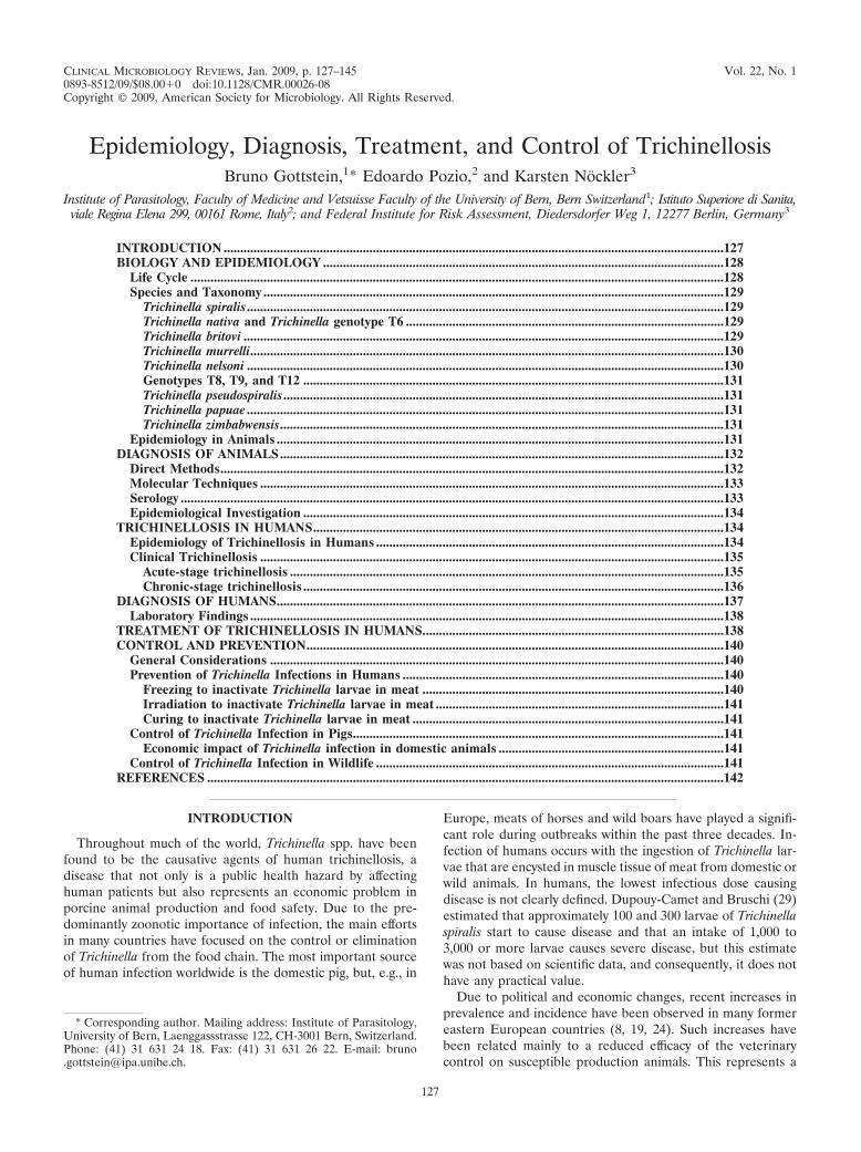
The life cycle of all species of the genus Trichinella princi-pally comprises two generations in the same host (Fig. 1) andincludes a very broad range of host species (mammals, birds,
and reptiles), although only humans become clinically affected.Following delivery by the gravid female worm, which liveswithin the intestinal mucosa of the host, newborn larvae (NBL)migrate directly into predominantly lymphatic and blood ves-sels of the host. This allows them to be transported to pre-dilection sites (highly oxygenated muscles), where they pene-trate. It is likely that NBL enter in the striated muscle cells bythe aid of its stylet. Penetration mechanisms involving enzymeshave been suspected but have not yet been ruled out (21). Inexperimental infections in which NBL were injected directlyinto muscles, the penetration occurred as early as 10 min afterinjection (22). Within such muscle nurse cells, NBL develop tothe infective muscle-stage larvae without molting (this L1 is0.65 to 1.45 mm in length and 0.026 to 0.040 mm in width). Thismaturation terminates within approximately 15 days. In musclenurse cells, parasite larvae can survive for years (up to 40 yearsin humans and over 20 years, e.g., in polar bears) (44, 77).After a period of time that is under the influence of the hostspecies, its immune response, which can change among indi-viduals within a given species, and the Trichinella species orgenotype, calcification of the collagen capsule first and of thenurse cell and larva can occur. This hypobiotic stage is main-tained until being ingested by a new host. Following such aningestion, parasite larvae are released upon gastric digestion inthe new host, and the first-stage larval parasite subsequentlyreaches the duodenum and, embedded in the intestinal mu-cosa, undergoes four molts, thus developing into the adultstage within a very short time of 2 days. Males and femalescopulate, and 5 to 7 days postinfection (p.i.), the females startto deliver new generations of NBL. Within several weeks, anintestinally immune-mediated host response becomes estab-lished, and immune effector mechanisms affect the viability ofthe female parasites, resulting in a continuous expulsion ofadult worms (122).
FIG. 1. Trichinella sp. life cycle. (A) Main sources of Trichinella sp. infections for humans (including pigs, horses, wild boars, dogs, walruses,foxes, and bears). (B) Trichinella sp. cycle in the host body. In the enteral phase, muscle tissues are digested in the stomach, and larvae are released(1); larvae penetrate the intestinal mucosa of the small intestine and reach the adult stage within 48 h p.i., and male and female mate (2); femaleworm releases newborn larvae in the lymphatic vessels (from the fifth day p.i. onwards; the length of newborn production, from 1 week to severalweeks, is under the influence of host immunity) (3). In the parenteral phase, the newborn larvae reach the striated muscle and actively penetratein the muscle cell (4); the larva grow to the infective stage in the nurse cell (the former muscle cell) (5); and, after a period of time (weeks, months,or years), a calcification process occurs (6). (Modified from www.iss.it/site/Trichinella/index.asp with permission of the publisher.)
128 GOTTSTEIN ET AL. CLIN. MICROBIOL. REV.
Species and Taxonomy
Conversely to the older conventional scientific recognitionof Trichinella spiralis as the only member of the genusTrichinella (13), more recent studies of the genetic diversityand zoogeographical and epidemiological peculiarities withinthis genus yielded a new Trichinella taxonomy encompassingeight species (Table 1) (66, 76, 126, 134). All 12 recognizedtaxa are genetically and biologically delineated into two dis-tinct clades characterized by the presence or absence of anintramuscular collagen capsule (167). Thus, one clade is rep-resented by all species and taxa that accordingly encapsulate inhost muscle tissue of mammals only, and the other one doesnot encapsulate after muscle cell dedifferentiation and infectsmammals, birds (one species), and even some reptiles (125).Today’s identification of samples to the species level and geno-typing are based primarily upon molecular means (133). Infor-mation on species and genotype distribution and host rangecan be downloaded from the website of the InternationalTrichinella Reference Centre (www.iss.it/site/Trichinella/index.asp).
Trichinella spiralis. Trichinella spiralis is the species mostadapted to domestic and wild swine but can also include synan-thropic rats in its life cycle. T. spiralis exhibits a wide and globaldistribution (Table 1 and Fig. 2). This species is also the mostimportant etiological agent to cause disease in humans (125).Conversely to the domestic cycle, the sylvatic cycle of T. spiralisincludes a broad range of wild carnivores, which may, however,become the origin of a life cycle introduction into a domestic
host population (23, 120). In the domestic cycle, pork scrapsfrom T. spiralis-infected pigs are the main source of infectionfor synanthropic animals (e.g., rats, horses, stray cats, and dogs[no Trichinella infection has been detected in urban wild ani-mals so far, such as the red fox]) (137).
Trichinella nativa and Trichinella genotype T6. Trichinellanativa affects predominantly sylvatic carnivores living in frigidzones of Asia, northern states of North America, and North-eastern Europe, while the closely related Trichinella genotypeT6 appears to be restricted to several regions of Canada (Brit-ish Columbia, Ontario, Manitoba, and Nunavut) and theUnited States (Alaska, Montana, Idaho, and Pennsylvania)(Table 1 and Fig. 3). The main hosts are terrestrial (e.g., brownand black bears, wolverines, raccoons, lynxes, wolves, andfoxes) and marine (e.g., polar bears, walruses, and seals) car-nivores (23, 39, 66, 80, 131). This species developed the abilityof muscle-stage larvae to survive in frozen muscles of carni-vores for up to 5 years (23). Humans who are at risk forinfection are meat-consuming people living in frigid zonesof Canada, Greenland, and Russia (98, 147, 149) or huntersfrom Europe and the United States who consume raw orundercooked meat from bears hunted in arctic or subarcticregions (2).
Trichinella britovi. Trichinella britovi is the most widely dis-tributed species within sylvatic life cycles of Europe, Asia, andNorthern and Western Africa (123, 136, 137). As is the casewith T. spiralis, T. britovi can also affect domestic pig popula-tions mainly via extensive grazing systems or feed with scraps
TABLE 1. Main epidemiological features of Trichinella species and genotypesa
Species or genotype Geographical distribution Host range Main source of infectionof humans
Resistance of larvae infrozen muscles
EncapsulatedT. spiralis Cosmopolitan Domestic and sylvatic
mammalsDomestic and sylvatic
swine horsesYes in horse muscles
T. nativa Arctic and subarctic areasof America, Asia,Europe
Sylvatic carnivores Bears, walruses Yes in carnivore muscles
Trichinella genotype T6 Canada, Alaska, RockyMountains, andAppalachian Mountainsin the United States
Sylvatic carnivores Carnivores Yes in carnivore muscles
T. britovi Temperate areas ofEurope and Asia,Northern and WesternAfrica
Sylvatic mammals andseldomly domesticpigs
Wild boars, domestic pigshorses, foxes, jackals
Yes in carnivore andhorse muscles
Trichinella T8 South Africa andNamibia
Sylvatic carnivores None documented No
T. murrelli United States andSouthern Canada
Sylvatic carnivores Bears, horses No
Trichinella genotype T9 Japan Sylvatic carnivores None documented NoT. nelsoni Eastern-Southern Africa Sylvatic mammals Warthogs, bush pigs NoTrichinella genotype T12 Argentina Cougars None documented Unknown
NonencapsulatedT. pseudospiralis Cosmopolitan Sylvatic mammals and
birds, domestic pigsDomestic and wild pigs No
T. papuae Papua New Guinea,Thailand
Wild pigs, saltwatercrocodiles
Wild pigs No
T. zimbabwensis Zimbabwe, Mozambique,Ethiopia, South Africa
Nile crocodiles, monitorlizards
None documented No
a Based on data from reference 125.
VOL. 22, 2009 TRICHINELLOSIS 129
or carrion originating from sylvatic carnivores. Zoonotically, T.britovi is the second-most common species of Trichinella thatmay affect human health.
Trichinella murrelli. Trichinella murelli is spread among syl-vatic carnivores across the United States and some southernregions of Canada. This species does not develop in swine. It isthe causative agent of infection in humans, especially following
the consumption of meat originating from hunted black bears.Valuable clinical information on this species was gained froma 1985 outbreak in France due to the consumption of horsemeat imported from the United States (3).
Trichinella nelsoni. Trichinella nelsoni has been detected inEastern Africa, from Kenya to South Africa (88). The hostrange includes sylvatic carnivores and, at least occasionally,
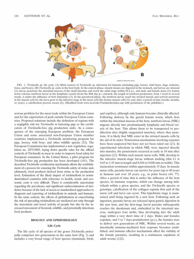
FIG. 2. World map showing the distribution areas of Trichinella spiralis (Tsp), Trichinella pseudospiralis from north America (TpsN), T.pseudospiralis from Europe and Asia (TpsP), T. pseudospiralis from Tasmania (TpsA), Trichinella papuae (Tpa), and Trichinella zimbabwensis (Tzi).(Modified from www.iss.it/site/Trichinella/index.asp with permission of the publisher.)
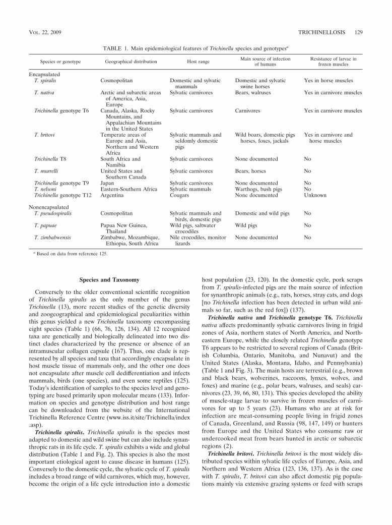
FIG. 3. World map showing the distribution areas of Trichinella nativa (Tna), Trichinella britovi (Tb), Trichinella murrelli (Tm), Trichinellanelsoni (Tne), Trichinella genotype T6 (T6), Trichinella genotype T8 (T8), and Trichinella genotype T9 (T9). In some regions, the distribution areasof these encapsulated species and genotypes overlap between them. (Modified from www.iss.it/site/Trichinella/index.asp with permission of thepublisher.)
130 GOTTSTEIN ET AL. CLIN. MICROBIOL. REV.

bush pigs and warthogs, some of which have been the source ofinfection for humans. Less than 100 human infections havebeen documented for this species in Kenya and Tanzania(123).
Genotypes T8, T9, and T12. Trichinella genotype T8, verysimilar to T. britovi, has been identified in wild animals ofSouth Africa and Namibia (88). No human case due to thisgenotype has been documented. Trichinella isolates from Jap-anese wildlife, originally identified as being T. britovi, are nowdesignated a separate genotype, named Trichinella genotypeT9, which is phylogenetically related to T. murrelli (125, 167).Finally, Trichinella genotype T12 is a new encapsulated geno-type of Trichinella recently detected in a mountain lion (Pumaconcolor) from Trapalco, Patagonia, Río Negro, Argentina.The only information available is the molecular structure oftwo noncoding sequences and one coding sequence that aredifferent from those of the 11 currently recognized speciesand/or genotypes of the genus Trichinella (76).
All species and/or genotypes described so far are character-ized by one common biological feature: they all induce thedevelopment of a thick collagen capsule, which can be detectedby light microscopy, during the muscle phase of infection.Conversely, three other species produce only a thin capsuledetectable by electron microscopy only.
Trichinella pseudospiralis. Trichinella pseudospiralis exhibits acosmopolitan distribution and infects both mammals and birds.Three genetically distinct populations can be distinguished,each referring to a specific geographical origin: Palaearctic,Nearctic, and Australian (Tasmania) origins (79, 165). T.pseudospiralis has been found in 14 mammalian host speciesincluding domestic and sylvatic swine and 13 avian species(121), where the number of reports for mammals is muchhigher than that for birds. Cases of trichinellosis in humanswith some deaths have been documented in Kamchatka, Thai-land, and France (125).
Trichinella papuae. Trichinella papuae circulates in bothmammals and reptiles (domestic sows, wild pigs, and farmedsaltwater crocodiles) of Papua New Guinea and Thailand (122,135). Infections in humans have been documented (113).
Trichinella zimbabwensis. Trichinella zimbabwensis, very sim-ilar to T. papuae, has been detected only in wild and farmedreptiles of Africa (Zimbabwe, Mozambique, South Africa, andEthiopia), although experimentally, it is able to infect mam-mals (129, 130). Human infections are not known so far.
Epidemiology in Animals
Parasites of the genus Trichinella are present on all conti-nents except Antarctica, where no report or investigation ofthese parasites has been released or carried out so far (123).Most of the species, with the exception of T. spiralis, parasitizepredominantly wild animals. A switch from wild animals todomestic animals can occur when there is an improper man-agement in segregating husbandry and wildlife. Domestic cy-cles and the sylvatic cycle can function either independentlyfrom each other or interactively (123).
The term “domestic cycle” refers to the transmission patternwhere the focus is on a swine herd being fed, e.g., uncookedpork scraps, carrion, garbage (i.e., garbage-fed pigs), or thepigs can feed on carcasses that are not promptly removed from
the farm; transmission can also become domestic via synan-thropic animals living near the swine herd (e.g., rats and mus-telides). Horses fattened with pork scraps or with carcasses offur animals became infected with Trichinella. Similarly, infec-tions in sled dogs fed with carcasses of other dogs or of gamefrom the arctic, including carcasses of slaughtered fur animals,were reported. The use of meat of slaughtered crocodiles tofeed other farmed crocodiles has been reported as well (135).With regard to the geographic distribution of the domesticcycle of Trichinella, since World War II, there have been noreports of infections on industrialized farms in Canada, theUnited States, and western Europe; the domestic cycle hasrecently been reported only among small swine herds of South-ern Finland and certain regions of Spain where control mea-sures were not adopted (119). In several countries of Central-Eastern Europe, the transient breakdown of governmentalveterinary services and state farms accompanied by economicproblems and war have resulted in sharp increases in the inci-dence of Trichinella infection among domestic pig herds, withprevalence rates reaching 50% in some villages in the 1990s(94). In Canada, the United States, and most European Unioncountries, Trichinella infection in domestic animals has virtu-ally disappeared, although sporadic foci do occur (6, 127). InSouth and Central America, Trichinella infection is still en-demic in Argentina, Chile, and Mexico in both humans andpigs (111, 144, 148). In East Asia, the domestic cycle occurs inChina (158). Foci of Trichinella infection involving swine andhumans are also widespread in Thailand, Indonesia, Laos, Ma-laysia, and Myanmar (123). Occasionally, T. britovi can betransmitted within the domestic cycle when humans feed pigswith game meat scraps or when “pasture” pigs had access todumps containing offal of sylvatic animals (120). T. pseudospi-ralis has also been transmitted to domestic pigs and rats onfarms in Croatia, Kamchatka, Russia, and the Slovak Republic(61).
The “sylvatic cycle” oscillates between wildlife hosts and alsoincludes all Trichinella species and genotypes, including T.spiralis for mammals, T. pseudospiralis for mammals and birds,and T. papuae and T. zimbabwensis for mammals and reptiles.Peroral infection occurs either after ingestion of muscle tissuefrom an infectious prey animal or by consumption of infectioustissue from a carrion of a homologous (the former actuallyrepresenting “cannibalism”) or a heterologous species. NaturalTrichinella infections have been reported for more than 100species of mammals, seven avian species, and three reptilespecies (121). Despite the potential broad host spectrum forTrichinella spp., the predominant biotic potential concerns car-nivores (13, 23) and porcine omnivores (mainly domestic pigs,different races of wild pigs, wild boars, bush pigs, and war-thogs) (13, 121). One of the most important biological factorspromoting transmission is the physiological ability of muscle-stage larvae to survive in decaying carcasses/carrion. Thus,even nonencapsulated larvae of T. papuae retained their infec-tivity in decaying tissues of a pig exposed at 35°C for 9 days(114). Encapsulated larvae of T. spiralis have been found to beinfective for laboratory animals up to 4 months in extremelyrotten meat (85). Encapsulated larvae of T. britovi and T.nelsoni in mouse carcasses packed in plastic vials have beenfound to be infective for laboratory animals up to 45 days atroom temperature even if the muscle tissues were completely
VOL. 22, 2009 TRICHINELLOSIS 131
liquefied (E. Pozio, unpublished data). The importance of thiswell-established environmental adaptation is underscored bysurvival even at low freezing temperatures, as, e.g., T. britovican survive in frozen carrion for up to 1 year, and T. nativa andTrichinella genotype T6 can survive for up to several years,maintaining infectivity for future hosts (23, 132). The anaero-bic metabolism favoring survival in putrefying flesh along withthe ability of larvae of some species to survive freezing are twoseparate mechanisms that strongly increase the survival of theparasite in nature. It is important to stress that the survival ofmuscle larvae after freezing occurs mainly when these larvaeparasitize striated muscles of carnivores (bears, wolves, andfoxes, etc.), whereas the survival time after freezing is stronglyreduced to a few days or weeks, when muscle larvae of thesame strain parasitize other mammalian hosts such as swineand rodents.
DIAGNOSIS OF ANIMALS
Direct Methods
Meat inspection for the detection of Trichinella larvae isdesigned to prevent clinical trichinellosis in humans but not toprevent infection. The identification of Trichinella larvae inmuscle samples from pigs and other animal species intendedfor human consumption (e.g., horses, wild boars, and bears) islimited to postmortem inspection of carcasses (49). Direct de-tection is also applied in wildlife monitoring, where indicatoranimals (e.g., foxes or raccoon dogs) are examined to assessthe prevalence of Trichinella infection among the wildlife res-ervoir and the risk of introduction into domestic animals.Methods to detect Trichinella larvae in muscle samples need tobe highly sensitive, and performance is greatly influenced bythe sample size, the muscle type selected for sampling, and thespecific method used (103).
Host animals ingesting even high numbers of Trichinellalarvae from infectious meat will not develop clinical symptomssuch as those observed in human patients. Therefore, “Trichinellainfection” rather than “trichinellosis” should be used for animals.
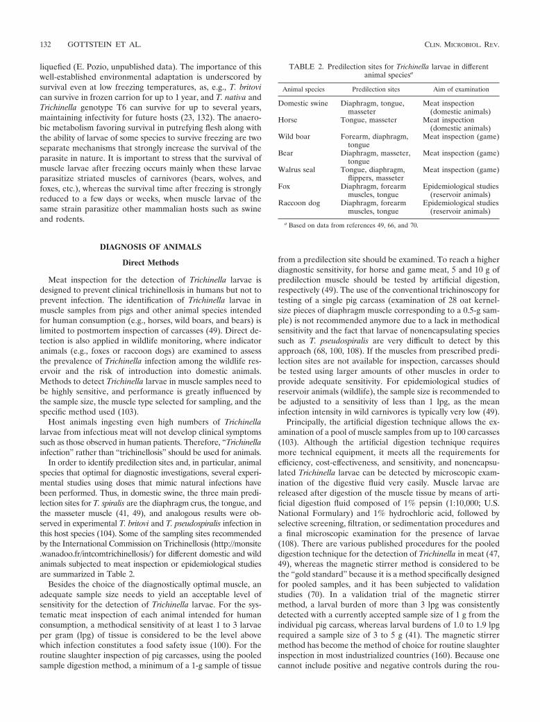
In order to identify predilection sites and, in particular, animalspecies that optimal for diagnostic investigations, several experi-mental studies using doses that mimic natural infections havebeen performed. Thus, in domestic swine, the three main predi-lection sites for T. spiralis are the diaphragm crus, the tongue, andthe masseter muscle (41, 49), and analogous results were ob-served in experimental T. britovi and T. pseudospiralis infection inthis host species (104). Some of the sampling sites recommendedby the International Commission on Trichinellosis (http://monsite.wanadoo.fr/intcomtrichinellosis/) for different domestic and wildanimals subjected to meat inspection or epidemiological studiesare summarized in Table 2.
Besides the choice of the diagnostically optimal muscle, anadequate sample size needs to yield an acceptable level ofsensitivity for the detection of Trichinella larvae. For the sys-tematic meat inspection of each animal intended for humanconsumption, a methodical sensitivity of at least 1 to 3 larvaeper gram (lpg) of tissue is considered to be the level abovewhich infection constitutes a food safety issue (100). For theroutine slaughter inspection of pig carcasses, using the pooledsample digestion method, a minimum of a 1-g sample of tissue
from a predilection site should be examined. To reach a higherdiagnostic sensitivity, for horse and game meat, 5 and 10 g ofpredilection muscle should be tested by artificial digestion,respectively (49). The use of the conventional trichinoscopy fortesting of a single pig carcass (examination of 28 oat kernel-size pieces of diaphragm muscle corresponding to a 0.5-g sam-ple) is not recommended anymore due to a lack in methodicalsensitivity and the fact that larvae of nonencapsulating speciessuch as T. pseudospiralis are very difficult to detect by thisapproach (68, 100, 108). If the muscles from prescribed predi-lection sites are not available for inspection, carcasses shouldbe tested using larger amounts of other muscles in order toprovide adequate sensitivity. For epidemiological studies ofreservoir animals (wildlife), the sample size is recommended tobe adjusted to a sensitivity of less than 1 lpg, as the meaninfection intensity in wild carnivores is typically very low (49).
Principally, the artificial digestion technique allows the ex-amination of a pool of muscle samples from up to 100 carcasses(103). Although the artificial digestion technique requiresmore technical equipment, it meets all the requirements forefficiency, cost-effectiveness, and sensitivity, and nonencapsu-lated Trichinella larvae can be detected by microscopic exam-ination of the digestive fluid very easily. Muscle larvae arereleased after digestion of the muscle tissue by means of arti-ficial digestion fluid composed of 1% pepsin (1:10,000; U.S.National Formulary) and 1% hydrochloric acid, followed byselective screening, filtration, or sedimentation procedures anda final microscopic examination for the presence of larvae(108). There are various published procedures for the pooleddigestion technique for the detection of Trichinella in meat (47,49), whereas the magnetic stirrer method is considered to bethe “gold standard” because it is a method specifically designedfor pooled samples, and it has been subjected to validationstudies (70). In a validation trial of the magnetic stirrermethod, a larval burden of more than 3 lpg was consistentlydetected with a currently accepted sample size of 1 g from theindividual pig carcass, whereas larval burdens of 1.0 to 1.9 lpgrequired a sample size of 3 to 5 g (41). The magnetic stirrermethod has become the method of choice for routine slaughterinspection in most industrialized countries (160). Because onecannot include positive and negative controls during the rou-
TABLE 2. Predilection sites for Trichinella larvae in differentanimal speciesa
Animal species Predilection sites Aim of examination
Domestic swine Diaphragm, tongue,masseter
Meat inspection(domestic animals)
Horse Tongue, masseter Meat inspection(domestic animals)
Wild boar Forearm, diaphragm,tongue
Meat inspection (game)
Bear Diaphragm, masseter,tongue
Meat inspection (game)
Walrus seal Tongue, diaphragm,flippers, masseter
Meat inspection (game)
Fox Diaphragm, forearmmuscles, tongue
Epidemiological studies(reservoir animals)
Raccoon dog Diaphragm, forearmmuscles, tongue
Epidemiological studies(reservoir animals)
a Based on data from references 49, 66, and 70.
132 GOTTSTEIN ET AL. CLIN. MICROBIOL. REV.

tine performance of the magnetic stirrer approach, it is com-pulsory to validate the test prior to using it at the slaughter-house.
Molecular Techniques
For epidemiological studies and improvement of knowledgeon the occurrence and spread of Trichinella spp. in the domes-tic and sylvatic cycles, all isolates should be identified to thespecies or genotype level. Since there are no morphologicalfeatures to specify larvae, molecular diagnosis is used to yieldthe species or genotype diagnostically recovered. For this pur-pose, a multiplex PCR has been developed for the simple andunequivocal differentiation of Trichinella species and geno-types. Partial DNA sequence data were generated from theinternal transcribed spacers ITS1 and ITS2 and from the ex-pansion segment V region of the rRNA repeat from differentTrichinella species and genotypes (166). This multiplex PCR isa sensitive, inexpensive, and rapid molecular approach that canunequivocally identify a single larva at the species and geno-type levels (133).
The identification of Trichinella isolates at the species andgenotype levels and data collection are provided by the Inter-national Trichinella Reference Centre (http://www.iss.it/site/Trichinella/index.asp).
Serology
Animals can be tested for the presence of anti-Trichinellaantibodies in the serum or in the meat juice either upon ante-mortem or upon postmortem examination (108). According tothe International Commission on Trichinellosis, indirect meth-ods such as the detection of anti-Trichinella antibodies in do-mestic and wild animals are not recommended as a substitutefor meat inspection of individual carcasses (49). However,Trichinella serology is considered to be suitable for the surveil-lance and epidemiological investigations of domestic animalsand wildlife (51).
The time of seroconversion after a primary Trichinella in-fection is dependent upon the infection dose and the larvalburden in the muscle, as has been demonstrated by compre-hensive experimental studies with different Trichinella speciesin various host animals such as pigs, horses, wild boars, andfoxes. Following ingestion of high numbers of T. spiralis larvae,anti-Trichinella immunoglobulin G (IgG) can be detected inanimals about 2 to 3 weeks p.i. Conversely, there is a delayedantibody response for several weeks if animals get a low infec-tion dose (Table 3). Analogous results are available for otherTrichinella species from experimental studies of pigs where anincreasing infection dose of 100, 1,000, and 20,000 larvae of T.pseudospiralis and T. britovi in Yorkshire and Iberian pigs wascorrelated with a shorter time of seroconversion (104).
Longitudinal studies revealed that anti-Trichinella antibod-ies may persist in pigs for a long and presumably indefinitetime (106), assuming that in slaughter pigs, it is unlikely that afalse-negative result will be obtained at a later stage of infec-tion because of putatively declining antibody levels (103). In anexperimental study carried out with wild boars, the antibodylevel against excretory/secretory (E/S) antigens remained sta-ble in animals infected with T. spiralis, T. britovi, and T. nelsoni,
but the decline of antibodies directed against T. nativa, T.murrelli, and Trichinella genotype T6 was associated with therapid disappearance of larvae in the muscle sample (67) due tothe lower susceptibility of wild boar to these species and/orgenotypes. Experimental and field studies of horses have dem-onstrated that the serological response to Trichinella infectionin this species is less consistent than that observed in pigs (51).For instance, in naturally infected horses, specific antibodieswere not detected in spite of the presence of a high larvalburden in muscles (128, 139). Therefore, serological methodscannot be recommended for the examination of single animalsor for monitoring horses for Trichinella infection.
In conclusion, from the diagnostic point of view, the earlystage of Trichinella infection in the host is characterized by a“diagnostic window” at which false-negative results may occurcompared with direct (larval detection) tests (108). During thisearly stage of infection, however, Trichinella larvae may not yethave reached maturity in the muscle cell (approximately 20days after infection) to allow infection of the next appropriatehost (103).
Blood serum is conventionally the preferred sample matrixfor conducting serological tests for Trichinella. However,serum samples of poor quality due to extensive hemolysis ormicrobial contamination, especially in samples obtained fromwild animals, may significantly influence the sensitivity andspecificity of the test (108). Other body fluids that can be usedfor serological testing include plasma, whole blood, and tissuefluids. Results from experimental studies indicate that tissuefluids such as meat juice from slaughtered pigs or from otheranimals (e.g., wild boars and foxes) may be suitable for sero-logical examinations (46, 71, 91). Comparable correlations and
TABLE 3. Relationship between time of seroconversion andinfection dose (T. spiralis) in pig, horse, wild boar,
and red foxes
Animalspecies
Infection dose(no. of larvae/
animal)No. of lpg
Time ofseroconversion
p.i. (wk)
Swine 100 1.62–6.50b 5–7500 18.4–48.6b 4–5
1,000 26.3–90.6e 4–62,500 87.6–99.5b 48,000 12.1–81.4c 3
20,000 699.2–1103.5e 3–464,000 221.4–466.6c 2.5–3
Horse 1,000 0.10–0.26b 3–44,000 0.39–7.8b 3–75,000 0.02–8.9e 2–4.5
10,000 6.6–60.0b 3–440,000 484–1060d 2–3
Wild boar 10,000 43–100e 3–4
Red fox 10,000 7.7–202.7 3
a Data for pig based on references 46, 104, and 152; data for horse based onreferences 47 and 157; data for wild boar based on reference 67; and data for redfoxes based on reference 91.
b Mean of tongue.c Mean of tongue, masseter, diaphragm, intercostal, psoas, and rectus abdo-
minis.d Mean of masseter.e Mean of diaphragm.
VOL. 22, 2009 TRICHINELLOSIS 133

dilution ratios between blood serum and muscle juice wereobserved in a comparative study addressing pigs, wild boars,and foxes (91).
Enzyme-linked immunosorbent assay (ELISA) is the mostcommonly used method for the detection of Trichinella infec-tion due mainly to the methodical sensitivity that allows thedetection of as low as 1 larva per 100 g of muscle tissue (108).Thus, a large series of experimental and/or field studies hasbeen carried out using pig serum and meat juice samples (46,47, 62, 95, 102, 106, 152, 155). The specificity of ELISA becamegreatly improved by utilizing metabolic E/S antigens releasedfrom Trichinella muscle larvae and generated upon in vitromaintenance. These E/S antigens consist of a group of struc-turally related glycoproteins (48, 52). The predominantTrichinella epitopes inducing humoral immunity are localizedon the so-called TSL-1 antigen, which is found in the stichocytecells and on the surface of the parasite’s cuticle and which issecreted by first-stage larvae in the muscle (5, 112). TSL-1antigen epitopes are highly conserved, and a high cross-reac-tivity between Trichinella antigens derived from different spe-cies and genotypes was revealed, allowing serology to detectinfection with all of these Trichinella species (5, 51, 69, 86, 104).Tyvelose has been identified as being a major carbohydrateepitope of the TSL-1 antigen, and a synthetic variant oftyvelose has been developed for use in ELISA (143, 161). Thissynthetic carbohydrate antigen offers the advantages of stabil-ity and standardization and shows a higher test specificity inmany host species (20). Unfortunately, the tyvelose antigensuffers from sensitivity problems in some instances (51). Re-sults from studies of Trichinella seroprevalence in sylvatic anddomestic animals demonstrated a less sensitive but very spe-cific antibody response against synthetic carbohydrate com-pared to E/S antigen. Therefore, tyvelose ELSIA can be usedfor confirmatory testing (91).
Multiple experimental and field studies have been carriedout using swine to evaluate ELISA using E/S antigens. Theinfection status of pigs was determined by the digestion ofdiaphragm muscle samples. The sensitivity of ELISA rangedfrom between 93.1 and 99.2%, whereas the specificity variedfrom 90.6 to 99.4% after examination of serum samples of pigswhich originated from Trichinella-free farms (95, 110, 155). Ina validation study for testing of pigs using tyvelose antigenELISA, the sensitivity and specificity were 94.3 and 96.7%,respectively, compared with the E/S ELISA, which had speci-ficity and sensitivity of 84.9 and 96.0%, respectively (42).
Epidemiological Investigation
Outbreaks of trichinellosis can cause severe harm to theaffected group of persons. By definition, a trichinellosis out-break requires at least one case that must be laboratory con-firmed (http://www.med.unipi.it/ict/outbr1.htm). Associatedcases should be reported as confirmed if the patient shared anepidemiologically implicated meal or ate an epidemiologicallyimplicated meat product and has either a positive serologicaltest for trichinellosis or a clinically compatible illness (http://www.cdc.gov/epo/dphsi/casedef/trichinosis_current.htm). If two ormore persons in the same household or a number of persons inthe same community have high fever, periorbital or facialedema, and myalgia, trichinellosis can be suspected. When
cases are sporadic or the clinical course is atypical, it is lesslikely that the infection will be suspected. Thus, the diagnosiscan be more complex, e.g., for people who have consumed verysmall amounts of infected meat over a period of several days.The clinical course will then be atypical, and the clinical diag-nosis will be arduous.
Once infection is suspected, information on the consump-tion of raw or undercooked meat or meat products, includingthe place and time of purchase (or receipt) and consumption,should be collected (29).
Reviewing past trichinellosis outbreaks provides evidence ofthe close relationship between clinical findings, diagnostic re-sults, and epidemiological uncoverings conducted by the localpublic health and veterinary public health authorities. In thiscontext, the fast and correct identification of the infectionsource and tracing it back to the animal farm are very impor-tant issues to assess the maximal number of people who mighthave been exposed to food contaminated with infectiveTrichinella larvae (105). Usually, such outbreak studies areconducted as case-control studies on the basis of a close con-nection between the time (appearance of clinical signs) andlocation of trichinellosis cases.
TRICHINELLOSIS IN HUMANS
Epidemiology of Trichinellosis in Humans
In approximately 20% of countries around the globe, in-cluding predominantly small islands or city-states, whereTrichinella sp. infections cannot develop for the lack of poten-tial reservoirs, case numbers are very low, as infections ofhumans only accidentally occur upon the (legal and illegal)importation of Trichinella-infected meat from abroad. Follow-ing reports from the main 55 countries where trichinellosisoccurs autochthonously, the yearly total number of clinicaltrichinellosis was estimated to be 10,000 cases, with a deathrate of 0.2% (123).
Trichinella infection in humans is strongly associated withthe consumption of raw or undercooked meat; thus, culturalfactors such as traditional dishes based on raw or undercookedmeat or meat-derived products play an important role in theepidemiology of the disease. Conversely, when a populationuniquely consumes well-cooked meat, trichinellosis cases arelacking or very scarce despite persistent wildlife transmission(123). Overall, domestic pork and related products remain themost important source of Trichinella infection in humans, es-pecially when pigs are raised under free-ranging or backyardproduction conditions. Another important source of infectionrelated to local meat consumption habit is found in France,where, for the past two decades, most trichinellosis cases havebeen due to the consumption of raw horse meat, a peculiarityrelated to the French culture (9). In Italy, human infectionsdue to the consumption of horse meat have been documentedin only two areas (Emilia Romagna and Lombardy regions innorthern Italy and the Apulia region in southern Italy), wherethe French habit to consume raw horse meat was introducedsome centuries ago (120). In China and the Slovak Republic,dog meat was the source of infection for several foci (25, 81).In Romania, the highest prevalence of trichinellosis in humansoccurred in the Transylvanian region, where the local ethnic
134 GOTTSTEIN ET AL. CLIN. MICROBIOL. REV.

group maintains the food habit of raw meat consumption (8).In Israel, Lebanon, and Syria, where the Judaic and Muslimreligions forbid the consumption of pork, human outbreaks oftrichinellosis have been documented only following the con-sumption of pork from wild boars among the Christian Arabpopulation and immigrants from Thailand (33, 54, 57, 89, 109).In Algeria and Senegal, since the majority of the human pop-ulation is Muslim, trichinellosis has been documented only inEuropeans (99, 123). However, the Muslim population is notexempt from acquiring trichinellosis, as shown by the occur-rence of a large outbreak of trichinellosis for the consumptionof minced beef illegally mixed with pork of unknown origin inTurkey (1). Hunters, their relatives, and their friends are at riskof trichinellosis infection when raw meat from game animals(e.g., bears, cougars, foxes, walruses, and wild pigs) is nottested for Trichinella before consumption (2, 17, 31, 39, 72, 87,92, 115, 147, 158, 164).
The migratory flow of humans with their own food practicesincluding the consumption of raw meat, the illegal importationof not-controlled meat from endemic to nonendemic coun-tries, and new food practices and dishes including raw meatresulted in outbreaks in Denmark, Germany, Italy, Spain, andthe United Kingdom (45, 101, 124, 154). The increasing num-ber of international travelers has resulted in many reports oftourists who acquired Trichinella infections while traveling orhunting in areas of endemicity and subsequently developeddisease after their return to their home countries. In mostinstances, diagnosis was difficult because infections appearedas isolated cases (28, 90, 96, 151).
Clinical Trichinellosis
Principally, a Trichinella infection in the human host can bedivided into two phases: an intestinal (or enteral) phase and amuscular (or parenteral or systemic) phase. Infections with lowintensities can remain asymptomatic, but parasite burdenshigher than a few hundred larvae can initially cause gastroen-teritis associated with diarrhea and abdominal pain approxi-mately 2 days p.i. (intestinal acute phase of disease). Thepathology is caused by the larvae released into the intestinalmucosa, which subsequently migrate to the blood vessels, bymeans of which they spread throughout the body until reachingtheir final location (i.e., the cells of the striated skeletal mus-cles).
Migrating Trichinella larvae and their metabolites provokean immediate reaction, which causes immunological, patholog-ical, and metabolic disturbances and the various clinical phe-nomena observed during the acute stage of the infection (14,73, 93). The immunological reaction is characterized by infil-trating inflammatory cells (i.e., mast cells, eosinophils, mono-cytes, and lymphocytes) and a rather Th2-oriented cytokineprofile. In this context, eosinophilia is a common characteristicin most cases of trichinellosis. Eosinophils may contribute toimmunopathology in trichinellosis due to the release of en-zymes such as histaminase and aryl sulfatase. However, eosin-ophils also induce damage to Trichinella larvae upon activity ofthe major basic protein, eosinophil cationic protein, and eo-sinophil peroxidase. The intensity and dynamics of the eo-siniphilic phenomenon are dependent on the dose of larvae,the species of Trichinella involved, susceptibility of the host to
infection, as well as the time at which the treatment (in par-ticular, the treatment with anthelmintics) has been started(73). The release of histamine, serotonin, a slow-reacting sub-stance of anaphylaxis, bradykinin, and prostaglandins (PGE2,PGD2, and PGJ2) results in an augmented permeability ofcapillaries and a leakage of fluids, electrolytes, albumins, andcell elements into the surrounding tissue (73). Thus, tissueedema, mainly around the eyes, will be found. Furthermore,another consequence of these inflammatory processes will bevasculitis and fine intravascular thrombi, which represent theprincipal pathology in the acute stage of trichinellosis.
The production of IgE is another Th2-oriented characteris-tic; however, an increase in total IgE levels is not a consistentphenomenon found in trichinellosis, and thus, it is not possibleto exclude trichinellosis on the basis of its absence. Clinicalobservations suggest that Trichinella-specific IgE is responsiblefor the allergic manifestations typical of the clinical picture oftrichinellosis, such as cutaneous rash or edemas (38, 159).
Dupouy-Camet and Bruschi (29) described the following thethree major cell modifications occurring during the acute stageof infection, the penetration, and, finally, the residence oflarvae in the striated skeletal muscle cells: (i) the host celltransformation into a new phenotype called “nurse cell,” ac-companied by the disappearance of sarcomere myofibrils; (ii)the encapsulation of the larvae (in the case of encapsulatedspecies); and (iii) the development of a capillary network sur-rounding the infected cell (15).
In addition to these three major modifications, the sarco-plasm becomes basophilic, the cell nucleus is displaced to thecenter of the cell, and the nucleoli increase in both number andsize. The cell becomes more permeable, resulting in an in-creased release of muscle enzymes (29). In humans, the calci-fication of encapsulated T. spiralis larvae may take place after6 months, but this process is not synchronized and will notsimultaneously include all parasites. The time point of calcifi-cation appears to depend on the anatomical localization of theparasites as well as other unknown reasons. Calcification of thecollagen capsule occurs first, followed by the nurse cell andthe larva. The whole process may lead to the death of thelarvae, but not all larvae, as some larvae may survive foryears in the same host.
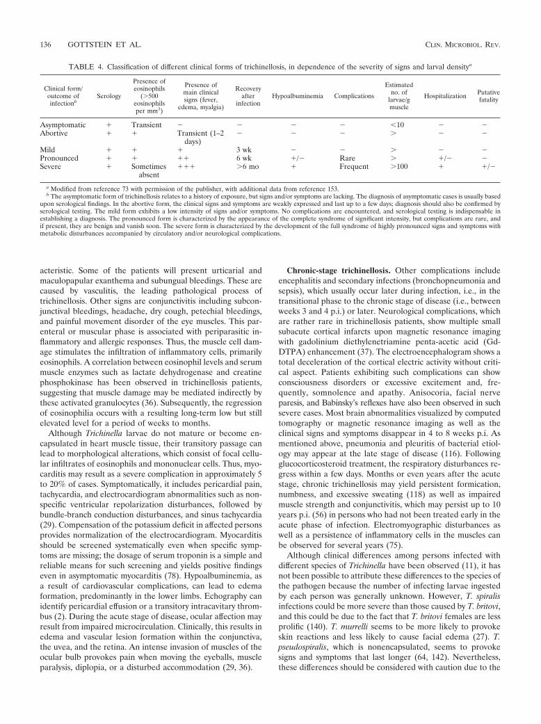
The severity of clinical disease upon infection with T. spiralis(and also other species) is strongly dependent on and directlycorrelated with the number of infective larvae ingested by theperson or patient. Thus, infection may result in a large spec-trum of clinical forms ranging from asymptomatic to fatality.An overview of such different forms is presented in Table 4.
Acute-stage trichinellosis. A Trichinella infection that be-comes clinically manifest in a human host starts with nonspe-cific signs such as uneasiness, headache, fever, fever-associatedchills, and, occasionally, gastrointestinal disorders. The feverusually persists for 1 to 3 weeks, depending on the infectiondose and severity of disease. Clinical features of this acutestage of infection and disease include pyrexia, eyelid or facialedema, and myalgia as the principal syndrome, occasionallycomplicated by myocarditis, thromboembolic disease, andencephalitis. One week p.i. (or up to 20 days p.i. in the case ofmild infections), but still during the acute stage, high fever (asmentioned above), shivering, myalgia, and symmetrical perior-bital or facial edema (invasive or myopathic phase) are char-
VOL. 22, 2009 TRICHINELLOSIS 135
acteristic. Some of the patients will present urticarial andmaculopapular exanthema and subungual bleedings. These arecaused by vasculitis, the leading pathological process oftrichinellosis. Other signs are conjunctivitis including subcon-junctival bleedings, headache, dry cough, petechial bleedings,and painful movement disorder of the eye muscles. This par-enteral or muscular phase is associated with periparasitic in-flammatory and allergic responses. Thus, the muscle cell dam-age stimulates the infiltration of inflammatory cells, primarilyeosinophils. A correlation between eosinophil levels and serummuscle enzymes such as lactate dehydrogenase and creatinephosphokinase has been observed in trichinellosis patients,suggesting that muscle damage may be mediated indirectly bythese activated granulocytes (36). Subsequently, the regressionof eosinophilia occurs with a resulting long-term low but stillelevated level for a period of weeks to months.
Although Trichinella larvae do not mature or become en-capsulated in heart muscle tissue, their transitory passage canlead to morphological alterations, which consist of focal cellu-lar infiltrates of eosinophils and mononuclear cells. Thus, myo-carditis may result as a severe complication in approximately 5to 20% of cases. Symptomatically, it includes pericardial pain,tachycardia, and electrocardiogram abnormalities such as non-specific ventricular repolarization disturbances, followed bybundle-branch conduction disturbances, and sinus tachycardia(29). Compensation of the potassium deficit in affected personsprovides normalization of the electrocardiogram. Myocarditisshould be screened systematically even when specific symp-toms are missing; the dosage of serum troponin is a simple andreliable means for such screening and yields positive findingseven in asymptomatic myocarditis (78). Hypoalbuminemia, asa result of cardiovascular complications, can lead to edemaformation, predominantly in the lower limbs. Echography canidentify pericardial effusion or a transitory intracavitary throm-bus (2). During the acute stage of disease, ocular affection mayresult from impaired microcirculation. Clinically, this results inedema and vascular lesion formation within the conjunctiva,the uvea, and the retina. An intense invasion of muscles of theocular bulb provokes pain when moving the eyeballs, muscleparalysis, diplopia, or a disturbed accommodation (29, 36).
Chronic-stage trichinellosis. Other complications includeencephalitis and secondary infections (bronchopneumonia andsepsis), which usually occur later during infection, i.e., in thetransitional phase to the chronic stage of disease (i.e., betweenweeks 3 and 4 p.i.) or later. Neurological complications, whichare rather rare in trichinellosis patients, show multiple smallsubacute cortical infarcts upon magnetic resonance imagingwith gadolinium diethylenetriamine penta-acetic acid (Gd-DTPA) enhancement (37). The electroencephalogram shows atotal deceleration of the cortical electric activity without criti-cal aspect. Patients exhibiting such complications can showconsciousness disorders or excessive excitement and, fre-quently, somnolence and apathy. Anisocoria, facial nerveparesis, and Babinsky’s reflexes have also been observed in suchsevere cases. Most brain abnormalities visualized by computedtomography or magnetic resonance imaging as well as theclinical signs and symptoms disappear in 4 to 8 weeks p.i. Asmentioned above, pneumonia and pleuritis of bacterial etiol-ogy may appear at the late stage of disease (116). Followingglucocorticosteroid treatment, the respiratory disturbances re-gress within a few days. Months or even years after the acutestage, chronic trichinellosis may yield persistent formication,numbness, and excessive sweating (118) as well as impairedmuscle strength and conjunctivitis, which may persist up to 10years p.i. (56) in persons who had not been treated early in theacute phase of infection. Electromyographic disturbances aswell as a persistence of inflammatory cells in the muscles canbe observed for several years (75).
Although clinical differences among persons infected withdifferent species of Trichinella have been observed (11), it hasnot been possible to attribute these differences to the species ofthe pathogen because the number of infecting larvae ingestedby each person was generally unknown. However, T. spiralisinfections could be more severe than those caused by T. britovi,and this could be due to the fact that T. britovi females are lessprolific (140). T. murrelli seems to be more likely to provokeskin reactions and less likely to cause facial edema (27). T.pseudospiralis, which is nonencapsulated, seems to provokesigns and symptoms that last longer (64, 142). Nevertheless,these differences should be considered with caution due to the
TABLE 4. Classification of different clinical forms of trichinellosis, in dependence of the severity of signs and larval densitya
Clinical form/outcome ofinfectionb
Serology
Presence ofeosinophils
(�500eosinophilsper mm3)
Presence ofmain clinicalsigns (fever,
edema, myalgia)
Recoveryafter
infectionHypoalbuminemia Complications
Estimatedno. of
larvae/gmuscle
Hospitalization Putativefatality
Asymptomatic � Transient � � � � �10 � �Abortive � � Transient (1–2
days)� � � � � �
Mild � � � 3 wk � � � � �Pronounced � � �� 6 wk �/� Rare � �/� �Severe � Sometimes
absent��� �6 mo � Frequent �100 � �/�
a Modified from reference 73 with permission of the publisher, with additional data from reference 153.b The asymptomatic form of trichinellosis relates to a history of exposure, but signs and/or symptoms are lacking. The diagnosis of asymptomatic cases is usually based
upon serological findings. In the abortive form, the clinical signs and symptoms are weakly expressed and last up to a few days; diagnosis should also be confirmed byserological testing. The mild form exhibits a low intensity of signs and/or symptoms. No complications are encountered, and serological testing is indispensable inestablishing a diagnosis. The pronounced form is characterized by the appearance of the complete syndrome of significant intensity, but complications are rare, andif present, they are benign and vanish soon. The severe form is characterized by the development of the full syndrome of highly pronounced signs and symptoms withmetabolic disturbances accompanied by circulatory and/or neurological complications.
136 GOTTSTEIN ET AL. CLIN. MICROBIOL. REV.
obvious lack of knowledge on the infecting dose for each pa-tient.
DIAGNOSIS OF HUMANS
In general, the early clinical diagnosis of trichinellosis israther difficult because pathognomonic signs or symptoms arelacking (29), and also, later chronic forms of the disease arenot easy to diagnose. Furthermore, physicians practicing innonendemic countries are usually unfamiliar with the diseaseand may thus experience problems in diagnosing trichinellosis.This problem becomes obvious when reviewing the extensivediagnostic delay found in cohort studies, indicating that thediagnosis was usually made at the late phase of the disease(63). This is of concern for the patients, because a delay indiagnosis and treatment favors the establishment of larvae in
muscle tissue and the development of a collagen capsule, whichleaves the larvae resistant to drugs (26, 138).
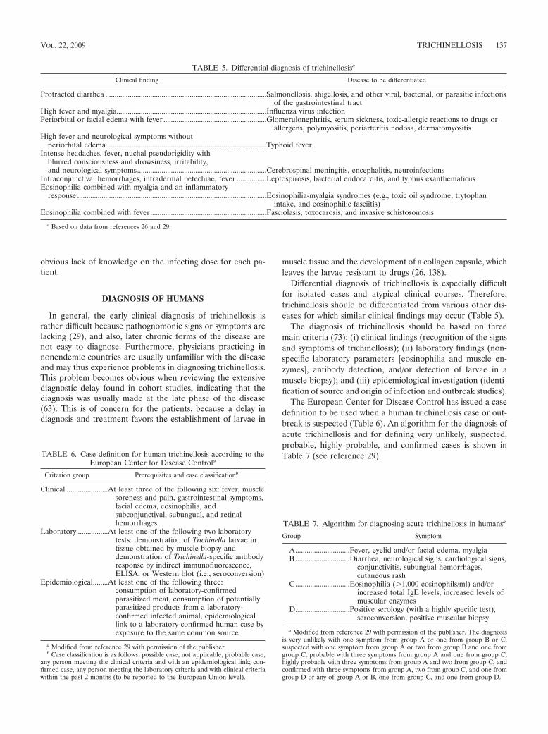
Differential diagnosis of trichinellosis is especially difficultfor isolated cases and atypical clinical courses. Therefore,trichinellosis should be differentiated from various other dis-eases for which similar clinical findings may occur (Table 5).
The diagnosis of trichinellosis should be based on threemain criteria (73): (i) clinical findings (recognition of the signsand symptoms of trichinellosis); (ii) laboratory findings (non-specific laboratory parameters [eosinophilia and muscle en-zymes], antibody detection, and/or detection of larvae in amuscle biopsy); and (iii) epidemiological investigation (identi-fication of source and origin of infection and outbreak studies).
The European Center for Disease Control has issued a casedefinition to be used when a human trichinellosis case or out-break is suspected (Table 6). An algorithm for the diagnosis ofacute trichinellosis and for defining very unlikely, suspected,probable, highly probable, and confirmed cases is shown inTable 7 (see reference 29).TABLE 6. Case definition for human trichinellosis according to the
European Center for Disease Controla
Criterion group Prerequisites and case classificationb
Clinical ......................At least three of the following six: fever, musclesoreness and pain, gastrointestinal symptoms,facial edema, eosinophilia, andsubconjunctival, subungual, and retinalhemorrhages
Laboratory ................At least one of the following two laboratorytests: demonstration of Trichinella larvae intissue obtained by muscle biopsy anddemonstration of Trichinella-specific antibodyresponse by indirect immunofluorescence,ELISA, or Western blot (i.e., seroconversion)
Epidemiological........At least one of the following three:consumption of laboratory-confirmedparasitized meat, consumption of potentiallyparasitized products from a laboratory-confirmed infected animal, epidemiologicallink to a laboratory-confirmed human case byexposure to the same common source
a Modified from reference 29 with permission of the publisher.b Case classification is as follows: possible case, not applicable; probable case,
any person meeting the clinical criteria and with an epidemiological link; con-firmed case, any person meeting the laboratory criteria and with clinical criteriawithin the past 2 months (to be reported to the European Union level).
TABLE 5. Differential diagnosis of trichinellosisa
Clinical finding Disease to be differentiated
Protracted diarrhea ......................................................................................Salmonellosis, shigellosis, and other viral, bacterial, or parasitic infectionsof the gastrointestinal tract
High fever and myalgia................................................................................Influenza virus infectionPeriorbital or facial edema with fever .......................................................Glomerulonephritis, serum sickness, toxic-allergic reactions to drugs or
allergens, polymyositis, periarteritis nodosa, dermatomyositisHigh fever and neurological symptoms without
periorbital edema .....................................................................................Typhoid feverIntense headaches, fever, nuchal pseudorigidity with
blurred consciousness and drowsiness, irritability,and neurological symptoms.....................................................................Cerebrospinal meningitis, encephalitis, neuroinfections
Intraconjunctival hemorrhages, intradermal petechiae, fever ................Leptospirosis, bacterial endocarditis, and typhus exanthematicusEosinophilia combined with myalgia and an inflammatory
response .....................................................................................................Eosinophilia-myalgia syndromes (e.g., toxic oil syndrome, trytophanintake, and eosinophilic fasciitis)
Eosinophilia combined with fever..............................................................Fasciolasis, toxocarosis, and invasive schistosomosis
a Based on data from references 26 and 29.
TABLE 7. Algorithm for diagnosing acute trichinellosis in humansa
Group Symptom
A.............................Fever, eyelid and/or facial edema, myalgiaB .............................Diarrhea, neurological signs, cardiological signs,
conjunctivitis, subungual hemorrhages,cutaneous rash
C.............................Eosinophilia (�1,000 eosinophils/ml) and/orincreased total IgE levels, increased levels ofmuscular enzymes
D.............................Positive serology (with a highly specific test),seroconversion, positive muscular biopsy
a Modified from reference 29 with permission of the publisher. The diagnosisis very unlikely with one symptom from group A or one from group B or C,suspected with one symptom from group A or two from group B and one fromgroup C, probable with three symptoms from group A and one from group C,highly probable with three symptoms from group A and two from group C, andconfirmed with three symptoms from group A, two from group C, and one fromgroup D or any of group A or B, one from group C, and one from group D.
VOL. 22, 2009 TRICHINELLOSIS 137

Laboratory Findings
If persons suffer from clinically manifest trichinellosis, leu-kocytosis accompanied with eosinophilia and increased levelsof muscle enzymes will usually occur as nonspecific laboratorysigns. Eosinophilia appears early prior the development ofclinical signs and symptoms, between the second and the fifthweek of infection in practically every case of trichinellosis, withonly few exceptions (26). Several studies have shown that eo-sinophilia is correlated with the degree of myalgia (29) and issignificantly higher in persons with neurological complications(43). Besides moderately elevated white blood cell counts, eo-sinophilia appears already early after infection and increasesbetween the second and the fifth week p.i. in various degrees:low (�1,000 white blood cells/mm3), moderate (1,000 whiteblood cells/mm3 to 3,000 white blood cells/mm3), and high(�3,000 white blood cells/mm3). During the acute stage ofinfection, a massive decrease in eosinophil levels in personswith severe trichinellosis can be considered to be a predictorfor a severe outcome. Eosinophilia regresses slowly and re-mains at lower levels for periods of 10, 9, and 7 weeks in 50, 30,and 20% of patients, respectively (116).
Elevated levels of muscle enzymes (e.g., creatinine phos-phokinase, lactate dehydrogenase, and aldolase) in blood,which indicate myositis, are found in 75 to 90% of infectedpersons between the second and fifth weeks of infection (15)and may persist for up to 4 months (64).
The detection of specific anti-Trichinella antibodies in bloodserum is of great diagnostic value. After primary infection withTrichinella larvae, antibody detection in the human patientdepends on seroconversion time, whereas differences betweenthe different immunoglobulin classes exist (82, 156). Duringthe acute stage of infection, an early increase in IgE levels canbe observed in most cases, suggesting that this antibody class isrelated to allergic reactions such as cutaneous rash or edema.However, trichinellosis cannot be excluded if this antibodyclass is absent. Therefore, IgE is not considered to exhibitdiagnostic relevance for routine diagnosis (12). Also, IgA andIgM presented no advantage to conventional IgG detection(74, 84, 93, 105). First, IgG antibodies can generally be dem-onstrated 12 to 60 days after infection. The time point ofseroconversion is dependent on several factors such as thenumber of ingested larvae, the Trichinella species involved, andthe individual immune response (12, 140).
Out of conventional serodiagnostic methods, ELISA is themost commonly used approach for the detection of Trichinellainfection in humans, while the indirect fluorescent antibodytest may be used as well. For the indirect fluorescent antibodytest, cryostat sections of infected rodent muscle, frozen sec-tions of free muscle larvae, or formalin-fixed whole larvae areemployed to detect anti-Trichinella antibodies in serum sam-ples (18). This test is more time-consuming and expensive thanELISA, and cross-reactions with antibodies directed againstfilariae (Onchocerca spp.) and Schistosoma mansoni have beenobserved in blood samples of Trichinella-negative patients(145). For the ELISA, and for any serological test, indepen-dent validation should be performed using sera from infectedand Trichinella-free humans representative of the local popu-lation where the test is being used. The cutoff value of theELISA, for example, can change based on host genotype and
exposure to antigens in the local environment and in food (51).An important aspect of ELISA and other serological tests isthe choice of the best antigens to be used. The validationprocess of an ELISA to detect anti-Trichinella IgG in humansera has revealed a large number of cross-reactions with serafrom persons affected by other diseases, mainly if these personsoriginate from developing countries where a high number ofparasitic diseases occur (53).
Immunoblotting showed promise for confirming positivecases or excluding false-positive serological results. Using E/Santigens of muscle larvae of T. spiralis, immunoblotting provedto be more sensitive in screening a population for trichinellosisthan conventional ELISA (107). This technique, however, maybe used as confirmatory rather than screening tests due to thehigh expenditure of labor and time and intensive costs. Thedirect detection of muscle-stage larvae based upon appropriateexamination of muscle biopsies etiologically proves the diag-nosis. Furthermore, isolated larvae allow the molecular iden-tification of the Trichinella species or genotype (see “Molecu-lar Techniques”), a procedure that is not possible by serology.The disadvantage of this method is that it requires a significantsurgical intervention in the affected person and that the sensi-tivity of the diagnosis depends on the amount of muscle sampletested (26). Usually, 0.2 to 0.5 g of muscle tissue without fatand skin is collected from the deltoid or another skeletal mus-cle. Examination of a muscle biopsy can be performed throughartificial digestion (identically to that described above for an-imals) or histological analysis upon hematoxylin-eosin staining(Fig. 4). The basophilic transformation of muscle cells repre-sents a valuable diagnostic criterion of Trichinella invasioneven when no larvae have been detected. This method is moresensitive than trichinoscopy at an early stage of muscle inva-sion, when larvae are very small and cannot easily be differen-tiated from muscle fibers (163).
TREATMENT OF TRICHINELLOSIS IN HUMANS
Upon appropriate diagnosis, therapy has to be initiated asearly as possible (Table 8). Drugs administered in trichinel-losis patients include anthelmintics, glucocorticosteroids,and preparations that compensate for protein and electro-lyte deficits. Anthelmintics are the principal drugs for thetreatment of trichinellosis. They include primarily albenda-zole (Zentel; Smith-Kline Beecham) and mebendazole(Vermox; Janssen Pharmaceutica, Beerse, Belgium) (4). Asexperienced with the treatment of echinococcosis (58), al-bendazole has the slight advantage that most patients reachrequested plasma levels and thus do not require monitoring,while mebendazole plasma levels can vary considerablyamong different patients and might require individual mon-itoring and dosing (65). Secondarily, pyrantel (Combantrin;Pfizer) can also be used (29).
The recommended dose for albendazole is 400 mg twicedaily for 8 to 14 days; for mebendazole, it is 200 to 400 mgthree times a day for 3 days, followed by 400 to 500 mg threetimes a day for 10 days (4). Both treatment schemes aresuitable for adult and pediatric dosages; however, they arecontraindicated during pregnancy and not recommended inchildren aged �2 years (60). Steroids, e.g., prednisone, admin-istered at a dose of 30 mg/day to 60 mg/day for 10 to 15 days
138 GOTTSTEIN ET AL. CLIN. MICROBIOL. REV.

for severe symptoms are the standard choice of chemotherapy.Prednisone was shown to be safe and to alleviate symptomsdue to active tissue larvae (150). Pyrantel (Combantrin) isgiven in a single dose of 10 to 20 mg/kg of body weight,repeated for 2 to 3 days, and may be used by pregnant womenand children, but it is active only against worms in the gut, andit has no effect against newborn and muscle larvae (29). Theapplication of anthelmintics at the stage of intestinal invasion
aims primarily at the elimination of intestinal forms ofTrichinella sp. from the lumen of the gastrointestinal tract.Such treatment is of basic importance for an early and effectivetherapy, particularly in the first 3 days following the infection(73). If applied, such therapy prevents subsequent muscularinvasion and the development of disease. However, in manycases, this option cannot be achieved anymore. The effect ofanthelmintic therapy on the course of a more advanced stageof disease, on lethality, and on already encysted larvae is poorlyelucidated so far. Prognosis for severe cases including cardiacor cerebral complications is not good. Despite therapy, lethal-ity in cases with high infection intensity was up to 5%. Inmilder cases, prognosis is good, and most patients exhibit adisappearance of symptoms within 2 to 6 months. Occasionally,chronic myalgia and rheumatalgia will remain in otherwisesuccessfully treated patients.
Principally, the effectiveness of chemotherapy using, e.g.,albendazole or mebendazole is strongly dependent upon thetime of administration; application only at the early stage ofinfection yields good effectiveness, although no sound case-control efficacy studies have been conducted to date. Unfortu-nately, most infected people are diagnosed only several weeksafter the infection, when the larvae have already establishedthemselves in the muscles.
As a general rule, the later a treatment is prescribed, thehigher the probability that the infected person will alreadyharbor viable larvae in their muscles, which can then survivefor years despite treatment, with possible persistent myalgia(138). As a consequence, in advanced stages of infection anddisease, medication should be administered for longer periodsof time. Nonetheless, anthelmintics may be useless againstlong-term sequelae and chronic trichinellosis. Attempts to in-crease the bioavailability of anthelmintics have been success-
FIG. 4. Histological section (hematoxylin-eosin staining) of a muscle biopsy from a patient involved in a trichinellosis outbreak (100).(A) Cellular infiltrates; (B) collagen capsule of a “nurse cell”; (C) intersected muscle larva. (Photograph courtesy of Dietrich-Bonhoeffer-Klinikum,Neubrandenburg, Germany.)
TABLE 8. Practical recommendations to handle clinicaltrichinellosis casesa
Severity code Recommendation for treatment
Severe and moderatelysevere diseases................................Hospitalization is compulsory for
severe forms and debatable formoderately severe forms
Administration of anthelmintics(albendazole or mebendazole)
Monitoring of thepharmacokinetics ofanthelmintics (if possible)
Administration ofglucocorticosteroids (e.g.,prednisolone), always withanthelmintics
Compensation of fluid andelectrolyte deficits
Administration of pain killers
Benign, abortive, andasymptomatic diseases...................Administration of anthelmintics
(albendazole or mebendazole)Administration of nonsteroidal
anti-inflammatory drugs ifnecessary
a Modified from reference 29 with permission of the publisher.
VOL. 22, 2009 TRICHINELLOSIS 139

fully approved in a mouse model by adding 2-hydroxypropyl-�-cyclodextrin (16). Cymetidine has yielded similar effects onhuman cystic echinococcosis (83), but experience is lacking forhuman trichinellosis.
Astonishingly, beside benzimidazoles, other groups of com-pounds have not been tested so far or did not reach the stageof clinical trials in human trichinellosis. Since the middle of the1970s, the new class of the macrocyclic lactones has revolu-tionized the animal health market. They can be divided intotwo subclasses: the milbemycins and the avermectins. Ivermec-tin, abamectin, eprinomectin, doramectin, milbemycin, mox-idectin, and selamectin belong to these classes. In human medi-cine, only ivermectin has been used against some nematodes.Macrocyclic lactones may thus exhibit good potential to actagainst Trichinella spp. at different stages. Thus, Ros-Morenoet al. (146) showed that the gamma-aminobutyric acid receptorof Trichinella as a target for ivermectin’s mode of action dis-played sufficient binding activity in vitro to suggest furtherinvestigation. El-Azzouni (34) demonstrated some effect ofivermectin against experimental T. spiralis infection in Swissalbino mice at an early stage of infection. In veterinary medi-cine, studies were undertaken to determine the efficacy ofmilbemycin oxime against T. spiralis in experimentally infecteddogs and cats (10). One of the most recent classes of com-pounds, that of cyclooctadepsipeptides, has entered the sceneof anthelmintic research in the early 1990s. Anthelminticallyactive molecules were isolated as a natural compound from thefungus Mycelia sterilia (55). The semisynthetic drug emodep-side has been shown to be active against T. spiralis larvae inmuscles. Electrophysiological studies revealed that emodep-side inhibits pharyngeal pumping of the nematodes (55). Fu-ture investigations and clinical trials will have to show if suchnew compounds provide a better efficacy against maturatedand encapsulated Trichinella muscle larvae.
CONTROL AND PREVENTION
General Considerations
In many European countries, trichinellosis has been de-clared a disease that must be monitored, and for EuropeanUnion Member States, this is obligatory according to the Eu-ropean Union directive on zoonoses, 2003/99/EC. Cases oftrichinellosis must be reported, but further actions are notnecessarily taken. However, the European Commission (EC)has edited legislation laying down rules for the official controlsfor Trichinella in meat in order to improve food safety forEuropean consumers (35). According to regulation (EC) no.2075/2005, meat inspections are required for domestic pigs,horses, wild boars, and other farmed and wild animal speciesthat are susceptible to Trichinella infection and that are slaugh-tered for consumption. It foresees the possibility of a deroga-tion from the testing program for herds, categories of herds, orregions where the risk of Trichinella infection in domesticswine is officially recognized as being negligible. Trichinella-free herds or categories of herds will have to meet severalrequirements. Most requirements are related to generalhygiene and rodent control. Additionally, fattening pigs are notallowed to have access to outdoor facilities as of their fourthweek and only if strict conditions are met during the first 4
weeks. Additional requirements apply to categories of herdsthat may be recognized as Trichinella free, including the needfor a 10-year national surveillance program that would havedetected a prevalence exceeding 0.0001%. In comparison tothe EC, the Office International des Epizooties recognizes thepossibility that a country or zone might be considered to befree from trichinellosis caused by T. spiralis in domestic swine.This may occur when trichinellosis is notifiable in the countryand an effective disease reporting system is in place and hasbeen shown to be capable of capturing the occurrences ofcases. Additionally, a surveillance program that must be inplace was described previously (35). For surveillance of theslaughter sow population, FAO/WHO/OIE guidelines (100)prescribe serological test methods, but the test method to beused for surveillance in slaughter pigs is not specified. Conclu-sively, the most appropriate and economically reasonable ap-proach that could be developed will be a risk-based assess-ment, surveillance, and management system.
Prevention of Trichinella Infections in Humans
The prevention of trichinellosis in humans is based on threemain approaches: (i) education of the consumer about the riskof consumption of raw or semiraw meat and meat productsfrom both domestic (e.g., pigs, horses, and dogs) and sylvatic(e.g., wild boars, bears, walruses, cougars, badgers, foxes, jack-als, armadillos, crocodiles, and monitor lizards) animals thatcan be carriers of Trichinella parasites if they are not properlytested for Trichinella larvae upon meat inspection; (ii) farmingof pigs (the most important source of Trichinella infection forhumans) in modern, industrialized, indoor pigsties under strictveterinary control and use of certified feedstuff (see below);and (iii) control of all susceptible animals (both domestic andsylvatic) by a standardized artificial digestion method atslaughtering or after hunting.
All meat from animals which might contain Trichinella lar-vae but cannot be tested by an appropriate laboratory methodshould be treated by a procedure that has been proven toinactivate Trichinella prior to distribution for human consump-tion (49, 50). This applies to both commercial and noncom-mercial sources of meat. Three methods have been shown toreliably inactivate Trichinella larvae in meat: (i) cooking toreach a core temperature of not less than 71°C (159.8°F) for atleast 1 min (i.e., the meat must change the color from pink togray, and muscle fibers are easily separated from each other),(ii) freezing (see below), and (iii) irradiation (see below).
Methods for the preparation of meat and meat products thatare not considered safe include (i) cooking by using microwaveovens and (ii) curing, drying, or smoking. Education of huntersfor the proper preparation of game meat should follow thesame guidelines issued to consumers. Particular caution shouldbe given to the putative presence of freeze-resistant Trichinellaspecies or genotypes in game meats.
Freezing to inactivate Trichinella larvae in meat. In the ab-sence of proper temperature and time control and monitoringsystems, processors and consumers of meat should ensure thatcuts or pieces of meat up to 15 cm in thickness are frozen solid(at least �15°C [5°F]) for no less than 3 weeks, and cuts andpieces of meat up to 50 cm in thickness should be frozen solidfor no less than 4 weeks. The requirements for freezing are
140 GOTTSTEIN ET AL. CLIN. MICROBIOL. REV.
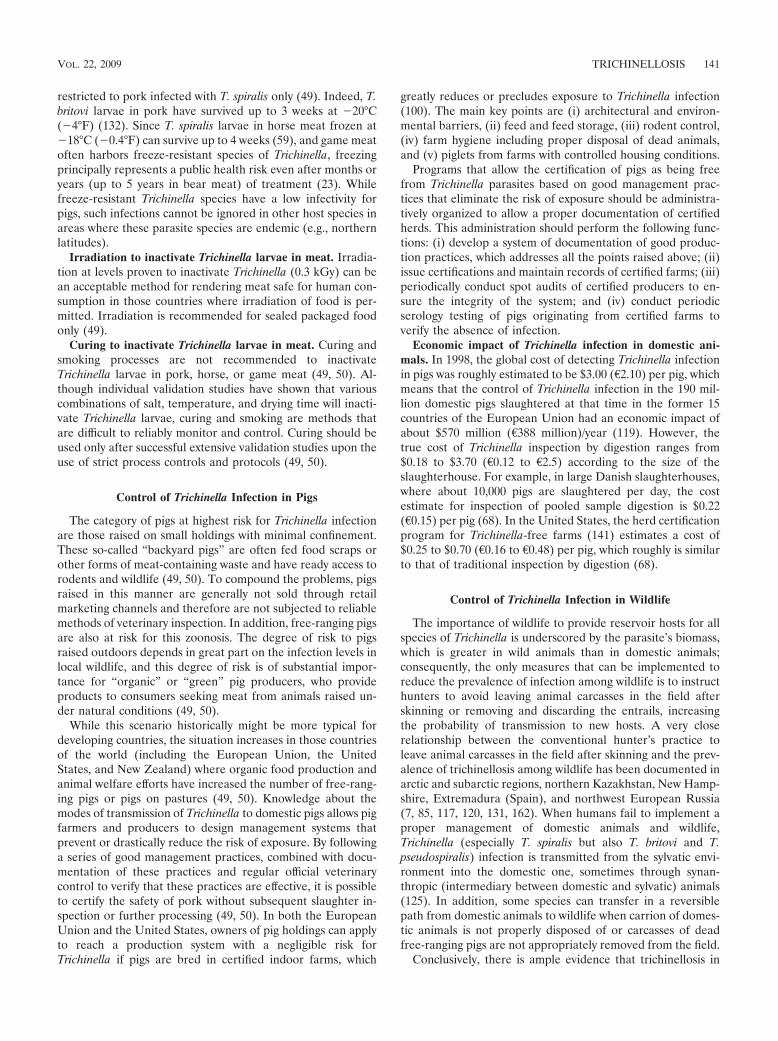
restricted to pork infected with T. spiralis only (49). Indeed, T.britovi larvae in pork have survived up to 3 weeks at �20°C(�4°F) (132). Since T. spiralis larvae in horse meat frozen at�18°C (�0.4°F) can survive up to 4 weeks (59), and game meatoften harbors freeze-resistant species of Trichinella, freezingprincipally represents a public health risk even after months oryears (up to 5 years in bear meat) of treatment (23). Whilefreeze-resistant Trichinella species have a low infectivity forpigs, such infections cannot be ignored in other host species inareas where these parasite species are endemic (e.g., northernlatitudes).
Irradiation to inactivate Trichinella larvae in meat. Irradia-tion at levels proven to inactivate Trichinella (0.3 kGy) can bean acceptable method for rendering meat safe for human con-sumption in those countries where irradiation of food is per-mitted. Irradiation is recommended for sealed packaged foodonly (49).
Curing to inactivate Trichinella larvae in meat. Curing andsmoking processes are not recommended to inactivateTrichinella larvae in pork, horse, or game meat (49, 50). Al-though individual validation studies have shown that variouscombinations of salt, temperature, and drying time will inacti-vate Trichinella larvae, curing and smoking are methods thatare difficult to reliably monitor and control. Curing should beused only after successful extensive validation studies upon theuse of strict process controls and protocols (49, 50).
Control of Trichinella Infection in Pigs
The category of pigs at highest risk for Trichinella infectionare those raised on small holdings with minimal confinement.These so-called “backyard pigs” are often fed food scraps orother forms of meat-containing waste and have ready access torodents and wildlife (49, 50). To compound the problems, pigsraised in this manner are generally not sold through retailmarketing channels and therefore are not subjected to reliablemethods of veterinary inspection. In addition, free-ranging pigsare also at risk for this zoonosis. The degree of risk to pigsraised outdoors depends in great part on the infection levels inlocal wildlife, and this degree of risk is of substantial impor-tance for “organic” or “green” pig producers, who provideproducts to consumers seeking meat from animals raised un-der natural conditions (49, 50).
While this scenario historically might be more typical fordeveloping countries, the situation increases in those countriesof the world (including the European Union, the UnitedStates, and New Zealand) where organic food production andanimal welfare efforts have increased the number of free-rang-ing pigs or pigs on pastures (49, 50). Knowledge about themodes of transmission of Trichinella to domestic pigs allows pigfarmers and producers to design management systems thatprevent or drastically reduce the risk of exposure. By followinga series of good management practices, combined with docu-mentation of these practices and regular official veterinarycontrol to verify that these practices are effective, it is possibleto certify the safety of pork without subsequent slaughter in-spection or further processing (49, 50). In both the EuropeanUnion and the United States, owners of pig holdings can applyto reach a production system with a negligible risk forTrichinella if pigs are bred in certified indoor farms, which
greatly reduces or precludes exposure to Trichinella infection(100). The main key points are (i) architectural and environ-mental barriers, (ii) feed and feed storage, (iii) rodent control,(iv) farm hygiene including proper disposal of dead animals,and (v) piglets from farms with controlled housing conditions.
Programs that allow the certification of pigs as being freefrom Trichinella parasites based on good management prac-tices that eliminate the risk of exposure should be administra-tively organized to allow a proper documentation of certifiedherds. This administration should perform the following func-tions: (i) develop a system of documentation of good produc-tion practices, which addresses all the points raised above; (ii)issue certifications and maintain records of certified farms; (iii)periodically conduct spot audits of certified producers to en-sure the integrity of the system; and (iv) conduct periodicserology testing of pigs originating from certified farms toverify the absence of infection.
Economic impact of Trichinella infection in domestic ani-mals. In 1998, the global cost of detecting Trichinella infectionin pigs was roughly estimated to be $3.00 (€2.10) per pig, whichmeans that the control of Trichinella infection in the 190 mil-lion domestic pigs slaughtered at that time in the former 15countries of the European Union had an economic impact ofabout $570 million (€388 million)/year (119). However, thetrue cost of Trichinella inspection by digestion ranges from$0.18 to $3.70 (€0.12 to €2.5) according to the size of theslaughterhouse. For example, in large Danish slaughterhouses,where about 10,000 pigs are slaughtered per day, the costestimate for inspection of pooled sample digestion is $0.22(€0.15) per pig (68). In the United States, the herd certificationprogram for Trichinella-free farms (141) estimates a cost of$0.25 to $0.70 (€0.16 to €0.48) per pig, which roughly is similarto that of traditional inspection by digestion (68).
Control of Trichinella Infection in Wildlife
The importance of wildlife to provide reservoir hosts for allspecies of Trichinella is underscored by the parasite’s biomass,which is greater in wild animals than in domestic animals;consequently, the only measures that can be implemented toreduce the prevalence of infection among wildlife is to instructhunters to avoid leaving animal carcasses in the field afterskinning or removing and discarding the entrails, increasingthe probability of transmission to new hosts. A very closerelationship between the conventional hunter’s practice toleave animal carcasses in the field after skinning and the prev-alence of trichinellosis among wildlife has been documented inarctic and subarctic regions, northern Kazakhstan, New Hamp-shire, Extremadura (Spain), and northwest European Russia(7, 85, 117, 120, 131, 162). When humans fail to implement aproper management of domestic animals and wildlife,Trichinella (especially T. spiralis but also T. britovi and T.pseudospiralis) infection is transmitted from the sylvatic envi-ronment into the domestic one, sometimes through synan-thropic (intermediary between domestic and sylvatic) animals(125). In addition, some species can transfer in a reversiblepath from domestic animals to wildlife when carrion of domes-tic animals is not properly disposed of or carcasses of deadfree-ranging pigs are not appropriately removed from the field.
Conclusively, there is ample evidence that trichinellosis in
VOL. 22, 2009 TRICHINELLOSIS 141

humans can be monitored and controlled to some extent witha rigorous reporting and testing system, an option that requiresa good interaction between the public health sector and thecorresponding veterinary sector. It is to be hoped that mostaffected countries will in time be able to join in the campaignto minimize human infection from this zoonotic and costlydisease.
REFERENCES
1. Akkoc, N., Z. Kuruuzum, S. Akar, A. Yuce, F. Onen, N. Yapar, O. Ozgenc,M. Turk, D. Ozdemir, M. Avci, Y. Guruz, A. M. Oral, and E. Pozio. 2008.A large scale outbreak of trichinellosis due to Trichinella britovi in Turkey.Zoonoses Public Health. doi:10.1111/j.1863-2378.2008.01158.x.
2. Ancelle, T., A. E. De Bruyne, and J. Dupouy-Camet. 2005. Outbreak oftrichinellosis due to consumption of bear meat from Canada, France, Sep-tember 2005. Euro Surveill. 10:EO51013.3.
3. Ancelle, T. 1998. History of trichinellosis outbreaks linked to horse meatconsumption 1975-1998. Euro Surveill. 3:86–89.
4. Anonymous. 2004. Drugs for parasitic infections. Med. Lett. 2004:1–12.5. Appleton, J. A., R. G. Bell, W. Homan, and F. Van Knapen. 1991. Consen-
sus on Trichinella spiralis antigens and antibodies. Parasitol. Today 7:190–192.
6. Appleyard, G. D., and A. A. Gajadhar. 2000. A review of trichinellosis inpeople and wildlife in Canada. Can. J. Public Health 91:293–297.
7. Batkaev, A. I., and V. G. Vakker. 1992. The role of corsac in circulation oftrichinelles in the Middle Irtysh territory, p. 24–26. In Proceedings of the6th Scientific Conference on Trichinellosis. Kirov, Moscow, Russia.
8. Blaga, R., B. Durand, S. Antoniu, C. Gherman, C. M. Cretu, V. Cozma, andP. A. Boireau. 2007. Dramatic increase in the incidence of human trichinel-losis in Romania over the past 25 years: impact of political changes andregional food habits. Am. J. Trop. Med. Hyg. 76:983–986.
9. Boireau, P., I. Vallee, T. Roman, C. Perret, L. Mingyuan, H. R. Gamble,and A. Gajadhar. 2000. Trichinella in horses: a low frequency infection withhigh human risk. Vet. Parasitol. 93:309–320.
10. Bowman, D. D., R. A. Darigrand, M. K. Frongillo, S. C. Barr, J. A.Flanders, and L. G. Carbone. 1993. Treatment of experimentally inducedtrichinosis in dogs and cats. Am. J. Vet. Res. 54:1303–1305.
11. Bruschi, F., and K. D. Murrell. 2002. New aspects of human trichinellosis:the impact of new Trichinella species. Postgrad. Med. J. 78:15–22.
12. Bruschi, F., C. Tassi, and E. Pozio. 1990. Parasite-specific antibody re-sponse in Trichinella sp. 3 human infection: a one year follow-up. Am. J.Trop. Med. Hyg. 43:186–193.
13. Campbell, W. C. 1983. Modes of transmission, p. 425–444. In W. C. Camp-bell (ed.), Trichinella and trichinosis. Plenum Press, London, United King-dom.
14. Capo, V., and D. D. Despommier. 1996. Clinical aspects of infection withTrichinella spp. Clin. Microbiol. Rev. 9:47–54.
15. Capo, V., D. Despommier, and R. I. Polvere. 1998. Trichinella spiralis:vascular endothelial growth factor is up-regulated within the nurse cellduring the early phase of its formation. J. Parasitol. 84:209–214.
16. Casulli, A., M. A. Gomez Morales, B. Gallinella, L. Turchetto, and E. Pozio.2006. HP-�-cyclodextrin improves the effectiveness of albendazole againstencapsulated larvae of Trichinella spiralis in a murine model. J. Antimicrob.Chemother. 58:886–890.
17. CDC. 2004. Trichinellosis associated with bear meat—New York and Ten-nessee, 2003. MMWR Morb. Mortal. Wkly. Rep. 53:606–610.
18. Cui, J., Z. Q. Wang, X. X. Jin, F. R. Mao, and F. Wu. 1995. Studies onindirect fluorescent antibody test (IFAT) for immunodiagnosis of humantrichinosis. Chin. J. Parasitic Dis. Control 8:137–139.
19. Cuperlovic, K., M. Djordjevic, and S. Pavlovic. 2005. Re-emergence oftrichinellosis in southeastern Europe due to political and economicchanges. Vet. Parasitol. 132:159–166.
20. Dea-Ayuela, M. A., F. Romaris, F. M. Uberira, S. Rama-Iniguez, A. Mar-tinez-Fernandez, and F. Bolas. 2001. Possible presence of commontyvelose-containing glycans in Trichinella L1 larvae and embryonated eggsof several helminths. Parasite 8:120–122.
21. Despommier, D. D. 1983. Biology, p. 75–151. In W. C. Campbell (ed.),Trichinella and trichinosis. Plenum Press, London, United Kingdom.
22. Despommier, D. D., L. Aron, and R. Thorson. 1975. Trichinella spiralis:growth of the intracellular (muscle) larva. Exp. Parasitol. 37:108–116.
23. Dick, T. A., and E. Pozio. 2001. Trichinella spp. and trichinellosis, p. 380–396. In W. M. Samuel, M. J. Pybus, and A. A. Kocan (ed.), Parasiticdiseases of wild mammals, 2nd ed. Iowa State University Press, Ames, IA.
24. Djordjevic, M., M. Bacic, M. Petricevic, K. Cuperlovic, A. Malakauskas,C. M. Kapel, and K. D. Murrell. 2003. Social, political, and economicfactors responsible for the reemergence of trichinellosis in Serbia: a casestudy. J. Parasitol. 89:226–231.
25. Dubinsky, P., A. Stefancikova, J. Kincekova, F. Ondriska, K. Reiterova, and
M. Medvedova. 2001. Trichinellosis in the Slovak Republic. Parasite8(Suppl. 2):100–102.
26. Dupouy-Camet, J., W. Kociecka, F. Bruschi, F. Bolas-Fernandez, and E.Pozio. 2002. Opinion on the diagnosis and treatment of human trichinel-losis. Expert Opin. Pharmacother. 3:1117–1130.
27. Dupouy-Camet, J., A. Paugam, G. De Pinieux, V. Lavarde, and A. Vieille-fond. 2001. Trichinella murrelli: pathological features in human muscles atdifferent delays after infection. Parasite 8(Suppl. 2):176–179.
28. Dupouy-Camet, J., S. Allegretti, and T. P. Truong. 1998. Enquete surl’incidence de la trichinellose en France (1994-1995). Bull. Epidemiol.Hebd. 28:122–123.
29. Dupouy-Camet, J., and F. Bruschi. 2007. Management and diagnosis ofhuman trichinellosis, p. 37–68. In J. Dupouy-Camet and K. D. Murrell (ed.),FAO/WHO/OIE guidelines for the surveillance, management, preventionand control of trichinellosis. World Organisation for Animal Health Press,Paris, France.
30. Reference deleted.31. Dworkin, M. S., H. R. Gamble, D. S. Zarlenga, and P. O. Tennican. 1996.
Outbreak of trichinellosis associated with eating cougar jerky. J. Infect. Dis.174:663–666.
32. EFSA. 2007. The Community Summary Report on trends and sources ofzoonoses, zoonotic agents, antimicrobial resistance and foodborne out-breaks in the European Union in 2006. EFSA J. 94:167–174.
33. Eisenman, A., and R. Einat. 1992. A family outbreak of trichinosis acquiredin Israel. Harefuah 122:702–704. (In Hebrew.)
34. El-Azzouni, M. Z. 1997. Effect of ivermectin on experimental trichinosis. J.Egypt. Soc. Parasitol. 27:331–340.
35. European Community. 2005. Regulation (EC) no. 2075/2005 of the Euro-pean Parliament and of the Council of 5 December 2005 laying downspecific rules on official controls for Trichinella in meat. Off. J. Eur. Com-mun. 338:60–82.
36. Ferraccioli, G. F., M. Mercadanti, F. Salaffi, F. Bruschi, M. Melissari, andE. Pozio. 1988. Prospective rheumatological study of muscle and jointsymptoms during Trichinella nelsoni infection. Q. J. Med. 69:973–984.
37. Feydy, A., E. Touze, Y. Miaux, F. Bolgert, N. Martin-Duverneuil, D.Laplane, and J. Chiras. 1996. MRI in a case of neurotrichinosis. Neuro-radiol. 38(Suppl. 1):S80–S82.
38. Finkelman, F. D., T. Shea-Donohue, S. C. Morris, L. Gildea, R. Strait, K. B.Madden, L. Schopf, and J. F. Urban, Jr. 2004. Interleukin-4- and interleu-kin-13-mediated host protection against intestinal nematode parasites. Im-munol. Rev. 201:139–155.
39. Forbes, L. B. 2000. The occurrence and ecology of Trichinella in marinemammals. Vet. Parasitol. 93:321–334.
40. Reference deleted.41. Forbes, L. B., and A. A. Gajadhar. 1999. A validated Trichinella digestion
assay and an associated sampling and quality assurance system for use intesting pork and horse meat. J. Food Prot. 62:1308–1313.
42. Forbes, L. B., G. D. Appleyard, and A. Gajadhar. 2004. Comparison ofsynthetic tyvelose antigen with excretory-secretory antigen for the detectionof trichinellosis in swine using enzyme-linked immunosorbent assay. J.Parasitol. 90:835–840.
43. Fourestie, V., H. Douceron, P. Brugieres, T. Ancelle, J. L. Lejonc, and R. K.Gherardi. 1993. Neurotrichinosis. A cerebrovascular disease associatedwith myocardial injury and hypereosinophilia. Brain 116:603–616.
44. Froscher, W., F. Gullotta, M. Saathoff, and W. Tackmann. 1988. Chronictrichinosis. Clinical, bioptic, serological and electromyographic observa-tions. Eur. Neurol. 28:221–226.
45. Gallardo, M. T., L. Mateos, J. Artieda, L. Wesslen, C. Ruiz, M. A. Garcia,A. Galmes-Truyols, A. Martin, G. Hernandez-Pezzi, Y. Andersson, T. Ga-rate, and D. Christensson. 2007. Outbreak of trichinellosis in Spain andSweden due to consumption of wild boar meat contaminated withTrichinella britovi. Euro Surveill. 12:E070315.1.
46. Gamble, H. R., and I. V. Patrascu. 1996. Whole blood, serum and tissuefluids in an EIA for swine trichinellosis. J. Food Prot. 59:1213–1217.
47. Gamble, H. R., A. A. Gajadhar, and M. B. Solomon. 1996. Methods for thedetection of trichinellosis in horses. J. Food Prot. 59:420–425.
48. Gamble, H. R., and C. E. Graham. 1984. Monoclonal antibody-purifiedantigen for the immunodiagnosis of trichinosis. Am. J. Vet. Res. 45:67–74.
49. Gamble, H. R., A. S. Bessonov, K. Cuperlovic, A. A. Gajadhar, F. vanKnapen, K. Noeckler, H. Schenone, and X. Zhu. 2000. International Com-mission on Trichinellosis: recommendations on methods for the control ofTrichinella in domestic and wild animals intended for human consumption.Vet. Parasitol. 93:393–408.
50. Gamble, H. R., P. Boireau, K. Nockler, and C. M. O. Kapel. 2007. Preven-tion of Trichinella infection in the domestic pig, p. 99–108. In J. Dupouy-Camet and K. D. Murrell (ed.), FAO/WHO/OIE guidelines for the surveil-lance, management, prevention and control of trichinellosis. WorldOrganisation for Animal Health Press, Paris, France.
51. Gamble, H. R., E. Pozio, F. Bruschi, K. Nockler, C. M. O. Kapel, and A. A.Gajadhar. 2004. International Commission on Trichinellosis: recommen-dations on the use of serological tests for the detection of Trichinellainfection in animals and man. Parasite 11:3–13.
142 GOTTSTEIN ET AL. CLIN. MICROBIOL. REV.

52. Gamble, H. R., W. R. Anderson, C. E. Graham, and K. D. Murrell. 1983.Serodiagnosis of swine trichinosis using an excretory-secretory antigen. Vet.Parasitol. 13:349–361.
53. Gomez-Morales, M. A., A. Ludovisi, M. Amati, S. Cherchi, P. Pezzotti, andE. Pozio. 2008. Validation of an enzyme-linked immunosorbent assay forthe diagnosis of human trichinellosis. Clin. Vaccine Immunol. 15:1723–1729.
54. Haim, M., M. Efrat, M. Wilson, P. M. Schantz, D. Cohen, and J. Shemer.1997. An outbreak of Trichinella spiralis infection in southern Lebanon.Epidemiol. Infect. 119:357–362.
55. Harder, A., H. P. Schmitt-Wrede, J. Krucken, P. Marinovski, F. Wunder-lich, J. Wilson, K. Amliwala, L. Holden-Dye, and R. Walker. 2003. Cy-clooctapeptides—an anthelminthically active class of compounds exhibitinga novel mode of action. Int. J. Antimicrob. Agents 22:318–331.
56. Harms, G., P. Binz, H. Feldmeier, K. Zwingenberger, D. Schleehauf, W.Dewes, I. Kress-Hermesdorf, C. Klindworth, and U. Bienzle. 1993. Tri-chinosis: a prospective controlled study of patients ten years after acuteinfection. Clin. Infect. Dis. 17:637–643.
57. Hefer, E., S. Rishpon, and I. Volovik. 2004. Trichinosis outbreak amongThai immigrant workers in the Hadera sub-district. Harefuah 143:656–660.(In Hebrew.)
58. Hemphill, A., M. Spicher, B. Stadelmann, J. Mueller, A. Naguleswaran, B.Gottstein, and M. Walker. 2007. Innovative chemotherapeutical treatmentoptions for alveolar and cystic echinococcosis. Parasitology 134:1657–1670.
59. Hill, D. E., L. Forbes, A. A. Gajadhar, and H. R. Gamble. 2007. Viabilityand infectivity of Trichinella spiralis muscle larvae in frozen horse tissue.Vet. Parasitol. 146:102–106.
60. Horton, J. 1993. The use of antiprotozoan and anthelmintic drugs duringpregnancy and contraindications. J. Infect. 26:104–105.
61. Hurníkova, Z., V. Snabel, E. Pozio, K. Reiterova, G. Hrckova, D. Halasova,and P. Dubinsky. 2005. First record of Trichinella pseudospiralis in theSlovak Republic. Vet. Parasitol. 128:91–98.
62. Jakob, H. P., J. Eckert, T. Jemmi, and B. Gottstein. 1994. Investigations ontrichinellosis in slaughter animals and game in Switzerland with a digestionmethod and a serological approach (E/S-ELISA). Schweiz. Arch. Tierheilk.136:298–308. (In German.)
63. Jansen, A., I. Schoneberg, K. Stark, and K. Nockler. 2008. Epidemiology oftrichinellosis in Germany, 1996-2006. Vector-Borne Zoon. Dis. 8:189–196.
64. Jongwutiwes, S., N. Chantachum, P. Kraivichian, P. Siriyasatien, C. Puta-porntip, A. Tamburini, G. La Rosa, C. Sreesunpasirikul, P. Yingyourd, andE. Pozio. 1998. First outbreak of human trichinellosis caused by Trichinellapseudospiralis. Clin. Infect. Dis. 26:111–115.
65. Kadry, Z., E. C. Renner, L. M. Bachmann, N. Attigah, E. L. Renner, R. W.Ammann, and P. A. Clavien. 2005. Evaluation of treatment and long-termfollow-up in patients with hepatic alveolar echinococcosis. Br. J. Surg.92:1110–1116.
66. Kapel, C. M. O. 2000. Host diversity and biological characteristics of theTrichinella genotypes and their effect on transmission. Vet. Parasitol. 93:263–278.
67. Kapel, C. M. O. 2001. Sylvatic and domestic Trichinella spp. in wild boars;infectivity, muscle larvae distribution, and antibody response. J. Parasitol.87:309–314.
68. Kapel, C. M. O. 2005. Changes in the EU legislation on Trichinella inspec-tion—new challenges in the epidemiology. Vet. Parasitol. 132:189–194.
69. Kapel, C. M. O., and H. R. Gamble. 2000. Infectivity, persistence, andantibody response to domestic and sylvatic Trichinella spp. in experimen-tally infected pigs. Int. J. Parasitol. 30:215–221.
70. Kapel, C. M. O., P. Webster, and R. Gamble. 2005. Muscle distribution ofsylvatic and domestic Trichinella larvae in production animals and wildlife.Vet. Parasitol. 132:101–105.
71. Kapel, C. M. O., P. Webster, P. Lind, E. Pozio, S. A. Henriksen, K. D.Murrell, and P. Nansen. 1998. Trichinella spiralis, T. britovi, and T. nativa:infectivity, larval distribution in muscle, and antibody response after exper-imental infection of pigs. Parasitol. Res. 84:264–271.
72. Khamboonruang, C. 1991. The present status of trichinellosis in Thailand.Southeast Asian J. Trop. Med. Public Health 22:312–315.
73. Kociecka, W. 2000. Trichinellosis: human disease, diagnosis and treatment.Vet. Parasitol. 93:365–383.
74. Kociecka, W., B. Mrozewicz, L. Gustowska, and D. Jakuszewska. 1997. Latesequele of trichinellosis: clinical and pathomorphological findings, p. 611–621. In G. Ortega-Pierres, H. R. Gamble, F. van Knappen, and D. Wakelin(ed.), Trichinellosis. Centro de Investigacion y Estudios Avanzados delInstituto Polytecnico Nacional Mexico, D.F., Mexico City, Mexico.
75. Kociecka W., K. Bombicki, Ł. Pielok, and L. Gustowska. 2001. New aspectsof clinical pathology and electrophysiological muscle disturbances in pa-tients with history of trichinellosis. Parasite 8:173–175.
76. Krivokapich, S. J., C. L. Prous, G. M. Gatti, V. Confalonieri, V. Molina, H.Matarasso, and E. Guarnera. 2008. Molecular evidence for a novel encap-sulated genotype of Trichinella from Patagonia, Argentina. Vet. Parasitol.156:234–240.
77. Kumar, V., E. Pozio, J. De Borchgrave, J. Mortelmans, and W. De
Meurichy. 1990. Characterization of a Trichinella isolate from polar bear.Ann. Soc. Belge Med. Trop. 70:131–135.
78. Lachkar, S., P. Abboud, G. Gargala, M. Etienne, E. Gauliard, C. Tron, L.Favennec, I. Gueit, F. Caron, and J. Dupouy-Camet. 2007. Troponin dosagein a patient with asymptomatic myocarditis due to trichinellosis. Rev. Med.Interne 29:246–248.
79. La Rosa, G., G. Marucci, D. S. Zarlenga, and E. Pozio. 2001. Trichinellapseudospiralis populations of the Palearctic region and their relationshipwith populations of the Nearctic and Australian regions. Int. J. Parasitol.31:297–305.
80. La Rosa, G., G. Marucci, D. S. Zarlenga, A. Casulli, R. L. Zarnke, and E.Pozio. 2003. Molecular identification of natural hybrids between Trichinellanativa and Trichinella T6 provides evidence of gene flow and ongoinggenetic divergence. Int. J. Parasitol. 33:209–216.
81. Liu, M., and P. Boireau. 2002. Trichinellosis in China: epidemiology andcontrol. Trends Parasitol. 18:553–556.
82. Ljungstrom, I. 1983. Immunodiagnosis in man, p. 403–424. In W. C. Camp-bell (ed.), Trichinella and trichinosis. Plenum Press, New York, NY.
83. Luder, P. J., B. Siffert, F. Witassek, F. Meister, and J. Bircher. 1986.Treatment of hydatid disease with high oral doses of mebendazole. Long-term follow-up of plasma mebendazole levels and drug interactions. Eur.J. Clin. Pharmacol. 31:443–448.
84. Machnicka, B., D. Prokopowicz, K. Madalinski, E. Dziemian, and K.Prokopowicz. 1994. Antibodies circulating antigens and circulating immunecomplexes in human trichinellosis, p. 427–432. In W. C. Campbell, E. Pozio,and F. Bruschi (ed.), Trichinellosis. Istituto Superiore di Sanita Press,Rome, Italy.
85. Madsen, H. 1974. The principles of the epidemiology of trichinellosis witha new view on the life cycle, p. 615–638. In C. W. Kim (ed.), Trichinellosis.Intext Educational Publishers, New York, NY.
86. Malakauskas, A., C. M. Kapel, and P. Webster. 2001. Infectivity, persis-tence and serological response of nine Trichinella genotypes in rats. Parasite8(Suppl. 2):216–222.
87. Margolis, H. S., J. P. Middaugh, and R. D. Burgess. 1979. Arctic trichinosis:two Alaskan outbreaks from walrus meat. J. Infect. Dis. 139:102–105.
88. Marucci, G., L. J. La Grange, G. La Rosa, and E. Pozio. 2008. Trichinellanelsoni and Trichinella T8 co-infection in a lion (Panthera leo) of theKruger National Park (South Africa). Vet. Parasitol. doi:10.1016/j.vetpar.2008.10.041.
89. Marva, E., A. Markovics, M. Gdalevich, N. Asor, C. Sadik, and A. Lev-enthal. 2005. Trichinellosis outbreak. Emerg. Infect. Dis. 11:1979–1981.
90. McAuley, J. B., M. K. Michelson, and P. M. Schantz. 1991. Trichinellainfection in travelers. J. Infect. Dis. 164:1013–1016.
91. Møller, L. N., E. Petersen, R. Gamble, and C. M. O. Kapel. 2005. Com-parison of two antigens for demonstration of Trichinella spp. antibodies inblood and muscle fluids of foxes, pigs and wild boars. Vet. Parasitol. 132:81–84.
92. Møller, L. N., E. Petersen, C. M. Kapel, M. Melbye, and A. Koch. 2005.Outbreak of trichinellosis associated with consumption of game meat inWest Greenland. Vet. Parasitol. 132:131–136.
93. Murrell, K. D., and F. Bruschi. 1994. Clinical trichinellosis, p. 117–150. InT. Sun (ed.), Progress in clinical parasitology. CRC Press, Boca Raton, FL.
94. Murrell, K. D., and E. Pozio. 2000. Trichinellosis: the zoonosis that won’t goquietly. Int. J. Parasitol. 30:1339–1349.
95. Murrell, K. D., W. R. Anderson, G. A. Schad, R. D. Hanbury, K. R.Kazacos, H. R. Gamble, and J. Brown. 1986. Field evaluation of the en-zyme-linked immunosorbent assay for swine trichinellosis: efficacy of theexcretory-secretory antigen. Am. J. Vet. Res. 47:1046–1049.
96. Nakamura, T., T. Miura, T. Nakaoka, I. Nagano, Y. Takahashi, and A.Iwamoto. 2003. A case of trichinellosis with spontaneous remission.Kansenshogaku Zasshi 77:839–843. (In Japanese.)
97. Reference deleted.98. Nelson, M., T. L. Wright, A. Pierce, and R. A. Krogwold. 2003. A common-
source outbreak of trichinosis from consumption of bear meat. J. Environ.Health 65:16–19.
99. Nezri, M., J. Ruer, A. De Bruyne, R. Cohen-Valensi, E. Pozio, and J.Dupouy-Camet. 2006. Premiere observation d’un cas de humain detrichinellose a Trichinella britovi en Algerie apres consummation de viandede chacal (Canis aureus). Bull. Soc. Pathol. Exot. 99:94–95.
100. Nockler, K., and C. M. O. Kapel. 2007. Detection and surveillance forTrichinella: meat inspection and hygiene, and legislation, p. 69–97. In J.Dupouy-Camet and K. D. Murrell (ed.), FAO/WHO/OIE guidelines for thesurveillance, management, prevention and control of trichinellosis. WorldOrganisation for Animal Health Press, Paris, France.
101. Nockler, K., H. Wichmann-Schauer, P. Hiller, A. Muller, and K.-H. Bogner.2007. Trichinellosis outbreak in Bavaria caused by cured sausage fromRomania, January 2007. Euro Surveill. 12:EO70823.2.
102. Nockler, K., A. Hamidi, R. Fries, J. Heidrich, R. Beck, and A. Marinculic.2004. Influence of methods for Trichinella detection in pigs from endemicand non-endemic European region. J. Vet. Med. B 51:297–301.
103. Nockler, K., E. Pozio, W. P. Voigt, and J. Heidrich. 2000. Detection ofTrichinella infection in food animals. Vet. Parasitol. 93:335–350.
VOL. 22, 2009 TRICHINELLOSIS 143

104. Nockler, K., F. J. Serrano Aguilera, P. Boireau, C. M. O. Kapel, and E.Pozio. 2005. Experimental studies in pigs on Trichinella detection in differ-ent diagnostic matrices. Vet. Parasitol. 132:85–90.
105. Nockler, K., I. Reiter-Owona, J. Heidrich, D. Protz, S. Rehmet, G. Sinn,and A. Ammon. 2001. Aspects of clinical features, diagnosis, notificationand tracing back in connection with two Trichinella outbreaks in NorthRhine-Westphalia, Germany. Parasite 8:183–185.
106. Nockler, K., W. P. Voigt, D. Protz, A. Miko, and K. Ziedler. 1995. IndirectELISA for the diagnosis of trichinellosis in living pigs. Berl. Munch. Tier-arztl. Wschr. 108:167–174. (In German.)
107. Nunez, G. G., S. L. Malmassari, S. N. Costantino, and S. M. Venturiello.2000. Immunoelectrotransfer blot assay in acute and chronic humantrichinellosis. J. Parasitol. 86:1121–1124.
108. Office International des Epizooties. 2004. Trichinellosis, chapter 2.2.9. InManual of standards for diagnostic tests and vaccines, 5th ed. Office Inter-national des Epizooties, Paris, France.
109. Olaison, L., and I. Ljungstrom. 1992. An outbreak of trichinosis in Lebanon.Trans. R. Soc. Trop. Med. Hyg. 86:658–660.
110. Oliver, D. G., P. Singh, D. E. Allison, K. D. Murrell, and H. R. Gamble.1989. Field evaluation of an enzyme immunoassay for detection of hogs ina high volume North Carolina abattoir, p. 439–444. In C. E. Tanner (ed.),Trichinellosis. Consejo Superior de Investigaciones Cientificas Press,Madrid, Spain.
111. Ortega Pierres, M. G., C. Arriaga, and L. Yepez-Mulia. 2000. Epidemiologyof trichinellosis in Mexico, Central and South America. Vet. Parasitol.93:201–225.
112. Ortega-Pierres, M. G., L. Yepez-Mulia, W. Homan, H. R. Gamble, P. Lim,Y. Takahashi, D. L. Wassom, and J. A. Appleton. 1996. Workshop on adetailed characterization of Trichinella spiralis antigens: a platform forfuture studies on antigens and antibodies to this parasite. Parasite Immu-nol. 18:273–284.
113. Owen, I. L., M. A. Gomez Morales, P. Pezzotti, and E. Pozio. 2005.Trichinella infection in a hunting population of Papua New Guinea suggestsan ancient relationship of Trichinella with human beings. Trans. R. Soc.Trop. Med. Hyg. 99:618–624.
114. Owen, I. L., and S. A. Reid. 2007. Survival of Trichinella papuae musclelarvae in a pig carcass maintained under simulated natural conditions inPapua New Guinea. J. Helminthol. 81:429–432.
115. Ozeretskovskaya, N. N., L. G. Mikhailova, T. P. Sabgaida, and A. S. Dov-galev. 2005. New trends and clinical patterns of human trichinellosis inRussia at the beginning of the XXI century. Vet. Parasitol. 132:167–171.
116. Pawlowski, Z. S. 1983. Clinical aspects in man, p. 367–402. In W. C. Camp-bell (ed.), Trichinella and trichinosis. Plenum Press, London, United King-dom.
117. Perez-Martin, J., F. J. Serrano, D. Reina, J. A. Mora, and I. Navarrete.2000. Sylvatic trichinellosis in southwestern Spain. J. Wildl. Dis. 36:531–534.
118. Pielok, L. 2001. Clinical analysis and evaluation of selected laboratoryparameters in patients examined in distant periods after trichinellosis. Wia-dom. Parazytol. 47:185–209. (In Polish.)
119. Pozio, E. 1998. Trichinellosis in the European Union: epidemiology, ecol-ogy and economic impact. Parasitol. Today 14:35–38.
120. Pozio, E. 2001. New patterns of Trichinella infections. Vet. Parasitol. 98:133–148.
121. Pozio, E. 2005. The broad spectrum of Trichinella hosts: from cold- towarm-blooded animals. Vet. Parasitol. 132:3–11.
122. Pozio, E. 2007. Taxonomy, biology and epidemiology of Trichinella para-sites, p. 1–35. In J. Dupouy-Camet and K. D. Murrell (ed.), FAO/WHO/OIE guidelines for the surveillance, management, prevention and control oftrichinellosis. World Organisation for Animal Health Press, Paris, France.
123. Pozio, E. 2007. World distribution of Trichinella spp. infections in animalsand humans. Vet. Parasitol. 149:3–21.
124. Pozio, E., and G. Marucci. 2003. Trichinella-infected pork products: a dan-gerous gift. Trends Parasitol. 19:338.
125. Pozio, E., and K. D. Murrell. 2006. Systematics and epidemiology ofTrichinella. Adv. Parasitol. 63:367–439.
126. Pozio, E., and D. S. Zarlenga. 2005. Recent advances on the taxonomy,systematics and epidemiology of Trichinella. Int. J. Parasitol. 35:1191–1204.
127. Pozio, E., P. Cossu, G. Marucci, M. Amati, M. Ludovisi, M. A. GomezMorales, G. La Rosa, and T. Firinu. 2008. The birth of a Trichinella britovifocus on the Mediterranean island of Sardinia (Italy). Vet. Parasitol. doi:10.1016/j.vetpar.2008.10.055.
128. Pozio, E., F. Paterlini, C. Pedarra, L. Sacchi, R. Bugarini, E. Goffredo, andP. Boni. 1999. Predilection sites of Trichinella spiralis larvae in naturallyinfected horses. J. Helminthol. 73:233–237.
129. Pozio, E., C. M. Foggin, T. Gelanew, G. Marucci, A. Hailu, P. Rossi, andM. A. Gomez Morales. 2007. Trichinella zimbabwensis in wild reptiles ofZimbabwe and Mozambique and in farmed reptiles of Ethiopia. Vet. Para-sitol. 143:305–310.
130. Pozio, E., C. M. Foggin, G. Marucci, G. La Rosa, L. Sacchi, S. Corona, P.Rossi, and S. Mukaratirwa. 2002. Trichinella zimbabwensis n. sp. (Nematoda),
a new non-encapsulated species from crocodiles (Crocodylus niloticus) inZimbabwe also infecting mammals. Int. J. Parasitol. 32:1787–1799
131. Pozio, E., A. Casulli, V. V. Bologov, G. Marucci, and G. La Rosa. 2001.Hunting practices increase the prevalence of Trichinella infection in wolvesfrom European Russia. J. Parasitol. 87:1498–1501.
132. Pozio, E., C. M. O. Kapel, A. A. Gajadhar, P. Boireau, J. Dupouy-Camet,and H. R. Gamble. 2006. Trichinella in pork: current knowledge on thesuitability of freezing as a public health measure. Euro Surveill. 11:E061116.1.
133. Pozio, E., and G. La Rosa. 2003. PCR-derived methods for the identifica-tion of Trichinella parasites from animal and human samples. Methods Mol.Biol. 216:299–309.
134. Pozio, E., G. La Rosa, K. D. Murrell, and J. R. Lichtenfels. 1992. Taxo-nomic revision of the genus Trichinella. J. Parasitol. 78:654–659.
135. Pozio, E., I. L. Owen, G. Marucci, and G. La Rosa. 2005. Inappropriatefeeding practice favors the transmission of Trichinella papuae from wild pigsto saltwater crocodiles in Papua New Guinea. Vet. Parasitol. 127:245–251.
136. Pozio, E., P. Pagani, G. Marucci, D. S. Zarlenga, E. P. Hoberg, D. DeMeneghi, G. La Rosa, and L. Rossi. 2005. Trichinella britovi etiologicalagent of sylvatic trichinellosis in the Republic of Guinea (West Africa) anda re-evaluation of geographical distribution for encapsulated species inAfrica. Int. J. Parasitol. 35:955–960.
137. Pozio, E., L. Rinaldi, G. Marucci, V. Musella, F. Galati, G. Cringoli, P.Boireau, and G. La Rosa. 2008. Hosts and habitats of Trichinella spiralis andTrichinella britovi in Europe. Int. J. Parasitol. doi:10.1016/j.ijpara.2008.06.006.
138. Pozio, E., D. Sacchini, L. Sacchi, A. Tamburrini, and F. Alberici. 2001.Failure of mebendazole in treating Trichinella spiralis infection in humansat the stage of encapsulating larvae. Clin. Infect. Dis. 32:638–642.
139. Pozio, E., L. Sofronic-Milosavljevic, M. A. Gomez Morales, P. Boireau, andK. Nockler. 2002. Evaluation of ELISA and Western blot analysis usingthree antigens to detect anti-Trichinella IgG in horses. Vet. Parasitol. 108:163–178.
140. Pozio, E., P. Varese, M. A. Gomez Morales, G. P. Croppo, D. Pelliccia, andF. Bruschi. 1993. Comparison of human trichinellosis caused by Trichinellaspiralis and by Trichinella britovi. Am. J. Trop. Med. Hyg. 48:568–575.
141. Pyburn, D. G., H. R. Gamble, E. A. Wagstrom, L. A. Anderson, and L. E.Miller. 2005. Trichinae certification in the United States pork industry. Vet.Parasitol. 132:179–183.
142. Ranque, S., B. Faugere, E. Pozio, G. La Rosa, A. Tamburrini, J. F. Pellis-sier, and P. Brouqui. 2000. Trichinella pseudospiralis outbreak in France.Emerg. Infect. Dis. 6:543–547.
143. Reason, A. J., L. A. Ellis, J. A. Appleton, N. Wisnewski, R. B. Grieve, M.McNeil, D. L. Wassom, H. R. Morris, and A. Dell. 1994. Novel tyvelose-containing tri- and tetra-antennary N-glycans in the immunodominant an-tigens of the intracellular parasite Trichinella spiralis. Glycobiology 4:593–603.
144. Ribicich, M., H. R. Gamble, A. Rosa, J. Bolpe, and A. Franco. 2005.Trichinellosis in Argentina: an historical review. Vet. Parasitol. 132:137–142.
145. Robert, F., B. Weil, N. Kassis, and J. Dupoy-Camet. 1996. Investigation ofimmunofluorescence cross-reactions against Trichinella spiralis by Westernblot (immunoblot) analysis. Clin. Diagn. Lab. Immunol. 3:575–577.
146. Ros-Moreno, R. M., M. J. Moreno-Guzman, A. Jimenez-Gonzalez, and F.Rodríguez-Caabeiro. 1999. Interaction of ivermectin with gamma-amino-butyric acid receptors in Trichinella spiralis muscle larvae. Parasitol. Res.85:320–323.
147. Schellenberg, R. S., B. J. Tan, J. D. Irvine, D. R. Stockdale, A. A. Gajadhar,B. Serhir, J. Botha, C. A. Armstrong, S. A Woods, J. M. Blondeau, and T. L.McNab. 2003. An outbreak of trichinellosis due to consumption of bearmeat infected with Trichinella nativa, in 2 northern Saskatchewan commu-nities. J. Infect. Dis. 188:835–843.
148. Schenone, H., A. Olea, H. Schenone, M. C. Contreras, R. Mercado, L.Sandoval, and C. Pavletic. 2002. Current epidemiological situation of trich-inosis in Chile, 1991-2000. Rev. Med. Chil. 130:281–285. (In Spanish.)
149. Serhir, B., J. D. MacLean, S. Healey, and L. Forbes. 1999. Outbreak oftrichinellosis associated with arctic walruses in northern Canada. Can.Commun. Dis. Rep. 27:31–36.
150. Shimoni, Z., Z. Klein, P. Weiner, M. V. Assous, and P. Froom. 2007. Theuse of prednisone in the treatment of trichinellosis. Isr. Med. Assoc. J.9:537–539.
151. Shiota, T., N. Arizono, T. Yoshioka, Y. Ishikawa, J. Fujitake, H. Fujii, Y.Tatsuoka, and Y. Kim. 1999. Imported trichinellosis with severe myositis—report of a case. Kansenshogaku Zasshi 73:76–82 (In Japanese.)
152. Smith, H. J. 1987. Evaluation of the ELISA for the serological diagnosis oftrichinosis in Canadian swine. Can. J. Vet. Res. 51:194–197.
153. Soule, C., and J. Dupouy-Camet. 1991. La trichinellose: une zoonose enevolution. Office International des Epizooties, Paris, France.
154. Stensvold, C. R., H. V. Nielsen, and K. Molbak. 2007. A case of trichinel-losis in Denmark, imported from Poland, June 2007. Euro Surveill. 12:E070809.3.
155. Van der Leek, M. L., J. B. Dame, C. L. Adams, K. D. Gillis, and R. C. Littell.
144 GOTTSTEIN ET AL. CLIN. MICROBIOL. REV.
1992. Evaluation of an enzyme-linked immunosorbent assay for diagnosis oftrichinellosis in swine. Am. J. Vet. Res. 53:877–882.
156. van Knapen, F., J. H. Franchimont, A. R. Verdonk, J. Stumpf, and K.Undeutsch. 1982. Detection of specific immunoglobulins (IgG IgM, IgA,IgE) and total IgE levels in human trichinosis by means of the enzyme-linked immunosorbent assay (ELISA). Am. J. Trop. Med. Hyg. 31:973–976.
157. Voigt, W. P., K. Nockler, B. Freischem, S. A. Henriksen, F. van Knapen, F.Martinez-Fernandez, G. Pfeiffer, E. Pozio, G. Reuter, C. Ring, C. Soule, andH. Weiss. 1998. Detection of low levels of Trichinella spiralis in experimen-tally infected horses, p. 629–634. In G. Ortega-Pierres, H. R. Gamble, F.van Knapen, and D. Wakelin (ed.), Trichinellosis. 9th International Con-ference on Trichinellosis. Germar Press, Nonoalco Tlateloco, Mexico.
158. Wang, Z. Q., J. Cui, and B. L. Xu. 2006. The epidemiology of humantrichinellosis in China during 2000-2003. Acta Trop. 97:247–251.
159. Watanabe, N., F. Bruschi, and M. Korenaga. 2005. IgE: a question ofprotective immunity in Trichinella spiralis infection. Trends Parasitol. 21:175–178.
160. Webster, P., C. Maddox-Hyttel, K. Nockler, A. Malakauskas, J. van derGiessen, E. Pozio, P. Boireau, and C. M. O. Kapel. 2006. Meat inspectionfor Trichinella in pork, horsemeat and game within EU: available technol-ogy and its present implementation. Euro Surveill. 11:50–55.
161. Wisnewski, N., M. McNeil, R. B. Grieve, and D. L. Wassom. 1993. Char-
acterization of novel fucosyl- and tyvelosyl-containing glycoconjugates fromTrichinella spiralis muscle stage larvae. Mol. Biochem. Parasitol. 61:25–35.
162. Worley, D. E., F. M. Seesee, D. S. Zarlenga, and K. D. Murrell. 1994.Attempts to eradicate trichinellosis from a wild boar population in a privategame park (USA), p. 611–616. In C. W. Campbell, E. Pozio, and F. Bruschi(ed.), Trichinellosis. ISS Press, Rome, Italy.
163. Wranicz, M. J., L. Gustowska, P. Gabryel, E. Kucharska, and W. Cabaj.1998. Trichinella spiralis: induction of the basophilic transformation of mus-cle cells by synchronous newborn larvae. Parasitol. Res. 84:403–407.
164. Yamaguchi, T. 1991. Present status of trichinellosis in Japan. SoutheastAsian J. Trop. Med. Public Health 22:295–301.
165. Zarlenga, D. S., R. A. Aschenbrenner, and J. R. Lichtenfels. 1996. Varia-tions in microsatellite sequences provide evidence for population differ-ences and multiple ribosomal gene repeats within Trichinella pseudospiralis.J. Parasitol. 82:534–538.
166. Zarlenga, D. S., M. B. Chute, A. Martin, and C. M. Kapel. 1999. A multi-plex PCR for unequivocal differentiation of all encapsulated and non-encapsulated genotypes of Trichinella. Int. J. Parasitol. 29:1859–1867.
167. Zarlenga, D. S., B. Rosenthal, G. La Rosa, E. Pozio, and E. P. Hoberg. 2006.An old genus learns new tricks: late tertiary colonization and speciation ofTrichinella nematodes among eutheria. Proc. Natl. Acad. Sci. USA 103:7354–7359.
VOL. 22, 2009 TRICHINELLOSIS 145