epidemiology and diagnosis of periodontal diseases: recent...
TRANSCRIPT

International Journal of Dentistry
Epidemiology and Diagnosis of Periodontal Diseases: Recent Advances and Emerging Trends
Guest Editors: S. Al-Mubarak, S. Ciancio, and J. K. Baskaradoss

Epidemiology and Diagnosis of PeriodontalDiseases: Recent Advances and Emerging Trends

International Journal of Dentistry
Epidemiology and Diagnosis of PeriodontalDiseases: Recent Advances and Emerging Trends
Guest Editors: S. Al-Mubarak, S. Ciancio,and J. K. Baskaradoss

Copyright © 2014 Hindawi Publishing Corporation. All rights reserved.
This is a special issue published in “International Journal of Dentistry.” All articles are open access articles distributed under the CreativeCommons Attribution License, which permits unrestricted use, distribution, and reproduction in any medium, provided the originalwork is properly cited.

Editorial Board
Ali I. Abdalla, EgyptYahya Acil, GermanyJasim M. Albandar, USAManal Awad, UAEAshraf F. Ayoub, UKSilvana Barros, USASema Belli, TurkeyMarilia Buzalaf, BrazilGiuseppina Campisi, ItalyFrancesco Carinci, ItalyLim Kwong Cheung, Hong KongBrian W. Darvell, KuwaitHugo De Bruyn, BelgiumDong Mei Deng, The NetherlandsShinn-Jyh Ding, TaiwanJ. D. Eick, USAAnnika Ekestubbe, SwedenCarla Evans, USAVincent Everts, The NetherlandsRoland Frankenberger, GermanyGerald Glickman, USAValeria V. Gordan, USARosa H. Grande, BrazilYoshitaka Hara, Japan
James K. Hartsfield, USAYumiko Hosoya, JapanSaso Ivanovski, AustraliaChia-Tze Kao, TaiwanElizabeth Kay, UKKristin Klock, NorwayKee-Yeon Kum, Republic of KoreaManuel Lagravere, CanadaDaniel M. Laskin, USAClaudio R. Leles, BrazilLouis M. Lin, USAA. D. Loguercio, BrazilTommaso Lombardi, SwitzerlandMartin Lorenzoni, AustriaAdriano Loyola, BrazilM. A. Moreira Machado, BrazilJukka H. Meurman, FinlandHendrik Meyer-Luckel, GermanyKonstantinos Michalakis, GreeceMasashi Miyazaki, JapanYasuhiro Morimoto, JapanCarlos A. Munoz-Viveros, USAHiroshi Murata, JapanToru Nikaido, Japan
Joseph Nissan, IsraelAthena Papas, USAPatricia Pereira, USARoberta Pileggi, USAMichael E. Razzoog, USAAndre Reis, BrazilGeorgios E. Romanos, USAKamran Safavi, USAGilberto Sammartino, ItalyRobin Seymour, UKTimo Sorsa, FinlandGianrico Spagnuolo, ItalyAndreas Stavropoulos, SwedenDimitris N. Tatakis, USAShigeru Uno, JapanJacques Vanobbergen, BelgiumMarcos Vargas, USAAhmadWaseem, UKIzzet Yavuz, TurkeyCynthia Yiu, Hong KongLi Wu Zheng, Hong KongQiang Zhu, USASpiros Zinelis, Greece

Contents
Epidemiology and Diagnosis of Periodontal Diseases: Recent Advances and Emerging Trends,S. Al-Mubarak, S. Ciancio, and J. K. BaskaradossVolume 2014, Article ID 953646, 2 pages
Common Periodontal Diseases of Children and Adolescents, Hayat Al-Ghutaimel, Hisham Riba,Salem Al-Kahtani, and Saad Al-DuhaimiVolume 2014, Article ID 850674, 7 pages
The Effect of OrthodonticTherapy on Periodontal Health: A Review of the Literature, Samah Alfuriji,Nora Alhazmi, Nasir Alhamlan, Ali Al-Ehaideb, Moatazbellah Alruwaithi, Nasser Alkatheeri,and Amrita GeevargheseVolume 2014, Article ID 585048, 8 pages
Risk Factors of Periodontal Disease: Review of the Literature, Yousef A. AlJehaniVolume 2014, Article ID 182513, 9 pages
ANew Classification of Endodontic-Periodontal Lesions, Khalid S. Al-FouzanVolume 2014, Article ID 919173, 5 pages
Diagnostic Applications of Cone-Beam CT for Periodontal Diseases, Yousef A. AlJehaniVolume 2014, Article ID 865079, 5 pages

EditorialEpidemiology and Diagnosis of Periodontal Diseases: RecentAdvances and Emerging Trends
S. Al-Mubarak,1 S. Ciancio,2 and J. K. Baskaradoss3
1 Dental Department, King Faisal Specialist Hospital & Research Centre, Riyadh, Saudi Arabia2Department of Periodontics and Endodontics, School of Dental Medicine, University at Buffalo, SUNY, Buffalo, NY, USA3Department of Dental Public Health, School of Dental Medicine, Case Western Reserve University, Cleveland, OH 44106, USA
Correspondence should be addressed to S. Al-Mubarak; [email protected]
Received 25 July 2014; Accepted 25 July 2014; Published 25 August 2014
Copyright © 2014 S. Al-Mubarak et al. This is an open access article distributed under the Creative Commons Attribution License,which permits unrestricted use, distribution, and reproduction in any medium, provided the original work is properly cited.
Periodontal diseases, including gingivitis and periodontitis,are the most commonly occurring, yet very specific, infec-tions of the oral cavity. When the infection is progressive,it changes from a reversible diagnosis of gingivitis to a lessfavorable, more difficult to reverse condition: periodontitis.Previously, research was focused on the microbiologicalaspects of the periodontitis. Currently, it has been well estab-lished that microorganisms alone are not sufficient for theinitiation of periodontal diseases. Factors like host response,health behaviour, and stress, among other risk factors, play animportant role in the presence of the disease.
Research during the past 50 years have markedlyimproved our understanding of the biological mechanisms ofperiodontal diseases. We believe that scientific partnershipsamong dedicated practitioners along with extensive interna-tional collaboration on research activity can further enhanceexisting knowledge in this field.
This special issue contains five review articles, fromauthors in different specialties in dentistry. In the paperentitled “Common periodontal diseases of children and ado-lescents,” Al-Ghutaimel et al. present the role of periodon-tal/gingival diseases in children and adolescents. This paper,highlights the importance of periodontal and gingival healthin the overall wellbeing of a child.
In the paper “The effect of orthodontic therapy on peri-odontal health: a review of the literature,” S. Alfuriji etal. present a review on the role of periodontal health inorthodontic therapy. Knowledge about this interaction isof great importance not only for clinicians but also for
researchers, as it has been shown that orthodontic forces tendto induce an inflammatory reaction in the periodontium.
The paper “Risk factors of periodontal disease: review ofthe literature,” by Y. A. AlJehani, provides an in-depth reviewof the current evidence on the potential roles of modifiableand nonmodifiable risk factors associated with periodontaldisease. This review article also briefly describes the variousrisk characteristics that are related to periodontal diseases.
In the paper “A new classification of endodontic-periodontal lesions,” K. S. Al-Fouzan presents a new andmodified method of classifying endo-perio lesions. Thedifferential diagnosis of endodontic and periodontal diseasescan sometimes be difficult, but it is of vital importanceto make a correct diagnosis for providing the appropriatetreatment.
The paper “Diagnostic applications of cone-beam CT forperiodontal diseases,” by Y. A. AlJehani, describes the advan-tages of using cone-beamCT (CBCT) in the diagnosis of peri-odontal diseases. CBCT is a relatively new imaging modalitythat is widely used in general and specialized dentistry. Thisreview is highly relevant since only few studies have appraisedthe role of CBCT in the diagnosis of periodontal diseases.Theauthor has highlighted the role of CBCT in the diagnosingbony craters and defects, in measuring bone levels and in thevisualization of the periodontal ligament space.
The published articles are not exhaustive representationof the recent advances in the field of epidemiology anddiagnosis of periodontal diseases. Nonetheless, the papersprovide a rich and many-faceted knowledge, that we have
Hindawi Publishing CorporationInternational Journal of DentistryVolume 2014, Article ID 953646, 2 pageshttp://dx.doi.org/10.1155/2014/953646

2 International Journal of Dentistry
the pleasure of sharing with the readers. We would like tothank the authors for their excellent contributions and all thereviewers for their patience and cooperation.
S. Al-MubarakS. Ciancio
J. K. Baskaradoss

Review ArticleCommon Periodontal Diseases of Children and Adolescents
Hayat Al-Ghutaimel, Hisham Riba, Salem Al-Kahtani, and Saad Al-Duhaimi
Department of Pediatric Dentistry, King Saud Bin Abdulaziz University for Health Science, Riyadh, Saudi Arabia
Correspondence should be addressed to Hisham Riba; [email protected]
Received 15 February 2014; Accepted 29 April 2014; Published 26 June 2014
Academic Editor: Jagan Kumar Baskaradoss
Copyright © 2014 Hayat Al-Ghutaimel et al. This is an open access article distributed under the Creative Commons AttributionLicense, which permits unrestricted use, distribution, and reproduction in any medium, provided the original work is properlycited.
Background. Since 2000, studies, experiments, and clinical observations revealed high prevalence of periodontal diseases amongchildren and adolescents. Therefore, this paper was designed to provide an update for dental practitioners on epidemiology,microbiology, pathology, prevention, diagnosis, and treatment of periodontal diseases in children and adolescents. Methods.This paper reviews the current literature concerning periodontal diseases in pediatric dentistry. It includes MEDLINE databasesearch using key terms: “periodontal diseases in children,” “Periodontal diseasesin adolescents,” “periodontal diseases risk factors,”“microbiology of periodontal diseases,” “classification of periodontal diseases,” “epidemiology of periodontal diseases,” and“treatment of periodontal diseases.” Articles were evaluated by title and/or abstract and relevance to pediatric dentistry. Sixty-fivecitations were selected by this method and by the references within the chosen articles. A review of the comprehensive textbooks onpediatric dentistry and periodontology was done. Some recommendations were based on the opinions of experienced researchersand clinicians, when data were inconclusive.
1. Periodontium of the Primary Dentition
In medical dictionaries, the word periodontium comes fromthe Greek terms peri-, which means “around,” and -odons,which means “tooth.” Literally, it means that which is aroundthe tooth. Periodontium includes the tissues that surroundand support the teeth. Those tissues are gingiva, cementum,periodontal ligaments, and alveolar bone [1, 2]. A long timeago, it has been found that periodontium of the primary den-tition differs from that of the permanent dentition in severalaspects [3]. The gingiva in primary dentition appears to bemore reddish, vascular, and flabby and to lack stippling [1, 4].And the periodontal ligaments in children are wider andhave less dense fibers [1, 3, 4]. The alveolar bone in primarydentition has less trabecula and calcification, more marrowspaces, and greater blood supply and lymphatic drainage[1, 3, 4]. At the molecular level, some investigators reportedthat periodontium of the primary dentition resorbed moreeasily because it containsmore sialoprotein and osteoprotein,which facilitate the binding of odontoclast [1, 5–7].
2. Periodontal Diseases
2.1. Definition. Periodontal diseases constitute a group ofconditions that are considered nowadays ubiquitous among
children, adolescents, and adults [3]. The term “periodontaldiseases” includes any inherited or acquired disorders of thetissues that are investing and supporting the teeth (gingiva,cementum, PDL, and alveolar bone) [2]. Another researcherdefined periodontal diseases as chronic infectious disorderscaused primarily by bacteria [14].
2.2. Epidemiology. In 1996, Albandar et al. assessed theprevalence of gingivitis among large group of adolescents inthe United States and found that 82.1% of the participatingsubjects were having gingivitis [15]. Similar findings of highprevalence of gingivitis among children and adolescents werereported by other studies worldwide [16, 17]. Albandar etal., in another study, assessed the prevalence of early-onsetforms of periodontitis among group of US adolescents andreported that 0.6% of the subjects were having juvenileperiodontitis at the age of 13–15, and 2.75% of the subjectswere having chronic periodontitis at the age of 16-17 [18]. Lowprevalence of periodontitis among children and adolescentswas reported by other studies in different populations [19].Many researchers have observed larger amount of plaqueand less inflammation in relation to the amount of plaquein children compared to the adults [3, 4, 14]. Furthermore,experts and clinicians noted that most of the periodontal
Hindawi Publishing CorporationInternational Journal of DentistryVolume 2014, Article ID 850674, 7 pageshttp://dx.doi.org/10.1155/2014/850674

2 International Journal of Dentistry
Gingivitis
Infectious gingivitis Noninfectious gingivitis
Bacterial origin
Nonbacterial origin(e.g., herpetic
Gingivitis caused bymechanical, thermal, and
chemical factors
Allergic reactions of thegingiva to restorative
materials, toothpaste, andfood additives
gingivostomatitis, HIV,and fungal infections)
Figure 1: Classification of gingivitis.
diseases that affect children and adolescents are reversible andcause little tissue damage compared to the adults [4].
2.3. Causes. Periodontal diseases are most commonly causedby pathogenic microorganism in the oral biofilm or dentalplaque that accumulated around the teeth due to poororal hygiene [3, 4]. The evidences indicate that periodon-tal diseases develop when the numbers of Gram-negativebacteria and anaerobes in subgingival plaque increased[20, 21]. Numerous research efforts were implementedin order to identify bacterial species that are associatedwith the periodontal diseases [22, 23]. The most com-mon periodontal-diseases-associated microorganisms wereAggregatibacter (Actinobacillus), Porphyromonas gingivalis,Tannerella forsythensis, and spirochaete Treponema denticola[2, 24–28]. Recent studies implicate fungi, such as Candidaalbicans, andHerpes viruses in the pathogenesis of periodon-tal diseases among immune-compromised children [29–31].However, genetic, developmental, traumatic, neoplastic, andmetabolic factors contributed to the cause of these diseases [9,11, 24]. Furthermore, some systemic diseases andmedicationsalso have periodontal manifestations [2–4].
2.4. Classification. Over the last few decades, the nomen-clature and classification of periodontal diseases changedperiodically [3]. Regardless of the causative factors, periodon-tal diseases are divided into destructive and nondestructiveform [14]. Gingivitis is a reversible and nondestructive formof periodontal diseases [14, 32, 33]. It is the inflammationof the marginal gingiva that may progress to include freeand attached gingiva but causing no attachment loss [9,11]. Based on clinical findings and diagnosis, gingivitis wassubdivided into infectious and noninfectious forms as inFigure 1 [14, 34–37]. On the other hand, the irreversibleand destructive form of periodontal diseases is periodontitis[14]. It is the inflammation of the tooth supporting tissue,which is accompanied by loss of connective tissue attachmentand breakdown of the supporting alveolar bone [9, 11].Periodontitis may progress to cause exposure of the roots,
mobility, and premature loss of the teeth [9]. In 1989, theAmerican Academy of Periodontology set criteria in order todistinguish various forms of periodontitis [3]. Those criteriaare (1) age at onset, (2) distribution of the sites affectedby the disease, (3) presence or absence of the systemicdiseases, (4) rate of the disease progression, (5) response totreatment, and (6) presence or absence of specific host ormicrobial factors (the consensus of the world workshop inclinical periodontics) [3]. The most recent classification ofperiodontal diseases was introduced in 1999 by internationalworkshop of periodontology and includes greater varietyof periodontal diseases categories [3, 38, 39]. However, thispaper will not follow specific classification system but ratherwill focus primarily on the periodontal diseases that are mostcommonly seen in pediatric dental patients.
2.4.1. Gingivitis. As mentioned earlier in this paper, gingivalproblems, either in acute or chronic nature, are nearlyuniversal among children and adolescents [19]. Diagnosisof various types of gingivitis relied mainly on the clinicalfindings and manifestations [3, 32]. Those findings includeredness and edema of the marginal gingiva and bleedingupon probing [2–4]. As disease persists, gingival marginmay become rolled, interdental papilla may become enlargedand bulbous, bleeding may start spontaneously, and probingdepth may increase as a consequence of gingival overgrowth(hyperplasia and hypertrophy) [2, 3, 32, 34].
Histologically, ulceration of the sulcular epithelium wasobserved both in children and in adolescents [32, 34]. How-ever, researchers have noted predominance of T-lymphocyteinfiltrate in gingivitis in children compared to B-cell (plasmacells) infiltrate in gingivitis in adolescents (Ranney et al., 1981,and Page and Schroeder, 1976) [3, 4]. Although the microbi-ological picture of gingivitis in children and adolescents hasnot been completely characterized, certain bacterial specieshave been found in experimental studies [4]. Those specieswere Aggregatibacter (Actinobacillus) sp., Capnocytophagasp., Leptotrichia sp., and Selenomonas sp. [27, 28].
Gingival problems that are commonly seen in childrenand adolescents are as follows.

International Journal of Dentistry 3
(1) Eruption Gingivitis. Some gingival inflammation normallyaccompanies eruption process [4, 9, 11]. Poor oral hygieneby neglect or as a consequence of malalignment of theerupting teeth will aggravate gingival inflammation [2–4, 9,11]. Usually, the condition will subside as the oral hygieneimproves and the tooth reaches normal occlusion [2, 4, 9, 11].Plaque control regimen is the treatment of eruption gingivitis[4].
(2) Pubertal Gingivitis. Pubertal gingivitiswhich isalso calledsteroid hormone-related gingivitis [3] is defined as exacerba-tion of gingivitis by fluctuation in gonadotrophic hormonelevels during puberty [3, 4, 11]. A similar condition isseen during pregnancy (Loe, 1965) and in females takingcontraceptives (Kalkwarf, 1978) [2–4, 11]. The phenomenonof this condition can be explained as any increase in the levelsof estrogen and progesterone in the gingival tissues resultingin vasodilatation and proliferation, increase in gingival vas-cularity, and increase in susceptibility of inflammation in thepresence of local factors [4, 11, 40, 41].
Pubertal gingivitis is characterized by swelling of theinterdental papilla, with spontaneous gingival hemorrhage[3, 4]. Professional prophylaxis and removal of the localfactors combined with good oral hygiene regimen at homewill result inmajor improvements [4]. In some cases, gingivalswelling becomes fibrotic and necessitates surgical excision inthe future [3].
(3) Gingivitis Related to Mouth-Breathing. Mouth-breathingcauses desiccation of the oral tissue and consequently gingivalinflammation and halitosis [4, 11]. Immediate managementfor the problem includes (1) maintaining good oral hygiene,(2) lubrication of the tissue, and (3) the use of the oralscreen to cover the tissue during sleeping [4]. Eliminationof the problem requires comprehensive treatment plan by anorthodontist and an otolaryngologist [3, 26].
(4) Drug Induced Gingival Overgrowth. Certain classes ofmedications have been approved to cause gingival over-growth and aggravate gingival inflammation in the presenceof local factors [3, 4, 41]. Those medications are cyclosporine(immune-depressant), phenytoin (anticonvulsant), and cal-cium channel blockers (antihypertensive) [2, 3, 11, 41].Gingival overgrowth was noted in 30% of patients takingcyclosporine, 50% of patients using phenytoin, and 15% ofpatients medicated with calcium channel blockers such asnifedipine, verapamil, and amlodipine [3, 41, 42]. This kindof gingival overgrowth usually starts at the interdental areaand then spreads to include marginal gingiva [3, 4, 41].Occasionally, it can be so severe to cover the incisal andocclusal surfaces of the teeth [11, 41]. However, its severity isclosely related to the amount of accumulated plaque [4, 11, 41].
The pathogenesis of this condition is uncertain yet[11]. However, the interaction between those drugs and/ormetabolites and fibroblast will lead to fibroepithelial gingivalovergrowth, epithelial acanthosis, increase in fibroblast num-ber, and increase in collagen production [43].
The management of this condition starts from improvingpatient’s oral hygiene by both mechanical and chemical
plaque control techniques [4, 11]. In addition, professionalscaling and polishing are required to remove all the localaggravating factors [2–4, 11]. Sometimes, gingivectomy andgingivoplasty are needed for gingival recontouring in orderto improve esthetic and hygiene [3, 4]. Dentist should nottry to stop or replace patient medications [4]. However,a consultation with the patient’s physician can be done todetermine the possibility of drug replacement [3, 44].
(5) Gingivitis Associated with Malnutrition. There is strongevidence that hypovitaminoses and mineral deficiency asso-ciated with specific manifestation in oral and perioral areamay lead to periodontal diseases [2, 11]. For example, vitaminC deficiency will cause scurvy, which is manifested as adecrease in the production and maintenance of collagen [11].Oral scurvy is characterized by painful gingival swelling,gingival edema, and hemorrhage on slight provocation [11,45]. “Scorbutic gingivitis” results when severe vitamin Cdeficiency is combined with poor oral hygiene [11, 46, 47].However, it is characterized by ulcerative gingivitis, fetidodor, rapid development of periodontal pocket, and toothloss [46, 47].
(6) Acute Necrotizing Ulcerative Gingivitis (ANUG). Trenchmouth or Vincent’s infection is an acute gingival inflam-mation caused mainly by a special bacterial species calledBorrelia vincentii [4, 9, 11]. Occasionally, other anaerobesand spirochetes such as Fusobacterium spp., Selenomonasspp., Prevotella spp., and Treponema spp. are observed inmicrobiological culture [48]. The risk factors include poororal hygiene, stress, decreased host resistance, and HIVinfection [4, 11]. ANUG is characterized by punched-outinterdental papilla that is covered with a grayish-whitepseudomembrane, which may extend to cover marginalgingiva [4, 48, 49]. Patients are usually suffering from strongcontinuous pain and fetid odor as a result of bacterial reac-tion’s end products, bacterial toxins, and tissue necrosis [4,49]. Generalized systemic manifestation including low-gradefever, lymphadenopathy, and malaise is often accompanyingANUG [48, 49].
Both local and systemic therapy are needed for the treat-ment of ANUG [4, 48, 49].The first step is professional gentlescaling to remove local deposits as well as necrotic tissue[4]. Patients are instructed to follow strict daily oral hygieneregimen [4, 49]. Oxidizing mouthwash such as chlorhexidinemay help to restoremicrobial balance [4, 48, 49]. 250–500mgper dose of penicillin or erythromycin was recommended forfive days [4, 48, 49]. Flagyl (metronidazole) is approved byevidence to help in eliminating the acute symptoms rapidly[11].
(7) Primary Herpetic Gingivostomatitis. Primary herpeticgingivostomatitis is defined as an acute gingival conditionthat is caused by Herpes simplex virus type I [3, 4].Its clinical picture is characterized by a painful gingivalinflammation and vesicles that are formed mainly on thedorsum of the tongue, hard palate, and gingiva [4]. Thosevesicles ruptured eventually, leaving a painful ulcer with a

4 International Journal of Dentistry
Table 1: Systemic and genetic disorders associated with periodontal diseases in children and adolescents.
Systemic or geneticdisorder Nature of the disorder Periodontal and other manifestations
Insulin dependentdiabetes mellitus(IDDM)
Decrease in insulin secretion oravailability caused by genetic defect inpancreatic beta-cells [8–10].
(i) Gingivitis, attachment loss, and bone loss are more prevalent inpoorly controlled cases [4].(ii) Reduced PMNs functions (chemotaxis, adhesion, andphagocytosis) [3, 11].(iii) Decreased collagen synthesis and increased collagenaseactivity [4].(iv) Delayed wound healing [3, 4].(v) Increased susceptibility to infections [8–10].
HIV/AIDSHIV/AIDS develops as a result ofinfection with human immunodeficiencyvirus [3].
(i) Linear gingival erythema [3, 4].(ii) Acute necrotizing ulcerative gingivitis [3, 4, 11].(iii) Acute necrotizing periodontitis [12, 13].
Leukocyte adhesiondeficiency (LAD)
Inherited as autosomal recessivecondition in which glycoprotein adhesionin leukocyte molecules is severelyreduced [3, 11].
(i) Poor immune response to bacterial infections [3, 4].(ii) Acute inflammation and rapid bone loss [3, 4, 11].(iii) Recurrent bacterial infections [3].(iv) Poor wound healing [3, 4].(v) Associated with prepubertal periodontitis [3, 8, 11].
Leukemia Uncontrolled proliferation of white bloodcells [3, 4].
(i) Gingival hyperplasia and hypertrophy [3, 4].(ii) Gingival pallor [3, 4, 11].(iii) Spontaneous gingival hemorrhage and petechiae [3, 8].
NeutropeniaThe number of PMNs in peripheral bloodis below 1000/mm3 in infants and1500/mm3 in children [3, 4].
(i) Severe gingivitis, gingival ulcerations, and periodontitis [3, 4].(ii) Recurrent infections such as otitis media and upper respiratoryinfections [3, 9, 11].
AcrodyniaAcrodynia is caused by mercurial toxicityreaction (mercury poisoning oridiosyncrasy to mercury) [3, 4, 11].
(i) Gingival and mucosal hyperplasia [3].(ii) Alveolar bone loss [3, 4].(iii) Early loss of primary teeth [3, 4].(iv) Profuse salivation and sweating [3, 11].
Histiocytosis XDisturbance of the reticuloendothelialsystem includes defects in PMNs andmonocyte [3, 4, 11].
(i) Increased susceptibility to bacterial infections [11].
Hypophosphatasia
Genetic disorder characterized by lowlevel of serum alkaline phosphatase andexcretion of phosphoethanolamine inurine [3, 4, 11].
(i) Premature loss of deciduous teeth and skeletal deformity[3, 4, 11].(ii) Defective bone/tooth mineralization [3, 4, 11].(iii) Cementum hypoplasia/aplasia [3, 4, 11].
Chediak-Higashisyndrome
Autosomal recessive disordercharacterized by impaired function ofcytoplasmic microtubules in PMNs[3, 4, 11].
(i) Recurrent infections [3].(ii) Severe gingivitis and periodontitis [4].(iii) Intraoral ulcerations [3, 11].
Papillon-Lefevresyndrome
Autosomal recessive condition associatedwith impaired neutrophil functions[3, 4, 11].
(i) Palmoplantar hyperkeratosis [3].(ii) Early-onset periodontitis affecting both primary dentition andpermanent dentition [3].
Down syndrome
Trisomy 21, mongolism, and autosomalchromosomal anomaly associated withimpaired PMNs functions, connectivetissue disorders, and gingivalhyperinnervation [3, 11].
(i) Gingivitis and periodontitis especially in lower anteriors [11].(ii) Enamel hypoplasia [3, 4, 11].(iii) Microdontia [3, 4, 11].(iv) Macroglossia [3, 4, 11].(v) Fissured tongue [3, 4, 11].
Ehlers-Danlossyndrome
Collage disorder affecting joints and skin.Ten type; type VIII is autosomaldominant and has periodontalimplications [11].
(i) Aggressive early-onset periodontitis [11].(ii) Prolonged bleeding [3].(iii) Easily traumatized mucosa. [11]
yellow gray floor and red halo [3, 4, 11]. Lymphadenopa-thy, fever, and malaise are common systemic features forherpetic gingivostomatitis [48, 49]. It is commonly affectingchildren under the age of ten with a peak incidence at
2–4 years of age [4]. The condition is self-limiting andrequired symptomatic treatment only [3, 4]. However, sys-temic antiviral therapy is needed in immunocompromisedpatients [11].

International Journal of Dentistry 5
2.4.2. Periodontitis
(1) Chronic Periodontitis (Incipient). Although this formof periodontitis is considered more prevalent in adults, itcan be seen occasionally in children and adolescents [9].Comparing to aggressive periodontitis, chronic periodontitisis characterized by a low to moderate rate of progressionthat may include episodes of rapid destruction [9, 11]. Itis subdivided according to the percentage of the involvedsites into localized (<30%) and generalized (>30%) [2, 3, 9].Furthermore, it can be subdivided according to the severityof the disease into mild (1-2 CAL), moderate (3-4 CAL), andsevere (≥5CAL) [9, 11].
(2) Aggressive Periodontitis. Aggressive periodontitis whichis also called “juvenile periodontitis” is considered to beprevalent in children and adolescents during circumpubertalperiod [3, 10, 11]. It is characterized by rapid loss of con-nective tissue attachment and alveolar bone with familialaggregation [11]. It is caused by both pathogenic microfloraand abnormality in host defense mechanisms [3]. Aggressiveperiodontitis can be subdivided into localized (LAgP) andgeneralized form (GAgP).
Localized aggressive periodontitis patients have inter-proximal attachment loss on no more than two teeth otherthan first permanent molars and incisors [3, 10, 11]. Atthe microbiological level, up to date, no single species ofmicroorganism has been found in all cases of LAgP [9, 50].However, Aggregatibacter (Actinobacillus) sp. in combinationwith Bacteroides-like sp. and Eubacterium sp. has been iso-lated frommost of LAgP cases [51–54]. It is well documentedthat LAgP is associated with a variety of functional defects inneutrophils [10, 55].
Generalized aggressive periodontitis patients have inter-proximal attachment loss on at least three teeth that arenot permanent first molars or incisors [3, 10]. It is usuallyaffecting the entire dentition and is considered as a diseaseof adolescents and young adults [10]. At the microbiologicallevel, Porphyromonas gingivalis and Treponema denticolawere isolated from most GAgP cases [3, 10]. Patients withGAgP have defective neutrophil functions and reduction inGP-110 [10, 56]. Furthermore, alteration in IgG was reportedto be present in both forms of aggressive periodontitis [10].IgG is known to have a protective and disease-limiting effect[10].
The successful treatment of aggressive periodontitisincludes surgical or nonsurgical periodontal therapy in com-bination with systemic antibiotic therapy [10, 57]. Accordingto the number of studies, the most successful antibiotic in thetreatment of aggressive periodontitis is tetracycline alone orwithmetronidazole [10, 57–59], followed bymetronidazole incombination with amoxicillin in the presence of tetracyclineresistance [10, 60].
(3) Periodontitis as a Manifestation of Systemic and GeneticDisorders. Include a group of rare diseases that predisposethe affected individual to highly destructive periodontalinfections [8, 9, 12]. Those diseases were characterized bydefective functions of neutrophils and/or other immune
cells [9]. The most common systemic diseases and geneticdisorders that are associated with periodontal conditions arelisted in Table 1.
The treatment of periodontitis as a manifestation ofsystemic diseases includes a combination of surgical andnonsurgical therapy in addition to antibiotic therapy [8, 9,61, 62]. However, the success of treatment of periodontitisas a manifestation of systemic diseases is considered unpre-dictable [8, 9].
Conflict of Interests
The authors declare that there is no conflict of interestsregarding the publication of this paper.
References
[1] E. Harokopakis-Hajishengallis, “Physiologic root resorption inprimary teeth: molecular and histological events,” Journal ofOral Science, vol. 49, no. 1, pp. 1–12, 2007.
[2] B. L. Pihlstrom, B. S. Michalowicz, and N. W. Johnson, “Peri-odontal diseases,”The Lancet, vol. 366, no. 9499, pp. 1809–1820,2005.
[3] T.-J. Oh, R. Eber, and H.-L. Wang, “Periodontal diseases in thechild and adolescent,” Journal of Clinical Periodontology, vol. 29,no. 5, pp. 400–410, 2002.
[4] J. R. Pinkham, P. S. Casamassimo, H. W. Fields, D. J. McTigue,and A. Nowak, Pediatric Dentistry, Elsevier Saunders, 2005.
[5] K. R. Davies, G. B. Schneider, T. E. Southard et al., “Decid-uous canine and permanent lateral incisor differential rootresorption,” American Journal of Orthodontics and DentofacialOrthopedics, vol. 120, no. 4, pp. 339–347, 2001.
[6] A. Lee, G. Schneider, M. Finkelstein, and T. Southard, “Rootresorption: the possible role of extracellular matrix proteins,”American Journal of Orthodontics and Dentofacial Orthopedics,vol. 126, no. 2, pp. 173–177, 2004.
[7] D. D. Bosshardt, T. Degen, and N. P. Lang, “Sequence ofprotein expression of bone sialoprotein and osteopontin at thedeveloping interface between repair cementum and dentin inhuman deciduous teeth,” Cell & Tissue Research, vol. 320, no. 3,pp. 399–407, 2005.
[8] J. Meyle and J. R. Gonzales, “Influences of systemic diseases onperiodontitis in children and adolescents,” Periodontology 2000,vol. 26, no. 1, pp. 92–112, 2001.
[9] The American Academy of Periodontology, “Periodontal dis-eases of children and adolescences,” Journal of Periodontology,vol. 67, pp. 57–62, 1996.
[10] The American Academy of Periodontology, “Diabetes andperiodontal diseases,” Journal of Periodontology, vol. 70, no. 8,pp. 935–949, 1999.
[11] V. Clerehugh and A. Tugnait, “Diagnosis and management ofperiodontal diseases in children and adolescents,” Periodontol-ogy 2000, vol. 26, no. 1, pp. 146–168, 2001.
[12] B. L.Mealey, “Periodontal implications:medicallycompromisedpatients,” Annals of Periodontology, vol. 1, no. 1, pp. 256–321,1996.
[13] P. A. Murray, “Periodontal diseases in patients infected byhuman immunodeficiency virus,” Periodontology 2000, vol. 6,no. 1, pp. 50–67, 1994.

6 International Journal of Dentistry
[14] J. M. Albandar, “Global risk factors and risk indicators forperiodontal diseases,” Periodontology 2000, vol. 29, no. 1, pp.177–206, 2002.
[15] J. M. Albandar, L. J. Brown, J. A. Brunelle, andH. Loe, “Gingivalstate and dental calculus in early-onset periodontitis,” Journal ofPeriodontology, vol. 67, no. 10, pp. 953–959, 1996.
[16] J. M. Albandar, M. B. Muranga, and T. E. Rams, “Prevalence ofaggressive periodontitis in school attendees in Uganda,” Journalof Clinical Periodontology, vol. 29, no. 9, pp. 823–831, 2002.
[17] P. Gjermo, C. K. Rosing, C. Susin, and R. Oppermann, “Peri-odontal diseases in Central and South America,” Periodontology2000, vol. 29, no. 1, pp. 70–78, 2002.
[18] J. M. Albandar, L. J. Brown, and H. Loe, “Clinical features ofearly-onset periodontitis,” The Journal of the American DentalAssociation, vol. 128, no. 10, pp. 1393–1399, 1997.
[19] J. M. Albandar and E. M. B. Tinoco, “Global epidemiology ofperiodontal diseases in children and young persons,” Periodon-tology 2000, vol. 29, no. 1, pp. 153–176, 2002.
[20] L. A. Ximenez-Fyvie, A. D. Haffajee, and S. S. Socransky, “Com-parison of the microbiota of supra- and subgingival plaque inhealth and periodontitis,” Journal of Clinical Periodontology, vol.27, no. 9, pp. 648–657, 2000.
[21] P. Ramberg, S. Sekino, N. G. Uzel, S. Socransky, and J. Lindhe,“Bacterial colonization during de novo plaque formation,”Journal of Clinical Periodontology, vol. 30, no. 11, pp. 990–995,2003.
[22] S. S. Socransky, C. Smith, and A. D. Haffajee, “Subgingivalmicrobial profiles in refractory periodontal disease,” Journal ofClinical Periodontology, vol. 29, no. 3, pp. 260–268, 2002.
[23] M. Sanz, L. Lau, D. Herrera, J. M. Morillo, and A. Silva,“Methods of detection of Actinobacillus actinomycetemcomi-tans, Porphyromonas gingivalis and Tannerella forsythensis inperiodontal microbiology, with special emphasis on advancedmolecular techniques: a review,” Journal of Clinical Periodontol-ogy, vol. 31, no. 12, pp. 1034–1047, 2004.
[24] T. E. Van Dyke and S. Dave, “Risk factors for periodontitis,”Journal of the International Academy of Periodontology, vol. 7,no. 1, pp. 3–7, 2005.
[25] M. Faveri, L. C. Figueiredo, P. M. Duarte, M. J. Mestnik, M. P.A. Mayer, and M. Feres, “Microbiological profile of untreatedsubjects with localized aggressive periodontitis,” Journal ofClinical Periodontology, vol. 36, no. 9, pp. 739–749, 2009.
[26] G. C. Armitage, “Comparison of the microbiological features ofchronic and aggressive periodontitis,” Periodontology 2000, vol.53, no. 1, pp. 70–88, 2010.
[27] V. Clerehugh, G. J. Seymour, P. S. Bird, M. Cullinan, D. B.Drucker, and H. V.Worthington, “The detection ofActinobacil-lus actinomycetemcomitans, Porphyromonas gingivalis and Pre-votella intermedia using an ELISA in an adolescent populationwith early periodontitis,” Journal of Clinical Periodontology, vol.24, no. 1, pp. 57–64, 1997.
[28] S. Hamlet, R. Ellwood, M. Cullinan et al., “Persistent colo-nization with Tannerella forsythensis and loss of attachment inadolescents,” Journal of Dental Research, vol. 83, no. 3, pp. 232–235, 2004.
[29] A. Kubar, I. Saygun, A. Ozdemir, M. Yapar, and J. Slots,“Real-time polymerase chain reaction quantification of humancytomegalovirus and Epstein-Barr virus in periodontal pocketsand the adjacent gingiva of periodontitis lesions,” Journal ofPeriodontal Research, vol. 40, no. 2, pp. 97–104, 2005.
[30] J. Slots, “Herpesviruses, the missing link between gingivitisand periodontitis?” Journal of the International Academy ofPeriodontology, vol. 6, no. 4, pp. 113–119, 2004.
[31] P. G. Robinson, “The significance and management of peri-odontal lesions in HIV infection,” Oral Diseases, vol. 8, no. 2,pp. 91–97, 2002.
[32] A. Mariotti, “Dental plaque-induced gingival diseases,” Annalsof Periodontology, vol. 4, no. 1, pp. 7–19, 1999.
[33] R. C. Page, “Gingivitis,” Journal of Clinical Periodontology, vol.13, no. 5, pp. 345–355, 1986.
[34] P. Holmstrup, “Non-plaque-induced gingival lesions,”Annals ofPeriodontology, vol. 4, no. 1, pp. 20–31, 1999.
[35] W. P. Holbrook, G. T. Gudmundsson, and K. T. Ragnarsson,“Herpetic gingivostomatitis in otherwise healthy adolescentsand young adults,” Acta Odontologica Scandinavica, vol. 59, no.3, pp. 113–115, 2001.
[36] C. Birek, “Herpesvirus-induced diseases: oral manifestationsand current treatment options,” Journal of the California DentalAssociation, vol. 28, no. 12, pp. 911–921, 2000.
[37] C. E. Barr, “Periodontal problems related to HIV-1 infection,”Advances in Dental Research, vol. 9, no. 2, pp. 147–151, 1995.
[38] G. C. Armitage, “Periodontal diagnoses and classification ofperiodontal diseases,” Periodontology 2000, vol. 34, no. 1, pp. 9–21, 2004.
[39] J. Highfield, “Diagnosis and classification of periodontal dis-ease,” Australian Dental Journal, vol. 54, supplement s1, pp. S11–S26, 2009.
[40] K. S. Kornman and W. J. Loesche, “The subgingival microbialflora during pregnancy,” Journal of Periodontal Research, vol. 15,no. 2, pp. 111–122, 1980.
[41] R. A. Seymour and P. A. Heasman, Drugs, Diseases, and thePeriodontium, Oxford Medical, Oxford, UK, 1992.
[42] R. A. Seymour, J. S. Ellis, and J. M. Thomason, “Risk factorsfor drug-induced gingival overgrowth,” Journal of ClinicalPeriodontology, vol. 27, no. 4, pp. 217–223, 2000.
[43] R. A. Seymour, J. M. Thomason, and J. S. Ellis, “The pathogen-esis of drug-induced gingival overgrowth,” Journal of ClinicalPeriodontology, vol. 23, no. 3, pp. 165–175, 1996.
[44] M. Aimetti, F. Romano, A. Marsico, and R. Navone, “Non-surgical periodontal treatment of cyclosporin A-induced gingi-val overgrowth: immunohistochemical results,” Oral Diseases,vol. 14, no. 3, pp. 244–250, 2008.
[45] A. K. Schlueter and C. S. Johnston, “Vitamin C: overview andupdate,” Journal of Evidence-Based Complementary & Alterna-tive Medicine, vol. 16, no. 2, pp. 49–57, 2011.
[46] L. Z. Touyz, “Vitamin C, oral scurvy and periodontal disease,”South AfricanMedical Journal, vol. 65, no. 21, pp. 838–842, 1984.
[47] M. Nishida, S. G. Grossi, R. G. Dunford, A.W. Ho, M. Trevisan,and R. J. Genco, “Dietary vitamin C and the risk for periodontaldisease,” Journal of Periodontology, vol. 71, no. 8, pp. 1215–1223,2000.
[48] P. Holmstrup and J. Westergaard, “Necrotizing periodontaldisease,” in Clinical Periodontology and Implant Dentistry, J.Lindhe, T. Karring, and N. P. Lang, Eds., pp. 258–278, Munks-gaard, Copenhagen, Denmark, 3rd edition, 1997.
[49] B. L. Mealey, “Periodontal implications: medically compro-mised patients,” Annals of Periodontology, vol. 1, no. 1, pp. 256–321, 1996.
[50] W. E. C.Moore and L. V. H.Moore, “The bacteria of periodontaldiseases,” Periodontology 2000, vol. 5, no. 1, pp. 66–77, 1994.
International Journal of Dentistry 7
[51] V. I. Haraszthy, G. Hariharan, E. M. B. Tinoco et al., “Evidencefor the role of highly leukotoxic Actinobacillus actinomycetem-comitans in the pathogenesis of localized juvenile and otherforms of early-onset periodontitis,” Journal of Periodontology,vol. 71, no. 6, pp. 912–922, 2000.
[52] J. J. Zambon, “Actinobacillus actinomycetemcomitans inhuman periodontal disease,” Journal of Clinical Periodontology,vol. 12, no. 1, pp. 1–20, 1985.
[53] W. E. C. Moore, L. V. Holdeman, E. P. Cato et al., “Comparativebacteriology of juvenile periodontitis,” Infection and Immunity,vol. 48, no. 2, pp. 507–519, 1985.
[54] W. E. C. Moore, “Microbiology of periodontal disease,” Journalof Periodontal Research, vol. 22, no. 5, pp. 335–341, 1987.
[55] M. A. Daniel and T. E. Van Dyke, “Alterations in phagocytefunction and periodontal infection,” Journal of Periodontology,vol. 67, no. 10, pp. 1070–1075, 1996.
[56] R. J. Genco, T. E. VanDyke,M. J. Levine, R. D. Nelson, andM. E.Wilson, “1985 Kreshover lecture. Molecular factors influencingneutrophil defects in periodontal disease,” Journal of DentalResearch, vol. 65, no. 12, pp. 1379–1391, 1986.
[57] K. S. Kornman and P. B. Robertson, “Clinical and microbiolog-ical evaluation of therapy for juvenile periodontitis,” Journal ofPeriodontology, vol. 56, no. 8, pp. 443–446, 1985.
[58] A. J. Van Winkelhoff, T. E. Rams, and J. Slots, “Systemicantibiotic therapy in periodontics,” Periodontology 2000, vol. 10,no. 1, pp. 45–78, 1996.
[59] J. J. Zambon, L. A. Christersson, andR. J. Genco, “Diagnosis andtreatment of localized juvenile periodontitis,”The Journal of theAmerican Dental Association, vol. 113, no. 2, pp. 295–299, 1986.
[60] A. J. VanWinkelhoff, J. P. Rodenburg, R. J. Goene, F. Abbas, E.G.Winkel, and J. deGraaff, “Metronidazole plus amoxycillin in thetreatment of Actinobacillus associated periodontitis,” Journal ofClinical Periodontology, vol. 16, no. 2, pp. 128–131, 1989.
[61] E. M. Rateitschak-Pluss and H. E. Schroeder, “History ofperiodontitis in a child with Papillon-Lefevre syndrome. A casereport,” Journal of Periodontology, vol. 55, no. 1, pp. 35–46, 1984.
[62] V. Kirstila, L. Sewon, and J. Laine, “Periodontal disease in threesiblings with familial neutropenia,” Journal of Periodontology,vol. 64, no. 6, pp. 566–570, 1993.
[63] G. C. Armitage, “Comparison of the microbiological features ofchronic and aggressive periodontitis,” Periodontology 2000, vol.53, no. 1, pp. 70–88, 2010.

Review ArticleThe Effect of Orthodontic Therapy on Periodontal Health:A Review of the Literature
Samah Alfuriji,1 Nora Alhazmi,1 Nasir Alhamlan,2 Ali Al-Ehaideb,3
Moatazbellah Alruwaithi,4 Nasser Alkatheeri,5 and Amrita Geevarghese6
1 College of Dentistry, King Saud bin Abdulaziz University for Health Sciences, Riyadh, Saudi Arabia2 King Saud bin Abdulaziz University for Health Sciences, Dental Services-CR, King Abdulaziz Medical City-Riyadh,National Guard Health Affairs, P.O. Box 22490, Riyadh 11426, Saudi Arabia
3 College of Dentistry, King Saud bin Abdulaziz University for Health Sciences, KAMC, National Guard Health Affairs,Riyadh, Saudi Arabia
4 Saudi Speciality Certificate Program in Orthodontics, King Abdulaziz Medical City, National Guard Health Affairs,Riyadh, Saudi Arabia
5 Endodontic Division, King Abdulaziz Medical City, National Guard Health Affairs, Riyadh, Saudi Arabia6Department of Dental Public Health, College of Dentistry, King Saud bin Abdulaziz University for Health Sciences,KAMC, National Guard Health Affairs, Riyadh, Saudi Arabia
Correspondence should be addressed to Nasir Alhamlan; [email protected]
Received 28 February 2014; Accepted 11 May 2014; Published 29 May 2014
Academic Editor: Jagan Kumar Baskaradoss
Copyright © 2014 Samah Alfuriji et al. This is an open access article distributed under the Creative Commons Attribution License,which permits unrestricted use, distribution, and reproduction in any medium, provided the original work is properly cited.
Objectives.This review aims to evaluate the effect of orthodontic therapy on periodontal health.Data.Original articles that reportedon the effect of orthodontic therapy on periodontal health were included. The reference lists of potentially relevant review articleswere also sought. Sources. A literature search was conducted using the databases, Medline, EMBASE, Cochrane Library, Web ofScience, Google Scholar, and Scopus databases for relevant studies. The search was carried out by using a combined text and theMeSH search strategies: using the key words in different combinations: “periodontal disease,” “orthodontics” and “root resorption.”This was supplemented by hand-searching in peer-reviewed journals and cross-referenced with the articles accessed. Articlespublished only in English language were included. Letters to the Editor, historical reviews and unpublished articles were not sought.Conclusions. Within the limitations of the present literature review, it was observed that there is a very close inter-relationshipbetween the periodontal health and the outcome of orthodontic therapy.
1. Introduction
Orthodontic treatment ensures proper alignment of the teethand improves the occlusal and jaw relationship.This not onlyaids in better mastication, speech, and facial aesthetics, butalso contributes to general and oral health, thereby improvingthe quality of life. Like any other treatment modalities,orthodontic treatment, in addition to its benefits, has alsoassociated risks and complications. However, the risk andcomplication associated with treatment are reported to beconsiderably lower compared to other surgical or nonsurgicalinterventions [1].
However, the most commonly reported adverse effects oforthodontic treatment can be both local and systemic. Thisincludes, tooth discolorations, decalcification, root resorp-tion, periodontal complications, psychological disturbances,gastrointestinal complications, allergic reactions, infectiveendocarditis, and chronic fatigue syndrome [1–4]. It hasbeen shown that orthodontic forces represent a physicalagent capable of inducing an inflammatory reaction in theperiodontium [5]. This reaction is necessary for orthodontictooth movement [6]. One of the challenges of orthodontics isto finish the orthodontic treatment with the least effects onthe root and periodontium.
Hindawi Publishing CorporationInternational Journal of DentistryVolume 2014, Article ID 585048, 8 pageshttp://dx.doi.org/10.1155/2014/585048

2 International Journal of Dentistry
This review aims to highlight themain coordinates of riskissues like periodontal complication and root resorption inorthodontics.
2. Periodontal Complication
Periodontal health is an important factor that may be usedto evaluate the success of orthodontic therapy. Periodontalcomplications are reported to be one of the most commonside effects linked to orthodontics [7]. Also, properly alignedteeth are easier to clean, and perhaps correct occlusion maypromote healthier periodontium. The periodontal compli-cations associated with orthodontic therapy mainly includegingivitis, periodontitis, gingival recession or hypertrophy,alveolar bone loss, dehiscences, fenestrations, interdentalfold, and dark triangles [1, 2, 8, 9]. Presence of microbialplaque is reported to be the most important factor in theinitiation, progression, and recurrence of periodontal diseasein reduced periodontium [10].
The reasons behind these periodontal complicationsinvolve patient factors and the technique used in the treat-ment [11]. Patient factors include past periodontal condition,increased susceptibility, and poor oral hygiene. Smoking isalso a known factor that affects the periodontal support [12,13].
Orthodontic treatment and the procedures are known toinduce both positive and negative local soft-tissue reactionsin the gingiva.The negative reaction ismainly associatedwithgingivitis.
The presence of plaque is the considered as one ofthe main factors in the development of gingivitis [11, 12].Orthodontic brackets and elastics might interfere with effec-tive removal of dental plaque, thereby increasing the risk ofgingivitis. Few clinical studies also reported poor periodontalhealth and greater loss of clinical attachment level distallyin the dental arches. This could be a result of poor oralhygiene in molar regions and the presence of molar bands,which favors food lodgment [14]. However, as a result ofthe orthodontic treatment a shift in the composition andtype of bacteria can be expected [15]. Orthodontic treatmentis known to affect the equilibrium of oral microflora byincreasing bacteria retention. In a study done by Ristic etal. [16] an increase in the value of periodontal indices andgrowth of periodontopathogenic bacteria were observed inadolescent patients undergoing fixed orthodontic treatment.In the majority of the patients, following placement of a fixedappliance, small amount gingival inflammation is visible,which could be transient in nature and does not lead toattachment loss [17]. Some reports support the fact that thefixed orthodontic treatment may result in localized gingivi-tis, which rarely progresses to periodontitis [18]. Gingivalinflammation around orthodontic bands leads to pseudo-pockets, which usually disappear with debanding of thebrackets. However, this is usually resolved within weeks ofdebanding. However, some of the published researches havereported reduced risk of gingivitis in the absence of plaque,orthodontic forces, and tooth movements [7, 19, 20]. If theorthodontic forces kept within the adequate limits in healthyreduced periodontal tissue support regions, the chances of
gingival inflammation will beminimal [21]. Alexander [14] inhis results has also reported lack of periodontal destructionover a longer period of time among patients wearing fixedappliances.
Published reports on human periodontal tissues statethat the orthodontic banding performed with great care andproper maintenance of oral hygiene can prevent permanentperiodontal destruction [22].
3. Pathophysiology
The periodontal ligament mainly consists of type I collagen,although type III collagen fibres are also present. The mainfunction of PDL is sending proprioceptive signals to the brainand withstanding compressive forces during chewing move-ments. Various studies have reported significant recruitmentof mononucleated cells, macrophages, dendritic cells, andMHC class II Ia-expressing cells in the pressure zone incidentto orthodontic tooth movement [23, 24]. In the tension zone,however, minimal changes in the number and distribution ofimmune cells have been reported [25]. Under stress from theorthodontic treatment, there would be changes to the bloodflow [26].
Neuropeptides are released from the periodontal nerveendings, which causes neurogenic inflammation in thecompressed periodontal ligament [27]. Furthermore, vari-ous immunoregulatory molecules, such as interleukin-1 a,interleukin-6, and tumour necrosis factor-a, are releasedduring inflammation and participate in the remodelling ofthe periodontium [28].
3.1. Root Resorption. One of the challenges of orthodonticsis to finish the orthodontic treatment with the least effectson the root and periodontium. Root resorption is consideredas undesirable but unavoidable iatrogenic consequence oforthodontic treatment [29]. Individual biologic variability,genetic predisposition, and the effect of mechanical factorsare believed to influence apical root resorption [30, 31].This undesirable complication of orthodontic treatment mayresult in toothmobility and even permanent tooth loss [32]. Itis an inflammatory process resulting in an ischemic necrosisin the periodontal ligament when the orthodontic force isapplied [33]. It appears that apical root resorption is not justa result of orthodontic force but instead a combination ofindividual biological variability and the effect of mechanicalfactors [31].
Root resorption is a common consequence associatedwith orthodontic treatment. It has received considerableattention because of medicolegal exposure. It appears thatapical root resorption results from a combination of individ-ual biological variability and the effect of mechanical factors[31]. Loss of apical root structure is not predictable; whenit progresses reaching the dentine is considered irreversible[34]. The cause of root resorption is still unknown, but thepossible etiological factors are known and considered to becomplex and multifactorial [30].
Severe root resorption after orthodontic treatment com-promises the outcome of successful orthodontic treatment.

International Journal of Dentistry 3
Generally, root resorption can affect the longevity of the den-tition. However, the majority of orthodontic root resorptiondoes not affect the functional capacity of the dentition [35–37].
Studies of root resorption date back to more than 150years. Bates in 1856 was the first to discuss root resorptionof permanent teeth [38]. Several studies on root resorptionhave been published in the last 20 years [30, 31, 34, 39–41].During this period, a better understanding of the process ofroot resorption has been achieved.The terms used to describeroot resorption in the literature were variable like (root short-ening, idiopathic root resorption, frequent complication, andcommon consequence).
Root resorption is defined as the destruction of thecementum or dentin by cementoclastic or osteoclastic activ-ity; it may result in the shortening or blunting of the root[42]. Root resorption is also defined as microscopic areasof resorption lacunae visualized with histological techniques[43]. Root resorption is a general term that describes thegeneral pathologic process which does not include anyexpression of the etiological factors.
Several etiological factors for root resorption are known(trauma, periodontal diseases, etc.), with almost similaroutcome of root structure loss. Orthodontic root resorptionis unique as compared to other types of root resorption.Brezniak and Wasserstein in 2002 suggested a new and moredescriptive term of orthodontic root resorption based onthe actual process and termed it orthodontically inducedinflammatory root resorption (OIIRR) [44].
Orthodontically induced inflammatory root resorption(OIIRR) is a sterile inflammatory process that is extremelycomplex and composed of various disparate componentsincluding forces, tooth roots, bone, cells, surroundingmatrix,and certain known biological messengers [44].
3.2. Prevalence and Diagnosis. Histological studies reportgreater than 90% occurrence of root resorption in orthodon-tically treated teeth with varying degree [45]; in most casesthe loss of root structure is minimal and clinically insignif-icant. When diagnostic radiographic techniques are usedlower percentages are reported of root resorption. Otherstudies reported that the average OIIRR is usually less than2.5 mm when using panoramic or periapical radiographs[46, 47].
Using graded scales, OIIRR is usually classified as minoror moderate in most orthodontic patients. Severe resorptiondefined as exceeding 4 mm or 1/3 of the original root length,is seen in 1-5% of orthodontically treated teeth [48, 49]. Therisk group where severe resorption may occur compromisesone to three percent of the population [50].
Lupi et al reported the incidence of root resorption beforeorthodontic treatment 15% and after orthodontic treatment73% [48].
3.3. Etiological Factors
3.3.1. Treatment Duration. Most studies have concluded thatthe risk and severity of external apical root resorptionincrease as the duration of orthodontic treatment increases
[39, 44, 51–56]. Sameshima and Sinclair looked at a sample of868 patients collected from 6 different specialist practitionersand found longer treatment times to be significantly asso-ciated with increased root resorption for maxillary centralincisors [54].The reasons for the longer duration in treatmentmay also have had an influence on the increased levels of rootresorption seen in these patients.
However, others found no significant association betweenOIIRR and treatment duration [51, 57, 58].
Many variables are associated with treatment durationsuch as complicated treatment plans or lack of the patientcompliance and these variablesmay also contribute toOIIRR.
3.3.2. Appliance Type. Fixed appliances have been shownto cause more root resorption than removable applianceswhich can be explained by the increased range of toothmovement afforded by fixed appliances [52]. The risk ofroot resorption associated with different bracket designs hasyielded inconclusive results [59, 60].
It is generally agreed that the use of a rapid maxillaryexpander is associatedwith increased levels of root resorption[61–64]. There are no other strong studies that investigatedthis correlation, but a case report has shown a significantOIIRR outcome with aligner treatment [44].
Kinzinger et al. studied the correlation between bondedHerbst functional appliance and root resorption. They con-cluded that banded Herbst appliance might deliver unphys-iologic forces to immediate anchor teeth, thereby exposingthese to a higher risk of root resorption than in other teethincorporated into the anchorage either directly via bands orindirectly via occlusal or approximal contacts [65].
3.3.3. Treatment Mechanics. When comparing straight wireand standard edgewise techniques, no statistically significantdifferences in the amount of tooth root loss or prevalence ofroot resorption were observed between groups [60]. Somestudies have suggested that the Begg technique may inducemore root resorption [60, 66, 67]. Other studies showedno significant difference between Begg, Tweed, or variousstraight wire edgewise techniques on root resorption [58, 59,68].
L. Linge and B. O. Linge suggested that the use of inter-maxillary elastics increased the amount of root resorption[52], but Sameshima and Sinclair did not find any correlation[54]. No difference has been found between the use ofsectional and continuous mechanics [68].
Bioefficient therapy using contemporary orthodonticmaterials was found to produce less root resorption than thestandard edgewise systems. The use of heat-activated andsuperelastic wires and a smaller rectangular stainless steelwire during incisor retraction and finishing played a role inthis finding [69]. When comparing conventional edgewisesystems to self-ligating systems, three studies concludedthat there are no statistically significant differences in rootresorption between systems [43, 70–72].
3.3.4. Force Magnitude. Human and animal studies agreethat there is an increase in severity of root resorption with

4 International Journal of Dentistry
increasing force magnitude [63, 73–78]. Harry and Simsused a scanning electron microscope to examine extractedhuman premolar teeth that had 50 g, 100 g, and 200 g ofintrusive force. They concluded that higher forces increasedroot resorption through an increase in the stress to the rootsurface which increased the rate of lacunae development[74]. The more recent studies have confirmed that the higherforces increase the amount of external root resorption, thusconfirming the previous studies. Chan and Darendelilerused a volumetric analysis of resorption craters on extractedhuman teeth to compare controls with a force of 25 g or225 g, with buccal displacement [79] or intrusion [80]. Reitan,on the other hand, found that external root resorption waspoorly correlated with force magnitude. He examined 72premolars after application of 25 g to 240 g of intrusive,extrusive, and tipping movement over a period of 10 to 47days [81].
A series of studies by Owman-Moll et al. agreed withthe findings of Reitan. They looked at tooth movement withregard to force magnitudes of 50 g, 100 g, and 200 g. Theyfound that there was a large interindividual variance, butno significant differences in the frequency and severity ofroot resorption could be detected. They concluded that rootresorption was independent of force magnitude, but thatindividual reactions may be more important [82].
3.3.5. Force Duration. Debate exists as to whether moreroot resorption is associated with continuous or intermittentforces. Many believe that discontinuous forces produce lessroot resorption because the pause in tooth movement allowsthe resorbed cementum to heal [83–88].
Acar et al. examined 22 human teeth. The patients wereexposed to a continuous tipping force of 100 g on one sideand on the other side an intermittent force was appliedthrough elastics for 12 hours per day over a period of 9 weeks.Their results showed that the intermittent forces resultedin less root resorption. The accuracy of these results isquestionable because the intermittent forces were subject topatient compliance [87].
Weiland [88] studied 84 premolars from patients whichhad been moved buccally with an orthodontic appliance. Onone side of the mouth, force on the premolar was appliedwith a stainless steel wire (0.016 inch), while force on thecontralateral premolar was applied with a superelastic wire(0.016 inch). Their results support the findings of Acar etal. [87] that continuous forces cause more resorption. Theyshowed that the teeth activated with the super elastic wiremoved significantlymore but had 140%more resorption thanthe teeth with stainless steel wire.
Contrary to these reports Owman-Moll et al. [82] foundno difference in the amount or severity of root resorptionbetween forces applied continuously or intermittently afterapplication of a buccally directed force of 50 g to humanpremolars.
3.3.6. Direction of Tooth Movement. Intrusion has beenconsistently implicated as the most likely type of toothmovement to cause root resorption [57, 81]. Displacement
of the root apex horizontally or torquing has been provenbeyond doubt to produce root resorption [54, 89]. Thehighest incidence of root resorption is reported to occurwhen 3 to 4.5mm of torquing movement was performed[54]. Reitan and Thilander and colleagues suggested that thestress distribution associatedwith tippingmovements ismorelikely to cause root resorption than the stress distributionassociated with bodily movement [64, 83].
3.3.7. Amount of Tooth Movement. Sameshima and Sinclair[54] found that severe root resorption occurred in theirsamples when the root apex was displaced lingually, witha mean difference of 1mm more than the control group.They concluded that root resorption is directly related to thedistance moved by the tooth roots. Maxillary incisors tendto be moved more than other teeth in orthodontic treatmentand therefore this is a possible explanation for why maxillaryincisors are at a high risk of root resorption.
3.3.8. Timing of Orthodontic Therapy. It is generally rec-ommended that orthodontics be preceded by periodontaltherapy based on the belief that orthodontics in the presenceof inflammation can lead to rapid and irreversible breakdownof the periodontium [90]. Scaling, root planning (if necessary,by open flap debridement procedures for access), and gingivalaugmentation should be performed as appropriate beforeany tooth movement. The corrective phase of periodontaltherapy, that is, osseous or pocket reduction/eliminationsurgery, ought to be delayed until the end of orthodontictherapy, because tooth movement may modify gingival andosseous morphology [91].
3.3.9. Extraction. Sameshima and Sinclair [54] examinedthe relationship of the extraction pattern in detail as afactor affecting the resorption process. They concluded thatextraction procedures (all first premolars, all second pre-molars, mandibular incisors, and asymmetric extractions)have the potential to produce root resorption during spaceclosure. They observed a statistically significant difference inthe resorption process when extraction and nonextractiongroups were compared; among the extraction groups, theextraction of all first premolars showed the greatest resorp-tion potential. Other studies that examined this factor did notfind it to be significant [92, 93].
There are many etiological possible factors that mayincrease susceptibility for OIIRR.The current evidence avail-able is conflicting and inconclusive. Weltman et al. [41]conducted a systematic review to this topic, where the factorshave been grouped into likely, unlikely, and unclear risk-relationship categories.
4. Conclusion
Periodontal health is essential for any form of dental treat-ment. Adult patients must undergo regular oral hygieneinstruction and periodontal maintenance in order to main-tain healthy gingival tissue during active orthodontic treat-ment. Close monitoring of adults with reduced periodontal

International Journal of Dentistry 5
support is mandatory. Orthodontic treatment is usuallycontraindicated in patients with active periodontal disease orpoor periodontal health as the chance of further periodontaldeterioration is high in such case. Therefore, a thoroughassessment of the periodontal health and level of attachedgingival is recommended prior to the orthodontic treatment.Also, it is equally important to lay emphasis on the necessityof good oral hygiene in order to achieve the best treatmentoutcome. Oral hygiene instructions should be given beforethe start of orthodontic treatment and it should be reinforcedduring every visit.
Conflict of Interests
The authors declare that there is no conflict of interestsregarding the publication of this paper.
References
[1] C. T. Preoteasa, E. Ionescu, and E. Preoteasa, Risks and Compli-cations Associated with Orthodontic Treatment, 2012.
[2] N. F. Talic, “Adverse effects of orthodontic treatment: a clinicalperspective,” Saudi Dental Journal, vol. 23, no. 2, pp. 55–59, 2011.
[3] N.K.Veien, E. Borchorst, T.Hattel, andG. Laurberg, “Stomatitisor systemically-induced contact dermatitis from metal wire inorthodontic materials,” Contact Dermatitis, vol. 30, no. 4, pp.210–213, 1994.
[4] S. Sonwane, P. Ganesh, and B. S. Kumar, “Is orthodontic treat-ment causes bacterial endocarditis? A review based randomstudy,” International Journal of Molecular Medical Science, vol.3, no. 2, 2013.
[5] P. Tripuwabhrut, P. Brudvik, I. Fristad, and S. Rethnam,“Experimental orthodontic toothmovement and extensive rootresorption: periodontal and pulpal changes,” European Journalof Oral Sciences, vol. 118, no. 6, pp. 596–603, 2010.
[6] A. Crescini, M. Nieri, J. Buti, T. Baccetti, and G. P. P. Prato,“Orthodontic and periodontal outcomes of treated impactedmaxillary canines: An appraisal of prognostic factors,” AngleOrthodontist, vol. 77, no. 4, pp. 571–577, 2007.
[7] A. Dannan, “An update on periodontic-orthodontic interrela-tionships,” Journal of Indian Society of Periodontology, vol. 14,no. 1, pp. 66–71, 2010.
[8] U. Bragger and N. P. Lang, “The significance of bone inperiodontal disease,” Seminars in Orthodontics, vol. 2, no. 1, pp.31–38, 1996.
[9] M. Romero, “Surgical solutions to periodontal complications oforthodontic therapy,” Journal of Clinical Pediatric Dentistry, vol.24, no. 3, pp. 159–163, 2000.
[10] R. J. Genco and W. S. Borgnakke, “Risk factors for periodontaldisease,” Periodontology 2000, vol. 62, no. 1, pp. 59–94, 2013.
[11] N. A.Meeran, “Iatrogenic possibilities of orthodontic treatmentand modalities of prevention,” Journal of Orthodontic Science,vol. 2, no. 3, pp. 73–86, 2013.
[12] V. Krishnan, R. Ambili, Z. Davidovitch, and N. C. Murphy,“Gingiva and Orthodontic Treatment,” Seminars in Orthodon-tics, vol. 13, no. 4, pp. 257–271, 2007.
[13] N. L. Sanders, “Evidence-based care in orthodontics and peri-odontics: a review of the literature,” Journal of the AmericanDental Association, vol. 130, no. 4, pp. 521–527, 1999.
[14] S. A. Alexander, “Effects of orthodontic attachments on thegingival health of permanent second molars,” The AmericanJournal of Orthodontics and Dentofacial Orthopedics, vol. 100,no. 4, pp. 337–340, 1991.
[15] S. Petti, E. Barbato, and A. Simonetti D'Arca, “Effect oforthodontic therapy with fixed and removable appliances onoral microbiota: A six-month longitudinal study,” New Micro-biologica, vol. 20, no. 1, pp. 55–62, 1997.
[16] M. Ristic, M. Vlahovic Svabic, M. Sasic, and O. Zelic, “Clinicaland microbiological effects of fixed orthodontic appliances onperiodontal tissues in adolescents,”Orthodontics & CraniofacialResearch, vol. 10, no. 4, pp. 187–195, 2007.
[17] E. Bimstein and A. Becker, Malocclusion, Orthodontic Inter-vention, and Gingival and Periodontal Health. Periodontal andGingival Health and Diseases: Children, Adolescents, and YoungAdults, 2001.
[18] J. van Gastel, M. Quirynen, W. Teughels, and C. Carels,“The relationships between malocclusion, fixed orthodonticappliances and periodontal disease. A review of the literature,”Australian Orthodontic Journal, vol. 23, no. 2, pp. 121–129, 2007.
[19] F. B. Naini and D. S. Gill, “Tooth fracture associated withdebonding a metal orthodontic bracket: a case report,” Worldjournal of orthodontics, vol. 9, no. 3, pp. e32–e36, 2008.
[20] I. Ericsson and B. Thilander, “Orthodontic forces and recur-rence of periodontal disease. An experimental study in the dog,”American Journal of Orthodontics, vol. 74, no. 1, pp. 41–50, 1978.
[21] I. Ericsson and B.Thilander, “Orthodontic relapse in dentitionswith reduced periodontal support: an experimental study indogs,” European Journal of Orthodontics, vol. 2, no. 1, pp. 51–57,1980.
[22] S. A. Alexander, “Effects of orthodontic attachments on thegingival health of permanent second molars,” The AmericanJournal of Orthodontics and Dentofacial Orthopedics, vol. 100,no. 4, pp. 337–340, 1991.
[23] V. Vandevska-Radunovic, I. H. Kvinnsland, S. Kvinnsland, andR. Jonsson, “Immunocompetent cells in rat periodontal liga-ment and their recruitment incident to experimental orthodon-tic toothmovement,” European Journal of Oral Sciences, vol. 105,no. 1, pp. 36–44, 1997.
[24] P. Brudvik and P. Rygh, “Non-clast cells start orthodontic rootresorption in the periphery of hyalinized zones,” EuropeanJournal of Orthodontics, vol. 15, no. 6, pp. 467–480, 1993.
[25] K. Nakamura, N. Sahara, and T. Deguchi, “Temporal changesin the distribution and number of macrophage-lineage cellsin the periodontal membrane of the rat molar in response toexperimental toothmovement,”Archives ofOral Biology, vol. 46,no. 7, pp. 593–607, 2001.
[26] M. Santamaria Jr., D. Milagres, A. Sasso Stuani, M. B. SassoStuani, and A. C. de Oliveira Ruellas, “Initial changes inpulpal microvasculature during orthodontic tooth movement:a stereological study,” European Journal of Orthodontics, vol. 28,no. 3, pp. 217–220, 2006.
[27] V. Vandevska-Radunovic, “Neural modulation of inflamma-tory reactions in dental tissues incident to orthodontic toothmovement. A review of the literature,” European Journal ofOrthodontics, vol. 21, no. 3, pp. 231–247, 1999.
[28] A. Bletsa, E. Berggreen, and P. Brudvik, “Interleukin-1𝛼 andtumor necrosis factor-𝛼 expression during the early phases oforthodontic tooth movement in rats,” European Journal of OralSciences, vol. 114, no. 5, pp. 423–429, 2006.

6 International Journal of Dentistry
[29] N. Brezniak and A. Wasserstein, “Orthodontically inducedinflammatory root resorption. Part I.The basic science aspects,”Angle Orthodontist, vol. 72, no. 2, pp. 175–179, 2002.
[30] E. F. Harris, Q. C. Robinson, and M. A. Woods, “An analysisof causes of apical root resorption in patients not treatedorthodontically,” Quintessence international, vol. 24, no. 6, pp.417–428, 1993.
[31] D. M. Killiany, “Root resorption caused by orthodontic treat-ment: an evidence-based review of literature,” Seminars inorthodontics, vol. 5, no. 2, pp. 128–133, 1999.
[32] Z. Ahangari, M. Nasser, M. Mahdian, Z. Fedorowicz, and M.A. Marchesan, “Interventions for the management of externalroot resorption,” Cochrane database of systematic reviews, vol.6, Article ID CD008003, 2010.
[33] G. Pizzo, M. E. Licata, R. Guiglia, and G. Giuliana, “Rootresorption and orthodontic treatment. Review of the literature,”Minerva stomatologica, vol. 56, no. 1-2, pp. 31–44, 2007.
[34] M. F. Martins-Ortiz and S. D. O. B. Franzolin, “Analysis ofpredictors of root resorption in orthodontic treatment,” Journalof Dentistry and Oral Hygiene, vol. 3, no. 3, pp. 46–52, 2011.
[35] D. N. Remington, D. R. Joondeph, J. Artun, R. A. Riedel,and M. K. Chapko, “Long-term evaluation of root resorptionoccurring during orthodontic treatment,”TheAmerican Journalof Orthodontics and Dentofacial Orthopedics, vol. 96, no. 1, pp.43–46, 1989.
[36] I. Hendrix, C. Carels, A. M. Kuijpers-Jagtman, and M. Van 'THof, “A radiographic study of posterior apical root resorptionin orthodontic patients,”The American Journal of Orthodonticsand Dentofacial Orthopedics, vol. 105, no. 4, pp. 345–349, 1994.
[37] W. S. Parker, “Root resorption–long-term outcome,”The Amer-ican Journal of Orthodontics and Dentofacial Orthopedics, vol.112, no. 2, pp. 119–123, 1997.
[38] S. Bates, “Absorption,” British Journal of Dental Science, vol. 1, p.256, 1856.
[39] V. Vlaskalic, R. L. Boyd, and S. Baumrind, “Etiology andsequelae of root resorption,” Seminars in orthodontics, vol. 4, no.2, pp. 124–131, 1998.
[40] N. Brezniak and A. Wasserstein, “Orthodontically inducedinflammatory root resorption. Part II. The clinical aspects,”Angle Orthodontist, vol. 72, no. 2, pp. 180–184, 2002.
[41] B. Weltman, K. W. L. Vig, H. W. Fields, S. Shanker, andE. E. Kaizar, “Root resorption associated with orthodontictooth movement: a systematic review,” American Journal ofOrthodontics and Dentofacial Orthopedics, vol. 137, no. 4, pp.462–476, 2010.
[42] T. J. Zwemer, Mosby’s Dental Dictionary, Mosby, St. Louis, Mo,USA, 2007.
[43] J. Hartsfield, E. Everett, and R. Al-Qawasmi, “Genetic factorsin external apical root resorption and orthodontic treatment,”Critical Reviews in Oral Biology & Medicine, vol. 15, no. 2, pp.115–122, 2004.
[44] N. Brezniak and A. Wasserstein, “Orthodontically inducedinflammatory root resorption. Part I. the basic science aspects,”Angle Orthodontist, vol. 72, no. 2, pp. 175–179, 2002.
[45] M. Harry and M. Sims, “Root resorption in bicuspid intrusion:a scanning electron microscope study,”The Angle Orthodontist,vol. 52, no. 3, pp. 235–258, 1982.
[46] B. O. Linge and L. Linge, “Apical root resorption in upperanterior teeth,”The European Journal of Orthodontics, vol. 5, no.3, pp. 173–183, 1983.
[47] M. Blake, D. Woodside, and M. Pharoah, “A radiographic com-parison of apical root resorption after orthodontic treatmentwith the edgewise and Speed appliances,”The American Journalof Orthodontics and Dentofacial Orthopedics, vol. 108, no. 1, pp.76–84, 1995.
[48] J. E. Lupi, C. S. Handelman, and C. Sadowsky, “Prevalenceand severity of apical root resorption and alveolar bone lossin orthodontically treated adults,” The American Journal ofOrthodontics andDentofacial Orthopedics, vol. 109, no. 1, pp. 28–37, 1996.
[49] E. Levander and O. Malmgren, “Long-term follow-up of max-illary incisors with sever apical root resorption,” The EuropeanJournal of Orthodontics, vol. 22, no. 1, pp. 85–92, 2000.
[50] J. Kaley and C. Phillips, “Factors related to root resorption inedgewise practice,” The Angle Orthodontist, vol. 61, no. 2, pp.125–132, 1991.
[51] E. Levander and O. Malmgren, “Evaluation of the risk of rootresorption during orthodontic treatment: A study of upperincisors,”European Journal of Orthodontics, vol. 10, no. 1, pp. 30–38, 1988.
[52] L. Linge and B. O. Linge, “Patient characteristics and treat-ment variables associated with apical root resorption duringorthodontic treatment,” The American Journal of Orthodonticsand Dentofacial Orthopedics, vol. 99, no. 1, pp. 35–43, 1991.
[53] S. Baumrind, E. L. Korn, and R. L. Boyd, “Apical root resorptionin orthodontically treated adults,” The American Journal ofOrthodontics and Dentofacial Orthopedics, vol. 110, no. 3, pp.311–320, 1996.
[54] G. T. Sameshima and P. M. Sinclair, “Predicting and preventingroot resorption: Part I. Diagnostic factors,” The AmericanJournal of Orthodontics andDentofacial Orthopedics, vol. 119, no.5, pp. 505–510, 2001.
[55] G. R. Segal, P. H. Schiffman, andO. C. Tuncay, “Meta analysis ofthe treatment-related factors of external apical root resorption,”Orthodontics & Craniofacial Research, vol. 7, no. 2, pp. 71–78,2004.
[56] N. Fox, “Longer orthodontic treatment may result in greaterexternal apical root resorption,” Evidence-Based Dentistry, vol.6, no. 1, p. 21, 2005.
[57] L. R. Dermaut and A. de Munck, “Apical root resorptionof upper incisors caused by intrusive tooth movement: aradiographic study,” The American Journal of Orthodontics andDentofacial Orthopedics, vol. 90, no. 4, pp. 321–326, 1986.
[58] B. W. Beck and E. F. Harris, “Apical root resorption inorthodontically treated subjects: analysis of edgewise and lightwire mechanics,” The American Journal of Orthodontics andDentofacial Orthopedics, vol. 105, no. 4, pp. 350–361, 1994.
[59] O. Malmgren, L. Goldson, C. Hill, A. Orwin, L. Petrini, andM. Lundberg, “Root resorption after orthodontic treatment oftraumatized teeth,” The American Journal of Orthodontics, vol.82, no. 6, pp. 487–491, 1982.
[60] M. Mavragani, A. Vergari, N. J. Selliseth, O. E. Bøe, and P.J. Wisth, “A radiographic comparison of apical root resorp-tion after orthodontic treatment with a standard edgewiseand a straight-wire edgewise technique,” European Journal ofOrthodontics, vol. 22, no. 6, pp. 665–674, 2000.
[61] A. F. Barber and M. R. Sims, “Rapid maxillary expansionand external root resorption in man: a scanning electronmicroscope study,” The American Journal of Orthodontics, vol.79, no. 6, pp. 630–652, 1981.
[62] L. Odenrick, E. L. Karlander, A. Pierce, and U. Kretschmar,“Surface resorption following two forms of rapid maxillary

International Journal of Dentistry 7
expansion,” European Journal of Orthodontics, vol. 13, no. 4, pp.264–270, 1991.
[63] A. D. Vardimon, T. M. Graber, L. R. Voss, and J. Lenke, “Deter-minants controlling iatrogenic external root resorption andrepair during and after palatal expansion,” Angle Orthodontist,vol. 61, no. 2, pp. 113–122, 1991.
[64] B. Thilander, P. Rygh, and K. Reitan, “Tissue reactions inorthodontics,” in Orthodontics: Current Principles and Tech-niques, T. Graber, Ed., 2005.
[65] G. S.M.Kinzinger, S. Savvaidis, U.Gross, N.Gulden, B. Ludwig,and J. Lisson, “Effects of class II treatment with a bandedHerbst appliance on root lengths in the posterior dentition,”TheAmerican Journal of Orthodontics and Dentofacial Orthopedics,vol. 139, no. 4, pp. 465–469, 2011.
[66] R.M.Mulie, “Cephalometry and theBegg technic,”L’Orthodon-tie francaise, vol. 47, pp. 351–361, 1976.
[67] S. McNab, D. Battistutta, A. Taverne, and A. L. Symons, “Exter-nal apical root resorption following orthodontic treatment,”Angle Orthodontist, vol. 70, no. 3, pp. 227–232, 2000.
[68] S. A. Alexander, “Levels of root resorption associated withcontinuous arch and sectional arch mechanics,” The AmericanJournal of Orthodontics and Dentofacial Orthopedics, vol. 110,no. 3, pp. 321–324, 1996.
[69] G. R. P. Janson, G. de Luca Canto, D. Rodrigues Martins, J. F.Castanha Henriquesand, and M. R. de Freitas, “A radiographiccomparison of apical root resorption after orthodontic treat-ment with 3 different fixed appliance techniques,”TheAmericanJournal ofOrthodontics andDentofacialOrthopedics, vol. 118, no.3, pp. 262–273, 2000.
[70] R. H. Haug, J. Abdul-Majid, G. H. Blakey, and R. P. White,“Evidenced-based decision making: the third molar,” DentalClinics of North America, vol. 53, no. 1, pp. 77–96, 2009.
[71] N. Pandis, M. Nasika, A. Polychronopoulou, and T. Eliades,“External apical root resorption in patients treated with con-ventional and self-ligating brackets,” The American Journal ofOrthodontics and Dentofacial Orthopedics, vol. 134, no. 5, pp.646–651, 2008.
[72] P. Scott, A. T. DiBiase, M. Sherriff, and M. T. Cobourne,“Alignment efficiency of Damon3 self-ligating and conventionalorthodontic bracket systems: a randomized clinical trial,” TheAmerican Journal of Orthodontics and Dentofacial Orthopedics,vol. 134, no. 4, pp. 470.e1–470.e8, 2008.
[73] E. L. Dellinger, “A histologic and cephalometric investigationof premolar intrusion in the Macaca speciosa monkey,” TheAmerican Journal of Orthodontics, vol. 53, no. 5, pp. 325–355,1967.
[74] M. R. Harry andM. R. Sims, “Root resorption in bicuspid intru-sion: a scanning electronmicroscope study,”AngleOrthodontist,vol. 52, no. 3, pp. 235–258, 1982.
[75] G. J. King and W. Fischlschweiger, “The effect of force mag-nitude on extractable bone resorptive activity and cementalcratering in orthodontic tooth movement,” Journal of DentalResearch, vol. 61, no. 6, pp. 775–779, 1982.
[76] R. M. Faltin, V. E. Arana-Chavez, K. Faltin, F. G. Sander,and A. Wichelhaus, “Root resorptions in upper first premolarsafter application of continuous intrusive forces. Intra-individualstudy,” Journal of Orofacial Orthopedics, vol. 59, no. 4, pp. 208–219, 1998.
[77] R. M. Faltin, K. Faltin, F. G. Sander, and V. E. Arana-Chavez, “Ultrastructure of cementum and periodontal ligamentafter continuous intrusion in humans: a transmission electron
microscopy study,” European Journal of Orthodontics, vol. 23,no. 1, pp. 35–49, 2001.
[78] M. A. Darendeliler, O. P. Kharbanda, E. K. Chan et al., “Rootresorption and its association with alterations in physicalproperties, mineral contents and resorption craters in humanpremolars following application of light and heavy controlledorthodontic forces,” Orthodontics & Craniofacial Research, vol.7, no. 2, pp. 79–97, 2004.
[79] E. Chan and M. A. Darendeliler, “Physical properties of rootcementum. Part 5. Volumetric analysis of root resorption cratersafter application of light and heavy orthodontic forces,” TheAmerican Journal of Orthodontics and Dentofacial Orthopedics,vol. 127, no. 2, pp. 186–195, 2005.
[80] D. A. Harris, A. S. Jones, and M. A. Darendeliler, “Physicalproperties of root cementum. Part 8. Volumetric analysis of rootresorption craters after application of controlled intrusive lightand heavy orthodontic forces: a microcomputed tomographyscan study,” The American Journal of Orthodontics and Dento-facial Orthopedics, vol. 130, no. 5, pp. 639–647, 2006.
[81] K. Reitan, “Initial tissue behavior during apical root resorption,”Angle Orthodontist, vol. 44, no. 1, pp. 68–82, 1974.
[82] P. Owman-Moll, J. Kurol, and D. Lundgren, “The effects ofa four-fold increased orthodontic force magnitude on toothmovement and root resorptions. An intra-individual study inadolescents,” European Journal of Orthodontics, vol. 18, no. 3, pp.287–294, 1996.
[83] K. Reitan, “Effects of force magnitude and direction oftooth movement on different alveolar bone types,” The AngleOrthodontist, vol. 34, no. 4, pp. 244–255, 1964.
[84] H. L. Dougherty, “The effect of mechanical forces upon themandibular buccal segments during orthodontic treatment,”TheAmerican Journal of Orthodontics, vol. 54, no. 2, pp. 83–103,1968.
[85] S. J. Oppenheimer and G. Snodgrass, “Neonatal rickets.Histopathology and quantitative bone changes,” Archives ofDisease in Childhood, vol. 55, no. 12, pp. 945–949, 1980.
[86] E. Levander, O. Malmgren, and S. Eliasson, “Evaluation of rootresorption in relation to two orthodontic treatment regimes. Aclinical experimental study,” European Journal of Orthodontics,vol. 16, no. 3, pp. 223–228, 1994.
[87] A.Acar, U. Canyurek,M.Kocaaga, andN. Erverdi, “Continuousvs. discontinuous force application and root resorption,” AngleOrthodontist, vol. 69, no. 2, pp. 159–164, 1999.
[88] F. Weiland, “Constant versus dissipating forces in orthodontics:the effect on initial tooth movement and root resorption,”European Journal of Orthodontics, vol. 25, no. 4, pp. 335–342,2003.
[89] R. J. Parker and E. F. Harris, “Directions of orthodontic toothmovements associated with external apical root resorptionof the maxillary central incisor,” The American Journal ofOrthodontics and Dentofacial Orthopedics, vol. 114, no. 6, pp.677–683, 1998.
[90] J. Lindhe and G. Svanberg, “Influence of trauma from occlusionon progression of experimental periodontitis in the beagle dog,”Journal of Clinical Periodontology, vol. 1, no. 1, pp. 3–14, 1974.
[91] H. M. Goldman and D. W. Cohen, Periodontal Therapy, Mosby,St. Louis, Mo, USA, 1968.
[92] J. Kaley and C. Phillips, “Factors related to root resorption inedgewise practice,” Angle Orthodontist, vol. 61, no. 2, pp. 125–132, 1991.

8 International Journal of Dentistry
[93] S. Baumrind, E. L. Korn, R. L. Boyd, and R. Maxwell, “Thedecision to extract. Part II. Analysis of clinicians’ stated reasonsfor extraction,” The American Journal of Orthodontics andDentofacial Orthopedics, vol. 109, no. 4, pp. 393–402, 1996.
Review ArticleRisk Factors of Periodontal Disease: Review of the Literature
Yousef A. AlJehani
Dental Health Department, College of Applied Medical Sciences, King Saud University, P.O. Box 10219, Riyadh 11433, Saudi Arabia
Correspondence should be addressed to Yousef A. AlJehani; [email protected]
Received 3 April 2014; Accepted 7 April 2014; Published 20 May 2014
Academic Editor: Jagan Kumar Baskaradoss
Copyright © 2014 Yousef A. AlJehani. This is an open access article distributed under the Creative Commons Attribution License,which permits unrestricted use, distribution, and reproduction in any medium, provided the original work is properly cited.
Objectives. This paper aims to review the evidence on the potential roles of modifiable and nonmodifiable risk factors associatedwith periodontal disease. Data. Original articles that reported on the risk factors for periodontal disease were included. Sources.MEDLINE (1980 to Jan 2014), PubMed (using medical subject headings), and Google Scholar were searched using the followingterms in different combinations: “periodontal disease,” “periodontitis,” “risk factors,” and “causal.”This was supplemented by hand-searching in peer-reviewed journals and cross-referenced with the articles accessed. Conclusions. It is important to understand theetiological factors and the pathogenesis of periodontal disease to recognize and appreciate the associated risk factors. As periodontaldisease is multifactorial, effective disease management requires a clear understanding of all the associated risk factors.
1. Introduction
Periodontitis is one of the most ubiquitous diseases andis characterized by the destruction of connective tissueand dental bone support following an inflammatory hostresponse secondary to infection by periodontal bacteria [1,2]. Severe periodontitis, which may result in tooth loss, isfound in 5–20% of most adult populations worldwide [3–5].Children and adolescents can have any of the several forms ofperiodontitis such as aggressive periodontitis, chronic peri-odontitis, and periodontitis as a manifestation of systemicdiseases [6–8].
It is now generally agreed that almost all forms of peri-odontal disease occur as a result of mixed microbial infec-tions within which specific groups of pathogenic bacteriacoexist [9–11]. Evidence is reviewed on the potential rolesof modifiable and nonmodifiable risk factors associated withperiodontal disease. An understanding of risk factors isessential for clinical practice.
1.1. Search Strategy. MEDLINE (1980 to Jan 2014), PubMed(using medical subject headings), and Google Scholar weresearched using the following terms in different combina-tions: “periodontal disease,” “periodontitis,” “risk factors,”and “causal.” This was supplemented by hand-searching inpeer-reviewed journals and cross-referenced with the articlesaccessed.
2. Risk Factors of Periodontal Disease
2.1. Modifiable Risk Factors
2.1.1. Microorganisms and Periodontal Disease. The oral bac-terial microbiome includes over 700 different phylotypes,with approximately 400 species found in subgingival plaque[12, 13]. The subgingival microflora in periodontitis can har-bor hundreds of bacterial species but only a small number hasbeen associated with the progression of disease and consid-ered etiologically important. Subgingival plaque from deep-ened periodontal pockets is dominated by gram-negativeanaerobic rods and spirochetes [14, 15]. Strong evidence hasimplicated Porphyromonas gingivalis [16] and Aggregatibac-ter actinomycetemcomitans [17, 18] to the pathogenesis ofadult periodontitis. In addition, Bacteroides forsythus [19],Prevotella intermedia [18], Peptostreptococcusmicros [20], andFusobacterium nucleatum [21] have been strongly linked withthe progression of adult periodontitis.
2.1.2. Tobacco Smoking. There is accumulating evidence for ahigher level of periodontal disease among smokers [22, 23].Tobacco smoking exerts a substantial destructive effect onthe periodontal tissues and increases the rate of periodontaldisease progression [24]. Risk factors including tobaccosmokingmodify the host response to the challenge of bacteria
Hindawi Publishing CorporationInternational Journal of DentistryVolume 2014, Article ID 182513, 9 pageshttp://dx.doi.org/10.1155/2014/182513

2 International Journal of Dentistry
inmicrobial dental plaque [25, 26]. Smokers with periodontaldisease seem to show less signs of clinical inflammationand gingival bleeding compared to nonsmokers [27]. Thatcould be explained by the fact that nicotine exerts localvasoconstriction, reducing blood flow, edema, and clinicalsigns of inflammation [28]. Nicotine acetylcholine receptorhas been found to play an important role in the developmentof nicotine related periodontitis [29].
2.1.3. Diabetes Mellitus. One of the important oral signsof diabetes is gingivitis and periodontitis. Patients withundiagnosed or poorly controlled diabetes mellitus type 1or type 2 are at higher risk for periodontal disease. Thereare many studies that demonstrate an association betweendiabetes and an increased susceptibility to oral infectionsincluding periodontal disease [30–34]. Periodontitis alsoprogresses more rapidly in poorly controlled diabetics [35],and early age of onset of the disease is seen as a risk factor formore severe diseases [36]. Conversely, most well-controlleddiabetic patients can maintain periodontal health and willrespond favorably to periodontal therapy [37].
Despite discrepancy regarding this issue in the scientificliterature, it seems that the effect of glycemic control isrelated to themode of periodontal therapy [38].Many studiesaddressed the effect of periodontal treatment on glycemiccontrol of diabetes patients [39–46].
2.1.4. Cardiovascular Disease. The biological plausibility ofthe association between periodontal diseases and cardio-vascular diseases is well studied and it includes some ofthe following possible mechanisms: high concentrations ofcholesterol and the action of oral bacteria in the process ofatherosclerosis or the participation of acute-phase proteinsthat may increase in chronic periodontitis [47, 48]. Severalbiological mechanisms have been proposed to explain therelationship between periodontal diseases and cardiovasculardiseases. Therefore, periodontitis can probably elicit a sys-temic inflammatory response and it deserves more attention[49].
Periodontal disease is capable of predisposing to vasculardisease due to the rich source of subgingivalmicrobial speciesand host’s response. Furthermore, we must be aware thatthese diseases share many risk factors and there are evidentsimilarities to the basic pathogenic mechanisms [50].
Periodontitis is associated with the increase in the levelof C-reactive protein and fibrinogen, irrespective of coronarydiseases. Furthermore, there is evidence that suggests that theincrease in the levels of systemic markers of inflammation,such as the C-reactive protein (CRP) and interleukin-6 (IL-6), is associated with cardiovascular diseases [51].
Bacteremia from periodontitis and dental disease isknown to be the primary cause of infective endocarditis[52]. In particular, patients who have undergone heart valvesurgery have a significant risk of life-threatening infectiveendocarditis. Epidemiological and microbiological studieshave lent credence to the concept that periodontal diseasemay be a separate risk factor for cardiovascular disease,
cerebrovascular disease [53], and preterm delivery of lowbirth weight infants [54].
Wu et al. [55] have shown that periodontal disease isanother putative and independent risk factor for cerebrovas-cular disease, particularly for ischemic stroke. Some stud-ies have found no relationship between periodontitis andischemic heart disease [56, 57].
2.1.5. Drug-Induced Disorders. Some medications signifi-cantly decrease salivary flow. These include antihyperten-sives, narcotic analgesics, some tranquilizers and sedatives,antihistamines, and antimetabolites. Other drugs, particu-larly those in liquid or chewable form that contain addedsugar, alter the pHand composition of plaque,making itmoreable to adhere to tooth surfaces [58].
Drugs can be a contributing factor in periodontal dis-eases. Drugs such as anticonvulsants, calcium channel block-ing agents, and cyclosporine may induce gingival overgrowth[59].
2.1.6. Stress. Patients with inadequate stress behavior strate-gies (defensive coping) are at greater risk for severe peri-odontal disease [60–65]. Stress is associated with poor oralhygiene, increased glucocorticoid secretion that can depressimmune function, increased insulin resistance, and poten-tially increased risk of periodontitis [66]. Men who reportedbeing angry on a daily basis had a 43% higher risk of devel-oping periodontitis compared with men who reported beingangry seldom [66]. Studies have found some periodontaldisease indicators such as tooth loss and gingival bleeding tobe associated with work stress [66, 67] and financial strains[68].
2.1.7. Obesity. Obesity has been reported to be an importantrisk factor for periodontal disease [69, 70]. Several expla-nations for the association between obesity and periodon-tal disease [71–73] in younger adults have been provided.Younger peoplemayhave different dietary patterns than olderstudy participants. Research in dietary trends in adolescent’sages from 11 to 18 reveals a significant decrease in raw fruitand nonpotato vegetables, which are sources of vitamin C.In addition, adolescents have decreased their calcium intakeand increased their intake of soft drinks and noncitrus juices.This is important to oral health because low dietary intake ofcalcium and vitamin C has been associated with periodontaldisease [74]. People who consume less than the recom-mended dietary allowance (RDA) for calcium and vitamin Chave slightly higher rates of periodontal disease [74].
2.2. Nonmodifiable Risk Factors
2.2.1. Osteoporosis. Many of the studies conducted to datesuggest there is a relationship between skeletal osteoporosisand bone loss [75–80] to the extent that postmenopausalosteoporosis may result in dental osteopenia involving thejaws, and particularly the mandible [81]. Osteoporosis wassignificantly associated with severe alveolar crestal bone lossand the prevalence of periodontitis cases in postmenopausal

International Journal of Dentistry 3
women [82]. A review of the relationship between osteopenia,oral bone loss, and periodontal disease [83, 84] concludedthat osteopenia does play a role in the expression of peri-odontal disease. The review indicated a direct associationbetween skeletal and mandibular osteopenia and loss ofalveolar crestal height and tooth loss in postmenopausalwomen. Taguchi et al. [85] have stressed that it is importantto distinguish among osteopenia, which has been defined ingeneral terms as a decrease in normalmineralized bone, post-menopausal osteoporosis, which is a disease caused by thecessation of estrogen production and characterized by spinalfractures that occur between the ages of 50 and 70 years, andosteoporosis, which affects an older population and results inproximal femur fractures [86]. Periodontitis and osteopeniamay have common etiological agents that may either directlyinfluence or modulate both disease processes [87].
2.2.2. Hematological Disorders. Hemorrhagic gingival over-growth with or without necrosis is a common early manifes-tation of acute leukemia [88]. Patients with chronic leukemiamay experience similar but less severe periodontal changes.Chemotherapy or therapy associated with bone marrowtransplantation may also adversely affect the gingival health[89, 90].
2.2.3. Host Response. Chronic periodontitis involves complexinteractions between microbial factors and susceptible hosts[91, 92]. The bacterial components such as lipopolysaccha-rides and cytokines activate the macrophages to producecytokines such as interleukin (IL)-1 and tumor necrosis factor(TNF) [39, 93]. These cytokines activate the fibroblasts thatreside in the periodontal tissues to the matrix metallopro-teinases (MMPs), a plasminogen activator, which can activateplasmin. Plasmin, in turn, can activate some other types oflatent MMPs, while tissue inhibitors of metalloproteinases(TIMPs) can inactivate the active MMPs [94]. Among sus-ceptible individuals, the prolonged and excessive bacterialpromotions of the MMPs induce the enhanced degradationof collagen, which is a primary component of the periodontalmatrix. MMP-8 and -9 are released from the polymorphonu-clear leukocytes (PMN) and are responsible for a substantialpart of the destruction caused by the host response. MMP-13also facilitates bone resorption by degrading the collagenousmatrix of the bone after the bone is demineralized by osteo-clasts [95]. Marcaccini et al. reported increased plasma levelsof MMP-8 and MMP-9 in chronic periodontitis patients andemphasized the importance of periodontal treatment to avoidelevatedMMP-8 and -9 levels which are associatedwithmanysystemic diseases, particularly cardiovascular disorders [96].A recent review on the modifiable risk factors concluded thatsmoking and excess caloric intake contribute to increasesin systemic markers of inflammation and can modify generegulation through a variety of biologic mechanisms [97].
2.2.4. Female Hormonal Alterations. Hormonal fluctuationsin the female patient may alter the status of periodontalhealth [98]. Such changes may occur during puberty, themenstrual cycle, pregnancy, ormenopause. Changesmay also
be associated with the use of oral contraceptives. The mostpronounced periodontal changes occur during pregnancy, asa significant proportion of pregnant women suffer from preg-nancy gingivitis. Women on hormonal replacement therapy(HRT) and oral contraceptives experience increased gingivalinflammation [81, 99]. With oral contraceptives, this increasein gingival inflammation is mainly related to the durationof use as it has been suggested that prolonged use of oralcontraceptives may detrimentally affect the periodontium.
2.2.5. Pregnancy. Offenbacher et al. [92] found significantlymore periodontal attachment loss among mothers of PLBWinfants compared with mothers of normal-term infants.
Similarly, several other studies have suggested an adverseinfluence of periodontal disease on the course of pregnancy[100–103]. It has been suggested that periodontal diseasemay increase the risk of having preterm low birth weight(PLBW) infants [104, 105]. This outcome is thought to be theeffect of biologicmediators of inflammatory processes such asprostaglandins E2 and TNF. The common bacterial productlipopolysaccharide also may have a triggering role in adversechange of the course of pregnancy.
3. Risk Characteristics
3.1. Age. Several studies show that the prevalence and sever-ity of periodontal disease increase with age [87, 106–112].Papapanou et al. demonstrated that the mean annual rateof bone loss among the initially 70-year-old subjects was0.28mm compared to 0.07 on the 25-year-old individuals[112]. The increased severity of periodontal disease and boneloss with age is probably related to the length of time, wherethe periodontal tissues have been exposed to bacterial plaque,and is considered to reflect individual’s cumulative oralhistory [113]. More studies carried out in some of the devel-oped countries show changing patterns of periodontal dis-ease progression. These studies have shown that advancedperiodontal destruction and bone loss are seldom seen inindividuals under the age of 40 [109, 114]. A similar findinghas been observed even in the elderly population. Studiesamong the elderly have shown that advanced periodontaldisease affects only a small fraction of this age group [108,114]. However, among those with advanced disease, furtherbreakdown does occur with increasing age.
3.2. Sex. Numerous studies reported higher periodontaldestruction amongmales compared to the female population[87, 115–118]. The reasons for these sex differences are notclear, but they are thought to be related to the ignorance oforal hygiene, which is usually observed among males [118,119]. However, the relationship observed between sex and thedisease is not apparent and is not considered as strong andconsistent. Thus, sex may be a demographic factor, whichmay interfere with the effects of other factors and it must becontrolled for investigating the disease.
3.3. Socioeconomic Status (SES). The possible relationshipbetween periodontal disease and socioeconomic status was

4 International Journal of Dentistry
found in several studies [108, 110, 120–122]. Gingival con-dition is clearly related to lower SES, but the relationshipbetween SES and periodontitis is less direct. It can be certainthat gingival health is better among individuals with highereducation and with more secure income. SES is a modifiablefactor and it can be examined in multivariate models for thedisease.
3.4. Education and Race. Periodontal disease has a reciprocalrelationship with educational level. The higher the educa-tional level, the lower the periodontal diseases (Departmentof Health Education and Welfare, 1966). Several studiesinvolving different racial populations have found some dif-ference in the expression of periodontal disease [108]. Onceagain, race is not a modifiable factor, and some discrepanciesin disease expressions may be explained by the differences inother risk factors between populations.
3.5. Genetic Considerations. Studies show genetic risk fac-tors associated with periodontitis [123–129]. McDevitt et al.demonstrate that the composite IL-1 genotype is significantlyassociated with the severity of adult periodontitis. They alsoconfirmed that both IL-1 genotyping and smoking historyprovide objective risk factors for periodontal disease ina private practice environment [130]. Currently, there aretwo major forms of periodontitis-chronic and aggressiveperiodontitis [6]. Risk for periodontitis is not shared equallyby the population. It is clear that periodontitis severely affectsa high-risk group representing around 10–15% of the popula-tion, in whom the disease quickly progresses from chronicgingivitis to destructive periodontitis [131]. This differentialrisk for periodontitis is consistent with heritable elements ofsusceptibility, but direct evidence for a differential geneticcontribution to periodontitis comes from several sources.
Many works of the literature report familial aggregationof periodontal diseases, but due to different terminology,classification systems, and lack of standardized methods ofclinical examination, it is difficult to compare reports directly.Although periodontal disease nosology has changed manytimes over the timeframe of these reports, most familialreports for periodontitis are for early-onset forms now calledaggressive periodontitis [132–139]. Reports of the familialnature of chronic forms of periodontitis are less frequent,although German studies of the familial nature of chronicforms of periodontitis from the early 20th century havebeen reviewed by [140]. This aggregation within familiesstrongly suggests a genetic predisposition. It must be borne inmind that familial patterns may reflect exposure to commonenvironmental factors within these families.Thus, it is impor-tant to consider the shared environmental and behavioralrisk factors in any family. These would include education,socioeconomic grouping, oral hygiene, possible transmissionof bacteria, incidence of chronic diseases such as diabetes,and environmental features, such as passive smoking andsanitation. Some of these factors, such as lifestyle, behavior,and education, may be under genetic control and may influ-ence the standards of oral hygiene. The complex interactionsbetween genes and the environment must also be consideredin the evaluation of familial risk for the periodontal diseases.
In chronic periodontitis, the phenotype or disease charac-teristics do not present significantly until the third decade oflife, whereas, in the aggressive forms of periodontal disease,the presentation can occur in the first, second, third, andfourth decades. This variability in presentation of significantsigns of disease makes diagnosis difficult, not only in declar-ing if a patient suffers from the disease but also in detectingpatients who do not suffer from the disease and differenti-ating between adult and aggressive forms of periodontitis.The problems associated with the clinical differentiation ofperiodontal disease are not uncommon in medical genetics,since similar problems arise in the study of other delayed-onset hereditary traits [133].
3.6. C-Reactive Protein. Cholesterol has long been knownto play a crucial role in predicting risk for heart attackin seemingly healthy people. But half of all heart attacksoccur among people who do not have high cholesterol.Also, the classical risk factors of CVD cannot account forall the variation in the incidence of CVD cases [141]. Asa result there is a growing interest to identify additionalmarkers of coronary risk. One likely candidate is the C-reactive protein (CRP), although this protein is part of thebody’s normal response to infection and inflammation. Someof the recent studies have reported elevated CRP levelsamong those with periodontitis [142–145]. In a study byEbersole et al., they reported significantly higher levels ofCRP among those with adult periodontitis, especially amongthose having more active sites [146]. The participants ofthe MI Life Study [143] also reported positive associationbetween elevated levels of CRP (>3mg/L) and severity ofperiodontitis. Periodontitis is an inflammatory reaction of thesupporting tissues of the teeth like the periodontal ligament,cementum, and alveolar bone to gram-negative anaerobicbacteria. As a response to bacterial endotoxins, the localhost inflammatory mediators are activated [94, 147] that inturn initiate localized inflammatory response [148, 149] andfinally result in serum antibody response to the bacteria[150, 151]. Bacterial infections may often provide a strongstimulus for a systemic acute phase response that may resultin increased production of acute-phase proteins like CRP,macroglobulin, and serum amyloid [152]. In a recent study,it was found that the concentrations of hs-CRP and IL-6 weresignificantly higher in the sera of patients with periodontitisand periodontal treatment decreased the levels of serumhs-CRP and IL-6 [153]. Elevation of CRP levels amongthose with periodontitis indicates that periodontitis may alsohave systemic cytokine mediated effects that may in turnparticipate in atherogenesis. This may in turn help to explainconditions where dental infections may stimulate systemicinflammatory response, thereby placing “apparently healthy”people at increased risk of cardiovascular disease.
Many works in the literature report familial aggregationof periodontal diseases, but due to different terminology,classification systems, and lack of standardized methods ofclinical examination, it is difficult to compare reports directly.Reports of the familial nature of chronic forms of periodonti-tis are less frequent, although German studies of the familialnature of chronic forms of periodontitis from the early 20th
International Journal of Dentistry 5
century have been reviewed by [140].This aggregation withinfamilies strongly suggests a genetic predisposition. It must beborne in mind that familial patterns may reflect exposure tocommon environmental factors within these families.
4. Conclusion
It is important to understand the etiological factors and thepathogenesis of periodontal disease to recognize and appre-ciate the associated risk factors. As periodontal disease ismultifactorial, effective disease management requires a clearunderstanding of all the associated risk factors.
Conflict of Interests
The author declares that there is no conflict of interestsregarding the publication of this paper.
References
[1] A. Bascones-Martınez, M. Munoz-Corcuera, S. Noronha, P.Mota, C. Bascones-Ilundain, and J. Campo-Trapero, “Hostdefencemechanisms against bacterial aggression in periodontaldisease: basic mechanisms,” Medicina Oral, Patologia Oral yCirugia Bucal, vol. 14, no. 12, pp. e680–e685, 2009.
[2] L. Zhang, B. S. Henson, P. M. Camargo, and D. T. Wong, “Theclinical value of salivary biomarkers for periodontal disease,”Periodontology 2000, vol. 51, no. 1, pp. 25–37, 2009.
[3] J. M. Albandar, “Epidemiology and risk factors of periodontaldiseases,”Dental Clinics of North America, vol. 49, no. 3, pp. 517–532, 2005.
[4] D. R. Haynes, “Emerging and future therapies for the treatmentof bone loss associated with chronic inflammation,” Inflam-mopharmacology, vol. 14, no. 5-6, pp. 193–197, 2006.
[5] J. Khalili, “Periodontal disease: an overview for medical practi-tioners,” Likars’ka sprava, no. 3-4, pp. 10–21, 2008.
[6] G. C. Armitage, “Development of a classification system forperiodontal diseases and conditions,” Annals of Periodontology,vol. 4, no. 1, pp. 1–6, 1999.
[7] D. Baldi, A. Izzotti, P. Bonica, P. Pera, and A. Pulliero,“Degenerative periodontal-diseases and oral osteonecrosis: therole of gene-environment interactions,” Mutation Research—Fundamental and Molecular Mechanisms of Mutagenesis, vol.667, no. 1-2, pp. 118–131, 2009.
[8] G. C. Armitage and M. P. Cullinan, “Comparison of the clinicalfeatures of chronic and aggressive periodontitis,”Periodontology2000, vol. 53, no. 1, pp. 12–27, 2010.
[9] G. Blandino, I. Milazzo, D. Fazio et al., “Antimicrobial suscep-tibility and 𝛽-lactamase production of anaerobic and aerobicbacteria isolated from pus specimens from orofacial infections,”Journal of Chemotherapy, vol. 19, no. 5, pp. 495–499, 2007.
[10] P. J. Pussinen, S. Paju, P. Mantyla, and T. Sorsa, “Serummicrobial-and host-derived markers of periodontal diseases: areview,” Current Medicinal Chemistry, vol. 14, no. 22, pp. 2402–2412, 2007.
[11] J. Ruby and J. Barbeau, “The buccale puzzle: the symbioticnature of endogenous infections of the oral cavity,” CanadianJournal of Infectious Diseases, vol. 13, no. 1, pp. 34–41, 2002.
[12] B. J. Paster, I. Olsen, J. A. Aas, and F. E. Dewhirst, “The breadthof bacterial diversity in the humanperiodontal pocket and otheroral sites,” Periodontology 2000, vol. 42, no. 1, pp. 80–87, 2006.
[13] A. B. Berezow andR. P. Darveau, “Microbial shift and periodon-titis,” Periodontology 2000, vol. 55, no. 1, pp. 36–47, 2011.
[14] A. D. Haffajee and S. S. Socransky, “Microbial etiological agentsof destructive periodontal diseases,” Periodontology 2000, vol. 5,pp. 78–111, 1994.
[15] W. E. Moore and L. V. Moore, “The bacteria of periodontaldiseases,” Periodontology 2000, vol. 5, pp. 66–77, 1994.
[16] Y. Kou, H. Inaba, T. Kato et al., “Inflammatory responses of gin-gival epithelial cells stimulated with Porphyromonas gingivalisvesicles are inhibited by hop-associated polyphenols,” Journalof Periodontology, vol. 79, no. 1, pp. 174–180, 2008.
[17] B. Dogan, A. S. Kipalev, E. Okte, N. Sultan, and S. E. Asikainen,“Consistent intrafamilial transmission of Actinobacillus actino-mycetemcomitans despite clonal diversity,” Journal of Periodon-tology, vol. 79, no. 2, pp. 307–315, 2008.
[18] J. M. Lovegrove, “Dental plaque revisited: bacteria associatedwith periodontal disease,” Journal of the New Zealand Society ofPeriodontology, no. 87, pp. 7–21, 2004.
[19] D. M. Huang, J. Q. Ling, C. H. Fu, H. X. Luo, and X. D. Zhou,“Colonization relationship between Porphyromonas gingivalisand Bacteroides forsythus in the infected root canals withchronic apical periodontitis,” Shanghai Kou Qiang Yi Xue, vol.14, no. 5, pp. 531–535, 2005.
[20] S. Tanabe, C. Bodet, and D. Grenier, “Peptostreptococcusmicros cell wall elicits a pro-inflammatory response in humanmacrophages,” Journal of Endotoxin Research, vol. 13, no. 4, pp.219–226, 2007.
[21] Y. Saito, R. Fujii, K.-I. Nakagawa, H. K. Kuramitsu, K. Okuda,and K. Ishihara, “Stimulation of Fusobacterium nucleatumbiofilm formation by Porphyromonas gingivalis,” Oral Microbi-ology and Immunology, vol. 23, no. 1, pp. 1–6, 2008.
[22] M. Kubota, M. Tanno-Nakanishi, S. Yamada, K. Okuda, andK. Ishihara, “Effect of smoking on subgingival microflora ofpatients with periodontitis in Japan,” BMC Oral Health, vol. 11,no. 1, article 1, 2011.
[23] I. D. Vouros, C. D. R. Kalpidis, T. Chadjipantelis, and A. B.Konstantinidis, “Cigarette smoking associated with advancedperiodontal destruction in a Greek sample population ofpatients with periodontal disease,” Journal of the InternationalAcademy of Periodontology, vol. 11, no. 4, pp. 250–257, 2009.
[24] A. Zini, H. D. Sgan-Cohen, andW.Marcenes, “Socio-economicposition, smoking, and plaque: a pathway to severe chronicperiodontitis,” Journal of Clinical Periodontology, vol. 38, no. 3,pp. 229–235, 2011.
[25] O. Ozcaka, N. Bicakci, P. Pussinen, T. Sorsa, T. Kose, and N.Buduneli, “Smoking and matrix metalloproteinases, neutrophilelastase and myeloperoxidase in chronic periodontitis,” OralDiseases, vol. 17, no. 1, pp. 68–76, 2011.
[26] A. Y. Shchipkova, H. N. Nagaraja, and P. S. Kumar, “Subgingivalmicrobial profiles of smokers with periodontitis,” Journal ofDental Research, vol. 89, no. 11, pp. 1247–1253, 2010.
[27] J. Bergstrom, “Oral hygiene compliance and gingivitis expres-sion in cigarette smokers,” Scandinavian Journal of DentalResearch, vol. 98, no. 6, pp. 497–503, 1990.
[28] J. A. Jensen, W. H. Goodson, H. W. Hopf, and T. K.Hunt, “Cigarette smoking decreases tissue oxygen,” Archives ofSurgery, vol. 126, no. 9, pp. 1131–1134, 1991.
[29] Q. Wang, C. Cai, Y. Duan, and X. Wang, “Nicotinic acetyl-choline receptor but not acetylcholinesterase plays an importantrole in nicotine-related periodontitis,”Medical Hypotheses, vol.74, no. 5, pp. 954–955, 2010.

6 International Journal of Dentistry
[30] G. Campus, A. Salem, S. Uzzau, E. Baldoni, and G. Tonolo,“Diabetes and periodontal disease: a case-control study,” Journalof Periodontology, vol. 76, no. 3, pp. 418–425, 2005.
[31] D. T. Graves, H. Al-Mashat, and R. Liu, “Evidence that dia-betes mellitus aggravates periodontal diseases and modifies theresponse to an oral pathogen in animal models,” Compendiumof Continuing Education in Dentistry, vol. 25, no. 7, supplement1, pp. 38–45, 2004.
[32] H. Meng, “Association between periodontitis and diabetesmellitus,” Beijing Da Xue Xue Bao, vol. 39, no. 1, pp. 18–20, 2007.
[33] F. Nishimura, Y. Soga, Y. Iwamoto, C. Kudo, and Y. Murayama,“Periodontal disease as part of the insulin resistance syndromein diabetic patients,” Journal of the International Academy ofPeriodontology, vol. 7, no. 1, pp. 16–20, 2005.
[34] C.A. Stegeman, “Oralmanifestations of diabetes,”HomeHealth-care Nurse, vol. 23, no. 4, pp. 233–242, 2005.
[35] B. Seppala, M. Seppala, and J. Ainamo, “A longitudinal study oninsulin-dependent diabetes mellitus and periodontal disease,”Journal of Clinical Periodontology, vol. 20, no. 3, pp. 161–165,1993.
[36] H. Thorstensson and A. Hugoson, “Periodontal disease expe-rience in adult long-duration insulin-dependent diabetics,”Journal of Clinical Periodontology, vol. 20, no. 5, pp. 352–358,1993.
[37] J. Pucher and J. Stewart, “Periodontal disease and diabetesmellitus,”CurrentDiabetes Reports, vol. 4, no. 1, pp. 46–50, 2004.
[38] S. G. Grossi and R. J. Genco, “Periodontal disease and diabetesmellitus: a two-way relationship,” Annals of Periodontology, vol.3, no. 1, pp. 51–61, 1998.
[39] S. Al-Mubarak, S. Ciancio, A. Aljada et al., “Comparativeevaluation of adjunctive oral irrigation in diabetics,” Journal ofClinical Periodontology, vol. 29, no. 4, pp. 295–300, 2002.
[40] S. G. Grossi, F. B. Skrepcinski, T. DeCaro et al., “Treatment ofperiodontal disease in diabetics reduces glycated hemoglobin,”Journal of Periodontology, vol. 68, no. 8, pp. 713–719, 1997.
[41] C. J. Gustke, “Treatment of periodontitis in the diabetic patient:a critical review,” Journal of Clinical Periodontology, vol. 26, no.3, pp. 133–137, 1999.
[42] S.-J. Janket, A. Wightman, A. E. Baird, T. E. van Dyke, and J. A.Jones, “Does periodontal treatment improve glycemic controlin diabetic patients? A meta-analysis of intervention studies,”Journal of Dental Research, vol. 84, no. 12, pp. 1154–1159, 2005.
[43] L. S. Miller, M. A.Manwell, D. Newbold et al., “The relationshipbetween reduction in periodontal inflammation and diabetescontrol: a report of 9 cases,” Journal of Periodontology, vol. 63,no. 10, pp. 843–848, 1992.
[44] M. A. Perrino, “Diabetes and periodontal disease: an exampleof an oral/systemic relationship,” The New York State DentalJournal, vol. 73, no. 5, pp. 38–41, 2007.
[45] A. Promsudthi, S. Pimapansri, C. Deerochanawong, and W.Kanchanavasita, “The effect of periodontal therapy on uncon-trolled type 2 diabetes mellitus in older subjects,” Oral Diseases,vol. 11, no. 5, pp. 293–298, 2005.
[46] U. Skaleric, R. Schara, M. Medvescek, A. Hanlon, F. Doherty,and J. Lessem, “Periodontal treatment by Arestin and its effectson glycemic control in type 1 diabetes patients,” Journal of theInternational Academy of Periodontology, vol. 6, supplement 4,pp. 160–165, 2004.
[47] W. Kamil, R. Al Habashneh, Y. Khader, L. Al Bayati, and D.Taani, “Effects of nonsurgical periodontal therapy onC-reactiveprotein and serum lipids in Jordanian adults with advanced
periodontitis,” Journal of Periodontal Research, vol. 46, no. 5, pp.616–621, 2011.
[48] A. Izumi, A. Yoshihara, T. Hirotomi, and H. Miyazaki, “Therelationship between serum lipids and periodontitis in elderlynon-smokers,” Journal of Periodontology, vol. 80, no. 5, pp. 740–748, 2009.
[49] K. Mattila, M. Vesanen, V. Valtonen et al., “Effect of treatingperiodontitis on C-reactive protein levels: a pilot study,” BMCInfectious Diseases, vol. 2, article 30, 2002.
[50] D. F. Kinane and G. D. O. Lowe, “How periodontal disease maycontribute to cardiovascular disease,” Periodontology 2000, vol.23, no. 1, pp. 121–126, 2000.
[51] J. Zhu, A. A. Quyyumi, J. E. Norman et al., “Effects of totalpathogen burden on coronary artery disease risk andC-reactiveprotein levels,”American Journal of Cardiology, vol. 85, no. 2, pp.140–146, 2000.
[52] Y. Nakamura, O. Tagusari, Y. Seike et al., “Prevalence ofperiodontitis and optimal timing of dental treatment in patientsundergoing heart valve surgery,” Interactive Cardiovascular andThoracic Surgery, vol. 12, no. 5, pp. 696–700, 2011.
[53] J. M. Stein, R. Smeets, S. Reichert et al., “The role of thecomposite interleukin-1 genotype in the association betweenperiodontitis and acute myocardial infarction,” Journal of Peri-odontology, vol. 80, no. 7, pp. 1095–1102, 2009.
[54] L. S. Geiss, W. H. Herman, M. G. Goldschmid et al., “Surveil-lance for diabetes mellitus—United States, 1980–1989,”Morbid-ity and Mortality Weekly Report, vol. 42, no. 2, pp. 1–20, 1993.
[55] T. Wu, M. Trevisan, R. J. Genco, J. P. Dorn, K. L. Falkner, andC. T. Sempos, “Periodontal disease and risk of cerebrovasculardisease: the First National Health and Nutrition ExaminationSurvey and its follow-up study,” Archives of Internal Medicine,vol. 160, no. 18, pp. 2749–2755, 2000.
[56] R. I. Garcia, “No association between ischemic heart disease andperiodontitis in women,” Evidence-Based Dentistry, vol. 11, no.1, pp. 20–21, 2010.
[57] M. Trevisan and J. Dorn, “The relationship between periodontaldisease (Pd) and cardiovascular disease (Cvd),” MediterraneanJournal of Hematology and Infectious Diseases, vol. 2, no. 3,Article ID e2010030, 2010.
[58] D. W. Cohen, “Periodontal medicine in the next millennium,”The International Journal of Periodontics & Restorative Den-tistry, vol. 20, no. 1, pp. 6–7, 2000.
[59] T. D. Rees andR. A. Levine, “Systematic drugs as a risk factor forperiodontal disease initiation and progression,” Compendium,vol. 16, no. 1, p. 20, 22, 26, 42, 1995.
[60] R. Akhter,M.A.Hannan, R.Okhubo, andM.Morita, “Relation-ship between stress factor and periodontal disease in a rural areapopulation in Japan,” European Journal of Medical Research, vol.10, no. 8, pp. 352–357, 2005.
[61] M. Dolic, J. Bailer, H. J. Staehle, and P. Eickholz, “Psychosocialfactors as risk indicators of periodontitis,” Journal of ClinicalPeriodontology, vol. 32, no. 11, pp. 1134–1140, 2005.
[62] R. J. Genco, A. W. Ho, S. G. Grossi, R. G. Dunford, and L. A.Tedesco, “Relationship of stress, distress, and inadequate copingbehaviors to periodontal disease,” Journal of Periodontology, vol.70, no. 7, pp. 711–723, 1999.
[63] A. Johannsen, I. Rydmark, B. Soder, and M. Asberg, “Gingivalinflammation, increased periodontal pocket depth and elevatedinterleukin-6 in gingival crevicular fluid of depressed womenon long-term sick leave,” Journal of Periodontal Research, vol.42, no. 6, pp. 546–552, 2007.

International Journal of Dentistry 7
[64] A. Johannsen, G. Rylander, B. Soder, and M. Asberg, “Den-tal plaque, gingival inflammation, and elevated levels ofinterleukin-6 and cortisol in gingival crevicular fluid fromwomen with stress-related depression and exhaustion,” Journalof Periodontology, vol. 77, no. 8, pp. 1403–1409, 2006.
[65] M. Aimetti, F. Romano, and F. Nessi, “Microbiologie analysisof periodontal pockets and carotid atheromatous plaques inadvanced chronic periodontitis patients,” Journal of Periodon-tology, vol. 78, no. 9, pp. 1718–1723, 2007.
[66] A. T. Merchant, W. Pitiphat, B. Ahmed, I. Kawachi, and K.Joshipura, “A prospective study of social support, anger expres-sion and risk of periodontitis in men,” Journal of the AmericanDental Association, vol. 134, no. 12, pp. 1591–1596, 2003.
[67] W. S. Marcenes and A. Sheiham, “The relationship betweenwork stress and oral health status,” Social Science and Medicine,vol. 35, no. 12, pp. 1511–1520, 1992.
[68] M. E. Moss, J. D. Beck, B. H. Kaplan et al., “Exploratory case-control analysis of psychosocial factors and adult periodontitis,”Journal of Periodontology, vol. 67, supplement 10, pp. 1060–1069,1996.
[69] J. Suvan, F. D’Aiuto, D. R. Moles, A. Petrie, and N. Donos,“Association between overweight/obesity and periodontitis inadults. A systematic review,” Obesity Reviews, vol. 12, no. 501,pp. e381–e404, 2011.
[70] B. W. Chaffee and S. J. Weston, “Association between chronicperiodontal disease and obesity: a systematic review and meta-analysis,” Journal of Periodontology, vol. 81, no. 12, pp. 1708–1724,2010.
[71] C. F. D. Vecchia, C. Susin, C. K. Rosing, R. V. Oppermann, andJ. M. Albandar, “Overweight and obesity as risk indicators forperiodontitis in adults,” Journal of Periodontology, vol. 76, no.10, pp. 1721–1728, 2005.
[72] N. Pischon, N. Heng, J.-P. Bernimoulin, B.-M. Kleber, S. N.Willich, and T. Pischon, “Obesity, inflammation, and periodon-tal disease,” Journal of Dental Research, vol. 86, no. 5, pp. 400–409, 2007.
[73] Y. Shimazaki, Y. Egami, T. Matsubara et al., “Relationshipbetween obesity and physical fitness and periodontitis,” Journalof Periodontology, vol. 81, no. 8, pp. 1124–1131, 2010.
[74] R. F. Neiva, J. Steigenga, K. F. Al-Shammari, and H. Wang,“Effects of specific nutrients on periodontal disease onset,progression and treatment,” Journal of Clinical Periodontology,vol. 30, no. 7, pp. 579–589, 2003.
[75] J. C. Baxter, “Osteoporosis: oral manifestations of a systemicdisease,” Quintessence International, vol. 18, no. 6, pp. 427–429,1987.
[76] E. Dervis, “Oral implications of osteoporosis,” Oral Surgery,Oral Medicine, Oral Pathology, Oral Radiology and Endodontol-ogy, vol. 100, no. 3, pp. 349–356, 2005.
[77] M. Jeffcoat, “The association between osteoporosis and oralbone loss,” Journal of Periodontology, vol. 76, supplement 11, pp.2125–2132, 2005.
[78] M. K. Jeffcoat and C. H. Chesnut 3rd., “Systemic osteoporosisand oral bone loss: evidence shows increased risk factors,” TheJournal of the American Dental Association, vol. 124, no. 11, pp.49–56, 1993.
[79] S. Sidiropoulou-Chatzigiannis, M. Kourtidou, and L. Tsalikis,“The effect of osteoporosis on periodontal status, alveolar boneand orthodontic tooth movement. A literature review,” Journalof the International Academy of Periodontology, vol. 9, no. 3, pp.77–84, 2007.
[80] S. Suresh, T. S. Kumar, P. K. Saraswathy, and K. H. Pani Shankar,“Periodontitis and bone mineral density among pre and postmenopausal women: a comparative study,” Journal of IndianSociety of Periodontology, vol. 14, no. 1, pp. 30–34, 2010.
[81] R. D. Zachariasen, “Oral manifestations of menopause,” Com-pendium, vol. 14, no. 12, pp. 1584–1592, 1993.
[82] R. Al Habashneh, H. Alchalabi, Y. S. Khader, A. M. Hazza’a,Z. Odat, and G. K. Johnson, “Association between periodontaldisease and osteoporosis in postmenopausal women in Jordan,”Journal of Periodontology, vol. 81, no. 11, pp. 1613–1621, 2010.
[83] Y. Tayeb, J. Goultschin, M. Fogel, and Z. Schwartz, “The rela-tionship between osteoporosis, osteopenia and periodontitis,”Refuat Hapeh Vehashinayim, vol. 20, no. 1, pp. 8–22, 78, 2003.
[84] J. Wactawski-Wende, S. G. Grossi, M. Trevisan et al., “The roleof osteopenia in oral bone loss and periodontal disease,” Journalof Periodontology, vol. 67, supplement 10, pp. 1076–1084, 1996.
[85] A. Taguchi, K. Tanimoto, Y. Suei, K. Otani, and T. Wada, “Oralsigns as indicators of possible osteoporosis in elderly women,”Oral Surgery,OralMedicine,Oral Pathology,Oral Radiology and,vol. 80, no. 5, pp. 612–616, 1995.
[86] K. D. Cashman, “Diet, nutrition, and bone health,” Journal ofNutrition, vol. 137, supplement 11, pp. 2507S–2512S, 2007.
[87] R. J. Genco, “Current view of risk factors for periodontaldiseases,” Journal of Periodontology, vol. 67, no. 10, pp. 1041–1049,1996.
[88] R. J. Genco and H. Loe, “The role of systemic conditions anddisorders in periodontal disease,” Periodontology 2000, vol. 2,pp. 98–116, 1993.
[89] P. Tiemann, M. Toelg, and F. M. Ramos, “Administration ofRatanhia-based herbal oral care products for the prophylaxis oforal mucositis in cancer chemotherapy patients: a clinical trial,”Evidence-Based Complementary and Alternative Medicine, vol.4, no. 3, pp. 361–366, 2007.
[90] T. Waltimo, S. Christen, J. H. Meurman, and A. Filippi, “Dentalcare of patients with leukemia,” Schweiz Monatsschr Zahnmed,vol. 115, no. 4, pp. 308–315, 2005.
[91] J. P. Bureau, L. Henry, A. Baz, K. Scherrer, and M. Chateau,“Prosomes (proteasomes) changes during differentiation arerelated to the type of inducer,” Molecular Biology Reports, vol.24, no. 1-2, pp. 57–62, 1997.
[92] S. Offenbacher, V. Katz, G. Fertik et al., “Periodontal infectionas a possible risk factor for preterm low birth weight,” Journal ofPeriodontology, vol. 67, supplement 10, pp. 1103–1113, 1996.
[93] H. Baradaran-Rahimi, M. Radvar, H. R. Arab, J. Tavakol-Afshari, and A. R. Ebadian, “Association of interleukin-1 recep-tor antagonist gene polymorphisms with generalized aggressiveperiodontitis in an iranian population,” Journal of Periodontol-ogy, vol. 81, no. 9, pp. 1342–1346, 2010.
[94] R. C. Page, “The role of inflammatory mediators in the patho-genesis of periodontal disease,” Journal of Periodontal Research,vol. 26, no. 3, part 2, pp. 230–242, 1991.
[95] G. Greenstein and M. Rethman, “The role of tetracycline-impregnated fibers in retreatment,” Periodontology 2000, vol. 12,no. 1, pp. 133–140, 1996.
[96] A. M. Marcaccini, A. B. Novaes Jr., C. A. Meschiari et al.,“Circulating matrix metalloproteinase-8 (MMP-8) andMMP-9are increased in chronic periodontal disease and decrease afternon-surgical periodontal therapy,” Clinica Chimica Acta, vol.409, no. 1-2, pp. 117–122, 2009.
[97] M. A. Reynolds, “Modifiable risk factors in periodontitis: at theintersection of aging and disease,” Periodontol 2000, vol. 64, no.1, pp. 7–19, 2014.
8 International Journal of Dentistry
[98] J. F. Lopez-Marcos, S. Garcıa-Valle, and A. A. Garcıa-Iglesias,“Periodontal aspects in menopausal women undergoing hor-mone replacement therapy,” Medicina Oral, Patologia Oral yCirugia Bucal, vol. 10, no. 2, pp. 132–141, 2005.
[99] A. Tilakaratne,M. Soory, A.W. Ranasinghe, S.M. X. Corea, S. L.Ekanayake, andM. de Silva, “Effects of hormonal contraceptiveson the periodontium, in a population of rural Sri-Lankanwomen,” Journal of Clinical Periodontology, vol. 27, no. 10, pp.753–757, 2000.
[100] A. Agueda, J. M. Ramon, C. Manau, A. Guerrero, and J. J.Echeverrıa, “Periodontal disease as a risk factor for adversepregnancy outcomes: a prospective cohort study,” Journal ofClinical Periodontology, vol. 35, no. 1, pp. 16–22, 2008.
[101] W. Kazmierczak, P. Fiegler, P. Wegrzyn, H. Fiegler-Mecik, B.Przybyłek, and K. Kaminski, “Risk assessment for pretermdelivery in pregnant women with active periodontitis,” Ginek-ologia Polska, vol. 76, no. 8, pp. 632–638, 2005.
[102] M.Vogt, A.W. Sallum, J. G. Cecatti, and S. S.Morais, “Periodon-tal disease and some adverse perinatal outcomes in a cohortof low risk pregnant women,” Reproductive Health, vol. 7, no. 1,article 29, 2010.
[103] J. K. Baskaradoss, A. Geevarghese, and V. R. Kutty, “Maternalperiodontal status and preterm delivery: a hospital based case-control study,” Journal of Periodontal Research, vol. 46, no. 5, pp.542–549, 2011.
[104] J. G. Collins, M. A. Smith, R. R. Arnold, and S. Offen-bacher, “Effects of Escherichia coli and Porphyromonas gingi-valis lipopolysaccharide on pregnancy outcome in the goldenhamster,” Infection and Immunity, vol. 62, no. 10, pp. 4652–4655,1994.
[105] C. M. Gazolla, A. Ribeiro, M. R. Moyses, L. A. M. Oliveira, L.J. Pereira, and A. W. Sallum, “Evaluation of the incidence ofpreterm low birth weight in patients undergoing periodontaltherapy,” Journal of Periodontology, vol. 78, no. 5, pp. 842–848,2007.
[106] P. Axelsson and J. Lindhe, “Effect of controlled oral hygieneprocedures on caries and periodontal disease in adults,” Journalof Clinical Periodontology, vol. 5, no. 2, pp. 133–151, 1978.
[107] V. Baelum, L. Wen-Min, O. Fejerskov, and C. Xia, “Tooth mor-tality and periodontal conditions in 60–80-year-old Chinese,”Scandinavian Journal of Dental Research, vol. 96, no. 2, pp. 99–107, 1988.
[108] J. D. Beck, G. G. Koch, R. G. Rozier, and G. E. Tudor,“Prevalence and risk indicators for periodontal attachmentloss in a population of older community-dwelling blacks andwhites,” Journal of Periodontology, vol. 61, no. 8, pp. 521–528,1990.
[109] A. Hugoson, L. Laurell, and D. Lundgren, “Frequency distri-bution of individuals aged 20–70 years according to severityof periodontal disease experience in 1973 and 1983,” Journal ofClinical Periodontology, vol. 19, no. 4, pp. 227–232, 1992.
[110] D. Locker and J. L. Leake, “Periodontal attachment loss inindependently living older adults in Ontario, Canada,” Journalof Public Health Dentistry, vol. 53, no. 1, pp. 6–11, 1993.
[111] F. Mack, P. Mojon, E. Budtz-Jørgensen et al., “Caries andperiodontal disease of the elderly in Pomerania, Germany:results of the Study ofHealth in Pomerania,”Gerodontology, vol.21, no. 1, pp. 27–36, 2004.
[112] P. N. Papapanou and J. L. Wennstrom, “Radiographic andclinical assessments of destructive periodontal disease,” Journalof Clinical Periodontology, vol. 16, no. 9, pp. 609–612, 1989.
[113] H. Loe, A. Anerud, H. Boysen, and E. Morrison, “Naturalhistory of periodontal disease in man. Rapid, moderate and noloss of attachment in Sri Lankan laborers 14 to 46 years of age,”Journal of Clinical Periodontology, vol. 13, no. 5, pp. 431–445,1986.
[114] P. D. Miller Jr., “Root coverage with the free gingival graft.Factors associated with incomplete coverage,” Journal of Peri-odontology, vol. 58, no. 10, pp. 674–681, 1987.
[115] S. G. Grossi, R. J. Genco, E. E. Machtei et al., “Assessment ofrisk for periodontal disease. II. Risk indicators for alveolar boneloss,” Journal of Periodontology, vol. 66, no. 1, pp. 23–29, 1995.
[116] P. Meisel, J. Reifenberger, R. Haase, M. Nauck, C. Bandt, and T.Kocher, “Women are periodontally healthier thanmen, but whydon’t they have more teeth than men?” Menopause, vol. 15, no.2, pp. 270–275, 2008.
[117] T. Mundt, C. Schwahn, F. Mack et al., “Risk indicators formissing teeth in working-age pomeranians—an evaluation ofhigh-risk populations,” Journal of Public Health Dentistry, vol.67, no. 4, pp. 243–249, 2007.
[118] G. D. Slade and A. J. Spencer, “Periodontal attachment lossamong adults aged 60+ in South Australia,” Community Den-tistry and Oral Epidemiology, vol. 23, no. 4, pp. 237–242, 1995.
[119] J. M. Albandar and A. Kingman, “Gingival recession, gingivalbleeding, and dental calculus in adults 30 years of age and olderin the United States, 1988–1994,” Journal of Periodontology, vol.70, no. 1, pp. 30–43, 1999.
[120] E. L. Erde, “Irrational and pregnant,”TheHastings Center report,vol. 22, no. 3, p. 45, 1992.
[121] G. H. Gilbert, “Racial and socioeconomic disparities in healthfrom population-based research to practice-based research: theexample of oral health,” Journal of Dental Education, vol. 69, no.9, pp. 1003–1014, 2005.
[122] C. Susin, R. V. Oppermann, O. Haugejorden, and J. M. Alban-dar, “Tooth loss and associated risk indicators in an adult urbanpopulation from south Brazil,”Acta Odontologica Scandinavica,vol. 63, no. 2, pp. 85–93, 2005.
[123] T. C. Hart, “Genetic considerations of risk in human periodon-tal disease,” Current Opinion in Periodontology, pp. 3–11, 1994.
[124] M. L. Laine, M. A. Farre, M. A. Garcıa-Gonzalez et al., “Riskfactors in adult periodontitis: polymorphism in the interleukin-1 gene family,” Nederlands Tijdschrift voor Tandheelkunde, vol.109, no. 8, pp. 303–306, 2002.
[125] B. G. Loos, R. P. John, and M. L. Laine, “Identification ofgenetic risk factors for periodontitis and possible mechanismsof action,” Journal of Clinical Periodontology, vol. 32, supplement6, pp. 159–179, 2005.
[126] N. J. Lopez, L. Jara, and C. Y. Valenzuela, “Association ofinterleukin-1 polymorphisms with periodontal disease,” Journalof Periodontology, vol. 76, no. 2, pp. 234–243, 2005.
[127] B. S. Michalowicz, “Genetic and heritable risk factors in peri-odontal disease,” Journal of Periodontology, vol. 65, supplement5, pp. 479–488, 1994.
[128] L. Quappe, L. Jara, and N. J. Lopez, “Association of interleukin-1 polymorphisms with aggressive periodontitis,” Journal ofPeriodontology, vol. 75, no. 11, pp. 1509–1515, 2004.
[129] T. Roshna, R. Thomas, K. Nandakumar, and M. Banerjee, “Acase-control study on the association of human leukocyteantigen-A∗9 and -B∗15 alleles with generalized aggressive peri-odontitis in an Indian population,” Journal of Periodontology,vol. 77, no. 12, pp. 1954–1963, 2006.
International Journal of Dentistry 9
[130] M. J. McDevitt, H. Wang, C. Knobelman et al., “Interleukin-1 genetic association with periodontitis in clinical practice,”Journal of Periodontology, vol. 71, no. 2, pp. 156–163, 2000.
[131] N. W. Johnson, G. S. Griffiths, J. M. A. Wilton et al., “Detectionof high-risk groups and individuals for periodontal diseases.Evidence for the existence of high-risk groups and individualsand approaches to their detection,” Journal of Clinical Periodon-tology, vol. 15, no. 5, pp. 276–282, 1988.
[132] S. D. Benjamin and P. N. Baer, “Familial patterns of advancedalveolar bone loss in adolescence (periodontosis),” Periodontics,vol. 5, no. 2, pp. 82–88, 1967.
[133] J. A. Boughman, T. H. Beaty, P. Yang, S. B. Goodman, R. K.Wooten, and J. B. Suzuki, “Problems of genetic model testingin early onset periodontitis,” Journal of Periodontology, vol. 59,no. 5, pp. 332–337, 1988.
[134] J. H. Butler, “A familial pattern of juvenile periodontitis (peri-odontosis),” Journal of Periodontology, vol. 40, no. 2, pp. 115–118,1969.
[135] D. W. Cohen and H. M. Goldman, “Clinical observations onthe modification of human oral tissue,” Annals of the New YorkAcademy of Sciences, vol. 85, pp. 68–95, 1960.
[136] J. Fourel, “Periodontosis: a periodontal syndrome,” Journal ofPeriodontology, vol. 43, no. 4, pp. 240–255, 1972.
[137] M. L. Marazita, J. A. Burmeister, J. C. Gunsolley, T. E. Koertge,K. Lake, and H. A. Schenkein, “Evidence for autosomal domi-nant inheritance and race-specific heterogeneity in early-onsetperiodontitis,” Journal of Periodontology, vol. 65, no. 6, pp. 623–630, 1994.
[138] S. Ohtonen, V. Kontturi Narhi, H. Markkanen, and S. Syrjanen,“Juvenile periodontitis—a clinical and radiological familialstudy,” Journal of Pedodontics, vol. 8, no. 1, pp. 28–33, 1983.
[139] L. Saxen and H. R. Nevanlinna, “Autosomal recessive inher-itance of juvenile periodontitis: test of a hypothesis,” ClinicalGenetics, vol. 25, no. 4, pp. 332–335, 1984.
[140] T. M. Hassell and E. L. Harris, “Genetic influences in cariesand periodontal diseases,” Critical Reviews in Oral Biology andMedicine, vol. 6, no. 4, pp. 319–342, 1995.
[141] F. A. Scannapieco, “Position paper of The American Academyof Periodontology: periodontal disease as a potential risk factorfor systemic diseases,” Journal of Periodontology, vol. 69, no. 7,pp. 841–850, 1998.
[142] M. I. Fredriksson, C. M. S. Figueredo, A. Gustafsson, K. G.Bergstrom, and B. E. Asman, “Effect of periodontitis and smok-ing on blood leukocytes and acute-phase proteins,” Journal ofPeriodontology, vol. 70, no. 11, pp. 1355–1360, 1999.
[143] B. Noack, R. J. Genco, M. Trevisan, S. Grossi, J. J. Zambon,and E. de Nardin, “Periodontal infections contribute to elevatedsystemicC-reactive protien level,” Journal of Periodontology, vol.72, no. 9, pp. 1221–1227, 2001.
[144] W. Pitiphat, W. Savetsilp, and N. Wara-Aswapati, “C-reactiveprotein associated with periodontitis in a Thai population,”Journal of Clinical Periodontology, vol. 35, no. 2, pp. 120–125,2008.
[145] G. Tuter, B. Kurtis, and M. Serdar, “Evaluation of gingivalcrevicular fluid and serum levels of high-sensitivity C-reactiveprotein in chronic periodontitis patients with or without coro-nary artery disease,” Journal of Periodontology, vol. 78, no. 12,pp. 2319–2324, 2007.
[146] J. L. Ebersole, R. L. Machen, M. J. Steffen, and D. E. Will-mann, “Systemic acute-phase reactants, C-reactive protein andhaptoglobin, in adult periodontitis,” Clinical and ExperimentalImmunology, vol. 107, no. 2, pp. 347–352, 1997.
[147] I. B. Lamster and M. J. Novak, “Host mediators in gingivalcrevicular fluid: implications for the pathogenesis of periodon-tal disease,” Critical Reviews in Oral Biology and Medicine, vol.3, no. 1-2, pp. 31–60, 1992.
[148] J. L. Ebersole and D. Cappelli, “Gingival crevicular fluid anti-body to Actinobacillus actinomycetemcomitans in periodontaldisease,” Oral Microbiology and Immunology, vol. 9, no. 6, pp.335–344, 1994.
[149] J. L. Ebersole, D. Cappelli, M. N. Sandoval, and M. J. Steffen,“Antigen specificity of serum antibody in A. actinomycetem-comitans-infected periodontitis patients,” Journal of DentalResearch, vol. 74, no. 2, pp. 658–666, 1995.
[150] J. L. Ebersole, “Systemic humoral immune responses in peri-odontal disease,” Critical Reviews in Oral Biology and Medicine,vol. 1, no. 4, pp. 283–331, 1990.
[151] W. P. McArthur and W. B. Clark, “Specific antibodies and theirpotential role in periodontal diseases,” Journal of Periodontol-ogy, vol. 64, no. 8, pp. 807–818, 1993.
[152] D. M. Steel and A. S. Whitehead, “The major acute phasereactants: C-reactive protein, serum amyloid P component andserum amyloid A protein,” Immunology Today, vol. 15, no. 2, pp.81–88, 1994.
[153] T. Nakajima, T. Honda, H. Domon et al., “Periodontitis-associated up-regulation of systemic inflammatory mediatorlevel may increase the risk of coronary heart disease,” Journalof Periodontal Research, vol. 45, no. 1, pp. 116–122, 2010.
Review ArticleA New Classification of Endodontic-Periodontal Lesions
Khalid S. Al-Fouzan
College of Dentistry, King Saud Bin Abdulaziz University, King Abdulaziz Medical City No. 1243, P.O. Box 22490,Riyadh 11426, Saudi Arabia
Correspondence should be addressed to Khalid S. Al-Fouzan; [email protected]
Received 25 November 2013; Accepted 12 January 2014; Published 14 April 2014
Academic Editor: Sultan A. Al-Mubarak
Copyright © 2014 Khalid S. Al-Fouzan.This is an open access article distributed under the Creative Commons Attribution License,which permits unrestricted use, distribution, and reproduction in any medium, provided the original work is properly cited.
The interrelationship between periodontal and endodontic disease has always aroused confusion, queries, and controversy.Differentiating between a periodontal and an endodontic problem can be difficult. A symptomatic tooth may have pain ofperiodontal and/or pulpal origin. The nature of that pain is often the first clue in determining the etiology of such a problem.Radiographic and clinical evaluation can help clarify the nature of the problem. In some cases, the influence of pulpal pathologymaycause the periodontal involvement and vice versa. The simultaneous existence of pulpal problems and inflammatory periodontaldisease can complicate diagnosis and treatment planning. An endo-perio lesion can have a varied pathogenesis which ranges fromsimple to relatively complex one. The differential diagnosis of endodontic and periodontal diseases can sometimes be difficult, butit is of vital importance to make a correct diagnosis for providing the appropriate treatment. This paper aims to discuss a modifiedclinical classification to be considered for accurately diagnosing and treating endo-perio lesion.
1. Introduction
The periodontal-endodontic lesions have been characterizedby the involvement of the pulp and periodontal disease inthe same tooth. This makes it difficult to diagnose becausea single lesion may present signs of both endodontic andperiodontal involvement. There is a general agreement todaythat the vast majority of pulpal and periodontal lesions arethe result of bacterial infection.This suggests that one diseasemay be the result or cause of the other or even originated fromtwo different and independent processes which are associatedwith their advancement [1]. Diagnosis is complicated bythe fact that these diseases are too frequently viewed asindependent entities. However, it is critical to recognize theinterrelationship for successful management of these lesions.The pathways for the spread of bacteria between pulpal andperiodontal tissues are still a subject of controversy [2–6].
The apical foramen is the main access route between thepulp and the periodontium, with the participation of all rootcanal system: accessory, lateral, and secondary canals, as wellas the dentinal tubules through which the bacteria and itsproducts contaminate the medium [7, 8]. It is known thatthe main cause of the periodontal lesions is the presence
of the bacterial plaque, formed by aerobic and anaerobicmicroorganisms [9–12]. Pulp exposures, periodontitis, andcaries lesions are of significant importance in the develop-ment of periodontal-endodontic lesions. If the lesions arenot well treated and the canals are not disinfected andsealed completely, they will house bacterial necrotic rests,which account for the progression of the lesion or evenfor the endodontic reinfection [13–15]. Another form of theinterrelationship is because of the iatrogenic perforations dueto either rotary instruments or improper handling of theendodontic instruments [16].
Vertical root fractures and cracks may serve as a “bridge”for pulp contamination. If the periodontium had a previousinflammation, it may lead to dissemination of the inflamma-tion which can result in pulp necrosis [17].
Several authors, through their studies, diverge on thecontamination routes. Rubach and Mitchell [18] suggestedthat the periodontal disease may affect the pulp healthwhen the accessory canal exposure occurs, allowing the peri-odontopathogenic bacteria to cause inflammatory reactionsfollowed by pulp necrosis.
Lindhe [19] also reported that bacterial infiltrates ofthe inflammatory process may reach the pulp when there
Hindawi Publishing CorporationInternational Journal of DentistryVolume 2014, Article ID 919173, 5 pageshttp://dx.doi.org/10.1155/2014/919173
2 International Journal of Dentistry
is accessory canal exposure, through apical foramens andcanaliculi of the furcation area. Adriaens et al. [8] demon-strated that bacteria coming from the periodontal pocketshave the capacity of reaching the root canals towards the pulp,suggesting that the dentinal tubules may serve as a reservoirfor these microorganisms and that a recolonization of thetreated root surface may occur.
It is highlighted that the root planning and scalingmay result in the rupture of the vessels and destruction ofthe neurovascular bundle in the lateral canals, provoking areduction of the blood supply and consequently leading topulp alterations.
Knowledge of these disease processes is essential incoming to the correct diagnosis. This is achieved by carefulhistory taking, examination, and performing special tests.
This paper is an attempt to provide a rational classificationto the endo-perio question in order to scientifically diagnoseand treat these lesions with predictable success.
The periodontal-endodontic lesions have received severalclassifications, among which is the classification of Simonet al. [20] separating lesions involving both periodontal andpulpal tissues into the following groups:
(i) primary endodontic lesions,(ii) primary endodontic lesions with secondary peri-
odontal involvement,(iii) primary periodontal lesions,(iv) primary periodontal lesions with secondary
endodontic involvement,(v) true combined lesions.
From the point of view of treating these cases efficaciously,another clinical classification was provided by Torabinejadand Trope in 1996 [21], based on the origin of the periodontalpocket:
(i) endodontic origin,(ii) periodontal origin,(iii) combined endo-perio lesion,(iv) separate endodontic and periodontal lesions,(v) lesions with communication,(vi) lesions with no communication.
Another classification was recommended by the world work-shop for classification of periodontal diseases (1999) [22],Periodontitis Associated with Endodontic Disease:
(i) endodontic-periodontal lesion,(ii) periodontal-endodontic lesion,(iii) combined lesion.
Based on these classifications, the most widely used clas-sification of endodontic-periodontal lesions is the one thathas been classified by Simon et al. [20], according to theprimary cause of disease. One of the main classificationitems was primary endodontic disease, which we believeshould be modified, since it has no periodontal relationship.
A new endodontic-periodontal interrelationship classifica-tion, based on the primary disease with its secondary effect,is suggested as follows:
(1) retrograde periodontal disease:
(a) primary endodontic lesion with drainagethrough the periodontal ligament,
(b) primary endodontic lesionwith secondary peri-odontal involvement;
(2) primary periodontal lesion;(3) primary periodontal lesion with secondary endodon-
tic involvement;(4) combined endodontic-periodontal lesion;(5) iatrogenic periodontal lesions.
(1) Retrograde Periodontal Disease. It could be of two subcat-egories.
(a) Primary Endodontic Lesion with Drainage through thePeriodontal Ligament. A deep narrow probing defect is notedon just one aspect of the tooth root. Acute exacerbation ofa chronic apical lesion on a tooth with a necrotic pulp maydrain coronally through the periodontal ligament into thegingival sulcus. This condition may mimic, clinically, thepresence of a periodontal abscess. In reality, it is a sinustract from pulpal origin that opens through the periodontalligament area. For diagnostic purposes, it is imperative for theclinician to insert a gutta-percha cone into the sinus tract andto take one or more radiographs to determine the origin ofthe lesion. When the pocket is probed, it is narrow and lackswidth. Primary endodontic diseases usually heal followingroot canal treatment.
(b) Primary Endodontic Lesion with Secondary PeriodontalInvolvement. There is a more extensive periodontal pocketwhich has occurred as a result of the drainage from noxiousagents present in an infected root canal system. Long-termexistence of the defect has resulted in deposits of plaque andcalculus in the pocket with subsequent advancement of theperiodontal disease.
The integrity of the periodontium will be reestablished ifroot canal treatment is done properly. If a draining sinus tractthrough the periodontal ligament is present before root canaltreatment, resolution of the probing defect is expected.
(2) Primary Periodontal Lesion. The periodontal disease hasgradually spread along the root surface towards the apex.The pulp may remain vital but may show some degenerativechanges over time. In such cases, it is advisable to treat theperiodontal tissues only.
(3) Primary Periodontal Lesion with Secondary EndodonticInvolvement. Progression of the periodontal disease and thepocket leads to pulpal involvement via either a lateral canalforamen or the main apical foramen. The pulp subsequently
International Journal of Dentistry 3
becomes necrotic and infected. In such cases, it is advisableto treat both tissues [23].
(4) Combined Endodontic-Periodontal Lesion. The tooth hasa pulpless, infected root canal system and a coexistingperiodontal defect. A simpler classificationwould be to defineany situation with both endodontic and periodontal diseasesas being a “combined endodontic-periodontal lesion.” Anattempt should be made to identify the primary cause of acombined lesion but this may not always be possible. In suchcases, it is not essential to determine which disease entityoccurred first as the treatment will involve both endodonticand periodontal management. If only one of the problemswas treated, then it would be expected that the lesion wouldnot heal adequately. It is generally advisable to treat bothtissues concurrently in order to create the most favorableenvironment for healing.
(5) Iatrogenic Periodontal Lesions. Lesions produced as aresult of treatment modalities include the following.
(A) Root Perforations. Iatrogenic root canal perforations: theyare serious complications during dental treatment and havea rather poor prognosis [24]. Perforations may be producedby powered rotary instruments during the attempt to gainaccess to the pulp or during preparation for a post. Impropermanipulation of endodontic instruments can also lead toa perforation of the root. When root perforation occurs,communications between the root canal system and eitherperiradicular tissues or the oral cavity may often reducethe prognosis of treatment. At the site of perforation, aninflammatory reaction in periodontal ligament occurs andleads to the formation of a lesion which can progress as aconventional primary endodontic lesion.
(B) Coronal Leakage. It is the leakage of bacterial elementsfrom the oral environment along the margin of the restora-tion to the endodontic filling. Studies have indicated that thisfactor may be an important cause of endodontic treatmentfailure [25–27]. Root canals may become recontaminatedby microorganisms due to delay in placement of a coronalrestoration and fracture of the coronal restoration and/or thetooth. Madison and Wilcox [13] found that exposure of rootcanals to the oral environment leads to coronal leakage, and insome cases along the entire length of the root canal. Ray andTrope [14] reported that defective restorations and adequateroot canal fillings had a higher incidence of failures than teethwith inadequate root canal fillings and adequate restorations.
(C) Dental Injuries or Trauma. They may take on manyshapes but generally can be classified as enamel fractures,crown fractures without pulp involvement, crown fractureswith pulp involvement, crown-root fracture, root fracture,luxation, and avulsion [28]. Treatment of traumatic dentalinjuries varies depending on the type of injury and it willdetermine pulpal and periodontal ligament healing prognosis[17, 29–33]. The most common cause of vertical root fracturein endodontically treated teeth is the excessive force usedduring lateral condensation of gutta-percha. Mild pain or
discomfort and swelling are themajor clinical symptoms, andsolitary pocket aroundone aspect of the suspected tooth is themajor clinical sign.
(D) Chemicals Used in Dentistry. They have the potential tocause root resorption. Clinical reports [34–36] have shownthat intracoronal bleaching with highly concentrated oxidiz-ing agents, such as 30–35% hydrogen peroxide, can induceroot resorption. The irritating chemical may diffuse throughthe dentinal tubules, and when combined with heat, they arelikely to cause necrosis of the cementum, inflammation of theperiodontal ligament, and subsequently root resorption [36,37]. Replacement resorption or ankylosis occurs followingextensive necrosis of the periodontal ligamentwith formationof bone onto a denuded area of the root surface. Thiscondition is most often seen as a complication of luxationinjuries, especially in avulsed teeth that have been out of theirsockets in dry conditions for several hours. The potential forreplacement resorption was also associated with periodontalwound healing. Granulation tissue derived from bone orgingival connective tissue may induce root resorption andankylosis [17, 31].
(E) Vertical Root Fractures. The artificial pathways betweenperiodontal and pulpal tissues are vertical root fractures.Vertical root fractures are caused by trauma and have beenreported to occur in both vital and nonvital teeth. In vitalteeth, vertical fractures can be continuations of coronalfractures in the “cracked tooth syndrome” or can occur solelyon root surfaces [30, 31].
2. Discussion
It is known that both the pulp and the periodontium areclosely linked to each other, through the apical foramen,accessory canals, and dentinal tubules of the root, and onecan interfere on the integrity of the other. Although thereis existence of these communication routes, the mechanismof direct transmission of the periodontal infection to thepulp is still controversial. Some authors such as Rubachand Mitchell [18] affirmed that the periodontal disease mayaffect the pulp when there is exposure of the accessorycanals through the apical foramina and the canaliculi inthe furcation. Adriaens et al. [8] reported that the bacteriacoming from the periodontal pockets may contaminate thepulp through the dentinal tubules that would be exposedduring root planning and scaling, serving as amicroorganismreservoir resulting in the recolonization of the treated rootsurface. Some studies [2, 38] have contradicted this idea,because even with the removal of the cementum duringthe periodontal therapy in vital teeth, the pulp tissue willbe protected against the harmful agents through formingreparative dentin.Moreover, the dentinal fluidsmove towardsthe exterior, thereby reducing the diffusion of the harmfulproducts of the bacteria on the exposed dentin. On the otherhand, Langeland et al. [6] affirmed that only pulp would beaffected by the periodontal disease if the apical foramen isinvolved.

4 International Journal of Dentistry
The differential diagnosis of endodontic and periodontaldiseases can sometimes be difficult but it is of vital impor-tance to make a correct diagnosis so that the appropriatetreatment can be provided. Endodontic-periodontal lesionspresent challenges to the clinician as far as diagnosis andprognosis of the involved teeth are concerned. Etiologicfactors such as bacteria, fungi, and viruses as well as othervarious contributing factors such as trauma, root resorptions,perforations, and dental malformations also play an impor-tant role in the development and progression of such lesions.
The endo-perio lesion is a condition characterized by theassociation of periodontal and pulpal disease in the samedental element. This highlights the importance of taking thecomplete clinical history and making the right diagnosisto ensure correct prognosis and treatment. Taking intoconsideration all these factors and the divergences regardingthe origin and direction that these infections developed, thenewmodified classification of these lesions has been justified.
3. Conclusions
Based on the current classification, it can be concluded thatit is of extreme importance that the dentist should knowhow to differentiate between the origins of the periodontal-endodontic lesions, including all the routes of communica-tion between the pulp and the periodontium which act aspossible “bridges” for the microorganisms, thereby enablingthe dissemination of the infection from one site to another.
Through this knowledge, the dentist will achieve thecorrect diagnosis and adequate treatment, resulting ingreater chances of obtaining success in the treatment of theperiodontal-endodontic lesions.
Due to the complexity of these infections, an interdis-ciplinary approach with a good collaboration betweenendodontists, Periodontist, and microbiologists is recom-mended.
Conflict of Interests
The authors declare that there is no conflict of interestsregarding the publication of this paper.
References
[1] R. T. Czarnecki and H. Schilder, “A histological evaluation ofthe human pulp in teeth with varying degrees of periodontaldisease,” Journal of Endodontics, vol. 5, no. 8, pp. 242–253, 1979.
[2] S. Seltzer, I. B. Bender, and M. Ziontz, “The interrelationship ofpulp and periodontal disease,”Oral Surgery, OralMedicine, OralPathology, vol. 16, no. 12, pp. 1474–1490, 1963.
[3] B. Mazur and M. Massler, “Influence of periodontal disease onthe dental pulp,” Oral Surgery, Oral Medicine, Oral Pathology,vol. 17, no. 5, pp. 592–603, 1964.
[4] I. B. Bender and S. Seltzer, “The effect of periodontal disease onthe pulp,” Oral Surgery, Oral Medicine, Oral Pathology, vol. 33,no. 3, pp. 458–474, 1972.
[5] M. Torabinejad and R. D. Kiger, “A histologic evaluation ofdental pulp tissue of a patient with periodontal disease,” Oral
Surgery, Oral Medicine, Oral Pathology, vol. 59, no. 2, pp. 198–200, 1985.
[6] K. Langeland, H. Rodrigues, andW. Dowden, “Periodontal dis-ease, bacteria, and pulpal histopathology,” Oral Surgery OralMedicine and Oral Pathology, vol. 37, no. 2, pp. 257–270, 1974.
[7] P. A. Adriaens, J. A. de Boever, and W. J. Loesche, “Bacterialinvasion in root cementum and radicular dentin of periodon-tally diseased teeth in humans: a reservoir of periodontopathicbacteria,” Journal of Periodontology, vol. 59, no. 4, pp. 222–230,1988.
[8] P. A. Adriaens, C. A. Edwards, J. A. de Boever, andW. J. Loesche,“Ultrastructural observations on bacterial invasion in cemen-tum and radicular dentin of periodontally diseased humanteeth,” Journal of Periodontology, vol. 59, no. 8, pp. 493–503,1988.
[9] M. Haapasalo, H. Ranta, K. Ranta, and H. Shah, “Black-pigmented Bacteroides spp. in human apical periodontitis,”Infection and Immunity, vol. 53, no. 1, pp. 149–153, 1986.
[10] M. Trope, L. Tronstad, E. S. Rosenberg, and M. Listgarten,“Darkfield microscopy as a diagnostic aid in differentiatingexudates from endodontic and periodontal abscesses,” Journalof Endodontics, vol. 14, no. 1, pp. 35–38, 1988.
[11] L. Jansson, H. Ehnevid, L. Blomlof, A. Weintraub, and S. Lind-skog, “Endodontic pathogens in periodontal disease augmenta-tion,” Journal of Clinical Periodontology, vol. 22, no. 8, pp. 598–602, 1995.
[12] U. R. Dahle, L. Tronstad, and I. Olsen, “Characterization of newperiodontal and endodontic isolates of spi rachetes,” EuropeanJournal of Oral Sciences, vol. 104, no. 1, pp. 41–47, 1996.
[13] S. Madison and L. R. Wilcox, “An evaluation of coronalmicroleakage in endodontically treated teeth. Part III: in vivostudy,” Journal of Endodontics, vol. 14, no. 9, pp. 455–458, 1988.
[14] H. A. Ray and M. Trope, “Periapical status of endodonticallytreated teeth in relation to the technical quality of the root fillingand the coronal restoration,” International Endodontic Journal,vol. 28, no. 1, pp. 12–18, 1995.
[15] W. P. Saunders and E. M. Saunders, “Assessment of leakagein the restored pulp chamber of endodontically treated multi-rooted teeth,” International Endodontic Journal, vol. 23, no. 1, pp.28–33, 1990.
[16] R. C. K. Jew, F. S. Weine, J. J. Keene Jr., and M. H. Smulson,“A histologic evaluation of periodontal tissues adjacent to rootperforations filled with Cavit,”Oral Surgery, OralMedicine, OralPathology, vol. 54, no. 1, pp. 124–135, 1982.
[17] J. O. Andreasen, F. M. Andreasen, A. Skeie, E. Hjørting-Han-sen, and O. Schwartz, “Effect of treatment delay upon pulpand periodontal healing of traumatic dental injuries: a reviewarticle,” Dental Traumatology, vol. 18, no. 3, pp. 116–128, 2002.
[18] W.C. Rubach andD. F.Mitchell, “Periodontal disease, accessorycanals and pulp pathosis,”The Journal of Periodontology, vol. 36,pp. 34–38, 1965.
[19] J. Lindhe, Tratado De Periodontia Clınicae Implantologia Oral,Guanabara Koogan, Rio de Janeiro, Brazil, 3rd edition, 1999.
[20] J. H. Simon, D. H. Glick, and A. L. Frank, “The relationship ofendodontic-periodontic lesions,” Journal of Periodontology, vol.43, no. 4, pp. 202–208, 1972.
[21] M. Torabinejad and M. Trope, “Endodontic and periodontalinterrelationships,” in Principles and Practice of Endodontics, R.E. Walton and M. Torabinejad, Eds., 1996.
[22] G. C. Armitage, “Development of a classification system for per-iodontal diseases and conditions,”Annals of Periodontology, vol.4, no. 1, pp. 1–6, 1999.

International Journal of Dentistry 5
[23] I. Rotstein and J. H. Simon, “The endo-perio lesion: a criticalappraisal of the disease condition,” Endodontic Topics, vol. 13,no. 1, pp. 34–56, 2006.
[24] S. Lee,M.Monsef, andM.Torabinejad, “Sealing ability of amin-eral trioxide aggregate for repair of lateral root perforations,”Journal of Endodontics, vol. 19, no. 11, pp. 541–544, 1993.
[25] W. P. Saunders and E. M. Saunders, “Coronal leakage as a causeof failure in root-canal therapy: a review,” Endodontics & DentalTraumatology, vol. 10, no. 3, pp. 105–108, 1994.
[26] L. R. Wilcox and A. Diaz-Arnold, “Coronal microleakage ofpermanent lingual access restorations in endodontically treatedanterior teeth,” Journal of Endodontics, vol. 15, no. 12, pp. 584–587, 1989.
[27] M. Goldman, P. Laosonthorn, and R. R. White, “Microleakage-full crowns and the dental pulp,” Journal of Endodontics, vol. 18,no. 10, pp. 473–475, 1992.
[28] L. K. Bakland, F. M. Andreasen, and J. O. Andreasen, “Man-agement of traumatized teeth,” in Principles and Practice ofEndodontics, R. E. Walton and T. Torabinejad, Eds., pp. 445–465, WB Saunders, Philadelphia, Pa, USA, 3rd edition, 2002.
[29] F. M. Andreasen, E. Flugge, J. Daugaard-Jensen, and E. C.Munksgaard, “Treatment of crown fractured incisors with lam-inate veneer restorations: an experimental study,” Endodontics& Dental Traumatology, vol. 8, no. 1, pp. 30–35, 1992.
[30] M. K. Nair, U. D. P. Nair, H. Grondahl, R. L. Webber, and J.A. Wallace, “Detection of artificially induced vertical radicularfractures using Tuned Aperture Computed Tomography,” Euro-pean Journal of Oral Sciences, vol. 109, no. 6, pp. 375–379, 2001.
[31] F. M. Andreasen, J. O. Andreasen, and T. Bayer, “Prognosisof root-fractured permanent incisors: prediction of healingmodalities,” Endodontics & Dental Traumatology, vol. 5, no. 1,pp. 11–22, 1989.
[32] B. U. Zachrisson and I. Jacobsen, “Long term prognosis of66 permanent anterior teeth with root fracture,” ScandinavianJournal of Dental Research, vol. 83, no. 6, pp. 345–354, 1975.
[33] F. M. Andreasen, “Pulpal healing after luxation injuries androot fracture in the permanent dentition,”Endodontics &DentalTraumatology, vol. 5, no. 3, pp. 111–131, 1989.
[34] G. S. Heithersay, S. W. Dahlstrom, and P. D. Marin, “Incidenceof invasive cervical resorption in bleached root-filled teeth,”Australian Dental Journal, vol. 39, no. 2, pp. 82–87, 1994.
[35] M.Cvek andA.M. Lindvall, “External root resorption followingbleaching of pulpless teeth with oxygen peroxide,” Endodontics& Dental Traumatology, vol. 1, no. 2, pp. 56–60, 1985.
[36] S. Madison and R. Walton, “Cervical root resorption followingbleaching of endodontically treated teeth,” Journal of Endodon-tics, vol. 16, no. 12, pp. 570–574, 1990.
[37] I. Rotstein, Y. Torek, and I. Lewinstein, “Effect of bleachingtime and temperature on the radicular penetration of hydrogenperoxide,” Endodontics & Dental Traumatology, vol. 7, no. 5, pp.196–198, 1991.
[38] S. Seltzer, I. B. Bender, H. Nazimov, and I. Sinai, “Pulpitis-induced interradicular periodontal changes in experimentalanimals,” Journal of Periodontology, vol. 38, no. 2, pp. 124–129,1967.

Review ArticleDiagnostic Applications of Cone-Beam CT forPeriodontal Diseases
Yousef A. AlJehani
Dental Health Department, College of Applied Medical Sciences, King Saud University, P.O. Box 10219, Riyadh 11433, Saudi Arabia
Correspondence should be addressed to Yousef A. AlJehani; [email protected]
Received 20 January 2014; Accepted 3 February 2014; Published 3 April 2014
Academic Editor: Jagan Kumar Baskaradoss
Copyright © 2014 Yousef A. AlJehani. This is an open access article distributed under the Creative Commons Attribution License,which permits unrestricted use, distribution, and reproduction in any medium, provided the original work is properly cited.
Objectives. This paper aims to review the diagnostic application of cone beam computed tomography (CBCT) in the field ofperiodontology. Data. Original articles that reported on the use of CBCT for periodontal disease diagnosis were included. Sources.MEDLINE (1990 to January 2014), PubMed (usingmedical subject headings), andGoogle Scholar were searched using the followingterms in different combinations: “CBCT,” “volumetric CT,” “periodontal disease ,” and “periodontitis.” This was supplemented byhand-searching in peer-reviewed journals and cross-referenced with the articles accessed. Conclusions. Bony defects, caters, andfurcation involvements seem to be better depicted on CBCT, whereas bone quality and periodontal ligament space scored betteron conventional intraoral radiography. CBCT does not offer a significant advantage over conventional radiography for assessingthe periodontal bone levels.
1. Introduction
Periodontal disease is a chronic bacterial infection that affectsthe gingiva and bone supporting the teeth [1]. Treatment ofpatients with advanced periodontal diseases requires not onlyextensive clinical recording but also radiological examination[2]. Radiography provides vital information on the amountand type of damage to the alveolar bone [3].The current diag-nostic approaches including clinical probing and intraoralradiography have shown several limitations in their reliability[4–6].
Intraoral radiography is the most commonly usedimaging technique for the diagnosis of periodontal bonedefects. However, intraoral radiography provides only a 2-dimensional (2D) view of 3-dimensional (3D) structureswhich can lead to underestimation of bone loss and errorsin identifying reliable anatomical reference points [4, 5,7]. Three-dimensional (3D) diagnostic imaging of the jawshas been of interest from the introduction of computerizedtomography (CT) as a clinical tool. However, due to thefactors like high cost and high radiation dosage, use of thistechnology in dentistry has been limited.
Cone-beam computed tomography (CBCT) is a relativelynew imagingmodality andwith the introduction of dedicated
dentomaxillofacial CBCT scanners in the late 1990s [8, 9],there has been an explosion of interest in these devices in thefield dentistry. It has the obvious advantage of relatively low-cost and low-dose [10].
CBCT differs from CT in that it uses a single X-raysource that produces a cone beam of radiation (rather thana fan beam, as with CT) [11]. CBCT uses a single, relativelyinexpensive, flat-panel or image intensifier radiation detector.CBCT imaging is performed using a rotating platform towhich the X-ray source and detector are fixed. As the X-raysource and detector rotate around the object, it producesmul-tiple, sequential, and planar images that are mathematicallyreconstructed into a volumetric dataset. A single rotationalsequence would capture enough data for volumetric imageconstruction. The entire scanning of the target region isperformed in a single rotation thereby significantly reducingthe radiation exposure. Further, the exposure is reduced by50% (0.0037mGy) if a 180∘ scan is performed instead of360∘ [12]. In comparison, the radiation exposure in a digitalpanoramic radiograph is around 0.0063mGy and around0.0012mGy in a periapical radiograph [13]. It has beenreported that for an intraoral status of the entire dentitionan effective dose ranging from 33 to 84 Sv is required[14].
Hindawi Publishing CorporationInternational Journal of DentistryVolume 2014, Article ID 865079, 5 pageshttp://dx.doi.org/10.1155/2014/865079
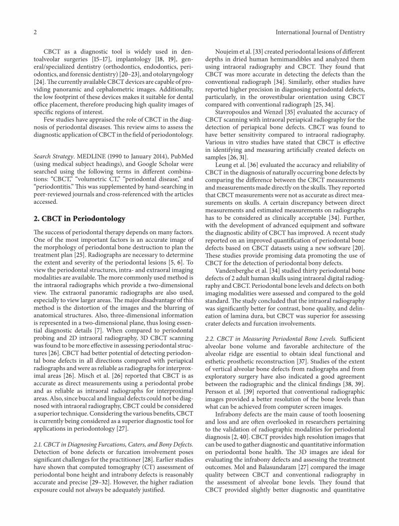
2 International Journal of Dentistry
CBCT as a diagnostic tool is widely used in den-toalveolar surgeries [15–17], implantology [18, 19], gen-eral/specialized dentistry (orthodontics, endodontics, peri-odontics, and forensic dentistry) [20–23], and otolaryngology[24].The currently available CBCTdevices are capable of pro-viding panoramic and cephalometric images. Additionally,the low footprint of these devices makes it suitable for dentaloffice placement, therefore producing high quality images ofspecific regions of interest.
Few studies have appraised the role of CBCT in the diag-nosis of periodontal diseases. This review aims to assess thediagnostic application ofCBCT in the field of periodontology.
Search Strategy. MEDLINE (1990 to January 2014), PubMed(using medical subject headings), and Google Scholar weresearched using the following terms in different combina-tions: “CBCT,” “volumetric CT,” “periodontal disease,” and“periodontitis.”This was supplemented by hand-searching inpeer-reviewed journals and cross-referenced with the articlesaccessed.
2. CBCT in Periodontology
The success of periodontal therapy depends on many factors.One of the most important factors is an accurate image ofthe morphology of periodontal bone destruction to plan thetreatment plan [25]. Radiographs are necessary to determinethe extent and severity of the periodontal lesions [5, 6]. Toview the periodontal structures, intra- and extraoral imagingmodalities are available.Themore commonly usedmethod isthe intraoral radiographs which provide a two-dimensionalview. The extraoral panoramic radiographs are also used,especially to view larger areas.Themajor disadvantage of thismethod is the distortion of the images and the blurring ofanatomical structures. Also, three-dimensional informationis represented in a two-dimensional plane, thus losing essen-tial diagnostic details [7]. When compared to periodontalprobing and 2D intraoral radiography, 3D CBCT scanningwas found to bemore effective in assessing periodontal struc-tures [26]. CBCT had better potential of detecting periodon-tal bone defects in all directions compared with periapicalradiographs andwere as reliable as radiographs for interprox-imal areas [26]. Misch et al. [26] reported that CBCT is asaccurate as direct measurements using a periodontal probeand as reliable as intraoral radiographs for interproximalareas. Also, since buccal and lingual defects could not be diag-nosed with intraoral radiography, CBCT could be considereda superior technique. Considering the various benefits, CBCTis currently being considered as a superior diagnostic tool forapplications in periodontology [27].
2.1. CBCT in Diagnosing Furcations, Caters, and Bony Defects.Detection of bone defects or furcation involvement posessignificant challenges for the practitioner [28]. Earlier studieshave shown that computed tomography (CT) assessment ofperiodontal bone height and intrabony defects is reasonablyaccurate and precise [29–32]. However, the higher radiationexposure could not always be adequately justified.
Noujeim et al. [33] created periodontal lesions of differentdepths in dried human hemimandibles and analyzed themusing intraoral radiography and CBCT. They found thatCBCT was more accurate in detecting the defects than theconventional radiograph [34]. Similarly, other studies havereported higher precision in diagnosing periodontal defects,particularly, in the orovestibular orientation using CBCTcompared with conventional radiograph [25, 34].
Stavropoulos and Wenzel [35] evaluated the accuracy ofCBCT scanning with intraoral periapical radiography for thedetection of periapical bone defects. CBCT was found tohave better sensitivity compared to intraoral radiography.Various in vitro studies have stated that CBCT is effectivein identifying and measuring artificially created defects onsamples [26, 31].
Leung et al. [36] evaluated the accuracy and reliability ofCBCT in the diagnosis of naturally occurring bone defects bycomparing the difference between the CBCT measurementsandmeasurementsmade directly on the skulls.They reportedthat CBCTmeasurements were not as accurate as direct mea-surements on skulls. A certain discrepancy between directmeasurements and estimated measurements on radiographshas to be considered as clinically acceptable [34]. Further,with the development of advanced equipment and softwarethe diagnostic ability of CBCT has improved. A recent studyreported on an improved quantification of periodontal bonedefects based on CBCT datasets using a new software [20].These studies provide promising data promoting the use ofCBCT for the detection of periodontal bony defects.
Vandenberghe et al. [34] studied thirty periodontal bonedefects of 2 adult human skulls using intraoral digital radiog-raphy and CBCT. Periodontal bone levels and defects on bothimaging modalities were assessed and compared to the goldstandard.The study concluded that the intraoral radiographywas significantly better for contrast, bone quality, and delin-eation of lamina dura, but CBCT was superior for assessingcrater defects and furcation involvements.
2.2. CBCT in Measuring Periodontal Bone Levels. Sufficientalveolar bone volume and favorable architecture of thealveolar ridge are essential to obtain ideal functional andesthetic prosthetic reconstruction [37]. Studies of the extentof vertical alveolar bone defects from radiographs and fromexploratory surgery have also indicated a good agreementbetween the radiographic and the clinical findings [38, 39].Persson et al. [39] reported that conventional radiographicimages provided a better resolution of the bone levels thanwhat can be achieved from computer screen images.
Infrabony defects are the main cause of tooth looseningand loss and are often overlooked in researchers pertainingto the validation of radiographic modalities for periodontaldiagnosis [2, 40]. CBCT provides high resolution images thatcan be used to gather diagnostic and quantitative informationon periodontal bone health. The 3D images are ideal forevaluating the infrabony defects and assessing the treatmentoutcomes. Mol and Balasundaram [27] compared the imagequality between CBCT and conventional radiography inthe assessment of alveolar bone levels. They found thatCBCT provided slightly better diagnostic and quantitative

International Journal of Dentistry 3
information on periodontal bone levels in three dimensionsthan conventional radiography.They found that the accuracyin the anterior aspect of the jaws is limited in both imagingtechniques, obtained with traditional means.
Alternatively, numerous studies have reported that theCBCT images provided comparable measurements of peri-odontal bone levels and defects as intraoral radiography[26, 41]. Vandenberghe et al. [34] reported that CBCTimages demonstrated more potential in the morphologicaldescription of periodontal bone defects, while the digitalradiography provided more bone details.
2.3. CBCT in the Visualization of Periodontal Ligament Space.A break in the continuity of lamina dura and a wedge-shaped radiolucent area at the mesial or distal aspect of theperiodontal ligament space are one of the earliest signs ofperiodontal disease [42].However, it is clear that this does notoccur until sometime after the loss of soft tissue attachment[43]. Therefore, only a sensitive imaging technique would beable to detect the earliest changes in the periodontal ligamentspace. The conventional intraoral radiographs have somesignificant disadvantages including the overlap of anatomicalstructures due to the positioning of theX-ray tube. Also, therecould be errors related to the chemical processing and patientpositioning [44]. Considering the potential advantages ofusing CBCT for assessing the periodontal structures, onlyvery few studies have used it for visualization of the peri-odontal ligament space [45, 46]. In terms of image quality, theCBCT scans were found to be superior to the CT scans withparticular reference to the periodontal ligament space [32].Various studies have stated that conventional radiographs arebetter thanCBCT in assessing the periodontal ligament space[34, 47]. Ozmeric et al. [47] created a phantom model withartificial periodontal ligament space to compare betweenCBCT and conventional radiographs. They found that theCBCT was inferior to conventional radiographs in termsof the clarity of the artificial periodontal ligament space.However, conflicting views were reported in an in vitro studythat found CBCT to be better than conventional radiographyin visualizing the periodontal ligament space [48]. A phan-tom mimicking variable periodontal ligament spaces wasradiographed using CBCT and intraoral radiographs [48].This study found that CBCT provided better visualization ofsimulated periodontal ligament space in this phantom.
2.4. Other Periodontal Applications of CBCT. Even withinthe field of periodontology, CBCT has found numerousapplications. CBCT has been widely used in the detectionof periapical pathology. Various studies have reported on theeffectiveness of using CBCT for the diagnosis of periapicalpathology [49–51]. The literature shows that CBCT imagesare superior for the detection of apical periodontitis thanconventional radiographs [49–51]. Apical periodontitis is oneof themost common endodontic diseases, and it is consideredto be the primary indication for root canal treatment anda sequela of inadequate or failing treatment [52]. CBCThas found application even in epidemiological surveys. Arecent study [53] used CBCT images from a database to
determine the prevalence of apical periodontitis. Dutta et al.[23] investigated the prevalence of periradicular periodontitisusing CBCT scans in a retrospective cross-sectional epidemi-ological study in a Scottish subpopulation. CBCT is a radio-logical technique that has been more successful in detectingperiradicular changes than conventional radiography [54]. Ina recent case-report, CBCT was used in the diagnosis of apalatogingival groove [55]. Another recent study evaluatedbone resorption at the extraction sites of a group of patientsunder orthodontic treatment using CBCT to evaluate theperiodontal and bone support loss after tooth extraction [56].CBCT was preferred due to its higher precision in detectingbone changes.
3. Conclusion
As the radiation dosage of CBCT is substantially higher thanthat of other routine dental imaging techniques, appropriatepatient selection criteria must be adopted [57]. Also, theinfluence of the technical conditions on the image quality isrelatively higher for CBCT. CBCT has the potential to gatheraccurate diagnostic and quantitative information about peri-odontal bone condition. Bony defects, caters, and furcationinvolvements seem to be better depicted on CBCT, whereasbone quality and periodontal ligament space scored betteron conventional intraoral radiography. CBCT does not offera significant advantage over conventional radiography forassessing the periodontal bone levels. Decision pertaining tothe use of CBCT in the field of periodontology should betaken after careful consideration of its advantages, limita-tions, and risks.
Conflict of Interests
The author declares that there is no conflict of interestsregarding the publication of this paper.
References
[1] B. L. Pihlstrom, B. S. Michalowicz, and N. W. Johnson, “Peri-odontal diseases,”The Lancet, vol. 366, no. 9499, pp. 1809–1820,2005.
[2] A. Tugnait, V. Clerehugh, and P. N. Hirschmann, “The useful-ness of radiographs in diagnosis and management of periodon-tal diseases: a review,” Journal of Dentistry, vol. 28, no. 4, pp.219–226, 2000.
[3] G. C. Armitage, “The complete periodontal examination,”Periodontology 2000, vol. 34, pp. 22–33, 2004.
[4] D. K. Benn, “A review of the reliability of radiographicmeasure-ments in estimating alveolar bone changes,” Journal of ClinicalPeriodontology, vol. 17, no. 1, pp. 14–21, 1990.
[5] P. Eickholz and E.Hausmann, “Accuracy of radiographic assess-ment of interproximal bone loss in intrabony defects usinglinear measurements,” European Journal of Oral Sciences, vol.108, no. 1, pp. 70–73, 2000.
[6] U. Bragger, “Radiographic parameters: biological significanceand clinical use,” Periodontology 2000, vol. 39, pp. 73–90, 2005.

4 International Journal of Dentistry
[7] M. K. Jeffcoat, I. C. Wang, and M. S. Reddy, “Radiographicdiagnosis in periodontics,” Periodontology 2000, vol. 7, pp. 54–68, 1995.
[8] Y. Arai, E. Tammisalo, K. Iwai, K. Hashimoto, and K. Shinoda,“Development of a compact computed tomographic apparatusfor dental use,” Dentomaxillofacial Radiology, vol. 28, no. 4, pp.245–248, 1999.
[9] P. Mozzo, C. Procacci, A. Tacconi, P. Tinazzi Martini, andI. A. Bergamo Andreis, “A new volumetric CT machine fordental imaging based on the cone-beam technique: preliminaryresults,” European Radiology, vol. 8, no. 9, pp. 1558–1564, 1998.
[10] K. Tsiklakis, C. Donta, S. Gavala, K. Karayianni, V.Kamenopoulou, and C. J. Hourdakis, “Dose reduction inmaxillofacial imaging using low dose cone beam CT,” EuropeanJournal of Radiology, vol. 56, no. 3, pp. 413–417, 2005.
[11] W. Kalender and K. O. Khadivi, “Computed tomography:fundamentals, system technology, image quality, applications,”Medical Physics, vol. 33, p. 3076, 2006.
[12] R. A. Danforth, I. Dus, and J. Mah, “3-D volume imaging fordentistry: a new dimension,” Journal of the California DentalAssociation, vol. 31, no. 11, pp. 817–823, 2003.
[13] J. B. Ludlow, L. E. Davies-Ludlow, and S. L. Brooks, “Dosimetryof two extraoral direct digital imaging devices: newtom conebeam CT and orthophos plus DS panoramic unit,” Dentomax-illofacial Radiology, vol. 32, no. 4, pp. 229–234, 2003.
[14] R. A. Danforth and D. E. Clark, “Effective dose from radiationabsorbed during a panoramic examination with a new genera-tionmachine,”Oral Surgery, OralMedicine, Oral Pathology, OralRadiology, and Endodontics, vol. 89, no. 2, pp. 236–243, 2000.
[15] Y. Nakagawa, H. Ishii, Y. Nomura et al., “Third molar position:reliability of panoramic radiography,” Journal of Oral andMaxillofacial Surgery, vol. 65, no. 7, pp. 1303–1308, 2007.
[16] D.-G. Liu, W.-L. Zhang, Z.-Y. Zhang, Y.-T. Wu, and X.-C.Ma, “Localization of impacted maxillary canines and observa-tion of adjacent incisor resorption with cone-beam computedtomography,” Oral Surgery, Oral Medicine, Oral Pathology, OralRadiology and Endodontology, vol. 105, no. 1, pp. 91–98, 2008.
[17] M. Martorelli, P. Ausiello, and R. Morrone, “A new method toassess the accuracy of a cone beam computed tomography scan-ner by using of a non-contact reverse engineering technique,”Journal of Dentistry, 2014.
[18] S. D. Ganz, “Conventional CT and cone beam CT for improveddental diagnostics and implant planning,” Dental ImplantologyUpdate, vol. 16, no. 12, pp. 89–95, 2005.
[19] M. E. Guerrero, R. Jacobs, M. Loubele, F. Schutyser, P. Suetens,and D. van Steenberghe, “State-of-the-art on cone beam CTimaging for preoperative planning of implant placement,” Clin-ical Oral Investigations, vol. 10, no. 1, pp. 1–7, 2006.
[20] J. Fleiner, C. Hannig, D. Schulze, A. Stricker, and R. Jacobs,“Digital method for quantification of circumferential periodon-tal bone level using cone beam CT,” Clinical Oral Investigations,vol. 17, no. 2, pp. 389–396, 2013.
[21] K. de Faria Vasconcelos, K. M. Evangelista, C. D. Rodrigues,C. Estrela, T. O. De Sousa, and M. A. G. Silva, “Detectionof periodontal bone loss using cone beam CT and intraoralradiography,” Dentomaxillofacial Radiology, vol. 41, no. 1, pp.64–69, 2012.
[22] K. Nakajima, T. Yamaguchi, and K.Maki, “Surgical orthodontictreatment for a patient with advanced periodontal disease: eval-uation with electromyography and 3-dimensional cone-beamcomputed tomography,” The American Journal of Orthodonticsand Dentofacial Orthopedics, vol. 136, no. 3, pp. 450–459, 2009.
[23] A. Dutta, F. Smith-Jack, and W. P. Saunders, “Prevalence ofperiradicular periodontitis in a Scottish subpopulation foundon CBCT images,” International Endodontic Journal, 2013.
[24] W. de vos, J. Casselman, and G. R. J. Swennen, “Cone-beamcomputerized tomography (CBCT) imaging of the oral andmaxillofacial region: a systematic review of the literature,”International Journal of Oral and Maxillofacial Surgery, vol. 38,no. 6, pp. 609–625, 2009.
[25] X. Braun, L. Ritter, P.-M. Jervøe-Storm, and M. Frentzen,“Diagnostic accuracy of CBCT for periodontal lesions,”ClinicalOral Investigations , 2013.
[26] K. A.Misch, E. S. Yi, andD. P. Sarment, “Accuracy of cone beamcomputed tomography for periodontal defect measurements,”Journal of Periodontology, vol. 77, no. 7, pp. 1261–1266, 2006.
[27] A. Mol and A. Balasundaram, “In vitro cone beam computedtomography imaging of periodontal bone,” DentomaxillofacialRadiology, vol. 37, no. 6, pp. 319–324, 2008.
[28] D. A. Tyndall and S. Rathore, “Cone-beamCT diagnostic appli-cations: caries, periodontal bone assessment, and endodonticapplications,”Dental Clinics of North America, vol. 52, no. 4, pp.825–841, 2008.
[29] R. A. Fuhrmann, A. Bucker, and P. R. Diedrich, “Assessment ofalveolar bone loss with high resolution computed tomography,”Journal of Periodontal Research, vol. 30, no. 4, pp. 258–263, 1995.
[30] A. Pistorius, C. Patrosio, B.Willershausen, P.Mildenberger, andG. Rippin, “Periodontal probing in comparison to diagnosis byCT-Scan,” International Dental Journal, vol. 51, no. 5, pp. 339–347, 2001.
[31] H. M. Pinsky, S. Dyda, R. W. Pinsky, K. A. Misch, and D.Sarment, “Accuracy of three-dimensional measurements usingcone-beamCT,”Dentomaxillofacial Radiology, vol. 35, no. 6, pp.410–416, 2006.
[32] R. Mengel, M. Candir, K. Shiratori, and L. Flores-de-Jacoby,“Digital volume tomography in the diagnosis of periodontaldefects: an in vitro study on native pig and human mandibles,”Journal of Periodontology, vol. 76, no. 5, pp. 665–673, 2005.
[33] M. Noujeim, T. J. Prihoda, R. Langlais, and P. Nummikoski,“Evaluation of high-resolution cone beam computed tomogra-phy in the detection of simulated interradicular bone lesions,”Dentomaxillofacial Radiology, vol. 38, no. 3, pp. 156–162, 2009.
[34] B. Vandenberghe, R. Jacobs, and J. Yang, “Detection of peri-odontal bone loss using digital intraoral and cone beamcomputed tomography images: an in vitro assessment of bonyand/or infrabony defects,”Dentomaxillofacial Radiology, vol. 37,no. 5, pp. 252–260, 2008.
[35] A. Stavropoulos and A. Wenzel, “Accuracy of cone beam dentalCT, intraoral digital and conventional film radiography for thedetection of periapical lesions. An ex vivo study in pig jaws,”Clinical Oral Investigations, vol. 11, no. 1, pp. 101–106, 2007.
[36] C. C. Leung, L. Palomo, R. Griffith, and M. G. Hans, “Accu-racy and reliability of cone-beam computed tomography formeasuring alveolar bone height and detecting bony dehiscencesand fenestrations,” The American Journal of Orthodontics andDentofacial Orthopedics, vol. 137, supplement 4, pp. S109–S119,2010.
[37] L. Schropp, A. Wenzel, L. Kostopoulos, and T. Karring, “Bonehealing and soft tissue contour changes following single-toothextraction: a clinical and radiographic 12-month prospectivestudy,” International Journal of Periodontics and RestorativeDentistry, vol. 23, no. 4, pp. 313–323, 2003.
[38] A. Bolin, S. Lavstedt, L. Frithiof, and C. O. Henrikson,“Proximal alveolar bone loss in a longitudinal radiographic

International Journal of Dentistry 5
investigation. IV. Smoking and some other factors influencingthe progress in individuals with at least 20 remaining teeth,”Acta Odontologica Scandinavica, vol. 44, no. 5, pp. 263–269,1986.
[39] R. E. Persson, L. G. Hollender, and G. R. Persson, “Assessmentof alveolar bone levels from intraoral radiographs in subjectsbetween ages 15 and 94 years seeking dental care,” Journal ofClinical Periodontology, vol. 25, no. 8, pp. 647–654, 1998.
[40] D. E. Deas, A. J. Moritz, B. L. Mealey, H. T. McDonnell, andC. A. Powell, “Clinical reliability of the “furcation arrow” as adiagnostic marker,” Journal of Periodontology, vol. 77, no. 8, pp.1436–1441, 2006.
[41] B. Vandenberghe, R. Jacobs, and J. Yang, “Diagnostic validity(or acuity) of 2D CCD versus 3D CBCT-images for assessingperiodontal breakdown,” Oral Surgery, Oral Medicine, OralPathology, Oral Radiology and Endodontology, vol. 104, no. 3, pp.395–401, 2007.
[42] F. Carranza, “Radiographic and other aids in the diagnosis ofperiodontal disease,” in Carranza’s Clinical Periodontology, M.G. Newman, H. H. Takei, P. R. Klokkevold, and F. A. Carranza,Eds., pp. 364–365, Saunders, Philadelphia, Pa, USA, 8th edition,1996.
[43] P. N. Hirschmann, “Radiographic interpretation of chronicperiodontitis,” International Dental Journal, vol. 37, no. 1, pp. 3–9, 1987.
[44] A. Mol, “Imaging methods in periodontology,” Periodontology2000, vol. 34, pp. 34–48, 2004.
[45] T. Tammisalo, T. Luostarinen, K. Vahatalo, and M. Neva,“Detailed tomography of periapical and periodontal lesions.Diagnostic accuracy compared with periapical radiography,”Dentomaxillofacial Radiology, vol. 25, no. 2, pp. 89–96, 1996.
[46] K. Ito, N. Yoshinuma, E. Goke, Y. Arai, andK. Shinoda, “Clinicalapplication of a new compact computed tomography system forevaluating the outcome of regenerative therapy: a case report,”Journal of Periodontology, vol. 72, no. 5, pp. 696–702, 2001.
[47] N. Ozmeric, I. Kostioutchenko, G. Hagler, M. Frentzen, andP.-M. Jervøe-Storm, “Cone-beam computed tomography inassessment of periodontal ligament space: in vitro study onartificial tooth model,” Clinical Oral Investigations, vol. 12, no.3, pp. 233–239, 2008.
[48] P.-M. Jervøe-Storm, M. Hagner, J. Neugebauer et al., “Com-parison of cone-beam computerized tomography and intraoralradiographs for determination of the periodontal ligament in avariable phantom,”Oral Surgery, Oral Medicine, Oral Pathology,Oral Radiology and Endodontology, vol. 109, no. 2, pp. e95–e101,2010.
[49] S. Lofthag-Hansen, S. Huumonen, K. Grondahl, and H.-G.Grondahl, “Limited cone-beam CT and intraoral radiographyfor the diagnosis of periapical pathology,” Oral Surgery, OralMedicine, Oral Pathology, Oral Radiology, and Endodontics, vol.103, no. 1, pp. 114–119, 2007.
[50] C. Estrela, M. R. Bueno, C. R. Leles, B. Azevedo, and J. R.Azevedo, “Accuracy of cone beam computed tomography andpanoramic and periapical radiography for detection of apicalperiodontitis,” Journal of Endodontics, vol. 34, no. 3, pp. 273–279, 2008.
[51] R. Ordinola-Zapata, C. M. Bramante, M. H. Duarte et al., “Theinfluence of cone-beam computed tomography and periapicalradiographic evaluation on the assessment of periapical bonedestruction in dog’s teeth,” Oral Surgery, Oral Medicine, OralPathology, Oral Radiology and Endodontology, vol. 112, no. 2, pp.272–279, 2011.
[52] M. A. Al-Omari, A. Hazaa, and F. Haddad, “Frequency anddistribution of root filled teeth and apical periodontitis in aJordanian subpopulation,” Oral Surgery, Oral Medicine, OralPathology, Oral Radiology and Endodontology, vol. 111, no. 1, pp.e59–e65, 2011.
[53] L. M. Paes da Silva Ramos Fernandes, R. Ordinola-Zapata, M.A. Hungaro Duarte, and A. L. Alvares Capelozza, “Prevalenceof apical periodontitis detected in cone beam CT images of aBrazilian subpopulation,”Dentomaxillofacial Radiology, vol. 42,no. 1, pp. 8017–9163, 2013.
[54] S. Patel, R.Wilson, A.Dawood, and F.Mannocci, “Thedetectionof periapical pathosis using periapical radiography and conebeam computed tomography—part 1: pre-operative status,”International Endodontic Journal, vol. 45, no. 8, pp. 702–710,2012.
[55] A. Rajput, S. Talwar, S. Chaudhary, and A. Khetarpal, “Success-fulmanagement of pulpo-periodontal lesion inmaxillary lateralincisor with palatogingival groove using CBCT scan,” IndianJournal of Dental Research, vol. 23, no. 3, pp. 415–418, 2012.
[56] L. Lombardo, R. Bragazzi, C. Perissinotto, D. Mirabella, andG. Siciliani, “Cone-beam computed tomography evaluation ofperiodontal and bone support loss in extraction cases,” Progressin Orthodontics, vol. 14, no. 1, p. 29, 2013.
[57] D. A. Tyndall, J. B. Price, S. Tetradis et al., “Position statementof the American academy of oral andmaxillofacial radiology onselection criteria for the use of radiology in dental implantologywith emphasis on cone beam computed tomography,” OralSurgery, Oral Medicine, Oral Pathology and Oral Radiology, vol.113, no. 6, pp. 817–8126, 2012.