epidemic ribotypes of clostridium (now clostridioides
TRANSCRIPT
RESEARCH ARTICLE Open Access
Epidemic ribotypes of Clostridium (nowClostridioides) difficile are likely to be morevirulent than non-epidemic ribotypes inanimal modelsJohn C. Vitucci1, Mark Pulse1, Leslie Tabor-Simecka2 and Jerry Simecka1*
Abstract
Background: Clostridioides difficile infections have become more frequently diagnosed and associated with greaterdisease severity, which has resulted in an increase burden on the healthcare system. These increases are attributedto the increased prevalence of hypervirulent strains encompassing select ribotypes. These epidemic ribotypes werecharacterized as hypervirulent due to higher in vitro spore and toxin production, as well as increased incidence, severity andmortality within patients. However, it is unclear whether epidemic ribotypes are truly more virulent than non-epidemicribotypes in vivo. Furthermore, there is conflicting evidence about the ability of a strain’s in vitro phenotype to be predictive oftheir in vivo virulence. The goals of the current studies were to determine if epidemic ribotypes are more virulent than otherribotypes in animal models, and whether the in vitro virulence phenotype of an isolate or ribotype predict in vivo virulence.
Results: To determine if epidemic strains were truly more virulent than other non-epidemic strains, the in vivo virulence of 13C. difficile isolates (7 non-epidemic and 6 epidemic ribotype isolates) were determined in murine and hamster models of CDI.The isolates of epidemic ribotype of C. difficile were found to be more virulent in both the murine and hamster models thannon-epidemic isolates. In particular, the group of epidemic ribotypes of C. difficile had lower LD50 values in hamsters. Theincreased severity of disease was associated with higher levels of Toxin A and Toxin B production found in fecal samples, butnot numbers of organisms recovered. The isolates were further characterized for their in vitro virulence phenotypes, e.g. toxinproduction, growth rates, spore formation and adherence of spores to intestinal epithelial cell lines. Although there were higherlevels of toxins produced and greater adherence for the group of epidemic ribotypes, the in vitro profiles of individual isolateswere not always predictive of their in vivo virulence.
Conclusions: Overall, the group of epidemic ribotypes of C. difficile were more virulent in vivo despite individual isolates havingsimilar phenotypes to the non-epidemic isolates in vitro.
Keywords: Clostridium, Clostridioides, Difficile, Animal models, Virulence, In vitro phenotype, Ribotype, Epidemic, Toxin
Clostridioides difficile, a spore forming bacillus, is thecause of C. difficile-associated disease. In the UnitedStates of America (US), the occurrence of C. difficile in-fections (CDI) increased by a factor of 400% between2000 and 2007 [1]. C. difficile is estimated to cause 500,000 infections in the US each year that results in 29,000deaths and associated annual healthcare costs of
approximately $3 billion [2, 3]. Clostridial endospores areessential for the environmental transmittance of C. difficilein humans and are resistant to a broad variety of physicaland chemical treatments [4, 5]. Within the host, C. difficilespores germinate into vegetative cells, which enablescolonization of the intestinal tract, toxin production, andeventual disease [6, 7]. Stages of disease progression includeintestinal inflammation, perforation, toxic megacolon,pseudo-membranous colitis, and death [7, 8]. Mortality as-sociated with CDI is approximately 5% but has been as highas 20% during particular outbreaks [9]. C. difficile is capable
© The Author(s). 2020 Open Access This article is distributed under the terms of the Creative Commons Attribution 4.0International License (http://creativecommons.org/licenses/by/4.0/), which permits unrestricted use, distribution, andreproduction in any medium, provided you give appropriate credit to the original author(s) and the source, provide a link tothe Creative Commons license, and indicate if changes were made. The Creative Commons Public Domain Dedication waiver(http://creativecommons.org/publicdomain/zero/1.0/) applies to the data made available in this article, unless otherwise stated.
* Correspondence: [email protected] of Pharmaceutical Sciences and UNTHSC Preclinical Services,University of North Texas System College of Pharmacy, University of NorthTexas Health Science Center, Fort Worth, TX, USAFull list of author information is available at the end of the article
Vitucci et al. BMC Microbiology (2020) 20:27 https://doi.org/10.1186/s12866-020-1710-5

of producing two different Rho glucosylating exotoxins,TcdA (toxin A) and TcdB (toxin B) [10, 11], which are re-sponsible for the pathology typically associated with CDI[12, 13]. Toxin A and B both produce multiple cytopathicand cytotoxic effects on the targeted cells [10]. These can in-clude disruption of Rho-dependent signaling, disruption ofthe actin cytoskeleton and of the tight adherence junctions,all causes of increased epithelial permeability which causethe diarrhea associated with C. difficile associated disease[10]. C. difficile isolates can produce another toxin, binarytoxin, which can disrupt normal cytoskeletal function of cells[14]; however, studies have yet to show that binary toxinplays a significant role in disease severity or virulence [15,16]. Therefore, both C. difficile spores and toxins play an im-portant role in disease transmission and pathogenesis, andthese virulence determinates have been shown to vary be-tween different C. difficile ribotypes [10, 11, 13, 17].The increase in the number and severity of CDI in the
United States is largely attributed to the emergence ofthe epidemic C. difficile clinical isolates, e.g. BI/NAP1/027 (type 027) and ribotype 078 [18, 19]. Interestingly,ribotype 027 is common among healthcare-associatedCDI cases, while the type 078 is more commonly associ-ated with community-acquired CDI [19]. Ribotype 027 isresponsible for 19 to 22.5% of hospital acquired CDIcases, and most of these cases are significantly associatedwith increased disease severity, recurrence, and mortality[19–21]. It was recently suggested that one possibilitywhy ribotypes 027 and 078 have become epidemicstrains was due to their ability to utilize low concentra-tions of the sugar trehalose [18]. The increased usage oftrehalose as a food additive in both the US and Europecoincides with the emergence of both ribotype 027 and078 outbreaks. Thus, the ability to utilize this sugar mayprovide a competitive advantage over other ribotypes,resulting in the increased frequency of infection within acomplex host environment [18]. Still, this does not ac-count for the increased frequency of diagnosis of diseaseassociated with infection with epidemic ribotypes, as wellas the increased severity of disease associated with themwhen compared to other non-epidemic ribotypes.The apparent increased severity of disease due to the
epidemic ribotypes of C. difficile suggests that these iso-lates may be more virulent than other ribotypes, and if so,this is likely linked to enhanced expression of virulencedeterminates, such as spores and toxins A and B [22].There are limited studies examining in vivo virulence ofmultiple isolates of the epidemic ribotypes using animalmodels [23, 24]. However, there are multiple in vitro stud-ies that characterize type 027’s spore and toxin produc-tion, but these studies have produced conflicting results.Some in vitro studies indicate that ribotype 027 has in-creased spore and toxin production [17, 22, 25, 26]. In-creased toxin production was highlighted in a study by
Warny et al., which found a ribotype 027 isolate express-ing 16 times more toxin A and 23 times more toxin B thatother ribotype isolates [22]. In contrast, other in vitrostudies found that spore production for other ribotype027 isolates were not significantly different from otherribotypes, and toxin production by ribotype 027 is not asrobust as shown in the study by Warny et al. [27, 28].These studies, as well as other studies, have not defini-tively compared the in vitro profiles of various C. difficileisolates with their ability to cause disease in vivo, leadingothers to speculate that clinical outcomes may be isolatedependent. Thus, it is unclear whether epidemic ribotypesare more virulent than other ribotypes, and whether thein vitro virulence phenotype of an isolate or ribotype isuseful in predicting in vivo virulence of individual isolates.To examine the virulence of epidemic isolates, we ini-
tially determined the in vivo virulence of 13 C. difficileisolates (7 non-epidemic and 6 epidemic) in two differ-ent animal models of CDI. The first animal model thatwas used in these studies was the murine model of CDI[23]. Being that mice are less susceptible to C. difficile,this model is an excellent shedding model and has beenused, with some success, as a survival model [23, 29].Also, due to this decreased sensitivity to C. difficile, themouse model is better suited for determining subtle dif-ferences between isolates that pose an issue in more sen-sitive animal models, such as toxin production overextended periods of time [20]. The second animal modelthat was used in these studies is the hamster model ofCDI. In contrast to mice, hamsters are very sensitive toC. difficile and, though there are differences (i.e., the in-creased sensitivity), closely parallels the characteristics ofclinical C. difficile-associated disease in humans [20].This enhanced sensitivity makes the hamster model ofCDI a strong choice for survival studies and the subse-quent calculation of LD50 values for C. difficile strains[29–31], whereas the murine model can be useful in dis-secting more subtle differences in virulence, such asin vivo toxin production and shedding of organismsother than lethality [20]. By using this approach, wefound collectively that the epidemic isolates had in-creased virulence in both experimental animal modelswhen compared to non-epidemic isolates. In particular,the group of epidemic ribotypes of C. difficile had lowerLD50 values in hamsters. Additionally, we also examinedthe in vitro production of toxins A and B, growth rates,spore formation and adherence of spores to intestinalepithelial cell lines, and although there was increase pro-duction of toxins and adherence for the group of epi-demic isolates, the in vitro profiles of individual isolateswere not predictive of their in vivo virulence. Overall,the group of epidemic ribotypes of C. difficile were morevirulent in vivo despite individual isolates having similarphenotypes to the non-epidemic isolates in vitro.
Vitucci et al. BMC Microbiology (2020) 20:27 Page 2 of 14
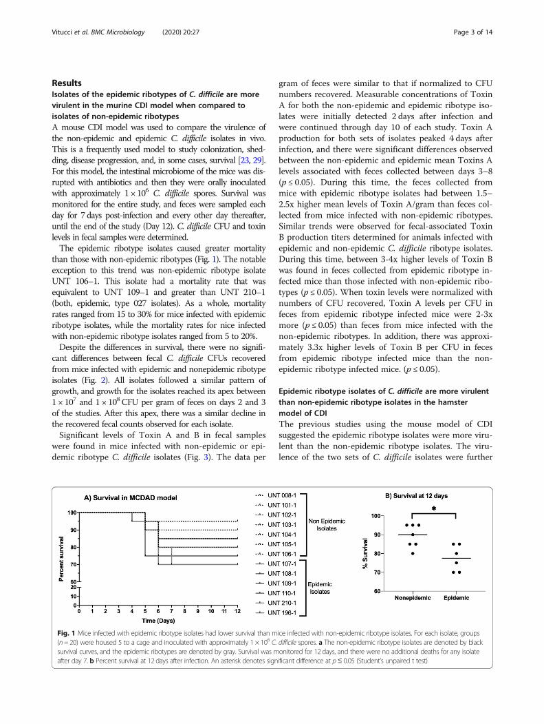
ResultsIsolates of the epidemic ribotypes of C. difficile are morevirulent in the murine CDI model when compared toisolates of non-epidemic ribotypesA mouse CDI model was used to compare the virulence ofthe non-epidemic and epidemic C. difficile isolates in vivo.This is a frequently used model to study colonization, shed-ding, disease progression, and, in some cases, survival [23, 29].For this model, the intestinal microbiome of the mice was dis-rupted with antibiotics and then they were orally inoculatedwith approximately 1 × 10⁶ C. difficile spores. Survival wasmonitored for the entire study, and feces were sampled eachday for 7 days post-infection and every other day thereafter,until the end of the study (Day 12). C. difficile CFU and toxinlevels in fecal samples were determined.The epidemic ribotype isolates caused greater mortality
than those with non-epidemic ribotypes (Fig. 1). The notableexception to this trend was non-epidemic ribotype isolateUNT 106–1. This isolate had a mortality rate that wasequivalent to UNT 109–1 and greater than UNT 210–1(both, epidemic, type 027 isolates). As a whole, mortalityrates ranged from 15 to 30% for mice infected with epidemicribotype isolates, while the mortality rates for nice infectedwith non-epidemic ribotype isolates ranged from 5 to 20%.Despite the differences in survival, there were no signifi-
cant differences between fecal C. difficile CFUs recoveredfrom mice infected with epidemic and nonepidemic ribotypeisolates (Fig. 2). All isolates followed a similar pattern ofgrowth, and growth for the isolates reached its apex between1 × 107 and 1 × 10⁸ CFU per gram of feces on days 2 and 3of the studies. After this apex, there was a similar decline inthe recovered fecal counts observed for each isolate.Significant levels of Toxin A and B in fecal samples
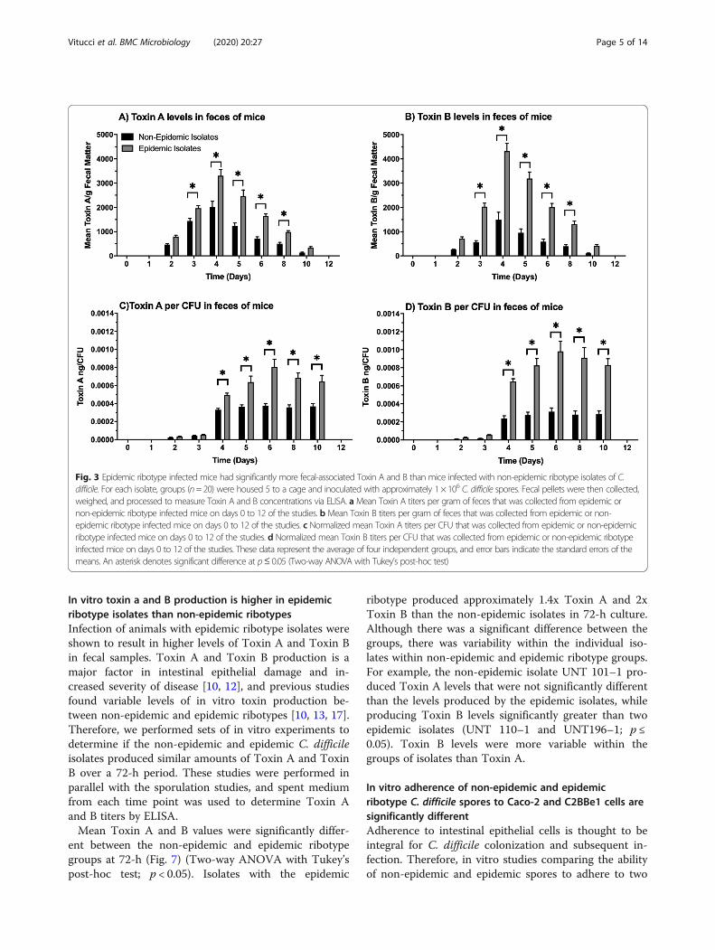
were found in mice infected with non-epidemic or epi-demic ribotype C. difficile isolates (Fig. 3). The data per
gram of feces were similar to that if normalized to CFUnumbers recovered. Measurable concentrations of ToxinA for both the non-epidemic and epidemic ribotype iso-lates were initially detected 2 days after infection andwere continued through day 10 of each study. Toxin Aproduction for both sets of isolates peaked 4 days afterinfection, and there were significant differences observedbetween the non-epidemic and epidemic mean Toxins Alevels associated with feces collected between days 3–8(p ≤ 0.05). During this time, the feces collected frommice with epidemic ribotype isolates had between 1.5–2.5x higher mean levels of Toxin A/gram than feces col-lected from mice infected with non-epidemic ribotypes.Similar trends were observed for fecal-associated ToxinB production titers determined for animals infected withepidemic and non-epidemic C. difficile ribotype isolates.During this time, between 3-4x higher levels of Toxin Bwas found in feces collected from epidemic ribotype in-fected mice than those infected with non-epidemic ribo-types (p ≤ 0.05). When toxin levels were normalized withnumbers of CFU recovered, Toxin A levels per CFU infeces from epidemic ribotype infected mice were 2-3xmore (p ≤ 0.05) than feces from mice infected with thenon-epidemic ribotypes. In addition, there was approxi-mately 3.3x higher levels of Toxin B per CFU in fecesfrom epidemic ribotype infected mice than the non-epidemic ribotype infected mice. (p ≤ 0.05).
Epidemic ribotype isolates of C. difficile are more virulentthan non-epidemic ribotype isolates in the hamstermodel of CDIThe previous studies using the mouse model of CDIsuggested the epidemic ribotype isolates were more viru-lent than the non-epidemic ribotype isolates. The viru-lence of the two sets of C. difficile isolates were further
Fig. 1 Mice infected with epidemic ribotype isolates had lower survival than mice infected with non-epidemic ribotype isolates. For each isolate, groups(n= 20) were housed 5 to a cage and inoculated with approximately 1 × 10⁶ C. difficile spores. a The non-epidemic ribotype isolates are denoted by blacksurvival curves, and the epidemic ribotypes are denoted by gray. Survival was monitored for 12 days, and there were no additional deaths for any isolateafter day 7. b Percent survival at 12 days after infection. An asterisk denotes significant difference at p≤ 0.05 (Student’s unpaired t test)
Vitucci et al. BMC Microbiology (2020) 20:27 Page 3 of 14

investigated using the hamster model of CDI. The ham-ster model is well established and shares some commonfeatures of C. difficile disease associated with the humanclinical condition [29, 32]. Like humans, hamsters alsoexhibit increased susceptibility to C. difficile infectionafter administration of a broad spectrum antibiotic thatoften leads to consistent clinical disease outcomes in theexperimental model [31, 32]. To perform these studies,groups of hamsters were inoculated with a range ofspore titers per isolate, and then treated with clindamy-cin to facilitate infection and subsequent disease estab-lishment. After this, the condition of the hamsters wasassessed multiple times a day, and fecal samples werecollected daily until the conclusion of the study on day7. Fecal samples were processed for CFU and assayed forToxin A and B concentration via ELISA.When LD50 values were compared between the iso-
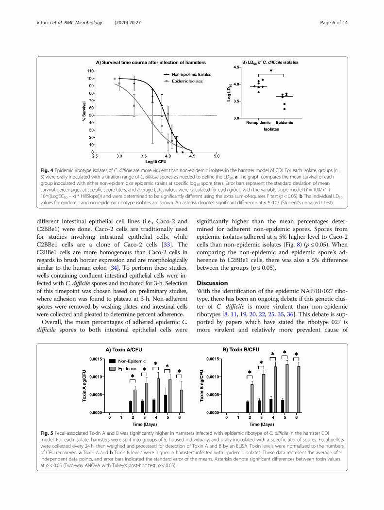
lates in the hamster CDI model, the epidemic isolateshad a lower mean LD50 value than the non-epidemic iso-lates did in the model (Fig. 4). The average LD50 valuewas 3.57 ± 0.025 log CFU for hamsters infected with epi-demic strains, and hamsters infected with non-epidemicstrains had a LD50 value of 3.94 ± 0.051 log CFU (p ≤0.05). As a whole, the LD50 values ranged from 3.27–3.72 log CFU for the hamsters infected with epidemicribotype strains, while the LD50 values for the hamstersinfected with non-epidemic ribotype isolates rangedfrom 3.76–4.13 log CFU.For this model, we chose not to compare fecal-
associated CFU counts, because determining the LD50
values led to varying inoculation doses for each isolate.Due to differences observed between the isolate’s toxinproduction in the mouse model, we chose to examine
fecal-associated Toxin A and B concentrations to deter-mine if this was similar in the hamster model. To dothis, toxin levels/CFU was assayed from the fecalsamples collected daily for 6 days after infection, andthe results were separated into multiple groups forcomparison purposes. Fecal-associated Toxin A and Bwere initially detected 2 days after infection for boththe non-epidemic and the epidemic ribotype infectedanimals (Fig. 5). When comparing non-epidemic andepidemic ribotype infected groups that survived, theepidemic isolate infected hamsters had approximately2-3x more Toxin A/CFU in their feces than did non-epidemic isolate infected hamsters (p ≤ 0.05), and thefeces collected from epidemic ribotype infected ani-mals had approximately 3-4x Toxin B/CFU higherlevels than hamsters infected with isolates of the non-epidemic ribotype (p ≤ 0.05).
In vitro growth and spore production are similar betweennon-epidemic and epidemic ribotype isolates of C. difficileEpidemic isolates were shown to be more virulent thannon-epidemic isolates in vivo, despite having no differ-ences in recovered CFU. To confirm that there are noinherent differences in growth and spore production ofthe isolates, in vitro growth and spore formation of allthe C. difficile isolates were determined over a 72-hperiod, and, it was found that non-epidemic and epi-demic strains exhibited similar in vitro growth patterns.Furthermore, when placed into sporulation medium,there was no difference over a 72-h period between epi-demic and non-epidemic isolates in spore formation orthe numbers of remaining vegetative cells (Fig. 6, Add-itional file 1: Figure S1).
Fig. 2 In vivo fecal-associated CFU counts were not different between isolates. For each isolate, groups (n = 20) were housed 5 to a cage andinoculated with approximately 1 × 10⁶ C. difficile spores. Fecal pellets were then collected, weighed, and processed to measure CFU countsthroughout the study. Mean fecal counts were not significantly different between the non-epidemic and epidemic ribotypes, and CFU countspeaked 3 days after infection which declined until the end of the study. These data represent the average of four independent groups, and errorbars indicate the standard errors of the means
Vitucci et al. BMC Microbiology (2020) 20:27 Page 4 of 14
In vitro toxin a and B production is higher in epidemicribotype isolates than non-epidemic ribotypesInfection of animals with epidemic ribotype isolates wereshown to result in higher levels of Toxin A and Toxin Bin fecal samples. Toxin A and Toxin B production is amajor factor in intestinal epithelial damage and in-creased severity of disease [10, 12], and previous studiesfound variable levels of in vitro toxin production be-tween non-epidemic and epidemic ribotypes [10, 13, 17].Therefore, we performed sets of in vitro experiments todetermine if the non-epidemic and epidemic C. difficileisolates produced similar amounts of Toxin A and ToxinB over a 72-h period. These studies were performed inparallel with the sporulation studies, and spent mediumfrom each time point was used to determine Toxin Aand B titers by ELISA.Mean Toxin A and B values were significantly differ-
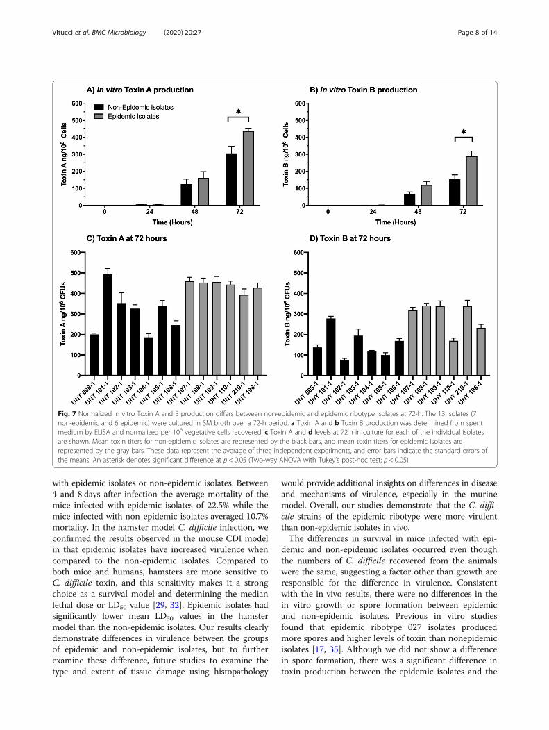
ent between the non-epidemic and epidemic ribotypegroups at 72-h (Fig. 7) (Two-way ANOVA with Tukey’spost-hoc test; p < 0.05). Isolates with the epidemic
ribotype produced approximately 1.4x Toxin A and 2xToxin B than the non-epidemic isolates in 72-h culture.Although there was a significant difference between thegroups, there was variability within the individual iso-lates within non-epidemic and epidemic ribotype groups.For example, the non-epidemic isolate UNT 101–1 pro-duced Toxin A levels that were not significantly differentthan the levels produced by the epidemic isolates, whileproducing Toxin B levels significantly greater than twoepidemic isolates (UNT 110–1 and UNT196–1; p ≤0.05). Toxin B levels were more variable within thegroups of isolates than Toxin A.
In vitro adherence of non-epidemic and epidemicribotype C. difficile spores to Caco-2 and C2BBe1 cells aresignificantly differentAdherence to intestinal epithelial cells is thought to beintegral for C. difficile colonization and subsequent in-fection. Therefore, in vitro studies comparing the abilityof non-epidemic and epidemic spores to adhere to two
Fig. 3 Epidemic ribotype infected mice had significantly more fecal-associated Toxin A and B than mice infected with non-epidemic ribotype isolates of C.difficile. For each isolate, groups (n=20) were housed 5 to a cage and inoculated with approximately 1 × 10⁶ C. difficile spores. Fecal pellets were then collected,weighed, and processed to measure Toxin A and B concentrations via ELISA. aMean Toxin A titers per gram of feces that was collected from epidemic ornon-epidemic ribotype infected mice on days 0 to 12 of the studies. b Mean Toxin B titers per gram of feces that was collected from epidemic or non-epidemic ribotype infected mice on days 0 to 12 of the studies. c Normalized mean Toxin A titers per CFU that was collected from epidemic or non-epidemicribotype infected mice on days 0 to 12 of the studies. d Normalized mean Toxin B titers per CFU that was collected from epidemic or non-epidemic ribotypeinfected mice on days 0 to 12 of the studies. These data represent the average of four independent groups, and error bars indicate the standard errors of themeans. An asterisk denotes significant difference at p≤ 0.05 (Two-way ANOVA with Tukey’s post-hoc test)
Vitucci et al. BMC Microbiology (2020) 20:27 Page 5 of 14
different intestinal epithelial cell lines (i.e., Caco-2 andC2BBe1) were done. Caco-2 cells are traditionally usedfor studies involving intestinal epithelial cells, whileC2BBe1 cells are a clone of Caco-2 cells [33]. TheC2BBe1 cells are more homogenous than Caco-2 cells inregards to brush border expression and are morphologicallysimilar to the human colon [34]. To perform these studies,wells containing confluent intestinal epithelial cells were in-fected with C. difficile spores and incubated for 3-h. Selectionof this timepoint was chosen based on preliminary studies,where adhesion was found to plateau at 3-h. Non-adherentspores were removed by washing plates, and intestinal cellswere collected and pleated to determine percent adherence.Overall, the mean percentages of adhered epidemic C.
difficile spores to both intestinal epithelial cells were
significantly higher than the mean percentages deter-mined for adherent non-epidemic spores. Spores fromepidemic isolates adhered at a 5% higher level to Caco-2cells than non-epidemic isolates (Fig. 8) (p ≤ 0.05). Whencomparing the non-epidemic and epidemic spore’s ad-herence to C2BBe1 cells, there was also a 5% differencebetween the groups (p ≤ 0.05).
DiscussionWith the identification of the epidemic NAP/BI/027 ribo-type, there has been an ongoing debate if this genetic clus-ter of C. difficile is more virulent than non-epidemicribotypes [8, 11, 19, 20, 22, 25, 35, 36]. This debate is sup-ported by papers which have stated the ribotype 027 ismore virulent and relatively more prevalent cause of
Fig. 5 Fecal-associated Toxin A and B was significantly higher in hamsters infected with epidemic ribotype of C. difficile in the hamster CDImodel. For each isolate, hamsters were split into groups of 5, housed individually, and orally inoculated with a specific titer of spores. Fecal pelletswere collected every 24 h, then weighed and processed for detection of Toxin A and B by an ELISA. Toxin levels were normalized to the numbersof CFU recovered. a Toxin A and b Toxin B levels were higher in hamsters infected with epidemic isolates. These data represent the average of 5independent data points, and error bars indicated the standard error of the means. Asterisks denote significant differences between toxin valuesat p < 0.05 (Two-way ANOVA with Tukey’s post-hoc test; p < 0.05)
Fig. 4 Epidemic ribotype isolates of C. difficile are more virulent than non-epidemic isolates in the hamster model of CDI. For each isolate, groups (n =5) were orally inoculated with a titration range of C. difficile spores as needed to define the LD50. a The graph compares the mean survival of eachgroup inoculated with either non-epidemic or epidemic strains at specific log10 spore titers. Error bars represent the standard deviation of meansurvival percentages at specific spore titers, and average LD50 values were calculated for each group with the variable slope model (Y = 100/ (1 +10^((LogEC50 – x) * HillSlope))) and were determined to be significantly different using the extra sum-of-squares F test (p < 0.05). b The individual LD50
values for epidemic and nonepidemic ribotype isolates are shown. An asterisk denotes significant difference at p≤ 0.05 (Student’s unpaired t test)
Vitucci et al. BMC Microbiology (2020) 20:27 Page 6 of 14

disease because it hyper-produces toxins and sporesin vitro [17, 19, 24, 25]. Whereas, other papers have statedthere is little differences between the 027 ribotype andother non-027 ribotypes in vitro [8, 11, 37]. However,there is also a question whether in vitro characterizationsaccurately predict the in vivo virulence of individual C. dif-ficile isolate or a group of isolates of the same ribotype.Therefore, we undertook a comprehensive set of in vitroand in vivo studies of 13 C. difficile isolates (7 of non-epidemic ribotypes and 6 of epidemic ribotypes) to exam-ine whether isolates of the epidemic ribotype are morevirulent than non-epidemic isolates in vivo. To do this, wenot only characterized the isolates in vitro, but also used aunique approach of characterizing the same isolates’in vivo virulence within two different animal models of C.difficile infection. Each of the animal models are valuablein understanding various contributing factors of C. difficile
disease. There are strength and weaknesses of each animalmodel [29, 32], and using both models decreased the poten-tial skewing of the data associated with the weaknesses andstrengths of each model. With this approach, we were ableto answer questions about C. difficile’s epidemic ribotype incomparison to other non-epidemic ribotypes. Such as, isthere truly a difference between non-epidemic and epi-demic isolate’s in vivo virulence, and is an isolate’s in vitrovirulence phenotype predictive of its in vivo virulence?As a group, isolates of an epidemic ribotype were more
virulent than those from non-epidemic ribotypes, al-though there was variability within each group of ribo-types. Difference in in vivo virulence was found usingtwo animal models, murine and hamster. The mousemodel is an excellent shedding model and has beenused, with some success, as a survival model [23, 29]. Inmice, there were differences in survival after infection
Fig. 6 Mean vegetative CFUs and spore recovery between non-epidemic and epidemic ribotype isolates did not differ over 72-h. The 13 isolates(7 non-epidemic and 6 epidemic) were incubated in SM broth over a 72-h period. A representative sample was then taken from each culture andplated on an agar medium ±0.1% taurocholate. The non-epidemic isolates are represented by the black bars, and the epidemic isolates arerepresented by the gray bars. This data represents the average of three independent experiments and error bars indicate the standard errors ofthe means. a Mean vegetative CFU’s recovered from 72-h SM broth cultures. b Mean spores/mL recovered from 72-h SM broth culture. c Meannumber of spores recovered from SM broth cultures normalized per 1000 vegetative cells recovered at the corresponding time point
Vitucci et al. BMC Microbiology (2020) 20:27 Page 7 of 14
with epidemic isolates or non-epidemic isolates. Between4 and 8 days after infection the average mortality of themice infected with epidemic isolates of 22.5% while themice infected with non-epidemic isolates averaged 10.7%mortality. In the hamster model C. difficile infection, weconfirmed the results observed in the mouse CDI modelin that epidemic isolates have increased virulence whencompared to the non-epidemic isolates. Compared toboth mice and humans, hamsters are more sensitive toC. difficile toxin, and this sensitivity makes it a strongchoice as a survival model and determining the medianlethal dose or LD50 value [29, 32]. Epidemic isolates hadsignificantly lower mean LD50 values in the hamstermodel than the non-epidemic isolates. Our results clearlydemonstrate differences in virulence between the groupsof epidemic and non-epidemic isolates, but to furtherexamine these difference, future studies to examine thetype and extent of tissue damage using histopathology
would provide additional insights on differences in diseaseand mechanisms of virulence, especially in the murinemodel. Overall, our studies demonstrate that the C. diffi-cile strains of the epidemic ribotype were more virulentthan non-epidemic isolates in vivo.The differences in survival in mice infected with epi-
demic and non-epidemic isolates occurred even thoughthe numbers of C. difficile recovered from the animalswere the same, suggesting a factor other than growth areresponsible for the difference in virulence. Consistentwith the in vivo results, there were no differences in thein vitro growth or spore formation between epidemicand non-epidemic isolates. Previous in vitro studiesfound that epidemic ribotype 027 isolates producedmore spores and higher levels of toxin than nonepidemicisolates [17, 35]. Although we did not show a differencein spore formation, there was a significant difference intoxin production between the epidemic isolates and the
Fig. 7 Normalized in vitro Toxin A and B production differs between non-epidemic and epidemic ribotype isolates at 72-h. The 13 isolates (7non-epidemic and 6 epidemic) were cultured in SM broth over a 72-h period. a Toxin A and b Toxin B production was determined from spentmedium by ELISA and normalized per 106 vegetative cells recovered. c Toxin A and d levels at 72 h in culture for each of the individual isolatesare shown. Mean toxin titers for non-epidemic isolates are represented by the black bars, and mean toxin titers for epidemic isolates arerepresented by the gray bars. These data represent the average of three independent experiments, and error bars indicate the standard errors ofthe means. An asterisk denotes significant difference at p < 0.05 (Two-way ANOVA with Tukey’s post-hoc test; p < 0.05)
Vitucci et al. BMC Microbiology (2020) 20:27 Page 8 of 14

non-epidemic isolates in the animal models of C. difficileinfection. In both mice and hamsters, there were two tothree times higher levels of both toxins after infectionwith the epidemic isolates. Consistent with the previouspublished studies [17, 22], higher levels of toxin produc-tion, by epidemic isolates, was also found during in vitroculture, but was only significant at 72-h in culture. Ap-proximately two times more toxin production was associ-ated with the epidemic isolates in in vitro cultures whencompared to the non-epidemic isolates. It is worth notingincreased toxin production for some ribotype 027 isolatesis associated with genetic mutations within its pathogen-icity island, this could also play a role in the epidemic iso-lates’ increased virulence in vivo [25, 38, 39]. Thus, theincreased virulence of the epidemic isolates was linked tothe higher production of Toxin A and Toxin B.Although toxin levels may be the most critical factor
involved in increased disease severity, there may beother factors. For example, one factor that is speculatedto contribute to C. difficile virulence is an isolate’s abilityto adhere to intestinal epithelium, but although it is ac-cepted that adherence is an important step for otherpathogens, it is currently not clear what the significanceis of adherence for this C. difficile in clinical disease.Studies do suggest it may play a role. Adherence of C.difficile spores to epithelium is dependent on the charac-teristics of exosporium, and the composition of this out-most layer can vary between strains [40–42]. Recently,two cysteine-rich proteins, cdeC and cdeM, were shownto influence the ability of C. difficile spores to adhere tointestinal epithelium [40]. In the mouse model of infec-tion, spores lacking the CdeC protein had increasedcolonization rates, recurrence rate, and were correlatedwith higher toxin titers during disease [40]. These results
suggest that adherence mediated factors could play arole in the increased virulence associated with the epi-demic isolates. In the current studies, the ability of C.difficile spores to adhere to two sets of human epithelialcells, Caco-2 and C2BBe1, in vitro was investigated, andthe epidemic isolates had about 5% greater adherence toboth cell lines than non-epidemic isolates. The ability ofthe epidemic strains to better bind to the epithelium sug-gests that these strains will more easily reach the inocula-tion threshold needed for the establishment of disease. Inaddition to adherence mediated factors, the spore coatalso harbors varying receptors for germination which re-spond to germanites and co-germinates [43]. Work byCarlson et al. has shown that epidemic isolates respond tomore optimized conditions for germinations, and, in turn,this led to more severe disease due to these ribotypes [43].Though the exact reasons for this has not been elucidated,it is hypothesized that more efficient germination couldlead to lower inoculation doses of spores needed to causedisease [43]. In support, lower doses of epidemic ribotypeisolates are needed to cause disease, e.g. LD50, in the ham-ster, but further studies are needed.In vitro virulence phenotypes of individual C. difficile
isolates were not predictive of their in vivo virulence. Al-though the group of epidemic isolates had higher levels oftoxin production in vitro, the level of toxin productionin vitro did not predict in vivo virulence for each individ-ual isolate. For example, UNT 101–1, a non-epidemic iso-late, expressed Toxin A and Toxin B at levels similar tothose of the epidemic isolates in in vitro cultures. In con-trast, in vitro characterizations showed that UNT 110–1and 210–1, two epidemic isolates, had toxin levels thatwere approximately equal with non-epidemic isolates.However, UNT 101–1, though producing high levels of
Fig. 8 Spores of epidemic ribotype adhere significantly different than those from the non-epidemic ribotype in vitro to Caco-2 and C2BBe1 Cells.C. difficile isolates (7 non-epidemic and 6 epidemic) were incubated with either Caco-2 or C2BBe1 cells for 3-h, washed, plated and counted todetermine the adhesion for each isolate. The non-epidemic isolates are denoted by the black symbols and the epidemic isolates by the graysymbols. a The isolates were incubated with Caco-2 cells and the mean adhesion percentages were determined as the percentage of sporesbound after washing as compared to the original inoculum dose. b The isolates were incubated with C2BBe1 cells and the mean adhesionpercentages were determined as the percentage of the spores bound after washing as compared to the original inoculum dose. These datarepresent the average of three independent experiments and error bars indicate the standard errors of the means, and a statistically significantdifference between each group at p < 0.05 (One-way ANOVA with Tukey’s post-hoc test; p < 0.05)
Vitucci et al. BMC Microbiology (2020) 20:27 Page 9 of 14

toxin in vitro, was one of the least virulent isolates in vivo,while UNT 110–1 and 210–1 were equal to other epi-demic isolates’ observed virulence in the mouse and ham-ster CDI models. Not only does this suggest that theevaluation of an individual isolate’s virulence should bedone using an in vivo model, but it is a strong possibilitythat factors in the in vivo environment influence an iso-late’s toxin production and virulence [40, 44, 45]. In fact,previous studies demonstrate that C. difficile epidemicribotype isolates can have increased in vivo fitness com-pared to non-epidemic isolates [18, 24]. They are capableof interacting more efficiently with metabolites producedby the host’s GI microbiome and have the ability to utilizeadditional nutrients that other ribotypes are unable to use.In addition, other factors may contribute to the in vivovirulence of C. difficile. For example, although the role ofbinary toxin in virulence is unclear [15, 16], a study sug-gests that binary toxin may suppress host immune re-sponses which results in enhanced virulence of epidemicribotype 027 strains in a mouse model [46]. Most likelycomplex combinations of factors of C. difficile influencesthe outcome of infection, and to further complicate theability to assess virulence solely using in vitro studies, thelevel and types of factors may be differentially expressedin the in vivo environment. Thus, in vitro characterizationof virulence factors produced by C. difficile alone is not re-liable approach to assess the potential to cause disease byindividual isolates, but this approach may still be useful incomparing the potential of different groups, e.g. ribotypes,of organisms to cause disease.Overall, these studies demonstrated that epidemic ribo-
types of C. difficile are likely to be more virulent than non-epidemic ribotypes. Within the last 10 years, C. difficile hasbecome an ever-increasing threat, even being designated anurgent threat level organism in 2013 by the Centers for Dis-ease Control, and the major reason for this is linked to therise of the epidemic NAP/BI/027 ribotype, along with other“hyper-virulent” ribotypes [19, 26]. Results described in thesestudies provide a comprehensive examination of virulencebetween different C. difficile isolates through multiplemethods and provides an important contribution in furtherunderstanding what causes the NAP/BI/027 ribotype to belabelled as, epidemic, hyper-virulent, and such a prevalentthreat to healthcare. Previous studies debated whether thecurrent epidemic ribotypes are more virulent than the non-epidemic ribotypes [11, 17, 19, 23, 25, 35]. This appears tobe the first study to compare the abilities of isolates of epi-demic and non-epidemic ribotypes to cause disease in boththe mice and hamster models of CDI. Although all C. difficileisolates examined were able to cause disease in both ham-sters and mice, the group of isolates with epidemic ribotypecaused more severe disease than the non-epidemic group ofisolates, providing a compelling case that the epidemic ribo-type is indeed more virulent. Additionally, the in vivo and
in vitro data supports the idea that the levels of toxins A andB production are likely to contribute to the increased viru-lence of the epidemic isolates. Other factors, such as the abil-ity to adhere to epithelial cells, may also play a role.However, there was variability in disease severity between in-dividual isolates within the group of epidemic and non-epidemic ribotypes, with one non-epidemic isolate causeddisease as severe as one of the epidemic strains. Furthermore,in vitro expression of virulence factors, such as toxin produc-tion and adherence to epithelial cells, corresponded with dis-ease potential of the ribotype groups, but was not a reliableapproach to assess the potential to cause disease by individ-ual isolates. These results suggest a link between the abilityto cause disease and the likelihood of a ribotype’s ability tobe epidemic and more easily transmissible between hosts.However, further studies are needed to directly link the ribo-type with increased virulence and spread of infection.
MethodsBacterial strains and Ribotype confirmationAll C. difficile isolates used in this study are listed in Table 1.C. difficile UNT 101–1 to UNT-110-1 were kindly providedby Dr. Curtis Donskey (Cleveland VA); UNT 008–1, UNT210–1, and UNT 196–1 were obtained from the AmericanType Culture Collection (ATCC). The source of relevant char-acteristics of each isolate can be found in Table 1. Ribotypeswere confirmed by running polymerase chain reaction (PCR)ribotyping with primers found in Bidet et. al. [47]. PCR frag-ments were analyzed in a Hitachi 3500xL genetic analyzerwith a 36 cm capillary loaded with a POP4 gel (Applied Bio-systems). The size of each peak was determined using PeakScanner software (Applied Biosystems). A database was gener-ated from the results of the capillary gel electrophoresis-basedPCR ribotyping result of each strain (http://webribo.ages.at).An error margin of ±4 bp was incorporated into the analysisalgorithm of the database [48].
MediaSporulation medium (SM) contained 90 g Trypticase Pep-tone, 5 g Proteose Peptone no. 3, 1 g Ammonium Sulfate,and 1.5 g of Tris in 1 l of distilled water. The pH was ad-justed to 7.4 at 37o with 1M NaOH. SM is a broth mediummade according to what has been previously described [49].TSA with 5% blood agar was made with 1 L of distilled
water (DI), 30 g of TSB, and 15 g of granulated agar withconstant mixing over low heat. Once the granulated agarwas dissolved, the mixture was autoclaved (20 min,121 °C, 15 psi). Once cooled to approximately 50 °C, 50mL of the medium was removed, and 50 mL of steriledefibrinated sheep blood (Remel, Lenexa, KS) was addedand mixed into the medium. Approximately 12 mL ofmedium was then poured into petri dishes and cooledovernight to solidify and stored in a 4 °C refrigeratoruntil used.
Vitucci et al. BMC Microbiology (2020) 20:27 Page 10 of 14

TGY-vegetative medium contained 5 g Tryptone, 5 g Yeastextract, 1 g Glucose, 1 g Potassium Phosphate, 15 g agar, and1 l of distilled water. This liquid-based medium was made ac-cording to what has been previously published [50].Columbia horse blood agar with 0.1% sodium tauro-
cholate was made by adding 869 mL of distilled water, incombination with 35 g of Columbia broth (Remel), and15 g of Difco Agar, granulated (BD). The mixture wasautoclaved (20 min, 121 °C, 15 psi). Once cooled, 70 mLof horse blood and 50 mL of a 20 mg/mL stock of so-dium taurocholate, 10 mL of a 50 mg/Ml stock of cyclo-serine and 1mL of a 15.5 mg/mL stock of cefoxitin werealso added.
Preparation of C. difficile spore stocksSpore stocks of each C. difficile strain were generated for usein the cellular adherence assay and the experimental animalmodels of CDI. These stocks were generated by growing eachstrain on 5% TSAb plates incubated at 37 °C in anaerobic con-ditions for 7 days. Plate growth was collected in a 1X PBS so-lution containing 1% (V/V) Tween-80 (ST-80), andsuspensions were washed 3 times in equal volumes of ST-80.Suspensions were incubated for 1 h at 65 ± 2 °C, washed withST-80, and re-suspended in 4mL of sterile nanopore water.Suspensions were then stored overnight at 4 °C in order topromote the maturation of endospores for each strain. Sporeswere separated from vegetative cells and residual debris bydensity gradient centrifugation (10min at 4500 x g) with a25% (W/V) HistoDenz solution. Spore pellets were washed 3times with ST-80 and suspended in sterile nanopore water toa final volume of 2mL. Spore stocks for each strain werestored at − 80 °C until used in in vitro or in vivo studies, andthe numbers of organisms given for infection or used inin vitro studies were confirmed for each study.
Mouse C. difficile associated disease modelFemale C57 BL/6 mice that were 7 to 8 weeks old were ob-tained from Charles River Laboratory and housed in sterilecaging for the in-life portion of each study. Animals wererandomly organized into groups of 20 (n = 20) and placedon drinking water supplemented with a cocktail of antibi-otics immediately upon arrival. These antibiotics and theirconcentrations were: Kanamycin (0.4mg/mL), Colistin(850 units/mL), Gentamicin (0.035mg/mL), Metronidazole(.215mg/mL), Vancomycin (0.045mg/mL) [23]. Animalswere left on the antibiotic supplemented water for 5 days,and then switched to normal water for 24 h. Mice were or-ally inoculated with 1 × 10⁶ C. difficile spores, and clinda-mycin was administered subcutaneously at 10mg/kg ofbody weight. Starting the day of infection, and each dayafter, approximately 0.1–0.2 g of feces was collected fromcages to determine C. difficile counts and associatedamounts of toxin A and B. Bedding was changed daily toensure fresh feces were collected for analysis, and census ofsurvivors were recorded daily for 14 days after infection.Feces were weighed before sterile 1x PBS was added to therecovered feces, this solution was then homogenized, and 1mL was separated for each total CFU recovery, spore recov-ery, and toxin A and B expression. Viable cell counts, sporecounts, and toxin expression were quantified as describedin the Material and Methods. The homogenized solutionseparated for spore quantification was heated to 65 ± 2 °Cfor 1 h to facilitate the isolation of only spores, while thefecal matter separated for toxin expression was diluted ap-proximately 100x - 500x for quantification. This allowed itto fall within detection range of the ELISA used to deter-mine toxin concentration.
Table 1 Clostridioides difficile Strain Designation, Sources, andCharacteristics. This table denotes the source of the individualisolates, other designations for each isolate, and some of themajor characteristics associated with each of the isolates
C. difficile Isolates and Sources
UNTStrain#
Source Relevant Characteristics
UNT101–1
Ohio VA MedicalCenter (Curtis Donskey)
Non-epidemic (Ribotype 014/0), OtherDesignation VA1
UNT102–1
Ohio VA MedicalCenter (Curtis Donskey)
Non-epidemic (Ribotype 660), OtherDesignation VA10
UNT103–1
Ohio VA MedicalCenter (Curtis Donskey)
Non-epidemic (Ribotype 428), REA J-type strain, binary toxin negative,non-epidemic, Other Designation VA11
UNT104–1
Ohio VA MedicalCenter (Curtis Donskey)
Non-epidemic (Ribotype 428), OtherDesignation UH15
UNT105–1
Ohio VA MedicalCenter (Curtis Donskey)
Non-epidemic (Ribotype 053), OtherDesignation UH18
UNT106–1
Ohio VA MedicalCenter (Curtis Donskey)
Epidemic (BI/NAP1, binary toxinpositive, Ribotype 027), OtherDesignation VA5
UNT107–1
Ohio VA MedicalCenter (Curtis Donskey)
Epidemic (BI/NAP1, binary toxinpositive, Ribotype 027), OtherDesignation VA17
UNT108–1
Ohio VA MedicalCenter (Curtis Donskey)
Epidemic (BI/NAP1, binary toxinpositive, Ribotype 027), OtherDesignation VA20
UNT109–1
Ohio VA MedicalCenter (Curtis Donskey)
Epidemic (BI/NAP1, binary toxinpositive, Ribotype 027), OtherDesignation CC20
UNT110–1
Ohio VA MedicalCenter (Curtis Donskey)
NAP-1, Epidemic, Other DesignationL32
UNT196–1
ATCC Epidemic (Ribotype 078), BAA-1875(Other Designation: 5325), Binary toxinpositive, Toxinotype V PFGE tye NAP7,REA type BI 8
UNT210–1
ATCC Epidemic (Ribotype 027) BAA-1870;Binary toxin positive, Toxinotype IIIbPFGE tye NAP1, REA type BI 8
UNT008–1
ATCC Non-epidemic (Ribotype 012), BAA-1382
Vitucci et al. BMC Microbiology (2020) 20:27 Page 11 of 14

Hamster LD-50/Survival C. difficile associated diseasemodelsMale Golden Syrian hamsters that were 6 to 7 weeks oldwere purchased from Envigo RMS Inc., and individuallyhoused in sterile cages. Up to 30 hamsters were used ineach study with 5 animals in each group that were orallyinoculated with a designated spore titer of each strain.The animals were inoculated with 0.5 mL of C. difficilespores from a spore preparation culture though oral gav-age. The inoculation dose for all strains ranged from 800to 30,000 spores/mL, and the exact titers chosen foreach strain were based on previously conducted studiesand observation of higher titers with non-epidemic andepidemic strains. Clindamycin was administered sub-cutaneously to each animal at 10 mg/kg per body weightapproximately 24 h after infection. Starting the day of in-fection, and each day after, approximately 0.1 to 0.2 g offeces was collected individually from each cage to deter-mine C. difficile counts and associated amounts of toxinA and B. Bedding was changed daily to ensure fresh feceswere collected for analysis, and census of survivors were re-corded daily for 7 days after infection. Cecal fluid was col-lected from deceased hamsters for C. difficile enumerationand toxin A and B quantification. Feces were weighed be-fore sterile 1x PBS was added to the recovered feces, thissolution was then homogenized, and 1mL was separatedfor each total CFU recovery, spore recovery, and toxin Aand B expression. Viable cell counts, spore counts, andtoxin expression were quantified as described in the Mater-ial and Methods. The homogenized solution separated forspore quantification was heated to 65 ± 2 °C for 1 h to facili-tate the isolation of only spores, and the fecal matter sepa-rated for toxin expression was diluted approximately 100x -500x for quantification. Cecal fluid was processed identi-cally to the fecal samples, with the exception that they werenot homogenized. This allowed it to fall within detectionrange of the ELISA used to determine toxin concentration.
In vitro growth of C. difficile vegetative cells and sporeformationPlate growth of each C. difficile isolate was transferredinto TGY-veg broth and anaerobically incubated at 37 °Cfor 24 h. TGY-veg associated growth for each strain wasadjusted to an optical density of 0.1 (600 nm) in eitherSM or TGY-veg broth, which were anaerobically incu-bated at 37 °C. Samples from each broth culture werecollected in triplicate every 24 h through 72 h of total in-cubation, and these samples were 10-fold serially dilutedand plated onto Columbia horse blood agar. Addition-ally, a second sample from each culture were possessedfor spore counts by incubating each sample in an equalvolume of 200 proof ethanol for 30 min, and then incu-bating the samples at 65 ± 2 °C for 1 h. The ethanol andheat-treated samples were centrifuged, washed with PBS,
and the spore-containing pellets were suspended in avolume of PBS equal to the original volume of the sam-ple. Ethanol and heat-treatment at 65 ± 2 °C were testedand sufficient to remove all viable vegetative cells duringthis stage. The spore suspension of each sample was 10-fold serially diluted and plated on Columbia horse bloodagar supplemented with 0.1% sodium taurocholate. Bothsets of plates were anaerobically incubated at 37 °C for48 h and colony counts were used to calculate the vege-tative CFU or spore counts per mL at each time point.In addition to determining spore counts associated with
each culture by counting the colonies recovered on agarmedia, the Schaeffer-Fulton endospore staining methodwas used to visually enumerate spores associated in 72-hcultures of each C. difficile isolate. This was done by gen-erating heat-fixed smears of samples taken from each cul-ture every 24 h on glass slides and staining with 0.5% (W/V) malachite green as each slide was being steamed for 5min. Slides were counterstained with Gram’s safranin for2min in order to contrast vegetative cells from endo-spores and spores in each sample. The number of endo-spores and free spores were visually counted among 100non-sporulating vegetative cells with a bright-field micro-scope at 1000x total magnification, and the percentage ofcells that had undergone sporulation was calculated foreach C. difficile strain in triplicate at each 24-h time point.At the time of the viable cell quantification, 1.0 mL from
the same sample vials were pipetted into 1.5 mL centrifugetubes and centrifuged at 10,000 x g for 5 min. The super-natant was pipetted into a new 1.5mL centrifuge tube andstored at − 80 °C until the quantification was performed.
Quantification of toxinsThe levels of toxins A (TcdA) and B (TcdB) in fecal andculture samples were determined using an enzyme-linked immunosorbent assay kit purchased from tgcBIO-MICS (Bingen, Germany). Samples were centrifuged at10,000 x g for 5 min, and the recovered supernatantswere diluted in kit supplied sample buffer. Toxin A andB concentration values for each sample were interpo-lated from standard curves generated for each toxin bynon-linear regression analysis.
In vitro C. difficile adhesion assayThe Caco-2 cell line (ATCC HTB-37) and the C2BBe1cell line were purchased from the ATCC. The Caco-2cells were cultured in Eagles Minimal Essential Medium(EMEM) supplemented with 20% (V/V) fetal bovineserum (FBS), which was heat-inactivated, and 2mML-glutamine. The C2BBe1 cells were cultured in Dulbec-co’s Modified Eagle’s Medium (DMEM) supplementedwith 0.01 mg/mL human transferrin and 10% (V/V) FBS.Other than the use of different growth media, the celllines were grown and treated the same during the
Vitucci et al. BMC Microbiology (2020) 20:27 Page 12 of 14

studies. The cells were grown at 37 °C in an atmosphereof 5% CO2/95% O2, and spent media was replaced everyother day until the cells reached 80–90% confluency.Caco-2 or C2BBe monolayers were removed from thegrowth flask with trypsin and transferred into 12-welltissue culture plates, which were placed into an incuba-tor for 2 days, 37 °C in 5% CO2/95% O2, to allow thecells to adhere to the wells.To prepare for the assay, four aliquots of prepared C.
difficile spore suspension of were washed twice by cen-trifugation and resuspended in PBS. For the adhesionassay, non-supplemented EMEM or DMEM replaced themedium currently in the wells containing the Caco-2and C2BBe1 cells at least 1 h prior to the assay, and C.difficile spores were seeded at a concentration of roughly5 × 103 spores per well in triplicate. A negative controlwith PBS containing no bacteria was also added to add-itional wells in triplicate. Plates were incubated at 37 °Cin 5% CO2/95% O2 for 3 h. Plates were removed fromthe incubator and the wells were washed twice with 1xPBS then the Caco-2 cell monolayer was detached fromeach well by adding a 1% (W/V) trypsin solution and an-aerobically incubating the plates for 5 min at 37 °C. Thewells were, again, washed with 1x PBS, and the effluentwas centrifuged at 8000 x g for 5 min. Supernatants werediscarded and each pellet suspended in 1 mL of 1x PBSthat was ten-fold serially diluted and plated ontoColumbia horse blood agar. To enumerate spores the so-lution was plated on Columbia horse blood agar con-taining 0.1% sodium taurocholate.
Statistical analysesData were evaluated by One- or Two-way ANOVA withTukey’s post-hoc test or unpaired Student’s t test. A pvalue ≤0.05 was considered statistically significant. Rep-resentation of survival rate against Log10 [daily dose].LD50 values were calculated with the variable slopemodel (Y = 100/ (1 + 10 ((LogEC50 – x) * HillSlope))) (Curvefitting, Prism 8, Graphpad Software, La Jolla, CA) andwere compared for statistical significance using the extrasum-of-squares F test (p ≤ 0.05). Analyses were per-formed using Prism 8 software (Graphpad Software).
Supplementary informationSupplementary information accompanies this paper at https://doi.org/10.1186/s12866-020-1710-5.
Additional file 1. Supplementary data.
AcknowledgmentsWe would like to thank Kiahrae Carter, David Valtierra and Phung Nguyen forall the technical support and advice they provided.
Authors’ contributionsJCV performed and participated in designing and analyses of most of thestudies and was a major contributor to writing the manuscript. MP helped
design the studies, provided technical expertise for the performance ofexperiments and analyses of the survival studies, and a major contributor towriting the manuscript. LTS helped develop the spore adherence assays andprovided technical expertise in cell culture and the performance of theseassays. LTS also contributed to writing of the manuscript. JWS providedoverall guidance in the direction, design, analyses and interpretation of thestudies and was a major contributor to writing the manuscript. All authorsread and approved the final manuscript.
FundingThis study was funded by UNTHSC Preclinical Services. All aspects of thedescribed studies were designed and performed by the authors of thismanuscript. The authors are members of UNTHSC Preclinical Services. Noone else, besides the authors, participated in these studies.
Availability of data and materialsThe datasets generated and analyzed during the current study are availablein the Dryad repository (https://doi.org/10.5061/dryad.jdfn2z36v).
Ethics approval and consent to participateAnimal studies were conducted in accordance with protocols 2016–0015and 2017–0002 approved by the Institutional Animal Care and UseCommittee (IACUC) at the University of North Texas Health Science Center(UNTHSC). IACUC established guidelines ensuring that approved protocolsare in compliance with federal and state laws regarding animal care and useactivity at UNTHSC. The UNTHSC animal program in USDA registered (74-R0081) and fully AAALAC accredited.
Consent for publicationNot applicable.
Competing interestsThe authors declare that they have no competing interests.
Author details1Department of Pharmaceutical Sciences and UNTHSC Preclinical Services,University of North Texas System College of Pharmacy, University of NorthTexas Health Science Center, Fort Worth, TX, USA. 2Department of Research,Reata Pharmaceuticals, Irving, TX, USA.
Received: 27 September 2019 Accepted: 21 January 2020
References1. Dubberke ER, Olsen MA. Burden of Clostridium difficile on the healthcare
system. Clin Infect Dis. 2012;55(Suppl 2):S88–92.2. Rupnik M, Wilcox MH, Gerding DN. Clostridium difficile infection: new
developments in epidemiology and pathogenesis. Nat Rev Microbiol. 2009;7(7):526–36.
3. Cloud J, Kelly C. Update on Clostridium difficile associated disease. Curr OpinGastroenterol. 2007;23(1):4–9.
4. Setlow P. I will survive: DNA protection in bacterial spores. Trends Microbiol.2007;15(4):172–80.
5. Calie D, Lee P, et al. Biocide resistance and transmission of Clostridiumdifficile spores spiked onto clinical surfaces from an American healthcarefacility. Appl Environ Microbiol. 2019;85:e01090-19. https://doi.org/10.1128/AEM.01090-19.
6. Depestel DD, Aronoff DM. Epidemiology of Clostridium difficile infection. JPharm Pract. 2013;26(5):464–75.
7. Barbut F, Petit JC. Epidemiology of Clostridium difficile-associated infections.Clin Microbiol Infect. 2001;7(8):405–10.
8. Akerlund T, Svenungsson B, Lagergren A, Burman LG. Correlation of diseaseseverity with fecal toxin levels in patients with Clostridium difficile-associateddiarrhea and distribution of PCR ribotypes and toxin yields in vitro ofcorresponding isolates. J Clin Microbiol. 2006;44(2):353–8.
9. Evans CT, Safdar N. Current trends in the epidemiology and outcomes ofClostridium difficile infection. Clin Infect Dis. 2015;60(Suppl 2):S66–71.
10. Di Bella S, Ascenzi P, Siarakas S, Petrosillo N, di Masi A. Clostridium difficileToxins A and B: Insights into Pathogenic Properties and ExtraintestinalEffects. Toxins. 2016;8(5):134. https://doi.org/10.3390/toxins8050134.
Vitucci et al. BMC Microbiology (2020) 20:27 Page 13 of 14

11. Sirard S, Valiquette L, Fortier LC. Lack of association between clinicaloutcome of Clostridium difficile infections, strain type, and virulence-associated phenotypes. J Clin Microbiol. 2011;49(12):4040–6.
12. Voth DE, Ballard JD. Clostridium difficile toxins: mechanism of action and rolein disease. Clin Microbiol Rev. 2005;18(2):247–63.
13. Kuehne SA, Cartman ST, Heap JT, Kelly ML, Cockayne A, Minton NP. The roleof toxin a and toxin B in Clostridium difficile infection. Nature. 2010;467(7316):711–3.
14. Aktories K, Papatheodorou P, Schwan C. Binary Clostridium difficile toxin (CDT) -a virulence factor disturbing the cytoskeleton. Anaerobe. 2018;53:21–9.
15. Marvaud JC, Quevedo-Torres S, Eckert C, Janoir C, Barbut F. Virulence ofnew variant strains of Clostridium difficile producing only toxin a or binarytoxin in the hamster model. New Microbes New Infect. 2019;32:100590.
16. Reigadas E, Alcala L, Marin M, Martin A, Iglesias C, Bouza E. Role of binarytoxin in the outcome of Clostridium difficile infection in a non-027 ribotypesetting. Epidemiol Infect. 2016;144(2):268–73.
17. Merrigan M, Venugopal A, Mallozzi M, Roxas B, Viswanathan VK, Johnson S,Gerding DN, Vedantam G. Human hypervirulent Clostridium difficile strainsexhibit increased sporulation as well as robust toxin production. J Bacteriol.2010;192(19):4904–11.
18. Collins J, Robinson C, Danhof H, Knetsch CW, van Leeuwen HC, Lawley TD,Auchtung JM, Britton RA. Dietary trehalose enhances virulence of epidemicClostridium difficile. Nature. 2018;553(7688):291–4.
19. Goorhuis A, Bakker D, Corver J, Debast SB, Harmanus C, Notermans DW,Bergwerff AA, Dekker FW, Kuijper EJ. Emergence of Clostridium difficileinfection due to a new hypervirulent strain, polymerase chain reactionribotype 078. Clin Infect Dis. 2008;47(9):1162–70.
20. O'Connor JR, Johnson S, Gerding DN. Clostridium difficile infection caused bythe epidemic BI/NAP1/027 strain. Gastroenterology. 2009;136(6):1913–24.
21. Control CfD: 2015 annual report for the emerging infections program forClostridium difficile infection. In. Edited by Control CfD; 2015.
22. Warny M, Pepin J, Fang A, Killgore G, Thompson A, Brazier J, Frost E,McDonald LC. Toxin production by an emerging strain of Clostridium difficileassociated with outbreaks of severe disease in North America and Europe.Lancet. 2005;366(9491):1079–84.
23. Chen X, Katchar K, Goldsmith JD, Nanthakumar N, Cheknis A, Gerding DN,Kelly CP. A mouse model of Clostridium difficile-associated disease.Gastroenterology. 2008;135(6):1984–92.
24. Robinson CD, Auchtung JM, Collins J, Britton RA. Epidemic Clostridiumdifficile strains demonstrate increased competitive fitness compared tononepidemic isolates. Infect Immun. 2014;82(7):2815–25.
25. Vohra P, Poxton IR. Comparison of toxin and spore production in clinicallyrelevant strains of Clostridium difficile. Microbiology. 2011;157(Pt 5):1343–53.
26. Hunt JJ, Ballard JD. Variations in virulence and molecular biology amongemerging strains of Clostridium difficile. Microbiol Mol Biol Rev. 2013;77(4):567–81.
27. Burns DA, Heap JT, Minton NP. The diverse sporulation characteristics ofClostridium difficile clinical isolates are not associated with type. Anaerobe.2010;16(6):618–22.
28. Burns DA, Heeg D, Cartman ST, Minton NP. Reconsidering the sporulationcharacteristics of hypervirulent Clostridium difficile BI/NAP1/027. PLoS One.2011;6(9):e24894.
29. Hutton ML, Mackin KE, Chakravorty A, Lyras D. Small animal models for thestudy of Clostridium difficile disease pathogenesis. FEMS Microbiol Lett. 2014;352(2):140–9.
30. Buckley AM, Spencer J, Candlish D, Irvine JJ, Douce GR. Infection ofhamsters with the UK Clostridium difficile ribotype 027 outbreak strainR20291. J Med Microbiol. 2011;60(Pt 8):1174–80.
31. Buckley AM, Spencer J, Maclellan LM, Candlish D, Irvine JJ, Douce GR.Susceptibility of hamsters to Clostridium difficile isolates of differingtoxinotype. PLoS One. 2013;8(5):e64121.
32. Best EL, Freeman J, Wilcox MH. Models for the study of Clostridium difficileinfection. Gut Microbes. 2012;3(2):145–67.
33. Basson M, et al. Effect of tyrosine kinase inhibition on basal and epidermalgrowth factor-stimulated human Caco-2 enterocyte sheet migration andproliferation. J Cell Physiol. 1994;160(3):491–501.
34. Peterson M, Mooseker M. Characterization of the enterocyte-like brushborder cytoskeleton of the C2BBe clones of the human intestinal cell line,Caco-2. J Cell Sci. 1992;102:581–600.
35. Akerlund T, Persson I, Unemo M, Noren T, Svenungsson B, Wullt M, BurmanLG. Increased sporulation rate of epidemic Clostridium difficile type 027/NAP1. J Clin Microbiol. 2008;46(4):1530–3.
36. Lanis JM, Heinlen LD, James JA, Ballard JD. Clostridium difficile 027/BI/NAP1encodes a hypertoxic and antigenically variable form of TcdB. PLoS Pathog.2013;9(8):e1003523.
37. Morgan OW, Rodrigues B, Elston T, Verlander NQ, Brown DF, Brazier J,Reacher M. Clinical severity of Clostridium difficile PCR ribotype 027: a case-case study. PLoS One. 2008;3(3):e1812.
38. Curry SR, Marsh JW, Muto CA, O'Leary MM, Pasculle AW, Harrison LH. tcdCgenotypes associated with severe TcdC truncation in an epidemic cloneand other strains of Clostridium difficile. J Clin Microbiol. 2007;45(1):215–21.
39. Dupuy B, Govind R, Antunes A, Matamouros S. Clostridium difficile toxinsynthesis is negatively regulated by TcdC. J Med Microbiol. 2008;57(Pt 6):685–9.
40. Calderon-Romero P, Castro-Cordova P, Reyes-Ramirez R, Milano-Cespedes M,Guerrero-Araya E, Pizarro-Guajardo M, Olguin-Araneda V, Gil F, Paredes-SabjaD. Clostridium difficile exosporium cysteine-rich proteins are essential for themorphogenesis of the exosporium layer, spore resistance, and affect C.difficile pathogenesis. PLoS Pathog. 2018;14(8):e1007199.
41. Mora-Uribe P, Miranda-Cardenas C, Castro-Cordova P, Gil F, Calderon I,Fuentes JA, Rodas PI, Banawas S, Sarker MR, Paredes-Sabja D.Characterization of the adherence of Clostridium difficile spores: the integrityof the outermost layer affects adherence properties of spores of theepidemic strain R20291 to components of the intestinal mucosa. Front CellInfect Microbiol. 2016;6:99.
42. Paredes-Sabja D, Shen A, Sorg JA. Clostridium difficile spore biology:sporulation, germination, and spore structural proteins. Trends Microbiol.2014;22(7):406–16.
43. Carlson PE Jr, Kaiser AM, McColm SA, Bauer JM, Young VB, Aronoff DM,Hanna PC. Variation in germination of Clostridium difficile clinical isolatescorrelates to disease severity. Anaerobe. 2015;33:64–70.
44. Kochan TJ, Somers MJ, Kaiser AM, Shoshiev MS, Hagan AK, Hastie JL,Giordano NP, Smith AD, Schubert AM, Carlson PE Jr, et al. Intestinal calciumand bile salts facilitate germination of Clostridium difficile spores. PLoSPathog. 2017;13(7):e1006443.
45. Theriot CM, Young VB. Interactions between the gastrointestinalmicrobiome and Clostridium difficile. Annu Rev Microbiol. 2015;69:445–61.
46. Cowardin CA, Buonomo EL, Saleh MM, Wilson MG, Burgess SL, Kuehne SA,Schwan C, Eichhoff AM, Koch-Nolte F, Lyras D, et al. The binary toxin CDTenhances Clostridium difficile virulence by suppressing protective coloniceosinophilia. Nat Microbiol. 2016;1(8):16108.
47. Bidet P, Barbut F, et al. Development of a new PCR-ribotyping method forClostridium difficile based on ribosomal RNA gene sequencing. MicrobiolLett. 1999;175:261–6.
48. Fawley WN, Knetsch CW, MacCannell DR, Harmanus C, Du T, Mulvey MR,Paulick A, Anderson L, Kuijper EJ, Wilcox MH. Development and validationof an internationally-standardized, high-resolution capillary gel-basedelectrophoresis PCR-ribotyping protocol for Clostridium difficile. PLoS One.2015;10(2):e0118150.
49. Wilson K, Kennedy M, Fekety F. Use of sodium Taurocholate to enhancespore recovery on a medium selective for Clostridium difficile. J ClinMicrobiol. 1982;15(3):443–6.
50. Paredes-Sabja D, Bond C, Carman RJ, Setlow P, Sarker MR. Germination ofspores of Clostridium difficile strains, including isolates from a hospitaloutbreak of Clostridium difficile-associated disease (CDAD). Microbiology.2008;154(Pt 8):2241–50.
Publisher’s NoteSpringer Nature remains neutral with regard to jurisdictional claims inpublished maps and institutional affiliations.
Vitucci et al. BMC Microbiology (2020) 20:27 Page 14 of 14