ephb4 kinase inactivating mutations cause autosomal...
TRANSCRIPT
1
EPHB4 kinase inactivating mutations cause autosomal dominant
lymphatic-related hydrops fetalis
Silvia Martin-Almedina,1* Ines Martinez-Corral,2* Rita Holdhus,3 Andres Vicente,4 Elisavet
Fotiou,1 Shin Lin,5,6 Kjell Petersen,7 Michael A Simpson,8 Alexander Hoischen,3,9 Christian
Gilissen,9 Heather Jeffery,1 Giles Atton,10 Christina Karapouliou,1 Glen Brice,10 Kristiana
Gordon,11 John W Wiseman,12 Marianne Wedin,12 Stanley G Rockson,5 Steve Jeffery,1 Peter S
Mortimer,1 Michael P Snyder,6 Siren Berland,13 Sahar Mansour,10 Taija Makinen,2 Pia
Ostergaard1
1Lymphovascular Research Unit, Cardiovascular and Cell Sciences Institute, St George’s
University of London, London, UK
2Department of Immunology, Genetics and Pathology, Uppsala University, Uppsala, Sweden.
3Genomics Core Facility, Department of Clinical Science, University of Bergen, Norway.
4Lymphatic Development Laboratory, Cancer Research UK London Research Institute,
London, UK.
5Division of Cardiovascular Medicine, Stanford University, Stanford, California, USA
6Department of Genetics, Stanford University, Stanford, California, USA
7Computational Biology Unit, Department of Informatics, University of Bergen, Norway
8Division of Genetics and Molecular Medicine, Kings College London School of Medicine,
Guy's Hospital, London, UK
9Department of Human Genetics, Radboud University Medical Center and Donders Centre
for Neuroscience, Radboud University Medical Center, Nijmegen, The Netherlands
10South West Thames Regional Genetics Unit, St George's University of London, London, UK

2
11Department of Dermatology, St George’s University Hospital NHS Foundation Trust,
London, UK
12Discovery Sciences, RAD-Transgenics, AstraZeneca R&D, Mölndal, Sweden
13Center for Medical Genetics and Molecular Medicine, Haukeland University Hospital,
Bergen, Norway
* Authorship note: Silvia Martin-Almedina and Ines Martinez-Corral contributed equally to
this work.
The authors have declared that no conflict of interest exists.
Corresponding author: Pia Ostergaard, Lymphovascular Research Unit, Cardiovascular & Cell
Sciences Institute, St George’s University of London, Cranmer Terrace, London SW17 0RE,
UK. Phone +44 (0)208 725 0192; Email: [email protected]
3
Abstract
Hydrops fetalis describes fluid accumulation in at least 2 fetal compartments, including
abdominal cavities, pleura, and pericardium, or in body tissue. The majority of hydrops
fetalis cases are nonimmune conditions that present with generalized edema of the fetus,
and approximately 15% of these nonimmune cases result from a lymphatic abnormality.
Here, we have identified an autosomal dominant, inherited form of lymphatic-related
(nonimmune) hydrops fetalis (LRHF). Independent exome sequencing projects on 2 families
with a history of in utero and neonatal deaths associated with nonimmune hydrops fetalis
uncovered 2 heterozygous missense variants in the gene encoding Eph receptor B4 (EPHB4).
Biochemical analysis determined that the mutant EPHB4 proteins are devoid of tyrosine
kinase activity, indicating that loss of EPHB4 signaling contributes to LRHF pathogenesis.
Further, inactivation of Ephb4 in lymphatic endothelial cells of developing mouse embryos
led to defective lymphovenous valve formation and consequent subcutaneous edema.
Together, these findings identify EPHB4 as a critical regulator of early lymphatic vascular
development and demonstrate that mutations in the gene can cause an autosomal
dominant form of LRHF that is associated with a high mortality rate.
4
Introduction
Hydrops fetalis decribesis defined as excessive fluid accumulation or edema in at least two
fetal compartments. Non-immune hydrops fetalis is the cause in more than 85% of cases, of
which 15% have been reported to have a lymphatic related abnormality (1). In 20% of non-
immune hydrops fetalis cases the cause is not known. Lymphatic-related (non-immune)
hydrops fetalis (LRHF) has been included in a subgroup of primary lymphedemas under the
umbrella term Generalised Lymphatic Dysplasia (GLD) by Connell et al. (2). In this
classification, GLD was defined as lymphedema associated with systemic or visceral
involvement (including hydrops fetalis), even if the lymphedema was not widespread. The
GLD group includes patients with a widespread developmental abnormality of the lymphatic
system, often presenting prenatally with hydrothoraces or non-immune hydrops fetalis.
Hennekam syndrome (OMIM#235510) is an example of a GLD which is inherited in an
autosomal recessive manner. Mutations in collagen and calcium binding EGF domain 1
(CCBE1) and FAT atypical cadherin 4 (FAT4) have been identified as causal (3-5). Another
recessively inherited form of GLD with a high incidence of LRHF has recently been reported
as caused by mutations in piezo-type mechanosensitive ion channel component 1 (PIEZO1)
(6), adding to the genetic heterogeneity of the GLD group.
We have recently ascertained two families with a history of non-immune hydrops and
postnatal lymphatic dysfunction, but with a pattern suggestive of autosomal dominant
inheritance, and with sporadic occurrence in one of the families. We have identified
heterozygous inactivating mutations in the kinase domain of EPHB4 as causative for this
condition. This suggests not only is LRHF/GLD genetically heterogeneous, but it should also
be considered in both dominant and recessive forms. The importance of Ephb4 for

5
lymphatic vascular development is further supported by analysis of a genetic mouse model
with lymphatic endothelial specific deletion of Ephb4.
6
Results
Genetic analysis of LRHF identifies causative mutations in EPHB4. We report two
multigenerational families (one from Norway, GLDNOR, and one from the UK, GLDUK) (Figure
1). Clinical findings in these families include antenatal non-immune hydrops fetalis or
bilateral hydrothoraces, and neonatal chylothoraces of variable severity (Table 1) and a high
number of first trimester miscarriages (GLDNOR). The non-immune hydrops fetalis is
associated with a high mortality, but babies surviving the neonatal period improve with
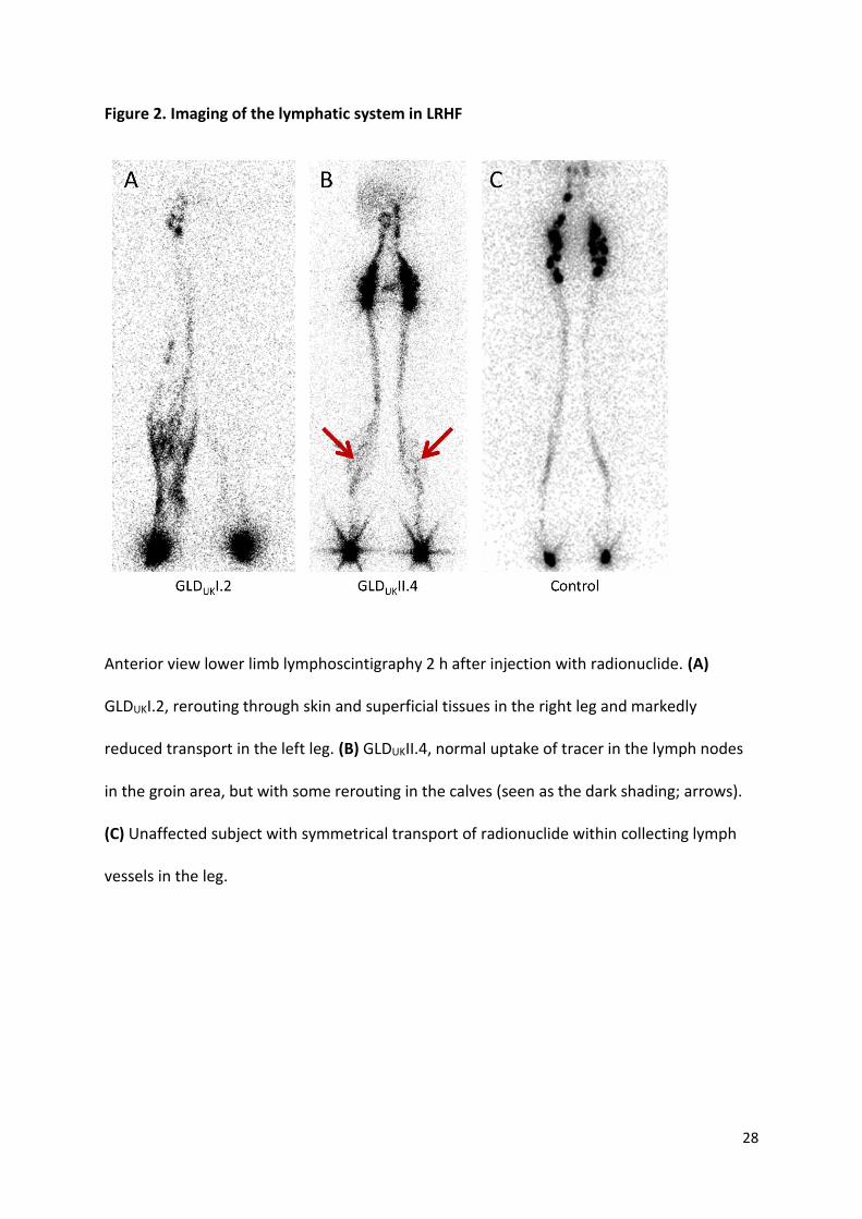
time, with spontaneous resolution of the hydrops and pleural effusions. Only one (GLDUK:I.2)
of five adults has developed peripheral edema of the lower extremities in adolescence
(Figure 2) but three of the five adults (GLDNOR:II.2, GLDNOR:II.3 and GLDUK:I.2) have varicose
veins. Although GLDUK:II.2 and GLDNOR:II.2 currently have no clinical evidence of peripheral
lymphedema, lymphoscintigraphy showed mild bilateral impairment of lymph drainage with
retention of tracer in the lower limbs in both. In addition, GLDUK:II.2 had an atrial septal
defect, diaphragmatic hernia and cystic hygroma at birth. There appears to be an
association with atrial septal defect (ASD) in both families (n=7).
Sanger sequencing identified no pathogenic variants in the genes known to be associated
with congenital primary lymphedema (i.e. CCBE1, VEGFR3 and VEGFC) in the UK proband
(GLDUK:I.2). Whole-exome sequencing (WES) was performed on the family to identify
pathogenic variants. When filtering the WES data of the UK family for variants in genes
known to have relevance to the lymphatic system and lymphangiogenesis, an unreported
variant (NM_004444.4: c.2216G>A, p.Arg739Glu) in EPHB4 was identified. Initially, it was
thought that the variant did not fully co-segregate with the disorder status in the family
(Figure 1A), as two clinically unaffected family members were found to carry the variant
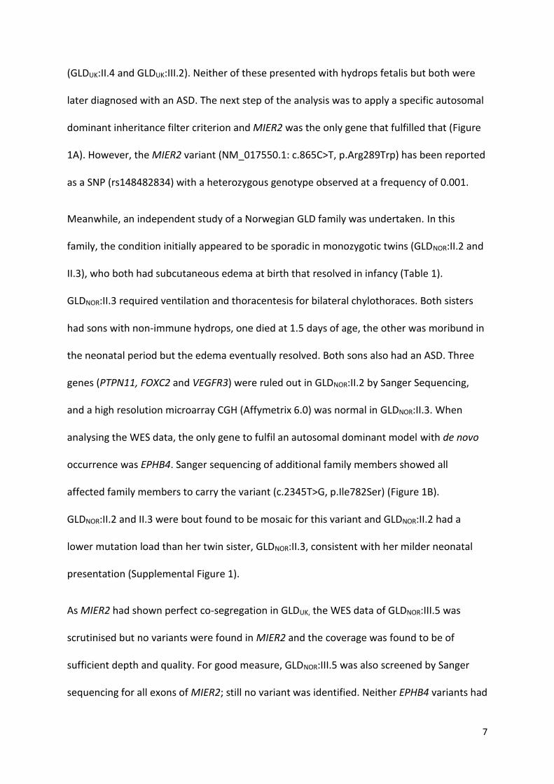
7
(GLDUK:II.4 and GLDUK:III.2). Neither of these presented with hydrops fetalis but both were
later diagnosed with an ASD. The next step of the analysis was to apply a specific autosomal
dominant inheritance filter criterion and MIER2 was the only gene that fulfilled that (Figure
1A). However, the MIER2 variant (NM_017550.1: c.865C>T, p.Arg289Trp) has been reported
as a SNP (rs148482834) with a heterozygous genotype observed at a frequency of 0.001.
Meanwhile, an independent study of a Norwegian GLD family was undertaken. In this
family, the condition initially appeared to be sporadic in monozygotic twins (GLDNOR:II.2 and
II.3), who both had subcutaneous edema at birth that resolved in infancy (Table 1).
GLDNOR:II.3 required ventilation and thoracentesis for bilateral chylothoraces. Both sisters
had sons with non-immune hydrops, one died at 1.5 days of age, the other was moribund in
the neonatal period but the edema eventually resolved. Both sons also had an ASD. Three
genes (PTPN11, FOXC2 and VEGFR3) were ruled out in GLDNOR:II.2 by Sanger Sequencing,
and a high resolution microarray CGH (Affymetrix 6.0) was normal in GLDNOR:II.3. When
analysing the WES data, the only gene to fulfil an autosomal dominant model with de novo
occurrence was EPHB4. Sanger sequencing of additional family members showed all
affected family members to carry the variant (c.2345T>G, p.Ile782Ser) (Figure 1B).
GLDNOR:II.2 and II.3 were bout found to be mosaic for this variant and GLDNOR:II.2 had a
lower mutation load than her twin sister, GLDNOR:II.3, consistent with her milder neonatal
presentation (Supplemental Figure 1).
As MIER2 had shown perfect co-segregation in GLDUK, the WES data of GLDNOR:III.5 was
scrutinised but no variants were found in MIER2 and the coverage was found to be of
sufficient depth and quality. For good measure, GLDNOR:III.5 was also screened by Sanger
sequencing for all exons of MIER2; still no variant was identified. Neither EPHB4 variants had
8
been previously reported in public databases or in 900 in-house controls and it was
therefore concluded that mutations in EPHB4 are the likely cause of the LRHF/GLD
phenotype seen in these two families despite the variable expression observed.
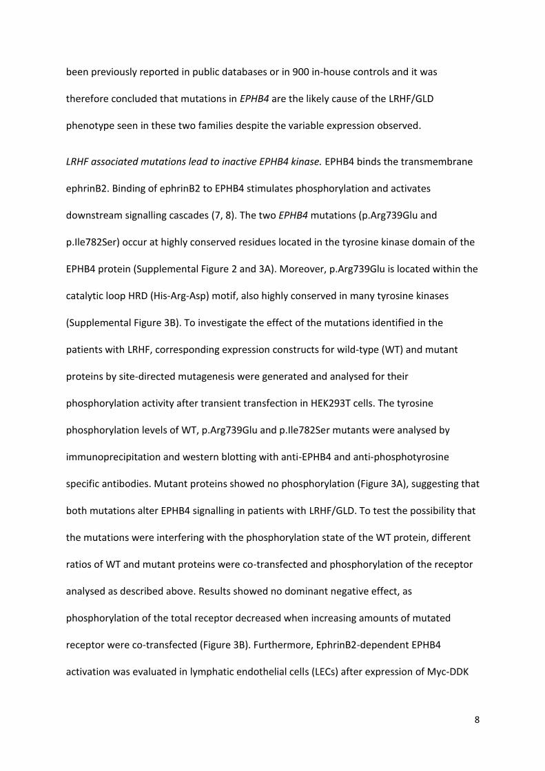
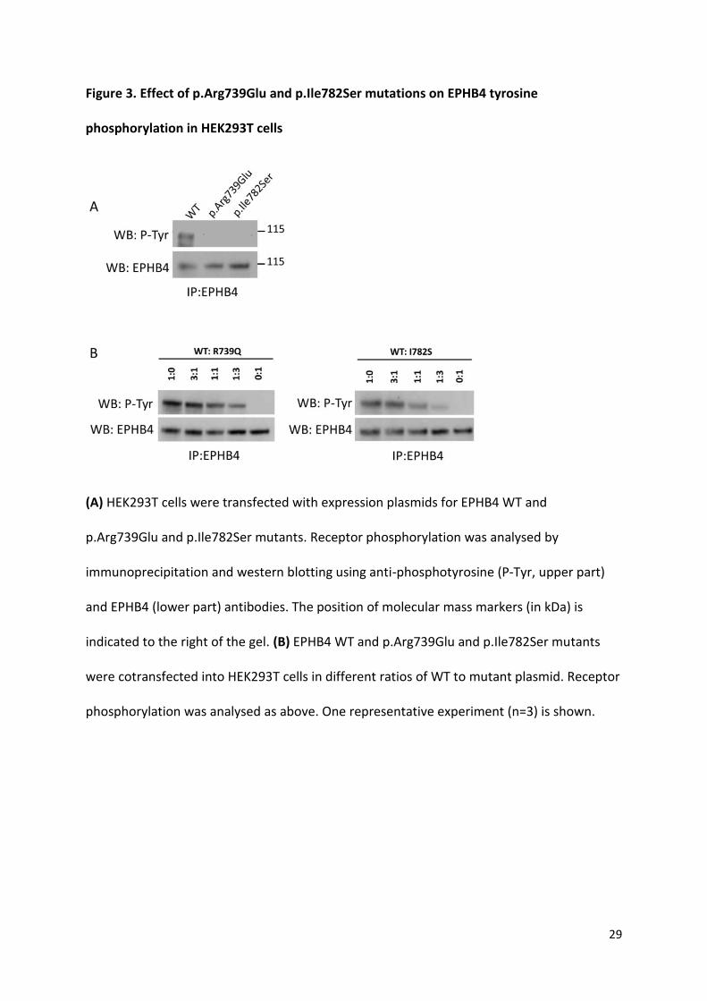
LRHF associated mutations lead to inactive EPHB4 kinase. EPHB4 binds the transmembrane
ephrinB2. Binding of ephrinB2 to EPHB4 stimulates phosphorylation and activates
downstream signalling cascades (7, 8). The two EPHB4 mutations (p.Arg739Glu and
p.Ile782Ser) occur at highly conserved residues located in the tyrosine kinase domain of the
EPHB4 protein (Supplemental Figure 2 and 3A). Moreover, p.Arg739Glu is located within the
catalytic loop HRD (His-Arg-Asp) motif, also highly conserved in many tyrosine kinases
(Supplemental Figure 3B). To investigate the effect of the mutations identified in the
patients with LRHF, corresponding expression constructs for wild-type (WT) and mutant
proteins by site-directed mutagenesis were generated and analysed for their
phosphorylation activity after transient transfection in HEK293T cells. The tyrosine
phosphorylation levels of WT, p.Arg739Glu and p.Ile782Ser mutants were analysed by
immunoprecipitation and western blotting with anti-EPHB4 and anti-phosphotyrosine
specific antibodies. Mutant proteins showed no phosphorylation (Figure 3A), suggesting that
both mutations alter EPHB4 signalling in patients with LRHF/GLD. To test the possibility that
the mutations were interfering with the phosphorylation state of the WT protein, different
ratios of WT and mutant proteins were co-transfected and phosphorylation of the receptor
analysed as described above. Results showed no dominant negative effect, as
phosphorylation of the total receptor decreased when increasing amounts of mutated
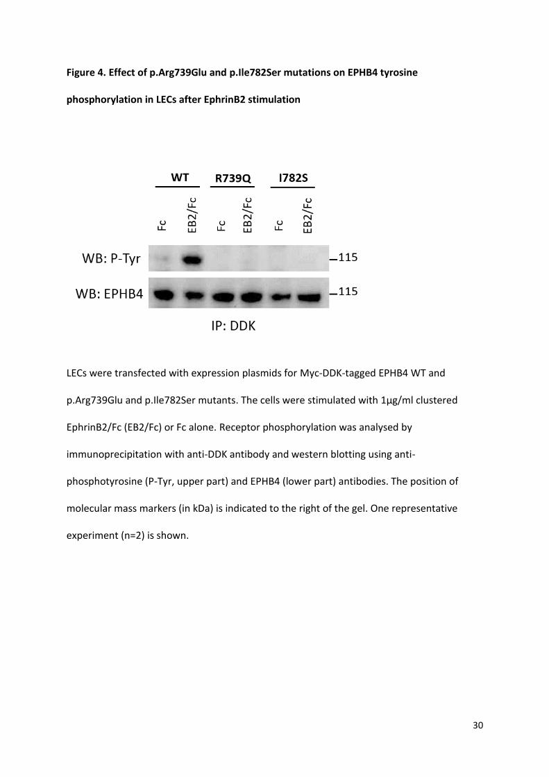
receptor were co-transfected (Figure 3B). Furthermore, EphrinB2-dependent EPHB4
activation was evaluated in lymphatic endothelial cells (LECs) after expression of Myc-DDK
9
tagged EPHB4. To distinguish phosphorylation levels of Myc-DDK tagged exogenous
expressed WT and mutant EPHB4 from endogenous expressed WT EPHB4, an anti DDK
antibody was used for the immunoprecipitation and isolation of only the overexpressed
forms of Myc-DDK tagged EPHB4. EphrinB2 treatment increased phosphorylation level of
WT Myc-DDK tagged EPHB4 but not mutant proteins (Figure 4), confirming the negative
effect of both mutations on the receptor activity after ligand stimulation in LECs.
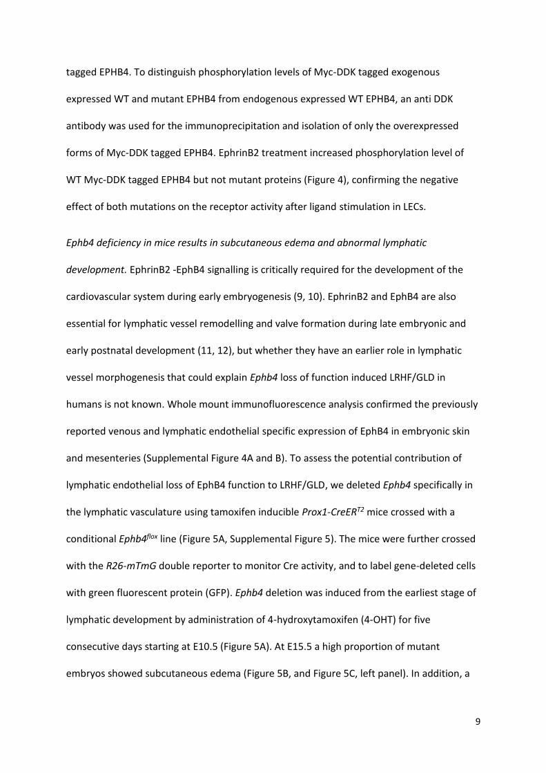
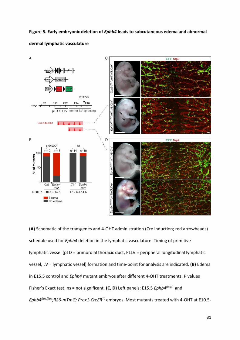
Ephb4 deficiency in mice results in subcutaneous edema and abnormal lymphatic
development. EphrinB2 -EphB4 signalling is critically required for the development of the
cardiovascular system during early embryogenesis (9, 10). EphrinB2 and EphB4 are also
essential for lymphatic vessel remodelling and valve formation during late embryonic and
early postnatal development (11, 12), but whether they have an earlier role in lymphatic
vessel morphogenesis that could explain Ephb4 loss of function induced LRHF/GLD in
humans is not known. Whole mount immunofluorescence analysis confirmed the previously
reported venous and lymphatic endothelial specific expression of EphB4 in embryonic skin
and mesenteries (Supplemental Figure 4A and B). To assess the potential contribution of
lymphatic endothelial loss of EphB4 function to LRHF/GLD, we deleted Ephb4 specifically in
the lymphatic vasculature using tamoxifen inducible Prox1-CreERT2 mice crossed with a
conditional Ephb4flox line (Figure 5A, Supplemental Figure 5). The mice were further crossed
with the R26-mTmG double reporter to monitor Cre activity, and to label gene-deleted cells
with green fluorescent protein (GFP). Ephb4 deletion was induced from the earliest stage of
lymphatic development by administration of 4-hydroxytamoxifen (4-OHT) for five
consecutive days starting at E10.5 (Figure 5A). At E15.5 a high proportion of mutant
embryos showed subcutaneous edema (Figure 5B, and Figure 5C, left panel). In addition, a
10
proportion of dermal lymphatic vessels contained blood in 71% of edematous mutant
embryos (n=14), but not in non-edematous mutants (n=5) or in control embryos (n=20)
(Figure 5C and data not shown). Whole-mount immunofluorescence of the skin revealed
tortuous and dilated dermal lymphatic vessels in the Ephb4 mutants (Figure 5C, right
panels). Notably, abnormal vessel morphology was also observed in vessels that showed a
low contribution of GFP+ (i.e. Ephb4 deficient cells) (Figure 5C), suggesting that edema
and/or blood filling of lymphatic vessels secondarily caused vessel dilation. In support of a
non-cell-autonomous effect of early embryonic deletion of Ephb4 to dermal lymphatic
vasculature, inactivation of Ephb4 from E12.5, when dermal lymphatic vessel formation
begins (13) (Figure 5A), resulted in normal vasculature despite efficient gene targeting
(Figure 5D). These results suggest E10-E12 as a critical time-window for EphB4 function
during lymphatic development.
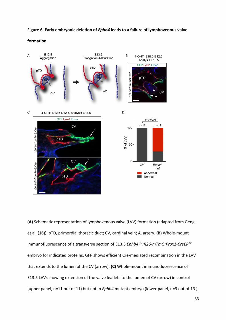
Ephb4 is required for the formation of lymphovenous and lymphatic valves. Previous studies
have shown that between E10.5-E13.5 formation of specialized lymphovenous valves (LVV)
occurs at the connection sites between the primordial thoracic duct (pTD) and the cardinal
vein (14-16) (Figure 6A). It was therefore reasoned that edema in Ephb4 mutants might be
due to defective LVVs leading to inefficient lymph drainage. To investigate this, we induced
Cre recombination in the developing LVVs in Ephb4flox;R26-mTmG;Prox1-CreERT2 embryos by
4-OHT treatment between E10.5-E12.5. Analysis of immunostained transverse vibratome
sections of E13.5 control embryos showed preferential and efficient targeting of the dual
LVVs by the Prox1-CreERT2 transgene, while pTD endothelium exhibited mosaic labeling
(Figure 6B). Control LVVs (11 out of 11) consisted of two well-defined leaflets extending to
the lumen of the cardinal vein (Figure 6C and 6D, Supplemental Movie 1). In contrast, the
11
majority of Ephb4 deficient LVVs (9 out of 13) did not show extended leaflets but instead
consisted of abnormal clusters of GFP+ cells (Figure 6C and 6D, Supplemental Movie 2).
Interestingly, studies using a function blocking antibody and a chemical genetic approach
showed that EphB4 kinase signalling regulates lymphatic valve formation (11), while genetic
studies have demonstrated an important function for its ligand EphrinB2 in the formation of
both lymphatic and venous valves (12, 17). Using our genetic loss-of-function model we
confirmed the essential role of EphB4 in lymphatic valve morphogenesis. Deletion of Ephb4
during embryonic valve formation led to a complete absence of valves that form in control
mesenteries by E18.5 by lymphatic endothelial cells (LECs) expressing high levels of Prox1
(Supplemental Figure 4C). In addition, early postnatal deletion of Ephb4 led to a complete
loss of lymphatic valves (Supplemental Figure 4D). These results demonstrate a critical role
of Ephb4 in the formation and early postnatal maintenance of lymphatic valves, and
highlight conserved mechanisms regulating the formation of valves at different anatomical
sites.
12
Discussion
This study identifies the EPHB4 receptor tyrosine kinase as a critical regulator of early
lymphatic vessel development and a novel causative gene for LRHF and primary
lymphedema. We have shown here that kinase inactivating mutations in EPHB4 can produce
a lymphatic phenotype in humans that can be classified as LRHF/GLD. However, this
phenotype shows highly variable expression. Some individuals present with severe in utero
swelling, which may cause perinatal demise (or fully resolve to become completely
asymptomatic), others with no edema but only an atrial septal defect. It can be
distinguished from the majority of Hennekam Syndrome cases, where the swelling presents
in the antenatal period but persists throughout life (3-5). The large number of miscarriages
in GLDNOR may well be related to this disorder. In this regard, it is of interest that EphB4 and
ephrin-B2 have been shown to be instrumental in the human placental development (18).
Invasive cytotrophoblasts uses the EPHB4 expression on veins to ensure that migration of
these cells into EPHB4 expressing uterine veins is limited and instead biased toward the
arterial side of the circulation (19). Expression of EPHB4 at half the levels normally
encountered may disturb the complex migration patterns seen in the process of
placentation. A failure of the invasive cytotrophoblasts to take on an arterial phenotype is
suspected to lead to the loss of pregnancy during the late first or early second trimester
(19). Perinatal deaths were also of a higher frequency in the autosomal recessive form of
LRHF/GLD caused by PIEZO1 mutations but, in this condition, were probably related to the
hydrops fetalis (6).
Two GLDUK family members (GLDUK:II.4 and her son, GLDUK:III.2) carry the variant but have
no clinical history of pre- or postnatal swelling. On lymphoscintigraphy (GLDUK:II.4),
13
quantification shows entirely normal levels of transport of lymph within the legs, but
imaging is suggestive of rerouting through skin and superficial tissues rather than a main
lymphatic tract as seen in the control (Figure 2B). She has a small ASD and interestingly her
son had large, multiple ASDs requiring surgical closure. Variable expression has been
observed in other primary lymphedemas e.g. PIEZO1-related LRHF/GLD (6).
Like other forms of GLD (4, 6), this novel condition presents antenatally with non-immune
hydrops or pleural effusions. The swelling may completely resolve with no residual
lymphatic phenotype, similar to observations in the recently identified PIEZO1 related GLD
(6). However, the report of one affected individual with bilateral lower limb edema
(GLDUK:I.2) with abnormal lymph scans suggests that there may be residual, lymphatic
weakness in the survivors. Further studies will be needed to investigate the specific nature
and extent of the lymphatic dysfunction in these patients.
LEC specific deletion of Ephb4 in mouse embryos led to subcutaneous edema and abnormal
lymphatic vessel morphology, thus recapitulating aspects of the human LRHF phenotype.
Temporal analysis of Ephb4 function demonstrated a critical requirement of Ephb4 during
early stages of lymphatic development. Specifically, we found that Ephb4 regulates the
formation of lymphovenous valves that are critical for efficient lymphatic function by
maintaining unidirectional flow of lymph into blood (14, 20, 21). We additionally confirmed
the previously reported critical role of Ephb4 in both formation and maintenance of
lymphatic valves (11). Lymphatic valve defects are, however, an unlikely cause of in utero
swelling due to their late embryonic development (22, 23). We postulate, therefore, that
defective lymphovenous valve formation, caused by the lack of EPHB4 could contribute to
the LRHF seen in the GLDNOR and GLDUK patients. In agreement with this, defective LVVs
14
were recently demonstrated in mouse models of primary lymphedemas caused by loss of
function of Foxc2, Connexin37 and Gata2 (16, 24).
Edema in the mouse embryos lacking Ephb4 specifically in the lymphatic endothelia appears
to be milder than that observed in the patients, suggesting that defective lymphovenous
valves may only partially explain human LRHF/GLD. It is well known that EphB4 is also
expressed in venous and capillary endothelium (9, 10), and therefore the impact of the
mutations on the venous system needs to be considered. In accordance with this hypothesis
some of the patients (GLDNOR:II.2 and II.3; GLDUK:I.2) showed varicose veins, which would be
consistent with a valve defect in the venous system and would support the contribution of
vascular deficiency to the observed phenotype. Together with the varicose veins, the ASD
observed in the patients could also be an important part of this phenotype. Both have been
reported in Lymphedema Distichiasis Syndrome with a frequency of approximately 7%
(ASDs) (25) and 100% (varicose veins) (26).
Kinase activity is critical for EphB4 forward signalling in lymphatic endothelia (11). Our in
vitro data show that EPHB4 mutants carrying the LRHF associated mutations p.Arg739Glu
and p.Ile782Ser are kinase-dead but do not have a dominant negative effect on WT protein.
In contrast, VEGFR3 mutants in Milroy Disease have a slower turnover (27), which may
affect the signalling capacity of the WT tyrosine kinase receptor due to accumulation of
mutant receptors on the cell surface. Unlike typical receptor tyrosine kinases that dimerise
upon ligand stimulation, Ephrin receptors form higher order clusters, with cluster size being
an important determinant of the quality and strength of cellular response (28, 29). Inclusion
of a kinase-dead receptor may thus significantly weaken the signalling strength of higher
order clusters and thereby alter cellular responses. EphB2 receptor-mediated endocytosis
15
requires the kinase activity of the receptor (30), so that kinase-dead EphB4 could also show
defective endocytosis, influencing clustering dynamics and cellular responses. Further
studies will aim to investigate those and other functional consequences of the LRHF/GLD
associated mutations.
In conclusion, we report on kinase inactivating EPHB4 mutations in eleven individuals from
two extended family pedigrees presenting with a phenotypic spectrum from severe, lethal
non-immune hydrops to ASD only. The inheritance pattern is typical of autosomal dominant
inheritance with variable expression. Using a genetic mouse model, we have further shown
that Ephb4 deficiency in lymphatic endothelium leads to defective lymphovenous valves,
which may critically contribute to edema formation in LRHF/GLD patients. This is the first
report in the literature of a human phenotype associated with EPHB4 mutations and also
the first report of an autosomal dominant form of LRHF.
16
Methods
Exome sequencing. For GLDUK, sequencing libraries were made following the protocol from
Roche/Nimblegen's SeqCap EZ Exome Library v2.0 kit. The libraries were then sequenced on
HiSeq2000 (Illumina) machines. Sequence reads were aligned to the reference genome
(hg19) using Novoalign (Novocraft Technologies). Duplicate reads, resulting from PCR
clonality or optical duplicates, and reads mapping to multiple locations were excluded from
downstream analysis. Depth and breadth of sequence coverage were calculated with
custom scripts and the BedTools package (31).
All variants were annotated using a custom annotation pipeline. Single-nucleotide
substitutions and small indel variants were identified and quality filtered within the
SamTools software package (32) and in-house software tools (33). Variants were annotated
with respect to genes and transcripts with the Annovar tool (34). Variants were filtered for
novelty by comparing them to dbSNP135 and 1000 Genomes SNP calls and to variants
identified in 900 control exomes (primarily of European origin), which were sequenced and
analyzed by the same method. Summary statistics for the exome sequencing is given in
Supplemental Tables 1 and 2. Analysis of the exome-variant profiles was performed under a
model of a rare autosomal-dominant disorder; this model required one previously
unobserved heterozygous variant for all affected individuals.
For GLDNOR the sequencing analysis was performed using the SOLID 5500xl platform (Life
Technologies). Exon sequences were enriched by SureSelect Human All Exon v5 (Agilent
Technologies), which targets ~21,500 human genes, covering a total of 50Mb of genomic
sequence. Read alignment and variant calling were performed with Lifescope v1.3 software.
All variants were annotated using a custom annotation pipeline. Variants from the exome
17
were filtered for known variants in dbSNP, intronic and UTR variants, synonymous variants
and variants in our in-house database. Variants with less than 5 variation reads were also
omitted. Summary statistics for the exome sequencing is given in Supplemental Table 3.
Confirmation Sequencing. Samples of available family members were analyzed by Sanger
Sequencing. Primers were designed for the coding regions and associated splice sites for
exons 13 and 14 of EPHB4 and all exons of MIER2 using Primer3 software (35) or ExonPrimer
(helmholtz-muenchen.de). Primer sequences listed in Supplemental Table 4. PCR products
were sequenced using BigDye Terminator v1.1 and v3.1 chemistry (Life Technologies) and an
ABI3130xla Genetic Analyser or 3730 DNA analyser (Life Technologies). Sequencing traces
were visually inspected in Finch TV v1.4 (Geospiza Inc) and SeqScape® Software v2.5 (Life
Technologies).
The twins in GLDNOR were also examined for mosaicism, by Sanger sequencing on DNA
extracted directly from blood (new sample), urine, saliva, and skin biopsy (fibroblasts).
The variants from the 2 families have been submitted to the Leiden Open Variation
Database (LOVD3) (genomic variants 0000095140 [c.2216G>A; p.(Arg739Gln)] and
0000095141 [c.2345T>G; p.(Ile782Ser)]; http://databases.lovd.nl/shared/genes/EPHB4).
Site-directed mutagenesis of EPHB4 constructs. The human EPHB4 mammalian expression
plasmids pCMV6-XL6-EPHB4 (SC117357) and Myc-DDK-tagged pCMV6-Entry-EPHB4
(RC208559) were obtained from OriGene and used as templates for site-directed
mutagenesis using the QuikChange II XL Site-Directed Mutagenesis Kit (Agilent). All primers
were designed using QuikChange Primer Design (Agilent) and are listed in Supplemental
Table 5. All constructs were verified by DNA sequencing.
18
Cell culture and transfection. HEK293T cells (kindly provided by Dr Tris McKay, St George’s
University of London, UK) were maintained in DMEM supplemented with 10% FBS.
Transfection of HEK293T cells was performed with GeneJuice transfection reagent (Merk)
following the manufacturer’s protocol. 6x10⁵ cells/well were seeded in 6-well plates the day
before transfection, and then they were transfected with 3 μl GeneJuice and 1 µg of DNA.
Human dermal lymphatic endothelial cells (C-12217) were obtained from Promocell and
maintained in supplemented endothelial cell growth medium MV2 (C-22022, Promocell)
containing recombinant human VEGF-C (2179-VC-025, R&D systems). Transfection of LECs
was performed with Viromer YELLOW (VY-01LB-01, Lipocalyx) following the manufacturer’s
recommendations. 2x10⁵ cells/well were seeded in fibronectin (F1141, Sigma)-coated 6-well
plates the day before transfection and then transfected with 1 µg of DNA. For both cell
types, lysates were collected 24 hours post-transfection and subjected to
immunoprecipitation and western blot analysis.
Ligand activation of EPHB4 receptor. EphrinB2/Fc (7397-EB-050, R&D systems) or Fc
fragment alone (CSB-NP005401h, Stratech) were clustered by incubation with a goat anti-
human IgG Fc antibody (40C-CR2022G-FIT, Stratech) at a 1:2 ratio for 1 hour at 4°C. 18 hours
post-transfection LECs were serum-starved for 6 hours and then treated with 1µg/ml
clustered EphrinB2/Fc or Fc alone for 30 minutes before cell lysates collection.
Immunoprecipitation and western blot analysis. For immunoprecipitation of overexpressed
WT and mutant EPHB4, transfected cells were harvested in 100 µl of lysis buffer (20 mM Tris
pH 7.5, 150 mM NaCl, 0.5%Triton X-100) supplemented with protease and phosphatase
inhibitors (Sigma). After clarification by centrifugation, 100 µg of total HEK293T cells protein
lysate was incubated with 0.8 µg of goat anti-human EPHB4 antibody (AF3038, R&D
19
systems) overnight at 4°C. After incubation with protein A sepharose beads (Sigma) for 4
hours at 4°C, immune complexes were precipitated by centrifugation. 100 µl of total LECs
protein lysate was incubated with 4 µg of mouse anti-DDK antibody (clone OTI4C5,
TA50011, Origene) overnight at 4°C and immune complexes precipitated with protein G
sepharose beads (Sigma). Immunoprecipitates were separated by SDS-PAGE and transferred
to PVDF membranes. Immunoblot analysis was performed with goat anti-human EPHB4
(AF3038, R&D Systems) and mouse anti-phosphotyrosine (clone 4G10, 05-321, Millipore)
antibodies. All uncropped western blots are shown in Supplemental Figures 6 and 7.
Mouse lines. R26-mTmG mice were acquired from the Jackson Laboratory (36). Prox1-
CreERT2 mice were previously described (17). For the generation of the Ephb4flox line, a
conditional knock-out strategy, flanking Ephb4 exons 2 and 3 with LoxP sites, was used to
target the Ephb4 locus. The targeting vector was built using homologous recombination in
bacteria (37). A C57BL/6 mouse BAC, served as template for the extraction of the homology
arms of the targeting vector. The targeting vector contained a frt flanked neomycin
phosphotransferase, Neo, selectable marker cassette. After linearization, the targeting
construct was electroporated into AZX1, a C57Bl/6JOlaHsd derived embryonic stem cell line.
PCR screens and Southern blot analyses revealed clones that had undergone the desired
homologous recombination event. Several of these clones were expanded and injected into
Balb/cOlaHsd blastocysts to generate chimeric males which were then bred to
C57Bl/6JOlaHsd females and black-coated offspring were genotyped on both sides of the
homology arms for correct integration into the EphB4 locus. The neomycin
phosphotransferase selectable marker cassette, which was flanked by frt sites, was deleted
after subsequent breeding to mice expressing flp recombinase under the CAG promoter.
20
For embryonic induction of Cre recombination, 4-hydroxytamoxifen (4-OHT, Sigma) was
dissolved in peanut oil (10 mg/ml). 1 mg was administered to pregnant females by
intraperitoneal injection at the indicated developmental stages. For induction during early
postnatal development, mice were administered by intraperitoneal injection at P1 and P2
with 50 µg of 4-OHT dissolved in Ethanol. All strains were maintained and analysed on
C57BL/6J background. Experimental procedures were approved by the United Kingdom Home
Office and the Uppsala Laboratory Animal Ethical Committee.
Immunofluorescence. For whole-mount immunostaining, tissues were fixed in 4%
paraformaldehyde (PFA) for 2h at RT followed by permeabilization in 0.3% Triton X-100 PBS
(PBSTx) and blocking in PBSTx 3% milk. Primary antibodies were incubated at 4C overnight
in blocking buffer. After washing several times, the samples were incubated with
fluorocrome-conjugated secondary antibodies in blocking buffer for 2 hours, before further
washing and mounting in Mowiol. The following primary antibodies were used: goat anti-
mouse EphB4 (AF446, R&D Systems), goat anti-human VE-Cadherin (sc-6458, Santa Cruz
Biotechnology), rabbit anti-human Prox1 (generated against human Prox1 C-terminus (567-
737aa)), rabbit anti-GFP (A11122, Invitrogen), chicken anti-GFP (Ab13970, Abcam), rat anti-
PECAM-1 (553370, BD Pharmigen), rat anti-Endomucin (sc-65495, Santa Cruz) and rabbit anti-
Lyve1 (103-PA50AG, Reliatech). Secondary antibodies conjugated to AF488, Cy3 or AF647
were obtained from Jackson ImmunoResearch. Controls were littermate embryos and mice
of the following genotypes: Ephb4flox/+;R26-mTmG+;Prox1-CreERT2+, Ephb4flox/flox;R26-
mTmG+;Prox1-CreERT2- or Ephb4flox/flox;R26-mTmG-;Prox1-CreERT2-.
For analysis of LVVs, 100μm coronal vibratome sections of E13.5 embryos were cut and
stained as described above. Single plane images of the valve were taken where the valve was
21
clearly visible. Only valves that appeared intact were included in the classification. 5 control
embryos (Ephb4flox/+;R26-mTmG+;Prox1-CreERT2+ and Ephb4+/+;R26-mTmG+;Prox1-CreERT2+)
were cut and 11 valves were imaged and included in the quantification. For mutant embryos
(Ephb4flox/flox;R26-mTmG+;Prox1-CreERT2+) 13 valves from 9 embryos were imaged.
Image acquisition. Stereomicroscope images were acquired using a Leica MZ16F fluorescence
microscope with a Leica DFC420C camera and Leica Microsystems software. Confocal images
were acquired using Zeiss LSM 780 confocal microscope and Zen 2009 software, or Leica SP8
confocal microscope and Leica Application Suite X software. Figures 5C and 5D were acquired
as 4x2 tile scans with a HCX PL APO CS 10x/0.40 DRY objective. Figure 6 was acquired using a
Fluotar VISIR 25x/0.95 Water objective. Supplemental figure 4A was taken with a HC PL APO
CS2 63x/1.30 GLYC objective; supplemental figure 4B (P6 mesentery) using a C-Aphochromat
40x/1.20 W Korr M27 objective and supplemental figures 4B (E18 mesentery), 4C and 4D were
acquired using a Plan-Apochromat 20x/0.8 Ph2. All confocal images represent maximum
intensity projections of z-stacks except images in Figure 6.
Lymphoscintigraphy. Lymphoscintigraphy is the imaging of the lymphatic system by injecting
radioactive isotope (technetium-99m) into the web spaces between the toes and/or fingers
and quantification of uptake into the inguinal lymph nodes for foot injections and axillary
nodes for hand injection after 2 h with a gamma camera.
Statistical analysis. P values representing difference in proportion of edematous and non-
edematous mutant versus control littermates (Figure 5B) and proportion of normal and
abnormal LVVs (Figure 6D) were calculated using Chi square/Fisher’s Exact (2-tailed). A P
value less than 0.05 was considered significant.

22
Study approval. Subjects in this study were recruited through genetic and lymphovascular
clinics in Norway and the United Kingdom. Ethical approval for this study was obtained from
the Norwegian Regional Committees for Medical and Health Research Ethics, West (REC ref:
2011/2453) and the South West London Research Ethics Committee (REC Ref:
05/Q0803/257). Written informed consent was obtained for all subjects. All affected
individuals and family members underwent a detailed physical examination. Animal study
was approved by the United Kingdom Home Office and the Uppsala Laboratory Animal
Ethical Committee.
23
Author contributions
Designing research study: SMA, IMC, SB, SJ, TM, PO
Conducting experiments: SMA, IMC, EF, RH, AV, SL, HJ, CK
Providing NGS pipeline: MAS, KP, SL, CG, AH
Analysing data: SMA, IMC, RH, AV, SL, SB, SJ, SM, TM, PO
Providing reagents, material, patients: GA, GB, KG, MW, JW, SB, PSM, SGR, SM, TM, PO
Writing the manuscript: SMA, IMC, SB, SJ, SM, TM, PO
Supervised the research: PSM, SGR, SJ, SM, MPS, TM, PO
24
Acknowledgements
We extend our thanks to the patients and their families. This work was supported by the
British Heart Foundation to SMA/IMC (SP/13/5/30288), KG (FS/11/40/28739), CK
(FS/15/39/31526) and PO (PG/10/58/28477); the Newlife Foundation for Disabled Children
to EF (12-13/01); the National Institute for Health Research (NIHR) Biomedical Research
Centre based at Guy's and St Thomas' NHS Foundation Trust and King's College London to
MAS; the National Institutes of Health (NIH) to SL (Fellowship Grants F32HL110473 and
K99HL119617); the Swedish Cancer Foundation and the Swedish Research Council to TM;
Cancer Research UK to AV/TM.
25
References – need moving to here
1. Bellini C, et al. Etiology of Non-Immune Hydrops Fetalis: An Update. Am J Med Genet A. 2015;167A(5):1082-1088.
2. Connell FC, et al. The classification and diagnostic algorithm for primary lymphatic dysplasia: an update from 2010 to include molecular findings. Clin Genet. 2013;84(4):303-314.
3. Alders M, et al. Mutations in CCBE1 cause generalized lymph vessel dysplasia in humans. Nat Genet. 2009;41(12):1272-1274.
4. Connell F, et al. Linkage and sequence analysis indicate that CCBE1 is mutated in recessively inherited generalised lymphatic dysplasia. Hum Genet. 2010;127(2):231-241.
5. Alders M, et al. Hennekam syndrome can be caused by FAT4 mutations and be allelic to Van Maldergem syndrome. Hum Genet. 2014;133(9):1161-1167.
6. Fotiou E, et al. Novel mutations in PIEZO1 cause an autosomal recessive generalized lymphatic dysplasia with non-immune hydrops fetalis. Nat Commun. 2015;6:8085.
7. Kuijper S, et al. Regulation of angiogenesis by Eph-Ephrin interactions. Trends Cardiovasc Med. 2007;17(5):145-151.
8. Pitulescu ME, Adams RH. Eph/ephrin molecules-a hub for signaling and endocytosis. Genes Dev. 2010;24(22):2480-2492.
9. Wang HU, et al. Molecular distinction and angiogenic interaction between embryonic arteries and veins revealed by ephrin-B2 and its receptor Eph-B4. Cell. 1998;93(5):741-753.
10. Adams RH, et al. Roles of ephrinB ligands and EphB receptors in cardiovascular development: demarcation of arterial/venous domains, vascular morphogenesis, and sprouting angiogenesis. Genes Dev. 1999;13(3):295-306.
11. Zhang G, et al. EphB4 forward signalling regulates lymphatic valve development. Nat Commun. 2015;6:6625.
12. Makinen T, et al. PDZ interaction site in ephrinB2 is required for the remodeling of lymphatic vasculature. Genes Dev. 2005;19(3):397-410.
13. Martinez-Corral I, et al. Nonvenous origin of dermal lymphatic vasculature. Circ Res. 2015;116(10):1649-1654.
14. Srinivasan RS, Oliver G. Prox1 dosage controls the number of lymphatic endothelial cell progenitors and the formation of the lymphovenous valves. Genes Dev. 2011;25(20):2187-2197.
15. Hagerling R, et al. A novel multistep mechanism for initial lymphangiogenesis in mouse embryos based on ultramicroscopy. EMBO J. 2013;32(5):629-644.
16. Geng X, et al. Multiple mouse models of primary lymphedema exhibit distinct defects in lymphovenous valve development. Dev Biol. 2016;409(1):218-233.
17. Bazigou E, et al. Genes regulating lymphangiogenesis control venous valve formation and maintenance in mice. J Clin Invest. 2011;121(8):2984-2992.
18. Chennakesava CS, et al. Differential expression of the receptor tyrosine kinase EphB4 and its ligand Ephrin-B2 during human placental development. Placenta. 2006;27(9-10):959-967.
19. Red-Horse K, et al. EPHB4 regulates chemokine-evoked trophoblast responses: a mechanism for incorporating the human placenta into the maternal circulation. Development. 2005;132(18):4097-4106.
20. Turner CJ, et al. Integrin-alpha 5 beta 1 is not required for mural cell functions during development of blood vessels but is required for lymphatic-blood vessel separation and lymphovenous valve formation. Dev Biol. 2014;392(2):381-392.
21. Hess PR, et al. Platelets mediate lymphovenous hemostasis to maintain blood-lymphatic separation throughout life. J Clin Invest. 2014;124(1):273-284.
22. Bazigou E, et al. Integrin-alpha 9 Is Required for Fibronectin Matrix Assembly during Lymphatic Valve Morphogenesis. Dev Cell. 2009;17(2):175-186.
26
23. Sabine A, et al. Mechanotransduction, PROX1, and FOXC2 Cooperate to Control Connexin37 and Calcineurin during Lymphatic-Valve Formation. Dev Cell. 2012;22(2):430-445.
24. Kazenwadel J, et al. GATA2 is required for lymphatic vessel valve development and maintenance. J Clin Invest. 2015;125(8):2979-2994.
25. Brice G, et al. Analysis of the phenotypic abnormalities in lymphoedema-distichiasis syndrome in 74 patients with FOXC2 mutations or linkage to 16q24. J Med Genet. 2002;39(7):478-483.
26. Mellor RH, et al. Mutations in FOXC2 are strongly associated with primary valve failure in veins of the lower limb. Circulation. 2007;115(14):1912-1920.
27. Karkkainen MJ, et al. Missense mutations interfere with VEGFR-3 signalling in primary lymphoedema. Nat Genet. 2000;25(2):153-159.
28. Seiradake E, et al. Structurally encoded intraclass differences in EphA clusters drive distinct cell responses. Nat Struct Mol Biol. 2013;20(8):958-964.
29. Schaupp A, et al. The composition of EphB2 clusters determines the strength in the cellular repulsion response. J Cell Biol. 2014:409-422.
30. Zimmer M, et al. EphB-ephrinB bi-directional endocytosis terminates adhesion allowing contact mediated repulsion. Nat Cell Biol. 2003;5(10):869-878.
31. Quinlan AR, Hall IM. BEDTools: a flexible suite of utilities for comparing genomic features. Bioinformatics. 2010;26(6):841-842.
32. Li H, et al. The Sequence Alignment/Map format and SAMtools. Bioinformatics. 2009;25(16):2078-2079.
33. Simpson MA, et al. Mutations in NOTCH2 cause Hajdu-Cheney syndrome, a disorder of severe and progressive bone loss. Nat Genet. 2011;43(4):303-305.
34. Wang K, et al. ANNOVAR: functional annotation of genetic variants from high-throughput sequencing data. Nucleic Acids Res. 2010;38(16):e164.
35. Rozen S, Skaletsky H. Primer3 on the WWW for general users and for biologist programmers. Methods Mol Biol. 2000;132:365-386.
36. Muzumdar MD, et al. A global double-fluorescent Cre reporter mouse. Genesis. 2007;45(9):593-605.
37. Datsenko KA, Wanner BL. One-step inactivation of chromosomal genes in Escherichia coli K-12 using PCR products. Proc Natl Acad Sci U S A. 2000;97(12):6640-6645.
27
Figures and figure legends
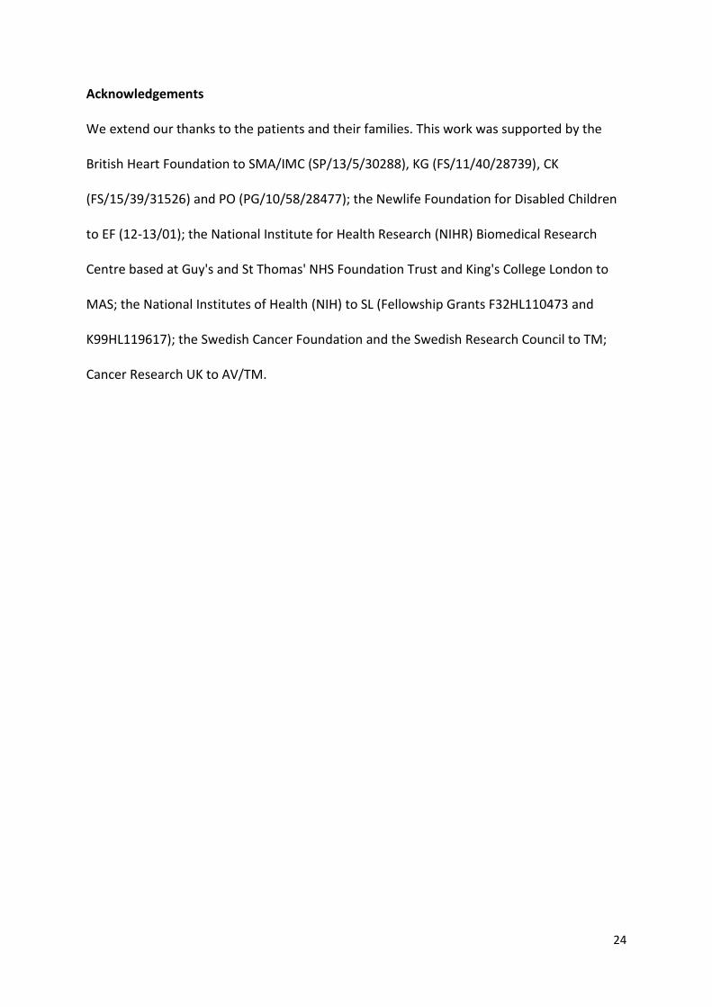
Figure 1. Mutations in EPHB4 cause Lymphatic-Related Hydrops Fetalis (LRHF)
(A) Pedigree of UK GLD family, (B) pedigree of Norwegian GLD family. Stars indicate which
samples have been exome sequenced. Genotypes indicated by hyphen (-) is the wildtype
allele and a plus (+) the mutant allele. Top row of genotypes in GLDUK genogram is for EPHB4
and bottom row of genotypes is MIER2. Triangles, first trimester miscarriages; IUD, intra-
uterine death. GLDNOR:III.3 had trisomy 18.
C D E F
28
Figure 2. Imaging of the lymphatic system in LRHF
Anterior view lower limb lymphoscintigraphy 2 h after injection with radionuclide. (A)
GLDUKI.2, rerouting through skin and superficial tissues in the right leg and markedly
reduced transport in the left leg. (B) GLDUKII.4, normal uptake of tracer in the lymph nodes
in the groin area, but with some rerouting in the calves (seen as the dark shading; arrows).
(C) Unaffected subject with symmetrical transport of radionuclide within collecting lymph
vessels in the leg.
29
Figure 3. Effect of p.Arg739Glu and p.Ile782Ser mutations on EPHB4 tyrosine
phosphorylation in HEK293T cells
(A) HEK293T cells were transfected with expression plasmids for EPHB4 WT and
p.Arg739Glu and p.Ile782Ser mutants. Receptor phosphorylation was analysed by
immunoprecipitation and western blotting using anti-phosphotyrosine (P-Tyr, upper part)
and EPHB4 (lower part) antibodies. The position of molecular mass markers (in kDa) is
indicated to the right of the gel. (B) EPHB4 WT and p.Arg739Glu and p.Ile782Ser mutants
were cotransfected into HEK293T cells in different ratios of WT to mutant plasmid. Receptor
phosphorylation was analysed as above. One representative experiment (n=3) is shown.
(C) (D) (E) (F)
30
Figure 4. Effect of p.Arg739Glu and p.Ile782Ser mutations on EPHB4 tyrosine
phosphorylation in LECs after EphrinB2 stimulation
LECs were transfected with expression plasmids for Myc-DDK-tagged EPHB4 WT and
p.Arg739Glu and p.Ile782Ser mutants. The cells were stimulated with 1µg/ml clustered
EphrinB2/Fc (EB2/Fc) or Fc alone. Receptor phosphorylation was analysed by
immunoprecipitation with anti-DDK antibody and western blotting using anti-
phosphotyrosine (P-Tyr, upper part) and EPHB4 (lower part) antibodies. The position of
molecular mass markers (in kDa) is indicated to the right of the gel. One representative
experiment (n=2) is shown.
(C) (D) (E) (F)
31
Figure 5. Early embryonic deletion of Ephb4 leads to subcutaneous edema and abnormal
dermal lymphatic vasculature
(A) Schematic of the transgenes and 4-OHT administration (Cre induction; red arrowheads)
schedule used for Ephb4 deletion in the lymphatic vasculature. Timing of primitive
lymphatic vessel (pTD = primordial thoracic duct, PLLV = peripheral longitudinal lymphatic
vessel, LV = lymphatic vessel) formation and time-point for analysis are indicated. (B) Edema
in E15.5 control and Ephb4 mutant embryos after different 4-OHT treatments. P values
Fisher’s Exact test; ns = not significant. (C, D) Left panels: E15.5 Ephb4flox/+ and
Ephb4flox/flox;R26-mTmG; Prox1-CreERT2 embryos. Most mutants treated with 4-OHT at E10.5-

32
E14.5 showed subcutaneous edema (white arrowhead) and blood-filled lymphatic vessels
(black arrowheads). Boxed area indicates the area of the skin imaged on the right. Right
panels: Whole-mount immunofluorescence of E15.5 thoracic skin for Nrp2 (red) and GFP
(green) to stain lymphatic vessels and gene targeted cells, respectively. Scale bars: 200 μm
(C and D).
33
Figure 6. Early embryonic deletion of Ephb4 leads to a failure of lymphovenous valve
formation
(A) Schematic representation of lymphovenous valve (LVV) formation (adapted from Geng
et al. (16)). pTD, primordial thoracic duct; CV, cardinal vein; A, artery. (B) Whole-mount
immunofluorescence of a transverse section of E13.5 Ephb4+/+;R26-mTmG;Prox1-CreERT2
embryo for indicated proteins. GFP shows efficient Cre-mediated recombination in the LVV
that extends to the lumen of the CV (arrow). (C) Whole-mount immunofluorescence of
E13.5 LVVs showing extension of the valve leaflets to the lumen of CV (arrow) in control
(upper panel, n=11 out of 11) but not in Ephb4 mutant embryo (lower panel, n=9 out of 13 ).

34
(D) Quantification of LVV morphology in control and Ephb4 mutant embryos. Normal,
elongated leaflets; abnormal, no leaflets. P value Fisher’s Exact test. Scale bars: 50 μm (B),
25 μm (C).
35
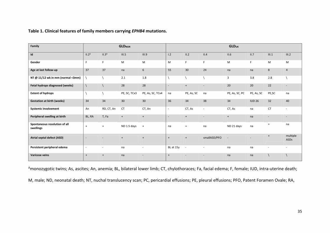
Table 1. Clinical features of family members carrying EPHB4 mutations.
Family GLDNOR GLDUK
Id II.2A II.3A III.5 III.9 I.2 II.2 II.4 II.6 II.7 III.1 III.2
Gender F F M M M F F M F M M
Age at last follow-up 37 37 na 6 55 30 24 na na 8 4
NT @ 11/12 wk in mm (normal <3mm) \ \ 2.1 1.8 \ \ \ 3 3.8 2.8 \
Fetal hydrops diagnosed (weeks) \ \ 28 28 - + - 20 20 22 -
Extent of hydrops \ \ PE, SC, TCx3 PE, As, SC, TCx4 na PE, As, SC na PE, As, SC, PC PE, As, SC PE,SC na
Gestation at birth (weeks) 34 34 30 30 36 34 38 34 IUD 26 32 40
Systemic involvement An RD, CT, An CT CT, An - CT, As - CT, As na CT -
Peripheral swelling at birth BL, RA T, Fa + + - + - + na - -
Spontaneous resolution of all swellings
+ + ND 1.5 days + na + na ND 21 days na + na
Atrial septal defect (ASD) - - + + + + smallASD/PFO - - + multiple
ASDs
Persistent peripheral edema - - na - BL at 15y - - na na - -
Varicose veins + + na - + - - na na \ \
Amonozygotic twins; As, ascites; An, anemia; BL, bilateral lower limb; CT, chylothoraces; Fa, facial edema; F, female; IUD, intra-uterine death;
M, male; ND, neonatal death; NT, nuchal translucency scan; PC, pericardial effusions; PE, pleural effusions; PFO, Patent Foramen Ovale; RA,

36
right arm edema; RD, respiratory distress; SC, subcutaneous edema; T, Truncal; TC, thoracocentesis in utero; \ not recorded; na, not applicable;
-, no; +, yes.