enrichment strategies for improving mass spec … enrichment strategies for improving mass spec...
TRANSCRIPT
1Rosa Viner, 2Ryan Bomgarden, 2Kratika Singhal, 2Sergei Snovida, 3Craig Gutierrez, 3Lan Huang 1Thermo Fisher Scientific, San Jose, CA; 2Thermo Fisher Scientific, Rockford, IL; 3University of California, Irvine, Irvine, CA
RESULTS
Figure 3. BSA crosslinked peptides enriched by Size Exclusion Chromatography (SEC).
Separation profiles shows collected fractions, number of total and unique non-redundant crosslinked
peptides identified for DSS (A), DSSO(B) and DSBU(C) crosslinkers vs original sample using
instrument methods as in Table 1. For each SEC experiment 100 µg of BSA tryptic digest was loaded
on the column and 1 µg of each fraction was analyzed by LC/MS. Most crosslinked peptides were
identified in first fractions (e.g. 0 and 1) as expected.4 Depending upon crosslinker used, we
observed an increase in identification of non-redundant peptides from 10% to 30% after SEC
fractionation and used these numbers to benchmark SCX spin column performance.
ABSTRACT
Purpose: To evaluate multiple, widely used enrichment/fractionation techniques and benchmark
newly developed strong cation exchange (SCX) spin columns for cross-linked peptide analysis using
a Thermo Scientific™ Orbitrap Fusion™ Lumos™ Tribrid™ mass spectrometer.
Methods: Different amine-reactive, homobifuctional crosslinkers were used to crosslink BSA
(monomer) and yeast enolase (homodimer) proteins. Cross-linked samples were reduced, alkylated
and digested with trypsin before MS analysis. Cross-linked peptides were pre-fractionated on SCX
stage tips, SCX spin columns or a peptide size exclusion chromatography (SEC) column. An Orbitrap
Fusion Lumos mass spectrometer was used for crosslinked peptide analysis. Data analysis was
performed with Thermo Scientific™ Proteome Discoverer™ 2.2 software using a XlinkX1 software
node.
Results: Newly designed spin columns containing a polymer-based strong cation exchange resin
were used for selective enrichment of cross-linked peptides. Our simplified, 2 fractions salt step
gradient, enabled identification a similar number of crosslinked peptides as SEC based fractionation.
INTRODUCTION Chemical cross-linking in combination with mass spectrometry is a powerful method to determine
protein-protein interactions. This method has been applied to recombinant and native protein
complexes and, more recently, to whole cell lysates or intact unicellular organisms in efforts to identify
protein-protein interactions on a global scale. However, this method suffers from low identification
rates without enrichment/fractionation, as the typical yield of cross-linked peptides is less than 1 % of
total identified peptides. In this study, we evaluated multiple, widely used enrichment/fractionation
techniques and benchmarked newly developed SCX spin columns for cross-linked peptide analysis
using an Orbitrap Fusion Lumos mass spectrometer.
MATERIALS AND METHODS
Sample Preparation
Disuccinimidyl suberate (DSS), disuccinimidyl sulfoxide (DSSO)2 and disuccinimidyl dibutyric
urea (DSBU)3 ( Figure 1) were used to crosslink 2mg/ml BSA and yeast enolase (Sigma Aldrich)
solubilized in 50mM HEPES pH 8 for 1hr at 100 molar excess of crosslinker to protein. After
crosslinking, reactions were quenched with 1M Tris pH 8. BSA was reduced, alkylated, acetone
precipitated and digested with trypsin before MS analysis. Enolase was first desalted using an
Amicon® centrifugal filter unit (30 kDa, EMD Millipore) and then digested with trypsin. Protein and
peptide concentrations were determined using the Pierce™ BCA Protein Assay Kit and the Pierce™
Quantitative Colorimetric Peptide Assay, respectively. Peptides were fractionated using a polymer-
based SCX spin columns with an increasing step gradient of sodium chloride (e.g. 20mM, 100mM,
500mM). Cross-linked peptides were also pre-fractionated on SCX stage tips (Thermo Fisher
Scientific) according to manufacturing instructions and SEC Superdex Peptide PC column (GE
Health) as described.4 SCX fractionated samples were desalted using a 70% ACN elution step on
Pierce™ High pH Reversed-Phase Peptide spin columns before LC-MS/MS analysis.
Liquid Chromatography and Mass Spectrometry
Samples were separated by RP-HPLC using a Thermo Scientific™ Dionex™ Ultimate 3000 system
connected to a Thermo Scientific™ EASY-Spray™ column, 50 cm × 75 µm over a 45 min 2-28%
gradient (A: water, 0.1% formic acid; B: acetonitrile, 0.1% formic acid) at 300 nL/min flow rate. The
crosslinked BSA and enolase samples were analyzed on the Orbitrap Fusion Lumos mass
spectrometer. Specific LC and MS settings are shown in Table 1.
Data Analysis
Spectral data files were analyzed using Proteome Discoverer 2.2 software using the XlinkX
node(Figure 2) for crosslinked peptides and SEQUEST®HT search engine for unmodified and dead-
end-modified peptides. Carbamidomethylation (+57.021 Da) used as a static modification for
cysteine. Different crosslinked mass modifications for lysine were used as variable modifications for
lysine in addition to methionine oxidation (+15.996 Da). Data were searched against a database
contained the Uniprot/SwissProt entries of the model proteins with/out common contaminants with a
1% FDR criteria for protein spectral matches. For MS2-MS3 methods, a linear–peptide search option
(using MS3 scans for identification and MS2 scan for detection of crosslinked peptides) was used for
XlinkX database searching. The XlinkX standard enumeration search option was used for data
acquired using non-cleavable crosslinker DSS and the MS2 methods (e.g. CID, ETD, EThcD).1
Results visualization and distance restraints were performed using xiNET5 and PyMOL 1.8
(Schrodinger LLC).
CONCLUSIONS
MSn analysis of DSSO cross-linked peptides resulted in comparable number of BSA
and enolase cross-linked peptides to those obtained from MS2 identification of non-
cleavable DSS and cleavable DSBU cross-linked peptides. The differences in
identification is more likely due to differences in MS acquisition, data analysis,
crosslinker length and/or solubility.
Cleavable cross-linkers are more advantageous than noncleavable cross-linkers for
diluted and complex samples due to improved FDR calculations based on MSn
integration and/or the presence of reporter ions for MS cleavable crosslinkers.
A simple 2 step salt gradient SCX fractionation enriched crosslinked peptide similar to
SEC fractionation for BSA and enolase.
Proteome Discoverer 2.2 software with XlinkX was used to confidently identify
crosslinked peptides for PyMol structure overlay.
REFERENCES
1. Liu F, Rijkers D, Post H, Heck AJ. Proteome-wide profiling of protein assemblies by cross-linking
mass spectrometry. 2015. Nat Methods, 12(12):1179-84.
2. Kao A, Chiu CL, Vellucci D, Yang Y, Patel VR, Guan S, Randall A, Baldi P, Rychnovsky SD,
Huang L. Development of a novel cross-linking strategy for fast and accurate identification of
cross-linked peptides of protein complexes. 2011. Mol Cell Proteomics, 10(1): M110.002212.
3. Müller MQ, Dreiocker F, Ihling CH, Schäfer M, Sinz A. Cleavable cross-linker for protein structure
analysis: reliable identification of cross-linking products by tandem MS. 2010. Anal Chem.
82(16):6958-68.
4. Leitner A, Reischl R, Leitner A, Reischl R, Walzthoeni T, Herzog F, Bohn S, Förster F, Aebersold
R. Expanding the chemical cross-linking toolbox by the use of multiple proteases and enrichment
by size exclusion chromatography. 2012. Mol Cell Proteomics, 11(3):1-12.
5. Combe CW, Fischer L, Rappsilber J. xiNET: cross-link network maps with residue resolution. 2015
Mol Cell Proteomics, 14(4):1137-47.
ACKNOWLEDGEMENTS
The authors would like to thank Kai Fritzemeier (Thermo Fisher Scientific, Germany) and Richard
Scheltema (University of Utrecht) for their work on integrating XLinkX software as a node in
Proteome Discoverer 2.2.
TRADEMARKS/LICENSING For Research Use Only. Not for use in diagnostic procedures.
© 2017 Thermo Fisher Scientific Inc. All rights reserved. SEQUEST is a registered trademark of the
University of Washington. All other trademarks are the property of Thermo Fisher Scientific and its
subsidiaries. This information is not intended to encourage use of these products in any manner that
might infringe the intellectual property rights of others.
Enrichment Strategies for Improving Mass Spec Analysis of Chemically Crosslinked Peptides
Figure 1. Structures of non-cleavable and MS-cleavable crosslinkers used in the study.
Figure 2. The processing (A) and consensus (B) XlinkX workflows in Proteome Discoverer 2.2
software includes a separate crosslinkers results tab (C), spectra annotation (D) and xiNET
data visualization (E) using csv and database files generated by Xlink Crosslink export node.
Table 1. Orbitrap Fusion Lumos MS crosslinker specific LC-MS acquisition parameter
settings.
Figure 4. BSA crosslinked peptides enrichment by SCX spin columns. A) SCX enrichment
workflow. B) Non-redundant crosslinked peptides identified from original BSA digest or from mixture
of control BSA digest (no crosslink) with BSA crosslinked sample in 100: 1 ratio to mimic typical 1%
presence of crosslinked peptides. For each experiment, 1 µg of BSA digest was analyzed by LC/MS
using methods as shown in Table 1. Using newly developed SCX spin columns, we identified a
similar number of crosslinked peptides in only 2 fractions (C) as by SEC ( Figure 3).
DSS
Spacer Arm 11.4Å
DSSO
Spacer Arm 10.3Å
DSBU
Spacer Arm 12.5Å
Settings DSSO
MS2 –MS3
DSS
MS2
DSBU
MS2
LC gradient 2-28% in 45min 2-28% in 45min 2-28% in 45min
MS 1 OT OT OT
Resolution 120K 120K 120K
Target value 4e5 4e5 4e5
Max injection time 100 100 100
Top Speed 5 sec 5 sec 5 sec
MS2 OT CID OT EThcD OT HCD
Charge states 3-8 3-8 3-8
Isolation width 1.6 1.6 1.6
NCE 25 SA15 SCE 30±5%
Resolution 30K 60K 30K
Target value 5e4 2e5 5e4
Max injection time 100ms 250ms 70ms
Targeted Mass MS3 IT HCD n/a n/a
Isolation width 2
NCE 30
Resolution Rapid
Target value 2e4
Max injection time 150 ms
A. B. C.
D.
E.
A. B.
C.
- SCX
+ SCX
A.
B.
C.
- SEC
+SEC
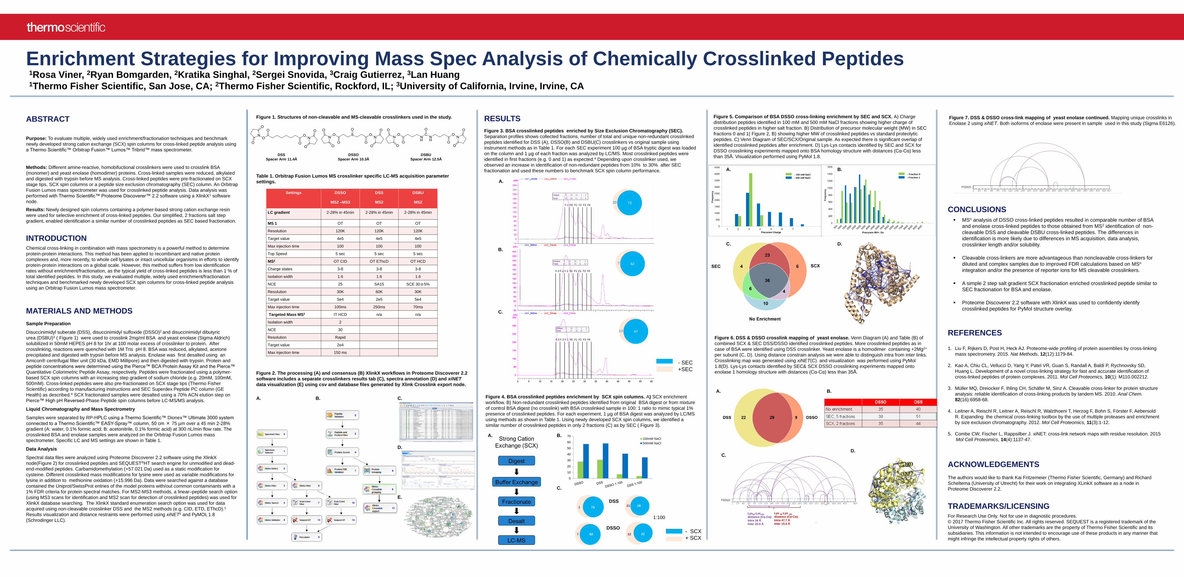
Figure 6. DSS & DSSO crosslink mapping of yeast enolase. Venn Diagram (A) and Table (B) of
combined SCX & SEC DSS/DSSO identified crosslinked peptides. More crosslinked peptides as in
case of BSA were identified using DSS crosslinker. Yeast enolase is a homodimer containing +2Mg2+
per subunit (C, D). Using distance constrain analysis we were able to distinguish intra from inter links.
Crosslinking map was generated using xiNET(C) and visualization was performed using PyMol
1.8(D). Lys-Lys contacts identified by SEC& SCX DSSO crosslinking experiments mapped onto
enolase 1 homology structure with distances (Cα-Cα) less than 35Å.
Figure 5. Comparison of BSA DSSO cross-linking enrichment by SEC and SCX. A) Charge
distribution peptides identified in 100 mM and 500 mM NaCl fractions showing higher charge of
crosslinked peptides in higher salt fraction. B) Distribution of precursor molecular weight (MW) in SEC
fractions 0 and 1( Figure 2, B) showing higher MW of crosslinked peptides vs standard proteolytic
peptides. C) Venn Diagram of SEC/SCX/Original sample. As expected there is significant overlap of
identified crosslinked peptides after enrichment. D) Lys-Lys contacts identified by SEC and SCX for
DSSO crosslinking experiments mapped onto BSA homology structure with distances (Cα-Cα) less
than 35Å. Visualization performed using PyMol 1.8.
- 100 mM NaCl
- 500 mM NaCl
- Fraction 0
- Fraction 1
0
10
20
30
40
50
60
70100mM NaCl
500mM NaCl
7311821
627 2222
DSS
DSSO
1:100
A. B.
A. B.
C. D.
7320
627Unique 5 12 16 2 3
Total 22 42 40 22 22
Unique 0 36 16 3 0
Total 16 68 51 21 6
4713Unique 0 18 17 3 0
Total 2 27 39 19 7
Lys 56-Lys 241
distance (Cα-Cα):
Intra 47.7 Å
Inter 15.5 Å
Lys60-Lys199
distance (Cα-Cα):
Intra 34 Å
Inter 20.5 Å
C. D.
Figure 7. DSS & DSSO cross-link mapping of yeast enolase continued. Mapping unique crosslinks in
Enolase 2 using xiNET. Both isoforms of enolase were present in sample used in this study (Sigma E6126).