enhanced sensitivity to 1-@-d-arabinofuranosylcytosine and...
TRANSCRIPT

[CANCER RESEARCH56. 5211-5216. November IS, 19961
ABSTRACT
We used human tumor cell lines from the National Cancer Institute's InVitro Antineoplastic Drug Screen to assess whether sensitivity to any of the—45,000compounds tested previously correlated with the presence of ama oncogene. Among these cell lines, the mutations in Ki-ras2 clustered in
non-small cell lung and colon carcinoma subpanels, and five of the sixleukemia lines contained mutations in either N-ras or Ki-ras2. Theseanalyses revealed a striking correlation with i-(l-D-arabinofuranosylcy.tosine (Ara-C) and 2,2'-O-cyclocytidine sensitivity in the cell lines harboring ras mutations compared to the tumor lines with wild-type ras alleles.Strong correlations were also found with topoisomerase (topo) IJ inhibitors, especially 3'-hydroxydaunorubicin and an olivacine derivative.These differential sensitivities persisted in an additional 22 non-small cell
lung carcinoma lines (ras mutations, n 12 and wild-type ras, n 10).Thus, the association with Ara-C sensitivity was greatest while topo IIinhibitors showed a lower, but significant, correlation. These results suggest that the ras oncogene may play a determinant role in rendering tumor
cells sensitive to deoxycytidine analogues and topo II inhibitors.
INTRODUCTION
Tumor progression is a multistep process that is driven by geneticinstability (1, 2). Defects in specific genes like DNA repair genes orthe frequently mutated tumor suppressor genes and activated oncogenes have been implicated in the genetic instability of tumor cells(reviewed in Refs. 3—9).In addition, profound changes occur incheckpoint function as well as in the regulation of apoptosis (reviewedin Refs. 5—12).Cancer cells in general display decreased sensitivity toa wide variety of therapeutic agents (13—16),but we and others (7—10)have proposed that through compromising cell cycle checkpoint functions, the oncogene and tumor suppressor gene of cancer cells may beresponsible for their preferential sensitivity to certain antineoplasticagents. To begin to test this hypothesis, we determined whether thepresence of mutations in ras alleles in the 60 human tumor cell linesof the NCI-ADS4 (17) correlated with enhanced sensitivity to any ofthe —45,000natural and synthetic compounds that have been tested inthe screen. We chose to examine the ras alleles since ras oncogeneactivation is the most frequently occurring gain-of-function mutationdetected in human tumors (18, 19).
Received 7/1/96; accepted 9/I 9/96.The costs of publication of this article were defrayed in part by the payment of page
charges. This article must therefore be hereby marked advertisement in accordance with18 U.S.C. Section 1734 solely to indicate this fact.
I Supported in part by the National Cancer Institute, Department of Health and Human
Servies, under contract with ABL.2 Present address: Fred Hutchinson Cancer Research Center, I 124 Columbia Street,
Cl-OlS. Seattle. Washington 98104.3 To whom requests for reprints should be addressed, at ABL-Basic Research Program,
National Cancer Institute-Frederick Cancer Research and Development Center, P. 0. BoxB, Frederick, MD 21702-1201.
4 The abbreviations used are: NCI-ADS, National Cancer Institute In Vitro Antineo
plastic Drug Screen; G130 and Gl@0,50 and 30% inhibition of cell growth, respectively;PCC, Pearson correlation coefficient; MDR, multidrug resistance; Ara-C, l-@3-o-arabinofuranosylcytosine; topo, topoisomerase; NSCL, non-small cell lung; FBS, fetal bovineserum; DGGE. denaturing gradient gel electrophoresis; AML, acute myeloid leukemia.
The NCI-ADS database presently contains the differential sensitivity patterns of the 60 tumor cell lines to over 45,000 compounds. Thesensitivity of each cell line to a given compound can be displayed as
a mean graph, based on the drug concentration that gives a 50%inhibition of cell growth (G150) in 2-day assay (20, 21). Individualpatterns can be compared to the entire database using the COMPAREpattern recognition program (21). COMPARE searches for similaritiesin mean graph patterns and ranks the similarity between patterns by aPCC (2 1). COMPARE analyses of the NCI-ADS database has identified compounds with similar chemical structures and/or relatedbiochemical mechanisms of action (21—29) and, recently, substrates
and inhibitors of the P-glycoprotein, which mediates MDR, have beenidentified (30, 31).
We used COMPARE to assess whether compounds present in thedatabase displayed selective growth inhibition against the tumor celllines with ras oncogenes. The compounds identified by COMPARE
were further tested against an independent set of tumor cell lines. Ourresults revealed that the presence of a ras oncogene in the tumor cell
lines correlated with enhanced sensitivity to deoxycytidine analoguesincluding Ara-C, 2,2'-O-cyclocytidine, and gemcitabine. Low butsignificant correlation was also shown to topo II inhibitors, especially
3'-hydroxydaunorubicin and an olivacine derivative.
MATERIALS AND METHODS
Compounds and Cell Lines. Compoundsused in this studywereobtainedthrough the Drug Synthesis and Chemistry Branch, NC!. The human tumor cell
lines of NCI-ADS and culture conditions has been described previously (20).The tumor cell lines derived from NSCL carcinoma (32) were maintained andassayed for drug sensitivity in RPM! supplemented with 10% FBS and 2 mM
L-glutamine.
Sequence Determination of ras Mutations. GenomicDNAs wereisolatedfrom the cell lines of NCI-ADS using a Cell Culture DNA kit (Qiagen). Theexons I and 2 of each ras gene were amplified by PCR from the genomic
DNAs using the primer pairs specific for each exon/intron junction. The PCRamplifications were performed in a GeneAmp PCR System 9600 (Perkin
Elmer) and were all preceded with one cycle of denaturation at 94°C for 3 mm
and finished with the incubation at 72°Cfor 3 mm. The PCR conditions usedwere 40 cycles of: 94°C(15 s), 48°C(15 s), 72°C(15 s with 1 s extension percycle) for Ki-ras2 exon I and N-ras exon I ; 94°C ( 15 s), 52°C ( I 5 s), 72°C (15
5 with 1 s extension per cycle) for Ki-ras2 exon 2 and Ha-rasl exon 2; 94°C
(15 s), 48°C(15 s), 70°C(25 s with I s extensionper cycle) for N-ras exon 2;and 94°C(15 s), 56°C(15 s), 72°C(15 s with I s extension per cycle) forHa-rasl exon 1. The products were then purified from a 6% polyacrylamidegel. DNA sequence was determined for both strands directly from the purified
PCR products using the same primers as those for PCR amplification. DNAsequencing reactions were carried out with the PRISM Ready Reaction
DyeDeoxy Terminator cycle sequencing kit (ABI), and the products wereanalyzed on an ABI 373A DNA sequencer. The DNA sequences of primersused for PCR amplification and sequencing were: KR15, GGCCTGCT
GAAAATGACTGA and KRI3, GTCCTGCACCAGTAATATGCfor Ki-ras2exon 1; KR25, CCAGACTGTG11TCTCCC'ITC and KR23, CACAAAGAAAGCCCTCCCCA for Ki-ras2 exon 2; NR15, GACTGAGTACAAACT
5211
Enhanced Sensitivity to 1-@-D-Arabinofuranosylcytosine and Topoisomerase IIInhibitors in Tumor Cell Lines Harboring Activated ras Oncogenes'
Han-Mo Koo, Anne Monks, Andrei Mikheev,2 Larry V. Rubinstein, Marcia Gray-Goodrich, Mary Jane McWilliams,W. Gregory Alvord, Herbert K. Oie, Adi F. Gazdar, Kenneth D. Paull, Helmut Zarbl,2 and George F. Vande Woude3
ABL-Basic Research Program, National Cancer Institute-Frederick Cancer Research and Development Center, Frederick. Maryland 21702-1201 IH-M. K., M. J. M., G. F. V. WI;Science Applications International Corporation-Frederick. National Cancer Institute-Frederick Cancer Research and Development Center, Frederick. Maryland 2! 702 (A. M..M. G-G.J; Massachusetts Institute of Technology. Cambridge. Massachusetts 02139 [A. M.. H. ii: Division of Cancer Treatment. National Cancer Institute, NIH, Bethesda,Maryland 20892 IL V. R., H. K. 0., K. D. P.!: Data Management Services, Inc., National Cancer Institute-Frederick Cancer Research and Development Center. Frederick,Maryland 21 702 [W. G. A.J: and Simmons Cancer Center and Department of Pathology, Universiti of Texas Southwestern Medical Center at Dallas, Dallas, Texas 75235[A. F. G./
on March 6, 2019. © 1996 American Association for Cancer Research. cancerres.aacrjournals.org Downloaded from

Table 1 Activating mutations of ras genes in the cell lines of leukemia. NSCLandcoloncancer subpanels of the NCI-ADSMutation
Dc.iciE°Sequence―K1: mutlwtK I2:GAT(Asp)Nl:
mut/wtNl2:GTF(Val)N2:mutN61:CTA(Leu)wtwtNl:
mut/wtN12:TGT(Cys)KI: mutlwtK I2:GCT(Ala)NI : mutlwtN 12:TGT(Cys)KI:
mutKl2:AGT(Ser)WIWtKI:
mut>wtKl2:TGT(Cys)WIWtWIWtKl:
mut>WtKl2:TGT(Cys)WtWtK2:
mutK61:CAT(His)wtWI
Subpanel
Leukemia
NSCL cancer
Colon cancer
ras AND SENSITIVITY TO Ara-C AND TOPO 11 INHIBITORS
GGTGGandNR13, GGGCCTCACCTCTATGGTGfor N-ras exon 1;NR25,GGTGAAACCTG1TrGTTGGA and NR23, ATACACAGAGGAAGCCT
TCG for N-ras exon 2; HR15, CAGGCCCCTGAGGAGCGATGand HR13,UCGTCCACAAAATGGTTCT for Ha-rasl exon 1; and HR25, TCCTG
CAGGArFCCTACCGG and HR23, GGTFCACCTGTACTGGTGGA forHa-rasl exon 2.
DGGE. The PCR amplification of each exon from genomic DNA, purification and end-labeling of the PCR products, heteroduplex formation, andDGGE procedure were described elsewhere (33). To facilitate separation of theDNA fragments by DGGE, one primer from each pair used for the direct DNAsequencing was linked at its 5-end to a 54-bp long, thermostable, OC-rich
clamp sequence (33).Drug Sensitivity Assays, Data Calculation, and COMPARE Algorithm.
The assay methodology, sensitivity data calculation, and the COMPAREalgorithm have been described in detail previously (20, 21). Briefly, cellularresponse to a compound was evaluated using a sulforhodamine-B assay after48 h continuous exposure of cells with a range of the compound concentration,and the sensitivity of each cell line was calculated for the compound concentration yielding a GI50 (20). The mean graph pattern generated from therelative sensitivities of cell lines tested was used by the COMPARE patternrecognition program to calculate PCCs for all compounds in NCI-ADS database (21).
Statistical Analysis. The 1-sided Ps by Wilcoxon rank sum test werecalculated by exact distribution methods using STATXACT software (CYTEL
Software Co.).
RESULTS
Determination of Activating Mutations of ras Genes in TumorCell Lines of NCI-AD5. The mutationstatus of ras alleles in eachtumor cell line of NCI-ADS was determined from PCR-amplifiedgenomic DNA prepared from each cell line. Each PCR-amplifiedDNA sample was then analyzed for the presence of an activatingmutation in the 12th, 13th and 61st codons of the Ki-, N- and Ha-rasalleles (18, 19) by direct DNA sequencing and DGGE. Among the 60cell lines analyzed, 17 cell lines were found to contain activatingmutations in at least one of the ras alleles. Most of the cell lines withactivating mutations in ras genes clustered in three of the ninecancer-subpanels of the NCI-ADS; mutations in the Ki-ras2 allelewere found in four cell lines of the NSCL cancer subpanel and in threecell lines of the colon cancer subpanel, whereas five of six leukemialines contained mutations in either the N-ras or Ki-ras2 allele (Table1). The additional mutant ras alleles were detected in cell lines of theremaining six cancer subpanels: N..rasArg6l in SK-MEL-2 (melanoma); K-ras@12 in OVCAR-5 (ovarian carcinoma); K@rasAst@3inSN12C (renal carcinoma); and K@rasA@@1@l3in MDA-MB-231 andH@rasAI@@l2in HS578T (breast carcinomas). The frequency of activating mutations in ras genes among the NCI-ADS cell lines werecomparablewith that found previouslyin other human tumors andtumor cell lines (18, 19).
Compounds That Are Preferentially Active against Tumor CellLines with Activated ras Oncogenes. We used COMPARE to assesswhether any compounds in the database of NCI-ADS displayed selective growth inhibition of the tumor cell lines in the NSCL andcolon carcinoma subpanels containing activated ras oncogenes. Weinitially focused on the 16 cell lines among which the distribution ofmutant and wild-type ras allele-containing cell lines was approximately equal (Table 1), allowing an optimal analysis of the databaseusing COMPARE. To establish the ras mean graph for this analysis,we assigned the cell lines with activated ras oncogenes a positivevalue to equate with preferential drug sensitivity, whereas those withwild-type ras alleles were given a negative value. The COMPAREanalysis was performed to identify compounds in the database that:(a) were tested at least three times against all sixteen cell lines; (b)gave a PCC of >0.75 with the ras mean graph pattern (under these
CelllineCCRF-CEM
HL-60(TB)K562MOLT-4RPMI-8226SR
A549/ATCCEKVXHOP-62HOP-92NCI-H226NCI-H23NCI-H322MNCI-H460NCI-H522
COLO-205 wt WIHCC2998 WI WtHCT-l 16 Kl: mut/Wt K13: GAC(Asp)HCT- I5 K 1: mut/WI K I3: GAC(Asp)HT-29 WI wtKM-12 WI wtSW620 Kl: mut Kl2: G1T(Val)
a The allelic status of ras genes determined by DGGE analysis of PCR-amplified
genomic DNA. The exon number containing a mutation is also indicated: K, Ki-ras2; N,
N-ras; mut, allele With a mutation; Wt, wild-type allele; >, elevated DGGE signal for themutant allele compared to the wild-type signal.
b The sequence of a mutation independently determined by direct sequencing of
PCR-amplified genomic DNA. The codon number harboring a mutation is indicated Withthe sequence and amino acid encoded.
C A silent mutation at 59th codon of Ha-rasl (GCC—+GCT) was also found.
conditions, i.e., n = 16 cell lines, the probability of an observingcorrelation is <0.0008); and (c) exhibited a wide range of activities onthese cell lines (greater than one order of magnitude by G150 drug
concentration). Compounds in the database under a confidentialagreement were excluded from this analysis.
Within these parameters, the compounds showing the strongestcorrelation with the ras pattern were deoxycytidine analogues that arecommonly used in the treatment of acute leukemia (34). In theNCI-ADS, Ara-C was tested at three different concentration ranges,all of which were identified by the COMPARE analysis (PCC = 0.93,0.78, and 0.77, respectively). At the concentration range where Ara-Chas been tested 122 times and displayed the widest range of activityagainst the cell lines, the ras oncogene-containing NSCL andcolon carcinoma cell lines were two orders of magnitude more sensitive to Ma-C than the cell lines with wild-type ras alleles(G150 = 8.5 1 x l0@ M and 1.1 X l0@ M, respectively; Fig. la).Similar response patterns were observed in the other test concentration ranges, although the actual differential in response could not bedetermined because the GI50 was not achieved for the less sensitivecell lines (data not shown). The preferential sensitivity of the rasoncogene-containing cell lines to Ara-C was further supported by thehigh correlation found for 2,2'-O-cyclocytidine (PCC = 0.81), whichis a latent prodrug form of Ara-C (35, 36). For 2,2'-O-cyclocytidine,the ras oncogene-containing NSCL and colon tumor cell lines wereapproximately 85-fold more sensitive to the drug than the cell lineswith wild-type ras alleles (G150 = 1 X lO_6 M and 8.6 X l0@ M,respectively; Fig. lb). To extend this correlation further, we alsodetermined whether the cell lines with activated ras oncogenes display preferential sensitivity to a new deoxycytidine analogue, gemcitabine (2',2'-difluorodeoxycytidine). Gemcitabine has pharmacokinetic properties superior to Ara-C both in vitro and in vivo (37—41)and, unlike Ara-C, shows activity against solid tumors, especiallyagainst carcinomas of the pancreas, lung, and colon (40—46).The test
5212
on March 6, 2019. © 1996 American Association for Cancer Research. cancerres.aacrjournals.org Downloaded from

ras AND SENSITIVITYTO Ara-CAND TOPOII INHIBITORS
1r8
1@
4
II
.
.
.7
•1..
$
I.I
.
..
.;
..
2•3#@
5
I
.
..7
:1
10.6.
1@
1o@'@
a@ ____________________b . ____________________C
1@
—@ 10.6.@
!. !,@, :@‘
:@@ .@ :@
.Th@ .@ .@@ . c C
a)@ a) a,Cl) : C/) (I)
io'.
.@
10@-
io-@ , ‘@@ I@ I IMut Wt Mut Wt Mut Wt
Fig. I . Sensitivity of the 16 NSCL and colon carcinoma cell lines of NCI-ADS to deoxycytidine analogues. Mean sensitivities of the cell lines with ras oncogenes (Mut. •) or
wild-type ras alleles (Wt. •) to Ara-C from 122 independent tests (a), 2.2'-O-cyclocytidine from 16 independent tests (b) and gemcitabine from 7 independent tests (c) are shown.Mean sensitivities (+) for each group ofcell lines are shown; bars, SE. Cell lines containing ras oncogenes are marked with numbers: I, NCI-H460; 2. A549: 3. HOP-62; 4. HCT-l 16;5, NCI-H23; 6, SW620; and 7, HCT- I5.
tions with leukemia lines in which the K562 line with wild-type rasalleles was generally more sensitive to the compounds than rasoncogene-containing lines (data not shown). The RPMI-8226 cells,which were less sensitive to deoxycytidine analogues and topo II
of gemcitabine on the cell lines of NSCL and colon cancer subpanelsshowed that cell lines with the enhanced sensitivity to Ara-C and2,2'-O-cyclocytidine were also the most sensitive to gemcitabine (Fig.
1, compare a and b with c). Thus, despite the many reported differences between Ara-C and gemcitabine (37, 38), their cytotoxic activities against these cell lines were similar.
The leukemia lines in the NCI-ADS displayed generally highersensitivity to cytotoxic compounds than adherent cells (20), and the
presence of oncogenic ras mutations in five of six cell lines in thesubpanel (Table 1) introduced a bias to the mean graph. Theseleukemia lines, therefore, were not a suitable data set to generate astatistically relevant differential pattern for COMPARE analysis. Nevertheless, consistent with the findings in NSCL and colon carcinomacell lines, four of the five ras oncogene-containing leukemia lines(Table 1) were highly sensitive to the deoxycytidine analogues cornpared to K562 line with wild-type ras alleles (Table 2).
In addition to the deoxycytidine analogues, the COMPARE analysis also revealed significant correlations between the presence of ras
oncogenes and enhanced sensitivity to the topo II inhibitors, 3'-hydroxydaunorubicin (47), and an olivacine derivative, NSC 659687(Ref. 48; PCC = 0.82 and 0.8 1, respectively; Fig. 2). The range ofresponse for these compounds was, however, narrower than for thedeoxycytidine analogues (compare Fig. I with Fig. 2), but as with thedeoxycytidine compounds, the same four ras oncogene-containingleukemia lines were more sensitive to the topo II inhibitors thanwild-type K562 (Table 2).
Positive correlations with the ras mutant NSCL and colon carcinoma cell lines were also observed for a 3-deazauridine derivativecompound (PCC = 0.77), the lipophilic antifolate trimetrexate(PCC = 0.77), and a bioreductive alkylating agent, carboquone
(PCC = 0.76). However, these compounds displayed poor correla
b
>
(I,Ca,Cl)
a
>
(I,Ca,Cl)
1 @-@—
1 @-@-
10@@—
•i
T+ $2.1.@t!I53 .
.
•1
4
io@—
i@-@
i!i•1•@
5$7
Mut Wt Mut Wt
Fig. 2. Sensitivity of the NSCL and colon carcinoma cell lines of NCI-ADS to topo IIinhibitors 3'-hydroxydaunorubicin (a) and olivacine derivative NSC 659687 (b). Symbolsand cell line number are as described in the legend to Fig. 1.
5213
on March 6, 2019. © 1996 American Association for Cancer Research. cancerres.aacrjournals.org Downloaded from
ras AND SENSITIVITYTO Am-C AND TOPOII INHIBITORS
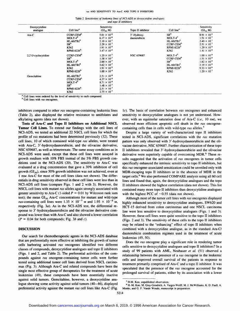
Table 2 Sensitivities of leukemia lines of NCI-ADS to deoxycytidine analoguesand topo Ii inhibitors
DeoxycytidineanalogueCell line―Sensitivity (Gl,0 M)Topo II inhibitorCell line―Sensitivity (GI@0M)Ara-CCCRF-CEM―
MOLT@4bHL-60(TB)―SRbK562RPMI-8226―5.01
X 10.86.17 X 10.82.19 X [email protected] X [email protected] X 10'3.47 X l0@3'-Hydroxy
daunorubicinSRb MOLT-4―HL-6O(TB)―CCRF-CEM―RPMI-8226―K5628.91
X 10'1.51 X [email protected] X [email protected] x [email protected] X l0―1.41 xItt―2,2'O-cyclocytidineCCRF-CEM―
SR―MOLT@4bHL-60(TB)―K562RPMI-8226―1
.20 X [email protected] X i072.00 X [email protected] X iO-71.91 X l0'1.00 X l0@NSC
659687MOLT-4―CCRF-CEM―SR―HL-60(TB)―RPMI-8226―K5621
.00 X 10'1.51 X Itt'1.82 X 10'2.19 x 10'9.33 x Itt'I .20 Xl0@GemcitabineHL-6O(TB)―
CCRF-CEM―MOLT-4―SR―RPMI-8226―K5622.51
X I0'4.37 X ltt'8.71x 10'1.48 X l0@@2.51 X [email protected] X l0@
a Cell lines were ordered by the level of sensitivity to each compound.
b Cell lines with ras oncogenes.
inhibitors compared to other ras oncogene-containing leukemia lines(Table 2), also displayed the relative resistance to antifolates andalkylating agents (data not shown).
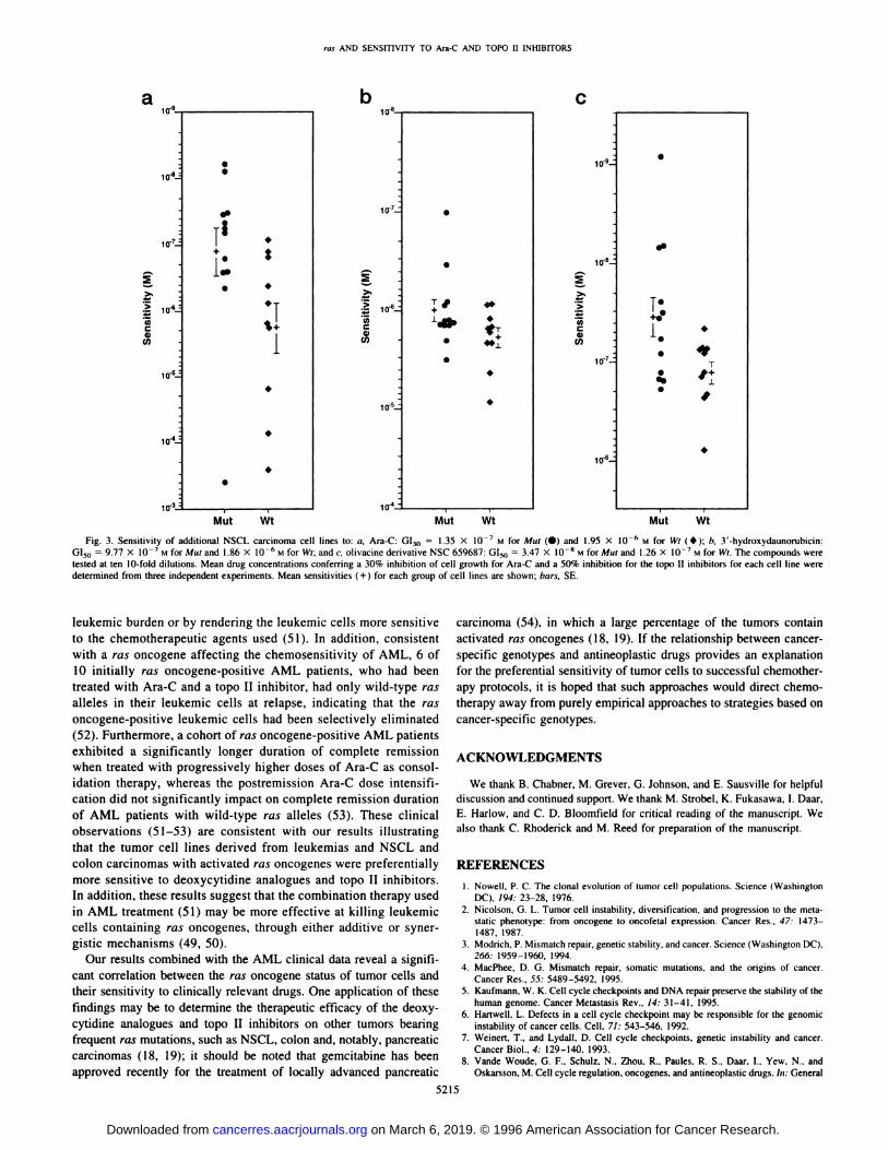
Tests of Am-C and Topo II Inhibitors on Additional NSCLTumor Cell Lines. To extend our findings with the cell lines ofNCI-ADS, we tested an additional 22 NSCL cell lines for which theprofileof ras mutationshad been determinedpreviously(32).Thesecell lines, 10 of which contained wild-type ras alleles, were treatedwith Ma-C, 3'-hydroxydaunorubicin, and the olivacine derivative,NSC 659687, as well as trimetrexate. The same assay conditions as inNCI-ADS were used, except that these cell lines were assayed ingrowth medium with 10% FBS instead of the 5% FBS growth conditions used in the NCI-ADS (20). The sensitivity to Ara-C wasevaluated at a drug concentration that gave a 30% inhibition of cellgrowth (G130), since 50% growth inhibition was not achieved, even at1 mM Ara-C for most of the cell lines (data not shown). The differentials in drug sensitivity observed in these cell lines were less than inNCI-ADS cell lines (compare Figs. 1 and 2 with 3). However, theNSCL cell lines with mutant ras alleles again strongly associated withgreater sensitivity to Ara-C (1-sided P = 0.01 by Wilcoxon rank sumtest); the mean GI30 Ara-C concentrations for mutant and wild-typeras-containing cell lines were 1.35 X i07 M and 1.95 X lO_6 M,respectively (Fig. 3a). As in the NCI-ADS test, the differential response to 3'-hydroxydaunorubicin and the olivacine derivative compound was lower than with Ara-C and also showed a lower correlation(P = 0.04 for both compounds; Fig. 3b and c).
DISCUSSION
Our search for chemotherapeutic agents in the NCI-ADS databasethat are preferentially more effective at inhibiting the growth of tumorcells harboring activated ras oncogenes identified two differentclasses of compounds, deoxycytidine analogues and topo II inhibitors
(Figs. 1 and 2, and Table 2). The preferential activities of the compounds against ras oncogene-containing tumor cells were furthertested using additional tumor cell lines derived from NSCL carcinomas (Fig. 3). Although Ara-C and related compounds have been thesingle most effective group of therapeutics for the treatment of acuteleukemias (49), these compounds have been essentially inactiveagainst solid tumors. Gemcitabine, however, a deoxycytidine analogue showing some activity against solid tumors (40—46),displayedpreferential activity against the mutant ras cell lines like Ara-C (Fig.
lc). The basis of correlation between ras oncogenes and enhancedsensitivity to deoxycytidine analogues is not yet understood. However, with an equimolar saturation dose of Ara-C (i.e., 10 nM), weobserved more efficient apoptotic cell death in the ras oncogenecontaining cells than in cells with wild-type ras alleles.5
Despite a large variety of well-characterized topo II inhibitorstested in NCI-ADS, significant correlations with the ras mutationpattern was only observed with 3'-hydroxydaunorubicin and the ohvacine derivative, NSC 659687. Further characterization of these topoII inhibitors revealed that 3'-hydroxydaunorubicin and the ohivacinederivative were superiorly capable of overcoming MDR.―These results suggested that the activation of ras oncogenes in tumor cells
specifically enhanced the intrinsic sensitivity to topo II inhibitors, butthis ras oncogene-associated sensitization could be unveiled only with
MDR-escaping topo II inhibitors or in the absence of MDR in thetarget cells.6 We also performed COMPARE analysis using all 60 celllines and found that, again, the deoxycytidine analogues and the topoII inhibitors showed the highest correlation (data not shown). This listcontained many more topo II inhibitors than deoxycytidine analogueswith the highest PCC of —0.55(data not shown).
Although most of the tumor cell lines with ras oncogenes displayedhighly enhanced sensitivity to deoxycytidine analogues, SW620 andHCT- 15 derived from colon carcinomas and one NSCL carcinomaline were less sensitive to deoxycytidine analogues (Figs. 1 and 3).However, these cell lines were quite sensitive to the topo II inhibitors(Figs. 2 and 3). The sensitivity of these cells to the topo II inhibitors
may be related to the “enhancing―effect of topo II inhibitors whencombined with a deoxycytidine analogue, as in the standard Ara-C/daunorubicin combination regimen used in the treatment of acuteleukemias (49, 50).
Does the ras oncogene play a significant role in rendering tumorcells sensitive to deoxycytidine analogues and topo II inhibitors? In astudy of 99 patients with AML, Neubauer et a!. (5 1) observed arelationship between the presence of a ras oncogene in the heukemiccells and improved overall survival of the patients in response totreatment primarily comprised of Ara-C and a topo II inhibitor. It wasspeculated that the presence of the ras oncogene accounted for theprolonged survival of patients, either by its association with a lower
5 H-M. Koo, unpublished observation.
6 H.-M. Koo, M. Gray-Goodrich, A. Vaigro-Wolff, M. J. McWilliams, K. D. Paull, A.Monks, and G. F. Vande Woude, manuscript in preparation.
5214
on March 6, 2019. © 1996 American Association for Cancer Research. cancerres.aacrjournals.org Downloaded from
ras AND SENSITIVITY TO Ma-C AND TOPO II INHIBITORS
a
10.8.
1 @-@-
>.
.@ 10.6@
U,Ca,Cl)
10@-
-—-3
@@-8
S
@5S
.1S
S
TS .Ã '+
S
.
SS
iiI •+
1..S •
.TI
.
.
.S
S
S
@‘ ..
.
S
.
.
Mut Wt Mut Wt Mut Wt
b _____ C
io@
io-7—
10.8_
@:@,
;@@
U) .@C Ca, a,
Cl) Cl)
io@—
10-s—
10.6_
10@-
Fig. 3. Sensitivity of additional NSCL carcinoma cell lines to: a, Ara-C: GI30 = 1.35 X l0@ M for Mut (•)and 1.95 X l0_6 M for Wt ( •); b, 3'-hydroxydaunorubicin:GI50 9.77x l0—@MforMutand 1.86X l0_6 si for Wt;andc, olivacinederivativeNSC659687:GI50= 3.47X 10' MforMutand 1.26X l0@ Mfor Wt.Thecompoundsweretested at ten 10-fold dilutions. Mean drug concentrations conferring a 30% inhibition of cell growth for Ara-C and a 50% inhibition for the topo II inhibitors for each cell line weredetermined from three independent experiments. Mean sensitivities (+) for each group of cell lines are shown; bars, SE.
heukemic burden or by rendering the leukemic cells more sensitiveto the chemotherapeutic agents used (5 1). In addition, consistentwith a ras oncogene affecting the chemosensitivity of AML, 6 of10 initially ras oncogene-positive AML patients, who had beentreated with Ara-C and a topo II inhibitor, had only wild-type rasalleles in their leukemic cells at relapse, indicating that the rasoncogene-positive leukemic cells had been selectively eliminated(52). Furthermore, a cohort of ras oncogene-positive AML patientsexhibited a significantly longer duration of complete remissionwhen treated with progressively higher doses of Ara-C as consolidation therapy, whereas the postremission Ara-C dose intensification did not significantly impact on complete remission durationof AML patients with wild-type ras alleles (53). These clinicalobservations (51—53) are consistent with our results illustratingthat the tumor cell hines derived from heukemias and NSCL andcolon carcinomas with activated ras oncogenes were preferentiallymore sensitive to deoxycytidine analogues and topo II inhibitors.In addition, these results suggest that the combination therapy usedin AML treatment (51) may be more effective at killing leukemiccells containing ras oncogenes, through either additive or synergistic mechanisms (49, 50).
Our results combined with the AML clinical data reveal a significant correlation between the ras oncogene status of tumor cells andtheir sensitivity to clinically relevant drugs. One application of thesefindings may be to determine the therapeutic efficacy of the deoxycytidine analogues and topo II inhibitors on other tumors bearingfrequent ras mutations, such as NSCL, colon and, notably, pancreaticcarcinomas (18, 19); it should be noted that gemcitabine has beenapproved recently for the treatment of locally advanced pancreatic
carcinoma (54), in which a large percentage of the tumors containactivated ras oncogenes (18, 19). If the relationship between cancerspecific genotypes and antineophastic drugs provides an explanation
for the preferential sensitivity of tumor cells to successful chemotherapy protocols, it is hoped that such approaches would direct chemotherapy away from purely empirical approaches to strategies based oncancer-specific genotypes.
ACKNOWLEDGMENTS
We thank B. Chabner, M. Grever, G. Johnson, and E. Sausville for helpfuldiscussion and continued support. We thank M. Strobel, K. Fukasawa, I. Daar,
E. Harlow, and C. D. Bloomfield for critical reading of the manuscript. Wealso thank C. Rhoderick and M. Reed for preparation of the manuscript.
REFERENCES
I . Nowell, P. C. The clonal evolution of tumor cell populations. Science (WashingtonDC), 194: 23—28,1976.
2. Nicolson, G. L. Tumor cell instability, diversification, and progression to the metastatic phenotype: from oncogene to oncofetal expression. Cancer Res., 47: 1473—1487,1987.
3. Modrich, P. Mismatch repair, genetic stability, and cancer. Science (Washington DC),266: 1959—1960,1994.
4. MacPhee, D. G. Mismatch repair, somatic mutations, and the origins of cancer.Cancer Res., 55: 5489—5492, 1995.
5. Kaufmann, W. K. Cell cycle checkpoints and DNA repair preserve the stability of thehuman genome. Cancer Metastasis Rev.. 14: 31—41,1995.
6. HarIwell, L. Defects in a cell cycle checkpoint may be responsible for the genomicinstability of cancer cells. Cell, 71: 543—546,1992.
7. Weinert, T., and Lydall, D. Cell cycle checkpoints, genetic instability and cancer.Cancer Biol., 4: 129—140,1993.
8. Vande Woude, G. F., Schulz, N., Thou, R., Paules. R. S., Daar, I., Yew, N., andOskarsson, M. Cell cycle regulation. oncogenes, and antineoplastic drugs. In: General
5215
on March 6, 2019. © 1996 American Association for Cancer Research. cancerres.aacrjournals.org Downloaded from

ras AND SENSITIVITY TO Ara-C AND TOPO II INHIBITORS
Motors Cancer Research Foundation: 1990 Views of Cancer Research, pp. 128—143.Philadelphia, PA: J. B. Lippincott Co., 1990.
9. Murakami,M. S., Strobel,M. C., and VandeWoude,G. F. Cellcycleregulation,oncogenes, and antineoplastic drugs. In: P. M. Howley, M. A. Israel, and L. A. Liotta(eds.), The Molecular Basis of Cancer, pp. 3—17.Philadelphia: W. B Saunders, Co.,1994.
10. Hartwell,L. H., and Kastan,M. B. Cellcyclecontroland cancer.Science(Washington DC), 266: 1821—1828,1994.
11. Thompson, C. B. Apoptosis in the pathogenesis and treatment of disease. Science(Washington DC), 267: 1456—1462, 1995.
12. Kastan, M. B., Canman, C. E., and Leonard, C. J. p53, cell cycle control andapoptosis: implications for cancer. Cancer Metastasis Rev., 14: 3—15,1995.
13. Lowe, S. W., Bodis, S., McClatchey, A., Remington, L., Ruley. H. E., Fisher, D. E.,Housman, D. E., and Jacks, T. p53 status and the efficacy of cancer therapy in vivo.Science (Washington DC), 266: 807—810, 1994.
14. Schaefer, D. I., Livanos, E. M., White, A. E., and Tlsty, T. D. Multiple mechanismsof N-(phosphonoacetyl)-L-asparlate drug resistance in SV4O-infected precrisis humanfibroblasts. Cancer Res., 53: 4946—4951, 1993.
15. Fan, S., El-Deity, W. S., Bae, I., Freeman, J., Jondle, D., Bhatia, K., Fomace, A. J.,Jr., Magrath, I., Kohn, K. W., and O'Connor, P. M. p53 gene mutations are associatedwith decreased sensitivity of human lymphoma cells to DNA damaging agents.Cancer Res., 54: 5824—5830, 1994.
16. Morgan,W. F., and Murnane,J. P. A role for genomicinstabilityin cellularradioresistance? Cancer Metastasis Rev., 14: 49—58,1995.
17. Grever, M. R., Schepartz, S. A., and Chabner, B. A. The National Cancer Institute.Cancer Drug and Discovery and Development Program. Scm. Oncol., 19: 622—638,1992.
18. Bos, J. L. ras oncogenes in human cancer: a review. Cancer Res., 49: 4682—4689,1989.
19. Barbacid, M. ras oncogenes: their role in neoplasia. Eur. J. Clin. Invest., 20: 225—235,1990.
20. Monks, A., Scudiero, D., Skehan, P., Shoemaker, R., Paull, K., Vistica, D., Hose, C.,Langley, J., Cronise, P., Vaigro-Wolff, A., Gray-Goodrich, M., Campbell, H., Mayo,J., and Boyd,M. Feasibilityof a high-fluxanticancerdrugscreenusinga diversepanel of cultured human tumor cell lines. J. Nail. Cancer. Inst., 83: 757—766,1991.
21. Paull, K. D., Hamel, E., and Malspeis, L. Prediction of biochemical mechanism ofaction from the in vitro antitumor screen of the National Cancer Institute. In: W. 0.Foye (ed.) Cancer Chemotherapeutic Agents, pp. 9—45.Washington, DC: AmericanChemical Society, 1995.
22. Gupta, M., Abdel-Megeed, M., Hold, Y., Kohihagen, G., Paull, K., and Pommier, Y.Eukaryotic DNA topoisomerases mediated DNA cleavage induced by a new inhibitor:NSC 665517. Mol. Pharmacol., 48: 658—665,1995.
23. Solary, E., Leteurtre, F., Paull, K., Scudiero, 0., Hamel, E., and Pommier, Y. Dualinhibition of topoisomerase II and tubulin polymerization by azatoxin, a novelcytotoxic agent. Biochem. Pharmacol., 45: 2449—2456, 1993.
24. Leteurtre, F., Sackeu, D. L., Madalengoitia, J., Kohihagen, G., MacDonald, T.,Hansel, E., Paull, K. D., and Pommier, Y. Azatoxin derivatives with potent andselective action on topoisomerase II. Biochem. Pharmacol., 49: 1283—1290,1995.
25. Leteurtre, F., Kohlhagen, G., Paull, K. D., and Pommier, Y. Topoisomerase IIinhibition and cytotoxicily of the anthrapyrazoles DuP 937 and DuP 941 (Losoxantrone) in the National Cancer Institute preclinical antitumor drug discovery screen.J. Nat!.CancerInst.,86: 1239—1244,1994.
26. Cleaveland, E. S., Monks, A., Vaigro-Wolfe, A., Zaharevitz, D. W., Paull, K.,Ardalan, K., Cooney, D. A., and Ford, H. J. Site of action of Iwo novel pyrimidinebiosynthesis inhibitors accurately predicted by the COMPARE program. Biochem.Pharmacol., 49: 947—954,1995.
27. Bai, R., Paull, K. D., Herald, C. L., Malspeis, L., Pettit, G. R., and Hansel, E.Halichondrin B and homohalichondrin B: marine natural products binding in theVinca domain of tubulin. J. Biol. Chem., 266: 15882—15889,1991.
28. Paull, K. D., Lin, C. M., Malspeis, L., and Hamel, E. Identification of novelantimitotic agents acting at the tubulin level by computer-assisted evaluation ofdifferential cytotoxicity data. Cancer Res., 52: 3892—3900, 1992.
29. Kuo, S-C., Lee, H-Z., Juang, J-P., Lin, Y-T., Wu, T-S., Chang, J-J., Lednicer, D.,Paul), K. D., Lin, C. M., Hamel, E., and Lee, K-H. Synthesis and cytotoxicily of1,6,7,8-substituted 2-(4'-substituted phenyl)-4-quinolones and related compounds:identification as antimitotic agents interacting with tubulin. J. Med. Chem., 36:1146—1156,1993.
30. Alvarez, M., Paull, K., Monks, A., Hose, C., Lee, J-S., Weinstein, J., Grever, M.,Bates, S., and Fojo, T. Generation of a drug resistance profile by quantitation ofmdr-1/P-glycoprotein in the cell lines of the National Cancer Institute AnticancerDrug Screen. J. Clin. Invest., 95: 2205—2214,1995.
31. Lee, J-S., Paul!, K., Alvarez, M., Hose, C., Monks, A., Grever, M., Fojo, A. T., and
Bates, S. E. Rhodamine efflux patterns predict P-glycoprotein substrates in theNational Cancer Institute Drug Screen. Mol. Pharmacol., 46: 627—638, 1994.
32. Mitsudomi, T., Viallel, J., Mulshine, J. L., Linnoila, R. I., Minna, J. D., and Gazdar,A. F. Mutations of ras genes distinguish a subset of non-small-cell lung cancer celllines from small-cell lung cancer cell lines. Oncogene, 6: 1353—1362,1991.
33. Mikheev, A., Cha, R. S., and Zarbl, H. Detection of point mutations in Ras in tumorcell lines by denaturant gradient gel electrophoresis. Methods Enzymol., 255: 442—451,1995.
34. Chabner, B. A. Anticancer Drugs. In: V. T. DeVita, Jr., S. HeIlman, and S. A.Rosenberg (eds.), Cancer: Principles & Practice of Oncology, Ed. 4, pp. 325—417.Philadelphia: J. B. Lippincott Co., 1993.
35. Hoshi, A., KanzaWa, F., Kubetani, K., Saneyoshi, M., and Arai, Y. 2,2'-O-Cyclocytidine: antitumor cytidine analog resistant to cytidine deaminase. Gann. 62: 145—146,I971.
36. Novotny, L., Farhall, H., Janku, I., and Beranek, J. Comparative study of cyclocytidine and arabinosylcytosine disposition in rats. Neoplasma. 35: 707—714,1988.
37. Heinemann, V., Hertel, L. W., Grindey, G. B., and Plunkett, W. Comparison of thecellular pharmacokinetics and toxicity of 2',2'-difluorodeoxycytidine and 1-@3-o-arabinofuranosylcytosine. Cancer Res., 48: 4024—4031, 1988.
38. van Haperen, V. W. T. R., and Peters, G. J. New targets for pyrimidine antimetabolites for the treatment of solid tumours. Pharmacy World Sci., JO: 104—112, 1994.
39. Grunewald, R., Kantarjian, H., Du, M., Faucher, K., Tarassoff, P., and Plunkett, W.Gemcitabine in leukemia: a phase I clinical, plasma, and cellular pharmacology study.J. Clin.Oncol.,10:406—413,1992.
40. Abbruzzese, J. L., Grunewald, R., Weeks, E. A., Gravel, D.. Adams, T., NOWak,B..Mineishi, S., Tarassoff. P., Satterlee, W., Raber, M. N., and Plunkeu, W. A phase Iclinical, plasma, and cellular pharmacology study of gemcitabine. J. Clin. Oncol., 9:491—498,1991.
41. Lund, B., Kristjansen, P. E. G., and Hansen, H. H. Clinical and preclinical activity of2',2'-difluorodeoxycytidine (gemcitabine). Cancer Treatment Rev., 19: 45—55,1993.
42. Casper, E. S., Green, M. R., Kelsen, D. P., Heelen, R. T., Brown, T. D., Flombaum,C. D., Trochanowski, B., and Tarassoff, P. G. Phase H trial of gemcitabine (2,2'-difluorodeoxycytidine) in patients with adenocarcinoma of the pancreas. Invest. NewDrugs, 12: 29—34,1994.
43. Carmichael, J., Fink, U., Russell, R. C. G., Spittle, M. F., Harris, A. L., Spiessi, G.,and Blalter, J. Phase II study of gemcitabine in patients with advanced pancreaticcancer. Br. J. Cancer, 73: 101—105,1996.
44. Shepherd, F. A., Gatzemeier, U., Gotfried, M., Weynants, P., Coltier, B., Groen, H.,Rosso, R., Mattson, K., Cornes-Funes, H., Tonato, M., Hatty, S., and Voi, M. Anextended phase II study or gemcitabine in non-small cell lung cancer. Proc. Am. Soc.Clin. Oncol., 12: 330, 1993.
45. Abratt, R. P., Bezwoda, W. R., Falkson, G., Goedhals, L., Hacking, D., and Rugg,T. A. Efficacy and safety profile of gemcitabine in non-small-cell lung cancer: a phaseII study.J. Clin.Oncol.,12: 1535—1540,1994.
46. Anderson, H., Lund, B., Bach, F., Thatcher, N., Walling, J., and Hansen, H. H.Single-agent activity of weekly gemcitabine in advanced non-small-cell lung cancer:a phase II study. J. Clin. Oncol., 19: 1821—1826,1994.
47. Fuchs, E.-F., Horton, D., Weckerle, W., and Winter-Mihaly, E. Synthesis and antitumor activity of sugar-ring hydroxyl analogues of daunorubicin. J. Med. Chem., 22:406—411, 1979.
48. Jasztold-Howorko, R., Landras, C., Pierre, A., Alassi, G., Guilbaud, N., KrausBerthier, L., Leonce, S., Rolland, Y., Prost, J-F., and Bisagni, E. Synthesis andevaluation of 9-hydroxy-5-methyl-(and 5,6-dimethyl-6H-pyrido[4,3-b]carbazole- 1-N-[(dialkylamino)alkyl]carboxamides, a new promising series of antitumor olivacinederivatives. J. Med. Chem., 37: 2445—2452, 1994.
49. Keating, M. J., Estey, E., and Kantarjian, H. Acute leukemia. In: V. T. DeVita, Jr., S.Hellman, and S. A. Rosenberg (eds.), Cancer: Principles & Practice of Oncology, Ed.4, pp. 1938—1964.Philadelphia: J. B. Lippincott Co., 1993.
50. Rohatiner, A. Z. S., and Lister, T. A. The treatment of acute myelogenous leukemia.In: E. S. Henderson and T. A. Lister (eds.), Leukemia, Ed. 5, pp. 485—514.Philadelphia: W. B. Saunders Co., 1990.
51. Neubauer, A., Dodge, R. K., George, S. L., Davey, F. R., Silver, R. T., Schiffer, C. A.,Mayer, R. J., Ball, E. D., Wurster-Hill, D., Bloomfield, C. D., and Liu, E. T.Prognostic importance in the rex proto-oncogenes in de novo acute myeloid leukemia.Blood,83: 1603—1611,1994.
52. Liu, E. T. The role of ras gene mutations in myeloproliferalive disorders. Clinics Lab.Med., 10: 797—807, 1990.
53. Liu, E. T., Dodge, R., Meyer, A., Thang, X. X., Schiffer, C., Mayer, R., andBloomfield, C. 0. De novo acute myeloid leukemias harboring ma mutations exhibitpreferential sensitivity to dose intensive cytarabine as consolidation therapy. Blood,86: (Suppl. 1): 2380, 1995.
54. ODAC okays gemcitabine for pancreatic cancer. Cancer LeIt., 21: 6—7,1995.
5216
on March 6, 2019. © 1996 American Association for Cancer Research. cancerres.aacrjournals.org Downloaded from

1996;56:5211-5216. Cancer Res Han-Mo Koo, Anne Monks, Andrei Mikheev, et al.
OncogenesrasActivated Topoisomerase II Inhibitors in Tumor Cell Lines Harboring
-d-Arabinofuranosylcytosine andβEnhanced Sensitivity to 1-
Updated version
http://cancerres.aacrjournals.org/content/56/22/5211
Access the most recent version of this article at:
E-mail alerts related to this article or journal.Sign up to receive free email-alerts
Subscriptions
Reprints and
To order reprints of this article or to subscribe to the journal, contact the AACR Publications
Permissions
Rightslink site. Click on "Request Permissions" which will take you to the Copyright Clearance Center's (CCC)
.http://cancerres.aacrjournals.org/content/56/22/5211To request permission to re-use all or part of this article, use this link
on March 6, 2019. © 1996 American Association for Cancer Research. cancerres.aacrjournals.org Downloaded from