engineering approaches to illuminating brain structure and dynamics
TRANSCRIPT
Neuron
Perspective
Engineering Approaches to IlluminatingBrain Structure and Dynamics
Karl Deisseroth1,2,5,6,* and Mark J. Schnitzer3,4,5,6,*1Department of Bioengineering2Department of Psychiatry and Behavioral Sciences3Department of Biology4Department of Applied Physics5Howard Hughes Medical Institute6CNC ProgramStanford University, Stanford, CA 94305, USA*Correspondence: [email protected] (K.D.), [email protected] (M.J.S.)http://dx.doi.org/10.1016/j.neuron.2013.10.032
Historical milestones in neuroscience have come in diverse forms, ranging from the resolution of specific bio-logical mysteries via creative experimentation to broad technological advances allowing neuroscientists toask new kinds of questions. The continuous development of tools is driven with a special necessity by thecomplexity, fragility, and inaccessibility of intact nervous systems, such that inventive technique develop-ment and application drawing upon engineering and the applied sciences has long been essential to neuro-science. Here we highlight recent technological directions in neuroscience spurred by progress in optical,electrical, mechanical, chemical, and biological engineering. These research areas are poised for rapidgrowth and will likely be central to the practice of neuroscience well into the future.
IntroductionRecent years have witnessed the intriguing and rapidly expand-
ing embodiment of an engineering approach to the study of ner-
vous systems, via influx of ideas, methods, investigators, and
scholarly traditions linked to applied and technical fields that
were historically far separated from neuroscience. In some
ways reminiscent of earlier contributions from theoretical and
computational scientists that helped frame aspects of systems
neuroscience, we are currently observing a wave of influence
from the applied sciences and engineering that is beginning to
transform the field. Engineering principles have always been
important in neuroscience, but the opportunities today seem
greater than ever before due to an especially fertile conceptual
and experimental landscape. Because we cannot capture here
the full breadth of this ongoing transformation (including the
vast realm of biomedical engineering of devices and instrumen-
tation specifically for clinical purposes), we focus instead on
specific recent advances and new directions that illustrate how
multiple major and distinct fields of engineering are becoming
crucial for basic neuroscience research.
Bioengineering
Bioengineering integrates engineering with the life sciences by
fusing quantitative and technological approaches with raw ma-
terials from the biological domain or focusing on biological
applications. Recently, bioengineering principles have found
particular traction in neuroscience. For example, although the
history of tissue engineering with neural cells has been chal-
lenged by the structural complexity of neurons and nervous
systems, recent advances have leveraged the self-assembly
capabilities of stem or progenitor cells, which, under the right
conditions, can form differentiated structures ranging from neu-
ral tube-like ellipsoids or neurospheres useful for studying neu-
568 Neuron 80, October 30, 2013 ª2013 Elsevier Inc.
ral stem cell biology (Reynolds and Weiss, 1992) to brain-like
organoids that mimic detailed features of brain morphology
(Lancaster et al., 2013). Despite (or even building upon) the
incomplete stability, consistency, and activity of these artificial
structures, it is likely that insights into normal and pathological
patterning of nervous systems may result from continued
research into such assembly of engineered neural structures
in vitro.
Protein engineering (a field of bioengineering in which the
raw materials are proteins rather than cells) has exerted a ma-
jor influence on neuroscience over the past 25 years, exempli-
fied by the process of engineering green fluorescent protein
(GFP) and related molecules for improved fluorescence prop-
erties via a diverse array of targeted molecular engineering
and high-throughput mutation/screening approaches (Heim
et al., 1995). This process not only delivered a panel of robust
and versatile genetically targetable tools for anatomical and
structural investigation of nerve cells and nervous systems
but also enabled the development of GFP-based reporters of
cellular activity dynamics (Akerboom et al., 2013; Wu et al.,
2013b). Various strategies for modification of GFP conferred
the ability to report intracellular Ca2+ concentration, allowing
tracking of this correlate of neural activity in genetically target-
able fashion and culminating over the ensuing 10–15 years in
the successful engineering of the GCaMP family of Ca2+ activ-
ity probes. These newest Ca2+ indicators cover a range of
excitation and emission bands in the visible spectrum and
approach single spike detection sensitivity in many neuron
types, such as pyramidal cells with relatively low spike rates;
resolution of spike timing is presently in the �10–250 ms
range (Akerboom et al., 2013; Ohkura et al., 2012; Wu et al.,
2013b).

Neuron
Perspective
What do we expect from the future in protein engineering for
activity readout? Cognizant that prior efforts have not always
considered the dictates of signal detection theory, we note that
indicators (for either Ca2+ or voltage dynamics) with ultralow
background emissions hold particular importance because
background photons often represent the chief impediment to
reliable event detection and timing estimation (Wilt et al.,
2013). Indicators with ultralow background emission and large
signaling dynamic range will also improve the imaging depths
that can be attained deep within brain tissue. Likewise, red or
near-infrared optical indicators would also improve imaging
depths in scattering tissues due to the increased optical attenu-
ation lengths at these wavelengths (Kobat et al., 2009; Lecoq
and Schnitzer, 2011; Zhao et al., 2011).
We also anticipate advances in the bioengineering of protein
sensors of neuronal transmembrane voltage; sufficient progress
in such indicators would permit voltage imaging with single-cell
resolution in the living mammalian brain. The latest generation
of genetically encoded voltage indicators can now reliably report
actionpotentials in culturedneuronsandappear tobeon thebrink
of gaining practical utility in mammalian brain tissue slices (Cao
et al., 2013; Gong et al., 2013; Jin et al., 2012; Kralj et al., 2012;
Lam et al., 2012). Ideally, improved voltage indicators should
dovetail with concurrent advances in targeting proteins to partic-
ular cell types or subcellular compartments and would reveal
neuronal spiking with millisecond-scale timing resolution, den-
dritic voltage dynamics, subthreshold inhibition and excitation,
and high-frequency oscillations. The improved voltage indicators
may well be genetically encoded, but other approaches from
chemistry and nanotechnology should also be considered (Alivi-
satos et al., 2013; Hall et al., 2012;Marshall and Schnitzer, 2013).
While engineered GFP-based tools have transformed neuro-
science by enabling the genetically targeted readout of both
static anatomy and dynamical activity, experimental strategies
to read-in (control) activity dynamics have typically relied on a
different class of engineered proteins (Fenno et al., 2011).
Devising methods for safely and effectively expressing in
neurons members of the microbial opsin gene family, which pre-
viously had been studied for many years by physiologists inves-
tigating membrane properties of organisms such as algae and
archaebacteria (reviewed in Zhang et al., 2011), has opened
the door to optical and genetically targetable control of neurons
with millisecond resolution within systems as complex as freely
behaving mammals. This optogenetic approach, based (as
with GFP strategies for imaging) on a single delivered protein
component, has likewise benefited enormously from protein
engineering (Deisseroth, 2011).
For example, the excitatory channelrhodopsin tools have been
engineered to confer many-orders-of-magnitude-increased light
sensitivity to neurons (compared with the original wild-type
forms) via mutations that selectively lengthen the intrinsic time
constant of deactivation of the channelrhodopsin photocurrent
(Berndt et al., 2009; Bamann et al., 2010; Yizhar et al., 2011a,
2011b; Mattis et al., 2012). Cells expressing these mutant
‘‘step-function’’ channelrhodopsins become photon integrators,
and extraordinarily low-intensity light can be used to increase
neural activity in deep-brain genetically targeted cells without
penetrating brain tissue with optical hardware (Mattis et al.,
2012; Yizhar et al., 2011b). These engineered step-function tools
have now found broad application in modulating complex be-
haviors within systems ranging from flies to worms to mice
(Carter et al., 2012; Haikala et al., 2013; Tanaka et al., 2012; Yiz-
har et al., 2011b; Bepari et al., 2012; Schultheis et al., 2011).
Other forms of protein engineering have (1) accelerated deac-
tivation of photocurrents for improved temporal precision
(Gunaydin et al., 2010; Berndt et al., 2011), (2) altered action
spectra for red- or blue-shifted control to facilitate the use ofmul-
tiple bands of the visible-light spectrum in the same experiment
(Yizhar et al., 2011b), or (3) enhanced expression of the microbial
opsins (Gradinaru et al., 2008, 2010; Zhao et al., 2008; Lin et al.,
2009; Wang et al., 2009; Yizhar et al., 2011b; Mattis et al., 2012).
The two major protein engineering strategies that led to
improved expression have been (1) addition of membrane traf-
ficking tags and (2) chimeric-opsin formation; the first strategy
(including addition of tags such as endoplasmic reticulum-
export motifs and trafficking signals that guide protein accumu-
lation in axons and dendrites) has enhanced the functionality of
every microbial opsin tested, including channelrhodopsins (Yiz-
har et al., 2011b), chloride pumps (Gradinaru et al., 2008,
2010; Zhao et al., 2008), and proton pumps (Mattis et al.,
2012). The resulting many-fold-greater currents also promote
application of the most versatile form of optogenetic targeting,
‘‘projection targeting,’’ in which light is delivered to the axon
termination field (and the axonally trafficked opsins therein) of
a transduced population in order to recruit cells for behavioral
control defined by possessing a particular spatially defined pro-
jection pattern (Gradinaru et al., 2010); similar trafficking strate-
gies are also reported to have benefited genetically encoded
voltage sensors.
The secondmajor protein engineering strategy (thus far partic-
ularly successful for the channelrhodopsins) has involved the
generation of chimeras by swapping transmembrane helices
among various known channelrhodopsins from different micro-
bial genes. This strategy, beginning in 2009 (Lin et al., 2009;
Wang et al., 2009), led to the generation ofmany high-expressing
channelrhodopsins, one of which (C1C2, a shortened form of a
chimera between the Chlamydomonas reinhardtii channelrho-
dopsin-1 and channelrhodopsin-2) enabled the 2.3A crystal
structure of channelrhodopsin to be obtained (Kato et al.,
2012). Other chimeras were then combined with point mutations
for additional optimization, culminating in tools such as CHIEF
(with high expression levels, fast kinetics, and reduced desensi-
tization) (Lin et al., 2009) and C1V1 (with high expression, red-
light activation, and raster-scanning two-photon optogenetic
activation suitability in vivo) (Yizhar et al., 2011b).
What do we expect for the coming years in this realm? The
crystal structure (Kato et al., 2012) along with future structures
capturing different stages of the photocycle, and in the presence
of different permeating or pore-blocking ions, should help drive
the directed engineering of opsin genes for new classes of func-
tion involving kinetic properties, spectral sensitivity, and ion
selectivity; a major goal on this front should be the development
of inhibitory channelrhodopsins, which will exceed the utility of
the inhibitory pumps by providing decreased membrane resis-
tance as well as hyperpolarization. Molecular dynamics simula-
tions will be of great value, capitalizing on the availability of
Neuron 80, October 30, 2013 ª2013 Elsevier Inc. 569

Neuron
Perspective
ever-increasing computational power. Many more opsin genes
will be identified in the coming years as numerous genomes
are sequenced across the kingdoms of life (Zhang et al., 2011),
and protein engineering will be accelerated by the shuffling
of motifs among these opsins and other tools (including modula-
tors of biochemical and electrical events) and by development
of high-throughput screening methodologies building upon
random and combinatorial mutagenesis strategies.
Protein engineering will also bring us other classes of tools for
information exchange with nervous systems. Development of
nonoptical (e.g., molecular) readouts of neural events will
greatly accelerate and will come to include single-neuron tran-
scriptomics, proteomics, and epigenomics, either in cells iso-
lated by high-throughput disassembly strategies or via in situ
methods that maintain the assembly of nervous systems while
allowing access for molecular and optical interrogation (Chung
et al., 2013). Additionally, proteins and particles designed to
serve as antennae for sources of information beyond light
(e.g., magnetic, acoustic, and thermal energy) will continue to
be explored (e.g., Anikeeva et al., 2012). We note that turnkey
delivery of these engineered protein tools to arbitrarily defined
elements within nervous systems will require in itself future feats
of molecular biological engineering; we expect this field to drive,
and build heavily upon, major new advances in high-throughput
promoter/enhancer screening, viral serotyping and pseudotyp-
ing, combinatorial and intersectional access to specific cell
types, and genome engineering tools for versatile targeting of
endogenous genetic loci (Konermann et al., 2013). And, finally,
genetic targeting of protein-based tools will powerfully syner-
gize with spatial targeting of the input stream of information
(exemplified by light targeting with increasingly sophisticated
optics and photonics, a distinct field of engineering discussed
next).
Optics and Photonics
Recent years have witnessed rapid advances in the engineering
realms of optics and photonics. Optimal application of these
tools to neuroscience demands a holistic view of optical exper-
imentation; the capabilities and limitations of optical hardware
in the neuroscience setting should be taken into consideration
when developing new optical sensor and control molecules
and vice versa, because the collective optical system is what
ultimately should be optimized according to the principles of
signal detection theory, estimation theory, or other appropriate
aspects of theoretical engineering.
Multiple branches of light microscopy have undergone ex-
citing progress. First, there has been rapid development of
new methods for superresolution fluorescence imaging (Dani
et al., 2010; Testa et al., 2012; Urban et al., 2011; Wilt et al.,
2009; Xu et al., 2013). Thesemethods are allowing ultrastructural
studies of synaptic content and structure using the light
microscope, complementary to traditional electron microscopy
methods. A key advantage of using fluorescence to study
synaptic ultrastructure is that multiple protein species can be
labeled and monitored concurrently (Micheva and Smith,
2007), including in live neurons. We expect rapid advances in
this arena, with high-content studies of synaptic molecular orga-
nization leveraging new labeling strategies and chemical biology
methods.
570 Neuron 80, October 30, 2013 ª2013 Elsevier Inc.
There has also been dramatic progress in nonlinear optical
microscopy. Today, neuroscientists widely appreciate the phe-
nomenon of two-photon excitation, but two-photon effects
were once considered esoteric aspects of optical physics. It
was the development of solid-state ultrafast lasers, chiefly the
development of titanium sapphire laser technology, that pro-
pelled the two-photon microscope innovated by Webb and
Denk to become broadly usable by biologists and to permeate
neuroscience. We expect continued improvement of not just
the hardware elements that comprise the two-photon micro-
scope, but also general optical strategies for laser scanning,
for scanless approaches to laser illumination, and for other
new approaches for imaging faster and deeper into tissue (Kobat
et al., 2011; Oron et al., 2005; Quirin et al., 2013; Schrodel et al.,
2013).
Beyond the current depths presently attainable by two-photon
microscopy, nonlinear optical microscopy modalities relying on
multiphoton excitation and long-wavelength, ultrashort-pulsed
lasers promise to reveal fundamental features of nervous tissue
(Farrar et al., 2011; Horton et al., 2013; Kobat et al., 2011; Mahou
et al., 2012). Due to reduced light scattering at longer wave-
lengths, three-photon excitation with an illumination wavelength
of 1.7 mm has been demonstrated in proof-of-concept studies to
reach into even the hippocampus in a live mouse (Horton et al.,
2013). However, much work remains to make this a practical
technique for day-to-day experimental studies of nervous sys-
tem structure.
For deep studies of nervous system dynamics, further devel-
opment of red fluorescent sensors of neural activity will be
required, which presently lag behind green fluorescent sensors
such as the highly successful GCaMP6 Ca2+ sensors. There
are also crucial issues of optical aberrations to consider when
imaging 1 mm or more into dense brain tissue. Adaptive optical
methods may help, offering the possibility of correcting aberra-
tions online, and have already made inroads into neuroscience
(particularly for ophthalmic imaging of the retina and visualization
of single human photoreceptor cells) (Godara et al., 2010; Hunter
et al., 2010). Adaptive optics have also shown utility for two-
photon microscopy by improving the resolution of two-photon
imaging deep in tissue (Ji et al., 2012). When combined with
long-wavelength laser illumination, such as for three-photon
excitation, adaptive optics may become even more important.
One-photon fluorescence imaging methods are also making
notable progress, including techniques based on selective
planar or light-sheet illumination, which has recently been used
to scan a plane of fluorescence excitation across the entire
zebrafish nervous system at 0.8 Hz (Ahrens et al., 2013). Holo-
graphic methods for fluorescence imaging are also emerging,
applicable to either one- or two-photon microscopy (Watson
et al., 2010). Engineering progress in the spatial light modulators
that are a key component for holographic imaging will help drive
progress in this area (Quirin et al., 2013). Light-field fluorescence
microscopy has now been applied to biology for the first time
(L. Grosenick et al., 2013, Society for Neuroscience, abstract),
allowing extremely fast three-dimensional image acquisition
without scanning (Broxton et al., 2013; A. Andalman et al.,
2013, Society for Neuroscience, abstract; L. Grosenick et al.,
2013, Society for Neuroscience, abstract). This speed and

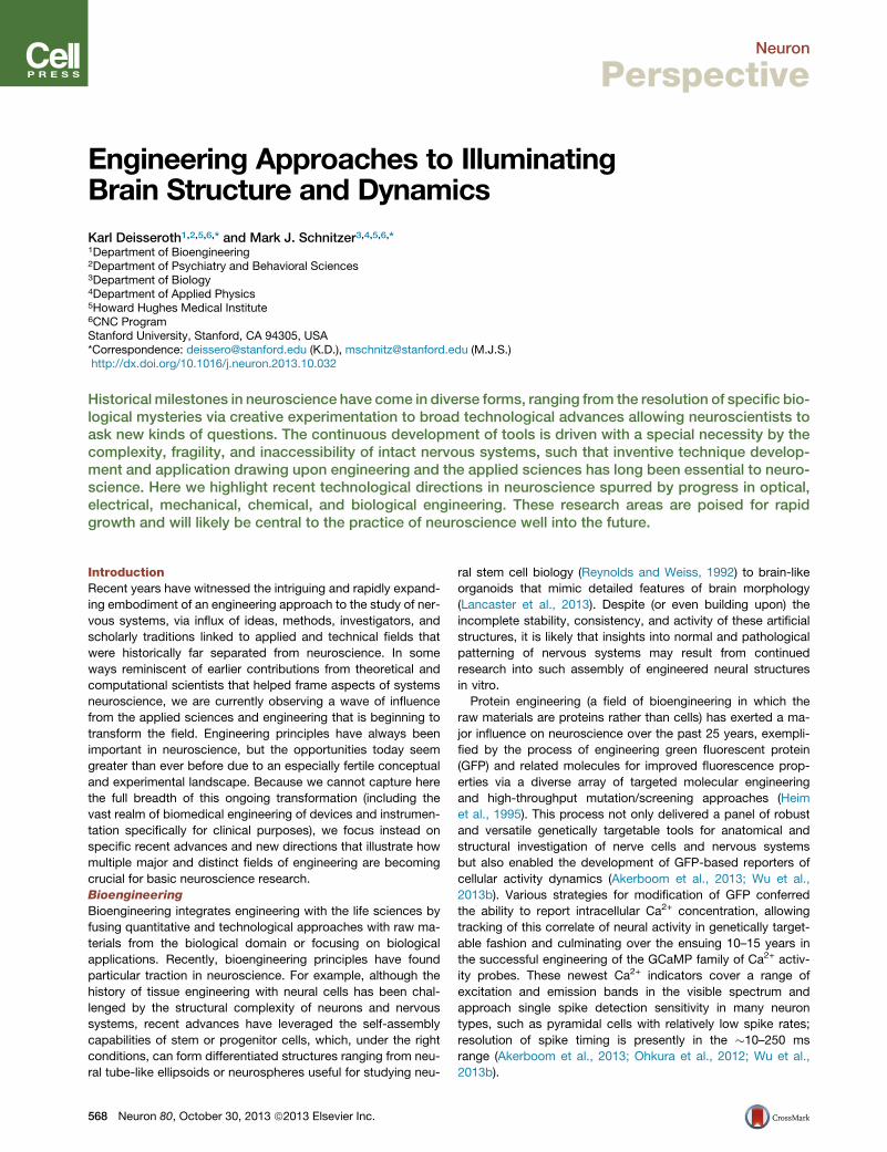
Figure 1. Optics and Protein Engineering Converge for Ca2+ Imagingin >1,200 CA1 Pyramidal Cells in Freely Moving Mice(A) An integrated microscope is equipped with a microendoscope to imageCA1 neurons expressing the engineeredCa2+ indicator GCaMP3 under controlof the Camk2a promoter. The base plate and microendoscope are fixed to thecranium for repeated access to the same field of view. Republished from Ziv etal., 2013.(B) Shown are 1,202 CA1 pyramidal cells (red somata) identified by Ca2+
imaging in a freely moving mouse atop a mean fluorescence image (green) ofCA1. Vessels appear as dark shadows. Image courtesy of Yaniv Ziv and LaceyKitch, Stanford University.(C) Example traces of [Ca2+]i dynamics from 15 cells. Scale bars denote 5%DF/F (vertical) and 10 s (horizontal).
Neuron
Perspective
volumetric information comes at the cost of somewhat reduced
lateral resolution but still permits resolution of individual neurons
within intact and functioning vertebrate nervous systems (L.
Grosenick et al., 2013, Society for Neuroscience, abstract; A.
Andalman et al., 2013, Society for Neuroscience, abstract).
Going forward, we expect continuous improvement in light-field,
holographic, and selective planar illumination methods for
improved acquisition rates, resolution, and coverage volume
applied to intact nervous systems. We also expect holographic
and light-field methods for optogenetic activity manipulation to
develop in tandem with corresponding methods for activity im-
aging.
The resulting large optical data sets require massive improve-
ments in data handling and computational analysis. Optical engi-
neering applied to the nervous system will also continue to
benefit from computational methods that improve the capabil-
ities to look through turbid media. In the brain, light attenuation
is chiefly due to light scattering (turbidity), rather than light
absorption; emerging methods for correcting for effects of light
scattering through a combination of computational approaches
and optical manipulations (Bertolotti et al., 2012) have yet to
havemajor impacts on neuroscience experimentation, but future
years may reveal a role for these computational techniques for
imaging deep into turbid brain tissue.
Progress in the engineering of optical hardware continually
propels improvements in optical systems. The invention of the
charge-coupled device (CCD) camera led to pioneering studies
of intracellular Ca2+ dynamics in neurons. Today, scientific-
grade cameras routinely monitor neuronal dynamics, but the
more recently developed complementary metal oxide semicon-
ductor (CMOS) image sensor has made substantial inroads into
experimental terrain previously dominated by the CCD camera.
The most recent CMOS image sensors have enabled a new gen-
eration of fluorescence imaging experiments.
Commonplace CMOS sensors, such as those in mobile
telephones, are now so advanced that they have enabled the
engineering and application of miniaturized fluorescence micro-
scopes, small enough to be mounted on the head of a mouse
during free behavior (Figure 1) (Ghosh et al., 2011; Ziv et al.,
2013). Such CMOS-based miniature microscopes can now pro-
vide recordings of up to �1,200 neurons concurrently during
active mouse behavior (Figure 1). This promises to be a useful
tool in the study of rodent models of human brain disorders,
and perhaps even in primate models. We expect continued
progress in camera technology and image sensor chips, leading
to larger sensors, faster image-frame acquisition rates, on-chip
imaging analyses, wireless imaging, and even capabilities for
three-dimensional imaging. Further improvements in tiny light
emitting diodes (LEDs) in combination with CMOS image sen-
sors should enable a new generation of devices capable of
both optogenetic manipulation and fluorescence imaging
concurrently. This need will provide additional impetus for the
ongoing engineering of spectrally compatible sets of optoge-
netic control probes and fluorescence-based sensors of neural
activity.
Even as next-generation optical tools offer increasingly so-
phisticated technological capabilities, the practice of systems
neuroscience will have to remain grounded in rigorous, clever,
and insightful behavioral paradigms. Here, digital imaging may
help advance the field, as many emerging opportunities exist
for high-throughput and high-resolution video tracking of animal
behavior. To maximally leverage the newfound capabilities for
optically monitoring individual cells over many weeks in the live
brain, new behavioral assays should be compatible with long-
term tracking and quantification of behavior. Machine-learning
approaches to scoring digital image sequences of animal
behavior (Kabra et al., 2013) might facilitate the combined auto-
mation of both brain imaging and behavioral data analyses.
Finally, we note that for in vivo animal experimentation, the
demands of small animal surgery often remain a limiting factor
on the rate of experimental progress. In recent years there has
been exploration of laser surgical methods to perform highly pre-
cise surgeries. One candidate approach involves the use of
regenerative laser amplifiers that emit high-energy ultrashort
pulses of light for highly precise tissue ablation (down to the sub-
micron scale, to cut or ablate individual axons, neurons, and
even organelles) (Jeong et al., 2012; Samara et al., 2010). How-
ever, the fine spatial scale of the cutting action is a limiting factor
for performing dissections over broad tissue regions. An alterna-
tive approach is to make use of ultraviolet lasers, such as those
commonly used in clinical ophthalmology for reshaping the
cornea (Sinha et al., 2013). Ultraviolet excimer lasers can cut pre-
cision holes down to the sub-10-mm scale, with clean-cut edges
straight to <1 mm, and at much faster cutting rates than the
regenerative laser amplifiers. These properties enable
Neuron 80, October 30, 2013 ª2013 Elsevier Inc. 571

Figure 2. Materials Science andOptoelectronics Converge for NeuralInterface ResearchLeft: exploded-view layout of injectable semi-conductor device for integrated stimulation/sensing/actuation, highlighting distinct layers forelectrophysiological measurement (1), opticalmeasurement (2), optical stimulation (3, micro-ILED array), and temperature sensing (4), allbonded to a releasable base for injection with amicroneedle. Top right: injection and release of themicroneedle. After insertion (left), artificial cere-brospinal fluid dissolves an external silk-basedadhesive (middle) and the microneedle can beremoved (right), leaving the active device in thebrain. Bottom right: SEM of an injectable micro-LED array, 8.5 mm thick; flexible and rigid formsshown. Adapted with permission from Kim et al.,2013.
Neuron
Perspective
automated forms of laser surgery in insects, nematodes, and
even rodents on the seconds timescale, offering intriguing pos-
sibilities for increasing the throughput of neuroscience experi-
mentation (Sinha et al., 2013).
Electrical Engineering
Electrical engineering has long influenced neuroscience, dating
back to the contributions of cable theory and radio electronics
on the pioneering research by Cole, Curtis, Hodgkin, and Huxley
on the squid giant axon. This influence has persisted, as in
the advancement of low-noise electronic amplifiers driving
improved electrophysiological instrumentation for single chan-
nel biophysics. Recent years have seen an acceleration of
technologies for performing large-scale multielectrode record-
ings—not just expanding arrays of electrodes to numbers and
densities beyond those previously feasible, but also novel sur-
face electrodes and mechanically flexible recording devices
that can be bent to match the brain’s curvature and could be
used to monitor dynamics and help detect phenomena such
as those involved in epilepsy at the neocortical surface (Figure 2)
(Viventi et al., 2011).
We expect continued progress in the development of large-
scale and flexible electronic technology platforms (Kim et al.,
2012, 2013). Active electrodes or smart electronics will be inter-
nally incorporated to permit in situ signal amplification, reducing
the impact of noise and allowing immediate extraction of specific
physiological signals. It may become commonplace to incorpo-
rate closed-loop capabilities within devices that allow both
measurement and manipulation—the latter being electrical,
optical, or pharmacologic. Such capabilities could have an
important clinical impact as well as an impact in basic science;
for example, early detection of epileptic episodes could trigger
immediate preventive action, perhaps taken by the same device.
We expect continued progress in the area of hybrid probes
such as optrodes (Gradinaru et al., 2007, 2008), which allow
optogenetic stimulation of neurons along with electrical record-
ings from the very same cells. Advanced forms of optrodes
have enabled the recording of neural circuit dynamics simulta-
neously with high-speed optical control and behavior (Anikeeva
et al., 2012; Wu et al., 2013a; Ozden et al., 2013; Kim et al., 2013)
and will also facilitate the identification or tagging of spikes from
572 Neuron 80, October 30, 2013 ª2013 Elsevier Inc.
cells that express opsins. Integration of electrical and optical
capabilities in the same devices will continue to improve; for
example, flexible electronics will be combined with high-density
multicolor miniature light sources and optical detectors, and
optrodes will become smaller, easier to fabricate, and better
integrated for more ready implementation in behaving animals.
Unconventional optrode designs such as new types of metal-
coated, tapered fiberoptics may likewise serve to facilitate
combined electrophysiological measurement and light delivery
(Dufour et al., 2013; Ozden et al., 2013). More generally, we fore-
see an expansion of new types of multifaceted probes for
electrophysiological recording and stimulation that might incor-
porate not only capabilities for light detection or delivery, but
also drug delivery or microfluidic sampling.
Another major area in which electrical engineering is exerting
a strong influence on neuroscience concerns brain-machine
interfaces. An established class of such interfaces concerns
sensory perception, with the cochlear implant as a paradigmatic
example. Likewise, there has been sustained progress toward
retinal prosthetics for restoring vision (Mathieson et al., 2012)
and toward motor prosthetics for achieving artificial-limb control
using neural signals sent from the brain and transduced into
electronic commands. Recent progress has conferred the
ability to control a computer cursor or robotic arm by motor-
impaired patients (Hochberg et al., 2012). This realm of pros-
thesis engineering is building heavily upon concepts from
computational and analytical aspects of electrical engineering
and computer science, including dynamical systems modeling,
state space analysis, dimensionality reduction, and adaptive
filtering (Dangi et al., 2013; Gilja et al., 2011; Shenoy et al.,
2013). We note that the notion of a neural prosthetic is concep-
tually broad, and nonelectrical prosthetics (e.g., optical or mag-
netic) might be developed to augment or correct aspects of
cognition or behavior. For basic neuroscience experimentation,
all-optical approaches to brain-machine interfaces should also
be feasible (optical readouts combined with optical manipula-
tion of neural dynamics). We expect to see increased
complexity in this prosthetics-focused fusion of engineering
and systems neuroscience, as the needs and opportunities
are enormous.

Neuron
Perspective
For imaging the human brain, engineering and physics have
long played key roles; for example, magnetic resonance imaging
(MRI) arose from nuclear magnetic resonance spectroscopy. We
expect continued major progress in the realm of MRI, with new
computational approaches and instrumentation allowing un-
precedented levels of detail to be revealed concerning the
human brain and cognition. This will include not just instrumen-
tation advances such as higher magnetic field strengths, but
also improved computational approaches for registration of
brain anatomy across different individuals and new methods
for interpreting with high confidence the nature of the signals
seen, as with diffusion tractography. And for controlling human
nervous systems, there has been recent engineering progress
in the design and development of optogenetic interfaces that
may be useful for bidirectional modulation of activity, such as
for major peripheral nerves (Liske et al., 2013).
Finally, we take note of miniaturization, which involves
electrical, mechanical, and materials engineering, among other
domains. Major industries such as telecommunications, con-
sumer electronics, and defense will continue to drive rapid
innovation in miniaturization of optics, electronics, wireless
technology, and computational elements, all of which have
contributed to superior instrumentation for neuroscience exper-
imentation. Major virtues of miniaturized systems for use in freely
moving animals include compatibility with behavioral assays that
have already been deployed and validated over decades of
neuroscience research. Akin to EEGand EMG telemetry systems
in present usage, wireless and miniaturized brain imaging sys-
tems may come to permit around-the-clock studies of brain
activity, e.g., for monitoring neural activity and brain states
across sleeping, eating, and other behaviors, in substantial
numbers of animals (e.g., for large behavioral cohorts in basic
neuroscience laboratory investigations or in drug screening)
without constant human supervision.
Chemical and Materials Engineering
The chemistry- and physics-based engineering of materials has
accelerated several exciting and important technologies for
neuroscience research (beyond miniaturization and electrode
design, already discussed above). Here we touch on only two
of many categories of chemical engineering that seem well
poised to grow with neuroscience into the future: (1) the
engineering of materials into which organisms and cells are
placed and (2) the engineering of materials from within intact
organisms.
Small organisms such as nematodes, fruit flies, and mamma-
lian embryos could be amenable to high-throughput investiga-
tions of nervous system development, structure, physiology,
and behavior. However, only recently have technologies been
developed to allow high-throughput positioning and interroga-
tion of small, intact organisms. Microfabrication and microflui-
dics, often with computer-aided design (CAD) molding, and
soft lithography with an elastomer such as polydimethylsiloxane
(PDMS), which is poured or spun into the micropatterned mold,
have been applied to the positioning of Caenorhabditis elegans
and mouse embryos (Albrecht and Bargmann, 2011; Chung
et al., 2011a, 2011b). While zebrafish are too large for typical
high-throughput microfabricated devices, approaches based
on multiple well plates are coming of age (Chang et al., 2012).
Chemical engineering and applied chemistry efforts have led
to the development of materials, nanoparticles, and polymers
for the study of central nervous system regeneration and repair
(Tam et al., 2013), delivery of small interfering RNAs for causal
testing of specific transcripts (Chan et al., 2013), and construc-
tion of hydrogel environments into which nervous system cells
(or stem/progenitor cells) may be seeded to study proliferation,
differentiation, survival, and other properties (Cha et al., 2012;
Ferreira et al., 2007; Owen et al., 2013; Tibbitt and Anseth,
2012). Going forward, we expect growth areas in this domain
to include increasingly sophisticated and modular engineering
of these scaffolds and environments, for example using versatile
click chemistry as well as novel conductive or transparent poly-
mers for compatibility with optical or electrical interrogation of
the resulting construct (Chung and Deisseroth, 2013; Keplinger
et al., 2013). This general approach may also synergize with
the studies of the development and assembly of neural struc-
tures beginning from the other direction, with biology rather
than chemistry, as in the stem cell/brain organoid (Lancaster
et al., 2013) approach described above.
A newly emerged concept at the interface of neuroscience and
chemical engineering, CLARITY (Figure 3), involves the chemical
construction of new physical forms from within biological sys-
tems such as the brain (Chung and Deisseroth, 2013; Chung
et al., 2013). For example, hydrogel infrastructures can be con-
structed from within intact brains to covalently stabilize native
proteins and nucleic acids in preparation for stringent removal
of membrane phospholipids with strong ionic detergents and
active electrophoresis of the entire brain. This lipid removal, in
turn, allows interrogation of the intact brain with photons (which
no longer scatter heavily due to removal of the lipid-aqueous in-
terfaces) and macromolecules (such as antibodies and oligonu-
cleotide probes, which can at that point penetrate the tissue
without interference from intact plasmamembranes). We expect
this kind of approach to find utility in mapping volumetric
anatomical features from animal models as well as clinical sam-
ples; moreover, many kinds of gels and scaffolds could be con-
structed in this way with a range of passive and active properties
for a broad range of different kinds of structural and functional
studies of nervous systems. Finally, distinct from gel and scaffold
diversity, there also exists a broad diversity of macromolecular
probe type that can be used to interrogate the resulting nanopo-
rous hybrid structures, including functionalized proteins and
active enzymes.
Outlook
As exciting as these domains of neuroscience have become, the
future may hold even greater opportunities—for example, via
combinations of multiple engineering subdisciplines (e.g.,
computer science with chemical engineering, or optical instru-
mentation with bioengineering, for applications to increasingly
sophisticated questions in increasingly complex nervous
systems; Figure 3). CLARITY is already being used in human
tissue, and advanced electrical and optical interfaces have
already been designed for human and nonhuman primate
applications.
Emerging optical methods may bring among the most excit-
ing synergistic possibilities for integrative studies of neural
circuit dynamics, connectivity, cytoarchitecture, and molecular
Neuron 80, October 30, 2013 ª2013 Elsevier Inc. 573

Figure 3. Chemical Engineering, Bioengineering, and PhotonicsGive Rise to Circuit-Probing Hardware and WetwareTop: construction of a hydrogel from within tissue (CLARITY) creates atransparent mammalian brain for intact-system anatomical analysis; adaptedfrom Chung et al., 2013. Bottom left: 2.3A crystal structure of the channel-rhodopsin optogenetic control tool enables directed protein engineering forenhanced interventional functionality; adapted from Kato et al., 2012. Bottomright: optogenetic neural interfaces deliver light from laser diodes or advancedLEDs; flexible fiberoptic control in freely moving mouse shown. Photo creditInbal Goshen and Karl Deisseroth.
Neuron
Perspective
composition. Specifically, in vivo optical recordings of neural
activity and optogenetic manipulations in cells defined geneti-
cally or by anatomical projections can be naturally combined
and registered with technologically advanced studies of cir-
cuitry, synaptic structure, and other macromolecular information
(e.g., using CLARITY). By comparison, due to the challenges
inherent to large-scale in vivo electrical recordings regarding
unambiguous assignments of cells’ types and postmortem
registration of their identities, circuit reconstructions in the very
same cell ensembles recorded electrically are likely to be far
more challenging.
Integrated optical studies in larger brains exacerbate the ‘‘big
data’’ problem, which is already becoming a notable challenge in
multiple subareas of neuroscience. Collaborations between neu-
roscientists and computer scientists will become increasingly
important, and even essential, for the challenges of the next
25 years—not only for generating testable hypotheses arising
from models of brain dynamics or machine learning research,
but also for storing, handling, processing, and making acces-
sible these vast data streams concurrent with the emergence
574 Neuron 80, October 30, 2013 ª2013 Elsevier Inc.
of integrated and computational optical approaches. For
example, large-scale Ca2+ recordings in mice will come to pro-
duce gigabytes per second of data, while CLARITY data sets
for individual whole rodent brains can be �1–10 terabytes in
size, depending on the number of color channels (Figures 1
and 3). These optical data sets will soon grow to the �10 peta-
byte scale and beyond, especially when larger brains including
those of humans are examined at high resolution. However, con-
ventional ‘‘cloud storage’’ approaches for large data sets are in
many ways suboptimal for the kinds of data encountered in
neuroscience, and computational/analytical methods will have
to be profoundly accelerated simply to keep pace with the exhil-
arating new rate of data acquisition in neuroscience.
Lastly, we close with some remarks on how engineers and
neuroscientists might fruitfully interact in the coming years.
Traditionally, there often have not been conventional career
paths, at least in academics, for engineers playing critical sup-
porting roles in neuroscience research. In many cases, engineer-
ing departments might not view such activity as breaking
sufficient ground in the engineering realm, whereas biology
departments might not appreciate the crucial but underlying
links to biological discovery. As the engineering challenges
become increasingly severe for neuroscientists in the years
ahead, with an upcoming deluge of sophisticated instrumenta-
tion and massive data sets, the neuroscience community will
need to consider carefully how best to engage and retain the
best, brightest, and most ambitious engineers.
Both the engineering and neuroscience communities might be
well served by further appreciation of each other’s intellectual
traditions and modus operandi. Engineers are typically moti-
vated to address wide sets of problems that share central fea-
tures, permitting common tools and approaches. Biologists are
usually motivated to solve specific mysteries in detail. These
are distinct intellectual mind sets, and the two communities
can sometimes talk past each other. Engineers are generally
well served by learning which classes of problems are the true
stumbling blocks in biological science, rather than looking for
suitable puzzles to solve after inventing a new widget. Biologists
can benefit from enhanced appreciation of the intellectual
potency of simultaneously examining all problems of a given
category, an approach that has yielded many technologies that
form the bedrock of modern biological research practice and
infrastructure. In the coming years, neuroscientists and engi-
neers will need (and want) to work more closely together than
ever before, making ‘‘cross-cultural’’ exchange of ideas and
working modes increasingly important for, and part of, the natu-
ral fabric of neuroscience.
ACKNOWLEDGMENTS
K.D. acknowledges support from the Wiegers Family Fund, NIMH, NIDA, NSF,the DARPA REPAIR Program, and the Gatsby Charitable Foundation. M.J.S.acknowledges support from NIMH, NSF, the Paul Allen Family Foundation,DARPA, the Ellison Foundation, the Keck Foundation, NIDA, and NIBIB.M.J.S. is a cofounder and consults scientifically for Inscopix Inc., which hascommercialized the miniature integrated microscope technology of Figure 1.K.D. is a cofounder and consults for Circuit Therapeutics Inc., which is usingoptogenetics to screen for medications and build devices for treating diseasesin the peripheral nervous system; optogenetics tools, training, and protocolsare freely available (http://www.optogenetics.org).

Neuron
Perspective
REFERENCES
Ahrens, M.B., Orger, M.B., Robson, D.N., Li, J.M., and Keller, P.J. (2013).Whole-brain functional imaging at cellular resolution using light-sheet micro-scopy. Nat. Methods 10, 413–420.
Akerboom, J., Carreras Calderon, N., Tian, L., Wabnig, S., Prigge, M., Tolo, J.,Gordus, A., Orger, M.B., Severi, K.E., Macklin, J.J., et al. (2013). Geneticallyencoded calcium indicators for multi-color neural activity imaging and combi-nation with optogenetics. Front Mol Neurosci 6, 2.
Albrecht, D.R., and Bargmann, C.I. (2011). High-content behavioral analysis ofCaenorhabditis elegans in precise spatiotemporal chemical environments.Nat. Methods 8, 599–605.
Alivisatos, A.P., Andrews, A.M., Boyden, E.S., Chun, M., Church, G.M., Dei-sseroth, K., Donoghue, J.P., Fraser, S.E., Lippincott-Schwartz, J., Looger,L.L., et al. (2013). Nanotools for neuroscience and brain activity mapping.ACS Nano 7, 1850–1866.
Anikeeva, P., Andalman, A.S., Witten, I., Warden,M., Goshen, I., Grosenick, L.,Gunaydin, L.A., Frank, L.M., and Deisseroth, K. (2012). Optetrode: a multi-channel readout for optogenetic control in freely moving mice. Nat. Neurosci.15, 163–170.
Bamann, C., Gueta, R., Kleinlogel, S., Nagel, G., and Bamberg, E. (2010).Structural guidance of the photocycle of channelrhodopsin-2 by an interhelicalhydrogen bond. Biochemistry 49, 267–278.
Bepari, A.K., Sano, H., Tamamaki, N., Nambu, A., Tanaka, K.F., and Takebaya-shi, H. (2012). Identification of optogenetically activated striatal medium spinyneurons by Npas4 expression. PLoS ONE 7, e52783.
Berndt, A., Yizhar, O., Gunaydin, L.A., Hegemann, P., and Deisseroth, K.(2009). Bi-stable neural state switches. Nat. Neurosci. 12, 229–234.
Berndt, A., Schoenenberger, P., Mattis, J., Tye, K.M., Deisseroth, K., Hegem-ann, P., and Oertner, T.G. (2011). High-efficiency channelrhodopsins for fastneuronal stimulation at low light levels. Proc. Natl. Acad. Sci. USA 108,7595–7600.
Bertolotti, J., van Putten, E.G., Blum, C., Lagendijk, A., Vos, W.L., and Mosk,A.P. (2012). Non-invasive imaging through opaque scattering layers. Nature491, 232–234.
Broxton, M., Grosenick, L., Yang, S., Cohen, N., Andalman, A., Deisseroth, K.,and Levoy, M. (2013). Wave optics theory and 3-D deconvolution for the lightfield microscope. Opt. Express 21, 25418–25439.
Cao, G., Platisa, J., Pieribone, V.A., Raccuglia, D., Kunst, M., and Nitabach,M.N. (2013). Genetically targeted optical electrophysiology in intact neural cir-cuits. Cell 154, 904–913.
Carter, M.E., Brill, J., Bonnavion, P., Huguenard, J.R., Huerta, R., and deLecea, L. (2012). Mechanism for Hypocretin-mediated sleep-to-wake transi-tions. Proc. Natl. Acad. Sci. USA 109, E2635–E2644.
Cha, C., Liechty, W.B., Khademhosseini, A., and Peppas, N.A. (2012).Designing biomaterials to direct stem cell fate. ACS Nano 6, 9353–9358.
Chan, D.P., Deleavey, G.F., Owen, S.C., Damha, M.J., and Shoichet, M.S.(2013). Click conjugated polymeric immuno-nanoparticles for targeted siRNAand antisense oligonucleotide delivery. Biomaterials 34, 8408–8415.
Chang, T.Y., Pardo-Martin, C., Allalou, A., Wahlby, C., and Yanik, M.F. (2012).Fully automated cellular-resolution vertebrate screening platform with parallelanimal processing. Lab Chip 12, 711–716.
Chung, K., and Deisseroth, K. (2013). CLARITY for mapping the nervous sys-tem. Nat. Methods 10, 508–513.
Chung, K., Kim, Y., Kanodia, J.S., Gong, E., Shvartsman, S.Y., and Lu, H.(2011a). A microfluidic array for large-scale ordering and orientation ofembryos. Nat. Methods 8, 171–176.
Chung, K., Zhan, M., Srinivasan, J., Sternberg, P.W., Gong, E., Schroeder,F.C., and Lu, H. (2011b). Microfluidic chamber arrays for whole-organismbehavior-based chemical screening. Lab Chip 11, 3689–3697.
Chung, K., Wallace, J., Kim, S.Y., Kalyanasundaram, S., Andalman, A.S.,Davidson, T.J., Mirzabekov, J.J., Zalocusky, K.A., Mattis, J., Denisin, A.K.,
et al. (2013). Structural and molecular interrogation of intact biological sys-tems. Nature 497, 332–337.
Dangi, S., Orsborn, A.L., Moorman, H.G., and Carmena, J.M. (2013). Designand analysis of closed-loop decoder adaptation algorithms for brain-machineinterfaces. Neural Comput. 25, 1693–1731.
Dani, A., Huang, B., Bergan, J., Dulac, C., and Zhuang, X. (2010). Superreso-lution imaging of chemical synapses in the brain. Neuron 68, 843–856.
Deisseroth, K. (2011). Optogenetics. Nat. Methods 8, 26–29.
Dufour, S., Lavertu, G., Dufour-Beausejour, S., Juneau-Fecteau, A., Calakos,N., Deschenes, M., Vallee, R., and De Koninck, Y. (2013). A multimodal micro-optrode combining field and single unit recording, multispectral detection andphotolabeling capabilities. PLoS ONE 8, e57703.
Farrar, M.J., Wise, F.W., Fetcho, J.R., and Schaffer, C.B. (2011). In vivo imag-ing of myelin in the vertebrate central nervous system using third harmonicgeneration microscopy. Biophys. J. 100, 1362–1371.
Fenno, L., Yizhar, O., and Deisseroth, K. (2011). The development and applica-tion of optogenetics. Annu. Rev. Neurosci. 34, 389–412.
Ferreira, L.S., Gerecht, S., Fuller, J., Shieh, H.F., Vunjak-Novakovic, G., andLanger, R. (2007). Bioactive hydrogel scaffolds for controllable vascular differ-entiation of human embryonic stem cells. Biomaterials 28, 2706–2717.
Ghosh, K.K., Burns, L.D., Cocker, E.D., Nimmerjahn, A., Ziv, Y., Gamal, A.E.,and Schnitzer, M.J. (2011). Miniaturized integration of a fluorescence micro-scope. Nat. Methods 8, 871–878.
Gilja, V., Chestek, C.A., Diester, I., Henderson, J.M., Deisseroth, K., and She-noy, K.V. (2011). Challenges and opportunities for next-generation intracorti-cally based neural prostheses. IEEE Trans. Biomed. Eng. 58, 1891–1899.
Godara, P., Dubis, A.M., Roorda, A., Duncan, J.L., and Carroll, J. (2010). Adap-tive optics retinal imaging: emerging clinical applications. Optom. Vis. Sci. 87,930–941.
Gong, Y., Li, J.Z., and Schnitzer, M.J. (2013). Enhanced ArchaerhodopsinFluorescent Protein Voltage Indicators. PLoS ONE 8, e66959.
Gradinaru, V., Thompson, K.R., Zhang, F., Mogri, M., Kay, K., Schneider, M.B.,and Deisseroth, K. (2007). Targeting and readout strategies for fast opticalneural control in vitro and in vivo. J. Neurosci. 27, 14231–14238.
Gradinaru, V., Thompson, K.R., and Deisseroth, K. (2008). eNpHR: a Natrono-monas halorhodopsin enhanced for optogenetic applications. Brain Cell Biol.36, 129–139.
Gradinaru, V., Zhang, F., Ramakrishnan, C., Mattis, J., Prakash, R., Diester, I.,Goshen, I., Thompson, K.R., and Deisseroth, K. (2010). Molecular and cellularapproaches for diversifying and extending optogenetics. Cell 141, 154–165.
Gunaydin, L.A., Yizhar, O., Berndt, A., Sohal, V.S., Deisseroth, K., and Hegem-ann, P. (2010). Ultrafast optogenetic control. Nat. Neurosci. 13, 387–392.
Haikala, V., Joesch, M., Borst, A., andMauss, A.S. (2013). Optogenetic controlof fly optomotor responses. J. Neurosci. 33, 13927–13934.
Hall, L.T., Beart, G.C., Thomas, E.A., Simpson, D.A., McGuinness, L.P., Cole,J.H., Manton, J.H., Scholten, R.E., Jelezko, F.,Wrachtrup, J., et al. (2012). Highspatial and temporal resolution wide-field imaging of neuron activity usingquantum NV-diamond. Sci Rep 2, 401.
Heim, R., Cubitt, A.B., and Tsien, R.Y. (1995). Improved green fluorescence.Nature 373, 663–664.
Hochberg, L.R., Bacher, D., Jarosiewicz, B., Masse, N.Y., Simeral, J.D., Vogel,J., Haddadin, S., Liu, J., Cash, S.S., van der Smagt, P., and Donoghue, J.P.(2012). Reach and grasp by people with tetraplegia using a neurally controlledrobotic arm. Nature 485, 372–375.
Horton, N.G., Wang, K., Kobat, D., Clark, C.G., Wise, F.W., Schaffer, C.B., andXu, C. (2013). In vivo three-photon microscopy of subcortical structures withinan intact mouse brain. Nat. Photonics 7, 205–209.
Hunter, J.J., Masella, B., Dubra, A., Sharma, R., Yin, L., Merigan, W.H., Palc-zewska, G., Palczewski, K., and Williams, D.R. (2010). Images of photorecep-tors in living primate eyes using adaptive optics two-photon ophthalmoscopy.Biomed. Opt. Express 2, 139–148.
Neuron 80, October 30, 2013 ª2013 Elsevier Inc. 575

Neuron
Perspective
Jeong, D.C., Tsai, P.S., and Kleinfeld, D. (2012). Prospect for feedback guidedsurgery with ultra-short pulsed laser light. Curr. Opin. Neurobiol. 22, 24–33.
Ji, N., Sato, T.R., and Betzig, E. (2012). Characterization and adaptive opticalcorrection of aberrations during in vivo imaging in the mouse cortex. Proc.Natl. Acad. Sci. USA 109, 22–27.
Jin, L., Han, Z., Platisa, J., Wooltorton, J.R., Cohen, L.B., and Pieribone, V.A.(2012). Single action potentials and subthreshold electrical events imaged inneurons with a fluorescent protein voltage probe. Neuron 75, 779–785.
Kabra, M., Robie, A.A., Rivera-Alba, M., Branson, S., and Branson, K. (2013).JAABA: interactive machine learning for automatic annotation of animalbehavior. Nat. Methods 10, 64–67.
Kato, H.E., Zhang, F., Yizhar, O., Ramakrishnan, C., Nishizawa, T., Hirata, K.,Ito, J., Aita, Y., Tsukazaki, T., Hayashi, S., et al. (2012). Crystal structure of thechannelrhodopsin light-gated cation channel. Nature 482, 369–374.
Keplinger, C., Sun, J.Y., Foo, C.C., Rothemund, P., Whitesides, G.M., and Suo,Z. (2013). Stretchable, transparent, ionic conductors. Science 341, 984–987.
Kim, D.H., Ghaffari, R., Lu, N., and Rogers, J.A. (2012). Flexible and stretchableelectronics for biointegrated devices. Annu. Rev. Biomed. Eng. 14, 113–128.
Kim, T.I., McCall, J.G., Jung, Y.H., Huang, X., Siuda, E.R., Li, Y., Song, J.,Song, Y.M., Pao, H.A., Kim, R.H., et al. (2013). Injectable, cellular-scale opto-electronics with applications for wireless optogenetics. Science 340, 211–216.
Kobat, D., Durst, M.E., Nishimura, N., Wong, A.W., Schaffer, C.B., and Xu, C.(2009). Deep tissue multiphoton microscopy using longer wavelength excita-tion. Opt. Express 17, 13354–13364.
Kobat, D., Horton, N.G., and Xu, C. (2011). In vivo two-photon microscopy to1.6-mm depth in mouse cortex. J. Biomed. Opt. 16, 106014.
Konermann, S., Brigham, M.D., Trevino, A.E., Hsu, P.D., Heidenreich, M.,Cong, L., Platt, R.J., Scott, D.A., Church, G.M., and Zhang, F. (2013). Opticalcontrol of mammalian endogenous transcription and epigenetic states. Nature500, 472–476.
Kralj, J.M., Douglass, A.D., Hochbaum, D.R., Maclaurin, D., and Cohen, A.E.(2012). Optical recording of action potentials in mammalian neurons using amicrobial rhodopsin. Nat. Methods 9, 90–95.
Lam, A.J., St-Pierre, F., Gong, Y., Marshall, J.D., Cranfill, P.J., Baird, M.A.,McKeown, M.R., Wiedenmann, J., Davidson, M.W., Schnitzer, M.J., et al.(2012). Improving FRET dynamic range with bright green and red fluorescentproteins. Nat. Methods 9, 1005–1012.
Lancaster, M.A., Renner, M., Martin, C.A., Wenzel, D., Bicknell, L.S., Hurles,M.E., Homfray, T., Penninger, J.M., Jackson, A.P., and Knoblich, J.A. (2013).Cerebral organoids model human brain development and microcephaly.Nature 501, 373–379.
Lecoq, J., and Schnitzer, M.J. (2011). An infrared fluorescent protein fordeeper imaging. Nat. Biotechnol. 29, 715–716.
Lin, J.Y., Lin, M.Z., Steinbach, P., and Tsien, R.Y. (2009). Characterization ofengineered channelrhodopsin variants with improved properties and kinetics.Biophys. J. 96, 1803–1814.
Liske, H., Towne, C., Anikeeva, P., Zhao, S., Feng, G., Deisseroth, K., andDelp, S. (2013). Optical inhibition of motor nerve and muscle activity in vivo.Muscle Nerve 47, 916–921.
Mahou, P., Zimmerley, M., Loulier, K., Matho, K.S., Labroille, G., Morin, X.,Supatto, W., Livet, J., Debarre, D., and Beaurepaire, E. (2012). Multicolortwo-photon tissue imaging by wavelength mixing. Nat. Methods 9, 815–818.
Marshall, J.D., and Schnitzer, M.J. (2013). Optical strategies for sensingneuronal voltage using quantum dots and other semiconductor nanocrystals.ACS Nano 7, 4601–4609.
Mathieson, K., Loudin, J., Goetz, G., Huie, P., Wang, L., Kamins, T.I., Galam-bos, L., Smith, R., Harris, J.S., Sher, A., and Palanker, D. (2012). PhotovoltaicRetinal Prosthesis with High Pixel Density. Nat. Photonics 6, 391–397.
Mattis, J., Tye, K.M., Ferenczi, E.A., Ramakrishnan, C., O’Shea, D.J., Prakash,R., Gunaydin, L.A., Hyun, M., Fenno, L.E., Gradinaru, V., et al. (2012). Princi-ples for applying optogenetic tools derived from direct comparative analysisof microbial opsins. Nat. Methods 9, 159–172.
576 Neuron 80, October 30, 2013 ª2013 Elsevier Inc.
Micheva, K.D., and Smith, S.J. (2007). Array tomography: a new tool for imag-ing the molecular architecture and ultrastructure of neural circuits. Neuron 55,25–36.
Ohkura, M., Sasaki, T., Sadakari, J., Gengyo-Ando, K., Kagawa-Nagamura, Y.,Kobayashi, C., Ikegaya, Y., and Nakai, J. (2012). Genetically encoded greenfluorescent Ca2+ indicators with improved detectability for neuronal Ca2+ sig-nals. PLoS ONE 7, e51286.
Oron, D., Tal, E., and Silberberg, Y. (2005). Scanningless depth-resolvedmicroscopy. Opt. Express 13, 1468–1476.
Owen, S.C., Fisher, S.A., Tam, R.Y., Nimmo, C.M., and Shoichet, M.S. (2013).Hyaluronic acid click hydrogels emulate the extracellular matrix. Langmuir 29,7393–7400.
Ozden, I., Wang, J., Lu, Y., May, T., Lee, J., Goo, W., O’Shea, D.J., Kalanithi,P., Diester, I., Diagne,M., et al. (2013). A coaxial optrode asmultifunction write-read probe for optogenetic studies in non-human primates. J. Neurosci.Methods 219, 142–154.
Quirin, S., Peterka, D.S., and Yuste, R. (2013). Instantaneous three-dimen-sional sensing using spatial light modulator illumination with extended depthof field imaging. Opt. Express 21, 16007–16021.
Reynolds, B.A., and Weiss, S. (1992). Generation of neurons and astrocytesfrom isolated cells of the adult mammalian central nervous system. Science255, 1707–1710.
Samara, C., Rohde, C.B., Gilleland, C.L., Norton, S., Haggarty, S.J., and Yanik,M.F. (2010). Large-scale in vivo femtosecond laser neurosurgery screen re-veals small-molecule enhancer of regeneration. Proc. Natl. Acad. Sci. USA107, 18342–18347.
Schrodel, T., Prevedel, R., Aumayr, K., Zimmer, M., and Vaziri, A. (2013). Brain-wide 3D imaging of neuronal activity in Caenorhabditis elegans with sculptedlight. Nat. Methods 10, 1013–1020.
Schultheis, C., Liewald, J.F., Bamberg, E., Nagel, G., and Gottschalk, A.(2011). Optogenetic long-term manipulation of behavior and animal develop-ment. PLoS ONE 6, e18766.
Shenoy, K.V., Sahani, M., and Churchland, M.M. (2013). Cortical control of armmovements: a dynamical systems perspective. Annu. Rev. Neurosci. 36,337–359.
Sinha, S., Liang, L., Ho, E.T.W., Urbanek, K.E., Luo, L., Baer, T.M., and Schnit-zer, M.J. (2013). High-speed laser microsurgery of alert fruit flies for fluores-cence imaging of neural activity. Proc. Natl. Acad. Sci. USA, in press.
Tam, R.Y., Fuehrmann, T., Mitrousis, N., and Shoichet, M.S. (2013). Regener-ative Therapies for Central Nervous System Diseases: a BiomaterialsApproach. Neuropsychopharmacology, in press. Published online September4, 2013.
Tanaka, K.F., Matsui, K., Sasaki, T., Sano, H., Sugio, S., Fan, K., Hen, R.,Nakai, J., Yanagawa, Y., Hasuwa, H., et al. (2012). Expanding the repertoireof optogenetically targeted cells with an enhanced gene expression system.Cell Rep 2, 397–406.
Testa, I., Urban, N.T., Jakobs, S., Eggeling, C., Willig, K.I., and Hell, S.W.(2012). Nanoscopy of living brain slices with low light levels. Neuron 75,992–1000.
Tibbitt, M.W., and Anseth, K.S. (2012). Dynamic microenvironments: the fourthdimension. Sci. Transl. Med. 4, 60ps24.
Urban, N.T., Willig, K.I., Hell, S.W., and Nagerl, U.V. (2011). STED nanoscopyof actin dynamics in synapses deep inside living brain slices. Biophys. J. 101,1277–1284.
Viventi, J., Kim, D.H., Vigeland, L., Frechette, E.S., Blanco, J.A., Kim, Y.S.,Avrin, A.E., Tiruvadi, V.R., Hwang, S.W., Vanleer, A.C., et al. (2011). Flexible,foldable, actively multiplexed, high-density electrode array for mapping brainactivity in vivo. Nat. Neurosci. 14, 1599–1605.
Wang, H., Sugiyama, Y., Hikima, T., Sugano, E., Tomita, H., Takahashi, T., Ish-izuka, T., and Yawo, H. (2009). Molecular determinants differentiating photo-current properties of two channelrhodopsins from chlamydomonas. J. Biol.Chem. 284, 5685–5696.

Neuron
Perspective
Watson, B.O., Nikolenko, V., Araya, R., Peterka, D.S., Woodruff, A., and Yuste,R. (2010). Two-photonmicroscopy with diffractive optical elements and spatiallight modulators. Front Neurosci 4.
Wilt, B.A., Burns, L.D., Wei Ho, E.T., Ghosh, K.K., Mukamel, E.A., and Schnit-zer, M.J. (2009). Advances in light microscopy for neuroscience. Annu. Rev.Neurosci. 32, 435–506.
Wilt, B.A., Fitzgerald, J.E., and Schnitzer, M.J. (2013). Photon shot noise limitson optical detection of neuronal spikes and estimation of spike timing.Biophys. J. 104, 51–62.
Wu, F., Stark, E., Im, M., Cho, I.J., Yoon, E.S., Buzsaki, G., Wise, K.D., andYoon, E. (2013a). An implantable neural probe with monolithically integrateddielectric waveguide and recording electrodes for optogenetics applications.J. Neural Eng. 10, 056012.
Wu, J., Liu, L., Matsuda, T., Zhao, Y., Rebane, A., Drobizhev, M., Chang, Y.F.,Araki, S., Arai, Y., March, K., et al. (2013b). Improved orange and red Ca2+ in-dicators and photophysical considerations for optogenetic applications. ACSChem Neurosci 4, 963–972.
Xu, K., Zhong, G., and Zhuang, X. (2013). Actin, spectrin, and associated pro-teins form a periodic cytoskeletal structure in axons. Science 339, 452–456.
Yizhar, O., Fenno, L.E., Davidson, T.J., Mogri, M., and Deisseroth, K. (2011a).Optogenetics in neural systems. Neuron 71, 9–34.
Yizhar, O., Fenno, L.E., Prigge, M., Schneider, F., Davidson, T.J., O’Shea, D.J.,Sohal, V.S., Goshen, I., Finkelstein, J., Paz, J.T., et al. (2011b). Neocorticalexcitation/inhibition balance in information processing and social dysfunction.Nature 477, 171–178.
Zhang, F., Vierock, J., Yizhar, O., Fenno, L.E., Tsunoda, S., Kianianmomeni, A.,Prigge, M., Berndt, A., Cushman, J., Polle, J., et al. (2011). The microbial opsinfamily of optogenetic tools. Cell 147, 1446–1457.
Zhao, S., Cunha, C., Zhang, F., Liu, Q., Gloss, B., Deisseroth, K., Augustine,G.J., and Feng, G. (2008). Improved expression of halorhodopsin for light-induced silencing of neuronal activity. Brain Cell Biol. 36, 141–154.
Zhao, Y., Araki, S., Wu, J., Teramoto, T., Chang, Y.F., Nakano,M., Abdelfattah,A.S., Fujiwara, M., Ishihara, T., Nagai, T., and Campbell, R.E. (2011). Anexpanded palette of genetically encoded Ca2+ indicators. Science 333,1888–1891.
Ziv, Y., Burns, L.D., Cocker, E.D., Hamel, E.O., Ghosh, K.K., Kitch, L.J., ElGamal, A., and Schnitzer, M.J. (2013). Long-term dynamics of CA1 hippocam-pal place codes. Nat. Neurosci. 16, 264–266.
Neuron 80, October 30, 2013 ª2013 Elsevier Inc. 577