endoxifen, 4-hydroxytamoxifen and an estrogenic derivative
TRANSCRIPT
1521-0111942812ndash822$3500 httpsdoiorg101124mol117111385MOLECULAR PHARMACOLOGY Mol Pharmacol 94812ndash822 August 2018Copyright ordf 2018 by The Author(s)This is an open access article distributed under the CC BY-NC Attribution 40 International license
Endoxifen 4-Hydroxytamoxifen and an Estrogenic DerivativeModulate Estrogen Receptor Complex Mediated Apoptosis inBreast Cancer s
Philipp Y Maximov Balkees Abderrahman Sean W Fanning Surojeet Sengupta Ping FanRamona F Curpan Daniela Maria Quintana Rincon Jeffery A GreenlandShyamala S Rajan Geoffrey L Greene and V Craig JordanDepartment of Breast Medical Oncology University of Texas MD Anderson Cancer Center Houston Texas (PYM BA PFDMQR JAG VCJ) The Ben May Department for Cancer Research University of Chicago Chicago Illinois (SWF SSRGLG) Department of Oncology Lombardi Comprehensive Cancer Center Washington DC (SS) and Institute of ChemistryRomanian Academy Timisoara Romania (RFC)
Received December 27 2017 accepted May 4 2018
ABSTRACTEstrogen therapy was used to treat advanced breast cancer inpostmenopausal women for decades until the introduction oftamoxifen Resistance to long-term estrogen deprivation (LTED)with tamoxifen and aromatase inhibitors used as a treatment ofbreast cancer inevitably occurs but unexpectedly low-doseestrogen can cause regression of breast cancer and increasedisease-free survival in some patients This therapeutic effect isattributed to estrogen-induced apoptosis in LTED breast cancerHere we describe modulation of the estrogen receptor (ER)liganded with antiestrogens (endoxifen and 4-hydroxytamoxifen)and an estrogenic triphenylethylene (TPE) ethoxytriphenylethy-lene (EtOXTPE) on estrogen-induced apoptosis in LTED breast
cancer cells Our results show that the angular TPE estrogen(EtOXTPE) is able to induce the ER-mediated apoptosis only at alater time compared with planar estradiol in these cells Usingreal-time polymerase chain reaction chromatin immunoprecip-itation western blotting molecular modeling and X-ray crystal-lography techniques we report novel conformations of the ERcomplex with an angular estrogen EtOXTPE and endoxifen Wepropose that alteration of the conformation of the ER complexeswith changes in coactivator binding governs estrogen-inducedapoptosis through the protein kinase regulated by RNA-likeendoplasmic reticulum kinase sensor system to trigger anunfolded protein response
IntroductionHigh-dose synthetic estrogen therapy was the first success-
ful chemical treatment of any cancer (Haddow et al 1944)Estrogen was used clinically to treat metastatic breast cancer(MBC) for 30 years prior to the successful introduction of theantiestrogen tamoxifen (Jordan 2003) Tamoxifen is a struc-tural derivative of the synthetic estrogen triphenylethylene
(TPE) During the time before tamoxifen clinical rules weredefined for the successful application of high-dose estrogentreatment Therapy was only effective in 30 ofMBC and onlyif applied more than 5 years past menopause (Haddow 1970)Mechanisms were unknownThe advantage of tamoxifen compared with high-dose
estrogen to treat MBC was not an increase in response ratebut a lower incidence of side effects (Cole et al 1971 Ingleet al 1981) Targeting tamoxifen to patients with estrogenreceptor (ER) positive breast cancer and the safety ofthe medicine permitted implementation of translationalresearch (Jordan 2008 2014b) to establish the value oftamoxifen as a long-term adjuvant therapy (Early BreastCancer Trialistsrsquo Collaborative Group 1998) The proposi-tion in the 1970s (Jordan et al 1979 Jordan and Allen 1980)that longer adjuvant therapy would be superior to shortertherapy also demanded an investigation of acquired resis-tance to long-term tamoxifen treatment in breast cancerNothing was known
This work was supported by the Department of Defense Breast Center ofExcellence Program [Award W81XWH-06-1-0590] the Susan G Komen for theCure Foundation [Award SAC100009] the National Institutes of HealthLombardi Comprehensive Cancer Center [Grant P30 CA051008] the NationalInstitutes of Health MD Andersonrsquos Cancer Center [Grant CA016672] theInstitute of Chemistry Timisoara of the Romanian Academy [Project 112015]the Romanian National Authority for Scientific Research and InnovationCNCSndashUEFISCDI [Project PN-II-RU-TE-2014-4-0422] the Department ofDefense BCRP [Breakthrough Award W81XWH-14-1-0360] the Susan GKomen Postdoctoral Fellowship [Grant PDF14301382] and the Virginia andD K Ludwig Fund for Cancer Research
httpsdoiorg101124mol117111385s This article has supplemental material available at molpharm
aspetjournalsorg
ABBREVIATIONS 4OHT 4-hydroxytamoxifen ANOVA analysis of variance ASU asymmetric unit ChIP chromatin immunoprecipitation E217b-estradiol ER estrogen receptor EtOXTPE ethoxytriphenylethylene LBD ligand-binding domain LTED long-term estrogen deprivation MBCmetastatic breast cancer PDB Protein Data Bank PEG polyethylene glycol PERK protein kinase regulated by RNA-like endoplasmic reticulumkinase qRT-PCR quantitative real-time polymerase chain reaction RT-PCR real-time polymerase chain reaction TPE triphenylethylene UPRunfolded protein response
812
httpmolpharmaspetjournalsorgcontentsuppl20180508mol117111385DC1Supplemental material to this article can be found at
at ASPE
T Journals on D
ecember 5 2021
molpharm
aspetjournalsorgD
ownloaded from
Acquired resistance to tamoxifen is unique Initially labo-ratory models in vivo demonstrated that tamoxifen actuallystimulated tumor growth within 1 to 2 years (Gottardis andJordan 1988 Gottardis et al 1989b) Nevertheless low-doseestrogen also stimulated tumor growth Mechanisms havesubsequently been deciphered using breast cancer cell modelsin vitro (Fan et al 2014abc) A new steroidal pure anties-trogen was subsequently developed following proof of efficacyin vivo to prevent tumor growth in tumors with acquiredresistance (Gottardis et al 1989a) Fulvestrant is nowapproved for the first line and second line treatment of MBC(Howell et al 2002 Osborne et al 2002Moscetti et al 2017)It therefore came as some surprise to find that low-dose
estrogen therapy triggered breast tumor regression followingthe development of acquired resistance to tamoxifen treatmentof 5 years (Yao et al 2000) This experimental model mimicsthe 5 years of adjuvant tamoxifen therapy that was thestandard of care at the time The rules described by Haddow(1970) applied to adjuvant tamoxifen therapy 5 years ofestrogen deprivation in ER-positive breast cancer is necessaryto sensitize selected cell populations to undergo estrogen-induced apoptosis (Jordan 2015) This created a generalprinciple of ER positive breast cancer cell biology Mostimportantly low -dose estrogen salvage therapy is effective inproducing a 30 clinical benefit rate in patients failing long-term adjuvant therapy with aromatase inhibitors (Ellis et al2009) Indeed the science of estrogen-induced apoptosis hasalso been linked to the antitumor effects of estrogen therapyalone in the Womenrsquos Health Initiative (Abderrahman andJordan 2016) and responsible for the ldquocarry over effectrdquo thatmaintains patients recurrence free after adjuvant therapy isterminated at 5 years (Jordan 2014a)The clinical significance of estrogen-induced apoptosis
requires an understanding of molecular mechanisms to de-cipher appropriate applications for clinical care We addressthe modulation of estrogen-induced apoptosis using estrogensof different shapes and related synthetic nonsteroidalantiestrogensHaddow et al (1944) documented antitumor activity with
both planar synthetic estrogens (diethylestibestrol) and an-gular estrogens (TPEs) In recent years these syntheticestrogens were classified based on the resulting ER complexto activate or block an estrogen-responsive gene Class Iestrogens are planar estrogens and class II estrogens areangular estrogens (Jordan et al 2001) based on theirestrogenic or antiestrogenic activity at the transforminggrowth factor a gene One explanation for the TPE estrogenshaving early antiestrogenic actions on estradiol-inducedapoptosis (Maximov et al 2011) was that the class II estrogencomplex initially induces an antiestrogenic ERclass II estro-gen complex (Obiorah et al 2014) which then evolves to anestrogenic ERclass I complex to trigger apoptosis (Obiorahet al 2014 Obiorah and Jordan 2014)Here we address the hypothesis using X-ray crystallogra-
phy of a novel type II estrogen known as ethoxytriphenyl-ethylene (EtOXTPE) (Maximov et al 2010 2011) andcompare and contrast the ER conformation with the TPEantiestrogen endoxifen which is the major biologically activesecondary metabolite of tamoxifen The biology of these ERcomplexes to modulate estrogen-induced apoptosis now opensup new opportunities to examine the elasticity of unfoldedprotein response (UPR) to trigger or block apoptosis through
the protein kinase regulated by RNA-like endoplasmic re-ticulum kinase (PERK) sensor in long-term estrogen depriva-tion (LTED) in breast cancer cell lines
Materials and MethodsCell Culture and Reagents The test compound was synthesized
and the details of the synthesis have been reported previously (Maximovet al 2010) The 17b-estradiol (E2) and 4-hydroxytamoxifen (4OHT)were acquired from Sigma-Aldrich (St LouisMO) Endoxifen (Z-isomer)waspurchased fromSantaCrusBiotechnology (SantaCruzCA) TheERpositive and LTED breast cancer cells MCF75C were derived fromMCF7cells as reportedpreviously (Jiang et al 1992 Lewis et al 2005b)MCF75C cells were maintained in phenol-red free RPMI 1610 mediacontaining 10 charcoal dextranndashtreated fetal bovine serum 6 ngmlbovine insulin L-glutamine penicillin and streptomycin and wereincubated at 37degC with 5 CO2 The cells were treated with indicatedcompounds for a specified time and then harvested
Growth Assays For the assays 10000 MCF75C cells were usedfor the 7-day growth assay and 5000 MCF75C cells were used for the14-day assays The cells were plated in each well of 24-well plates andthen treated the next day with specific concentrations of indicatedcompounds The assays were carried out as described previously(Maximov et al 2011 2014) using a fluorescent DNA quantitation kitpurchased from Bio-Rad (Hercules CA) with sonication of samplesafter harvesting in isotonic buffer All growth assays were performedin triplicate the results represent the average of all replicates anderror bars represent the SD in each treatment One-way analysis ofvariance (ANOVA) was used with a follow-up Tukeyrsquos test to de-termine the statistical significance of the treatments
Immunoblotting The MCF-75C cells were seeded on 5-cm Petridishes at a density of 2million cells per plate and incubated overnightThe cells were treated for specified times with the indicated com-pounds Protein isolation and immunoblotting were performed aspreviously described (Maximov et al 2014) The primary antibodiesused were anti-ERa clone G-20 (Santa Cruz Biotechnology) anti-eIF2a (9722) and anti-peIF2a (D9G8) (SantaCruzBiotechnology) andgoat anti-b-actin antibodies (Santa Cruz Biotechnology) diluted in 5dry milk in Tris-buffered salineTween 20 blocking buffer at ratiosrecommended by the supplier at 4degC All secondary antibodies werehorseradish peroxidase linked (Santa Cruz Biotechnology) Thesignals were visualized by enhanced chemiluminescence All immu-noblots were performed in three replicates data presented representone the biologic replicates Analysis was validated by densitometryusing Image J (National Institutes of Health Bethesda MD) and thedensitometry data are presented in Supplemental Tables 1ndash3
Chromatin Immunoprecipitation (ChIP) Assay ChIP wasperformed as described previously (Sengupta et al 2010 Obiorahet al 2014) with minor modifications The DNA fragments werepurified using Qiaquick PCR purification kit (Qiagen GermantownMD) Then 2 ml of eluted DNA was used for real-time polymerasechain reaction (RT-PCR) analysis The primer sequences used are asfollows TFF1 promoter 59TGGGCTTCATGAGCTCCTTC39 (for-ward) 59TTCATAGTGAGAGATGGCCGG39 (reverse) The data areexpressed as percent input of starting chromatin material aftersubtracting the percent input pull down of the negative control(normal rabbit IgG) The assay was performed in triplicate error barsrepresent the SD in each treatment One-way ANOVAwas used witha follow-up Tukeyrsquos test to determine the statistical significance of thetreatments
Annexin V Staining MCF-75C cells were seeded at 300000 cellsper 10-cm Petri dishes and treated the next day with test compoundsCells were treated with test compounds for 6 days and for 3 days with1 nM E2 Cells were harvested by aspirating media and washing cellswith warm phosphate-buffered saline twice and subsequently treatedwith accutase solution (Life Technologies Grand Island NY) for4 minutes at 37degC Cells were then harvested by pipetting after
Role of the ERa Complex in Apoptosis in Breast Cancer 813
at ASPE
T Journals on D
ecember 5 2021
molpharm
aspetjournalsorgD
ownloaded from
addition of phosphate-buffered saline and then transferred to centri-fuge tubes and precipitated Cells were put on ice afterward andstained using FITC Annexin V Apoptosis Detection Kit I (BDPharmingen San Diego CA) according to the manufacturerrsquos instruc-tions The assay was performed in triplicate data represent theaverage of the biologic replicates and error bars represent the SD ineach treatment One-way ANOVA was used with a follow-up Tukeyrsquostest to determine the statistical significance of the treatments
RT-PCR MCF-75C cells depending on the duration of treatmentwere seeded at a density of 100000ndash300000 per well into six-well platesCells were treated the next day with test compounds for specified timepoints RNA isolation cDNA synthesis and RT-PCR were performed aspreviously described (Obiorah et al 2014) The primer sequences thatwereused for human TFF1 cDNA amplification are 59-CATCGACGTCCCTC-CAGAAGA-39 sense and 59-CTCTGGGACTAATCACCGTGCTG-39 anti-sense human GREB1 gene 59-CAAAGAATAACCTGTTGGCCCTGC-39sense 59-GACATGCCTGCGCTCTCATACTTA-39 antisense humanBCL2L11 gene 59-TCGGACTGAGAAACGCAAG-39 sense 59-CTCGGT-CACACTCAGAACTTAC-39 antisense human TP63 gene 59-TTCGGA-CAGTACAAAGAACGG-39 sense 59-GCATTTCATAAGTCTCACGGC-39antisense human HMOX1 gene 59- TCAGGCAGAGGGTGATAGAAG-39sense 59-TTGGTGTCATGGGTCAGC-39antisense humanTNFa gene 59-ACTTTGGAGTGATCGGCC-39 sense 59-GCTTGAGGGTTTGCTACAAC-39 antisense and the reference gene RPLP0 59-GTGTTCGACAATGG-CAGCAT-39 sense 59-GACACCCTCCAGGAAGCGA-39 antisense Allprimers were obtained from Integrated DNA Technologies Inc (CoralvilleIA) All treatments were performed in triplicate data represent average ofthe replicates and error bars represent the SD in each treatment One-way ANOVA was used with a follow-up Tukeyrsquos test to determine thestatistical significance of the treatments
X-Ray Crystallography ERa ligand-binding domain (LBD) wasincubated with a mixture of both cis- and trans-EtOXTPE isomersprior to crystallization Separation of geometric isomers was notundertaken Inclusion of the glucocorticoid receptor interacting pro-tein 1 peptide was necessary to obtain diffraction quality crystals aswas the use of Y537S mutation (Nettles et al 2008) This mutationfavors the agonist state of the receptor by forming a hydrogen bondwith D351 Sitting drop was used for this crystallization Clearrectangular crystals were observed after 1 week in 015 M KBr 30polyethylene glycol (PEG) methyl ether 2000 Tris pH 83 Paratone-Nwas used as the cryoprotectant The structure was solved to 210 Aringusing molecular replacement (1GWR was used as the starting model)and one dimer was observed in the asymmetric unit (ASU) AllEtOXTPE molecules were well ordered in the hormone-bindingpocket Only the trans-isomer of EtOXTPE was present Coordinateshave been deposited in the Protein Data Bank (PDB) under accessioncode 5T1Z For estradiol complex ERa LBD Y537S was incubatedwith 1 mM ligand and 2 mM glucocorticoid receptor interactingprotein 1 peptide overnight at 4degC Hanging drop vapor diffusion witha Hampton VDX plate (Hampton Research Aliso Viejo CA) was usedfor crystallization Clear rectangular crystals were observed after2 days in 20 PEG 3350 100mMMgCl2 and Tris pH 80 Paratone-Nwas used as the cryoprotectant The structure was solved to 165 Aringwith one dimer in the ASU by molecular replacement with 1GWR asthe input model All estradiol ligands were resolved in the hormone-binding pocket Coordinates were deposited in the PDB underaccession code 6CBZ
An ERa LBD construct with mutations C381S C417S C530S andL536S was used to obtain co-crystal structures with endoxifen or4OHT Protein was expressed and purified as previously described(Fanning et al 2016) Protein was incubated with 2 mM ligand at 4degCovernight prior to crystallization Hanging drop at room temperaturewas used for these crystallizations Clear hexagonal crystals wereobserved for the endoxifen co-crystals in Tris pH 80 2mMMgCl2 and25 PEG 8000 after 5 days For the 4OHT co-crystal structure clearhexagonal crystals were observed after 1 week in Tris pH 65 2 mMMgCl2 and 30 PEG 8000 All 4OHT ligands are resolved in thehormone-binding pocket The endoxifen structure was solved with
molecular replacement (using 5ACC as a starting model) to 165 Aringwith one dimer in the ASU The 4OHT structure was solved bymolecular replacement (with 5ACC as the starting model) to 180 Aringwith two dimers in the ASU All endoxifen ligandswere resolved in thehormone-binding pocket Both structures were deposited in the PBDwith accession codes 5W9C for the 4OHT structure and 5W9D for theendoxifen structure
While structures of ERa LBD with E2 or 4OHT have beenpublished they have not been published using the same constructthatwewere using for EtOXTPEor endoxifen (with the 537Smutationfor EtOXTPE or 381S 417S and 530S for endoxifen) Obtaining thesestructures enabled a more appropriate comparison between theligands within the hormone-binding pocket Furthermore the pre-viously publishedER4OHT structure (PDB3ERT) possesses a crystalcontact just after helix 11 that perturbs the loop connecting helices11 and 12 and appears to alter how helix 12 sits in the activationfunction 2 cleft Our structure does not have this crystal contactTherefore to properly compare endoxifen and 4OHT it was especiallyimportant to obtain this new 4OHT structure The omit maps areshown in Supplemental Fig 1
ResultsEffect of Nonplanar Estrogen EtOXTPE Alone on
MCF-75C Cells To assess the activity of the test compoundson the viability of the MCF-75C cells (Fig 1) we employed theproliferation assay as described in Materials and MethodsFollowing the increases in DNA levels at low concentrations
Fig 1 Chemical structures of compounds used in this study EtOXPTEtest compound is a 11 mixture or geometric isomers
814 Maximov et al
at ASPE
T Journals on D
ecember 5 2021
molpharm
aspetjournalsorgD
ownloaded from
(Fig 2A) E2 started to induce apoptosis in cells at aconcentration of 10211 M reducing the amount of viable cellsin the wells by 60 after 7 days of treatment (Fig 2A) (P 005 vs vehicle control) At a concentration of 10210 M E2
inhibited cell growth further by more than 90 (Fig 2A) after1week of treatment All other test compoundswere usedwithina 10212 to 1026 M concentration range Compound EtOXTPEproduced partial agonist activity inhibiting cell growth onaverage by 30 at the highest concentration of 1026M (Fig 2A)after 1week of treatment Antiestrogen endoxifen produced noapoptotic activity with any statistically significant differencesat any concentrations tested (Fig 2A)Effect of Nonplanar Estrogen EtOXTPE in Combina-
tion with 1 nM E2 on MCF75C Cells To assess theantiestrogenic properties of the test compounds to block E2-induced apoptosis MCF-75C cells were treated with thecompounds in combination with 1 nM E2 for 7 days The resultsof the DNA quantification show that endoxifen completelyinhibits 1 nM E2-induced apoptosis in the 1027 to 1026 M rangewith no statistical significance between theDNA values at theseconcentrations and vehicle control (P 005) (Fig 2B) Com-pound EtOXTPE has only partial antiestrogenic propertiesinhibiting 1 nM E2 according to its intrinsic activity alone at1026 M and does not completely block E2 action (P 005compared with vehicle control) (Fig 2B)Effect of Nonplanar Estrogen EtOXTPE on MCF-75C
Cells after an Extended Treatment To assess the activityof the test compounds of the cellular viability alone after an
extended treatment we performed the same treatments asdescribed previously for 14 days As a result of the longertreatment the DNA quantification assay showed that non-planar estrogen is able to induce apoptosis in MCF-75C cellsCompound EtOXTPE is able to reduce the amount of cells bymore than 90 starting at a 1027 M concentration (Fig 2C)(P 005) with an IC50 value of approximately 2 1029 M(Fig 2C) Antiestrogen endoxifen is completely inactive aloneand is not able to produce any reduction of cells at any of theconcentration points (Fig 2C)Reversal of Nonplanar Estrogen EtOXTPE Effects by
an Antiestrogen in MCF-75C Cells To test the possibilityof reversal of the proapoptotic actions of the nonplanarestrogen EtOXTPE we treated MCF-75C cells with thecompound for various durations (Fig 2D) after which anties-trogen 4OHT was added at the concentration of 1026 M Thetreatments with E2 EtOXTPE and 4OHT alone were used ascontrols The cells were harvested for DNA fluorescentquantification assay as described in the Materials and Meth-ods section after a total of 14 days of treatment The resultsshow that the test compound EtOXTPE is able to induceapoptosis in the cells at 1026 M concentration after 14 days oftreatment alone (Fig 2D) by more than 90 compared withthe vehicle control (P 005) which is consistent with thedose-response curves with the same compound for equalduration of treatment (Fig 2D) Addition of 4OHT at anequimolar concentration at early time points almost com-pletely reversed the apoptotic actions of EtOXTPE after 1 and
Fig 2 Cell proliferation assays in MCF-75C antihormone-resistant breast cancer cells (A) Effects of test compounds alone after 7 days of treatmentResults show that the only compound able to completely inhibit the growth of the cells is E2 and the other test compounds possess no or only minorinhibitory effects (B) Antiestrogenic effects of test compounds in combination with 1 nM E2 after 7 days of treatment Results show that all compoundsexhibit antiestrogenic effects after 7 days of treatment with the nonsteroidal antiestrogens 4OHT and endoxifen completely inhibiting the effect of E2 andangular estrogen EtOXTPE being able to inhibit E2 according to its intrinsic activity alone (C) Effects of test compounds alone on cells after 14 days oftreatment Results show that besides E2 EtOXTPE can inhibit the cell growth after 14 days of treatment All nonsteroid antiestrogenic compounds(4OHT and endoxifen) did not produce any inhibitory growth effect (D) Reversal of EtOXTPE-induced inhibitory effect on MCF-75C cells by 4OHT afterpretreatment of the cells with the test compound for various durations All treatments were performed in triplicate data represent the average of thereplicates
Role of the ERa Complex in Apoptosis in Breast Cancer 815
at ASPE
T Journals on D
ecember 5 2021
molpharm
aspetjournalsorgD
ownloaded from
2 days of treatment (Fig 2D) However after 4 days oftreatment with EtOXTPE 4OHT was able to reverse theapoptotic actions of the estrogen only by 50 of vehicle control(Fig 2D) After 5 days of treatment with EtOXTPE 4OHTwasable to consistently reverse the apoptotic action of thecompound by 30 on average throughout the remaining daysof treatment in the experiment (Fig 2D) This result isconsistent with previously observed results with anotherTPE called bisphenol TPE (Obiorah and Jordan 2014)Compared with planar E2 both EtOXTPE and biphenol TPE(Obiorah and Jordan 2014) induce apoptosis consistently inMCF-75C cells later than E2 (Obiorah and Jordan 2014)The Apoptotic Effect of Nonplanar Estrogen
EtOXTPE on MCF-75C Cells Is Delayed Cells undergovisual morphologic changes during treatment with EtOXTPEat a delayed rate compared with E2 (Fig 3) The results ofannexin V staining demonstrate that EtOXTPE is able toinduce positive annexin V staining after 6 days of treatmentcompared with vehicle control (P 005) (Fig 4A) whereasendoxifen did not produce statistically significant changes inannexin V staining compared with vehicle control (Fig 4A) oranymorphologic changes (Fig 3) Besides the positive annexinV staining results with the test compound nonplanar estrogenis also able to induce activation of the proapaptotic genes suchas TNFa and BCL2L11 in MCF-75C cells after 120 hours oftreatment compared with the vehicle control (P 005) (Fig 4B and C) All these data indicate that nonplanar estrogenreduces the number of viable cells in the growth assay due toapoptosis and is delayed compared with E2 To assess the roleof PERK signaling in EtOXTPE-induced apoptosis we per-formed annexin V staining of the treated MCF-75C cells withthe compound alone and in combination with PERK inhibitorGSK2606414 (Fig 4D) We show that the inhibition of PERKwith a selective inhibitor completely abrogates the EtOXTPE-induced apoptosis when compared with the vehicle control or
to the PERK inhibitor alone treatment (Fig 4D) (P 005 inboth cases) which is consistent with previously publisheddata on E2-induced apoptosis in the same cell line (Fan et al2013)Activation of Estrogen-Regulated Genes by Nonpla-
nar Estrogen EtOXTPE To test the impact of the testcompounds on the transcription of the some of the estrogen-regulated genes in MCF-75C cells we treated cells with thetest compounds at the 1026 M concentration for 24 hours andused quantitative RT-PCR (qRT-PCR) as described in Mate-rials and Methods The results of the experiments show that1 nM E2 is able to induce mRNA production of TFF1 andGREB1 genes compared with vehicle control (P 005) (Fig5) Treatment with test compound EtOXTPE produced mRNAproduction in both TFF1 and GREB1 genes when comparedwith vehicle controls (P 005) but only partially comparedwith E2 treatment (P 005) at 24 hours (Fig 5)Delayed Effect on the Regulation of ERa mRNA and
Protein by Nonplanar Estrogen EtOXTPE in MCF-75CCells To assess the regulation of ERa protein and mRNAlevels we employed immunoblotting and qRT-PCR Theresults of immunoblotting show that E2 can downregulateERa protein level considerably as early as 12 hours oftreatment with subsequent downregulation maintained (Fig6A) Interestingly nonplanar estrogen EtOXTPE can down-regulate ERa protein levels to levels comparable with E2 onlyafter 48 hours of treatment (Fig 6A) Antiestrogens endoxifendid downregulate the levels of ERa protein compared withvehicle control but the results were equivalent to the 4OHTtreatment (Fig 6A) Interestingly 1mM4OHT can reverse theeffect of 100 nM EtOXTPE downregulation of ERa proteinafter 24 hours of treatment (Fig 6B) As for the regulation ofERa mRNA levels with test compounds we employed qRT-PCR The results demonstrate that E2 downregulated themRNA levels as soon as 6 hours of treatment although not
Fig 3 Bright-field microscopy photographs of MCF-75C cell morphology after being treated with test compounds for indicated durations
816 Maximov et al
at ASPE
T Journals on D
ecember 5 2021
molpharm
aspetjournalsorgD
ownloaded from
statistically significantly (Fig 6C) However after 12 hours oftreatment E2 downregulated the mRNA levels by 50 com-pared with vehicle control (P 005) and stayed consistently
low for the rest of the time points (Fig 6C) InterestinglyEtOXTPE downregulated the ERa mRNA expression onlyafter 24 hours of treatment (P 005 compared with vehicle
Fig 4 (A) Annexin V staining ofMCF-75C cells after 3 days of treatment with 1 nME2 and 6 days of treatment with EtOXTPE 4OHT and endoxifen at1 mM concentrations (B) effects of 1 mMEtOXTPE on TNFa gene mRNA inMCF75C cells after 120 hours of treatment (C) Effects of 1 mMEtOXTPE onBCL2L11 gene mRNA in MCF75C cells after 120 hours of treatment (D) Inhibition of EtOXTPE-induced apoptosis measured by annexin V staining inMCF75C cells by PERK inhibitor All treatments were performed in triplicate data represent the average of the replicates
Fig 5 Effects of well-documented estrogen-responsive gene mRNA expression in MCF-75C cells after treatment with test compounds at different timepoints at 1 nM concentration for E2 and 1 mM for other test compounds (A) TFF1 gene after 24 hours of treatment (B) GREB1 gene after 24 hours oftreatment All treatments were performed in triplicate data represent the average of the replicates
Role of the ERa Complex in Apoptosis in Breast Cancer 817
at ASPE
T Journals on D
ecember 5 2021
molpharm
aspetjournalsorgD
ownloaded from
control) (down by 50) and even further downregulated themRNA levels after 36 hours of treatment (P 005 comparedwith vehicle control down by 60) (Fig 6C) Fulvestrant (ICI182780) was used as a positive control for ERa proteindegradation in the immunoblotting experiments (Fig 6 Aand B) and as a negative control in the qRT-PCR experiments(Fig 6C) The results indicate that nonplanar estrogen down-regulates the ERa protein and mRNA levels as well as E2 inMCF-75C cells however this effect was delayed over timeInduction of Unfolded Protein Response through the
PERK Pathway by Nonplanar Estrogen EtOXTPE inMCF-75C Cells One of the hallmarks and triggers ofestrogen-induced apoptosis in MCF-75C cells is the inductionof the UPR at early time points of treatment with estrogens(Fan et al 2013) The unfolded protein response initiatesendoplasmic reticulum stress that activates PERK whichsubsequently activates eIF2a by phosphorylation The PERKpathway is essential for triggering estrogen-induced apopto-sis To assess the UPR through the phosphorylation of eIF2awe employed immunoblotting Our results show that E2 is ableto induce phosphorylation of eIF2a as soon as 24 hours whencompared with vehicle control (Fig 6D) however nonplanarEtOXTPE increases the phosphorylation levels of eIF2a equalto E2 treatment only after 36 hours of treatment whencompared with vehicle control (Fig 6D) Additionally anties-trogens endoxifen and ICI 182780 were used as negative
controls and did not induce activation of eIF2a and were ableto reverse the E2- and EtOXTPE-induced phosphorylation ofeIF2a (Fig 6D) Interestingly EtOXTPE was also able toreverse E2-induced activation of eIF2a but only at 24 hours oftreatment and at later time points both compounds inducedthe phosphorylation of eIF2a (Fig 6D)Recruitment of ERa and SRC3 on GREB1 Promoter
by Nonplanar Estrogen EtOXTPE in MCF-75C CellsTo assess and compare the effects of test compounds on therecruitment of ERa protein and SRC3 coactivator in MCF-75C cells we have employed ChIP assay The ChIP assays weredone after 45-minute and 36-hour time points At the45-minute time point EtOXTPE recruits only 25 of ER totheGREB1 promoter comparedwith E2 (Fig 7A) and even lessfor SRC3 after 45 minutes of treatment (Fig 7B) After36 hours of treatment EtOXTPE recruits about one-half ofERa compared with E2 (Fig 7C) Interestingly 4OHT alsorecruited asmuchER asEtOXTPEafter 36 hours of treatment(Fig 7C) but after 45 minutes of treatment 4OHT recruitedonly twice as much as vehicle control (Fig 7A) but no SRC3was associated with the ER (Fig 7 B and D) EtOXTPErecruited very little of the SRC3 coactivator when comparedwith vehicle control at the 36-hour time point (P 005)and not more than 4OHT (P 005) (Fig 7D) In summaryEtOXTPE behaves more like an antiestrogen in termsof coactivator recruitment to the estrogen-responsive gene
Fig 6 (A) Effects of treatments with indicated compounds on ERa protein levels in MCF-75C cells after different durations of treatments at 1 nMconcentration for E2 and 1 mM for other test compounds (B) Inhibition of ERa protein degradation induced by EtOXTPE and E2 treatment after 36 hoursof treatment in MCF-75C cells E2 was used at 1 nM concentration and EtOXTPE was used at 100 nM concentration which is the minimumconcentration at which the compound is able to still downregulate the ERa protein levels in the cells after 36 hours of treatment 4OHTwas used at 1 mMconcentration (C) Time course of treatments with indicated compounds and their effect on ERa mRNA gene expression in MCF-75C cells at 10 nMconcentration for E2 and 1 mM for other test compounds (C) Reversal of ERa protein degradation by estrogens after a 48-hour treatment includingEtOXTPE at suboptimal concentration by 1 mM 4OHT (D) Phosphorylation levels of eIF2a (p-eIF2a) after various treatment durations with indicatedcompounds or their combinations E2 and EtOXTPE both induce phosphorylation of eIF2a however EtOXTPE has a delayed response Antiestrogenssuch as endoxifen and ICI 182780 do not induce phosphorylation of eIF2a at any time points and inhibit activation of eIF2a by E2 or EtOXTPE At a24-hour time point EtOXTPE acts as an antiestrogen and inhibits E2-induced phosphorylation of eIF2a In earlier work it was determined that PERKsignaling protein in UPR was crucial for facilitating estrogen-induced apoptosis in MCF-75C cells via phosphorylation of eIF2a (Fan et al 2013) Allimmunoblots were performed in three replicates data presented represent one the biologic replicates Analysis was validated by densitometry usingImage J (National Institutes of Health) and the densitometry data are presented in Supplemental Tables 1ndash3
818 Maximov et al
at ASPE
T Journals on D
ecember 5 2021
molpharm
aspetjournalsorgD
ownloaded from
GREB1 however biologically EtOXTPE is a partial agonist asdemonstrated by growth assays and RT-PCR This can in-dicate a different mechanism of ER activation for transcrip-tion based on a different surface conformation of the ER boundwith the EtOXTPE compoundNovel Conformation of ERa LBDEtOXTPE Com-
pared with Estrogen Endoxifen and 4OHT Resolvedby X-Ray Crystallography To investigate the structuralbasis for EtOXTPE binding to the ERa LBD we obtained anX-ray crystal structure of the co-crystallized complex Cleardensity was observed in the ligand binding pocket correspond-ing to the trans-EtOXTPE isomer postrefinement (PBD code5T1Z) (Fig 8A) As such this model shows that the trans-isomer of EtOXTPE is preferred over the cis-isomer in the ERaLBD binding pocket The ethoxy moiety is not well ordered inthe map This is likely due to the absence of any stabilizinginteractions with the LBD making it free to adopt multipleconformations in the binding pocket EtOXTPE appears tomainly form nonpolar interactions within the binding pocketHowever the hydrogen bond network formed between the Aring of estradiol carboxylate group of E353 guanidiniumgroup of R394 and a water molecule is conserved for theanalogous phenol of EtOXTPE (Fig 8 A and B)To investigate structural differences between the EtOXTPE
structure and the planar estrogen E2 an X-ray crystalstructure of the ERa LBD Y537S mutant in complex with E2
and glucocorticoid receptor interacting protein 1 was solved to165 Aring using molecular replacement (PDB 5DTA) (Fig 8B)One dimerwas observed in the ASU Chain B of each structurewas chosen for all comparisons because it is not influenced bycrystal packing while chain A shows crystal contacts athelices 11 and 12 Overall both structures adopt the agonistconformation of the receptor with helix 12 (shown in yellow inFig 8 A and B) closing over the opening of the binding pocketHowever clear structural differences are apparent betweenthe E2 and EtOXTPE structures (Fig 8 A and B Supplemen-tal Fig 2A) Notably H524 is forced outside of the bindingpocket by the bulkier phenol group of EtOXTPE and faces thesolvent rather than adopting a conformation similar to that forE2 (Supplemental Fig 2A) Consequently the formation of ahydrogen bond with the ligand is no longer possible In theERaE2 complex this hydrogen bond is part of an extendedhydrogen bond network starting at the H524 side chain (helix11) and terminating at E339 (helix 3) and K531 (helix 11) viaE419 Thus E2 is arrested in the binding pocket and thisnetwork induces stability to the E2 agonist conformation of thereceptor This network is no longer formed in the EtOXTPEstructure and the receptorrsquos stability is affected The imidazolegroup of H524 forms hydrogen bonds with the backbone ofG521 and K520 while the amino group of K520 is involved information of a bridge salt with the carboxylate group of E523(Supplemental Fig 2A) The side chains of L525 and L540 are
Fig 7 ChIP assay showing recruitment of ERaand SRC3 at TFF1 estrogen-responsive elementpromoter after treatment (45 minutes and36 hours) with test compounds inMCF75C cellsat 10 nM concentration for E2 and 1mM for othertest compounds Recruitment of ERa (A) andSRC3 (B) after 45 minutes of treatment withindicated ligands Recruitment of ERa (C) andSRC3 (D) after 36 hours of treatment withindicated ligands Recruitment of ERa andSRC3 was calculated as percentage of the totalinput after subtracting the IgG recruitment Alltreatments were performed in triplicate datarepresent the average of the replicates
Role of the ERa Complex in Apoptosis in Breast Cancer 819
at ASPE
T Journals on D
ecember 5 2021
molpharm
aspetjournalsorgD
ownloaded from
shifted to accommodate and participate in Van Der Waalsinteractions with the ethoxybenzene group of EtOXTPE (Fig8A Supplemental Fig 2A) Together these changes propa-gate to alter the vector of helix 11 such that it is now closer tohelix 12 by 1876 Aring (Ca to Ca at C530) (Supplemental Fig 2A)As a result the carbonyl oxygen of Y526 breaks its hydrogenbond with the amide nitrogen of C530 placing the phi and psiangles of C530 outside the range for an alpha helix Togetherthese structures show that EtOXTPE induces an alternativeconformation of the ERa LBD compared with E2EtOXTPE and endoxifen are structurally similar except
that EtOXTPE is missing the terminal methylamine side-chain and additionally it has a phenolic hydroxyl group Cleardifferences are observed between the X-ray crystal struc-tures Specifically the bulkier terminalmethylamine group ofendoxifen forms hydrogen bondswithD351 andV533 (Fig 8C)and forces helix 12 into the activation function 2 cleft to blockcoregulator binding similar to 4OHT structure (Fig 8 C andD Supplemental Fig 2B) The sum of the smaller size andabsence of a hydrogen bond with D351 in the EtOXTPE armcombined with its binding orientation that favors hydrophobiccontacts with L536 and L540 of helix 12 enable the compoundto stabilize the agonist conformation of the receptor (Fig 8A)but to a lesser extent that E2 This conformation is not possiblefor endoxifen which similar to 4-OHT contains a large ethox-yamino substituent that protrudes out of the binding pocket(Fig 8D) and precludes the binding to the agonist conformationof ERa LBD due to steric clashes with helix 12 especially L536and L540 (Supplemental Fig 2C) All X-ray crystal structuredata collection and refinement statistics are given in Supple-mental Table 4
DiscussionAn understanding of the modulation of estrogen-induced
apoptosis through the ER has important implications forphysiology through bone remodeling in osteoporosis and tro-phoblast turnover during implantation of the fertilized egg inthe prepared uterine lining (Jordan et al 2016) Thesephysiologic programs are also exposed during LTED therapyused to treat breast cancer (Jordan 2015) We and others havepreviously reported (Song et al 2001 Lewis et al 2005ab)that antiestrogen-resistant breast cancer cells can triggerapoptosis with low-dose estrogen treatment This laboratorymodel has clinical significance since low- and high-dose estro-gen treatments have antitumor actions following LTED forbreast cancer (Jordan 2014a Coelingh Bennink et al 2017)Here we address the hypothesis that a class II (angular)
TPE-derived estrogen (Jordan et al 2001) has an initialantiestrogenic response at the LTED breast tumor cell ER(Maximov et al 2011) but subsequently triggers apoptosisdays later We have observed that despite the slowed trigger-ing of ER-mediated apoptosis by EtOXTPE X-ray crystallo-graphic studies demonstrate that helix 12 seals the ligandwithin the LBD in a similar way as observed with E2 (Fig 8)Nevertheless the complex with EtOXTPE referred to as5T1Z is distinct from the E2 complexThe delay in EtOXTPE-induced apoptosis is illustrated in
Fig 2 A and C and compared with endoxifen an importantantiestrogenic metabolite of tamoxifen (Jordan 2017) re-cently reported to be effective for salvage therapy followingthe failure of LTED therapy in breast cancer (Goetz et al
Fig 8 Overall structures and close-up views of the binding site of ERaLBD in complex with EtOXTPE (A) E2 (B) endoxifen (C) and 4OHT (D)On the left-hand side of each panel is shown the overall structure of ERaLBD in complex with the ligands represented as spheres with helix12 depicted in yellow activation function 2 site colored in blue andcoactivator proteins in the agonist conformations (A and B) shown ingreen On the right-hand side is presented a close-up view of the bindingsite of ERa LBD displaying the ligands and amino acids involved in theformation of hydrogen bonds and hydrophobic contacts Hydrogen bondsare depicted as blue dashed lines
820 Maximov et al
at ASPE
T Journals on D
ecember 5 2021
molpharm
aspetjournalsorgD
ownloaded from
2017) Endoxifen produces no apoptosis over the 14-day timecourse whereas EtOXTPE produced complete apoptosiswithin the same time period The X-ray crystallography ofendoxifen is reported for the first time and compared andcontrasted with 4OHT and EtOXTPE (Fig 8 SupplementalFig 1) Estradiol the natural ER binding ligand inducescomplete closure of the LBD (Fig 8A) via helix 12 whichfacilitates binding of coactivators and the formation of thetranscriptional complex (Fig 7) It is important to notehowever that the initial biologic response in the LTED breastcancer cells is growth stimulation with either E2 or EtOXTPE(Fig 2A) We have addressed this decision-making mecha-nism to either grow or die in LTED breast cancer cells in anearlier publication (Fan et al 2015) The interesting obser-vation is that the class II estrogen EtOXTPE has a longerdecision-making mechanism (Fig 2 A and C)EtOXTPE causes closure of the LBD with helix 12 locking
the ligand inside however the complex is different than theE2ER complex which may account for the rapidity ofestradiol-induced apoptosis (Fig 2C) Coactivator SRC3 wasnot rapidly recruited to the EtOXTPE complex (Fig 7)Indeed the fact that EtOXTPE recruited ER and SRC3 tothe TFF1 target gene promoter more like the antiestrogen4OHT than E2 provides an explanation for the delayed partialagonist actions of EtOXTPE at TFF1 and GREB1 (Fig 7)Indeed the related class II (angular) triphenylethylenebisphenol also has impaired recruitment of the ER to theTFF1 promoter (Sengupta et al 2013) and 25 of SRC3binding compared with E2 or the planar class I estrogenbisphenol A This accounts for the partial agonist actions atestrogen target genes in normal cell of the rat anteriorpituitary gland (Jordan and Lieberman 1984 Jordan et al1984) and MCF-7 breast cancer cells (Sengupta et al 2013Maximov et al 2014 Obiorah et al 2014)Estrogen-induced apoptosis is the ultimate result of a
protective mechanism embedded in the human genome topreserve fidelity of reproducing normal cells The UPR re-sponse that triggers PERK signaling through eIF2a is delayedby EtOXTPE (Fig 6D) In contrast apoptosis is not triggeredby endoxifen (Fig 2C) over 2-week exposure Additionallyapoptosis with EtOXTPE is rescued by addition of 4OHTwithin 48 hours after the addition of estrogenic EtOXTPE(Fig 2C) These data are consistent with previous resultsobserved with the class II synthetic angular estrogen bisphe-nol (Obiorah and Jordan 2014)In summary we provide the first studies to report the X-ray
crystallography of the potent antiestrogenicmetabolite endox-ifen (Fig 8C) and a class II angular estrogen EtOXTPE (Fig8A) Both have related structures based on the estrogen TPE(Fig 1) but contrasting pharmacologic actions in the LTEDcellsMCF-75C endoxifen does not induce apoptosis or triggerestrogen-responsive gene transcription whereas EtOXTPEis a partial agonist but is unable to mobilize full agonistresponses Nevertheless it is the relentless activation of theUPR sensor PERK that eventually initiates delayed apoptosisThe key to immediate efficient estrogen-induced apoptosis inLTED cells is the efficient recruitment of coregulators to theexternal surface of the TPEER complex This mechanism hasits origins in earlier work by the McDonnell group 20 yearsago (Paige et al 1999) A comparison of class II syntheticestrogens apoptosis and coregulator recruitment as previouslyreported by Han et al (2016) will aid further understanding
of the molecular modulation of estrogen-induced apoptosis viathe ERThese data illustrate the plasticity of the UPR system
which is dependent on the shape of the ligandER complex totrigger apoptosis However it is the translation of thesefindings to clinical utility for patient care that holds themost promise There is a concern that 5 years of adjuvanttherapy (Pan et al 2017) is not sufficient to control diseaserecurrence for high-risk patients ie in relation to largeprimary tumors andor multiple lymph nodes There is aproposal (Abderrahman and Jordan 2018) to deploy estrogen-induced apoptosis and other precision medicines as a pre-emptive salvage therapy to lower micrometastatic tumorburden in high-risk patients However medicinal chemistshave discovered (Xiong et al 2016) raloxifene analogs thatcan occupy the ER LBD and trigger apoptosis without thecollateral estrogenic activity Studies are ongoing to addressthis hypothesis and a phase I trial is planned
Authorship Contributions
Participated in research design Maximov JordanConducted experiments Maximov Abderrahman Fanning Sen-
gupta Fan Curpan Rincon Greenland Rajan GreenePerformed data analysis Maximov Fanning Curpan JordanWrote or contributed to the writing of the manuscript Maximov
Fanning Curpan Jordan
References
Abderrahman B and Jordan VC (2016) The modulation of estrogen-induced apoptosisas an interpretation of the womenrsquos health initiative trials Expert Rev EndocrinolMetab 1181ndash86
Abderrahman B and Jordan VC (2018) Rethinking extended adjuvant antiestrogentherapy to increase survivorship in breast cancer JAMA Oncol 415ndash16
Coelingh Bennink HJ Verhoeven C Dutman AE and Thijssen J (2017) The use ofhigh-dose estrogens for the treatment of breast cancer Maturitas 9511ndash23
Cole MP Jones CT and Todd ID (1971) A new anti-oestrogenic agent in late breastcancer An early clinical appraisal of ICI46474 Br J Cancer 25270ndash275
Early Breast Cancer Trialistsrsquo Collaborative Group (1998) Tamoxifen for early breastcancer an overview of the randomised trials Early Breast Cancer Trialistsrsquo Col-laborative Group Lancet 3511451ndash1467
Ellis MJ Gao F Dehdashti F Jeffe DB Marcom PK Carey LA Dickler MN Sil-verman P Fleming GF Kommareddy A et al (2009) Lower-dose vs high-dose oralestradiol therapy of hormone receptor-positive aromatase inhibitor-resistant ad-vanced breast cancer a phase 2 randomized study JAMA 302774ndash780
Fan P Agboke FA Cunliffe HE Ramos P and Jordan VC (2014a) A molecular modelfor the mechanism of acquired tamoxifen resistance in breast cancer Eur J Cancer502866ndash2876
Fan P Agboke FA McDaniel RE Sweeney EE Zou X Creswell K and Jordan VC(2014b) Inhibition of c-Src blocks oestrogen-induced apoptosis and restoresoestrogen-stimulated growth in long-term oestrogen-deprived breast cancer cellsEur J Cancer 50457ndash468
Fan P Cunliffe HE Griffith OL Agboke FA Ramos P Gray JW and Jordan VC(2014c) Identification of gene regulation patterns underlying both oestrogen- andtamoxifen-stimulated cell growth through global gene expression profiling inbreast cancer cells Eur J Cancer 502877ndash2886
Fan P Cunliffe HE Maximov PY Agboke FA McDaniel RE Zou X Ramos P RussellML and Jordan VC (2015) Integration of downstream signals of insulin-likegrowth factor-1 receptor by endoplasmic reticulum stress for estrogen-inducedgrowth or apoptosis in breast cancer cells Mol Cancer Res 131367ndash1376
Fan P Griffith OL Agboke FA Anur P Zou X McDaniel RE Creswell K Kim SHKatzenellenbogen JA Gray JW et al (2013) c-Src modulates estrogen-inducedstress and apoptosis in estrogen-deprived breast cancer cells Cancer Res 734510ndash4520
Fanning SW Mayne CG Dharmarajan V Carlson KE Martin TA Novick SJ Toy WGreen B Panchamukhi S Katzenellenbogen BS et al (2016) Estrogen receptoralpha somatic mutations Y537S and D538G confer breast cancer endocrine re-sistance by stabilizing the activating function-2 binding conformation eLife 5e12792
Goetz MP Suman VJ Reid JM Northfelt DW Mahr MA Ralya AT Kuffel MBuhrow SA Safgren SL McGovern RM et al (2017) First-in-human phase I studyof the tamoxifen metabolite Z-endoxifen in women with endocrine-refractorymetastatic breast cancer J Clin Oncol 353391ndash3400
Gottardis MM Jiang SY Jeng MH and Jordan VC (1989a) Inhibition of tamoxifen-stimulated growth of an MCF-7 tumor variant in athymic mice by novel steroidalantiestrogens Cancer Res 494090ndash4093
Gottardis MM and Jordan VC (1988) Development of tamoxifen-stimulated growth ofMCF-7 tumors in athymic mice after long-term antiestrogen administrationCancer Res 485183ndash5187
Role of the ERa Complex in Apoptosis in Breast Cancer 821
at ASPE
T Journals on D
ecember 5 2021
molpharm
aspetjournalsorgD
ownloaded from
Gottardis MM Wagner RJ Borden EC and Jordan VC (1989b) Differential ability ofantiestrogens to stimulate breast cancer cell (MCF-7) growth in vivo and in vitroCancer Res 494765ndash4769
Haddow A (1970) David A Karnofsky memorial lecture Thoughts on chemicaltherapy Cancer 26737ndash754
Haddow A Watkinson JM Paterson E and Koller PC (1944) Influence of syntheticoestrogens on advanced malignant disease BMJ 2393ndash398
Han SJ Begum K Foulds CE Hamilton RA Bailey S Malovannaya A Chan D QinJ and OrsquoMalley BW (2016) The dual estrogen receptor a inhibitory effects of thetissue-selective estrogen complex for endometrial and breast safety Mol Phar-macol 8914ndash26
Howell A Robertson JF Quaresma Albano J Aschermannova A Mauriac L Klee-berg UR Vergote I Erikstein B Webster A and Morris C (2002) Fulvestrantformerly ICI 182780 is as effective as anastrozole in postmenopausal women withadvanced breast cancer progressing after prior endocrine treatment J Clin Oncol203396ndash3403
Ingle JN Ahmann DL Green SJ Edmonson JH Bisel HF Kvols LK Nichols WCCreagan ET Hahn RG Rubin J et al (1981) Randomized clinical trial of di-ethylstilbestrol versus tamoxifen in postmenopausal women with advanced breastcancer N Engl J Med 30416ndash21
Jiang SY Wolf DM Yingling JM Chang C and Jordan VC (1992) An estrogenreceptor positive MCF-7 clone that is resistant to antiestrogens and estradiol MolCell Endocrinol 9077ndash86
Jordan VC (2003) Tamoxifen a most unlikely pioneering medicine Nat Rev DrugDiscov 2205ndash213
Jordan VC (2008) Tamoxifen catalyst for the change to targeted therapy Eur JCancer 4430ndash38
Jordan VC (2014a) Linking estrogen-induced apoptosis with decreases in mortalityfollowing long-term adjuvant tamoxifen therapy J Natl Cancer Inst 106dju296
Jordan VC (2014b) Tamoxifen as the first targeted long-term adjuvant therapy forbreast cancer Endocr Relat Cancer 21R235ndashR246
Jordan VC (2015) The new biology of estrogen-induced apoptosis applied to treat andprevent breast cancer Endocr Relat Cancer 22R1ndashR31
Jordan VC (2017) Endoxifen the end or are we at the beginning J Clin Oncol 353378ndash3379
Jordan VC and Allen KE (1980) Evaluation of the antitumour activity of the non-steroidal antioestrogen monohydroxytamoxifen in the DMBA-induced rat mam-mary carcinoma model Eur J Cancer 16239ndash251
Jordan VC Dix CJ and Allen KE (1979) The effectiveness of long-term treatment ina laboratory model for adjuvant hormone therapy of breast cancer in AdjuvantTherpy of Cancer II (Salmon SE and Jones SE eds) pp 19ndash26 Greene and StrattonNew York
Jordan VC Fan P Abderrahman B Maximov PY Hawsawi YM Bhattacharya Pand Pokharel N (2016) Sex steroid induced apoptosis as a rational strategy to treatanti-hormone resistant breast and prostate cancer Discov Med 21411ndash427
Jordan VC and Lieberman ME (1984) Estrogen-stimulated prolactin synthesisin vitro Classification of agonist partial agonist and antagonist actions based onstructure Mol Pharmacol 26279ndash285
Jordan VC Lieberman ME Cormier E Koch R Bagley JR and Ruenitz PC (1984)Structural requirements for the pharmacological activity of nonsteroidal anties-trogens in vitro Mol Pharmacol 26272ndash278
Jordan VC Schafer JM Levenson AS Liu H Pease KM Simons LA and Zapf JW(2001) Molecular classification of estrogens Cancer Res 616619ndash6623
Lewis JS Meeke K Osipo C Ross EA Kidawi N Li T Bell E Chandel NSand Jordan VC (2005a) Intrinsic mechanism of estradiol-induced apoptosis inbreast cancer cells resistant to estrogen deprivation J Natl Cancer Inst 971746ndash1759
Lewis JS Osipo C Meeke K and Jordan VC (2005b) Estrogen-induced apoptosis in abreast cancer model resistant to long-term estrogen withdrawal J Steroid BiochemMol Biol 94131ndash141
Maximov P Sengupta S Lewis-Wambi JS Kim HR Curpan RF and Jordan VC(2011) The conformation of the estrogen receptor directs estrogen-induced apo-ptosis in breast cancer a hypothesis Horm Mol Biol Clin Investig 527ndash34
Maximov PY Fernandes DJ McDaniel RE Myers CB Curpan RF and Jordan VC(2014) Influence of the length and positioning of the antiestrogenic side chain ofendoxifen and 4-hydroxytamoxifen on gene activation and growth of estrogen re-ceptor positive cancer cells J Med Chem 574569ndash4583
Maximov PY Myers CB Curpan RF Lewis-Wambi JS and Jordan VC (2010)Structure-function relationships of estrogenic triphenylethylenes related toendoxifen and 4-hydroxytamoxifen J Med Chem 533273ndash3283
Moscetti L Fabbri MA Natoli C Vici P Gamucci T Sperduti I Iezzi L Iattoni EPizzuti L Roma C et al (2017) Fulvestrant 500 milligrams as endocrine therapyfor endocrine sensitive advanced breast cancer patients in the real world theFul500 prospective observational trial Oncotarget 854528ndash54536
Nettles KW Bruning JB Gil G Nowak J Sharma SK Hahm JB Kulp K HochbergRB Zhou H Katzenellenbogen JA et al (2008) NFkB selectivity of estrogen re-ceptor ligands revealed by comparative crystallographic analyses Nat Chem Biol4241ndash247
Obiorah I Sengupta S Curpan R and Jordan VC (2014) Defining the conformation ofthe estrogen receptor complex that controls estrogen-induced apoptosis in breastcancer Mol Pharmacol 85789ndash799
Obiorah IE and Jordan VC (2014) Differences in the rate of oestrogen-induced apo-ptosis in breast cancer by oestradiol and the triphenylethylene bisphenol Br JPharmacol 1714062ndash4072
Osborne CK Pippen J Jones SE Parker LM Ellis M Come S Gertler SZ May JTBurton G Dimery I et al (2002) Double-blind randomized trial comparing theefficacy and tolerability of fulvestrant versus anastrozole in postmenopausalwomen with advanced breast cancer progressing on prior endocrine therapy re-sults of a North American trial J Clin Oncol 203386ndash3395
Paige LA Christensen DJ Groslashn H Norris JD Gottlin EB Padilla KM Chang CYBallas LM Hamilton PT McDonnell DP et al (1999) Estrogen receptor (ER)modulators each induce distinct conformational changes in ER a and ER b ProcNatl Acad Sci USA 963999ndash4004
Pan H Gray R Braybrooke J Davies C Taylor C McGale P Peto R Pritchard KIBergh J Dowsett M et al Early Breast Cancer Trialistsrsquo Collaborative Group(2017) 20-year risks of breast-cancer recurrence after stopping endocrine therapyat 5 years N Engl J Med 3771836ndash1846
Sengupta S Obiorah I Maximov PY Curpan R and Jordan VC (2013) Molecularmechanism of action of bisphenol and bisphenol A mediated by oestrogen receptoralpha in growth and apoptosis of breast cancer cells Br J Pharmacol 169167ndash178
Sengupta S Sharma CG and Jordan VC (2010) Estrogen regulation of X-box bindingprotein-1 and its role in estrogen induced growth of breast and endometrial cancercells Horm Mol Biol Clin Investig 2235ndash243
Song RX Mor G Naftolin F McPherson RA Song J Zhang Z Yue W Wang Jand Santen RJ (2001) Effect of long-term estrogen deprivation on apoptotic re-sponses of breast cancer cells to 17b-estradiol J Natl Cancer Inst 931714ndash1723
Xiong R Patel HK Gutgesell LM Zhao J Delgado-Rivera L Pham TND Zhao HCarlson K Martin T Katzenellenbogen JA et al (2016) Selective human estrogenreceptor partial agonists (ShERPAs) for tamoxifen-resistant breast cancer J MedChem 59219ndash237
Yao K Lee ES Bentrem DJ England G Schafer JI OrsquoRegan RM and Jordan VC(2000) Antitumor action of physiological estradiol on tamoxifen-stimulated breasttumors grown in athymic mice Clin Cancer Res 62028ndash2036
Address correspondence to Dr V Craig Jordan Department of BreastMedical Oncology University of Texas MD Anderson Cancer Center1515 Holcombe Blvd Unit 1354 Houston TX 77030 E-mail VCJordanmdandersonorg
822 Maximov et al
at ASPE
T Journals on D
ecember 5 2021
molpharm
aspetjournalsorgD
ownloaded from
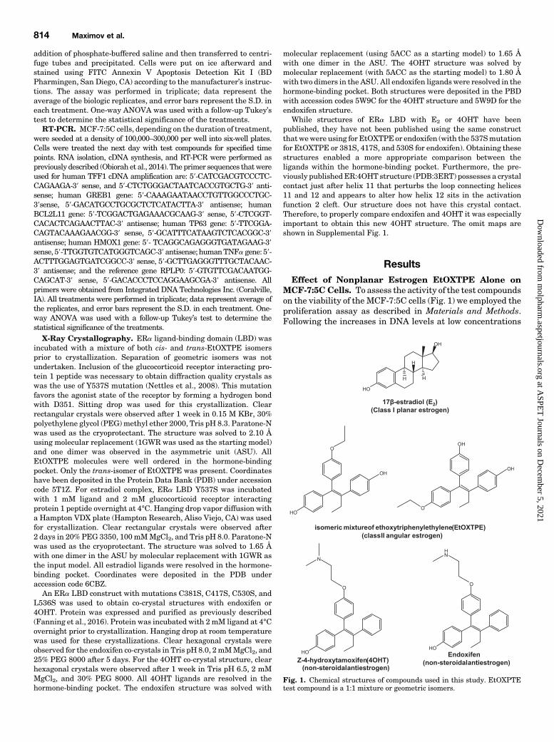
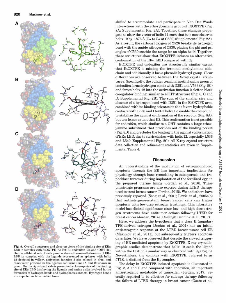