endophytic bacteria within the green siphonous seaweed bryopsis
TRANSCRIPT
Endophytic bacteria within
the green siphonous seaweed Bryopsis:
Exploration of a partnership
Joke Hollants
Promoters
Prof. Dr. Anne Willems
Prof. Dr. Olivier De Clerck
Dissertation submitted in fulfillment of the requirements for the degree of Doctor (Ph.D.) in Sciences, Biotechnology

______________________________________________________________________________
Joke Hollants - Endophytic bacteria within the green siphonous seaweed Bryopsis: Exploration of a
partnership
Copyright ©2012 Joke Hollants
ISBN-number: 978-94-6197-046-6
No part of this thesis protected by its copyright notice may be reproduced or utilized in any form, or
by any means, electronic or mechanical, including photocopying, recording or by any information
storage or retrieval system without written permission of the author and promoters.
Printed by University Press | www.universitypress.be
Ph.D. thesis, Faculty of Sciences, Ghent University, Ghent, Belgium.
This Ph.D. work was financially supported by ‘Fonds Wetenschappelijk Onderzoek’ FWO-Flanders.
Publicly defended in Ghent, Belgium, June 8th 2012

______________________________________________________________________________
Examination committee
Prof. Dr. Savvas Savvides (Chairman) L-Probe: Laboratory for protein Biochemistry and
Biomolecular Engineering, Faculty of Sciences, Ghent University, Ghent, Belgium
Prof. Dr. Anne Willems (Promoter) LM-UGent: Laboratory of Microbiology, Faculty of Sciences,
Ghent University, Ghent, Belgium
Prof. Dr. Olivier De Clerck (Co-promoter) Phycology Research Group, Faculty of Sciences,
Ghent University, Ghent, Belgium
Dr. Frederik Leliaert Phycology Research Group, Faculty of Sciences, Ghent University, Ghent,
Belgium
Prof. Dr. Paul De Vos LM-UGent: Laboratory of Microbiology, Faculty of Sciences, Ghent
University, Ghent, Belgium
Prof. Dr. Wim Bert Nematology Research Group, Faculty of Sciences, Ghent University, Ghent,
Belgium
Dr. Danny Vereecke Department of Plant production, Faculty of Applied Bioscience Engineering,
University College Ghent, Ghent, Belgium
Dr. Thomas Wichard Institute for Inorganic and Analytical Chemistry, Friedrich Schiller University
Jena, Jena, Germany


Preface
“Life did not take over the globe by combat, but by networking”
Lynn Margulis evolutionary biologist
Life itself is contained in this one sentence.
Besides Darwin's famous ‘natural selection' theory,
symbiosis, the cooperation between different organisms,
is necessary for the survival and evolution of species.
Looking at a human being, an animal, or a plant.
They all tell the same story.
Everything works according the same holistic principle of
interconnection and cooperation.
Our body contains trillions of bacterial cells, 10 times more than human cells.
Yes, you are more bacteria than human!
Without bacteria you would weigh 1.2 kg less, and yet you don’t want to lose them.
Bacteria digest our food and keep us healthy.
Also seaweeds undertake close collaborations with external (ectosymbiotic) and internal (endosym-
biotic) bacteria. Seaweeds are an unlimited source of oxygen and sugars which bacteria are happy to
take advantage of. In exchange for these nutrients, bacteria produce growth promoting minerals and
vitamins and they protect their host against environmental threats. As a result, many seaweed-
bacterial associations are essential for both symbiotic partners.
This thesis focuses on the association between the feathery-like alga Bryopsis and bacteria inside this
green seaweed. It has been known for over 40 years that Bryopsis houses bacteria, but nothing was
known about their identity and function. The following pages take you on an exploratory trip to the
hows and whys of this exciting partnership.
I hope that, while reading between the lines, I can tell you a story about the power of collaboration.
Not between two, but a lot of partners.
Each with their own talents and flaws.
Each in their own way.
“It takes two to tango, but a whole crowd to stage dive! ”


Voorwoord
“Life did not take over the globe by combat, but by networking”
Lynn Margulis evolutiebiologe
Het verhaal van het leven ligt vervat in deze ene zin.
Naast Darwins gekende ‘natuurlijke selectie’-theorie,
is symbiose, de samenwerking tussen verschillende organismen,
nodig voor de overleving van soorten.
Het is tevens de motor van hun evolutie.
Bekijk een mens, bekijk een dier, bekijk een plant.
Ze vertellen allen hetzelfde verhaal.
Alles functioneert volgens hetzelfde holistische principe van
onderlinge beïnvloeding en samenwerking.
In en op ons lichaam zitten biljoenen bacteriële cellen, 10x meer dan menselijke cellen.
Ja, je leest het goed, je bent meer bacterie dan mens!
Zonder bacteriën zouden wij maar liefst 1,2 kg minder wegen, geen onaangename gedachte.
En toch wil je ze niet kwijt. Bacteriën verteren ons voedsel en houden ons gezond.
Ook zeewieren gaan hechte samenwerkingsverbanden aan met uitwendige (ectosymbiontische) en
inwendige (endosymbiontische) bacteriën. Zeewieren zijn een onuitputtelijke bron van zuurstof en
suikers en daar maken bacteriën maar al te graag gebruik van. In ruil voor deze voedingsstoffen
maken bacteriën groeibevorderende mineralen en vitamines aan en beschermen ze hun gastheer
tegen bedreigingen van buitenaf. Vele zeewier-bacterie associaties zijn dan ook van levensbelang
voor beide symbiose partners.
Deze thesis focust op de associatie tussen het vederwier Bryopsis en bacteriën aanwezig binnenin het
wier. Het is al meer dan 40 jaar geweten dat het vederwier bacteriën huist, maar er was niets gekend
omtrent hun identiteit en functie. De volgende bladzijden nemen je mee op een verkennende tocht
naar het hoe en waarom van dit boeiend partnerschap.
Ik hoop dat ik jou, tussen de technische hoofdstukken door, een verhaal kan vertellen over de kracht
van samenwerking. Niet tussen twee, maar een heleboel partners. Elk met hun eigen talenten en
gebreken. Elk op hun eigen manier.
“It takes two to tango, but a whole crowd to stage dive! ”

Dankwoord
Tijdens mijn persoonlijke doctoraats-stage-dive heb ook ik gebruik kunnen maken van gezellige en nuttige symbioses met anderen, daarom gaat mijn dank uit naar
Iedereen van het labo microbiologie en algologie,
en in het bijzonder naar
Mijn promotoren Anne en Olivier voor de opdracht en het vertrouwen
Mijn top-begeleider Frederik
voor het vele kunst- en vliegwerk en zijn eeuwig optimisme
De vele studenten, vooral Helen en Lana voor de praktische hulp
Olivier
voor het inbed- en snijwerk en de knaagdiermoppen
Myriam voor de supersnelle EM-interventies
Heroen
voor de staalnames, artikel-hulp en opbouwende feedback
Lennert voor de staalnames en Bio-ORACLE hulp
Ellen en Caroline
om mij te leren extraheren en kloneren
Frederique om mij te leren duiken en Bryopsis herkennen in Franse wateren
Sofie, Tine, Annelien en Pieter
om het koude nulde met enkele graden te verwarmen
Annemie en Christelle om mij heel wat administratie-frustratie te besparen
Jeanine
voor de overvloed aan propere potjes en vuile praatjes
Margo voor de hulp bij het zoeken naar ongewone dingen

Dankwoord
Het sympathieke sequentieteam: Liesbeth, Evie en Jindrich voor het kortwieken van de vele sequenties en om zich gewillig te laten omkopen
Bjorn
voor de hulp bij die ver-draaide excelsheets
Wim
voor de symbiose tussen leek & PC en hoofd & voeten
Gwen en Anne
voor de zotheid en om samen het DGGEspook te temmen
Renata voor de eerste opvang en de blijvende vriendschap
Sofie en Karolien
om het drie-muskutier- én symbiose-motto "Eén voor allen, allen voor één!" alle eer aan te doen
Vrienden en familie Moeke, vake en meke
om mij te laten springen en indien nodig ook op te vangen
Dear crowd, It was a pleasure to PhD-dive with you!
Joke

Table of contents
Chapter 1: Literature 1
What to learn from sushi: a review on seaweed-bacterial associations 3
From kitchen secrets to sushi: a historical overview 4
Foundations 4
First cultivation and microscopy studies 6
Emergence of molecular techniques 7
Chemical interactions between seaweeds and bacteria 7
Seaweed partner 9
Bacterial partner 10
Endophytic seaweed-bacterial relationships 13
Bacterial diversity associated with seaweeds 13
Identity of bacteria associated with seaweeds: higher taxonomic ranks 14
Identity of bacteria associated with seaweeds: genus/species level 17
Linking identity to function 19
Conclusion 22
Chapter 2: Objectives 25
Chapter 3: Experimental work 29
Part 1: Optimization of the experimental design 31
3.1.1. Overview 31
Surface sterilization 31
Molecular work: full-cycle 16S rRNA gene approach 32
Cultivation work 33
Functional gene analysis 33
3.1.2. Surface sterilization of Bryopsis samples 35
Table of contents
Part 2: Endophytic bacterial communities of Bryopsis cultures 47
3.2.1. Endophytic bacterial diversity within Mexican Bryopsis samples 47
3.2.2. Uniqueness, temporal stability and symbiotic nature of Bryopsis endophytic
bacterial communities 67
3.2.3. Disentangling host phylogenetic, environmental and geographic signals in
intracellular bacterial communities of Bryopsis 83
3.2.4. Axenic cultivation of the Bryopsis host and in vitro isolation of intracellular bacteria 105
Part 3: Endophytic bacterial communities of natural Bryopsis samples 117
3.3.1. Flavobacteriaceae endosymbionts within natural Bryopsis samples: host specificity and 117
cospeciation
3.3.2. In situ hybridizations of Bryopsis intracellular bacteria with group- and species-specific 137
fluorescent probes
Chapter 4: Concluding discussion 143
Summary 155
Samenvatting 158
Bibliography 161
Curriculum vitae 175

Chapter
Literature
1


Literature | 3
What to learn from sushi: a review on seaweed-bacterial associations
Joke Hollants, Frederik Leliaert, Olivier De Clerck and Anne Willems. What to learn from sushi: a review on
seaweed-bacterial associations. Manuscript submitted as a mini-review to FEMS Microbiol Ecol. Author
contributions: The literature review was outlined, performed and written by Joke Hollants. Frederik Leliaert,
Olivier De Clerck and Anne Willems commented on the text.
If there is one thing we can learn from sushi, it is its digestion by an alga-associated bacterium. The
carbohydrate active enzyme porphyranase from the marine Bacteroidetes bacterium Zobellia
galactanivorans breaks down the sulphated polysaccharide porphyran from the red alga Porphyra (nori)
traditionally used to prepare sushi. Moreover, the genes coding for this porphyranase have been
horizontally transferred through dietary seaweed from Z. galactanivorans to the gut microbe Bacteroides
plebeius from particularly Japanese people, allowing them to digest the algae which wrap sushi rolls and
other delicacies [1]. This not only indicates that the human gut microbiota may become proficient at
using dietary polysaccharides by horizontal gene transfer; it also highlights the significance of macroalgal-
bacterial associations.
Like sushi, algae come in many forms and flavors ranging from microscopic unicells to gigantic kelps
inhabiting oceans, freshwater habitats, soils, rocks and even trees [2]. Consequently, this review
needed some delimitation and is restricted to studies of bacteria associated with marine macroalgae
(seaweed) belonging to the Chlorophyta (green algae), Rhodophyta (red algae) and Phaeophyceae
(brown algae). Seaweed and bacteria have come a long way since algal plastids originated from
endosymbiotic cyanobacteria [3 and see also Box 1]. Like their unicellular ancestors, marine
macroalgae form the modern-day playground for a wide diversity of bacterial associations ranging
from beneficial (mutualistic), harmful (parasitic) and neutral (commensal), over obligate and
facultative, to endo- and ectophytic interactions (Box 1). This, along with applied aspects of current
algal-bacterial symbioses (Box 2), makes their associations appealing for evolutionary, ecological and
biochemical studies. Nevertheless, investigations of macroalgal-bacterial associations lag behind these
of other marine eukaryotes [4]. Whereas the full cycle 16S rRNA approach [5] is well established to
characterize the microbial associates of unicellular algae, corals and sponges [6, 7], these molecular
techniques are just beginning to be applied to macroalgae [4 and references therein].

4 | Literature
BOX 1 - Symbiosis: highlighting the beauty in biology Symbiosis from the ancient Greek sýn ‘with’ and bíōsis ‘living’ stands for ‘living together’. In 1879 the German mycologist Heinrich Anton de Bary was the first to use the term to describe the relationship between fungi and algae in the formation of lichens. In this context, he defined the term symbiosis as ‘the living together of two dissimilar organisms, usually in intimate association, and usually to the benefit of at least one of them’. The last decades, the term has been used more widely to cover beneficial (mutualistic), harmful (parasitic) and neutral (commensal) interactions that can change over time for any given set of partners [8]. It has to be noted, however, that in practice the term symbiosis is often associated with mutualistic associations only [9]. Either way, symbiotic relationships encompass both long term, obligate associations in which the symbiotic partners entirely depend on each other for survival as well as transient, facultative interactions in which the partners can exist independently of one another. These symbiotic relationships can be epi- or endobiotic with one symbiotic partner (i.e. the symbiont) living on or within the other (i.e. the host), respectively. Even though symbiosis is generally assumed to involve only two partners, most hosts accommodate complex symbiont communities consisting of multiple species [8]. Accordingly, the list of symbiotic relationships is endless. Protective sea anemones that hitchhike on the back of hermit crabs, photosynthetic algae living inside coral hosts, small cleaner fish visiting larger clients, wood-digesting protozoans in termite stomachs, and tick-eating oxpeckers on the backs of zebra, elephants, hippopotamuses and other large African animals, are only some of nature’s best symbiosis examples. Besides these eukaryote-eukaryote interactions, also eukaryote-prokaryote symbiotic associations are widespread in nature. Well-known examples include the human microbiome, nitrogen fixing rhizobia inside root nodules, bioluminescent Vibrio species within squid light organs, chemosynthetic bacteria which associate with marine invertebrates and various insect-bacterial interactions (for an overview see: http://iss.cloverpad.org/Default.aspx?pageId= 552623). Compared to prokaryotes, eukaryotes are limited in their biochemical repertoire. Therefore, eukaryotic hosts associate with bacterial symbionts which expand their physiological capacities, allowing them to invade novel metabolic and ecological niches. Symbioses are thus the ultimate examples of success through collaboration and support fundamentally important processes [8]. The ‘endosymbiotic theory’ which claims that eukaryotic organelles like mitochondria and chloroplasts are of endosymbiotic origin [3], is a fine example of this essential significance of symbiosis throughout life’s history. Symbioses have been and are to this very day anything but a marginal or rare phenomenon. In fact, according to the late evolutionary biologist Lynn Margulis (1938-2011), “we abide in a symbiotic world”.
From kitchen secrets to sushi: a historical overview
Foundations
The first report of a seaweed-bacterium alliance – although artificial – is one that altered bacteriology
forever. In 1881, Walther Hesse, a German physician, joined Robert Koch’s laboratory to study the
bacteria responsible for his patients’ illnesses. But, like his colleagues, Hesse encountered major
technical problems attaining pure bacterial cultures on solid gelatin-based media. The gelatin often
liquefied due to bacterial enzymes or because of the temperature of the laboratory. When he vented
his frustrations to his wife Fanny, she suggested using a seaweed extract, agar-agar, which she had
used to thicken her jellies and puddings for years [10]. The practical application of this kitchen secret
accelerated bacteriological research greatly, opening the way also for real life macroalgal-bacterial
studies. In fact it was Walther Hesse himself who developed agar plate techniques to count bacteria
Literature | 5
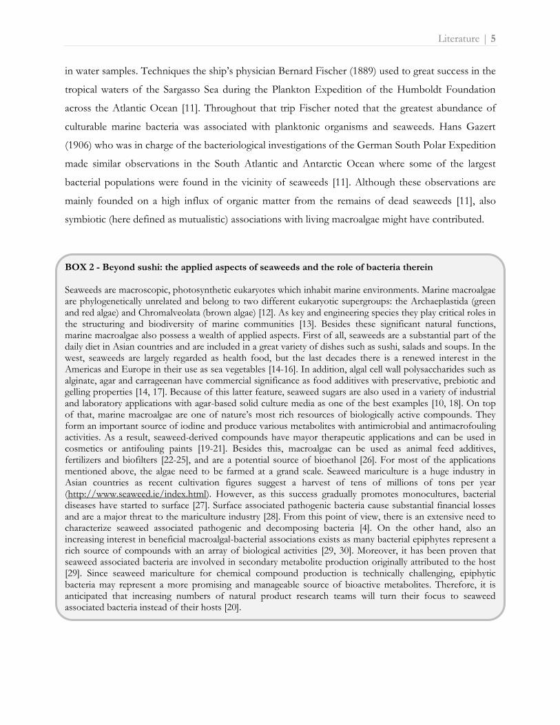
in water samples. Techniques the ship’s physician Bernard Fischer (1889) used to great success in the
tropical waters of the Sargasso Sea during the Plankton Expedition of the Humboldt Foundation
across the Atlantic Ocean [11]. Throughout that trip Fischer noted that the greatest abundance of
culturable marine bacteria was associated with planktonic organisms and seaweeds. Hans Gazert
(1906) who was in charge of the bacteriological investigations of the German South Polar Expedition
made similar observations in the South Atlantic and Antarctic Ocean where some of the largest
bacterial populations were found in the vicinity of seaweeds [11]. Although these observations are
mainly founded on a high influx of organic matter from the remains of dead seaweeds [11], also
symbiotic (here defined as mutualistic) associations with living macroalgae might have contributed.
BOX 2 - Beyond sushi: the applied aspects of seaweeds and the role of bacteria therein Seaweeds are macroscopic, photosynthetic eukaryotes which inhabit marine environments. Marine macroalgae are phylogenetically unrelated and belong to two different eukaryotic supergroups: the Archaeplastida (green and red algae) and Chromalveolata (brown algae) [12]. As key and engineering species they play critical roles in the structuring and biodiversity of marine communities [13]. Besides these significant natural functions, marine macroalgae also possess a wealth of applied aspects. First of all, seaweeds are a substantial part of the daily diet in Asian countries and are included in a great variety of dishes such as sushi, salads and soups. In the west, seaweeds are largely regarded as health food, but the last decades there is a renewed interest in the Americas and Europe in their use as sea vegetables [14-16]. In addition, algal cell wall polysaccharides such as alginate, agar and carrageenan have commercial significance as food additives with preservative, prebiotic and gelling properties [14, 17]. Because of this latter feature, seaweed sugars are also used in a variety of industrial and laboratory applications with agar-based solid culture media as one of the best examples [10, 18]. On top of that, marine macroalgae are one of nature’s most rich resources of biologically active compounds. They form an important source of iodine and produce various metabolites with antimicrobial and antimacrofouling activities. As a result, seaweed-derived compounds have mayor therapeutic applications and can be used in cosmetics or antifouling paints [19-21]. Besides this, macroalgae can be used as animal feed additives, fertilizers and biofilters [22-25], and are a potential source of bioethanol [26]. For most of the applications mentioned above, the algae need to be farmed at a grand scale. Seaweed mariculture is a huge industry in Asian countries as recent cultivation figures suggest a harvest of tens of millions of tons per year (http://www.seaweed.ie/index.html). However, as this success gradually promotes monocultures, bacterial diseases have started to surface [27]. Surface associated pathogenic bacteria cause substantial financial losses and are a major threat to the mariculture industry [28]. From this point of view, there is an extensive need to characterize seaweed associated pathogenic and decomposing bacteria [4]. On the other hand, also an increasing interest in beneficial macroalgal-bacterial associations exists as many bacterial epiphytes represent a rich source of compounds with an array of biological activities [29, 30]. Moreover, it has been proven that seaweed associated bacteria are involved in secondary metabolite production originally attributed to the host [29]. Since seaweed mariculture for chemical compound production is technically challenging, epiphytic bacteria may represent a more promising and manageable source of bioactive metabolites. Therefore, it is anticipated that increasing numbers of natural product research teams will turn their focus to seaweed associated bacteria instead of their hosts [20].

6 | Literature
Simultaneously with these initial notes of seaweed-bacterial alliances at sea, scientists in the
laboratory deduced similar conclusions from their preliminary late 19th century macroalgal culture
work. The German botanist Georg Klebs (1896) was aware of the presence of bacteria in his
seaweed cultures and tried to set up pure, axenic cultures of filamentous and siphonous algae. While
he was successful in growing the algae, he was not able to keep his cultures bacteria-free [31]. Even
though Klebs was a former assistant of Anton de Bary who first introduced the term ‘symbiosis’ in
biology, it was Johannes Reinke (1903) who was the first to suggest a true symbiotic marcoalgal-
bacterial partnership. The occurrence of Azotobacter as an epiphyte on marine algae let him to
propose that a symbiosis may exist in which the algae supply Azotobacter with carbohydrates and use
the nitrogen fixed by the bacteria [11, 32]. Also Edgar Johnson Allen (1910), director of the Marine
Biological Association of the United Kingdom, and his collaborator E.W. Nelson recognized a
symbiotic aspect in xenic marcoalgal cultures [31]. As they laid the foundations for seaweed culture,
they noticed good growth of algae only when small quantities of natural seawater were added to the
artificial culture media. Allen remarked that these effects may be caused by products of the
metabolism of bacteria [31].
First cultivation and microscopy studies
It took until after World War II for Luigi Provasoli and colleagues to establish the first bacteria-free
cultures of the green foliaceous seaweed Ulva using newly discovered antibiotics [31]. Provasoli,
however, observed that the typical foliose morphology of Ulva lactuca was lost in the absence of
bacteria and even more interesting that the normal thallus morphology was restored when
certain bacteria previously isolated from the algal surface were re-added to the culture medium [33,
34]. In 1955, Harold and Stanier [35] were the first to exhaustively describe the bacterium Leucothrix
mucor which was found consistently as an algal epiphyte, showing macroalgae not only to interact
with bacteria but also to represent a distinct source of new microbial taxa. With the introduction of
electron microscopy to study the macroalgal ultrastructure in the ‘70s, an intriguing new form of
seaweed-bacterial interactions was discovered. In addition to epiphytic bacteria, various siphonous
seaweeds such as Bryopsis, Caulerpa, Chlorodesmis, Halimeda, Penicillus and Udotea were also shown to
harbor intracellular bacteria within their cytoplasm and/or vacuolar systems (see Box 3) [36-41].
Simultaneously with these early microscopic observations, the first cultivation studies aiming to
examine the total diversity of bacteria associated with macroalgae arose. Although the bacteria were
initially identified only by morphological and biochemical tests, the epiphytic flora on seaweeds was
Literature | 7
clearly very diverse, covering numerous bacterial taxa [42-51]. Not only were these macroalgal
associated bacteria distinct from the surrounding seawater communities, they also appeared host-
specific with clear differences in occurrence among green, red and brown seaweeds [46, 48-50]. A
stable association between algal hosts and bacteria was observed [46, 49, 50], even though the
bacterial flora could vary between seasons and/or between different parts of the algal thallus [42, 45,
47]. From these and other studies in the ‘70s and ‘80s, Bolinches and coworkers [52] concluded the
existence of both positive and negative macroalgal-bacterial interactions based on the algal capacity
to produce organic compounds and oxygen that are utilized by bacteria. In turn, bacteria produce
morphogenic factors, fixed nitrogen, enzymes and vitamins which promote algal growth [34, 48, 53-
56]. In addition, epiphytic bacteria as well as the seaweed hosts themselves produce antibiotic
substances which prevent colonization of the algal surface by bacterial competitors and pathogens
[51, 57].
Emergence of molecular techniques
Although the number of macroalgal-bacterial studies risen steadily during the last two decades, these
have not significantly increased our understanding of macroalgal-bacterial interactions as postulated
above. Thanks to the improvement of analysis techniques, both symbiotic partners can be
characterized biochemically and phylogenetically in more detail. However, many questions remain
[4]. In the following sections we review the current knowledge on the diversity and functional
ecology of bacterial communities associated with green, red and brown marine macroalgae.
Chemical interactions between seaweeds and bacteria
The relationship between macroalgae and bacteria in which seaweeds provide nutrients, while the
bacterial community promotes algal growth and protects the host against pathogens, has been
elaborated over the last 20 years. Figure 1.1 depicts the complex, chemically mediated interplay of
beneficial and detrimental relations that exists between macroalgae and bacteria. The variety and
nature of these chemical interactions have been exhaustively reviewed by Goecke and coworkers [4],
and are summarized in the remainder of this section.

Figure 1.1: Overview of beneficial (green) and detrimental (red) interactions between macroalgae and bacteria.

Literature | 9
Seaweed partner
From the algal host perspective, macroalgal-bacterial interactions are not unexpected. Seaweed
surfaces provide a protected and nutrient rich ‘hot spot’ for opportunistic bacteria that are abound
wherever organic material is available [30]. In most cases molecular investigations have confirmed
the outcome of initial cultivation studies, i.e. that the attraction of bacteria by seaweeds turns out to
be highly specific. While the composition of the bacterial flora can change over seasons, life span and
different thallus-parts as a result of biotic and abiotic factors [58-60], marine macroalgae generally
associate with specific bacterial communities that differ significantly from those occurring in the
surrounding seawater [61, 62]. Recently, however, Burke and colleagues [13] found highly variable
bacterial species compositions among local individuals of Ulva australis by means of in-depth 16S
rRNA screening, suggesting each U. australis plant hosts a unique assemblage of bacterial species.
Moreover, using a metagenomic approach they subsequently showed that the bacterial community
composition on U. australis is driven by functional genes rather than the taxonomic or phylogenetic
composition of its species [63]. This implies that functional groupings (or “guilds”) of not
necessarily phylogenetically related bacterial species exist of which the composition on a single
algal individual is determined stochastically by recruitment from within those guilds. Even if the
specificity of a seaweed-associated bacterial community may be based on functional genes rather
than species, it is known that the physiological and biochemical properties of the algal host
predetermine the composition of the adhering bacterial communities. For example, algal cell wall
components and secondary metabolites can trigger specific interactions between seaweeds and
beneficial bacteria [64, reviewed in 65]. Algal bioactive compounds also have antimicrobial properties
with interesting biomedical and industrial applications (see Box 2) which protect the seaweed
surface from bacterial pathogens, grazers and biofouling, i.e. the undesirable accumulation of micro-
and macroorganisms as biofilms on the seaweed surface [4 (Table 5), 21, 28, 65-67]. Besides these
bioactive compounds, macroalgae control bacterial colonization by interfering with bacterial quorum
sensing (QS) systems which regulate bacterial cell-to-cell communication [4 (Table 6), 68-70]. In
addition to these induced defense mechanisms, seaweeds also possess non-specific defense responses
against bacterial pathogens similar to the ‘oxidative burst’ process of higher plants [71, 72].

10 | Literature
Bacterial partner
Many bacteria growing on seaweed surfaces are able to enzymatically decompose algal cell walls,
making them key players in biotransformation and nutrient recycling in the oceans [4 (Table 2), 18].
Also specific, beneficial bacterial-macroalgal interactions are based on the bacterial capacity to
mineralize algal organic substrates and subsequently supply the seaweed host with carbon dioxide,
minerals, vitamins and growth factors [30, 55, 56, 73, 74]. Several studies also revealed that seaweed
associated bacteria are important sources of fixed nitrogen and detoxifying compounds [4 and
references therein, 75, 76]. Besides nutritional and growth promoting effects, bacteria may shape the
morphology and life cycle of their algal host. Bacterial effects on morphogenesis have been reported
in foliaceous green macroalgae such as Ulva and Monostroma [34, 77-81], and have been shown to be
controlled by a highly potent differentiation inducer, thallusin, isolated from well-defined associated
bacteria [4 (Table 4), 82]. Thallusin and other bacterial metabolites, including QS molecules, also play
a role in the host’s life cycle completion as well as in algal spore release and germination [4 (Table 4),
82-87]. Furthermore, QS inhibitors and antimicrobial compounds produced by numerous epiphytic
bacteria work in concert with seaweed derived metabolites (see above) to protect the seaweed surface
from pathogens, herbivores and fouling organisms [4 (Table 4), 30, 88-93]. Pathogenic bacteria can
cause severe degradation of algal host cells or even lead to seaweed mortality, causing major financial
losses to seaweed mariculture every year (Box 2) [4 (Table 4), 94, 95]. Also biofouling forms a
permanent threat to macroalgae as bacterial biofilms increase the hydrodynamic drag on their host
and enhance the attachment of other fouling organisms and grazers. Biofilms may also compete for
nutrients, inhibit gaseous exchange or block light, essential for photosynthesis. Thus, both bacterial
and algal bioactive compounds are essential chemical mediators in macroalgal-bacterial associations
which jointly control the composition and density of bacterial biofilms thereby defending the
seaweed surfaces against biofouling [4 and references therein, 28]. In addition, these bacterial
bioactive compounds may represent a more promising and easier to handle source of natural
products with biotechnological applications in comparison with seaweed derived compounds (Box 2)
[20, 29, 96, 97].
Literature | 11
BOX 3 - Bryopsis, an underwater peacock with extraordinary features Bryopsis (Greek, moss like) is a 1-20 cm tall feathery-like, marine green macroalga (Ulvophyceae, Bryopsidales) which inhabits temperate and tropical seas (Fig. 1.2). Bryopsis algae usually attach to rocks or other seaweeds and grow in the intertidal to subtidal zone, generally down to 5 m depth [2, 12, 98]. Bryopsis possibly originated in the late Mesozoic, about 100 my ago [99]. The genus was described in 1809 by J.V. Lamouroux, and in the past 200 years more than 200 species and intraspecific taxa have been described, of which about 55 are currently accepted [100]. Species delineation in Bryopsis is problematic because of rampant morphological plasticity, which is intrinsically related to its simple body plan [101]. The entire plant, built up of feathers consisting of a holdfast (rhizoids) and a central stem (axis) with branches (pinnae) on either side, has a unicellular structure without any internal cross walls (Fig. 1.2B). On that account, Bryopsis belongs to a unique group of marine ‘giant-celled’ macroalgae which are composed of a single, tubular (siphonous) shaped cell. These siphonous seaweeds exhibit a typical intracellular architecture in which the multinucleate (coenocytic) cytoplasm is restricted to a thin cell-wall associated layer that surrounds a central vacuole which occupies most of the cell volume [102]. In Bryopsis the peripheral cytoplasmic layer is divided into two sublayers: an outer layer adjacent to the cell wall which contains most of the organelles excluding the chloroplasts, and an inner chloroplast layer next to the vacuole (Fig. 1.2I). Bryopsis algae are homoplastidic, which means that they only possess one type op plastid, namely chloroplasts. These chloroplasts contain pyrenoids with starch as the principal carbohydrate storage product [36]. Another interesting phenomenon is that Bryopsis chloroplasts can maintain activity inside the body of some herbivorous sea slugs, rendering the animals photosynthetic [103] (Fig. 1.2H). The plastid maintenance is thought to involve lateral gene transfer from the algal food source to the slugs [104], but this has been recently questioned [105]. Although the complete Bryopsis plastid genome has been recently sequenced, the plastidial autonomy seems to have little to do with the size and gene content of the cpDNA itself [106]. Furthermore, the Bryopsis cytoplasm exhibits vigorous streaming by which organelles and nutrients are transported throughout the siphonous thallus thereby enabling optimal photosynthesis and nutrient exchange [107]. In addition, Bryopsis has evolved several other features to overcome the physical limitations of being unicellular [108]. Bryopsis produces, for example, the bioactive [also therapeutic, see reference 109] metabolite kahalalide F which protects the vulnerable alga from fish predation [110]. Cell wounding triggers a complex, multistep wound response resulting in in loco plug formation and subsequent synthesis of a new cell wall [37, 111, 112]. To this, Bryopsis algae add a surprisingly feature, i.e. the formation of protoplasts [113]. These protoplasts, which are released upon injury, are membraneless and can survive in seawater for 10–20 minutes (Fig. 1.2D). Subsequently, phospholipid membranes and a cell wall are synthesized de novo surrounding each protoplast, which then develops into a new Bryopsis plant. Protoplast formation is thus a defense as well as an effective propagation mechanism. In Bryopsis, this vegetative reproduction by thallus fragmentation is accompanied with sexual reproduction modes alternating between a gametophytic and sporophytic phase [2]. Despite early reports on the simplicity of the Bryopsis life cycle, subsequent culture observations showed a wide variety of life history patterns, even within a single Bryopsis species [114]. For a nice overview of life history pathways, including fragmentation, parthenogenesis and differentiation of zoospores and gametes, see Rietema [114] and Morabito et al. [115]. Besides these well-studied morphological, regenerative and reproductive characteristics, Bryopsis has long been suspected to harbor intracellular bacteria inside its cytoplasm as well as vacuolar systems [36, 37]. Endophytic bacteria have been visualized in the Bryopsis cytoplasm by electron microscopy at every stage of development, including the gametes [36] (Fig. 1.2E). This indicates a natural, stable relationship between the algal host and its endophytes in which both partners may provide mutualistic ecological benefits. To date, this remarkable algal-bacterial partnership has received little or no attention. However, as it has already been proven that endosymbiotic bacteria inside Caulerpa algae share responsibility for the successful - though sometimes highly invasive - spread of siphonous seaweeds in oligotrophic waters [75], an enlightening characterization of the bacterial partner would be welcome.

12 | Literature
Figure 1.2: The green siphonous seaweed Bryopsis and its characteristic features. A: B. pennata forms soft, feathery clumps. Scale bar: 2.5 cm (photo by Linda Preskitt). B: Detail of one Bryopsis feather. The main axis shows no internal cross walls. Scale bar: 2.5 mm (photo: http://www.turtlejournal.com/?p=7629). C: Bryopsis loses its characteristic morphology in culture. Scale bar: 4 cm (photo by Anne Willems). D: Formation of protoplasts from B. hypnoides. Scale bar: 30 µm (photo by Lü et al. [106]). E: Electron micrograph of Bryopsis female gamete in longisection. Cluster of bacteria can be seen just above the chloroplast. Magnification: x20000, scale bar: 1 µm (photo by Burr & West [36]). F: Bryopsis rhizoids (holdfast). Scale bar: 5 mm (photo: http://www.turtlejournal.com/?p=7629). G: Release of gametes from a matured Bryopsis gametangium. Scale bar: 1.5 mm (photo by Joke Hollants). H: Elysia clarki, a ‘solar-powered sea slug’, sequesters chloroplasts from its Bryopsis food. Scale bar: 1.5 cm (photo by Curtis et al. [116]). I: Electron micrograph of Bryopsis vegetative thallus in longisection. Magnification: x8000, scale bar: 5 µm (photo by Olivier Leroux and Joke Hollants).

Literature | 13
Endophytic seaweed-bacterial relationships
Besides being epiphytic on algal surfaces, bacteria also live inside the thallus or cells. Seaweed grazers
or epiphytic bacteria capable of degrading algal cell walls (see above) can damage algal thalli and
provide an entrance for pathogenic and opportunistic bacteria [117-120]. These latter bacteria might
become detrimental if they are able to enter the algal tissue and contribute to further disintegration
of the host, finally leading to thallus rupture [4 and references therein]. In addition to these
pathogenic associations, also non-detrimental seaweed-associated endophytic bacteria are described.
Bacteria are present inside algal galls (i.e. abnormal tissue growths of seaweeds) reported on more
than 20 species of red and brown macroalgae [reviewed in 121]. In the red seaweed Prionitis,
endophytic bacteria are responsible for gall formation by overproduction of the phytohormone
indole-3-acetic acid (IAA), thereby creating a suitable microhabitat for their own proliferation [122,
123]. Even though the benefits for the seaweed partner are not well understood, coevolution
between Prionitis hosts and their gall-forming endobionts has been suggested [123]. Also in the red
macroalga Gracilaria dura endophytic bacteria enhance the algal bud induction by the production of
IAA and fixed nitrogen [74]. In various siphonous (single celled, multinucleate) green seaweeds,
endophytic bacteria have been reported over the past 40 years (see above and Box 3). Even though
these endophytic bacteria have been associated with detoxification, nitrogen fixation and
photosynthetic functions [75, 124, 125], the true physiological nature of these endobiotic siphonous
seaweed-bacterial symbioses remains unknown.
Bacterial diversity associated with seaweeds
Broad-spectrum seaweed-bacterial diversity studies identifying the total bacterial community are
scarce. This is not surprising given that the number of seaweed associated bacteria exceeds those in
the surrounding seawater by 100 to 10 000 times [42]. Total viable counts reach up to 107 bacterial
cells per gram dry algal weight using the agar spread plate method; a number that even increases by
two orders of magnitude when applying direct enumeration techniques [42, 47, 126]. Consequently,
most macroalgal-bacterial studies focus on the identification and characterization of specific bacterial
taxa, e.g. those with bioactive potential or pathogenic activity, rather than investigating the total
bacterial diversity [79, 88, 93, 120]. Until recently, most of these investigations used traditional
culture-based approaches, which are often considered insufficient since only 1% of all known
bacteria are estimated to be culturable [127]. However, current molecular methods such as clone

14 | Literature
libraries, denaturing gradient gel electrophoresis (DGGE), quantitative PCR (Q-PCR) and
fluorescent in situ hybridization (FISH) also have their limitations for grasping the entire diversity of
a microbial community, even in a single environmental sample, since they mainly reveal a snapshot in
time of the dominant bacterial community members only [128].
In the following paragraphs we review 149 studies from the last 55 years which dealt with bacteria
associated with a total of 159 seaweed species (36 green, 72 red and 51 brown marine macroalgae,
see Table S1.1 on http://www.phycology.ugent.be/). The bacterial diversity was compared between
brown, green and red seaweeds at all taxonomical levels. Wherever possible, the identity of the
associated bacteria was linked to their ecological function.
Identity of bacteria associated with seaweeds: higher taxonomic ranks
Bacteria described from seaweed surfaces or within algal thalli belong to the (super)phyla
Proteobacteria, Actinobacteria, Bacteroidetes (previously known as the Cytophaga-Flavobacteria-
Bacteroides (CFB) group), Cyanobacteria, Firmicutes, Planctomycetes, Verrucomicrobia, Chloroflexi,
Deinococcus-Thermus, Fusobacteria, Tenericutes and the candidate division OP11. In all studies
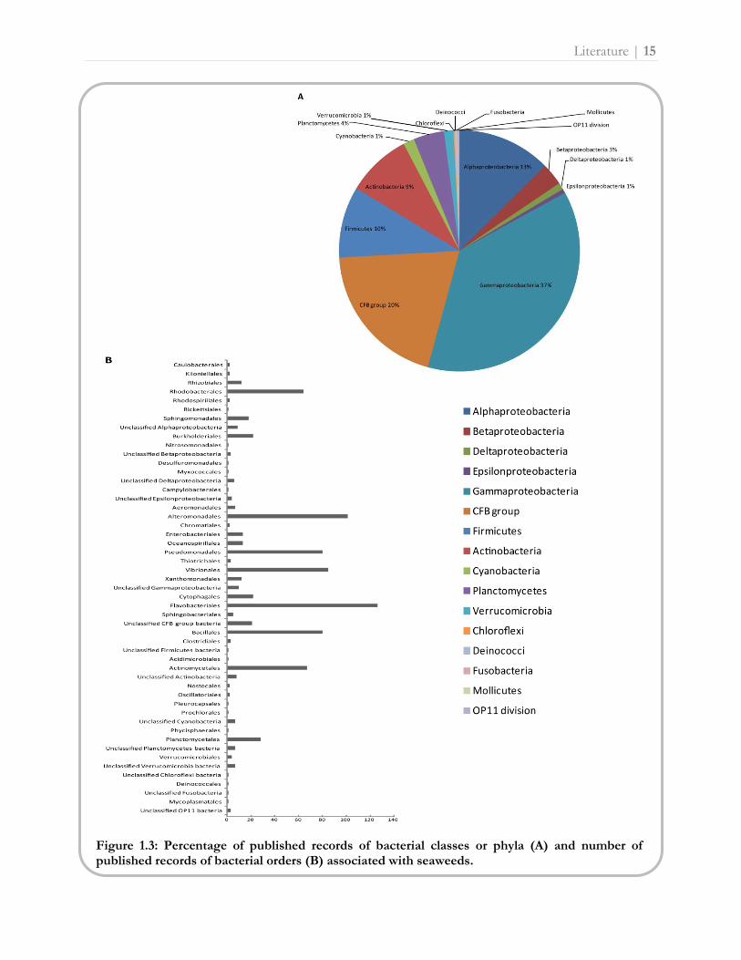
reviewed, Gammaproteobacteria were the most common bacterial clade associated with seaweeds
(37% relative abundance, i.e. percentage of published records), followed by the CFB group (20%),
Alphaproteobacteria (13%), Firmicutes (10%) and Actinobacteria (9%) (Fig. 1.3A). On a lower
taxonomic level, the orders Flavobacteriales (14% relative abundance), Alteromonadales (12%),
Vibrionales (10%), Pseudomonadales (9%), Bacillales (9%), Actinomycetales (8%) and
Rhodobacterales (7%) were most abundant in seaweed associated bacterial communities (Fig. 1.3B).
Comparing the relative abundance of bacterial taxa on brown, green and red macroalgae, bacterial
representatives of the major phylogenetic groups mentioned above were isolated from all three
seaweed groups (Fig. 1.4A). Despite this similarity, green macroalgae associated more with the CFB
group and Alphaproteobacteria compared to brown and red seaweeds. Brown and red macroalgae,
on the other hand, harbored more Firmicutes, Actinobacteria and Planctomycetes species,
respectively. Figure 1.4B shows that the discrepancy between brown, green and red seaweed
associated bacteria at the order level can mainly be attributed to differences in the number of
published records of Rhizobiales, Rhodobacterales, Alteromonadales, Vibrionales, Cythophagales,
Flavobacteriales, Bacillales and Actinomycetales species.
Literature | 15
Figure 1.3: Percentage of published records of bacterial classes or phyla (A) and number of published records of bacterial orders (B) associated with seaweeds.

16 | Literature
Figure 1.4: Percentage of published records of bacterial classes or phyla (A) and number of published records of bacterial orders (B) associated with brown, green and red seaweeds.
Literature | 17
Identity of bacteria associated with seaweeds: genus/species level
The similarities observed at high taxonomic ranks appear to decrease at lower ranks of both the host
and bacterial partner. Even though a consistent bacterial core community at higher taxonomic levels
(i.e. Alphaproteobacteria and Bacteroidetes) was observed on different Ulva australis and Saccharina
latissima samples [13, 58, 59], closely related seaweeds do not necessarily harbor the same bacterial
taxa (for example different species in the genera Fucus, Laminaria, Monostroma, Ulva, Gracilaria,
Polysiphonia and Porphyra, see Fig. S1.2 and Table S1.3 on http://www.phycology.ugent.be/).
Likewise, only 33 bacterial genera including Alteromonas, Bacillus, Flavobacterium, Pseudoalteromonas,
Pseudomonas and Vibrio have, to a greater or lesser extent, been described from green, red and brown
seaweeds (Fig. 1.5). Genera like Cytophaga, Planococcus and Tenacibaculum, on the other hand, are
regularly reported from green and red seaweeds, whereas they are virtually absent on brown
macroalgal surfaces. Also specific bacterial species have rarely been isolated from different seaweed
species, even within a single algal genus (see Table S1.3 on http://www.phycology.ugent.be/).
Exceptions are outlined in Table 1.1 and include for example certain Bacillus and Pseudoalteromonas
species that are present on or within a variety of brown, green and red seaweeds. This table also
illustrates that several of these bacterial species (Cellulophaga fucicola, Leucothrix mucor, Pseudoalteromonas
elyakovii, Tenacibaculum amylolyticum and Zobellia galactanovorans) were newly described from their algal
host, indicating marine macroalgae represent an important habitat for the discovery of novel bacterial
diversity. To date, more than 50 new bacterial species initially isolated from seaweeds have been
validly published [for an overview see reference 4, Table 1]. In contrast to the similarities in bacterial
communities at higher taxonomic levels, almost no individual species was consistently found on the
surface of different Ulva australis and Saccharina latissima samples [13, 58]. Consequently, there does
not appear to be a consistent core community of macroalgal associated bacterial species, suggesting
that a large number of bacterial species are able to colonize seaweed surfaces. This variability at the
species level appears to be an emerging feature of host-associated microbial communities in general
[13]. Endobiotic associations, on the other hand, seem to be more uniform at lower taxonomic ranks
compared to epiphytic bacteria. For example, different Prionitis species host similar bacteria of the
Roseobacter group inside their galls [123]. Also different species and geographical diverse algal samples
of the siphonous seaweed Caulerpa harbor one and the same Herbaspirillum species [125].

Table 1.1: Overview of bacterial species isolated from two or more host species/samples in independent macroalgal-bacterial studies. Type: EP = endophyte, FI = faecal indicator bacteria and SN = new bacterial species (sp. nov.) originally described from the algal host. Function: AB = antibacterial activity, AF = antifouling activity, AM = antimicrobial activity, AS = antisettlement of invertebrate larvae, D = disease, GF = growth enhancing activity, MG = morphogenesis activity, NF = nitrogen fixation, SZ = settlement of zoospores and QSI = quorum sensing inhibitory activity.
Bacterial species Host (bacterial type/bacterial function) References
Bacillus licheniformis Colpomenia sinuosa (QSI), Fucus serratus (AB), Palmaria palmate (AM) and Gracilaria dura (EP/GF, NF) [74, 129-131]
Bacillus pumilus Ecklonia cava (AM), Sargassum fusiforme (AM), Porphyra yezoensis (AM), Lomentaria catenata (AM), Chondrus oncellatus (AM), Colpomenia sinuosa (AM), Gracilaria dura (EP/GF, NF) and Delisea pulchra (AM)
[29, 129, 132-134]
Cellulophaga fucicola Ulva australis and Fucus serratus (SN) [90, 135-137]
Cobetia marina Antithamnion plumula, Cladophora rupestris, Ulva linza (GF, MG), Ulva compressa (GF, MG) and Ulva lactuca (GF, MG)
[80, 138]
Escherichia coli Monostroma undulatum (FI), Cladophora mats (FI), Kappaphycus alvarezii (FI), Laminaria religiosa (FI) and Ulva reticulate (FI)
[27, 94, 139-141]
Leucothrix mucor Ulva lactuca (SN), Clathromorphum and Sporolithon sp. [35, 142, 143]
Phaeobacter gallaeciensis Ulva australis (AF) and Delisea pulchra (AM) [29, 90, 135, 136]
Pseudoalteromonas citrea Ulva spp. (GF, MG) [80, 86]
Pseudoalteromonas elyakovii “Enteromorpha” sp. (SZ) and Laminaria japonica (SN/D) [86, 144, 145]
Pseudoalteromonas gracilis Ulva australis and Gracilaria gracilis (D) [90, 135, 136, 146]
Pseudoalteromonas tunicata Ulva australis (AF, AM) and Ulva lactuca (AF, AM, AS, SZ) [29, 89, 90, 135, 136]
Shewanella japonica Ulva australis (AM) [29, 147]
Tenacibaculum amylolyticum Ulva sp. (GF, MG), Monostroma sp. (GF, MG) and Avrainvillea riukiuensis (SN) [81, 82, 148]
Vibrio tasmaniensis Laminaria japonica, Polysiphonia urceolata and Plocamium telfairiae (AM) [134, 149]
Zobellia galactanovorans Ulva sp. (GF, MG), Monostroma sp. (GF, MG), Delesseria sanguine (SN) and “Enteromorpha” sp. (SZ) [81, 82, 86, 150]

Literature | 19
Linking identity to function
Although the ecological relevance of most bacterial associates on or within macroalgae remains
unclear, a number of beneficial and detrimental functions have been postulated for particular
bacterial species. For example, Alpha- and Gammaproteobacteria, Cyanobacteria, Actinobacteria and
CFB group species have been identified as the causative agent of various macroalgal diseases [for an
overview of macroalgal diseases caused by bacteria see reference 4, Table 3]. The sushi-alga nori
(Porphyra), for example, may be infected by species of Flavobacterium [Anaaki disease, 151], Pseudomonas
[green spot rotting, 152, 153] and Vibrio [green spot rotting and white rot disease, 152, 153-155]. In
addition, a wide variety of bacterial species isolated from seaweeds are capable of assimilating algal
cell wall sugars. Besides key players in nutrient recycling processes, they are thus also potential
pathogens as they can damage algal tissues and provide an entrance for opportunistic bacteria (see
Figure 1.5: Number of published records of bacterial genera isolated from all three macroalgal groups.

20 | Literature
above). These algal cell wall degrading bacteria mainly belong to the Alphaproteobacteria,
Gammaproteobacteria and the CFB group. Especially Alteromonas, Flavobacterium, Pseudoalteromonas,
Pseudomonas, Vibrio and Zobellia species possess sugar-degrading enzymes like agarases, carrageenases
and aliginases [for an overview of macroalgal cell wall degrading bacteria see reference 4, Table 2].
Also antimicrobial, including antisettlement and QS inhibiting, functions which protect the algal
surface from pathogens, herbivores and fouling organisms have been assigned to a broad range of
seaweed associated bacterial species. Not unexpectedly, nutrient-rich seaweed surfaces attract many
opportunistic micro- and macroorganisms, thereby creating a highly competitive environment in
which the production of defensive compounds can serve as a powerful tool for bacteria to
outcompete other surface colonizers [29, 30, 96]. As a result, the production of these antimicrobial
compounds is not restricted to a certain bacterial group but appears to be widespread across
alphaproteobacterial, betaproteobacterial, gammaproteobacterial, flavobacterial, actinobacterial and
bacilli clades (Fig. 1.6). In particular, Micrococcus, Phaeobacter, Pseudoalteromonas, Shewanella, Vibrio and
various Bacillus species are efficient producers of compounds with antimicrobial, antifouling and QS
inhibiting features, making them highly successful colonizers of seaweed surfaces [4, 133]. Besides
these defense functions, bacteria also sustain the normal morphology and life cycle of their algal
hosts. Morphogenesis and germination of foliaceous green macroalgae was linked to the production
of thallusin (see above) by an epiphytic Cytophaga species isolated from Monostroma [82]. But also
other bacterial species from the CFB group and members of the Alphaproteobacteria,
Gammaproteobacteria, Actinomycetales and Bacillales have been described as inducing morphogenic
effects [78-81, 156]. Likewise, Cytophaga, Polaribacter, Pseudoalteromonas, Pseudomonas, Psychroserpens,
Shewanella, Vibrio and Zobellia species have been shown to either stimulate or inhibit the zoospore
settlement of Ulva seaweeds (Fig. 1.6) [86, 157]. Growth promoting and nutritional effects, on the
other hand, have been attributed to endophytic Bacillus pumilus and B. licheniformis as well as to
epiphytic Exiguobacterium homiense, Pseudoalteromonas porphyrae, Azotobacter and various cyanobacterial
species (Fig. 1.6) [53, 54, 73]. These latter two bacterial taxa fix nitrogen and subsequently supply it
to their Codium host. In Caulerpa, another green siphonous seaweed, this nitrogen supply is provided
by an endosymbiotic Rhodopseudomonas species [75]. In addition, Caulerpa also hosts photosynthetic
Alphaproteobacteria in its cytoplasm [124]. These endosymbiotic associations may provide a
physiological explanation for the successful and sometimes invasive spread of siphonous green
algae in oligotrophic environments [75].

Figure 1.6: Potential host beneficial functions associated with certain bacterial genera.

Literature | 22
Conclusion
Seaweed-bacterial associations have been studied from the end of the 19th century onwards and were
shown to be highly diverse, covering a wide range of beneficial and detrimental interactions between
various macroalgal and bacterial partners. A rather complex chemically mediated interplay exists
among seaweeds and bacteria based on the exchange of nutrients and minerals (Figure 1.1).
Notwithstanding this diversity, all studies conducted so far have shown that seaweed associated
bacterial communities are highly specific as they differ significantly from those occurring in the
surrounding seawater. This specificity is predetermined by physiological and biochemical properties
of both the seaweed and bacterial partner, however, the taxonomic level at which to address this
specificity remains unknown. Lower levels seem not the answer as similar bacterial taxa are present
on different algal hosts and, on the other hand, samples from the same seaweed species harbor
distinct bacterial communities. Hence, it has been proposed that functional genes, rather than
taxonomic characteristics may be the appropriate perspective from which to understand these
specificity patterns [63]. Macroalgal associated bacterial communities appear to contain a consistent
functional profile with features related to an algal host-associated lifestyle. Most of these functions
can be performed by phylogenetically distinct bacterial taxa (Figure 1.6). Nevertheless, a definite
bacterial core community at higher taxonomic levels, mainly consisting of Gammaproteobacteria,
CFB group, Alphaproteobacteria, Firmicutes and Actinobacteria species, seems to exist which is
specifically (functionally) adapted to life on brown, green and/or red seaweed surfaces (Figure 1.3).
These three macroalgal groups, however, show some quantitative, rather than qualitative, differences
as they harbor the same higher bacterial taxa at dissimilar (relative) abundances (Figure 1.4). While
such an ecological coherence at high bacterial taxonomic ranks has also been observed in other
aquatic systems, intra- and intercellular bacterial communities generally show more specificity at
lower taxonomic levels [128]. Likewise, endobiotic macroalgal-bacterial relationships seem to be
highly species-specific.
Since both epi- and endobiotic seaweed-bacterial associations are appealing from evolutionary and
applied perspectives (Box 1 and 2), ecological and biochemical studies should be scaled up. Advances
in molecular techniques have, however, revealed that obtaining an accurate picture of the
composition of symbiotic bacterial communities presents an unusually difficult challenge [9].
Therefore, summarizing the immense bacterial diversity at the species level by integrating it into
higher levels of organization (both phylogenetic and functional) would provide a framework to study

Literature | 23
(epiphytic) macroalgal associated bacterial communities in a more practical way [128]. Nevertheless,
macroalgal-bacterial studies will always remain a difficult balancing act between examining the
seaweed and bacterial partner on their own or studying them as a whole (i.e. as a holobiont). Either
way, there is a strong need to integrate aspects of different biological disciplines such as
microbiology, phycology, ecology and chemistry in future macroalgal-bacterial studies.


Chapter
Objectives
2


Objectives | 27
Siphonous seaweeds are common in tropical and warm-temperate marine habitats where they form a
significant component of the marine flora and are among the major primary producers in coral reefs,
lagoons and seagrass beds [99]. Besides these constructive aspects, several siphonous taxa are also
vigorous invasive species (e.g. Caulerpa taxifolia and Codium fragile) which are known to profoundly
affect the ecology and native biota in their areas of introduction [158, 159]. While the cause of this
spread of siphonous green algae in a range of marine habitats is not known with certainty, unique
cellular innovations alongside interactions with intracellular bacteria may provide an explanation [75].
Indeed, many siphonous seaweeds have long been known to harbor endosymbiotic bacteria [36-41]
which may be associated with various metabolic functions including nitrogen fixation and
photosynthesis (Chapter 1). This dissertation aims to explore the association among siphonous
seaweeds and their intracellular bacterial communities, focusing on the green alga Bryopsis as host
organism. In contrast with other siphonous seaweed hosts, Bryopsis can be easily cultured in the
laboratory on account of its vegetative reproduction traits such as thallus regeneration and the
formation of protoplasts (Chapter 1, Box 3). Moreover, only in Bryopsis, intracellular bacteria have
been detected in both the vegetative thalli and gametes, suggesting an ancient, stable association
between the algal host and its bacterial endophytes [36]. This combination of features, combined
with the large collection of available cultures, makes the genus an ideal case study to address the
following specific objectives:
Phylogenetic identification of the intracellular bacterial diversity within Bryopsis algae
Examination of the symbiotic nature (i.e. facultative versus obligate) of the bacterial
endophytes
Characterization of the distinctiveness of the endobiotic bacterial communities from those
present in the surrounding seawater
Characterization of the temporal and spatial stability of the intracellular communities
Identification of the factors (i.e. ecology, geography and/or host phylogeny) shaping the
endobiotic bacterial communities
Examination of the host specificity of the Bryopsis endophytes
Investigation of the degree of interdependency between the algal host and the bacterial
partners
Exploration of the function of the endophytic bacteria

28 | Objectives
The methodology used to answer these research questions and the results of this study are outlined
in the following sections. Part 1 of Chapter 3 deals with the optimization of the experimental design.
As this thesis is the first to explore the Bryopsis-bacterial partnership, all methods had to be optimized
before the main objectives could be addressed. A surface sterilization protocol was designed to free
the Bryopsis surface from epiphytic bacteria and also the subsequent full-cycle 16S rRNA gene
approach was modified to meet the research questions. Part 2 presents the experimental work
performed on living Bryopsis samples which were kept in culture throughout this study. The identity,
diversity, uniqueness, stability, symbiotic nature and transmission modes of the endophytic bacterial
communities within Bryopsis cultures were examined by means of clone libraries, denaturing gradient
gel electrophoresis (DGGE) and fluorescent in situ hybridization (FISH). Statistical analyses were
performed to identify the factors responsible for the variation in endobiotic bacterial community
composition. In addition, attempts to culture both the Bryopsis host and its endophytes separate from
each other are reported at the end of Part 2. The last part of Chapter 3 describes the amplification of
species specific bacterial 16S rRNA genes form natural Bryopsis samples and addresses the host
specificity and evolution of Bryopsis Flavobacteriaceae endosymbionts. Also preliminary in situ
hybridization results of Bryopsis sections with group- and species-specific probes are reported. Finally,
the main results and future perspectives of this thesis study are discussed in Chapter 4.

Chapter
Experimental work
3


Optimization of the experimental
design
3.1.1. Overview
Figure 3.1: The full-cycle 16S rRNA gene approach used to study the Bryopsis-bacterial partnership.
Surface sterilization
Besides intracellular bacteria, marine macroalgae also harbor numerous epiphytic bacteria on their
surfaces (see Chapter 1). Elimination of these epiphytes is essential to study the bacterial endophytes.
Therefore, different mechanical (pipetting, sonication, vortexing, the use of beads, cytoplasm
isolation by centrifugation and the formation of protoplasts), enzymatic (different enzymes) and
chemical (several disinfectants and lysis buffers) surface sterilization procedures were compared to
successfully free the Bryopsis surface from epiphytic contamination. Only a combination of CTAB
buffer (cetyltrimethylammonium bromide), proteinase K and the bactericidal cleanser Umonium
Master proved to be highly effective. A full description of the surface sterilization protocol and its
evaluation can be found in section 3.1.2.
Part 3.1

32 | Optimization experimental design
Molecular work: full-cycle 16S rRNA gene approach
In addition to the surface sterilization step, several other protocols from the 16S rRNA gene
approach were optimized to address the objectives of this thesis. Different DNA extraction
techniques, 16S rDNA PCR protocols and dereplication methods were screened. The use of
different DNA extraction protocols (CTAB [160] versus Muyzer [161] protocol) had no significant
effect on the denaturing gradient gel electrophoresis (DGGE) and cloning efficiency, whereas a
nested rather than a direct PCR approach improved the identification rate of especially the low
abundant community members. Attempts to avoid the interference of chloroplast 16S rDNA
amplification by means of a specific primer pair (i.e. F799-R1492, [162, 163]) rather than universal
bacterial 16S rRNA gene primers, were unsuccessful. The intented non-amplification of chloroplast
16S rRNA genes was accompagnied by a failure to detect all bacteria present. Furthermore, short
fragment sequencing appeared a much more cost-effective technique over the RFLP method to
dereplicate the clone libraries. Consequently, CTAB DNA extraction [160], 16S rRNA gene
amplification with the universal primer pair F27-R1492 [164], short fragment sequencing
dereplication and the nested DGGE-PCR protocol were implemented in the 16S rRNA gene
approach applied on a total of 20 Bryopsis cultures (see Chapter 3, Part 2). To ‘close’ the 16S rRNA
cycle, the occurrence of bacterial 16S rRNA gene sequences in their respective samples needs to be
verified in situ [5, 165, 166]. Fluorescence in situ hybridization (FISH) with oligonucleotide probes
targeting rRNA molecules has been widely used to identify, quantify and localize bacteria in their
natural environment [127, 167]. Due to the high intrinsic autofluorescence of algal cells, however,
FISH applications on macroalgae are not straightforward [168]. During this thesis several FISH
attempts were undertaken on both whole mount and resin-embedded Bryopsis vegetative thalli and
gametes. A FISH protocol with the universal bacterial EUB338 probe mix [169] was optimized on
LR White sections of vegetative thalli and showed the presence of bacterial rRNA inside the Bryopsis
cytoplasm (see section 3.2.1). Preliminary results of fluorescent hybridizations with group-specific
16S rRNA probes and a newly designed Flavobacteriaceae endosymbiont species-specific probe are
reported in section 3.3.2.

Optimization experimental design | 33
Cultivation work
To examine the interdependency of the Bryopsis host and the endophytic bacterial partners, attempts
were made to culture them separately. Exploratory antibiotic experiments were performed to ‘cure’
the algae of endophytic bacteria. The antibiotic mixture and/or concentrations applied seemed not
sufficient to completely eliminate the bacterial epi- and endophytes without affecting the algal host
(see section 3.2.4). Also several attempts were made to culture the endophytic bacteria on media
mimicking the algal host. Tryouts by which Bryopsis cytoplasm was plated on solid agar media with
and without algal extract, were unsuccessful. Cultivation attempts in liquid media supplement with
Bryopsis extract and inhibitors for gram-positive bacteria, on the other hand, showed the growth of
Labrenzia and Phyllobacteriaceae bacteria. The cultivation methodology and results are described in
section 3.2.4. This section also reports Bryopsis epiphytes which were cultured during the cultivation
experiments.
Functional gene analysis
Preliminary attempts were made to amplify bacterial functional genes with the nifH protocol
described by De Meyer et al. [170]. Only from a small number of Bryopsis samples Rhizobiaceae
nitrogenase reductase and Phyllobacteriaceae nitrogenase-like light-independent protochlorophyllide
reductase genes (see section 3.2.1 and 3.2.3) could be successfully amplified. Most of the potential
amplicons, however, showed high sequence similarities with Bryopsis chloroplast genes.


Surface sterilization | 35
3.1.2. Surface sterilization of Bryopsis samples
Modified from: Joke Hollants, Frederik Leliaert, Olivier De Clerck and Anne Willems. (2010) How endo- is
endo-? Surface sterilization of delicate samples: A Bryopsis (Bryopsidales, Chlorophyta) case study. Symbiosis
51(1): 131-138. Author contributions: JH designed and performed the experiments, analyzed the data and
wrote the paper. FL maintained the algal cultures. ODC collected the Bryopsis (BR) specimen. FL, ODC and
AW commented on the manuscript.
Abstract
In the search for endosymbiotic bacteria, elimination of ectosymbionts is a key point of
attention. Commonly, the surface of the host itself or the symbiotic structures are
sterilized with aggressive substances such as chlorine or mercury derivatives. Although
these disinfectants are adequate to treat many species, they are not suitable for surface
sterilization of delicate samples. In order to study the bacterial endosymbionts in the
marine green alga Bryopsis, the cell wall of the host plant was mechanically, chemically
and enzymatically cleaned. Only a chemical and enzymatic approach proved to be highly
effective. Bryopsis thalli treated with cetyltrimethylammonium bromide (CTAB) lysis
buffer, proteinase K and bactericidal cleanser Umonium Master showed no bacterial
growth on agar plates or bacterial fluorescence when stained with a DNA fluorochrome.
Moreover, the algal cells were intact after sterilization, suggesting endophytic DNA is
still present within these algae. This new surface sterilization procedure opens the way
to explore endosymbiotic microbial communities of other, even difficult to handle,
samples.

36 | Surface sterilization
Introduction
Numerous eukaryotes maintain close associations with bacteria, either on their surface or within their
tissues or cells. To examine the latter alliance it is essential to remove the bacteria which inhabit the
host’s surface and form a main source of contamination. However, the ubiquity of bacterial biofilms
prevents the straightforward study of these endosymbionts [171]. In well established symbiosis
models the surface sterilization used is in general quite aggressive. Insect eggs, larvae and adults are
treated with hydrogen peroxide, formaldehyde, radiation, antibiotics or highly toxic mercury
derivatives like mercuric chloride and thiomersal [172, 173]. Land plants or their symbiotic structures
(e.g. root nodules) are mostly surface disinfected by means of beads, ethanol or sodium hypochlorite
[174]. Despite the use of these vigorous techniques, an effective surface sterilization remains a
balancing exercise. Few surface disinfection protocols result in complete removal of ectosymbionts
without penetrating interior tissues and thereby neutralizing internal bacteria; while an ineffective
sterilization may result in outer surface bacteria being mistaken for endosymbionts [174]. When the
host is delicate, as is the case for the siphonous green alga Bryopsis, finding the right balance becomes
even more challenging. Siphonous seaweeds are essentially single giant multinucleate cells
surrounded by a xylan-cellulose cell wall, a thin parietal layer of cytoplasm and a huge central vacuole
[2]. Like various other macroalgae [38, 40, 123], Bryopsis has long been suspected to harbor
endogenous bacteria in the cytoplasm [36]. The identity of these endosymbionts, however, remains
unknown. Further exploration of this algal-bacterial partnership requires an efficient surface
sterilization of the Bryopsis host. After all, many seaweeds live in close association with numerous
epiphytic bacteria, which control morphological development [34, 78, 80, 175] or are linked with
various metabolic functions [53, 55, 58, 75, 83], and Bryopsis seems no exception [176]. Whereas the
usage of axenic cultures is quite common for microalgae, for the study of marine macroalgae this is
limited. In general, axenic seaweed cultures are obtained by the addition of antibiotics to the growth
medium or a combination of antibiotic use and isolation of reproductive cells [31, 177, 178].
Reported attempts to efficiently remove epiphytes mechanically, chemically or enzymatically from
macroalgae are even scarcer. Only a few protocols have been published for the selective extraction
and subsequent application of epiphytic DNA from bacteria associated with seaweeds [171, 179].
Siphonous macroalgae, such as Bryopsis, offer some extra options for the elimination of epiphytes due
to their giant-cell morphology and regeneration mechanisms: the cytoplasm of these algae can be
isolated by centrifugation [180] and the formation of protoplasts can be easily attained through
wounding [113]. However, the objective of all techniques listed above was never to study the

Surface sterilization | 37
endophytic bacteria within these seaweeds, leaving the effect of these methods on the endophytes
unaddressed.
In this study, different mechanical, enzymatic and chemical procedures for the complete
elimination of epiphytes from Bryopsis plants, in order to study the internal bacterial communities,
were compared and evaluated. The aim was to develop a new, highly effective surface sterilization
technique which neither lyses the algal cells nor eliminates endophytic DNA, allowing further
molecular processing of the endosymbionts.
Materials and methods
Sampling and culturing
A Bryopsis hypnoides strain (BR) was collected from the lower intertidal zone in Roscoff, Brittany,
France in July 2008. The plant was grown in sterile 1x modified Provasoli enriched seawater [181] at
23°C under a 12:12 hours Light:Dark cycle with a photon flux rate of 25-30 µmol m-2 s-1. Unialgal
cultures were achieved by isolating apical fragments of the vegetative thalli under a binocular
dissecting microscope. The selected apical fragments were maintained under the same growth
conditions as described above. To obtain more material for further applications, unialgal cultures
were transferred to sterile 250 ml Erlenmeyer flasks with constant aeration.
Sterilization
Unialgal Bryopsis samples were submitted to a single or a combination of several mechanical,
enzymatic and chemical sterilization protocols listed in Table 3.1. Each protocol was followed by ten
washing and vortexing steps in sterile artificial seawater (ASW). Effective removal of epiphytes was
tested by incubation of the washing water and sterilized algal thalli on Marine Agar plates (Becton
Dickinson) for five days at 20°C. Because many bacteria are difficult to culture, the cleaned samples
were stained for 15 min with 5 µg.ml-1 4',6-diamidino-2-phenylindole (DAPI) and subsequently
viewed under a confocal and epifluorescence microscope (Zeiss) to determine whether the outer
surface bacteria were effectively eliminated by the sterilization protocol applied. Also the intactness
of the algal cells was microscopically verified.

38 | Surface sterilization
Table 3.1: List of protocols applied for the surface sterilization of Bryopsis plants. Sterilization technique Extended protocol
Mech
an
ical
Vortexing Repeatedly vortex the plants in 0.2 µm filtered ASW with five changes of washing water
Ultrasonic probe sonication Ultrasonic probe sonication of the samples in sterile ASW for 15 seconds at 30 kHz
Ultrasonic bath sonication Ultrasonic bath sonication of the samples in sterile ASW for 15 min at 47 kHz
Use of beads Add glass beads (0.5 mm, BioSpec Products) to the algal tissue and bead beat the mixture at 30 kHz for 3 x 85 seconds
En
zym
ati
c
Lysozyme Add 10 µl lysozyme (1 mg.ml-1 in 10 mM Tris-HCl) and 190 µl sterile ASW to the specimens and incubate for 5 min at room temperature
Proteinase K Incubate the algal thalli in a mixture of 1 µl 20 mg.ml-1 proteinase K and 99 µl ASW for 30 min at 60°C
Ch
em
ical
Ethanol Rinse plants in 80% ethanol for 5 min
Bleaching Sterilize algae in 3% sodium hypochlorite for 30 seconds
Alkaline lysis buffer Place thalli in 80 µl sterile ASW with 20 µl alkaline lysis buffer (1 M NaOH and 10% sodium dodecyl sulfate) for 15 min at 95°C
CTAB buffer Put plants directly into 100 µl CTAB buffer (2 g CTAB, 1 g PEG 8000, 1.5 M NaCl, 0.02 M EDTA and 0.1 M Tris-HCl) for 30 min at 60°C
UNSET buffer Place samples in 100 µl UNSET Lysis Buffer (8 M urea, 2% sodium dodecyl sulfate, 0.15 M NaCl, 0.001 M EDTA, 0.1 M Tris pH 7.5) for 15 min at 55°C [179]
Bactericidal cleanser Sterilize plants overnight in a 1:1 mixture of 0.2 µm filtered Umonium Master (Huckert's International) and sterile ASW
Combined approach 1. Place unialgal Bryopsis plants directly into CTAB buffer with 20 mg.ml-1 proteinase K for 30 min at 60°C
2. Wash the Bryopsis thalli with sterile ASW 3. Repeat step 2 two times 4. Incubate overnight the washed thalli in a 1:1 mixture of
0.2 μm filtered Umonium Master and sterile ASW 5. Wash thalli in sterile ASW 6. Repeat step 5 ten times with vigorous vortexing in between the
washing steps

Surface sterilization | 39
Denaturing Gradient Gel Electrophoresis
To compare the effectiveness of the different sterilization procedures, the remaining bacterial
diversity was examined by Denaturing Gradient Gel Electrophoresis (DGGE). Therefore the
cleaned Bryopsis plants were placed in liquid nitrogen and ground with a sterile pestle prior to a total
DNA extraction following a CTAB protocol modified from Doyle and Doyle [160]. The V3 region
of the 16S rRNA gene was amplified by a PCR with the universal bacterial primers F357
(5′-CCTACGGGAGGCAGCAG-3′) and R518 (5′-ATTACCGCGGCTGCTGG-3′) [182, 183].
A GC-clamp was coupled to the forward primer to improve DGGE separation. Amplifications were
performed in volumes of 50 μl containing 1 µl of target DNA, 1x PCR buffer (GeneAmp, Applied
Biosystems), 100 µM dNTPs, 0.05x BSA, 0.2 µM of both primers, and 1.25 units AmpliTaq DNA
polymerase (Applied Biosystems). After an initial denaturing step at 95°C for 5 min, 30 cycles of
denaturation (95°C, 30 seconds), annealing (55°C, 45 seconds) and extension (72°C, 1 min) were
completed, followed by a final amplification step at 72°C for 7 min. Successful amplification of the
V3 region was verified through agarose gel electrophoresis. DGGE analysis of PCR amplicons was
performed using the DCode Universal Mutation Detection System device (Bio-Rad) as described
previously [182]. Optimal electrophoretic separation was obtained using 35-70% denaturing gradient
polyacrylamide gels, running for 990 min at 70 V in 1x TAE buffer at a constant temperature of
60°C. The gels were stained with SYBR gold (Molecular Probes, Invitrogen) for 30 min followed by
visualization and digital capturing of the profiles via the Molecular Imager Gel Doc XR System (Bio-
Rad). Digital images were processed by means of the BioNumerics software (version 5.1, Applied
Maths). On each DGGE gel, a reference marker consisting of V3 16S rRNA gene amplicons of 12
different bacterial species was included for normalization to allow comparison between gels [184].
Results
Sterilization
Attempts to efficiently eliminate the epiphytes by means of vortexing, ultrasonic bath sonication,
beads, lysozyme, proteinase K or ethanol were unsuccessful. Incubation of the washing water on
Marine Agar plates indicated that the former techniques were able to reduce the amount of epiphytes
(data not shown), but bacteria were still visible when the sterilized algae themselves were plated or
stained with DAPI (Fig. 3.2b and c, Fig. 3.3d to i). In addition, the application of ultrasonic probe
sonication appeared to be too rough, the Bryopsis thalli were totally fragmented in a fraction of a
second. Also the use of sodium hypochlorite was too aggressive, causing elimination of the

40 | Surface sterilization
endosymbionts due to instant bleaching of the algae. When Bryopsis thalli treated with different lysis
buffers or the bactericidal cleanser Umonium Master were cultivated on agar plates, no bacterial
growth was noticeable (Fig. 3.2d and e); although, some bacterial fluorescence remained visible on
the plants after DNA staining (Fig. 3.3j to o). The results mentioned above show that no single
sterilization procedure was able to completely remove the epiphytes. Consequently, several
combinations of two or more protocols were tested and evaluated (data not shown). Only a
combination of CTAB buffer, proteinase K and the bactericidal cleanser Umonium Master proved to
be highly effective. Unialgal Bryopsis plants were directly placed into CTAB buffer (2 g CTAB, 1 g
PEG 8000, 1.5 M NaCl, 0.02 M EDTA and 0.1 M Tris-HCl) with 20 mg.ml-1 proteinase K for
30 min at 60°C. Subsequently, the thalli were washed three times with sterile ASW and incubated
overnight in a 1:1 mixture of 0.2 µm filtered Umonium Master and sterile ASW. After lysis of the
epiphytes, Bryopsis samples were washed ten times in sterile ASW with vigorous vortexing in between
the washing steps to remove the lysed bacterial DNA (step-by-step protocol of the combined
approach, Table 3.1). The absence of cultivable epiphytes and bacterial DNA on the sterilized
samples was verified as described above. Plating of these sterilized Bryopsis thalli on Marine agar
showed no bacterial growth (Fig. 3.2f). More significant, however, was the staining of the sterilized
Bryopsis samples with the DNA fluorochrome DAPI, revealing the absence of bacterial fluorescence
on the surface of the algae (Fig. 3.3p to r). The algal cells themselves, on the other hand, were not
lysed by the sterilization procedure as confirmed by light, epifluorescence and confocal microscopy,
suggesting endophytic DNA is still present within the algae after the used chemical and enzymatic
surface sterilization.
Figure 3.2: Incubation of untreated (a) and sterilized (b-f) Bryopsis thalli on Marine Agar plates. Like the untreated sample (a), the ethanol (b) or enzymatically (c) cleaned samples still show growth of epiphytic bacteria after five days incubation, indicating an unsuccessful surface sterilization. In contrast, Bryopsis plants treated with lysis buffers (d), Umonium Master (e) or the new combined approach (CTAB buffer, proteinase K and Umonium Master; f) showed no bacterial growth after plating, suggesting that (culturable) epiphytes are effectively eliminated. Scale bars: 5 mm (a, b, and c), 2.5 mm (d), and 1 mm (e and f). Arrowheads: Bryopsis thalli.

Surface sterilization | 41
Figure 3.3: Fluorescence microscopy images of untreated (a-c) and sterilized (d-r) Bryopsis thalli stained with DAPI. Rows display from left to right the result of progressively more aggressive treatments, columns show increasing magnification from top to bottom. When surface sterilization is more aggressive and consequently more effective (left-right), DAPI staining of the outer surface bacteria becomes less profuse. Images of the untreated sample (a-c), the ethanol (d-f) and enzymatically (g-i) sterilized samples show an intense staining of epiphytic DNA on their cell walls. This DAPI staining becomes gradually weaker on the images of plants treated with lysis buffer (j-l) or Umonium Master (m-o), and is missing on fluorescence pictures of Bryopsis thalli sterilized with the new combined approach (CTAB buffer, proteinase K and Umonium Master; p-r). The latter signifies an effective surface sterilization. In addition, algal cell walls become more permeable for the DAPI stain (e.g. more fluorescent foci from nuclei and chloroplasts at the inside of the algal cells, see asterisks) as surface sterilization is more aggressive, but they were never fully lysed. Scale bars: 100 µm (a, d, g, j, m, and p; confocal microscopy images), 50 µm (b, e, h, k, n, and q; confocal microscopy images), and 10 µm (c, f, i, l, o, and r; epifluorescence microscopy images).
Denaturing Gradient Gel Electrophoresis
Total DNA, of both algal and bacterial origin, was extracted from Bryopsis thalli using the CTAB
approach. This DNA mixture appeared to contain an excess of plant enzymes which interfere with
PCR amplification. In order to decrease the algal inhibitors in the DGGE PCR, a 1:10 dilution of
template DNA was used. Following electrophoresis, all samples, except the one treated with bleach,
displayed an expected band of approximately 200 bp on the agarose gel. Each band on the agarose
gel represents a mixture of fragments of 16S rRNA genes from potential remaining epiphytes,

42 | Surface sterilization
endophytes and chloroplasts. Hence, DGGE was used to separate these fragments and examine the
bacterial diversity surviving the various sterilization protocols applied. Figure 3.4 depicts a decrease
in bacterial diversity in proportion to the vigorousness of the used sterilization. Mechanically cleaned
samples show more individual DGGE bands, indicating an unsuccessful removal of epiphytes,
compared to enzymatically and chemically sterilized plants. The newly presented combined
sterilization protocol displayed the strongest reduction in bands and thus the most effective
elimination of outer surface bacteria. Taking together evidence from the fluorescence imaging along
with these molecular results strongly suggests that the remaining 16S rRNA gene diversity, including
the chloroplast 16S rDNA, is of endophytic origin.
Figure 3.4: Normalized DGGE profiles of 16S rDNA fragments amplified from untreated and sterilized Bryopsis plants. DGGE profiles represent the bacterial diversity of the untreated (lane 2) and treated (lane 3 till 11) samples. As the disinfection protocols applied are more effective (up-down), DGGE profiles become less complex, reflecting a more successful surface sterilization. The DGGE profile of the bleach-sterilized sample (lane 8) shows no remaining bacterial diversity. Furthermore, Bryopsis plants treated with the new combined approach (CTAB buffer, proteinase K and Umonium Master; lane 11) still show a clear occurrence of bands, indicating endophytic DNA is still present within these sterilized plants and suitable for further molecular processing. Lane 1 and 12 contain a molecular marker used for normalization. The black box indicates chloroplasts 16S rDNA fragments as verified by DNA sequencing.

Surface sterilization | 43
Discussion
Obtaining axenic macroalgal cultures while maintaining endophytic bacteria is challenging.
Established culture techniques relying on the usage of antibiotics are inadequate as antibiotics may
penetrate through the algal cell wall and eliminate the endophytes. Observations that antibiotics
clearly affect the growth of algae or even kill them indicate such diffusions [31]. Also the special
features of siphonous seaweeds, e.g. the formation of protoplasts and the ability to isolate their
cytoplasm, are insufficient to generate epiphytic-free algal material. Since these extraction techniques
both depend on cutting or squeezing the algae, the outer xenic membranes become damaged and
give rise to contaminations (personal observations). It has to be mentioned that the development of
protoplasts in some larger and therefore easier to manipulate siphonocladous algae, like for example
Boergesenia and Ventricaria, shows potential for the formation of epiphytic-free algal material [personal
observations, 102]. Hence, traditional algal cultivation and manipulation methods appear inadequate
for the removal of bacterial epiphytes from Bryopsis plants without affecting the endophytes.
Consequently, different mechanical, enzymatic and chemical surface sterilization protocols were
tested and compared. None of these techniques seems on its own able to effectively eliminate the
outer surface bacteria. The mechanical and enzymatic methods are highly insufficient, in accordance
with observations by Burke and coworkers [171]. Also the use of various lysis buffers and
disinfectants appears to be ineffective, in contrast with previous published studies [171, 179]. In
these studies, Fisher and coworkers [179] successfully sterilized filamentous green algae by placing
them directly in UNSET buffer (Table 3.1), and Burke and colleagues [171] fruitfully treated the
green and red seaweeds, Ulva australis and Delisea pulchra, with calcium- and magnesium-free artificial
seawater (CMFSW) supplemented with EDTA and a rapid multi-enzyme cleaner. These protocols,
which were designed for the selective extraction of epiphytic DNA from algae associated bacteria,
seem inefficient to completely sterilize Bryopsis externally with the aim of studying the bacterial
endophytes. This objective is achieved by a combined chemical and enzymatic approach as presented
here. Bryopsis thalli treated with CTAB lysis buffer, proteinase K and the bactericidal cleanser
Umonium Master are highly effectively sterilized. They show no bacterial growth on agar plates
(Fig. 3.2f) and no bacterial fluorescence on their cell wall when observed with confocal and
epifluorescence microscopy after DAPI staining (Fig. 3.3p to r). Although often neglected by
conventional surface sterilization protocols, the latter verification is essential since only 1% of all
known bacteria are suspected to be culturable [127]. For example, the untreated sample and the
ethanol and enzymatically cleaned plants all show growth when plated (Fig. 3.2a to c). On the other

44 | Surface sterilization
hand, fluorescence images allow a more detailed assessment of the outer surface community: the
untreated, ethanol-sterilized and enzymatically cleaned samples show, respectively, bacterial biofilms
(Fig. 3.3a to c), reduction of surface biofilms (Fig. 3.3d to f) and destruction of the biofilms into
unattached bacteria (Fig. 3.3g to i). Much less expected were the results of the DAPI staining of
Bryopsis plants sterilized with lysis buffer or Umonium Master. While both samples indicate no
bacterial existence on agar (Fig. 3.2d and e), fluorescence images prove the presence of DNA on the
surface of the algae (Fig. 3.3j to o). Even after vigorously vortexing during the several washing steps,
bacterial DNA remains trapped in the degraded algal cell wall which is still clearly outlined by the
blue DAPI stain. Despite the fact that these cell walls are gradually more damaged as surface
sterilization becomes more effective, they were never fully lysed after the different disinfectant
treatments. As shown in Figure 3.3 (internal fluorescent foci from nuclei and chloroplasts, a to r) the
weakened algal cell walls become permeable for the DAPI fluorochrome after chemical and
enzymatic surface sterilization. This does not only indicate that internal algal and bacterial DNA is
still present, this DNA also seems suitable for additional molecular processing like PCR amplification
and DGGE (Fig.3.4). This molecular approach is of great value for further research on the identity
and functionality of the – possibly unculturable – endosymbiotic bacteria in Bryopsis algae. Future
investigations will probably reveal that these bacteria have significant functions within their host.
Moreover, some of the compounds produced by these bacterial symbionts may have important
applications like for example the production of the anticancer drug kahalalide as suggested by Kan et
al. [176].
Conclusion
Although surface sterilization is a critical step in endosymbiosis research, it remains challenging,
especially in delicate organisms such as algae. Certainly in new symbiosis systems, it is worthwhile to
test and evaluate conventional sterilization techniques. This study demonstrates that small alterations
or combinations of established disinfection protocols permit an efficient sterilization. The protocol
presented here will likely be useful in studies of new and difficult to handle hosts, allowing
exploration of novel symbiosis systems.

Surface sterilization | 45
Acknowledgments
This research was funded by ‘Fonds Wetenschappelijk Onderzoek’ FWO-Flanders project
G.0045.08. We sincerely thank Valya Vassileva and Tom Beeckman for confocal microscopy
assistance. We acknowledge John West for useful comments.


Endophytic bacterial communities
of Bryopsis cultures
3.2.1. Endophytic bacterial diversity within Mexican Bryopsis samples
Modified from: Joke Hollants, Olivier Leroux, Frederik Leliaert, Helen Decleyre, Olivier De Clerck and Anne
Willems. (2011) Who is in there? Exploration of endophytic bacteria within the siphonous green seaweed
Bryopsis (Bryopsidales, Chlorophyta). PLoS ONE 6(10): e26458. Author contributions: JH designed the
experiments and wrote the paper. OL helped to design the FISH protocol. JH, OL and HD performed the
experiments. JH and FL analyzed the data. FL maintained the algal cultures and helped to draft the figures.
OL, FL, ODC and AW commented on the manuscript.
Abstract
Associations between marine seaweeds and bacteria are widespread, with endobiotic
bacterial-algal interactions being described for over 40 years. Also within the siphonous
marine green alga Bryopsis, intracellular bacteria have been visualized by electron
microscopy in the early ‘70s, but were up to now never molecularly analyzed. To study
this partnership, we examined the presence and phylogenetic diversity of microbial
communities within the cytoplasm of two Bryopsis species by combining fluorescence in
situ hybridization (FISH), denaturing gradient gel electrophoresis (DGGE) and 16S
rRNA gene clone libraries. Sequencing results revealed the presence of Arcobacter,
Bacteroidetes, Flavobacteriaceae, Mycoplasma, Labrenzia, Phyllobacteriaceae and
Xanthomonadaceae species. Although the total diversity of the endobiotic communities
was unique to each Bryopsis culture, Bacteroidetes, Mycoplasma, Phyllobacteriaceae, and in
particular Flavobacteriaceae bacteria, were detected in several Bryopsis samples collected
hundreds of kilometres apart. This suggests that Bryopsis closely associates with well-
defined endophytic bacterial communities of which some members possibly maintain an
endosymbiotic relationship with the algal host.
Part 3.2

48 | Endophytic identity and diversity
Introduction
Marine macroalgal-bacterial associations range from beneficial, harmful or neutral, over obligate or
facultative, to ecto- or endophytic interactions [4]. Elaborating the latter, endobiotic associations
between marine macroalgal hosts and bacteria have been reported over the past 40 years. Besides
reports of bacterial endosymbionts associated with red algal galls [95, 123, 185], endophytic bacteria
have been microscopically observed in the vacuolar as well as cytoplasmatic regions of various
bryopsidalean green algae, including Bryopsis, Penicillus, Halimeda, Udotea and Caulerpa [36, 38-40, 75,
124]. These seaweeds are composed of a single, giant tubular cell and form an interesting biotic
environment for bacterial communities. The giant cell contains millions of nuclei and chloroplasts in
a thin cytoplasmic layer surrounding a large central vacuole. The cytoplasm typically exhibits
vigorous streaming, enabling transport of nutrients, organelles and various biomolecules across the
plant [107]. In Bryopsis ‘bacteria-like particles’ have been visualized in the cytoplasm by means of
transmission electron microscopy in vegetative thalli as well as in the gametes, the latter suggesting
vertical transmission of the endophytic bacteria [36]. This implies a stable and specific relationship
between the algal host and its endobionts in which both partners may provide mutualistic ecological
benefits. To date, the diversity of the intracellular microbial communities associated with Bryopsis
remains unidentified. Up till now investigations of the bacterial endophytic diversity of siphonous
macroalgae have been limited to Caulerpa species and revealed endosymbiotic Alphaproteobacteria
with the potential to photosynthesize, detoxify and/or fix nitrogen [75, 124]. The endophytic
bacteria in Bryopsis may similarly possess ecologically significant functions and bioactive potential
since Bryopsis is a substantial source of bioactive compounds such as therapeutic kahalalides which
may be of bacterial origin [176, 186].
In order to explore these algal-endophytic bacterial interactions, we previously developed a
surface sterilization protocol for the complete elimination of bacterial epiphytes from the Bryopsis
surface (see section 3.1.2 [187]). We showed that Bryopsis samples treated with a combined chemical
and enzymatic approach (i.e. a mixture of cetyltrimethylammonium bromide (CTAB) lysis buffer,
proteinase K and the bactericidal cleanser Umonium Master) remained intact after sterilization and
showed no remaining bacterial fluorescence on their surface when stained with a DNA
fluorochrome. Successful 16S rRNA gene DGGE analysis following this surface sterilization
treatment showed that endophytic DNA was still present within the sterilized Bryopsis samples,
allowing specific molecular processing of the endophytes (section 3.1.2 [187]). In this study, we
verified the presence of bacteria inside two Bryopsis species from the Mexican west coast by a

Endophytic identity and diversity | 49
combination of fluorescence in situ hybridization (FISH), denaturing gradient gel electrophoresis
(DGGE) and clone libraries.
Materials and methods
Ethics Statement
No specific permits were required for the described field studies, i.e. the collection of algal samples
from the Mexican west coast, because marine algae are not included in the Convention on
International Trade in Endangered Species of Wild Fauna and Flora (CITES, http://www.cites.org/
eng/disc/species.shtml). The authors confirm that the location is not privately-owned or protected
in any way and that the field studies did not involve endangered or protected species.
Algal material
Five Bryopsis specimens were collected in February 2009 along the Pacific Mexican coast at different
sites located between Mazunte Beach (Oaxaca, southwest Mexico) and Playa Careyero (Nayarit,
central Mexico) (Figure 3.5). These five samples were classified in two different species with samples
MX19 and MX263 representing Bryopsis hypnoides J.V. Lamouroux and MX90, MX164, and MX344
representing Bryopsis pennata J.V. Lamouroux var. leprieurii (Kützing) Collins and Hervey individuals.
After sampling, living specimens were rinsed with sterile seawater and transferred to the laboratory in
plastic vessels containing a small amount of sterile seawater. In the laboratory, clean apical fragments
of the Bryopsis specimens were isolated and cultured in sterile 1x modified Provasoli enriched
seawater [181] at 23°C under 12:12 hours Light:Dark conditions with a photon flux rate of
25-30 µmol m-2 s-1. This isolation procedure was repeated for several months until the Bryopsis
cultures were free of eukaryotic contamination. Thus, the Bryopsis isolates were kept in culture for
eight months prior to molecular analyses in October 2009. After isolation, all five unialgal Bryopsis
cultures were maintained in the laboratory under the culture conditions described above.

50 | Endophytic identity and diversity
Figure 3.5: Bryopsis sampling sites along the Pacific Mexican coast. Bryopsis hypnoides () and Bryopsis
pennata var. leprieurii () samples were collected from following sites: Playa el Pantheon (MX19), Mazunte Beach (MX90), Acapulco (MX164), Playa las Gatas (MX263) and Playa Careyero (MX344).
Fluorescence in situ hybridization
Unialgal Bryopsis thalli were fixed in 4% paraformaldehyde and 0.25% glutaraldehyde in 50 mM
PIPES (piperazine-N,N′-bis(2-ethanesulfonic acid)) buffer, pH 7.2 for 2 hours. After dehydration
through a graded ethanol series from 30% to 80%, ethanol was subsequently replaced by LR white
embedding medium (London Resin, UK). Samples were loaded in gelatine capsules and allowed to
polymerize at 37°C for 3 days. Semithin sections were cut using glass knives on a Microm HM360
microtome (Microm International GmbH, Germany) and collected on Vectabond-coated (Vector
Laboratories, USA) slides. In situ hybridization was performed as described by Daims et al. [188] with
200 µl formamide per ml hybridization buffer, an incubation of 90 min at 46°C, and the universal
bacterial Cy3-labelled EUB338 probe mix [169]. Algal DNA and cell wall counterstaining was
performed by adding a mix of 4’,6-diamidino-2-phenylindole (DAPI) and calcofluor to the sections
for 7 min in the dark at room temperature. Sections were mounted in AF-1 antifadent (Citifluor,
UK) and viewed with an Olympus BX51 epifluorescence microscope fitted with a
DAPI/FITC/TRITC triple band filter. The Bryopsis specimens were not surface-sterilized prior to
hybridization due to potential morphological losses.

Endophytic identity and diversity | 51
Surface sterilization, DNA extraction and PCR
To identify the endophytic bacterial diversity, approximately 2 grams (ww) of each unialgal Bryopsis
sample was surface-sterilized as described in section 3.1.2 [187] prior to a total DNA extraction using
a CTAB protocol modified from Doyle and Doyle [160]. These extracts, containing both algal and
bacterial DNA, were subjected to rbcL and 16S rRNA gene PCR amplifications following protocols
outlined in Hanyuda et al. [189] and Lane [164] with, respectively, primer pairs 7F/R1391 and
27F/1492R. All obtained PCR amplicons were purified using a Nucleofast 96 PCR clean up
membrane system (Machery-Nagel, Germany) according to the manufacturer’s instructions.
Cloning and DGGE
To determine the bacterial diversity, purified 16S rRNA gene amplicons from the algal extracts were
cloned using the pGEM®-T Vector System (Promega Benelux, The Netherlands). For each Bryopsis
sample a clone library of 150 clones was prepared, the diversity of which was examined via short
fragment sequencing (see below). For dereplication, the short sequences of the clones were grouped
into the same operational taxonomic unit (OTU) when having ≥ 97% similarity. From each OTU,
representative clones were selected for full length (± 1450 bp) 16S rRNA gene sequencing (see
below). Coverage of the clone libraries was verified by DGGE analysis of each Bryopsis DNA extract
and its representative clones. A V3 PCR with primers F357-GC/R518 and subsequent DGGE
analysis were carried out as described in section 3.1.2 [187], with a denaturing gradient of 45-65%.
DGGE banding patterns were normalized and processed as outlined in section 3.1.2 [187]. DGGE
bands from the algal extracts which showed no correspondence with OTU band positions were
excised from the polyacrylamide gel following Van Hoorde et al. [190] and sequenced (± 150 bp) as
described below.
Sequencing
RbcL genes, DGGE bands as well as short and full length 16S rRNA genes were sequenced on an
ABI PRISM 3130xl Genetic Analyzer (Applied Biosystems, USA) by means of the BigDye®
xTerminator™ v.3.1 Cycle Sequencing and Purification Kit (Applied Biosystems, USA) according
the protocol of the supplier. Primers used were, respectively, 7F/R1391 [189], F357/R518 [190],
BKL1 [191] and T7/SP6 (Promega Benelux, The Netherlands). Sequences obtained were assembled
in BioNumerics, compared with nucleotide databases via BLAST and chimera-checked using
52 | Endophytic identity and diversity
Bellerophon [192]. The bacterial 16S rRNA gene and Bryopsis chloroplast 16S rRNA gene and rbcL
sequences were submitted to GenBank under accession numbers JF521593-JF521615 (Table 3.2).
Phylogenetic analyses
Two sets of alignments, made using MUSCLE [193], were considered for phylogenetic analyses. The
first one, consisting of a concatenated chloroplast 16S rRNA gene and rbcL dataset, was used for the
creation of a Bryopsis phylogram. A second set of alignments was assembled to assess 16S rRNA gene
phylogenetic relationships between the Bryopsis-associated bacterial endophytes and known bacterial
species, including BLAST hits and algae-associated bacteria described in literature. The most suitable
model for phylogenetic analysis was selected using the AIC criterion in jModelTest [194].
Subsequently, the Bryopsis host and bacterial datasets were analyzed by means of the maximum
likelihood (ML) algorithm in PhyML v3.0 [195] under a HKY + G4 model via the University of Oslo
Bioportal website [196]. Reliability of ML trees was evaluated based on 100 bootstrap replicates.
Output ML trees were subsequently visualized in Mega 4.0 [197] and edited with Adobe®
Illustrator® CS5.
Results
Fluorescence in situ hybridization
To confirm the observation of endogenous bacteria in Bryopsis made by Burr and West [36], Bryopsis
sections were hybridized with the universal bacterial EUB338 probe mix labelled with Cy3. Figures
3.6A-C depict clear binding of the red fluorescent probe mix to bacterial rRNA present throughout
the cytoplasm; both in the outer layer next to the cell wall, which contains most of the organelles
except the chloroplasts (Figures 3.6A-C), as well as in the inner chloroplast layer immediately
adjacent to the vacuole (Figures 3.6B-C). These hybridization results demonstrate the presence of
metabolically active bacteria within the Bryopsis cytoplasm. Since the Bryopsis thalli were not surface
sterilized before fixation, the EUB338 probe mix also hybridized with epiphytic bacterial rRNA on
the cell wall (Figures 3.6B-C).

Endophytic identity and diversity | 53
Figure 3.6: Epifluorescence microscopy images of Bryopsis sections hybridized with the universal bacterial Cy3-EUB338 probe mix (red). DAPI (light blue) and calcofluor (dark blue) were used as counter stains to visualize algal DNA in nuclei and chloroplasts and the algal cell wall, respectively. Metabolically active bacteria (red) are present throughout the Bryopsis cytoplasm: in the outer layer (OL) next to the cell wall (CW) which contains most of the organelles like mitochondria, endoplasmic reticulum, and nuclei (A-C), and in the inner chloroplast layer (CHL) immediately adjacent to the vacuole (V) (B-C). Since the Bryopsis thalli were not surface sterilized before fixation, the red probe also hybridized with epiphytic bacteria on the calcofluor stained cell wall (B-C). The scale bar on all images is 20 µm.

54 | Endophytic identity and diversity
Bacterial diversity within Bryopsis algae: Cloning
Five clone libraries were created using the amplified 16S rRNA gene fragments from samples MX19,
MX90, MX164, MX263 and MX344. After clone dereplication, 16S rRNA gene sequences from all
five clone libraries covered no more than seven unique OTUs. By far the most common OTU,
representing 72% of the total clones screened, showed ≥ 96% sequence similarity with the
B. hypnoides chloroplast 16S ribosomal RNA gene (AY221722). The six remaining OTUs, on the
other hand, contained bacterial sequences belonging to the phyla Bacteroidetes, Proteobacteria or
Tenericutes (Table 3.2). OTU-1 was detected in all five Bryopsis cultures and had 96% sequence
similarity with an uncultured Flavobacteriales bacterium (FJ203530) associated with the coral
Montastraea faveolata. OTU-2 and 3 were only present in the B. hypnoides samples. OTU-2 is related to
Mycoplasmataceae sequences amplified from the intestine of the small abalone Haliotis diversicolor
(GU070687, HQ393440). OTU-3 is allied to unclassified Bacteroidetes bacteria associated with
corals (GU118164, FJ202831) or Acanthamoeba species (EF140637). OTU-4 sequences were detected
in cultures MX19 and MX164, and showed high similarity (≥ 97%) with Phyllobacteriaceae bacteria
isolated from seawater (HM799061, FJ517108), dinoflagellates (AY258089), stromatolites (EU75366)
or corals (GU118131). OTU-5 and 6 were only present in B. pennata var. leprieurii sample MX164 and
are distantly related (93-94%) to, respectively, Luteibacter sp. (Xanthomonadaceae) present in soil
(EF612351, AM930508, FJ848571) and Arcobacter strains (Campylobacteraceae) recovered from
mussels (FR675874) and seawater surrounding seaweeds and starfish (EU512920).

Table 3.2: Taxonomic affiliation of the clones representing the bacterial OTUs, sorted per Bryopsis sample.
Host 16S rRNA gene sequence analysis of bacterial clones
Bryopsis sample
Chloroplast 16S rRNA gene1 and
rbcL gene
OTU no.2
OTU representative
clone name
Accession no. OTU library %/sample3
Higher taxonomic ranks
Three closest NCBI matches Accession no. (Query coverage/Maximum
identity)
MX19 JF521612
JF521594 OTU-3 MX19.8 JF521598 0.8% Bacteroidetes; unclassified
Bacteroidetes Uncultured bacterium clone Dstr_N15
GU118164 (99/94)
Uncultured bacterium clone SGUS845
FJ202831 (100/92)
Endosymbiont of Acanthamoeba sp. KA/E21
EF140637 (100/91)
OTU-2 MX19.9 JF521606 14.2% Tenericutes, Mollicutes, Mycoplasmatales, Mycoplasmataceae
Uncultured bacterium clone GB96 GU070687 (100/97)
Uncultured bacterium clone frc89 HQ393440 (100/93)
Uncultured bacterium isolate SRODG064
FM995178 (100/90)
OTU-4 MX19.12 JF521607 3% Proteobacteria; Alphaproteobacteria; Rhizobiales; Phyllobacteriaceae
Uncultured Rhizobiales bacterium clone PRTBB8661
HM799061 (99/99)
Uncultured Rhizobiaceae bacterium clone TDNP_Wbc97_42_3_189
FJ517108 (100/97)
Uncultured alpha proteobacterium clone D2F10
EU753666 (100/97)
OTU-1 MX19.14 JF521603 2.3% Bacteroidetes; Flavobacteria; Flavobacteriales
Uncultured bacterium clone SHFH601
FJ203530 (99/96)
Uncultured Bacteroidetes bacterium clone CN77
AM259925 (100/94)
Uncultured bacterium clone SINP825
HM127741 (99/89)
MX90 JF521615 JF521597
OTU-1 MX90.40 JF521602 6.5% Bacteroidetes; Flavobacteria; Flavobacteriales
Uncultured bacterium clone SHFH601
FJ203530 (99/96)
Uncultured Bacteroidetes bacterium clone CN77
AM259925 (100/94)
Uncultured bacterium clone SINP825
HM127741 (99/88)
MX164 JF521611 JF521593
OTU-5 MX164.9 JF521609 63.6% Proteobacteria; Gammaproteobacteria; Xanthomonadales; Xanthomonadaceae
Gamma proteobacterium strain OS-28
EF612351 (100/94)
Uncultured Luteibacter sp. clone SMa210
AM930508 (100/94)
"Luteibacter jiangsuensis” JW-64-1 FJ848571 (100/93)
OTU-1 MX164.14 JF521600 7.1% Bacteroidetes; Flavobacteria; Flavobacteriales
Uncultured bacterium clone SHFH601
FJ203530 (99/96)
Uncultured Bacteroidetes bacterium clone CN77
AM259925 (100/94)
Uncultured bacterium clone SINP825
HM127741 (99/89)

Host 16S rRNA gene sequence analysis of bacterial clones
Bryopsis sample
Chloroplast 16S rRNA gene1 and
rbcL gene
OTU no.2
OTU representative
clone name
Accession no. OTU library %/sample3
Higher taxonomic ranks
Three closest NCBI matches Accession no. (Query coverage/Maximum
identity)
OTU-6 MX164.20 JF521610 3.6% Proteobacteria;
Epsilonproteobacteria; Campylobacterales; Campylobacteraceae
Arcobacter marinus type strain CL-S1T
EU512920 (96/93)
“Arcobacter molluscorum” type strain CECT7696T
FR675874 (94/94)
Uncultured Arcobacter sp. clone bo13C09
AY862492 (96/93)
OTU-4 MX164.59 JF521608 5% Proteobacteria; Alphaproteobacteria; Rhizobiales; Phyllobacteriaceae
Phylobacteriaceae bacterium strain DG943
AY258089 (97/99)
Uncultured bacterium clone Apal_F11
GU118131 (99/98)
Uncultured bacterium clone MSB-2G6
EF125460 (100/97)
MX263 JF521613 JF521595
OTU-2 MX263.1 JF521605 22.6% Tenericutes, Mollicutes, Mycoplasmatales, Mycoplasmataceae
Uncultured bacterium clone GB96 GU070687 (100/97) Uncultured bacterium clone frc89 HQ393440 (100/93) Uncultured bacterium isolate SRODG064
FM995178 (100/90)
OTU-1 MX263.61 JF521604 4% Bacteroidetes; Flavobacteria; Flavobacteriales
Uncultured bacterium clone SHFH601
FJ203530 (99/96)
Uncultured Bacteroidetes bacterium clone CN77
AM259925 (100/94)
Uncultured bacterium clone SINP825
HM127741 (99/89)
OTU-3 MX263.73 JF521599 1.4% Bacteroidetes; unclassified Bacteroidetes
Uncultured bacterium clone Dstr_N15
GU118164 (99/94)
Uncultured bacterium clone SGUS845
FJ202831 (100/92)
Endosymbiont of Acanthamoeba sp. KA/E21
EF140637 (100/91)
MX344 JF521614 JF521596
OTU-1 MX344.2 JF521601 2.2% Bacteroidetes; Flavobacteria; Flavobacteriales
Uncultured bacterium clone SHFH601
FJ203530 (99/96)
Uncultured Bacteroidetes bacterium clone CN77
AM259925 (100/94)
Uncultured bacterium clone SINP825
HM127741 (99/89)
1 Chloroplast 16S rRNA gene sequences were derived from clones MX19.1, MX90.9, MX164.1, MX263.48 and MX344.10 with an OTU library percentage of, respectively, 79.7, 93.5, 20.7, 68 and 97.8 percent per sample. 2 All bacterial OTUs containing clones derived from different Bryopsis strains had minimal intra-OTU sequence similarities of ≥97% ranging from exactly 97% in OTU-4, over 99.3% and 99.7% in, respectively, OTU-2 and OTU-1, to no less than 99.9% pairwise similarity in OTU-3. 3 Especially noteworthy is the abundance of OTU-5 in the clone library of sample MX164. While the bacterial OTUs 1, 3, 4 and 6 have a low occurrence of 0.8-7.1% and OTU-2 a considerable presence of 14.2-22.6% in their respective clone libraries, OTU-5 amounts to a substantial percentage (63.6%) of the clones of sample MX164. In addition, only Bryopsis sample MX263 comprised chimeric Flavobacteriaceae-Bryopsis chloroplast 16S rRNA gene sequences which made up 4% of the clone library of the sample.

Endophytic identity and diversity | 57
Bacterial diversity within Bryopsis algae: DGGE
Coverage of the clone libraries was verified by comparing DGGE community profiles of the
different Bryopsis DNA extracts with the banding pattern of clones from their respective OTUs,
including representative clones with 16S rRNA gene chloroplast and chimeric sequences. As shown
in Figure 3.7 the OTUs DGGE bands overlap well with the individual bands of the DGGE profiles
of the MX extracts, indicating adequate clone library coverage. MX samples 19, 164 and 344,
however, all showed one band in their DGGE profile not represented by an OTU band.
Consequently, these three DGGE bands (A, B and C, respectively) were excised and sequenced. The
sequence of DGGE band A showed 100% similarity with the chimeric sequences detected in MX
sample 263, not unexpected given its corresponding band position with clone MX263.66. DGGE
band B was identified as forming part of the OTU-2 cluster with 100% sequence similarity with
clone MX19.9, whereas DGGE band C showed no correspondence with any bacterial OTU
detected. Hence, the latter DGGE band was assigned to a new OTU, i.e. OTU-7. BLAST searches
revealed that this OTU-7 is closely related to Labrenzia species isolated from the green seaweed Ulva
rigida (FN811315), crustose coralline red algae (HM178529) and the dinoflagellate Karlodinium micrum
(HM584720).
Figure 3.7: Normalized DGGE profiles of MX DNA extracts and their representative OTUs. DGGE bands marked with letters A, B and C, which did not match any of the individual OTU bands, were excised from the polyacrylamide gel and sequenced. The first and last lanes contain a known molecular marker [184] used for normalization.

58 | Endophytic identity and diversity
Figure 3.8 depicts the endophytic diversity results from the clone libraries and DGGE analyses
plotted on a phylogram representing the relations between the five Bryopsis samples. From Figure 3.8
we can deduce that Flavobacteriaceae (OTU-1), Mycoplasma (OTU-2), Bacteroidetes (OTU-3) and
Phyllobacteriaceae (OTU-4) species were present in more than one Bryopsis sample examined. Even
though the endobiotic community members were to a certain extent similar, the total diversity of the
endophytic community was unique to each Bryopsis sample. None of the Bryopsis samples harbored
the same number or range of bacterial endophytes.
Figure 3.8: Endophytic diversity results (right) plotted against the Bryopsis host phylogeny (left). The OTU diversity (1-7) displayed on the right summarizes the diversity results from the clone libraries and DGGE analyses. The concatenated chloroplast 16S rRNA gene - rbcL maximum likelihood tree on the left classifies the Bryopsis MX samples in two distinct species clades with 100% bootstrap support. The scale bar indicates 0.002 nucleotide changes per nucleotide position.
Bacterial diversity within Bryopsis algae: Phylogenetic analysis
A wide-range phylogenetic tree (Figure 3.9) was created, which includes bacterial OTUs determined
in this study (clones and DGGE bands), significant BLAST hits (Table 3.2), type strains from the
Bacteroidetes, Proteobacteria and Tenericutes division, and algae-associated bacteria described in the
literature (Supplementary Table S3.1, p. 65). As could be predicted from the BLAST maximum
identity scores (Table 3.2), none of the endobiotic bacterial sequences clustered tightly with
cultivated bacterial type strains. Consequently, all endophytic bacterial OTUs derived from Bryopsis
represent new species or genera which in some cases match previously sequenced unclassified
bacteria. These OTU sequences, however, all showed at least 93% sequence similarity with their best
BLAST hit which generally resulted in phylogenetic placements with good bootstrap support.
Accordingly, all OTU-1 sequences formed a distinct and well-supported (98%) clade within the
Flavobacteriaceae family and most likely represent a new genus given their low sequence similarities
(87% at most) with Flavobacteriaceae type strains. The similarity among the five OTU-1 sequences,
Endophytic identity and diversity | 59
however, was 99.7%, suggesting all sequences belong to the same new Flavobacteriaceae genus even
though they were derived from different Bryopsis samples collected several hundred kilometres apart.
Likewise, the Bacteroidetes OTU-3 clones were virtually identical displaying 99.9% pairwise
similarity. These OTU-3 clones, found in B. hypnoides samples MX19 and MX263, belong to a single
clade (100% bootstrap support) of unclassified Bacteroidetes, but are distantly related to other
unclassified Bacteroidetes symbionts. The OTU-2 clade, consisting of clones MX19.9 and MX263.1
and DGGE band B, fell into the genus Mycoplasma with 100% bootstrap support although these
clones showed low levels of similarity (≤ 90%) with Mycoplasma type strains. All OTU-2 sequences
presumably belong to one and the same new Mycoplasma species (99.7% intra-OTU sequence
similarity). The majority of the endophytic bacterial OTUs, however, were affiliated with the
Proteobacteria phylum and belonged to the Alpha-, Gamma- and Epsilonproteobacteria. Particularly,
OTU-5 and 6, both consisting of clones exclusively obtained from B. pennata var. leprieurii sample
MX164, most probably represent a new genus of Xanthomonadaceae and a new Arcobacter species,
respectively. OTU-4 and 7 are robustly affiliated (100% bootstrap support) with the
Alphaproteobacteria class and belong to the Rhizobiales and Rhodobacterales, respectively. Despite
the high sequence similarity of OTU-7 with algal-associated Labrenzia species, relatedness of DGGE
band C with the Labrenzia alexandrii type strain (AJ582083) and an uncultured Labrenzia bacterium
isolated from Caulerpa taxifolia (AF259594) lacks bootstrap support. The shortness of the DGGE
band C sequence (± 150 bp) and, consequently, the poor resolution within this clade, made it
difficult to conclude whether OTU-7 represents a new Labrenzia species. Finally, OTU-4 is the only
OTU containing clones derived from different Bryopsis samples in which the representative clones,
i.e. clone MX19.12 and MX164.59, did not cluster together. This is in agreement with the 97% intra-
OTU sequence similarity. Hence, both clones belong to the Phyllobacteriaceae clade with good
bootstrap support (80%), but most likely represent two different new species or genera because of
their low sequence similarities (96% at most) with Phyllobacteriaceae type strains.

60 | Endophytic identity and diversity

Endophytic identity and diversity | 61
Figure 3.9: A wide-range maximum likelihood tree showing the phylogenetic positions of endophytic clones and DGGE bands. Phylogenies were inferred from 16S rRNA gene sequences determined in this study (in bold), BLAST hits (see Table 3.2), Bacteroidetes, Proteobacteria and Mollicutes type strains, and algae-associated bacteria described in the literature (see Supplementary Table S3.1, p. 65). The tree was generated in PhyML according the HKY + G4 algorithmic model. Bootstrap values above 50% are indicated at the branch nodes and the scale bar shows 10 nucleotide substitutions per 100 nucleotides. Asterisks denote sequences previously isolated from micro* and macroalgae**.

62 | Endophytic identity and diversity
Discussion
Forty years after Burr and West [36] observed endogenous ‘bacteria-like particles’ in Bryopsis hypnoides,
this is the first study to verify the presence of metabolically active endophytic bacteria inside the
Bryopsis cytoplasm by means of the FISH technique. Mainly due to the intense background
autofluorescence of algal cells, reports of successful FISH applications on macroalgae are limited to
analyses of macroalgal surface-associated bacteria [168] and algal gall endosymbionts [123]. The use
in this study of semi thin algal sections and a triple band filter, however, made it possible to
discriminate bacterial FISH signals from autofluorescence of algal pigments using standard FISH
protocols in combination with epifluorescence microscopy. Even though Bryopsis samples were not
surface-sterilized prior to hybridization to avoid potential morphological losses, the solid embedding
at the start of the FISH protocol proved successful in immobilizing the epiphytes on the Bryopsis
surface (data not shown). This prevented the detachment and potential spread of surface bacteria
during sectioning. Consequently, our FISH results strongly suggest the presence of bacteria within
Bryopsis cells.
In this study, the first insights are provided into the identity and phylogenetic diversity of
endobiotic bacterial communities within Bryopsis. Despite the limited number of samples studied, our
results indicate that Bryopsis harbors endophytic bacterial communities which are not very complex
(i.e. only 7 bacterial OTUs detected), but taxonomically diverse including Arcobacter, Bacteroidetes,
Flavobacteriaceae, Mycoplasma, Labrenzia, Phyllobacteriaceae and Xanthomonadaceae members.
Although the composition of the total endophytic community seems unique to each Bryopsis culture,
Bacteroidetes, Flavobacteriaceae, Mycoplasma and Phyllobacteriaceae species were detected in two or
more Bryopsis samples. In particular OTU-1 Flavobacteriaceae species are present in all five Bryopsis
cultures, which were collected from diverse sites along the Mexican west coast. Delbridge and
colleagues [124] made similar observations when comparing the endosymbiotic communities within
four different Caulerpa species. While the endosymbiotic communities seemed unique to each
Caulerpa individual, all community members were photosynthetic Alphaproteobacteria.
Also within Bryopsis, Alphaproteobacteria appear well represented. This is not unexpected, since
Alphaproteobacteria are frequently associated with macroalgae [4, 125, 198], an alliance which may
be linked to dimethylsulfoniopropionate (DMSP) exchange [59]. Particularly OTU-7, belonging to
the marine phototrophic and CO-oxidizing Labrenzia genus [199, 200], is closely related to an
uncultured bacterium reported by Meusnier et al. [125] in their study on the total bacterial community
associated with Caulerpa taxifolia. Although Labrenzia species have not been reported as endophytes,
the presence of Rhizobiales-specific proteins in L. aggregata [201] may hint at potential endosymbiotic

Endophytic identity and diversity | 63
features. The Rhizobiales order contains various well-known nitrogen fixing plant symbionts, mainly
in terrestrial habitats but also in marine environments [202]. Moreover, Rhizobiales bacteria are
common epiphytes on green [59, 203], brown [58, 93] and red [203] macroalgae; and a
Rhodopseudomonas species with the potential to fix nitrogen was isolated from the inside of C. taxifolia
[75]. Also within Bryopsis, Rhizobiales species seem to be well established as clones MX19.12 and
MX164.59 (OTU-4) likely represent two different new Phyllobacteriaceae species or genera
clustering together with, respectively, a free-living marine Phyllobacteriaceae bacterium [204] and a
dinoflagellate-associated anoxygenic photosynthetic bacterial strain [205]. In addition, we amplified a
Phyllobacteriaceae nitrogenase-like light-independent protochlorophyllide reductase gene (submitted
to GenBank under accession number JN048464) from Bryopsis sample MX164 by the nifH protocol
described by De Meyer et al. [170], supporting the above suggested relatedness of OTU-4 to
photosynthetic bacteria.
Besides the presence of Alphaproteobacteria in three of the five Bryopsis cultures studied,
endophytes from the Gamma- and Epsilonproteobacteria order seem restricted to a single Bryopsis
sample. The latter endophytes (OTU-6) most likely belong to a new Arcobacter species within the
Campylobacteraceae family. Arcobacter species are mainly known as potential human and animal
pathogens, but have also been isolated from diverse marine environments including seawater
surrounding seaweeds [206, 207]. Despite their ecologically significant functions like nitrogen
fixation, denitrification, sulfide oxidation and manganese reduction [206, 208], they are not frequently
reported as endobionts [209, 210]. On the other hand, members of the Xanthomonadaceae family to
which OTU-5 belongs, are well-known plant endophytes [211] and have previously been isolated
from marine algae [93, 212]. Since many Xanthomonadaceae species cause plant diseases, the high
number of Xanthomonadaceae endophytes within Bryopsis MX164 could be a sign of infection. The
alga, however, showed no visible disease symptoms (e.g. bleaching), indicating a neutral or beneficial
relationship.
In the Bacteroidetes group, we found two distinct clusters (i.e. OTU-1 and OTU-3) of endophytic
bacteria, one within the Flavobacteriaceae family and one belonging to unclassified Bacteroidetes.
The Flavobacteriaceae endophytes (OTU-1) show an especially strong association with Bryopsis as
evidenced by their occurrence in all five samples. The phylum Bacteroidetes, and in particular the
family Flavobacteriaceae, forms one of the major components of marine bacterioplankton and
mediates a substantial proportion of the carbon flow and nutrient turnover in the sea during and
following algal blooms [213]. Moreover, many novel Bacteroidetes members, some of which were

64 | Endophytic identity and diversity
characterized as morphogenesis inducers [81], have been isolated from the surfaces of marine
macroalgae [4]. Whereas Bacteroidetes bacteria are obviously common epiphytes on macroalgae,
Meusnier and co-workers [125] suggested the existence of an endophytic Cytophaga-Flavobacteria-
Bacteroidetes (CFB) bacterium within Caulerpa taxifolia. In addition, Bacteroidetes bacteria are well-
known endosymbionts of amoebae, plant-parasitic nematodes and insects [214-216]. Phylogenetic
analysis, however, revealed that the Bacteroidetes endophytes of Bryopsis are more closely related to
bacteria tightly associated with corals and sponges [217-219] than to CFB sequences isolated from
green [81, 125], brown [93] and red [29, 220] macroalgae.
Finally, three Bryopsis samples (i.e. MX19, 164 and 263) contained Mycoplasma sequences (OTU-2).
Mycoplasmas are well-known human and animal parasites, but are also common members of the
intestinal bacterial flora of fishes and abalones where they may provide nutrients to their hosts [210,
221, 222]. Moreover, the close affiliation of Mycoplasma sequences isolated from Bryopsis and abalone
species is perhaps not at all surprising as the latter generally feeds on a broad selection of algae [223].
Also Huang and colleagues [221] postulated that the presence of Mycoplasma species in the intestinal
microflora of the abalone Haliotis diversicolor could be algal-food related. Additionally, this bacterial
link between Bryopsis and abalone species might be extrapolated to other marine gastropod mollusks,
supporting the hypothesis of Rao et al. [186] that the production of therapeutic kahalalides by the sea
slug Elysia rufescens as well as by its Bryopsis food could actually be performed through an associated
microorganism. Indeed, it has been shown that several metabolites initially assigned to eukaryotes are
in fact of microbial origin [4].
In summary, molecular analysis revealed, for the first time, that Bryopsis harbors relatively
restricted but taxonomically diverse communities of endophytic bacteria. The presence of
Phyllobacteriaceae, Bacteroidetes, Mycoplasma, and in particular Flavobacteriaceae endophytes in
several Bryopsis samples collected hundreds of kilometres apart indicates a close association between
these endophytes and Bryopsis plants. Even though these endophytic bacterial communities within
Bryopsis cultures might not fully represent those that are present within the alga in its natural
environment, the bacteria identified in this study are at least part of the natural Bryopsis endobiotic
flora. Future investigations of Bryopsis algae in natural environments, however, are necessary to
complete the Bryopsis-bacterial endobiosis picture.

Endophytic identity and diversity | 65
Acknowledgments
This research was funded by ‘Fonds Wetenschappelijk Onderzoek’ FWO-Flanders project
G.0045.08. Lana Vandersarren is gratefully acknowledged for carrying out parts of the laboratory
work. We sincerely thank Heroen Verbruggen and Lennert Tyberghein for collecting the specimens.
Supplementary Table S3.1: Bacterial 16S rRNA gene sequences isolated from algae (excluding
BLAST hits) included in the phylogenetic analysis.
Taxon label Accession no. Isolation source Reference
Bacte
roid
es
Bacteroidetes bacterium D295 FJ440982 Delisea pulchra [29]
Bacteroidetes bacterium LD83 AM913945 Saccharina latissima [93]
Flavobacteriaceae bacterium Cv4 EU278337 Ceramium virgatum [220]
Flavobacteriaceae bacterium I-1856 AB073589 Green macroalga [81]
Flavobacteriaceae bacterium Pf4 EU278339 Polysiphonia fucoides [220]
Flavobacteriaceae bacterium Rc6 EU278338 Rhodomela confervoides [220]
Uncultured CFB bacterium CtaxAus-4 AF259600 Caulerpa taxifolia [125]
Uncultured CFB bacterium CtaxMed-5 AF259614 Caulerpa taxifolia [125]
Pro
teo
bacte
ria
Alphaproteobacterium D323 FJ440988 Delisea pulchra [29]
Alphaproteobacterium L96 AM913948 Saccharina latissima [93]
Alphaproteobacterium RSHD3S10 AF190214 Pfiesteria-like dinoflagellate [224]
Phyllobacterium sp. MA2830 AF186702 Pfiesteria-like dinoflagellate [224]
Uncultured Alphaproteobacterium clone LSBS121
AM745991 Saccharina latissima [58]
Uncultured Alphaproteobacterium isolate DGGE band UA10
DQ229324 Ulva australis [59]
Uncultured bacterium clone OTU109 GU451440 Gracilaria vermiculophylla [203]
Uncultured bacterium clone OTU297 GU451628 Ulva intestinalis [203]
Uncultured Alphaproteobacterium CtaxAus-35 AF259594 Caulerpa taxifolia [125]
Endocytic bacterium Noc14 AF262740 Noctiluca scintillans [212]
Stenotrophomonas sp. L167 AM913974 Saccharina latissima [93]
Uncultured Epsilonproteobacterium clone CC7 DQ228213 Cladophora mats [139]
Uncultured Epsilonproteobacterium clone CC38 DQ228219 Cladophora mats [139]


Uniqueness, temporal stability and symbiotic nature | 67
3.2.2. Uniqueness, temporal stability and symbiotic nature of Bryopsis
endophytic bacterial communities
Modified from: Joke Hollants, Helen Decleyre, Frederik Leliaert, Olivier De Clerck and Anne Willems. (2011)
Life without a cell membrane: challenging the specificity of bacterial endophytes within Bryopsis (Bryopsidales,
Chlorophyta). BMC Microbiology 11: e255. Author contributions: JH designed the experiments, analyzed the
data and wrote the paper. JH and HD performed the experiments. FL maintained the algal cultures. FL, ODC
and AW commented on the manuscript.
Abstract
The siphonous green macroalga Bryopsis has some remarkable characteristics. Besides
hosting a rich endophytic bacterial flora, Bryopsis also displays extraordinary wound
repair and propagation mechanisms. This latter feature includes the formation of
protoplasts which can survive in the absence of a cell membrane for several minutes
before regenerating into new individuals. This transient ‘life without a membrane’ state,
however, challenges the specificity of the endophytic bacterial communities present and
raises the question whether these bacteria are generalists, which are repeatedly acquired
from the environment, or if there is some specificity towards the Bryopsis host. To
answer this question, we examined the temporal stability and the uniqueness of
endobiotic bacterial communities within Bryopsis samples from the Mexican west coast
after prolonged cultivation. DGGE analysis revealed that Bryopsis endophytic bacterial
communities are rather stable and clearly distinct from the epiphytic and surrounding
cultivation water bacterial communities. Although these endogenous communities
consist of both facultative and obligate bacteria, results suggest that Bryopsis owns some
intrinsic mechanisms to selectively maintain and/or attract specific bacteria after
repeated wounding events in culture. This suggests that Bryopsis algae seem to master
transient stages of life without a cell membrane well as they harbor specific and
possibly ecological significant endophytic bacteria.

68 | Uniqueness, temporal stability and symbiotic nature
Introduction
The marine green alga Bryopsis has long been suspected to harbor endogenous bacteria. These
intracellular bacteria have been repeatedly observed in the cytoplasm as well as vacuolar regions of
algal thalli and gametes by electron microscopy [36, 37] (and personal observations see
Supplementary Figure S3.1, p. 81), suggesting the presence of bacterial endophytes within Bryopsis is a
natural phenomenon. Recently, the first insights were provided into the identity and diversity of
these bacterial endophytes within two Bryopsis species from the Pacific Mexican coast (see section
3.2.1 [225]). Full length 16S rRNA gene analysis showed that the Bryopsis endophytic bacterial
communities are quite low in diversity (i.e. only 7 bacterial OTUs detected) but taxonomically wide-
ranging with the presence of Arcobacter, Bacteroidetes, Flavobacteriaceae, Mycoplasma, Labrenzia,
Phyllobacteriaceae and Xanthomonadaceae species. Moreover, the same Bacteroidetes, Mycoplasma,
Phyllobacteriaceae, and in particular Flavobacteriaceae bacteria, were detected in several Bryopsis
samples collected hundreds of kilometres apart. This apparent spatial stability of the Bryopsis-bacterial
endobiosis, however, raises the question whether these endophytes are a subset of the free-living
bacterial community or whether there is some specificity towards the Bryopsis host. Although the
distinctiveness between free-living and macroalgal-associated bacterial communities is well
established [4, 13, 125, 203, 226], the extraordinary morphological and physiological characteristics of
the Bryopsis host must have implications for the specificity of its bacterial endophytes. Bryopsis is a
marine siphonous macroalga composed of a single, tubular shaped cell which contains multiple
nuclei and chloroplasts in a thin cytoplasmic layer surrounding a large central vacuole [102]. While an
organism composed of a giant, single cell would be prone to damage, siphonous macroalgae possess
an intricate defense network that operates at various levels [4, 111]. In Bryopsis, for example, the
metabolite kahalalide F, which shows in vitro therapeutic activities, protects the alga from fish
predation [110]. Even if damage does occur, a complex, multistep wound response is triggered [111,
112] to which Bryopsis algae add a surprisingly feature, i.e. the formation of protoplasts [113]. These
protoplasts are membraneless structures that can survive in seawater for 10–20 minutes.
Subsequently, membranes and a cell wall are synthesized de novo surrounding each protoplast, which
then develop into new Bryopsis plants. This not only suggests Bryopsis can exist at least transiently
without a cell membrane, it also questions the nature of the association between the algal host and
the endophytic bacterial communities present. Are these bacteria Bryopsis-specific, obligate
endophytes (specialists) or are they rather generalists (facultative endogenous bacteria) which are
repeatedly acquired from the local environment (epiphytic communities and/or surrounding sea
water)?

Uniqueness, temporal stability and symbiotic nature | 69
To address this issue, we evaluated the temporal stability of the endobiotic bacterial communities
after prolonged cultivation of Bryopsis isolates. We also examined the diversity of the epiphytic and
surrounding water bacterial communities of five Bryopsis isolates in culture using the DGGE
technique and subsequently compared these DGGE profiles with previously obtained DGGE
banding patterns of Bryopsis endophytic bacterial communities (see section 3.2.1 [225]).
Materials and methods
Sample collection and DNA extraction
Bryopsis hypnoides (MX19 and MX263) and Bryopsis pennata var. leprieurii individuals (MX90, MX164
and MX344) were collected in February 2009 at five different sites along the Mexican west coast
(see Fig. 3.5, section 3.2.1 [225]). Living algal samples were transferred to the laboratory and unialgal
Bryopsis cultures were formed by repeatedly isolating clean apical fragments. To preserve these
unialgal cultures, apical fragments were monthly transferred to fresh sterile 1x modified Provasoli
enriched seawater [181]. All unialgal Bryopsis cultures were maintained in the laboratory at 23°C under
a 12:12 hours Light:Dark cycle with light intensities of 25-30 μmol m-2 s-1.
One year after the first endophytic community screening (see section 3.2.1 [225]), all five Bryopsis
MX samples were resubmitted to a total surface sterilization (section 3.1.2 [187]) and DNA
extraction [160] in October 2010 to evaluate the temporal stability of the endophytic bacterial
communities after prolonged cultivation. To address the specificity of the Bryopsis-bacterial
endobiosis in culture, 50 ml of 30 day old cultivation water was collected from each Bryopsis MX
culture that had been cultivated for two years (i.e. in February 2011). These cultivation water samples
were serially filtered over a syringe filter holder with sterile 11 µm and 0.2 µm cellulose acetate filters
(Sartorius Stedim Biotech GmbH, Germany) to remove small Bryopsis fragments and to retain the
planktonic microbial fraction, respectively. Bacterial DNA was extracted from the 0.2 µm filters
using the bead-beating method followed by phenol extraction and ethanol precipitation as described
by Zwart et al. [161]. Parallel with these cultivation water samples, washing water samples were
obtained from all five MX isolates by repeatedly vortexing the algae in 50 ml sterile artificial seawater
(ASW). These washing water samples, containing the loosely Bryopsis-associated bacterial fraction,
were processed as described above. Subsequently, approximately 1 gram of each washed Bryopsis MX
sample was placed in 500 µl cetyltrimethylammonium bromide (CTAB) lysis buffer supplemented
with 20 mg.ml−1 proteinase K and 2.5 µl filter-sterilized Umonium Master (Huckert’s International,

70 | Uniqueness, temporal stability and symbiotic nature
Belgium) to eliminate the epiphytic bacterial fraction from the Bryopsis surface (see section 3.1.2
[187]). Samples were incubated for 30 min at 60°C and subsequently vortexed in 500 µl sterile ASW
for 2 min. Algal material was removed by centrifugation and the DNA of the supernatants originated
from the epiphytic bacterial fraction was extracted using a CTAB protocol modified from Doyle and
Doyle [160].
DGGE and sequence analysis
The endophytic (EN-2010), epiphytic (EP), washing water (WW) and cultivation water (CW)
bacterial community extracts were subjected to a nested-PCR DGGE approach. First, full length 16S
rRNA gene amplification was carried out with the universal bacterial primers 27F/1492R following
the protocol outlined in Lane [164]. PCR amplicons were purified using a Nucleofast 96 PCR clean
up membrane system (Machery-Nagel, Germany) according to the manufacturer’s instructions and
subsequently submitted to a second PCR with primer pair F357-GC/R518 targeting the V3 region of
the 16S rRNA gene. The latter amplification reaction and subsequent DGGE analysis were carried
out as described in section 3.1.2 [187], with a denaturing gradient of 45-65%. DGGE banding
patterns were normalized using BioNumerics 5.1 software (Applied Maths, Belgium). As standard, a
marker containing the V3 16S rRNA gene fragments of all bacterial endophyte and chloroplast
OTUs formerly obtained from the five Bryopsis MX samples (see section 3.2.1 [225]) was used
(Supplementary Figure S3.2, p. 81). The temporal stability of the endophytic communities was
explored by visually comparing the normalized endophytic community profiles of the DNA extracts
of the MX samples made in October 2009 (EN-2009) versus October 2010 (EN-2010). To study the
specificity of the Bryopsis-bacterial endobiosis, normalized EP, WW and CW bacterial community
profiles of each Bryopsis sample were comparatively clustered with previously obtained endophytic
(EN-2009) DGGE banding patterns (see section 3.2.1 [225]) using Dice similarity coefficients. A
dendrogram was composed using the Unweighted Pair Group Method with Arithmetic Mean
(UPGMA) algorithm in BioNumerics to determine the similarity between the EP, WW, CW and
EN-2009 samples. The similarity matrix generated was also used for constructing a multi-
dimensional scaling (MDS) diagram in BioNumerics. MDS is a powerful data reducing method
which reduces each complex DGGE fingerprint into one point in a 3D space in a way that more
similar samples are plotted closer together [227]. Additionally, EP, WW and CW DGGE bands at
positions of endophytic (including chloroplast) marker bands were excised, sequenced and identified
as described in section 3.2.1 [225]. To verify their true correspondence with Bryopsis endophytes, the
sequences of the excised bands were aligned and clustered with previously obtained (see section 3.2.1
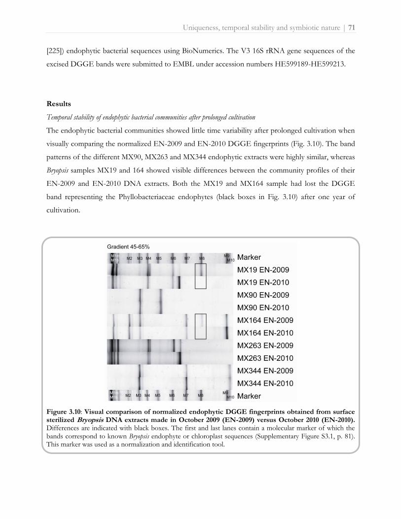
Uniqueness, temporal stability and symbiotic nature | 71
[225]) endophytic bacterial sequences using BioNumerics. The V3 16S rRNA gene sequences of the
excised DGGE bands were submitted to EMBL under accession numbers HE599189-HE599213.
Results
Temporal stability of endophytic bacterial communities after prolonged cultivation
The endophytic bacterial communities showed little time variability after prolonged cultivation when
visually comparing the normalized EN-2009 and EN-2010 DGGE fingerprints (Fig. 3.10). The band
patterns of the different MX90, MX263 and MX344 endophytic extracts were highly similar, whereas
Bryopsis samples MX19 and 164 showed visible differences between the community profiles of their
EN-2009 and EN-2010 DNA extracts. Both the MX19 and MX164 sample had lost the DGGE
band representing the Phyllobacteriaceae endophytes (black boxes in Fig. 3.10) after one year of
cultivation.
Figure 3.10: Visual comparison of normalized endophytic DGGE fingerprints obtained from surface sterilized Bryopsis DNA extracts made in October 2009 (EN-2009) versus October 2010 (EN-2010). Differences are indicated with black boxes. The first and last lanes contain a molecular marker of which the bands correspond to known Bryopsis endophyte or chloroplast sequences (Supplementary Figure S3.1, p. 81). This marker was used as a normalization and identification tool.

72 | Uniqueness, temporal stability and symbiotic nature
DGGE fingerprint cluster analysis: inside ≠ outside
DGGE cluster analysis showed that the endophytic (EN) banding patterns were significantly
different from the epiphytic (EP), washing water (WW) and cultivation water (CW) community
profiles of all five MX Bryopsis cultures studied. In the dendrogram (Fig. 3.11), the cluster containing
the EP, WW and CW community profiles is clearly separated from the endophytic banding patterns
(indicated in bold, Fig. 3.11). Also the multidimensional scaling (MDS) plot (Fig. 3.12A), which
reduces the complex DGGE patterns to one point per sample, shows that the EN samples (right)
are clearly apart from the epiphytic and surrounding water samples (left). Besides this, the MDS
diagram showed that the EN samples did not cluster together and are distributed over the y-axis of
the three-dimensional plot (Fig. 3.12A), while the EP, WW and CW samples were more or less
grouped per Bryopsis MX sample (Fig. 3.12B). Within one Bryopsis sample EP-WW-CW cluster
(clusters 1-5, Fig. 3.12B), however, no general grouping mode can be observed. Whereas the
epiphytic community samples within clusters 2, 3 and 4 (representing Bryopsis samples MX90, MX164
and MX263) were more apart from their corresponding WW and CW samples, this was not the case
for clusters 1 and 5 (i.e. Bryopsis cultures MX19 and MX344). These observations corresponded to
the results of the cluster analysis of all DGGE patterns (Fig. 3.11). In addition, Figure 3.11 also
shows a much larger diversity of DGGE bands in all epiphytic and surrounding water samples in
comparison with the endophytic DGGE profiles.
Figure 3.11: UPGMA dendrogam showing the similarities (≥ 70%) among the endophytic (EN-2009), epiphytic (EP), washing water (WW) and cultivation water (CW) normalized DGGE fingerprints. Cluster analysis was performed in BioNumerics using the band based Dice similarity coefficient with an optimization of 0.84% and a position tolerance of 0.48%. DGGE bands in the EN-2009 profiles identified as algal chloroplasts were excluded from the analysis. DGGE band patterns are graphically represented and similarity values above 70% are indicated above the branches.

Uniqueness, temporal stability and symbiotic nature | 73
Figure 3.12: Three-dimensional MDS plot seen from dimension X and Y (A) and Y and Z (B) visualizing the similarities among the endophytic (EN-2009), epiphytic (EP), washing water (WW) and cultivation water (CW) DGGE fingerprints. The MDS plot was derived from the similarity matrix generated during the DGGE cluster analysis (Fig. 3.11). Clusters 1 till 5 (B) surround the EP, WW and CW fingerprints (reduced into one point in the plot) of Bryopsis samples MX19, MX90, MX164, MX263 and MX344, respectively.
Figure 3.13: Normalized epiphytic (EP), washing water (WW) and cultivation water (CW) DGGE fingerprints obtained from Bryopsis samples MX19, MX90, MX164, MX263 and MX344. Numbers (1-27) indicate which bands were sequenced, and correspond to band numbers in Table 3.3 and Fig. 3.14. The first and last lanes contain a molecular marker of which each band (M1m, M1b, M2-M10) corresponds to a known Bryopsis endophyte or chloroplast sequence (see Supplementary Figure S3.2, p. 81). This marker was used as a normalization and identification tool.

74 | Uniqueness, temporal stability and symbiotic nature
DGGE band cluster analysis: inside ≈ outside
Although the community fingerprints of all EP, WW and CW samples were distinct from the EN
community profiles, some overlap was noticeable between individual bands from the EP, WW and
CW DGGE profiles and the EN (including chloroplast) marker bands. To examine this potential
overlap, EP, WW and CW DGGE bands at positions of marker bands (Fig. 3.13, bands 1-27) were
excised from the polyacrylamide gels and sequenced. Table 3.3 outlines the taxonomic identification
and phylogenetic affiliation of the excised bands. The last column in Table 3.3 shows the correlation
(positive+ or negative-) between the position of a certain EP, WW or CW DGGE band towards the
marker bands and its sequence identification. From this column we can deduce that most bands at
positions of marker bands M1m, M2, M8 and M10 showed sequences that matched those of the
marker bands and were thus identified as Mycoplasma, Arcobacter, Phyllobacteriaceae and Labrenzia
species, respectively. All EP, WW or CW bands at the height of Bacteroidetes (M1b), chloroplast
(M3 and M4), Flavobacteriaceae (M5-7) and Xanthomonadaceae (M9) marker bands, however,
showed a mismatch. Instead of being related to Bryopsis endophytic bacterial sequences, these latter
band sequences were affiliated with Alphaproteobacterial (Caulobacterales, Rhizobiales and
Sneathiellales), Gammaproteobacterial (Alteromonadales and Oceanospirillales) and Acanthopleuri-
bacterales sequences (see Table 3.3). To validate the true correspondence of excised EP, WW and
CW bands with endophytic sequences, band sequences were clustered with previously obtained
endophytic bacterial full length 16S rRNA gene sequences (see section 3.2.1 [225]). The UPGMA
dendrogram (Fig. 3.14) confirms that every one of the positively related bands (indicated with +) was
highly similar (≥ 99.2%) to endogenous sequences (indicated in bold). This dendrogram illustrates
that Arcobacter, Labrenzia, Mycoplasma and Phyllobacteriaceae endogenous sequences are also present
in the epiphytic, washing water and/or cultivation water bacterial communities of Bryopsis cultures,
whereas Bacteroidetes, Flavobacteriaceae and Xanthomonadaceae sequences were strictly
endogenous. In addition, Arcobacter and Mycoplasma sequences were only present in the EP, WW
and/or CW bacterial communities of those Bryopsis MX samples in which they were also
endogenously present. Labrenzia and Phyllobacteriaceae sequences, on the other hand, were also
found in the EP, WW and/or CW bacterial communities of algal samples in which these species
were not identified as being endophytic.

Table 3.3: Taxonomic identification and phylogenetic affiliation of the excised and sequenced epiphytic (EP), washing water (WW) and cultivation water (CW) DGGE bands. DGGE band number
Closest matching strain in BLAST (accession number) Query coverage/Maximum identity
Phylogenetic affiliation Correlation
MX19 EP 1 Uncultured Mycoplasma sp. clone MX19.9 (JF521606) 100/100 Tenericutes; Mollicutes; Mycoplasmatales; Mycoplasmataceae
M1m + M1b -
MX19 EP 2 Uncultured bacterium clone Del10081H12 (JF262029) 100/100 Proteobacteria; Alphaproteobacteria; Caulobacterales; Hyphomonadaceae
M4 -
MX19 EP 3 Uncultured Phyllobacteriaceae bacterium clone MX19.12 (JF521607) 100/100 Proteobacteria; Alphaproteobacteria; Rhizobiales; Phyllobacteriaceae
M8 +
MX19 EP 4 Uncultured bacterium isolate TTGE gel band N68 (JN185170) 100/100 Proteobacteria; Alphaproteobacteria; Rhizobiales; Rhizobiaceae
M9 -
MX19 EP 5 Uncultured Labrenzia sp. clone DGGE band C (HE599215) 100/100 Proteobacteria; Alphaproteobacteria; Rhodobacterales; Rhodobacteraceae
M10 +
MX90 EP 6 Uncultured bacterium clone CD02003D03 (HM768522) 100/96 Proteobacteria; Gammaproteobacteria; Alteromonadales; Alteromonadaceae
M5 -
MX90 EP 7 Uncultured Phyllobacteriaceae bacterium clone MX19.12 (JF521607) 100/100 Proteobacteria; Alphaproteobacteria; Rhizobiales; Phyllobacteriaceae
M8 +
MX90 EP 8 Uncultured alphaproteobacterium clone TH_d327 (EU272970) 100/98 Proteobacteria; Alphaproteobacteria; Rhizobiales, Hyphomicrobiaceae
M9 -
MX90 WW 9 Uncultured bacterium clone OTU017 (GU174663) 100/100 Proteobacteria; Alphaproteobacteria; Rhizobiales; Bartonellaceae
M2 -
MX164 EP 10 Uncultured Mycoplasma sp. clone MX19.9 (JF521606) 100/96 Tenericutes; Mollicutes; Mycoplasmatales; Mycoplasmataceae
M1m + M1b -
MX164 EP 11 Uncultured Arcobacter sp. clone MX164.20 (JF521610) 100/100 Proteobacteria; Epsilonproteobacteria; Campylobacterales; Campylobacteraceae
M2 +
MX164 EP 12 Uncultured proteobacterium clone Marsh_0_33 (JF980756) 100/100 Proteobacteria; Alphaproteobacteria; Caulobacterales; Hyphomonadaceae
M3 -
MX164 EP 13 Acanthopleuribacter pedis type strain NBRC 101209 (AB303221) 100/93 Acidobacteria; Holophagae; Acanthopleuribacterales
M5 -
MX164 EP 14 Hyphomicrobiaceae bacterium WPS10 (HQ638980) 100/98 Proteobacteria; Alphaproteobacteria; Rhizobiales; Bartonellaceae
M8 -
MX164 EP 15 Uncultured bacterium clone I3A_12H (EU352599) 100/98 Proteobacteria; Alphaproteobacteria; Rhizobiales; Methylobacteriaceae
M9 -

DGGE band number
Closest matching strain in BLAST (accession number) Query coverage/Maximum identity
Phylogenetic affiliation Correlation
MX164 EP 16 Stappia sp. enrichment culture clone NKiNSO2 (EU983274) 100/95 Proteobacteria; Alphaproteobacteria; Rhodobacterales;
Rhodobacteraceae
M10 -
MX164 WW 17 Uncultured Sneathiella sp. clone w-G7 (HQ727092) 100/97 Proteobacteria; Alphaproteobacteria; Sneathiellales; Sneathiellaceae
M7 -
MX263 EP 18 Thalassomonas sp. UST061013-012 (EF587959) 100/100 Proteobacteria; Gammaproteobacteria; Alteromonadales; Colwelliaceae
M7 -
MX263 EP 19 Uncultured Phyllobacteriaceae bacterium clone MX19.12 (JF521607) 100/100 Proteobacteria; Alphaproteobacteria; Rhizobiales; Phyllobacteriaceae
M8 +
MX263 EP 20 Uncultured Labrenzia sp. clone DGGE band C (HE599215) 100/100 Proteobacteria; Alphaproteobacteria; Rhodobacterales; Rhodobacteraceae
M10 +
MX263 WW 21 Uncultured Mycoplasma sp. clone MX263.1 (JF521605) 100/100 Tenericutes; Mollicutes; Mycoplasmatales; Mycoplasmataceae M1m + M1b -
MX263 CW 22 Uncultured bacterium isolate DGGE gel band B12 (HQ875697) 100/93 Proteobacteria; Gammaproteobacteria; Alteromonadales; Alteromonadaceae
M3 -
MX263 CW 23 Alcanivorax dieselolei strain PM07 (HM596594) 100/100 Proteobacteria; Gammaproteobacteria; Oceanospirillales; Alcanivoracaceae
M6 -
MX344 EP 24 Uncultured Labrenzia sp. clone DGGE band C (HE599215) 100/100 Proteobacteria; Alphaproteobacteria; Rhodobacterales; Rhodobacteraceae
M10 +
MX344 WW 25 Ruegeria mobilis strain F4122 (HQ338148) 100/99 Proteobacteria; Alphaproteobacteria; Rhodobacterales; Rhodobacteraceae
M8 -
MX344 CW 26 Uncultured bacterium clone EMar8 (FR667032) 100/94 Proteobacteria; Gammaproteobacteria; Alteromonadales
M4 -
MX344 CW 27 Uncultured bacterium clone W2-97 (HQ322761) 100/90 Proteobacteria; Alphaproteobacteria M7 -
The band numbers correspond to the numbers (1-27) in Fig 3.13. The last column shows the correlation (positive + or negative -) between the identification of a band and the sequence information of the marker band (M1m, M1b, M2-M10) at the same position.

Uniqueness, temporal stability and symbiotic nature | 77
Figure 3.14: UPGMA dendrogram showing the sequence similarities among the excised DGGE bands (numbers 1-27 in Fig. 3.13) V3 16S rRNA gene sequences and previously obtained (see section 3.2.1 [225]) endophytic bacterial full length 16S rRNA gene sequences (indicated in bold). Cluster analysis was performed in BioNumerics. Similarity values above 80% are given above the branches. The positive or negative correlation between the sequence identification of a certain excised DGGE band and its position towards the marker bands (see Table 3.3), is indicated with + or -, respectively.

78 | Uniqueness, temporal stability and symbiotic nature
Discussion
The existence of highly specific macroalgal-bacterial associations is no longer doubted [4]. Various
studies revealed that bacterial communities living on macroalgae clearly differ from those occurring
in the surrounding seawater [13, 58, 203, 226]. These studies, however, focused on the distinctiveness
of the epiphytic bacterial communities from the free-living environmental communities and never
studied the specificity of the endophytic bacteria associated with macroalgae. To our knowledge, this
is the first study to address the temporal variability of the endogenous (EN) bacterial communities of
Bryopsis isolates and their distinctiveness from the epiphytic (EP) and surrounding water (WW and
CW) bacterial communities after prolonged cultivation using the DGGE technique. Taken the
inherent limitations of the DGGE technique into account [228], we observed that the endophytic
bacterial community profiles were notably different from the fingerprints of bacterial communities
on and surrounding Bryopsis cultures. DGGE fingerprint cluster analysis (Fig. 3.11) and MDS
(Fig. 3.12) clearly indicate that the epiphytic and surrounding water samples in all Bryopsis cultures
were more similar to each other than to their corresponding endophytic community profile. This
suggests the existence of specialized endophytic bacterial communities within Bryopsis algae which are
clearly distinct from the outer surface and environmental bacterial communities. This apparent
specificity is supported by the observation that Bryopsis harbors rather stable endophytic bacterial
communities, which showed little time variability after one year cultivation of the algal samples
(Fig. 3.10). However, examination of individual DGGE bands did reveal some similarities between
intra- and extracellular bacteria. While Bacteroidetes, Flavobacteriaceae and Xanthomonadaceae
species seemed exclusively endobiotic, sequence cluster analysis confirmed that Arcobacter, Labrenzia,
Mycoplasma and Phyllobacteriaceae endophytes were also present in the epiphytic, washing water
and/or cultivation water extracts. This latter observation is consistent with the outcome of a study
conducted by Maki et al. [229] which revealed similar intracellular and extracellular bacterial
populations in and on the harmful marine microalga Heterocapsa circularisquama in culture.
Although the Bryopsis cultures used in this study have been kept in the laboratory for almost three
years due to experimental restrictions (see section 3.2.1 [225]), our data allow us to put forward some
hypotheses regarding the nature of the endophytic communities within natural Bryopsis populations.
Whereas we cannot rule out selection by artificial laboratory growth conditions, Arcobacter, Labrenzia,
Mycoplasma and Phyllobacteriaceae endophytes can at least survive without the Bryopsis host,
suggesting they might be facultative endogenous bacteria which are acquired from the local
environment. This is consistent with the general perception that most plant endophytes originate

Uniqueness, temporal stability and symbiotic nature | 79
from the surrounding environment and the outer plant surface [230, 231]. Bacteroidetes,
Flavobacteriaceae and Xanthomonadaceae endophytes, on the other hand, appear well adapted to an
endobiotic lifestyle as they persist within the Bryopsis interior after prolonged cultivation. Especially
Flavobacteriaceae endophytes, which are present in all five MX samples collected hundreds of
kilometres apart, might be obligate endophytes which are strictly dependent on the Bryopsis host for
their growth and survival. This co-occurrence of multiple facultative and obligate bacterial
endophytes is also well documented in many land plant and insect hosts [230, 232].
Furthermore, the Bryopsis endophytic communities seem also rather specific as the EP, WW and
CW extracts contained numerous Alphaproteobacterial, Gammaproteobacterial and Acanthopleuri-
bacterales species which are not present in the EN samples. This apparent specificity is confirmed by
our observations that EP, WW, CW (data not shown) and EN (see Fig. 3.10) extracts made at
different time points revealed largely consistent banding patterns even after the algal specimens were
repeatedly wounded and transferred to fresh, sterile cultivation medium (see Material and methods
section). Consequently, the Bryopsis host seems able to selectively maintain its endophytic flora
and/or to attract specific facultative endophytes after wounding. Although this may be the result of
more general physiological and biochemical processes [4], the characteristic properties of Bryopsis
might also contribute to this selectiveness. An interesting characteristic of Bryopsis is that following
cell wounding, the protoplasm can aggregate and regenerate into a mature individual. This process
involves a transient state of membrane-free protoplasts in seawater [113]. Although this transient ‘life
without a membrane’ state might seem anything but selective, Klotchkova and coworkers [233]
showed that an incompatibility barrier is present during protoplast formation to exclude foreign
inorganic particles or alien cell components. Only some chosen cells or particles could be
incorporated into Bryopsis protoplasts. Moreover, the lectins which play a key role in the aggregation
process during protoplast formation [234-237] might actually be ‘specificity mediators’. The
description of the Bryopsis specific lectin Bryohealin by Kim et al. [236], which contains an antibiotic
domain that protects the newly generated protoplasts from bacterial contamination [237], supports
this hypothesis. Lectins are known symbiosis mediators in, for example, legume-rhizobia and
sponge-bacterial symbioses [238, 239].
Besides the endophytic bacterial communities, also the epiphytic and the surrounding cultivation
water bacterial communities seemed unique to each Bryopsis culture as the EP, WW and CW
fingerprints of a given Bryopsis sample clearly clustered together. This is consistent with the general
perception of highly specific macroalgal-bacterial interactions as discussed above [4]. Additionally,

80 | Uniqueness, temporal stability and symbiotic nature
since all five Bryopsis cultures were maintained under similar laboratory conditions, the above
observation suggests that factors other than cultivation conditions contributed to the observed
specificity (see Material and methods section).
Conclusion
Our results indicate that Bryopsis samples harbor specific and rather stable endophytic bacterial
communities after prolonged cultivation which are clearly distinct from the epiphytic and
surrounding cultivation water bacterial communities. Even though Bryopsis algae are repeatedly being
exposed to a mix of marine bacteria, they seem to selectively maintain and/or attract their
endophytes after repeated wounding events in culture. Despite the limitations of the experimental
design, this indicates that Bryopsis has some intrinsic mechanisms to favour the entry of certain
bacteria of possible ecological importance within its cell, suggesting macroalgal-bacterial endobioses
might be as or even more specific than macroalgal-epiphytic bacterial associations. The use of
species-specific primers and probes may open the way to investigate the specificity, both spatially and
temporally, of the endophytic communities in natural Bryopsis populations.
Acknowledgments
This research was funded by ‘Fonds Wetenschappelijk Onderzoek’ FWO-Flanders project
G.0045.08. Myriam Claeys, Olivier Leroux and Wim Bert are gratefully acknowledged for electron
microscopy assistance. We sincerely thank Heroen Verbruggen and Lennert Tyberghein for
collecting the specimens.

Uniqueness, temporal stability and symbiotic nature | 81
Supplementary Figure S3.1: Transmission electron micrograph of vegetative Bryopsis thallus in longisection. Fig. A: the outer cytoplasmic layer (ol) adjacent to the Bryopsis cell wall (cw) contains most of the organelles excluding only the chloroplasts (chl), which are present in the inner layer next to the central vacuole (cv). Magnification: x 8000, scale bar: 3 µm. Fig. B (detail of Fig. A): besides mitochondria (m), endoplasmic reticulum and vacuolar evaginations (v), endogenous bacteria (ba) are present in the outer cytoplasmic layer. Magnification: x 25000, scale bar: 1 µm.
Supplementary Figure S3.2: The marker used as a normalization and identification tool in all DGGE analyses. This marker covers the full range of endophytic (including chloroplast) sequences previously obtained from Bryopsis samples MX19, MX90, MX164, MX263 and MX344 (see section 3.2.1 [225]). For each marker band, the band name (M1m, M1b, M2-M10), taxonomic identification, clone reference and accession number are represented.

Spatial stability, transmission modes and host/habitat influences | 83
3.2.3. Disentangling host phylogenetic, environmental and geographic signals
in intracellular bacterial communities of Bryopsis
Joke Hollants, Frederik Leliaert, Heroen Verbruggen, Anne Willems and Olivier De Clerck. Permanent
residents or temporary lodgers: characterizing intracellular bacterial communities of siphonous green algae.
Manuscript submitted to Mol Ecol. Author contributions: The first two authors have equally contributed to the
study. JH designed and performed the experiments and wrote the manuscript. FL maintained the algal
cultures, analyzed the data and outlined the figures. FL, HV and ODC collected the Bryopsis samples. FL, HV,
AW and ODC commented on the manuscript.
Abstract
The ecological success of giant celled, siphonous green algae has repeatedly been linked
to endophytic bacteria living within the cytoplasm of the hosts. Yet, virtually nothing is
known about the relative importance of evolutionary and ecological factors controlling
the intracellular bacterial flora of these seaweeds. Using the siphonous alga Bryopsis as a
model, we explore the diversity of the intracellular bacterial communities and investigate
whether their composition is controlled by ecological and biogeographical factors rather
than the evolutionary history of the host. Using a combination of 16S rDNA clone
libraries and DGGE analyses, we show that siphonous algae harbor a diverse and
complex mixture of generalist and specialist bacteria. Variation partitioning analyses
show a strong impact of local environmental factors on bacterial community
composition for generalist species, while specialists reflect a predominant imprint of
evolutionary history. The results highlight the importance of interpreting the presence
of individual bacterial phylotypes in the light of ecological and evolutionary principles
such as phylogenetic niche conservatism to understand complex endobiotic
communities and the parameters shaping them.
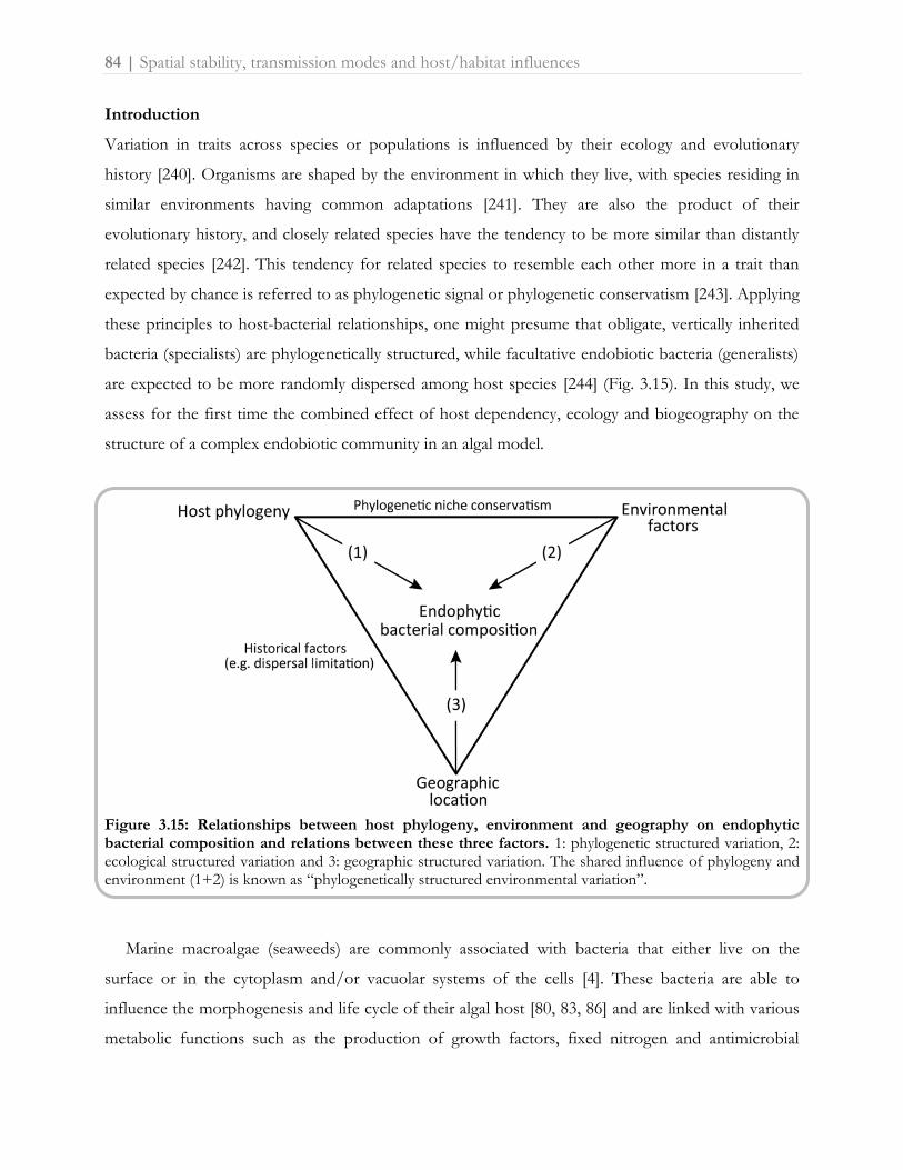
84 | Spatial stability, transmission modes and host/habitat influences
Introduction
Variation in traits across species or populations is influenced by their ecology and evolutionary
history [240]. Organisms are shaped by the environment in which they live, with species residing in
similar environments having common adaptations [241]. They are also the product of their
evolutionary history, and closely related species have the tendency to be more similar than distantly
related species [242]. This tendency for related species to resemble each other more in a trait than
expected by chance is referred to as phylogenetic signal or phylogenetic conservatism [243]. Applying
these principles to host-bacterial relationships, one might presume that obligate, vertically inherited
bacteria (specialists) are phylogenetically structured, while facultative endobiotic bacteria (generalists)
are expected to be more randomly dispersed among host species [244] (Fig. 3.15). In this study, we
assess for the first time the combined effect of host dependency, ecology and biogeography on the
structure of a complex endobiotic community in an algal model.
Figure 3.15: Relationships between host phylogeny, environment and geography on endophytic bacterial composition and relations between these three factors. 1: phylogenetic structured variation, 2: ecological structured variation and 3: geographic structured variation. The shared influence of phylogeny and environment (1+2) is known as “phylogenetically structured environmental variation”.
Marine macroalgae (seaweeds) are commonly associated with bacteria that either live on the
surface or in the cytoplasm and/or vacuolar systems of the cells [4]. These bacteria are able to
influence the morphogenesis and life cycle of their algal host [80, 83, 86] and are linked with various
metabolic functions such as the production of growth factors, fixed nitrogen and antimicrobial

Spatial stability, transmission modes and host/habitat influences | 85
compounds [54, 73, 93]. Siphonous green seaweeds, consisting of a single giant tubular cell, form a
benevolent biotic environment for endobiotic bacterial communities [39, 124]. The siphonous cells,
which range from centimeters to meters in length, typically exhibit vigorous cytoplasmic streaming to
transport organelles, photosynthates and nutrients [107]. Chisholm et al. [75] demonstrated that
siphonous algae take up nutrients from the sediment by a root-like system containing intracellular
bacteria and translocate them throughout the thallus. These cellular innovations alongside unique
mechanisms of wounding response [111, 112] and the close interactions with bacteria may provide a
physiological explanation for the successful spread of siphonous green algae in marine coastal
habitats [75, 125, 158].
Very little is known about the factors controlling the presence of bacteria inside siphonous
seaweeds. Two host-related mechanisms may affect the intracellular bacterial composition. Firstly,
siphonous seaweeds readily regenerate form protoplasts, facilitating environmental uptake of bacteria
into the cell [234]. Secondly, endogenous bacteria can persist by vertical inheritance through gametes
[36]. Beside the question of whether the endobionts are acquired vertically or from the environment,
ecological parameters and geographic aspects may also need to be considered to explain the bacterial
composition, as some bacteria (or hosts) are likely to be geographically restricted or occur only in
particular niches. Although a previous study suggested that seaweed-associated bacterial communities
are biogeographically structured [125], it is not known whether ecological or historical factors cause
this structure.
The goal of this study is to investigate the relative roles of host, environment and geography in
determining the intracellular bacterial flora of siphonous seaweeds, focusing on the genus Bryopsis as
a case study. This genus is known to harbor several types of endogenous bacteria and protocols are
in place to study them (see sections 3.1.2 and 3.2.1 [187, 225]). Bryopsis is known to possess
mechanisms for environmental uptake (see section 3.2.2 [245]) as well as vertical inheritance of
bacteria [36]. This combination of features, combined with the large collection of available cultures,
makes the genus an ideal case study to address our goal. The experimental approach consisted of
molecular characterization of host samples and their intracellular bacterial flora. The molecular
identification of bacterial phylotypes, along with the host phylogeny and environmental data, were
explored and analyzed with statistical techniques designed to disentangle the effects of host
phylogeny, geography and the external environment on the intracellular bacterial composition.

86 | Spatial stability, transmission modes and host/habitat influences
Material and methods
Algal material
The 20 Bryopsis samples analyzed in this study are listed in Supplementary Table S3.2 (p. 100) and
their sampling sites are depicted in Figure 3.16. All samples were transferred to and maintained as
unialgal cultures under the conditions described in section 3.2.1 [225].
Figure 3.16: Map of Bryopsis sampling sites. The collection sites are marked by black circles and labelled with the Bryopsis sample name. In addition to the 15 Bryopsis samples analyzed in this study, also the five Mexican Bryopsis samples MX19, MX90, MX164, MX263 and MX344, which were previously studied (see section 3.2.1 [225]), are depicted.
Molecular approach
Bryopsis samples were subjected to a surface sterilization step to eliminate epiphytic bacterial
contamination (see section 3.1.2 [187]) prior to total DNA extraction [160]. The host rbcL and
bacterial 16S rRNA genes were PCR amplified as described in section 3.2.1 [225]. The endophytic
bacterial diversity was assessed by creating 16S rRNA gene clone libraries and performing nested
PCR denaturing gradient gel electrophoresis (DGGE) analyses as described in sections 3.2.1 and
3.2.2 [225, 245]. Sequences were submitted to EMBL under accession numbers HE648924-
HE648948.

Spatial stability, transmission modes and host/habitat influences | 87
Sequence data analyses
Bryopsis rbcL and bacterial 16S rRNA gene sequences were assembled, checked for chimeras,
compared with nucleotide databases and aligned as described in section 3.2.1 [225]. Phylogenetic
trees were inferred with maximum likelihood (ML) implemented in PhyML v3.0 [195] and Bayesian
inference (BI) using MrBayes [246], via the University of Oslo Bioportal website [196]. Both analyses
were performed under a HKY+G model as determined by the Akaike Information Criterion in
JModeltest v0.1.1 [194].
Statistical analysis
The influence of environmental, geographic, and host phylogenetic factors on the endophytic
bacterial diversity in Bryopsis was analyzed using multivariate statistical and comparative phylogenetic
approaches. The response table was represented by a presence/absence matrix of the seven bacterial
phylotypes in the 20 host samples (Fig. 3.17). The three explanatory matrices (environment,
geography and phylogeny) were prepared as follows. The environmental component was represented
by seven macro-ecological variables extracted from Bio-ORACLE [247], a global environmental
dataset of satellite-based and in situ measured marine geophysical, biotic and climate information at a
final spatial resolution of 5 arcmin (9.2 km) (Supplementary Table S3.3, p. 100). The geographic
component was represented by a set of orthogonal spatial variables extracted from geographic
coordinates by Moran’s Eigenvector Maps (MEM) analysis [248] using ‘codep’ in R [249]. The
geographic matrix was represented by the first two eigenvectors, which were the only ones having
positive eigenvalues (6.54 and 1.52). The phylogenetic component was expressed as principal
coordinates via a principal coordinate analysis (PCoA) [250] computed from a distance matrix [251].
A corrected distance matrix of the Bryopsis rbcL alignment was calculated in MEGA [197]; the PCoA
analysis was performed in PCO [252]. The phylogenetic matrix was represented by the first four
principal coordinates, representing 98% of the total variation.
To study the influence of environment, geography and host phylogeny on the endophytic
bacterial diversity, we first performed data ordinations and calculated phylogenetic signals of the
bacterial community composition. Ordination of Bryopsis samples based on endophytic bacterial
community composition was performed using a principal component analysis (PCA) in CANOCO
for Windows 4.5 [253]. Environmental variables were plotted on the PCA graph as supplementary
information. Phylogenetic signal was assessed for (i) the environmental variables, (ii) geography,
(iii) the total endophytic bacterial community (i.e. represented by principal components 1 and 2
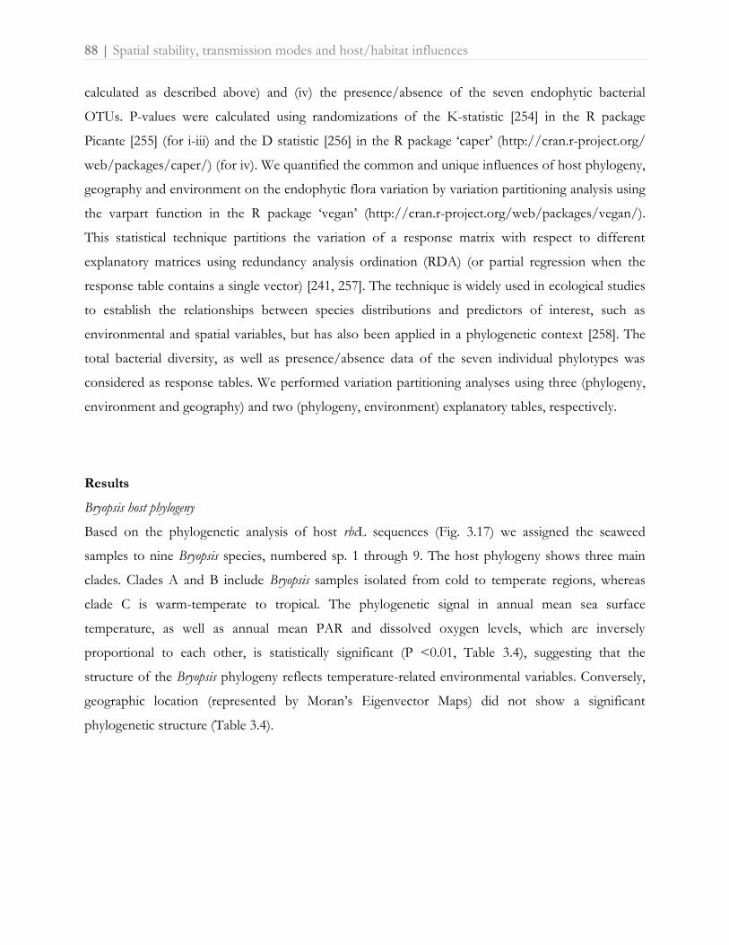
88 | Spatial stability, transmission modes and host/habitat influences
calculated as described above) and (iv) the presence/absence of the seven endophytic bacterial
OTUs. P-values were calculated using randomizations of the K-statistic [254] in the R package
Picante [255] (for i-iii) and the D statistic [256] in the R package ‘caper’ (http://cran.r-project.org/
web/packages/caper/) (for iv). We quantified the common and unique influences of host phylogeny,
geography and environment on the endophytic flora variation by variation partitioning analysis using
the varpart function in the R package ‘vegan’ (http://cran.r-project.org/web/packages/vegan/).
This statistical technique partitions the variation of a response matrix with respect to different
explanatory matrices using redundancy analysis ordination (RDA) (or partial regression when the
response table contains a single vector) [241, 257]. The technique is widely used in ecological studies
to establish the relationships between species distributions and predictors of interest, such as
environmental and spatial variables, but has also been applied in a phylogenetic context [258]. The
total bacterial diversity, as well as presence/absence data of the seven individual phylotypes was
considered as response tables. We performed variation partitioning analyses using three (phylogeny,
environment and geography) and two (phylogeny, environment) explanatory tables, respectively.
Results
Bryopsis host phylogeny
Based on the phylogenetic analysis of host rbcL sequences (Fig. 3.17) we assigned the seaweed
samples to nine Bryopsis species, numbered sp. 1 through 9. The host phylogeny shows three main
clades. Clades A and B include Bryopsis samples isolated from cold to temperate regions, whereas
clade C is warm-temperate to tropical. The phylogenetic signal in annual mean sea surface
temperature, as well as annual mean PAR and dissolved oxygen levels, which are inversely
proportional to each other, is statistically significant (P <0.01, Table 3.4), suggesting that the
structure of the Bryopsis phylogeny reflects temperature-related environmental variables. Conversely,
geographic location (represented by Moran’s Eigenvector Maps) did not show a significant
phylogenetic structure (Table 3.4).
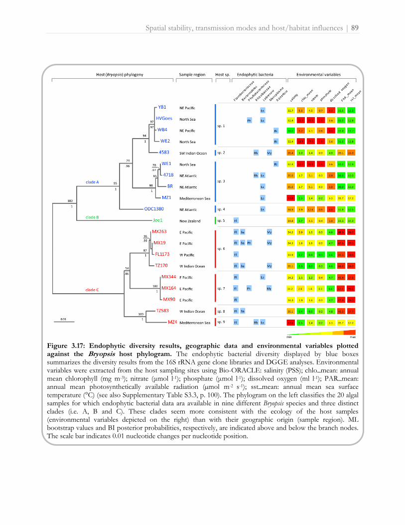
Spatial stability, transmission modes and host/habitat influences | 89
Figure 3.17: Endophytic diversity results, geographic data and environmental variables plotted against the Bryopsis host phylogram. The endophytic bacterial diversity displayed by blue boxes summarizes the diversity results from the 16S rRNA gene clone libraries and DGGE analyses. Environmental variables were extracted from the host sampling sites using Bio-ORACLE: salinity (PSS); chlo_mean: annual mean chlorophyll (mg m-3); nitrate (μmol l-1); phosphate (μmol l-1); dissolved oxygen (ml l-1); PAR_mean: annual mean photosynthetically available radiation (µmol m-2 s-1); sst_mean: annual mean sea surface temperature (°C) (see also Supplementary Table S3.3, p. 100). The phylogram on the left classifies the 20 algal samples for which endophytic bacterial data ara available in nine different Bryopsis species and three distinct clades (i.e. A, B and C). These clades seem more consistent with the ecology of the host samples (environmental variables depicted on the right) than with their geographic origin (sample region). ML bootstrap values and BI posterior probabilities, respectively, are indicated above and below the branch nodes. The scale bar indicates 0.01 nucleotide changes per nucleotide position.

90 | Spatial stability, transmission modes and host/habitat influences
Table 3.4: Phylogenetic signal values calculated for the environmental variables (Fig. 3.17), geography (Moran's eigenvector maps, MEM 1 and 2), total bacterial composition (principal components 1 and 2) (Fig. 3.18) and the presence of the seven endophytic bacterial OTUs (Fig. 3.17). P values were calculated from randomizations using Blomberg et al.'s K (K) and Fritz and Purvis’ D statistic (D). Statistical significant p-values ≤ 0.01 are indicated in bold.
Phylogenetic signal P-value
En
viro
nm
en
tal
vari
ab
les
chlo_mean 0.07 0.18 (K)
dissolved oxygen 0.16 0.00(K)
Phylogenetic signal P-value
nitrate 0.04 0.49(K)
Pre
sen
ce e
nd
op
hyti
c p
hylo
typ
es
Bacteroidetes -0.03 0.01 (D)
PAR_mean 0.73 0.00(K)
Flavobacteriaceae -0.54 0.00(D)
phosphate 0.05 0.29(K)
Labrenzia 1.16 0.67(D)
salinity 0.04 0.55(K)
Mycoplasma 0.63 0.12(D)
sst_mean 0.70 0.00(K)
Phyllobacteriaceae 1.75 0.98(D)
Geo
gra
ph
y MEM 1 0.07 0.07(K)
Rhizobiaceae 1.19 0.61(D)
MEM 2 0.08 0.20(K)
Rickettsia 1.24 0.66(D)
To
tal
bacte
rial
co
mp
osi
tio
n
PC 1 0.07 0.09(K)
PC 2 0.03 0.74(K)
Endophytic bacterial diversity
The results from the clone libraries and DGGE analyses showed the presence of seven unique
endophytic bacterial phylotypes or operational taxonomic units (OTUs) within Bryopsis (Table 3.5).
Five could be identified as Flavobacteriaceae (OTU-1), Mycoplasma (OTU-2), Bacteroidetes (OTU-3),
Phyllobacteriaceae (OTU-4) and Labrenzia (OTU-7) species, which were previously shown to occur
in Bryopsis (see section 3.2.1 [225] , Table 3.5 and Supplementary Figure S3.3 on page 101).
In addition, two new endophytic phylotypes were identified, OTU-5 and OTU-6 (Table 3.5 and
Supplementary Figure S3.3, p. 101). OTU-5 showed high sequence similarities with Rhizobiaceae
strains isolated from root nodules of leguminous plants, and represents two distinct clusters that
include Rhizobium leguminosarum and Ensifer meliloti type strains, respectively. OTU-6 is allied to

Spatial stability, transmission modes and host/habitat influences | 91
uncultured Rickettsiales bacteria associated with the coral Montastraea faveolata and the marine ciliate
Diophrys appendiculata. All OTU-6 sequences formed a distinct and well-supported clade closely related
to the genus Rickettsia and most likely represent at least a new species based on their low sequence
similarities (≤ 93%) with Rickettsia type strains.
Endophytic bacterial composition
Figure 3.17 schematizes the endophytic bacterial diversity (blue boxes) in Bryopsis. Composition of
the endophytic community varied between host species, and samples from the same host species
harbored diverse combinations of one to four different endophytic phylotypes. Different host
species with the same geographic origin commonly displayed differences in their intracellular
bacterial community composition (e.g. samples MZ1 and MZ4). This apparent lack of correlation
between total bacterial diversity and Bryopsis host species and geography is confirmed by the PCA
plot which illustrates that the ordination of the different Bryopsis species is not fully explained by their
similarity in endophytic bacterial community composition (Fig. 3.18). This PCA plot, however,
clearly indicates a correlation between the presence of individual endophytic phylotypes and certain
environmental variables. Flavobacteriaceae, Bacteroidetes and Mycoplasma endophytes were only
present in Bryopsis species isolated from tropical or warm-temperate seas, Labrenzia species were
more often found in algal samples isolated from temperate regions, and Rickettsia endophytes were
only present in Bryopsis species inhabiting seas with a low mean sea surface temperature (11.7-12.8°C)
and high chlorophyll, nitrate and phosphate levels (Figs. 3.17 and 3.18). These correlations suggest
that the distribution of individual bacterial OTUs may be more predictable than the total bacterial
community composition. Individual bacterial endophyte groups also appear to be more strongly
correlated with the host phylogeny than the overall bacterial composition. Flavobacteriaceae and
Bacteroidetes species displayed a significant phylogenetic signal (P ≤ 0.01, see Table 3.4) while
Rhizobiaceae, Phyllobacteriaceae, Mycoplasma, Rickettsia and Labrenzia species did not. Because the
host phylogeny is correlated with ecological features as a consequence of niche conservatism (see
Fig. 3.15), it is not obvious whether the latter pattern is due to ecological preferences of the
endophytic bacteria or their host.
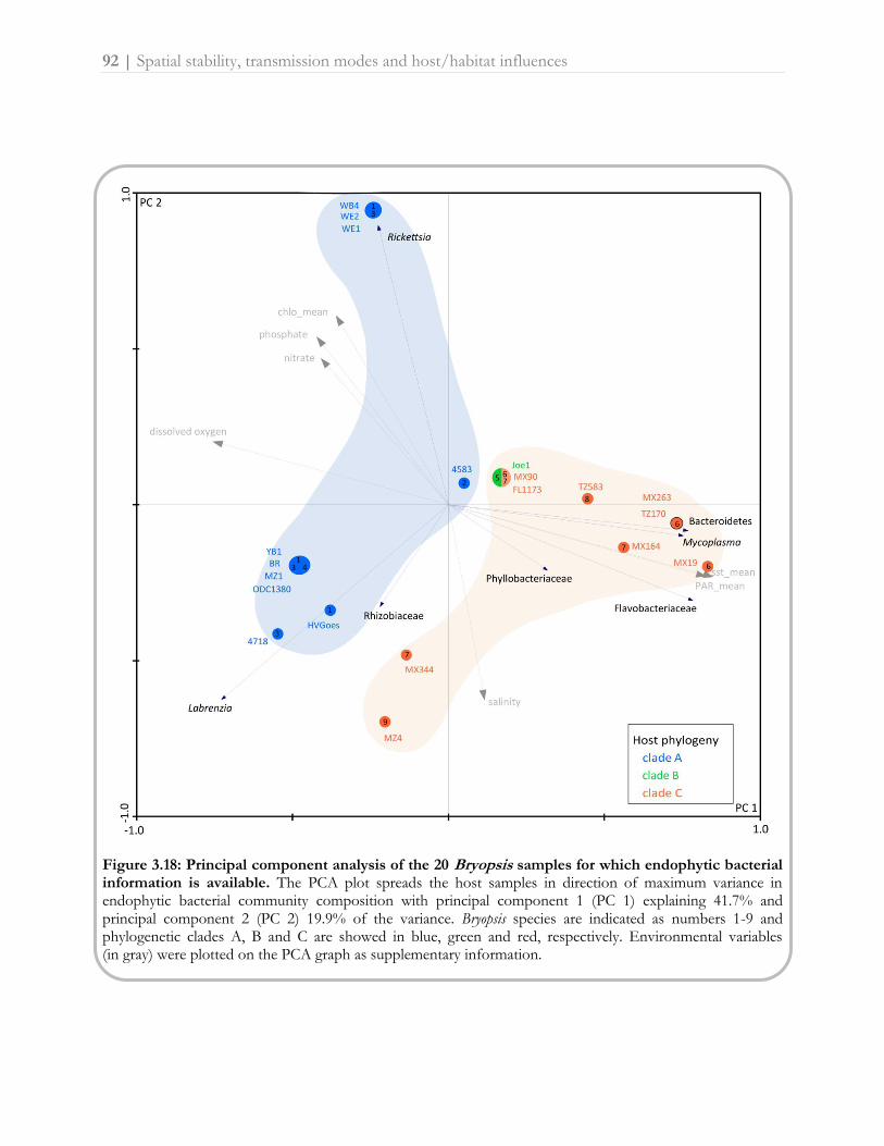
92 | Spatial stability, transmission modes and host/habitat influences
Figure 3.18: Principal component analysis of the 20 Bryopsis samples for which endophytic bacterial information is available. The PCA plot spreads the host samples in direction of maximum variance in endophytic bacterial community composition with principal component 1 (PC 1) explaining 41.7% and principal component 2 (PC 2) 19.9% of the variance. Bryopsis species are indicated as numbers 1-9 and phylogenetic clades A, B and C are showed in blue, green and red, respectively. Environmental variables (in gray) were plotted on the PCA graph as supplementary information.

Table 3.5: Taxonomic affiliation of the clones and DGGE bands representing the endophytic bacterial OTUs, sorted per Bryopsis sample.
Host 16S rRNA gene sequence analysis of bacterial clones and DGGE bands
Bryopsis sample OTU* no. OTU representative clone/DGGE band
Accession no.
Higher taxonomic ranks Closest NCBI match Accession no. (Query coverage/Maximum identity)
4583 OTU-2 DGGE band 4583a HE648924 Mollicutes, Mycoplasmatales, Mycoplasmataceae
Uncultured Mycoplasma sp. clone MX19.9 JF521606 (100/100)
OTU-5 DGGE band 4583I HE648925 Alphaproteobacteria; Rhizobiales; Rhizobiaceae
Ensifer meliloti strain RMP66 AB665549 (100/100)
OTU-5 DGGE band 4583II HE648926 Alphaproteobacteria; Rhizobiales; Rhizobiaceae
Rhizobium leguminosarum strain IPR-Pv1097 JN208903 (100/100)
4718 OTU-5 Clone 4718.68 HE648927 Alphaproteobacteria; Rhizobiales; Rhizobiaceae
Ensifer medicae WSM419 CP000738 (100/99)
OTU-7 Clone 4718.108 HE648928 Alphaproteobacteria; Rhodobacterales; Rhodobacteraceae
Uncultured bacterium clone SGUS723 FJ202588 (100/99)
BR OTU-7 Clone BR.63 HE648929 Alphaproteobacteria; Rhodobacterales; Rhodobacteraceae
Labrenzia alba strain CECT 5094 NR_042378 (100/99)
FL1173 OTU-1 DGGE band FL1173b HE648930 Bacteroidetes; Flavobacteria; Flavobacteriales Uncultured Flavobacteriaceae bacterium clone MX19.14
JF521603 (100/100)
HVGoes OTU-4 DGGE band HVGoesII HE648931 Alphaproteobacteria; Rhizobiales; Phyllobacteriaceae
Uncultured Phyllobacteriaceae bacterium clone MX164.59
JF521608 (100/100)
OTU-7 Clone HVGoes.14 HE648932 Alphaproteobacteria; Rhodobacterales; Rhodobacteraceae
Uncultured bacterium clone SGUS723 FJ202588 (100/99)
Joe1 OTU-1 Clone Joe1.40 HE648933 Bacteroidetes; Flavobacteria; Flavobacteriales Uncultured Flavobacteriaceae bacterium clone MX19.14
JF521603 (100/96)
MX19 OTU-1 Clone MX19.14 JF521603 Bacteroidetes; Flavobacteria; Flavobacteriales
Uncultured bacterium clone SHFH601 FJ203530 (99/96)
OTU-2 Clone MX19.9 JF521606 Mollicutes, Mycoplasmatales, Mycoplasmataceae
Uncultured bacterium clone GB96 GU070687 (100/97)
OTU-3 Clone MX19.8 JF521598 Bacteroidetes; unclassified Bacteroidetes
Uncultured bacterium clone Dstr_N15 GU118164 (99/94)
OTU-4 Clone MX19.12 JF521607 Alphaproteobacteria; Rhizobiales; Phyllobacteriaceae
Uncultured Rhizobiales bacterium clone PRTBB8661
HM799061 (99/99)
MX90 OTU-1 Clone MX90.40 JF521602 Bacteroidetes; Flavobacteria; Flavobacteriales
Uncultured bacterium clone SHFH601 FJ203530 (99/96)
MX164 OTU-1 Clone MX164.14 JF521600 Bacteroidetes; Flavobacteria; Flavobacteriales
Uncultured bacterium clone SHFH601 FJ203530 (99/96)
OTU-2 DGGE band MX164 B HE599214 Mollicutes, Mycoplasmatales, Mycoplasmataceae
Uncultured bacterium clone GB96 GU070687 (100/97)
OTU-4 Clone MX164.59 JF521608 Alphaproteobacteria; Rhizobiales; Phyllobacteriaceae
Phylobacteriaceae bacterium strain DG943
AY258089 (97/99)
MX263 OTU-1 Clone MX263.61 JF521604 Bacteroidetes; Flavobacteria; Flavobacteriales
Uncultured bacterium clone SHFH601 FJ203530 (99/96)
OTU-2 Clone MX263.1 JF521605 Mollicutes, Mycoplasmatales, Mycoplasmataceae
Uncultured bacterium clone GB96 GU070687 (100/97)
OTU-3 Clone MX263.73
JF521599 Bacteroidetes; unclassified Bacteroidetes
Uncultured bacterium clone Dstr_N15 GU118164 (99/94)

Host 16S rRNA gene sequence analysis of bacterial clones and DGGE bands
Bryopsis sample OTU* no. OTU representative clone/DGGE band
Accession no.
Higher taxonomic ranks Closest NCBI match Accession no. (Query coverage/Maximum identity)
MX344 OTU-1 Clone MX344.2 JF521601 Bacteroidetes; Flavobacteria; Flavobacteriales
Uncultured bacterium clone SHFH601 FJ203530 (99/96)
OTU-7 DGGE band MX344 C HE599215 Alphaproteobacteria; Rhodobacterales; Rhodobacteraceae
Labrenzia alba isolate CMS163 FR750958 (100/100)
MZ1 OTU-7 Clone MZ1.9 HE648934 Alphaproteobacteria; Rhodobacterales; Rhodobacteraceae
Labrenzia alba type strain CECT 5094T AJ878875 (100/99)
MZ4 OTU-1 Clone MZ4.22 HE648935 Bacteroidetes; Flavobacteria; Flavobacteriales Uncultured Flavobacteriaceae bacterium clone MX19.14
JF521603 (100/99)
OTU-5 Clone MZ4.102 HE648936 Alphaproteobacteria; Rhizobiales; Rhizobiaceae
Ensifer meliloti SM11 CP001830 (100/99)
OTU-5 Clone MZ4.43 HE648937 Alphaproteobacteria; Rhizobiales; Rhizobiaceae
Rhizobium leguminosarum bv. viciae strain BIHB 1160
EU730590 (100/99)
OTU-7 DGGE band MZ4 HE648938 Alphaproteobacteria; Rhodobacterales; Rhodobacteraceae
Uncultured Labrenzia sp. DGGE band MX344 C
HE599215 (100/100)
ODC1380 OTU-7 DGGE band ODC1380e HE648939 Alphaproteobacteria; Rhodobacterales; Rhodobacteraceae
Labrenzia aggregata strain KMO25 JF514325 (100/100)
TZ170 OTU-1 Clone TZ170.53 HE648940 Bacteroidetes; Flavobacteria; Flavobacteriales Uncultured Flavobacteriaceae bacterium clone MX19.14
JF521603 (100/99)
OTU-2 Clone TZ170.27 HE648941 Mollicutes, Mycoplasmatales, Mycoplasmataceae
Uncultured Mycoplasma sp. clone MX19.9 JF521606 (100/99)
OTU-3 Clone TZ170.55 HE648942 Bacteroidetes; unclassified Bacteroidetes Uncultured Bacteroidetes bacterium clone MX19.8
JF521598 (100/99)
TZ583 OTU-1 Clone TZ583.13 HE648943 Bacteroidetes; Flavobacteria; Flavobacteriales Uncultured Flavobacteriaceae bacterium clone MX19.14
JF521603 (100/99)
OTU-3 DGGE band TZ583c HE648944 Bacteroidetes; unclassified Bacteroidetes Uncultured Bacteroidetes bacterium clone MX19.8
JF521598 (100/99)
WE1 OTU-6 Clone WE1.5 HE648945 Alphaproteobacteria; Rickettsiales
Uncultured bacterium clone SHFG464 FJ203077 (99/98)
WE2 OTU-6 Clone WE2.2 HE648946 Alphaproteobacteria; Rickettsiales
Uncultured bacterium clone SHFG464 FJ203077 (99/97)
WB4 OTU-6 Clone WB4.44 HE648947 Alphaproteobacteria; Rickettsiales
Uncultured bacterium clone SHFG464 FJ203077 (99/98)
YB1 OTU-7 Clone YB1.1 HE648948 Alphaproteobacteria; Rhodobacterales; Rhodobacteraceae
Labrenzia aggregata strain 2PR58-2 EU440961 (100/99)
* OTUs were delineated at 97% sequence similarity

Spatial stability, transmission modes and host/habitat influences | 95
Host versus environmental influences
In order to disentangle the influences of different factors shaping the endophytic bacterial diversity,
we performed variation partitioning analyses. In the first set of analyses we partitioned the variation
of the bacterial diversity data with respect to the ecological, geographic and host-phylogenetic factors
into different portions: a part strictly influenced by environmental variables, a part strictly influenced
by the Bryopsis host phylogeny, a part strictly explained by geography, four parts explained by the
shared influence of these three factors, and an unexplained part of the variation. When considering
the total endophytic bacterial diversity, more or less equal parts of the variation (ca. 30%) were
explained by environmental and phylogenetic factors, while the strict influence of geography was
low; most of the variance, however, remained unexplained (Fig. 3.19A). Analyses of the seven
bacterial phylotypes separately showed that the influence of environment, phylogeny and geography
was very different between the seven phylotypes. The influence of geography was, in most cases, low
and highly correlated with environment and/or host phylogeny (Fig. 3.19A and Supplementary Table
S3.4, p. 103). For this reason, we excluded geography in a second set of analyses (Fig. 3.19B). The
independent effects of host phylogeny and environment had little influence on the presence of
Phyllobacteriaceae, Rhizobiaceae and Labrenzia phylotypes. The shared influence of host phylogeny
and environment was larger than their individual effects for these bacterial types. The occurrence of
Mycoplasma and Rickettsia species, on the other hand, was in part strictly determined by environmental
factors, whereas the distribution of Bacteroidetes could to a large extent be explained by host
phylogenetic factors only. Most of the variance in presence of these six endophytic phylotypes,
however, remained unexplained, suggesting that factors other than host phylogeny and environment
determine their occurrence within particular Bryopsis samples (Fig. 3.19). This is in contrast with the
situation for Flavobacteriaceae endophytes, whose presence could be entirely explained by host
phylogenetic factors, which partly overlapped with environmental factors.

96 | Spatial stability, transmission modes and host/habitat influences
Figure 3.19: Variation partitioning. Adjusted R2 values are given or illustrated. A. Results of the analysis with three explanatory tables: phylogeny, environment and geography. Venn diagram shows the influence of the three factors on the total bacterial diversity. Below are the variation explained by geography and the unexplained variation given for the seven bacterial phylotypes. B. Results of the analysis with two explanatory tables: phylogeny and environment. Diagrams show the unique and shared influence of both factors on the variation in total endophytic bacterial diversity and the individual endophytic phylotypes. Negative fractions (which indicate that two explanatory variables have strong and opposite effects on the dependent variable) are treated as zeros in the graphs. We refer to Supplementary Table S3.4 on page 103 for a detailed overview of the variation partitioning results.
Discussion
Community structure and variation in traits across species are the outcome of environmental,
geographical and historical factors which are clearly interwoven with each other. Bacterial
communities associated with eukaryotic hosts are influenced by similar factors which need to be
identified separately. Besides serving as baseline knowledge of the bacterial diversity occurring inside
the cells of siphonous seaweeds, our results provide insights into the various elements that
contribute to the composition of the endogenous bacterial flora of siphonous green seaweeds.

Spatial stability, transmission modes and host/habitat influences | 97
Diversity of endogenous bacteria
Besides the five bacterial phylotypes that were previously characterized in Bryopsis (Labrenzia,
Mycoplasma, Phyllobacteriaceae, Bacteroidetes and Flavobacteriaceae, see section 3.2.1 [225]), we
identified two additional phylotypes related to Rhizobiaceae and Rickettsia species. These bacteria
have been especially well studied from terrestrial habitats [202, 232], but have also been reported
from marine habitats. Rhizobiales are common epiphytes of Ulva seaweeds [13, 59, 61, 203] and have
also been isolated from the surface of kelps where they display antimicrobial activity [93].
Additionally, a Rhodopseudomonas species with the potential to fix nitrogen was isolated from the
rhizoidal cytoplasm of the siphonous green seaweed Caulerpa taxifolia [75]. We presume that also
Bryopsis hosts Rhizobiaceae species with nitrogen fixing capacities as we were able to amplify Ensifer-
like nitrogenase reductase genes (EMBL accession numbers HE649370-HE649371) from Bryopsis
samples 4718 and MZ4 by the nifH protocol described by De Meyer et al. [170]. Obligate intracellular
Rickettsia species, on the other hand, have not previously been described from macroalgae but have
been characterized through 16S rRNA gene analysis within freshwater green algae [259], marine
ciliates [260] and coral tissue [218].
Factors affecting bacterial composition
Even though each bacterial phylotype was encountered in at least three Bryopsis samples, the total
endophytic bacterial diversity per host sample showed no clear pattern. All algal samples harbored
diverse combinations of one to four endophytic phylotypes regardless of their phylogenetic
affiliation, geographic origin or macro-ecological niche. On the other hand, when the presence of
individual endophytic phylotypes rather than the total bacterial composition was analyzed, host
phylogenetic, geographic and environmental influences could be determined more clearly. These
three factors, however, are inevitably interrelated as a result of phylogenetic niche conservatism, i.e.
the tendency of closely related species to be ecologically similar [261], and historical factors such as
dispersal limitation, resulting in geographic proximity of closely related species (Fig. 3.15).
Disentangling the effects of host phylogeny, geography and environment shed light on the symbiotic
nature and transmission mode of the individual endophytic phylotypes.
The presence of endophytic Phyllobacteriaceae, Rhizobiaceae and Labrenzia phylotypes was not
separately determined by host phylogenetic, geographic and ecological factors, suggesting these
endophytes are true generalists adapted to both free-living and host associated lifestyles along with a
wide variety of environmental conditions. This is consistent with our previous observations that

98 | Spatial stability, transmission modes and host/habitat influences
Labrenzia and Phyllobacteriaceae endophytes can survive outside their Bryopsis host and are
reacquired from the local environment after repeated wounding events in culture (see section 3.2.2
[245]). Also the close phylogenetic relatedness of all three endophytic phylotypes with sequences
from free-living bacteria (Supplementary Figure S3.3, p. 101) indicates a recently initiated, facultative
relationship with the Bryopsis host. These generalist phylotypes may be selectively acquired by Bryopsis
hosts to fulfill specific metabolic requirements, such as nitrogen-fixation (Rhizobiaceae, [202]),
anoxygenic photosynthesis (Phyllobacteriaceae, see section 3.2.1 [225]) or CO-oxidation (Labrenzia,
[199]).
The occurrence of Mycoplasma and Rickettsia endophytes was to some extent strictly influenced by
environmental factors. Mycoplasma endophytes were only present in Bryopsis samples from tropical
regions, whereas Rickettsia bacteria were only found in algal samples isolated from temperate seas.
This environmental influence suggests the acquisition of habitat-specific endophytes by Bryopsis
hosts. In addition, the phylogenies of these more specialized endophytic phylotypes show a close
relatedness with symbiotic Rickettsia and Mycoplasma species isolated from the cytoplasm of the
marine ciliate Diophrys appendiculata [260] and the intestinal bacterial flora of the Bryopsis-feeding
abalone Haliotis diversicolor [221], respectively, suggesting the uptake of these endophytes could be
vector dependent. This hypothesis is likely as both endophytes belong to orders that are well-known
as obligate intracellular parasites of plants and animals [262, 263]. Also within sponge hosts,
horizontal symbiont transmission has been proposed to occur through vectors including sponge-
feeding animals [264].
The presence of Bacteroidetes species within Bryopsis was to a large degree influenced by host
phylogenetic factors, indicating that these endophytes may be vertically transmitted. This may take
place through reproduction via fragmentation of the Bryopsis thallus or by extruded protoplasts that
regenerate and develop into new Bryopsis plants [115]. Such asexual reproductive stages may act as
vehicles by which bacteria are inevitably transmitted without being exposed to strict host
phylogenetic influences only [265].
The presence of Flavobacteriaceae was found to be influenced by host phylogenetic factors only,
suggesting that these bacteria are true specialized and obligate endosymbionts, which are entirely
vertically transmitted via asexual and sexual reproductive stages [266]. This is in line with results from
culture experiments, which showed that these bacterial species are strictly dependent on the Bryopsis
host for their growth and survival (see section 3.2.2 [245]).

Spatial stability, transmission modes and host/habitat influences | 99
In conclusion, characterization of Bryopsis algae sampled worldwide revealed the presence of complex
endobiotic bacterial communities. Evaluation of host phylogenetic, geographic and ecological factors
revealed the presence of a mix of generalist and specialist bacteria. These observations, however,
were only evident when subdividing the total endophytic diversity into its individual bacterial
phylotypes, suggesting that both the whole community and individual community members need to
be considered in host-symbiont studies.
Acknowledgements
This research was funded by Research Foundation - Flanders project G.0045.08. We sincerely thank
Gayle Hansen, Lennert Tyberghein, John West and Joe Zuccarello for providing Bryopsis cultures or
collecting specimens. We acknowledge Frederik Hendrickx, Liam Revell and Guillaume Guénard for
useful comments on the statistical design. FL and HV are postdoctoral fellows of the Research
Foundation - Flanders.

100 | Spatial stability, transmission modes and host/habitat influences
Supplementary Table S3.2: Overview of the Bryopsis samples analyzed in this study, their collection sites and collection dates.
Bryopsis sample Collection site Collection date
Bryopsis 4583 Umhlanga Rocks KwaZulu Natal, South Africa August 2005
Bryopsis 4718 Roscoff, Brittany, France April 2008
Bryopsis BR Roscoff, Brittany, France July 2008
Bryopsis FL1173 Negros Oriental, Apo Island, Philippines September 2007
Bryopsis HVGoes Sas van Goes, The Netherlands June 2007
Bryopsis Joe1 Moa Dt, Wellington, New Zealand October 2008
Bryopsis MX19 Playa el Panteon, Puerto Angel, Oaxaca, Mexico February 2009
Bryopsis MX90 Mazunte Beach, Mazunte, Oaxaca, Mexico February 2009
Bryopsis MX164 Acapulco, Guerrero, Mexico February 2009
Bryopsis MX263 Playa las Gatas, Zihuatanejo, Guerrero, Mexico February 2009
Bryopsis MX344 Playa Careyero, Punta de Mita, Nayarit, Mexico February 2009
Bryopsis MZ1 and MZ4 Begur, Catalogna, Spain January 2008
Bryopsis ODC1380 Pointe de la Crèche, Boulogne, Nord-Pas-de-Calais, France April 2007
Bryopsis TZ170 N tip of peninsula, Ruvula, Mtwara, Tanzania January 2008
Bryopsis TZ583 E of lighthouse, Nungwi, Zanzibar, Tanzania February 2008
Bryopsis WB4 Willapa Bay, SW Washington, USA May 2008
Bryopsis WE1 and WE2 Wemeldinge, The Netherlands May 2008
Bryopsis YB1 Yaquina Bay, Oregon, USA May 2008
Supplementary Table S3.3: Overview of the macro-ecological variables extracted from Bio-ORACLE [247], their units, data handling and source.
Environmental variable Units Bio-ORACLE data handling Source
Annual mean chlorophyll (chlo_mean)
mg m-3 Temporal mean from monthly climatologies (2002-2009)
Satellite data from Aqua-MODIS2
Annual mean photosynthetically available radiation (PAR_mean)
µmol m-2 s-1 Temporal mean from monthly climatologies (1997-2009)
Satellite data from SeaWiFS2
Annual mean sea surface temperature (sst_mean)
°C Temporal mean from monthly climatologies (2002-2009)
Satellite data from Aqua-MODIS2
Dissolved oxygen ml l-1 DIVA1 interpolation of in situ measurements
In situ data from WOD 20093
Nitrate μmol l-1 DIVA1 interpolation of in situ measurements
In situ data from WOD 20093
Phosphate μmol l-1 DIVA1 interpolation of in situ measurements
In situ data from WOD 20093
Salinity PSS DIVA1 interpolation of in situ measurements
In situ data from WOD 20093
1 Data interpolating variational analysis [267] 2 Ocean-observing satellite sensors available at: http://oceancolor.gsfc.nasa.gov/ 3 World Ocean Database 2009 available at: http://www.nodc.noaa.gov/OC5/WOD09/pr_wod09.html
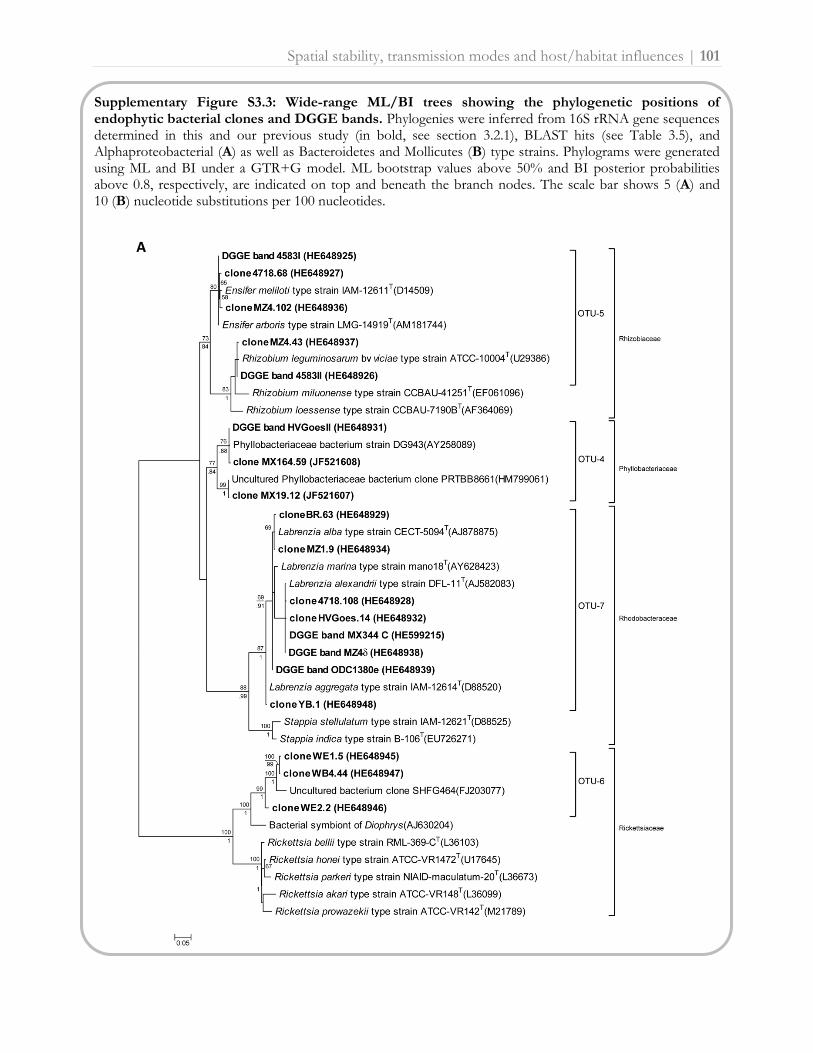
Spatial stability, transmission modes and host/habitat influences | 101
Supplementary Figure S3.3: Wide-range ML/BI trees showing the phylogenetic positions of endophytic bacterial clones and DGGE bands. Phylogenies were inferred from 16S rRNA gene sequences determined in this and our previous study (in bold, see section 3.2.1), BLAST hits (see Table 3.5), and Alphaproteobacterial (A) as well as Bacteroidetes and Mollicutes (B) type strains. Phylograms were generated using ML and BI under a GTR+G model. ML bootstrap values above 50% and BI posterior probabilities above 0.8, respectively, are indicated on top and beneath the branch nodes. The scale bar shows 5 (A) and 10 (B) nucleotide substitutions per 100 nucleotides.
A

102 | Spatial stability, transmission modes and host/habitat influences
B
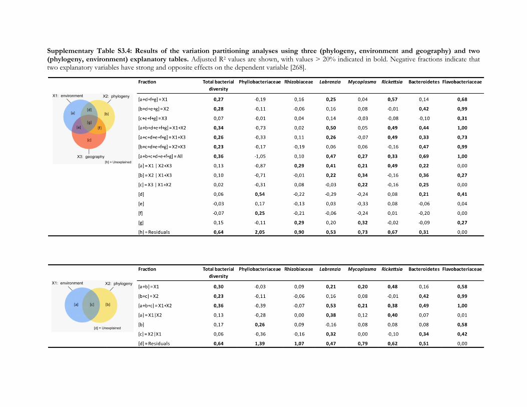
Supplementary Table S3.4: Results of the variation partitioning analyses using three (phylogeny, environment and geography) and two (phylogeny, environment) explanatory tables. Adjusted R2 values are shown, with values > 20% indicated in bold. Negative fractions indicate that two explanatory variables have strong and opposite effects on the dependent variable [268].


Interdependency of the partners | 105
3.2.4. Axenic cultivation of the Bryopsis host and in vitro isolation of
intracellular bacteria
Abstract
Interactions between different organisms can exhibit diverse forms of interdependency
among the partners. The symbiosis can be obligate, resulting in associates which are
mutually dependent for survival and can no longer be cultivated separately. In this case,
the host cannot be 'cured' of its symbionts by treatment with antibiotics without severe
consequences for its fitness and/or development, and neither can the symbionts
themselves be isolated in pure culture. Many associations among organisms, however,
have a more transient nature. In these facultative symbioses, the partners can survive
without each other and can be cultivated separately in vitro [8, 232]. Likewise, the
Bryopsis-intracellular bacterial partnership shows several interdependency modes. The
algal endophytic bacterial community consists of both generalist (e.g. Labrenzia,
Rhizobiaceae and Phyllobacteriaceae) and specialist (e.g. Flavobacteriaceae and
Bacteroidetes) species which display a facultative and more obligate endobiotic lifestyle,
respectively (see sections 3.2.2 [245] and 3.2.3). To examine the interdependency of the
Bryopsis host and the endophytic bacterial partners in more detail, several attempts were
made to culture them separately. This section describes preliminary experiments to
‘cure’ Bryopsis algae of endophytes and tryouts to culture the (facultative) endophytic
bacteria on solid and liquid media mimicking the algal host. While antibiotics seemed
not sufficient to ‘cure’ the Bryopsis host from its intracellular bacteria, cultivation
attempts in liquid media supplement with Bryopsis extract and inhibitors for gram-
positive bacteria yielded the growth of Labrenzia and Phyllobacteriaceae bacteria. In
addition, bacterial epi- and periphytes associated with Bryopsis algae which were cultured
during the cultivation tests are reported.

106 | Interdependency of the partners
Materials and methods
Axenic cultivation of the Bryopsis host
The interdependency among Bryopsis algae and bacteria as well as the physiological role of the
endophytic flora can only be determined when the host is free of epiphytic contamination and
(partially) ‘cured’ of its endophytes. Therefore, it is essential to cultivate Bryopsis axenically. A
successful surface sterilization protocol has been developed for Bryopsis cultures based on a
combined mechanical, chemical and enzymatic approach (see section 3.1.2 [187]). The utility of this
for cultivation purposes, however, is limited as non-viable algal samples are obtained at the end of
the treatment. Axenic culturing techniques that make use of antibiotics [31, 177, 178, 269], on the
other hand, might eliminate bacteria without affecting the viability of algal cells, allowing subsequent
in vitro experiments. To obtain axenic Bryopsis cultures, unialgal samples 4583, 4718, FL1173,
ODC1380 and TZ170 (see Supplementary Table S3.2, p. 100) were treated with an antibiotic mixture
following a protocol by Droop [177]. The antibiotic stock solution was prepared by adding Penicillin
(5000 µg/ml), Streptomycin (800 µg/ml), Chloramphenicol (400 µg/ml) and Kanamycin (100 µg/ml)
to 1x modified Provasoli enriched seawater (PES) [181]. This stock solution was sterilized through a
0.2 µm cellulose filter and serial diluted, i.e. 1:1, 2:1, 4:1, 8:1 and 16:1 (PES medium:antibiotic stock
solution). Bryopsis samples were treated with antibiotic dilutions for 24 hours under the culture
conditions described in section 3.1.2 [187] and subsequently transferred to fresh, sterile PES medium
for one week. Axenicity of Bryopsis cultures was tested by plating small algal filaments on PES
medium supplemented with 1% agar. Inoculated plates were incubated for two weeks under the
culture conditions described above and checked for bacterial growth and algal viability by means of a
binocular microscope. To verify the cultivation results at a molecular level, denaturing gradient gel
electrophoresis (DGGE) analyses were performed on algal extracts as described in section 3.1.2
[187].
Cultivation of Bryopsis (facultative) endophytic bacteria
Experiment 1: Plating of Bryopsis cytoplasm on solid agar media
In this experiment the cytoplasm of rinsed Bryopsis samples 4583, 4718, BR, HVGoes, MX164,
MX344 and ODC1380 (see Supplementary Table S3.2, p. 100) was isolated by centrifugation as
described by Berger & Kaever [180] and subsequently plated on four different agar media, i.e. Marine
Agar (MA, Becton Dickinson), Nutrient Agar (NA, Oxoid), Tryptone Soya Agar (TSA, Oxoid) and
Yeast Mannitol Agar (YMA), a medium proposed by Vincent [270] to isolate traditional rhizobia. All
Interdependency of the partners | 107
plates were incubated at 20°C and colonies with different morphologies were isolated and purified.
Pure cultures were cryopreserved at -80°C in broth medium supplemented with 15% glycerol.
Genomic DNA of each isolate was extracted using the alkaline lysis method following Baele et al.
[271] and these DNA extracts were subsequently subjected to DGGE analyses as described in
section 3.1.2 [187] to dereplicate the isolates and to obtain an initial identification. To achieve this
latter, DGGE analyses were performed with a known marker containing V3 16S rRNA gene
fragments of Bryopsis bacterial endophytes as a standard (see Supplementary Figure S3.2, p. 81).
DGGE banding patterns were normalized and comparatively clustered using Dice similarity
coefficients in the software package BioNumerics v5.1 (Applied Maths, Belgium). To determine the
phylogenetic position of the isolates, full length 16S rRNA gene sequences of representatives of each
cluster were amplified and sequenced as described in section 3.2.1 [225].
Experiment 2: Plating of Bryopsis cytoplasm on solid agar media mimicking the algal host
Based on the results of experiment 1, Bryopsis samples HVGoes and ODC1380 as well as the media
NA and TSA were omitted from this new experiment. Accordingly, the cytoplasm of samples 4583,
4718, BR, MX164 and MX344 was re-isolated by centrifugation as described above and plated on
MA and YMA plates supplemented with Bryopsis extract. Inoculated plates were incubated at 20°C
and bacterial isolates were screened as described above.
Experiment 3: Cultivation of Bryopsis endophytes in liquid media mimicking the algal host
To simulate the Bryopsis host environment even more and to tackle epiphytic contamination, attempts
were made to culture the bacterial endophytes in liquid media (Table 3.6) supplemented with Bryopsis
extract and a component (i.e. eosine/methylene blue or deoxycholic acid sodium salt) that inhibits
growth of Gram-positive bacteria. Since it has been shown that Bryopsis facultative bacterial
endophytes are also present in the epiphytic and surrounding cultivation water (see section 3.2.2
[245]), liquid media were inoculated with four subsamples of Bryopsis cultures 4718 and MX19 (see
Supplementary Table S3.2, p. 100): (i) cytoplasm, (ii) cytoplasm + epiphytes, (iii) cultivation water
and (iv) washing water. Subsample (i) (cytoplasm) was obtained by washing the Bryopsis thalli by
repeatedly vortexing in sterilized artificial seawater (ASW) for two minutes. Surface sterilization was
performed following a protocol modified from Burke et al. [171]. Bryopsis thalli were placed into 485
µl phosphate buffered saline (PBS) supplemented with 10 µl ethylenediaminetetraacetic acid (EDTA)
and 5 µl filter-sterilized multienzyme cleaner (ox-gall soap, Dr. Beckmann). Samples were incubated

108 | Interdependency of the partners
for 2 hours at room temperature and 80 rpm and subsequently vortexed for 2 min. Algal material
was separated from the remaining liquid, rinsed and crushed with a pestel to release the cytoplasm.
To acquire subsample (ii) (cytoplasm + epiphytes), Bryopsis thalli were washed as described above and
directly crushed. The washing water of both subsamples (i) and (ii) was collected to form subsample
(iv) (washing water). The cultivation water subsample (iii) was obtained by collecting seawater in
which Bryopsis samples were cultured for a period of one month. Of subsample (iii) and (iv) 150 ml
was centrifuged and the pellet obtained was resuspended in 4 ml ASW and used as inoculum. One
ml of each subsample was homogenized in 9 ml of all liquid media (see Table 3.6). Subsequently,
serial dilutions were made (10-1 till 10-3 for subsamples i and ii, and 10-1 till 10-5 for subsamples iii and
iv) and incubated at 20°C. Liquid cultures were screened for (endophytic) bacterial growth by
DGGE as described above and 100 µl of the ‘endophytic growth positive’ tubes was plated on solid
agar media. Inoculated plates were incubated at 20°C and bacterial isolates were screened as
described above. In addition, DGGE bands at positions of endophytic marker bands were excised,
sequenced and identified as described in section 3.2.1 [225]. To verify their true correspondence with
Bryopsis endophytes, the 16S rRNA gene sequences of the excised bands and isolates were aligned
and clustered with previously obtained endophytic bacterial sequences (see sections 3.2.1 [225] and
3.2.3) using BioNumerics.
Table 3.6: Composition of liquid cultivation media applied in experiment 3.
Medium Nutrient composition Gram-positive growth inhibitor pH
A 720 ml water
0.274 g Marine broth (Difco) 90 ml Bryopsis extract
0.324 g eosine Y 0.0526 g methylene blue
7
B 720 ml natural seawater 90 ml Bryopsis extract
0.324 g eosine Y 0.0526 g methylene blue
7
C 720 ml Bryopsis cultivation water 90 ml Bryopsis extract
0.324 g eosine Y 0.0526 g methylene blue
7
D 720 ml water 0.274 g Marine broth (Difco) 90 ml Bryopsis extract
2 g deoxycholic acid sodium salt 7

Interdependency of the partners | 109
Results and discussion
Axenic cultivation of the Bryopsis host
Only Bryopsis samples treated with the lowest antibiotic dilution (i.e. 1 part PES medium at 1 part
antibiotic stock solution) showed no bacterial growth on agar plates. Higher dilutions 2:1, 4:1, 8:1
and 16:1 were ineffective in eliminating epiphytic bacterial contamination. Microscopic observations
revealed all samples were viable after the antibiotic treatment and showed no reduction in growth
rate. However, when Bryopsis samples treated with the 1:1 antibiotic solution were screened with
DGGE, some epiphytic bacteria still seemed present on the Bryopsis surface (Fig. 3.20). The antibiotic
treatment was effective in reducing the epiphytic communities (treated sample versus untreated
control sample, Fig. 3.20), but the DGGE fingerprints of treated samples still showed several non-
endophytic DGGE bands (sample treated with antibiotics versus chemical and enzymatic sterilized
sample, Fig. 3.20). These remaining epiphytic bacterial bands (asterisks on Fig. 3.20) mainly
correspond with gammaproteobacterial, i.e. Alteromonas, Pseudoalteromas and Thalassomonas, species as
determined by sequence analysis.
Figure 3.20: Visual comparison of normalized DGGE fingerprints obtained from untreated control, treated (antibiotic treatment 1:1) as well as surface sterilized (endophytes) Bryopsis samples. Bacterial endophytes are indicated with Ba (Bacteroidetes), En (Ensifer), Fl (Flavobacteriaceae), La (Labrenzia), My (Mycoplasma) and Rh (Rhizobium). White asterisks denote bands which were excised from the polyacrylamide gel and subsequently sequenced. The black box indicates chloroplasts V3 16S rRNA gene fragments as verified by DNA sequencing.

110 | Interdependency of the partners
Significantly, none of the Bryopsis endophytes present (e.g. Ensifer, Labrenzia, Mycoplasma,
Rhizobium, Bacteroidetes and Flavobacteriaceae species) were eliminated by the antibiotic mixture
used, proving this antibiotic treatment is inadequate to ‘cure’ Bryopsis algae of their endophytic flora.
Longer exposure times, other antibiotic mixtures and concentrations, or a combination of antibiotic
use with cell wall penetrating substances might be more effective in future axenicity experiments.
Also the giant-cell morphology and special regeneration mechanisms of Bryopsis (see Chapter 1, Box
3) may offer some axenic cultivation options. Although it has been shown that these characteristics
are insufficient to generate true axenic algal cultures (section 3.1.2 [187]), a combined approach of
these features with antibiotic use might successfully eliminate epiphytes and (partially) ‘cure’ Bryopsis
algae from bacterial endophytes. Furthermore, the observation that Bryopsis cultures can lose their
Phyllobacteriaceae endophytes after one year cultivation (section 3.2.2 [245]), may hint at a potential
to ‘cure’ algal samples from endophytes by adjustments of the cultivation medium and/or serial
wounding events. Instead of axenicity experiments on Bryopsis gametophytes only, also other life
cycle phases, such as gametes, could be axenically cultivated. Wichard and colleagues (unpublished
data), for example, developed an effective protocol to obtain axenic Ulva gametes by means of a
phototaxis regulated separation between gametes and bacterial cells.
Cultivation of Bryopsis (facultative) endophytic bacteria
All cultivation experiments yielded growth of mostly Bryopsis epi- and periphytic bacteria (Fig. 3.21A-
C, Table 3.7). Only the liquid culture attempts of experiment 3 were successful in growing Labrenzia
and Phyllobacteriaceae endophytes in vitro. Of these latter bacterial species, merely Labrenzia could be
isolated in pure culture. Endophytic Labrenzia isolates were cultivated on media A, B and C (see
Table 3.6) inoculated with all types of Bryopsis subsamples and cluster together with Labrenzia
sequences previously obtained from Bryopsis samples (see section 3.2.3 and Fig. 3.22).
Phyllobacteriaceae endophytes, on the other hand, could be grown in a mixed liquid culture (medium
D) as shown by clustering of DGGE band ‘liquid culture MX19 (i) D’ with endophytic
Phyllobacteriaceae sequences in Figure 3.22, but could not be subsequently isolated on agar plates.
This suggests Phyllobacteriaceae endophytes may require some metabolites and/or signaling
molecules from other co-cultivated bacteria for their growth. Consequently, diffusion chambers [272,
273] or the augmentation of solid growth media with specific nutrients and/or cell-free extracts
derived from helper strains [274] might stimulate the monoculture of these Phyllobacteriaceae
endophytes in further experiments.

Interdependency of the partners | 111
Figure 3.21: Overview of bacterial isolates obtained from cultivation experiments on solid media (A), on solid media with Bryopsis extract (B) and on solid media after an initial liquid culture step (C). Media applied: Marine Agar (MA), Marine Agar supplemented with Bryopsis extract (MA + B), Nutrient Agar (NA), Tryptone Soya Agar (TSA), Yeast Mannitol Agar (YMA), Yeast Mannitol Agar supplemented with Bryopsis extract (YMA + B), and three newly designed media A, B and C (see Table 3.6). Inocula: (A and B) cytoplasm of Bryopsis samples 4583, 4718, BR, HVGoes, MX164, MX344 and ODC1380; and (C) subsamples of Bryopsis cultures 4718 and MX19: (i) cytoplasm, (ii) cytoplasm + epiphytes, (iii) cultivation water and (iv) washing water.

112 | Interdependency of the partners
Over all, experiment 3 allowed the most growth of Bryopsis associated bacteria (Fig. 3.21C). Not
unexpected, as liquid cultivation techniques using media with low organic matter concentrations have
been demonstrated to considerably improve the bacterial culturability [275 and references therein].
The solid media used in experiments 1 and 2 were apparently too nutrient-rich, resulting in an
overgrowth of mainly Gram-positive Bacillus species at the expense of endophytic bacterial
cultivation [274]. This drawback has been overcome in experiment 3 by the use of Gram-positive
growth inhibitors such as eosine/methylene blue and deoxycholic acid sodium salt as well as dilute
nutrient media. Nevertheless, cultivation attempts could be further elaborated and optimized in the
future to better suit the growth of Bryopsis endophytic bacteria. For example, the natural environment
of the endophytic bacteria, i.e. the host internal conditions, could be mimicked even more by adding
photosynthetic metabolites to the culture media. Watanabe et al. [276] developed such an effective
artificial medium supplemented with organic carbon and nitrogen which imitates the nutritional
conditions surrounding algae to favor the growth of photosynthate-dependent epiphytic bacteria.
The cultivation of bacteria with an intracellular life-style, however, presents a particular challenge as
it remains difficult to determine and reproduce the environmental conditions required for metabolic
activity [274].
Figure 3.22: UPGMA dendrogram of endophyte 16S rRNA gene sequences previously determined (in bold, see section 3.2.3) as well as sequenced endophytic isolates and liquid culture DGGE bands. Numbers at the branch nodes represent sequence similarity values.

Interdependency of the partners | 113
Cultivation of Bryopsis epi- and periphytic bacteria
Besides the targeted endophytic bacterial growth, cultivation experiments also yielded the growth of
Alcanivorax, Bacillus, Croceibacter, Halomonas, Knoellia, Marinobacter, Microbacterium, Nitratireductor,
Phaeobacter, Ponticoccus, Pseudoalteromonas, Roseovarius, Sphingomonas, Stappia, Thalassospira, and
undetermined Rhodobacteraceae bacterial species associated with the Bryopsis surface and/or
surroundings (Fig. 3.21A-C, Table 3.7). Most of these bacterial species have been previously isolated
from seaweed surfaces (see Chapter 1) and are known to possess cell wall degrading (e.g. Alcanivorax
[277], Bacillus [278], Halomonas [279], Marinobacter [280], Pseudoalteromonas [279] and Sphingomonas [58]),
pathogenic (Halomonas [120] and Pseudoalteromonas [120, 144]), morphogenic (Bacillus [80] and
Pseudoalteromonas [80]), growth promoting (Bacillus [74] and Pseudoalteromonas [73]) and antimicrobial
(Bacillus [29, 93, 134], Microbacterium [134], Phaeobacter [29, 90] and Pseudoalteromonas [29, 88, 93])
properties. As several of these isolates are related to up to now uncultivated clones and possibly
represent new bacterial species (Table 3.7), they may offer great opportunities for future biodiscovery
research [272].
In conclusion, it is only through the isolation of individual (facultative) symbiotic partners that a
comprehensive characterization of their interdependency and physiological properties can be
undertaken. Axenic cultivation of the host as well as the isolation of the bacterial partners in vitro may
open the way for further infection studies [281] which address the interdependency of the Bryopsis-
bacterial association and can give an insight into the uptake mode and potential function(s) of the
intracellular bacteria. In addition, even in this age of high-throughput sequencing, the establishment
of pure bacterial cultures remains essential for a full physiological and taxonomic characterization
and provides almost the only way to discover the applied potential of bacterial species [272].
Acknowledgements
This research was funded by ‘Fonds Wetenschappelijk Onderzoek’ FWO-Flanders project
G.0045.08. Vaibhav Mantri, Frederik Leliaert, Helen Decleyre and Fien Calliauw are gratefully
acknowledged for practical assistance.

Table 3.7: Taxonomic identification of cluster representative isolates obtained in all three cultivation experiments.
Isolate Closest matching strains in BLAST AN QC MI Ecology
4718 (iii) A1 t3 Halomonas sp. BCw150 FJ889581 99% 99% Arctic seawater
R-49267 Halomonas sp. BCw077 FJ889579 99% 99% Arctic seawater
Halomonas sp. Ko501 AF550585 99% 99% Basalt at Kolbeinsey Ridge
MX19 (i) D2 t1 Sphingomonas sp. SKJH-30 AY749436 100% 100% LN2 vessels for long term banking of genome resources
R-49268 Uncultured bacterium clone S25_1428 EF575084 99% 100% Site S25 near Coco's Island (Costa Rica)
Uncultured bacterium clone sb21.43 HQ904214 99% 100% Anaerobic degradation of Microcystis blooms
4718 (ii) B1 t1.2 Alcanivorax sp. TE-9 AB055207 100% 100% Sea water sampled from the Sea of Japan
R-49269 Alcanivorax dieselolei strain 2W806 AB453732 99% 100% Ago Bay (Japan)
Alcanivorax dieselolei strain N1203 AB453731 99% 100% Ago Bay (Japan)
4718(iii) C5 t5 Marinobacter alkaliphilus strain aa-11 EU652042 99% 99% Ocean sediment (hot springs close to Xiamen Sea)
R-49270 Marinobacter sp. E407-9 FJ169969 99% 99% Sediment of the South China sea
Marinobacter alkaliphilus strain 2PR56-13 EU440994 99% 99% Southwest Indian Ocean deep sea water column
4718 MA + B f Marinobacter sp. PR52-13 EU440976 99% 100% Southwest Indian Ocean deep sea water column
R-49271 Marinobacter sp. 2PR57-9 EU440985 99% 99% Southwest Indian Ocean deep sea water column
Marinobacter sp. MARC4S DQ768634 99% 99% Deep sea sediments of the Middle Atlantic Ridge
4583 MA c Uncultured Pseudoalteromonas sp. clone IerC24-21 HQ161521 100% 100% Chemotactic enrichment + carbon substrates sample
R-49272 Bacterium 4746K8-B14 HQ640932 100% 100% Cold-water coral Lophelia pertusa
Bacterium 4873K4-B11 HQ640926 100% 100% Cold-water coral Lophelia pertusa
MX164 YMA + B f Uncultured bacterium clone OTU9 AB576903 100% 99% Denitrifying PEG pellet samples
R-49273 Uncultured Stappia sp. clone MJ33 GU212810 100% 99% Ballast water at Ningbo port
Uncultured bacterium clone 15A7 FJ711763 100% 99% Tropical brackish water system
4718 (i) B1 t1 Alphaproteobacterium MBIC3993 AB025419 99% 100% Isolated from Ponape Island (western Pacific Ocean)
R-49274 Alphaproteobacterium MBIC1535 AB023435 99% 100% Isolated from Ponape Island (western Pacific Ocean)
Labrenzia aggregata strain 2PR58-2 EU440961 99% 100% Southwest Indian Ocean deep sea water column
MX19 (iii) A1 t3.2 Nitratireductor aquibiodomus strain PR57-9 EU440986 100% 99% Southwest Indian Ocean deep sea water column
R-49275 Nitratireductor aquibiodomus type strain NL21T AF534573 100% 99% Marine methanol-fed denitrification reactor
Mesorhizobium sp. TUT1018 AB098586 100% 99% Fed-batch reactor
MX19 (iv) A1 t3 Roseovarius aestuarii type strain SMK-122T EU156066 100% 95% Tidal flat of the Yellow Sea in Korea
R-49276 Uncultured marine bacterium clone BM1-F-85 FJ826183 99% 95% Sea water after diatom bloom in the Yellow Sea
Pseudoruegeria aquimaris type strain SW-255T DQ675021 100% 95% Seawater of the East Sea in Korea
4718 (iii) C5 t3 Uncultured bacterium clone A8W_70 HM057815 99% 99% Ocean water from the Yellow Sea
R-49277 Bacterium DG874 AY258075 96% 100% Gymnodinium catenatum
Bacterium DG878 AY258077 95% 100% Gymnodinium catenatum
4718 (iii) C5 t2 Uncultured Rhodobacteraceae bacterium clone xmg-9 HM116852 99% 98% Phenathrene-enriched consortium in sea water
R-49278 Phaeobacter caeruleus type strain LMG 24369T AM943630 98% 97% Marine electroactive biofilm
Leisingera aquimarina type strain LMG 24366T
AM900415 98% 97% Marine electroactive biofilm

Isolate Closest matching strains in BLAST AN QC MI Ecology
MX344 MA + B b Rhodobacteraceae bacterium CSQ-8 EF512131 95% 99% Isochrysis galbana
R-49279 Ponticoccus litoralis type strain CL-GR66 EF211829 95% 98% Coastal seawater
Uncultured alpha proteobacterium clone 06-03-31 DQ153131 100% 96% Surface biofilm in estuarine seawater
MX344 MA + B c Thalassospira sp. H94 FJ903195 100% 99% Seawater
R-49280 Thalassospira sp. H88 FJ903193 100% 99% Seawater
Thalassospira sp. MCCC 1A02060 EU440820 99% 99% Ocean seawater
4718 MA + B b Knoellia subterranea strain CCGE2276 EU867301 100% 99% Phaseolus vulgaris endophyte
R-49281 Uncultured actinobacterium clone D3E05 EU753661 98% 99% Dry stromatolite
Knoellia sp. DMZ1 HQ171909 97% 99% Soil sample in Korea
MX164 MA + B e Microbacterium schleiferi strain 2PR54-18 EU440992 99% 98% Southwest Indian Ocean deep sea water column
R-49282 Catechol-degrading Microbacterium sp. Atl-19 EF028128 99% 98% Unknown
Microbacterium lacticum strain 3388 EU714364 99% 98% Clinical specimen
MX344 MA + B h Croceibacter atlanticus type strain HTCC2559T AY163576 100% 100% Atlantic Ocean
R-49283 Uncultured Bacteroidetes bacterium clone DBS1h1 GQ984357 100% 99% Surface water in the Northern Bering Sea
Marine arctic deep-sea bacterium FI7 AJ557873 99% 99% Arctic deep sea
MX19 (i) D1 t1 Bacillus sp. 3559BRRJ JF327782 100% 99% Unknown
R-49284 Bacillus sp. BZ85 HQ588864 100% 99% Soil containing high amounts of oil and heavy metals
Bacillus arsenicus strain S8-14 EU624418 99% 99% Palk Bay sediments
MX19 (i) A2 t3 Bacillus sp. strain CCMM B645 FR695470 100% 99% Salt marsh Lower Loukkos (Larache, Morocco)
R-49285 Bacillus sp. strain CCMM B655 FR695469 100% 99% Salt marsh Lower Loukkos (Larache, Morocco)
Bacillus firmus strain MC1B-14 AY833571 100% 99% Epilithic biofilms from a subtropical rocky shore
4718 (iii) D1 t2 Bacillus sp. F15(2011) HQ323453 99% 98% Air in the Mogao Grottoes, Dunhuang, China
R-49286 Bacillus sp. 19495 AJ315063 100% 97% Mural paintings in the Servilia tomb
Bacillus sp. 19493 AJ315061 100% 97% Mural paintings in the Servilia tomb
4718 MA + B c Uncultured Firmicutes bacterium clone M0027_082 EF071363 100% 99% Human colonic mucosal biopsy
R-49287 Eubacterium sp. 11-12 EU571159 99% 99% Soil from the Amazonas, outside the city of Manaus
Uncultured bacterium clone Hg1bB9 EU236314 98% 99% Haliclona cf. Gellius sp.
BR YMA + B h Bacillus licheniformis strain AIS70 GU967451 100% 99% Clay mine
R-49288 Bacillus licheniformis strain AIS53 GU967448 100% 99% Clay mine
Bacillus licheniformis strain AIS39 GU967447 100% 99% Clay mine
AN: accession number QC: query coverage MI: maximum identity


Endophytic bacterial communities
of natural Bryopsis samples
3.3.1. Flavobacteriaceae endosymbionts within natural Bryopsis samples: host specificity and cospeciation
Joke Hollants, Frederik Leliaert, Heroen Verbruggen, Olivier De Clerck and Anne Willems. Complex pattern
of coevolution of a Flavobacteriaceae endosymbiont and its green algal host, Bryopsis. Manuscript in preparation.
Author contributions: The first two authors have equally contributed to the study. JH designed and
performed the experiments. FL analyzed the data and outlined the figures. JH and FL wrote the manuscript.
FL, HV and ODC collected the algal specimens. ODC and AW commented on the manuscript.
Abstract
The siphonous green seaweed Bryopsis harbors complex intracellular bacterial
communities, of which certain Flavobacteriaceae species form a close, obligate
association with the algal host. Culture studies have indicated a strict vertical
transmission of Flavobacteriaceae endosymbionts from one host generation to the next,
suggestive of host-symbiont cospeciation. To address this hypothesis we optimized a
PCR protocol to directly and specifically amplify Flavobacteriaceae endosymbiont 16S
rRNA gene sequences, which allowed us to screen a large number of algal samples
without the need for cultivation or surface sterilization. We analyzed 146 samples
belonging to the genus Bryopsis, and 92 additional samples belonging to the Bryopsidales
and other orders within the class Ulvophyceae. Our results indicate that the
Flavobacteriaceae endosymbionts are restricted to Bryopsis, and only occur within
specific warm-temperate and tropical clades of the genus. Comparative analyses of the
bacterial 16S rRNA and Bryopsis rbcL gene datasets show a complex host-symbiont
evolutionary association with some degree of cospeciation. Our results provide evidence
for a tight, highly specific symbiosis between the partners in which the endosymbionts
are likely to fulfill significant functions.
Part 3.3

118 | Host specificity and cospeciation
Introduction
Bacteria living within the body or cells of eukaryotes are extremely abundant and widespread [216,
230, 232]. These endosymbiotic bacteria often contribute to diverse metabolic host functions,
making their presence favorable or even essential [8]. Eventually, both the bacterial partner and the
host may lose their autonomy and become dependent on each other, resulting in an obligate
association [232, 282]. Obligate endosymbiotic bacteria have been shown to form highly host-
specific interactions that are maintained across host generations over long periods of time by vertical
transmission [283, 284]. This latter process might give rise to cospeciation, an evolutionary process
resulting in congruent host and bacterial phylogenies [285].
In seaweed-bacterial associations, coevolution has only been suggested between the red alga
Prionitis and its gall-forming Roseobacter symbionts [123]. In the siphonous green seaweed Bryopsis,
bacteria have been visualized by electron microscopy in both vegetative thalli and gametes,
suggesting a close, specific association between the algal host and bacterial endophytes [36]. Recently,
molecular results showed that geographically diverse Bryopsis samples harbor well-defined and rather
stable endophytic bacterial communities consisting of a mix of generalist and specialist species
(sections 3.2.1, 3.2.2 [225, 245] and 3.2.3). Of these bacteria, only Flavobacteriaceae symbionts
displayed an obligate endobiotic lifestyle and were never isolated from the Bryopsis surface and
surrounding seawater (section 3.2.2 [245]). The family Flavobacteriaceae is a large group of bacteria
with diverse eco-physiological characteristics [286]. They are known to decompose polysaccharides
such as agar, cellulose and carrageenan, making them key players in biotransformation and nutrient
recycling processes in the marine environment [4 and references therein, 286]. Because of these
traits, Flavobacteriaceae bacteria often inhabit seaweed surfaces where they have been shown to
possess antimicrobial [29, 93], pathogenic [94, 151, 287], and algal morphogenic as well as zoospore
settlement inducing [78-81, 86] capabilities. Many members of the family Flavobacteriaceae, like
Algibacter, Fucobacter, Maribacter, and Ulvibacter species, have been initially isolated from marine
macroalgal surfaces [4]. In addition, several intracellular bacterial symbionts of insects belong to the
family Flavobacteriaceae and were shown to affect their hosts’ reproduction [286 and references
therein]. In Bryopsis, the presence of Flavobacteriaceae was found to be highly congruent with the
host phylogeny of two warm-temperate to tropical clades (see section 3.2.3). Testing the hypothesis
of cospeciation, however, requires a rich and geographically diverse sampling. The experimental
design used previously, i.e. labor-intensive unialgal culturing, surface sterilization, clone libraries, and
DGGE analyses (see Chapter 3, Part 2 [187, 225, 245]), prevented high throughput screening of
Bryopsis associated Flavobacteriaceae endosymbionts.

Host specificity and cospeciation | 119
In this study, we aimed to assess the host specificity and evolution of Flavobacteriaceae
endosymbionts in Bryopsis. To achieve this goal we developed a PCR protocol to specifically amplify
Flavobacteriaceae endophytic sequences in non-surface sterilized, natural Bryopsis samples. To assess
the distribution of these Flavobacteriaceae endosymbionts outside Bryopsis, we also screened a large
number of Bryopsidales and other ulvophytes. Phylogenetic and statistical analyses were performed
to address the Bryopsis-Flavobacteriaceae cospeciation hypothesis.
Material and methods
Algal material
Supplementary Table S3.5 (p. 130) lists the algal samples which were screened for the presence of
Flavobacteriaceae endophytes in this study. The list contains 146 Bryopsis samples covering 23
different species and 92 additional samples of Bryopsidales (genera Avrainvillea, Boodleopsis, Caulerpa,
Chlorodesmis, Codium, Derbesia, Halimeda, Rhipilia, Tydemania and Udotea), Dasycladales (Acetabularia,
Bornetella and Neomeris), Cladophorales (Aegagropila, Anadyomene, Apjohnia, Boergesenia, Boodlea,
Chaetomorpha, Cladophora, Cladophoropsis, Dictyosphaeria, Ernodesmis, Microdictyon, Rhizoclonium,
Siphonocladus and Valonia) and Ulvales (Ulva). Algal samples were collected during different field
expeditions and clean portions of the thalli were preserved in silica-gel.
DNA extraction and PCR amplification
Algal samples were subjected to a total DNA-extraction following a CTAB protocol modified from
Doyle and Doyle [160]. To create a Bryopsis host phylogeny, chloroplast-encoded rbcL genes were
amplified as described in section 3.2.1 [225]. For the specific amplification of Flavobacteriaceae
endosymbiont 16S rRNA genes, we designed species-specific primers in Kodon v3.5 (Applied
Maths, Belgium) with as only target group full length Flavobacteriaceae 16S rRNA gene sequences
(JF521600-JF521604, HE648933, HE648935, HE648940, and HE648943) obtained in our previous
studies (sections 3.2.1 [225] and 3.2.3). However, due to the large non-target group (i.e. all other
bacterial 16S rRNA gene sequences other than Flavobacteriaceae endosymbiont sequences) during
primer design, only one suitable region (from position 690 to 720) for specific primer annealing
could be found. Consequently, we designed one species-specific primer which we subsequently used
in both the forward (F695: 5’-GGCAGGTTGCTAAGCCTTAA-3’) as well as reverse (R695:
5’-TTAAGGCTTAGCAACCTGCC-3’) direction together with the 16S rRNA gene universal

120 | Host specificity and cospeciation
primers 1492R and 27F [164], respectively. Bryopsis DNA extracts from previous studies (see sections
3.2.1 [225] and 3.2.3), with Flavobacteriaceae endosymbiont DNA present or not present, were used
as templates for the initial PCR optimization experiments. Thermocycling conditions were
investigated using gradient-PCR with the following reaction mix: 1x AmpliTaq Gold reaction buffer
(Applied Biosystems), 2.5 mM MgCl2, 0.2 mM dNTPs, 0.5 µM of each primer and 1.25 units/µl
AmpliTaq Gold DNA polymerase (Applied Biosystems). Optimized thermocycling conditions were
as follows: one cycle of 95°C for 5 min; 25 cycles of 95°C for 1 min, 59°C for 1 min, 72°C for 1 min;
one final extension cycle at 72°C for 10 min. PCR amplicons were purified using a Nucleofast 96
PCR clean up membrane system (Machery-Nagel, Germany) according to the manufacturer’s
instructions and sequenced as described in section 3.2.1 [225]. Flavobacteriaceae endosymbiont 16S
rRNA gene sequences were assembled using the BioNumerics 5.1 software (Applied Maths,
Belgium), compared with nucleotide databases via BLAST (http://blast.ncbi.nlm.nih.gov/Blast.cgi)
and chimera-checked using Bellerophon [192]. Sequences were submitted to EMBL under accession
numbers HE775438-HE775517.
Phylogenetic analyses of host and symbiont
Two alignments were created for phylogenetic analyses. The Bryopsis alignment consisted of 146 rbcL
sequences and was 1363 bp long, including 100 variable and 85 parsimony informative positions.
The 80 Flavobacteriaceae 16S rRNA gene sequences obtained from Bryopsis samples were aligned
with 15 Flavobacteriaceae type strains and closest BLAST hits using MUSCLE [193]. The resulting
alignment was 1470 bp long, including a small number of gaps, and 500 variable and 398 parsimony
informative positions.
Models of nucleotide substitution were selected using the Akaike information criterion with
JModelTest v0.1.1 [194]. Phylogenetic trees were reconstructed by maximum likelihood (ML) using
PhyML v3.0 [195], via the University of Oslo Bioportal website [196]. The Bryopsis rbcL and bacterial
16S rRNA gene alignment were analysed under a GTR + G model. Trees were visualized in Mega
4.0 [197] and annotated with Adobe® Illustrator® CS5.
Based on the resulting Bryopsis phylogram, 23 species were identified as clades of closely related
sequences that are preceded by relatively long, well supported branches [288, 289]. Phylogenetic
analysis of the Flavobacteriaceae 16S rRNA gene dataset resulted in a tree with three well supported
clades (Fig. 3.23B: A, B1 and B2). Because the internal branches of clade B2 were largely unresolved,
the genetic variation within this clade could be represented more appropriately by a network [290].
Statistical parsimony networks [291] were constructed with TCS 1.21 [292], with calculated maximum

Host specificity and cospeciation | 121
connection steps at 95% and alignment gaps treated as missing data. Sequence similarity between the
16S rRNA gene sequences was determined in BioNumerics v5.1 (Applied Maths, Belgium).
Analysis of host-symbiont co-evolution and biogeography
We used different statistical techniques to assess codivergence between Flavobacteriaceae
endosymbionts of clade B and the Bryopsis host, and to investigate to which degree the bacterial
genetic variation was geographically structured.
First, analysis of molecular variance (AMOVA) of Flavobacteriaceae was used to investigate the
percentage of variation within and between populations, which were predefined as the different host
species (Bryopsis spp. 20, 21, 22, 23, 24, 28 and B. myosuroides) or geographical regions (E Pacific,
Atlantic-Mediterranean, Indian Ocean and W Pacific). Patterns of genetic structuring among Bryopsis
species and between geographical regions were estimated using Arlequin v3.5.1.3. [293]. Population
pairwise ΦST values, a measure of population differentiation or genetic distance, were calculated
using Tamura–Nei distances. Because of small sample sizes, Bryopsis spp. 25 and 26 were excluded
from the analyses.
Results and discussion
Restricted phylogenetic distribution of Flavobacteriaceae endosymbionts
The newly designed PCR protocol was successful in amplifying Flavobacteriaceae sequences directly
from algal DNA extracts. Sequencing resulted in unambiguous electropherograms, indicating the
primer designed (i.e. F/R695) is highly specific for the targeted endosymbionts, and suggesting the
exclusive presence of one flavobacterial genotype per host plant. This allowed for screening of a
large number of algal samples without the need for culturing, surface sterilization, or molecular
cloning. Of the 146 Bryopsis samples examined, 80 displayed an amplicon on agarose gel. The 16S
rRNA gene sequences were most similar (99% BLAST similarity) to Flavobacteriaceae endosymbiont
sequences previously obtained from Bryopsis (see sections 3.2.1 [225] and 3.2.3). None of the other
Bryopsidales and Ulvophyceae algal samples yielded positive amplifications (Supplementary Table
S3.5, p. 130), indicating a strong host specificity and an intimate association of the Flavobacteriaceae
endosymbionts towards Bryopsis.

122 | Host specificity and cospeciation

Host specificity and cospeciation | 123
Figure 3.23: Flavobacteriaceae endosymbiont data (B and C) plotted on the Bryopsis host phylogram (A) and geographical distribution of Flavobacteriaceae 16S rRNA types (D). Green colored branches denote positive amplification of Flavobacteriaceae endosymbiont 16S rRNA genes within the respective algal samples. The TCS parsimony network (C) visualizes phylogentic relations among the different Flavobacteriaceae 16S rRNA gene types (numbers 1-29) and each black node represents 1 nucleotide mutation separating genotypes. Colored circles (numbers on these circles refer to sequence types) on pictures B and C indicate endosymbiont genotypes and are in picture C proportionally sized to the number of sequences (i.e. Flavobacteriaceae strains) they represent. These distributions are also represented in the pie charts (B and D) in which the numbers again correspond to the endosymbiont 16S rRNA gene types. ML bootstrap values are indicated at the branch nodes (A and B). The scale bar indicates 0.02 (A) and 0.001 (B) nucleotide changes per nucleotide position.
Mapping of the positive amplifications on the Bryopsis host phylogram revealed that
Flavobacteriaceae endosymbiont presence is restricted to two separate clades (green branches,
Fig. 3.23A): a large clade (B) containing Bryopsis species from tropical and warm-temperate regions
and a smaller clade (A) including B. vestita and B. foliosa samples from New Zealand and southern
Australia, respectively. The non-monophyly of the Bryopsis species containing Flavobacteriaceae
(although not strongly supported) either indicates that these endosymbionts were acquired twice
independently, or that these bacteria have been lost in one or more Bryopsis lineages. Regardless of
the robustness of the host phylogeny, the close relationships among the endosymbionts (Fig. 3.24)
would suggest a single acquisition of Flavobacteriaceae in Bryopsis.
Although our data suggest a preference of Flavobacteriaceae endosymbionts for high
temperatures, it is difficult to distinguish if this results from an actual temperature preference of the
bacteria or ecological preferences of the host. Host ecological preferences likely play an important
role as seaweed species distributions are known to be overwhelmingly limited by seawater
temperature regimes [294]. For Bryopsis, we previously showed (see section 3.2.3), using variation
partitioning analysis, that most of the Flavobacteriaceae endosymbiont presence variation could be
explained by host phylogenetic factors, which are inevitably interrelated with environmental factors
as a result of ‘phylogenetic niche conservatism’ [261]. This indicates that the occurrence of
Flavobacteriaceae endobionts is mainly structured by their algal host phylogeny instead of an
individual ecological preference, and is in agreement with the presumably vertical transmission of
these endosymbionts from one Bryopsis generation to the next by sexual reproductive stages
(see section 3.2.3). Niche conservatism of hosts resulting in temperature-dependent variation of
endosymbionts has been described in other eukaryotes, including sponges, squids and insects [295-
298].
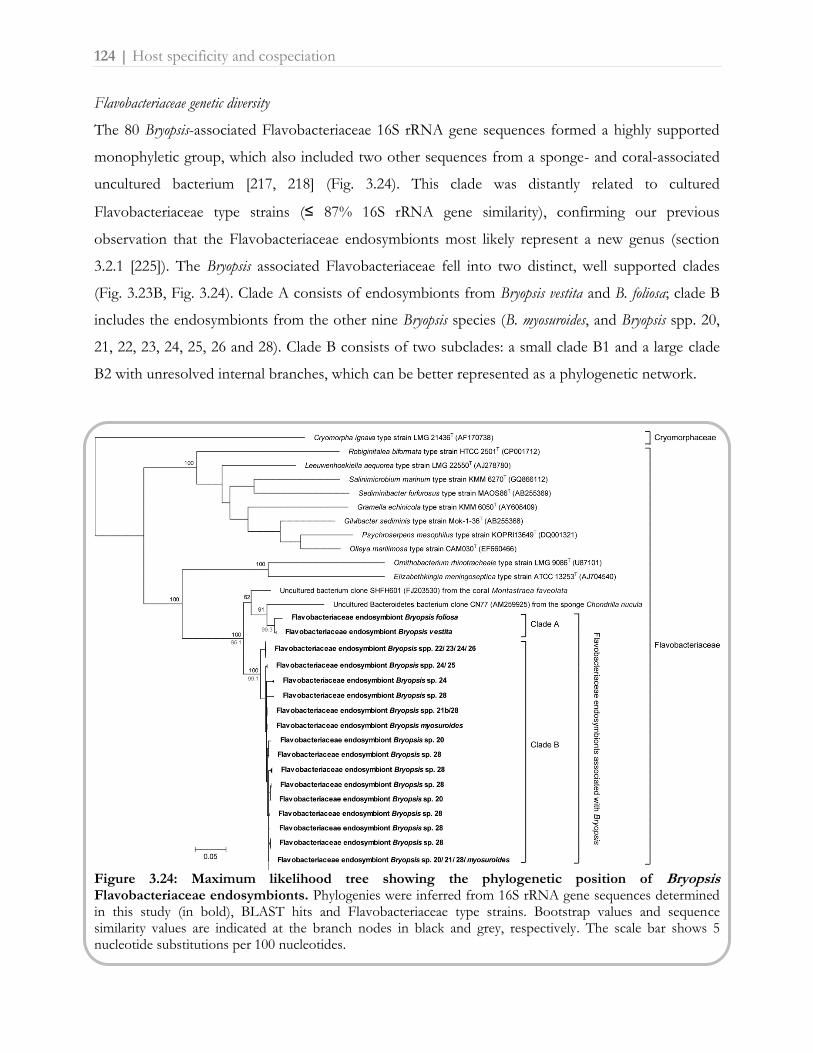
124 | Host specificity and cospeciation
Flavobacteriaceae genetic diversity
The 80 Bryopsis-associated Flavobacteriaceae 16S rRNA gene sequences formed a highly supported
monophyletic group, which also included two other sequences from a sponge- and coral-associated
uncultured bacterium [217, 218] (Fig. 3.24). This clade was distantly related to cultured
Flavobacteriaceae type strains (≤ 87% 16S rRNA gene similarity), confirming our previous
observation that the Flavobacteriaceae endosymbionts most likely represent a new genus (section
3.2.1 [225]). The Bryopsis associated Flavobacteriaceae fell into two distinct, well supported clades
(Fig. 3.23B, Fig. 3.24). Clade A consists of endosymbionts from Bryopsis vestita and B. foliosa; clade B
includes the endosymbionts from the other nine Bryopsis species (B. myosuroides, and Bryopsis spp. 20,
21, 22, 23, 24, 25, 26 and 28). Clade B consists of two subclades: a small clade B1 and a large clade
B2 with unresolved internal branches, which can be better represented as a phylogenetic network.
Figure 3.24: Maximum likelihood tree showing the phylogenetic position of Bryopsis Flavobacteriaceae endosymbionts. Phylogenies were inferred from 16S rRNA gene sequences determined in this study (in bold), BLAST hits and Flavobacteriaceae type strains. Bootstrap values and sequence similarity values are indicated at the branch nodes in black and grey, respectively. The scale bar shows 5 nucleotide substitutions per 100 nucleotides.

Host specificity and cospeciation | 125
Statistical parsimony analysis resulted in two unconnected networks, corresponding to clade A (three
16S genotypes) and B (16 genotypes). The unresolved relationships within clade B were reflected in a
highly interconnected network (Fig. 3.23C), which either result from homoplasies or may be
indicative of recombination [290] (see below). Pairwise sequence similarity of the 16S rRNA gene
sequences (1445 bp) was 99.3-99.9% within clade A, 99.1-100% within clade B, and 96.1% between
clades A and B (Fig. 3.24).
Host-symbiont coevolution and biogeography
We applied different methods for examining possible coevolution between Flavobacteriaceae
endosymbionts and Bryopsis hosts, and for assessing the role of geography on codivergence. First, a
possible correlation between endosymbiont and host genetic variation was visually explored by
comparing host and symbiont trees and by mapping the Flavobacteriaceae genotypes on the host
phylogeny (Fig. 3.23) or vice versa (Fig. 3.25A). Strict topological congruence was observed between
Bryopsis vestita and B. foliosa (clade A) and their associated endosymbionts. However, correlation
between Flavobacteriaceae of clade B and Bryopsis was less obvious for three reasons. First, several
bacterial genotypes were present in different Bryopsis hosts. For example, genotype 1 was found in
four Bryopsis species (spp. 22, 23, 24 and 26), genotype 11 was present in three species (spp. 20, 21
and 28), and genotype 7 in two species (spp. 21, 28). Secondly, most Bryopsis species contained
multiple Flavobacteriaceae genotypes, with Bryopsis sp. 28 possessing as much as 14 different
genotypes. Thirdly, relationships among Flavobacteriaceae genotypes were largely unresolved,
hampering the reconstruction of reconciled trees.
Because of these complicating factors, we applied statistical approaches that do not require a well
resolved host and symbiont phylogeny for assessing codivergence. AMOVA revealed that 57% of
the genetic variation in endosymbiont 16S rRNA gene sequences was attributable to the host species
clade divisions and subsequent permutation tests pointed out that this difference was significant
(p < 0.0001, Table 3.8A), indicating genetic differentiation between Bryopsis species. Pairwise ΦST-
values between the species are highest between more distantly related species, while genetic
differentiation was found to be insignificant between some closely related species (Table 3.8A). On
the other hand, our data indicate that genetic diversity of endosymbionts is also to a large extent
geographically structured, with most 16S genotypes being restricted to one geographical region
(Fig. 3.23D, 3.25B). This is supported by AMOVA and pairwise ΦST-values that showed significant
genetic differentiation between the East Pacific, Atlantic-Mediterranean and Indo-Pacific

126 | Host specificity and cospeciation
(Table 3.8B). This geographical signal, however, is at least partly due to dispersal limitation of the
host, resulting in confined geographical ranges for most host species. Several observations favor the
hypothesis that endosymbiont genetic diversity is primarily structured by host phylogeny. As
described above, Flavobacteriaceae endosymbionts are restricted to two Bryopsis clades (clade A and
B), irrespective of host biogeography. For example, of the five Bryopsis species from the
Mediterranean Sea, only the two species from clade A harbor Flavobacteriaceae endosymbionts
(Fig. 3.23A). A similar strict phylogenetic distribution of endosymbionts is observed for the different
Bryopsis species from Pacific Mexico, Pacific Nicaragua, South Africa and the Seychelles.
A phylogenetic rather than geographic effect on endosymbiont genetic differentiation is also
apparent when examining specific Flavobacteriaceae genotypes within Bryopsis clade B. For example,
genotype 2 is widely distributed in the Atlantic, Mediterranean and Indo-Pacific, but clearly restricted
to a single clade including Bryopsis spp. 22, 23, 24 and 26.
Figure 3.25: TCS parsimony network of 16S rRNA gene sequences of Flavobacteriaceae endosymbionts. Circles depict endosymbiont genotypes and are proportionally sized to the number of sequences (i.e. Flavobacteriaceae strains) they represent. Colors within the network correspond to (A) Bryopsis species as depicted in the host phylogram on the left and (B) geographical location of the host samples as depicted in the map on the right. Each black node represents 1 nucleotide mutation separating genotypes.

Host specificity and cospeciation | 127
Table 3.8: A: Pairwise ΦST values of Flavobacteriaceae endosymbionts between host species (clade B).
B. sp 22 B. sp 23 B. sp 24 B. myosuroides B. sp 21 B. sp 20
B. sp 22 B. sp 23 0.10
B. sp 24 0.51 0.41 B. myosuroides 0.94 0.91 0.27
B. sp 21 0.96 0.94 0.45 0.59 B. sp 20 0.92 0.88 0.27 0.32 0.03
B. sp 28 0.74 0.72 0.36 0.19 0.04 0.02
B: Pairwise ΦST values of Flavobacteriaceae endosymbionts between the four geographical regions.
Atl -Med E Pacific Indian Ocean
Atlantic-Mediterranean
E Pacific 0.66 Indian Ocean 0.45 0.26
W Pacific 0.44 0.18 0.05 Values in bold are significantly different from zero after Bonferroni correction
The observation that phylogenetically related Bryopsis species harbor the same or closely related
endosymbiont genotypes is suggestive of a vertical inheritance of Flavobacteriaceae species from one
host generation to the next, suggesting that cospeciation does occur to some extent. However, as
described above, several factors within the Bryopsis/Flavobacteriaceae clade B are obscuring this
pattern of cospeciation. The diversity of Flavobacteriaceae genotypes within a single Bryopsis species
can be explained by recent and ongoing divergence of endosymbionts. Also contact with co-
occurring facultative endophytes (see sections 3.2.1, 3.2.2 [225, 245] and 3.2.3) and/or with other
bacteria during asexual reproductive stages (i.e. thallus fragmentation and protoplast formation, see
Chapter 1, Box 3) might increase Flavobacteriaceae heterogeneity through recombination [299]. The
fact that some endosymbiont genotypes (genotypes 1, 7 and 11, Fig. 3.23) are distributed among
different Bryopsis species can be explained by two different scenarios. A first explanation might be the
persistence of ancestral Flavobacteriaceae genotypes in different host lineages (Fig. 3.26, scenario 1).
A second, alternative scenario is lateral gene transfer of Flavobacteriaceae endosymbionts between
different Bryopsis species, known as host-switching (Fig. 3.26, scenario 2). Given that the
Flavobacteriaceae endophytes have never been encountered free-living in seawater (see section 3.2.2
[245]), host-switching would require host-specific mechanisms. For example, hybridization between
closely related Bryopsis species could result in a mixing effect of the associated Flavobacteriaceae
genotypes. Hybridization in green algae is not well studied, but might be more widespread than

128 | Host specificity and cospeciation
anticipated [300]. Also sea slugs, which are known to graze on siphonous green algae, could act as
effective carriers of bacteria between different Bryopsis species [301]. The observation that Bryopsis
Flavobacteriaceae endosymbionts are related to bacterial sequences encountered in sponge and coral
hosts (see above, Fig. 3.24), might even suggest this host-switching may occur among distantly
related eukaryotes [262]. However, the fact that Flavobacteriaceae endosymbionts are
phylogenetically, rather than geographically, restricted, makes the host-switching scenario between
Bryopsis species less plausible.
Figure 3.26: Two possible scenarios for the occurrence of the same Flavobacteriaceae endosymbiont 16S rRNA gene type in different Bryopsis species.
Host specificity and cospeciation | 129
In addition, these incongruent host-symbiont coevolution patterns might also be biased by
ambiguous algal host and endosymbiont species delimitation. For example, the low level of 16S
rRNA gene sequence variability proves that this molecular marker offers limited phylogenetic
resolution at lower taxonomic scales [296]. Other markers may exist, which are evolving faster than
the 16S rRNA region, and their sequences would provide more polymorphic sites and suitable
information to assess coevolution patterns.
Despite these limitations, our results strongly point toward a tight, highly specific association
between the algal host and Flavobacteriaceae endosymbionts. The physiological ground for this
alliance, however, remains unknown from both the Bryopsis host and obligate Flavobacteriaceae
endosymbiont viewpoint. It is possible that Flavobacteriaceae endosymbionts offer the algal host
some adaption to elevated sea water temperatures. Such endosymbionts affecting host tolerance to
temperature stresses have been reported within various insect hosts [298, 302-304].
Acknowledgements
This research was funded by Research Foundation - Flanders project G.0045.08. We sincerely thank
Rob Anderson, Andrea Bernecker, Barrett Brooks, Eric Coppejans, Olivier Dargent, Kyatt Dixon,
Cindy Fernández, Fred Gurgel, Gayle Hansen, John Huisman, Wiebe Kooistra, Dian & Mark Littler,
Christine Maggs, Klaas Pauly, Claude Payri, Lennert Tyberghein, Eric Verheij, John West, Brian
Wysor and Joe Zuccarello for collecting the algal specimens. FL and HV are postdoctoral fellows of
the Research Foundation - Flanders.

Supplementary Table S3.5: Overview of algal samples (taxonomic affiliation, sample name and geographic location) that were screened for
the presence of Flavobacteriaceae endophytes.
Order Genus/species Sample name Country Geography
Bryopsidales Avrainvillea asarifolia LL0044 Belize Atlantic Ocean Bryopsidales Avrainvillea nigricans LL0005 Belize Atlantic Ocean Bryopsidales Avrainvillea silvana LL0045 Belize Atlantic Ocean Bryopsidales Boodleopsis pusilla LL0046 Belize Atlantic Ocean Bryopsidales Bryopsis corticulans HV1535 USA Pacific Ocean Bryopsidales Bryopsis corymbosa HEC4772 France Mediterranean Sea Bryopsidales Bryopsis corymbosa HV1237 Spain Mediterranean Sea Bryopsidales Bryopsis corymbosa ODCMZ1 Spain Mediterranean Sea Bryopsidales Bryopsis corymbosa ODCMZ2 Spain Mediterranean Sea Bryopsidales Bryopsis foliosa F0001 Australia Indian Ocean Bryopsidales Bryopsis foliosa F0002 Australia Indian Ocean Bryopsidales Bryopsis muscosa HV1238 Spain Mediterranean Sea Bryopsidales Bryopsis myosuroides F.0172 South Africa Indian Ocean Bryopsidales Bryopsis myosuroides F.0175 South Africa Indian Ocean Bryopsidales Bryopsis myosuroides KZN0156 South Africa Indian Ocean Bryopsidales Bryopsis myosuroides ODC1185 South Africa Indian Ocean Bryopsidales Bryopsis myosuroides ODC1186 South Africa Indian Ocean Bryopsidales Bryopsis myosuroides ODC1187a South Africa Indian Ocean Bryopsidales Bryopsis myosuroides F.0174 South Africa Indian Ocean Bryopsidales Bryopsis myosuroides KZN2318 South Africa Indian Ocean Bryopsidales Bryopsis sp. 1 F.0173 South Africa Indian Ocean Bryopsidales Bryopsis sp. 1 FL62 South Africa Atlantic Ocean Bryopsidales Bryopsis sp. 1 HEC10851 South Africa Atlantic Ocean Bryopsidales Bryopsis sp. 1 HEC10881 South Africa Atlantic Ocean Bryopsidales Bryopsis sp. 1 KZN0920 South Africa Atlantic Ocean Bryopsidales Bryopsis sp. 1 TS133 South Africa Atlantic Ocean Bryopsidales Bryopsis sp. 1 TS172 South Africa Atlantic Ocean Bryopsidales Bryopsis sp. 1 F0006 Argentina Atlantic Ocean Bryopsidales Bryopsis sp. 1 SEY477 Seychelles Indian Ocean Bryopsidales Bryopsis sp. 1 KZN931 South Africa Indian Ocean Bryopsidales Bryopsis sp. 1 SEY382 Seychelles Indian Ocean Bryopsidales Bryopsis sp. 1 Sn10839 Indonesia Pacific Ocean Bryopsidales Bryopsis sp. 1 JH001 France Atlantic Ocean Bryopsidales Bryopsis sp. 1 JH002 France Atlantic Ocean Bryopsidales Bryopsis sp. 1 JH003 France Atlantic Ocean Bryopsidales Bryopsis sp. 1 West4583 South Africa Indian Ocean Bryopsidales Bryopsis sp. 10 MX0359 Mexico Pacific Ocean Bryopsidales Bryopsis sp. 16 F.0112 Nicaragua Pacific Ocean Bryopsidales Bryopsis sp. 16 HV1559 Mexico Pacific Ocean Bryopsidales Bryopsis sp. 16 HV1757 Mexico Pacific Ocean Bryopsidales Bryopsis sp. 16 HV1779 Mexico Pacific Ocean

Order Genus/species Sample name Country Geography
Bryopsidales Bryopsis sp. 16 HV1780 Mexico Pacific Ocean Bryopsidales Bryopsis sp. 16 MX0254 Mexico Pacific Ocean Bryopsidales Bryopsis sp. 18 HEC15265 Madagascar Indian Ocean Bryopsidales Bryopsis sp. 18 KZN0800 South Africa Indian Ocean Bryopsidales Bryopsis sp. 18 ODC1187b South Africa Indian Ocean Bryopsidales Bryopsis sp. 2 EE4 (FJ715718) Netherlands Atlantic Ocean Bryopsidales Bryopsis sp. 2 HVGoes Netherlands Atlantic Ocean Bryopsidales Bryopsis sp. 2 WB3 USA Pacific Ocean Bryopsidales Bryopsis sp. 2 WB4 USA Pacific Ocean Bryopsidales Bryopsis sp. 2 WE2 Netherlands Atlantic Ocean Bryopsidales Bryopsis sp. 2 WE3 Netherlands Atlantic Ocean Bryopsidales Bryopsis sp. 2 YB2 USA Pacific Ocean Bryopsidales Bryopsis sp. 2 YB1 USA Pacific Ocean Bryopsidales Bryopsis sp. 20 F.0176 Malaysia Pacific Ocean Bryopsidales Bryopsis sp. 20 HEC14151 Tanzania Indian Ocean Bryopsidales Bryopsis sp. 20 HEC8671 Kenya Indian Ocean Bryopsidales Bryopsis sp. 20 KE1 Kenya Indian Ocean Bryopsidales Bryopsis sp. 20 HEC14796 Mauritius Indian Ocean Bryopsidales Bryopsis sp. 20 HEC16048 Sri Lanka Indian Ocean Bryopsidales Bryopsis sp. 21 FL1173 Philippines Pacific Ocean Bryopsidales Bryopsis sp. 21 HV1682 Mexico Pacific Ocean Bryopsidales Bryopsis sp. 21 HV1686 Mexico Pacific Ocean Bryopsidales Bryopsis sp. 21 MX0036 Mexico Pacific Ocean Bryopsidales Bryopsis sp. 21 MX0156 Mexico Pacific Ocean Bryopsidales Bryopsis sp. 21 MX0253 Mexico Pacific Ocean Bryopsidales Bryopsis sp. 21 MX19 (JF521594) Mexico Pacific Ocean Bryopsidales Bryopsis sp. 21b TZ170 Tanzania Indian Ocean Bryopsidales Bryopsis sp. 22 HV2122 Australia Pacific Ocean Bryopsidales Bryopsis sp. 22 MZ4 Spain Mediterranean Sea Bryopsidales Bryopsis sp. 22 ODCMZ3 Spain Mediterranean Sea Bryopsidales Bryopsis sp. 22 ODCMZA Spain Mediterranean Sea Bryopsidales Bryopsis sp. 22 HV1227 Spain Mediterranean Sea Bryopsidales Bryopsis sp. 22 HV1228 Spain Mediterranean Sea Bryopsidales Bryopsis sp. 22 HV1229 Spain Mediterranean Sea Bryopsidales Bryopsis sp. 22 HV1240 Spain Mediterranean Sea Bryopsidales Bryopsis sp. 22 HV1241 Spain Mediterranean Sea Bryopsidales Bryopsis sp. 23 HV967 USA Atlantic Ocean Bryopsidales Bryopsis sp. 23 HV968 USA Atlantic Ocean Bryopsidales Bryopsis sp. 24 HEC9474 Kenya Indian Ocean Bryopsidales Bryopsis sp. 24 HEC10690 Tanzania Indian Ocean Bryopsidales Bryopsis sp. 24 HEC11314 Tanzania Indian Ocean Bryopsidales Bryopsis sp. 24 HEC12192 Tanzania Indian Ocean

Order Genus/species Sample name Country Geography
Bryopsidales Bryopsis sp. 24 HEC14026 Tanzania Indian Ocean Bryopsidales Bryopsis sp. 24 HEC14932 Madagascar Indian Ocean Bryopsidales Bryopsis sp. 24 TZ583 Tanzania Indian Ocean Bryopsidales Bryopsis sp. 24 ODC679 Tanzania Indian Ocean Bryopsidales Bryopsis sp. 25 HV1983 Japan Pacific Ocean Bryopsidales Bryopsis sp. 26 HEC9417 Kenya Indian Ocean Bryopsidales Bryopsis sp. 26 HEC14026 Tanzania Indian Ocean Bryopsidales Bryopsis sp. 28 F.0096 New Caledonia Pacific Ocean Bryopsidales Bryopsis sp. 28 F.0097 New Caledonia Pacific Ocean Bryopsidales Bryopsis sp. 28 F.0104 Nicaragua Pacific Ocean Bryopsidales Bryopsis sp. 28 F.0105 Costa Rica Pacific Ocean Bryopsidales Bryopsis sp. 28 F.0107 Costa Rica Pacific Ocean Bryopsidales Bryopsis sp. 28 F.0108 Costa Rica Pacific Ocean Bryopsidales Bryopsis sp. 28 F.0109 Nicaragua Pacific Ocean Bryopsidales Bryopsis sp. 28 F.0110 Nicaragua Pacific Ocean Bryopsidales Bryopsis sp. 28 F.0111 Nicaragua Pacific Ocean Bryopsidales Bryopsis sp. 28 F.0113 Nicaragua Pacific Ocean Bryopsidales Bryopsis sp. 28 F.0114 Nicaragua Pacific Ocean Bryopsidales Bryopsis sp. 28 F.0115 Panama Pacific Ocean Bryopsidales Bryopsis sp. 28 F.0116 Panama Pacific Ocean Bryopsidales Bryopsis sp. 28 F.0117 Panama Pacific Ocean Bryopsidales Bryopsis sp. 28 F.0119 Panama Pacific Ocean Bryopsidales Bryopsis sp. 28 F.0120 Panama Pacific Ocean Bryopsidales Bryopsis sp. 28 HEC10527 Tanzania Indian Ocean Bryopsidales Bryopsis sp. 28 HEC10657 Tanzania Indian Ocean Bryopsidales Bryopsis sp. 28 HEC11198 Tanzania Indian Ocean Bryopsidales Bryopsis sp. 28 HEC6728 Kenya Indian Ocean Bryopsidales Bryopsis sp. 28 HEC9490 Kenya Indian Ocean Bryopsidales Bryopsis sp. 28 HEC9510 Kenya Indian Ocean Bryopsidales Bryopsis sp. 28 HOD-RUN98-33 Reunion Indian Ocean Bryopsidales Bryopsis sp. 28 HOD-RUN98-34 Reunion Indian Ocean Bryopsidales Bryopsis sp. 28 HV1609 Mexico Pacific Ocean Bryopsidales Bryopsis sp. 28 HV1614 Mexico Pacific Ocean Bryopsidales Bryopsis sp. 28 HEC14609b Mauritius Indian Ocean Bryopsidales Bryopsis sp. 28 ODC1747 Philippines Pacific Ocean Bryopsidales Bryopsis sp. 28 MX0086 Mexico Pacific Ocean Bryopsidales Bryopsis sp. 28 MX0314 Mexico Pacific Ocean Bryopsidales Bryopsis sp. 28 MX164 (JF521593) Mexico Pacific Ocean Bryopsidales Bryopsis sp. 28 MX344 (JF521596) Mexico Pacific Ocean Bryopsidales Bryopsis sp. 28 HEC12942 Tanzania Indian Ocean Bryopsidales Bryopsis sp. 28 SEY323 Seychelles Indian Ocean Bryopsidales Bryopsis sp. 28 HV566 Philippines Pacific Ocean Bryopsidales Bryopsis sp. 28 PH167 Philippines Pacific Ocean

Order Genus/species Sample name Country Geography
Bryopsidales Bryopsis sp. 28 PH222 Philippines Pacific Ocean Bryopsidales Bryopsis sp. 28 HV679 Philippines Pacific Ocean Bryopsidales Bryopsis sp. 28 SEY357 Seychelles Indian Ocean Bryopsidales Bryopsis sp. 28 TZ0053 Tanzania Indian Ocean Bryopsidales Bryopsis sp. 28 TZ0088 Tanzania Indian Ocean Bryopsidales Bryopsis sp. 3 HV880 (FJ432637) France Atlantic Ocean Bryopsidales Bryopsis sp. 3 ODC1380 France Atlantic Ocean Bryopsidales Bryopsis sp. 4b BR France Atlantic Ocean Bryopsidales Bryopsis sp. 4c BY Netherlands Atlantic Ocean Bryopsidales Bryopsis sp. 4c HV1340 Spain Atlantic Ocean Bryopsidales Bryopsis sp. 4c HV1341 Spain Atlantic Ocean Bryopsidales Bryopsis sp. 4c HV1370 Spain Atlantic Ocean Bryopsidales Bryopsis sp. 4c WE1 Netherlands Atlantic Ocean Bryopsidales Bryopsis sp. 4c West4718 France Atlantic Ocean Bryopsidales Bryopsis sp. 5 HV1388 France Atlantic Ocean Bryopsidales Bryopsis sp. 9 HEC1637 France Mediterranean Sea Bryopsidales Bryopsis sp. 9 JH021 France Mediterranean Sea Bryopsidales Bryopsis sp. 9 JH022 France Mediterranean Sea Bryopsidales Bryopsis sp. 9 JH023 France Mediterranean Sea Bryopsidales Bryopsis sp. 9 JH025 France Mediterranean Sea Bryopsidales Bryopsis vestita Joe1 New Zealand Pacific Ocean Bryopsidales Bryopsis vestita Joe2 New Zealand Pacific Ocean Bryopsidales Bryopsis vestita Joe3 New Zealand Pacific Ocean Bryopsidales Bryopsis vestita F0082b New Zealand Pacific Ocean Bryopsidales Caulerpa cupressoides MX0382 Mexico Pacific Ocean Bryopsidales Caulerpa mexicana LL0104 Bahamas Atlantic Ocean Bryopsidales Caulerpa peltata HV2030 Japan Pacific Ocean Bryopsidales Caulerpa prolifera LL0113 Belize Atlantic Ocean Bryopsidales Caulerpa racemosa MX0174 Mexico Pacific Ocean Bryopsidales Caulerpa racemosa LL0118 Martinique Atlantic Ocean Bryopsidales Caulerpa serrulata LL0010 Fiji Pacific Ocean Bryopsidales Caulerpa sertularioides MX0316 Mexico Pacific Ocean Bryopsidales Caulerpa taxifolia LL0131 Fiji Pacific Ocean Bryopsidales Chlorodesmis sp. HV1774 Sri Lanka Indian Ocean Bryopsidales Chlorodesmis sp. MX0081 Mexico Pacific Ocean Bryopsidales Codium arabicum TZ0517 Tanzania Indian Ocean Bryopsidales Codium decorticatum G.371 Brazil Atlantic Ocean Bryopsidales Codium duthiae HEC10919 South Africa Atlantic Ocean Bryopsidales Codium dwarkense TZ0818 Tanzania Indian Ocean Bryopsidales Codium fragile HEC1554 South Africa Atlantic Ocean Bryopsidales Codium fragile HV1099 USA Atlantic Ocean Bryopsidales Codium fragile HV1392 France Atlantic Ocean Bryopsidales Codium fragile HV1786 Mexico Pacific Ocean

Order Genus/species Sample name Country Geography
Bryopsidales Codium geppiorum TZ0370 Tanzania Indian Ocean Bryopsidales Codium isabelae G.083 Panama Pacific Ocean Bryopsidales Codium spongiosum HV2489 Australia Indian Ocean Bryopsidales Derbesia sp. HV1600 Mexico Pacific Ocean Bryopsidales Derbesia sp. HV1448 Netherlands Atlantic Ocean Bryopsidales Derbesia sp. TZ0612 Tanzania Indian Ocean Bryopsidales Derbesia sp. HV1079 Netherlands Atlantic Ocean Bryopsidales Derbesia sp. MX0021 Mexico Pacific Ocean Bryopsidales Halimeda borneensis W0168 Micronesia Pacific Ocean Bryopsidales Halimeda copiosa LL0417 Belize Atlantic Ocean Bryopsidales Halimeda cuneata G.905 South Africa Indian Ocean Bryopsidales Halimeda discoidea LL0020 Panama Pacific Ocean Bryopsidales Halimeda distorta H.0097 British Indian Ocean Territory Indian Ocean Bryopsidales Halimeda gigas W0162 Micronesia Pacific Ocean Bryopsidales Halimeda gigas HA0238 Australia Pacific Ocean Bryopsidales Halimeda macroloba LPT0034 Thailand Pacific Ocean Bryopsidales Halimeda opuntia LL0459 Panama Atlantic Ocean Bryopsidales Halimeda opuntia HA0373 Australia Pacific Ocean Bryopsidales Halimeda opuntia LPT0030 Thailand Indian Ocean Bryopsidales Halimeda tuna H.0086 British Indian Ocean Territory Indian Ocean Bryopsidales Halimeda tuna HV889 Spain Mediterranean Sea Bryopsidales Rhipilia orientalis HEC10402 Papua New Guinea Indian Ocean Bryopsidales Tydemania expeditionis HV873 Philippines Pacific Ocean Bryopsidales Udotea unistratea LL0051 Belize Atlantic Ocean Cladophorales Aegagropila sp. Aeg 1 probably Ukraine Freshwater, aquarium trade Cladophorales Anadyomene sp. FL1113 Philippines Pacific Ocean Cladophorales Anadyomene sp. TZ0177 Tanzania Indian Ocean Cladophorales Apjohnia laetevirens HV2291 Australia Pacific Ocean Cladophorales Apjohnia laetevirens HV2342 Australia Pacific Ocean Cladophorales Boergesenia forbesii Boerg1 Seychelles Indian Ocean Cladophorales Boergesenia forbesii FL1114 Philippines Pacific Ocean Cladophorales Boergesenia sp. JAP073 Japan Pacific Ocean Cladophorales Boodlea sp. Huisman nov2006 sn Australia Indian Ocean Cladophorales Boodlea sp. FL1110 Philippines Pacific Ocean Cladophorales Boodlea sp. TZ0147 Tanzania Indian Ocean Cladophorales Chaetomorpha antennina MX275.branched_culture Mexico Pacific Ocean Cladophorales Chaetomorpha crassa FL1132 Philippines Pacific Ocean Cladophorales Chaetomorpha crassa ODC1640 Kenya Indian Ocean Cladophorales Chaetomorpha sp. FL1092 Philippines Pacific Ocean Cladophorales Chaetomorpha sp. TZ0877 Tanzania Indian Ocean Cladophorales Cladophora vagabunda HEC15734 Madeira Atlantic Ocean Cladophorales Cladophora vagabunda Bernecker 73493 Costa Rica Pacific Ocean

Order Genus/species Sample name Country Geography
Cladophorales Cladophora vagabunda TZ0203 Tanzania Indian Ocean Cladophorales Cladophoropsis vaucheriiformis TZ0826 Tanzania Indian Ocean Cladophorales Cladophoropsis vaucheriiformis HEC7547 Papua New Guinea Pacific Ocean Cladophorales Cladophoropsis vaucheriiformis HEC10097 Japan Pacific Ocean Cladophorales Dictyosphaeria cavernosa FL1091 Philippines Pacific Ocean Cladophorales Dictyosphaeria cavernosa TZ0197 Tanzania Indian Ocean Cladophorales Dictyosphaeria sericea HV2275 Australia Pacific Ocean Cladophorales Dictyosphaeria sp. Bernecker86 Costa Rica Pacific Ocean Cladophorales Dictyosphaeria versluysii TS253 Hawaii Pacific Ocean Cladophorales Dictyosphaeria versluysii TZ0156 Tanzania Indian Ocean Cladophorales Ernodesmis verticillata Bernecker 73483 Costa Rica Pacific Ocean Cladophorales Microdictyon boergesenii BW00392 Panama Indian Ocean Cladophorales Microdictyon tenuius HEC16007 Sri Lanka Indian Ocean Cladophorales Rhizoclonium africanum TZ0781 Tanzania Indian Ocean Cladophorales Rhizoclonium sp. FL1164 Philippines Pacific Ocean Cladophorales Siphonocladus tropicus Siph3 Canary Islands Atlantic Ocean Cladophorales Valonia macrophysa BW00825 Panama Pacific Ocean Cladophorales Valonia sp. FL1120 Philippines Pacific Ocean Cladophorales Valonia sp. IT028B Hawaii Pacific Ocean Cladophorales Valonia sp. TZ0148 Tanzania Indian Ocean Dasycladales Acetabularia dentata HEC12349 Philippines Pacific Ocean Dasycladales Acetabularia ryukyuensis var. philippinensis HEC12329 Philippines Pacific Ocean Dasycladales Bornetella oligospora FL1108 Philippines Pacific Ocean Dasycladales Neomeris annulata HEC12327 Philippines Pacific Ocean Dasycladales Neomeris vanbosseae TZ0198 Tanzania Indian Ocean Ulvales Ulva sp. Qingdao1 China Pacific Ocean Ulvales Ulva sp. JH3epi France Atlantic Ocean

Location and function | 137
3.3.2. In situ hybridizations of Bryopsis intracellular bacteria with group- and
species-specific fluorescent probes
Abstract
Fluorescence in situ hybridization (FISH) with oligonucleotide probes targeting bacterial
16S ribosomal RNA has been widely used in environmental microbiology [165-167].
Two applications of the technique are common: (i) the identification, quantification
and/or localization of certain phylogenetic groups with previously designed and tested
probes [127], and (ii) the in situ presence verification of sequences within their respective
source of origin using newly designed species-specific probes [5]. In the search for
intracellular bacteria within the green siphonous seaweed Bryopsis, the presence of
metabolically active bacteria inside the algal cytoplasm was previously confirmed by
means of the universal bacterial probe mix EUB338 (section 3.2.1 [225]). In this section,
preliminary hybridization experiments are reported with alphaproteobacterial and CFB
group-specific fluorescent probes to reveal the detailed location of certain bacterial
endophytes within the Bryopsis cell. Also exploratory hybridizations with the newly
designed F695 probe are described to specifically locate Flavobacteriaceae
endosymbionts within the Bryopsis interior. Whereas Labrenzia and Rhizobiaceae
endophytes are mainly located in the vacuole and in the compact cytoplasm at the tip of
the Bryopsis plant, Rickettsia and Flavobacteriaceae endophytes are situated in the close
vicinity of and/or within the algal chloroplasts. Besides providing some insight into the
location and function of endogenous bacteria, hybridizations with endophyte specific
probes may facilitate the future detection and identification of intracellular bacteria in
natural Bryopsis samples.

138 | Location and function
Materials and Methods
To study the detailed location of certain endophytes (i.e. Labrenzia, Rickettsia, Rhizobiaceae and
Flavobacteriaceae species) within the Bryopsis interior, fresh thalli of the algal samples 4718, MX90
and WB4 (see Supplementary Table S3.2, p. 100) were fixed, embedded in LR white resin and
sectioned as described in section 3.2.1 [225]. Subsequently, six serial sections from each Bryopsis
sample were hybridized with group-specific fluorescent probes which were selected by comparing
the endophytic bacterial sequences (see sections 3.2.1 [225], 3.2.3 and 3.3.1) with previously designed
and tested probes in probeBase [305]. In situ hybridization was performed according to Daims et al.
[188] with FLUOS-labelled probes ALF968, CF319a and Rick_527 (Table 3.9) matching the
alphaproteobacterial (i.e. Rhizobiaceae and Labrenzia), Flavobacteriaceae and Rickettsia endophytes,
respectively. Additionally, the universal bacterial Cy3-labelled EUB338 probe mix [169] was used as a
positive control. To exclusively locate the Flavobacteriaceae endosymbionts, the species-specific
primer previously designed (see section 3.3.1) was transformed into a Cy3-labelled probe and trial
hybridizations were performed with 200 ml formamide per ml hybridization buffer and an
incubation of 90 min at 46°C. All hybridized sections were mounted in AF-1 antifadent (Citifluor,
UK) and viewed with an Olympus BX51 epifluorescence microscope fitted with a
DAPI/FITC/TRITC triple band filter. Bryopsis specimens were not surface-sterilized prior to
hybridization due to potential loss of morphology (see section 3.2.1 [225]).
Table 3.9: Fluorescence in situ hybridization probes. Probe Target Target site Sequence 5’-3’ %FA* Ref.
EUB338 mix ** All Bacteria 338-355 GCT GCC TCC CGT AGG AGT GCA GCC ACC CGT AGG TGT GCT GCC ACC CGT AGG TGT
0-50 [169]
ALF968 Alphaproteobacteria, except of Rickettsiales
968-985 GGT AAG GTT CTG CGC GTT 20 [306]
CF319a Most Flavobacteria, some Bacteroidetes, some Sphingobacteria
319-336 TGG TCC GTG TCT CAG TAC 35 [307]
Rick_527 Members of the Rickettsiaceae family
527-542 CCC CTC CGT CTT ACC G 0 [260]
F695 Flavobacteriaceae endosymbiont of Bryopsis
695-714 GGC AGG TTG CTA AGC CTT AA t.b.d. This study
Probe targets, sequences and hybridization conditions were obtained from probeBase [305] * Percent formamide in the hybridization buffer for optimal hybridization conditions in FISH experiments ** Use of probes EUB338 I, II and III in an equimolar mixture
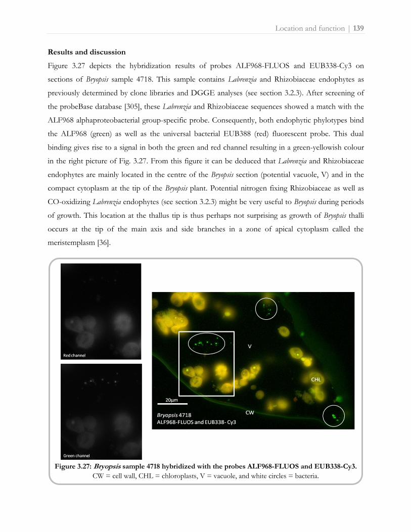
Location and function | 139
Results and discussion
Figure 3.27 depicts the hybridization results of probes ALF968-FLUOS and EUB338-Cy3 on
sections of Bryopsis sample 4718. This sample contains Labrenzia and Rhizobiaceae endophytes as
previously determined by clone libraries and DGGE analyses (see section 3.2.3). After screening of
the probeBase database [305], these Labrenzia and Rhizobiaceae sequences showed a match with the
ALF968 alphaproteobacterial group-specific probe. Consequently, both endophytic phylotypes bind
the ALF968 (green) as well as the universal bacterial EUB388 (red) fluorescent probe. This dual
binding gives rise to a signal in both the green and red channel resulting in a green-yellowish colour
in the right picture of Fig. 3.27. From this figure it can be deduced that Labrenzia and Rhizobiaceae
endophytes are mainly located in the centre of the Bryopsis section (potential vacuole, V) and in the
compact cytoplasm at the tip of the Bryopsis plant. Potential nitrogen fixing Rhizobiaceae as well as
CO-oxidizing Labrenzia endophytes (see section 3.2.3) might be very useful to Bryopsis during periods
of growth. This location at the thallus tip is thus perhaps not surprising as growth of Bryopsis thalli
occurs at the tip of the main axis and side branches in a zone of apical cytoplasm called the
meristemplasm [36].
Figure 3.27: Bryopsis sample 4718 hybridized with the probes ALF968-FLUOS and EUB338-Cy3.
CW = cell wall, CHL = chloroplasts, V = vacuole, and white circles = bacteria.

140 | Location and function
Rickettsia endophytes, on the other hand, seem to be more closely associated with the Bryopsis
chloroplast (Fig. 3.28), suggesting they and/or their biochemical pathways require the presence of
photosynthetic metabolites. Figure 3.28 shows the shared binding of the Rick_527-FLUOS and
EUB338-Cy3 probe on Bryopsis WB4 sections resulting in a signal in both the red and green channel.
A yellowish (in this case rather orange due to the high Cy3 intensity) colour is observed in the right
picture in Figure 3.28. In addition, the universal bacterial EUB338 probe hybridized with no other
bacterial rRNA than Rickettsia sequences, confirming our previous molecular results that Bryopsis
sample WB4 only harbors Rickettsia endophytes (section 3.2.3). This apparent mutual exclusion
among Rickettsia and other bacterial endophytes has also been observed within insect hosts [308].
Figure 3.28: Bryopsis sample WB4 hybridized with the probes Rick_527-FLUOS and EUB338-Cy3. CW = cell wall, CHL = chloroplasts, and white circles = bacteria.
Figure 3.29A displays the hybridization results of probes CF319a-FLUOS and EUB338-Cy3
probe on sections of Bryopsis sample MX90 known to harbor only Flavobacteriaceae endosymbionts
(sections 3.2.1 [225] and 3.2.3). Because the CF319a and EUB338 probe hinder each other sterically
(i.e. the binding sites of both probes on the 16S rRNA are too close to each other), Flavobacteriaceae
rRNA present only binds the CFB group-specific CF319a probe. Little or no binding of the EUB338
probe occurred (red channel, Fig. 3.29A), resulting in a vivid green signal in the picture on the right
of Fig. 3.29A. This signal indicates that Flavobacteriaceae endosymbionts are located within the
Bryopsis chloroplast and/or in close proximity with the chloroplastidal membranes. Also Figure 3.29B

Location and function | 141
which depicts the preliminary hybridization results of the Flavobacteriaceae endosymbiont specific
probe F695-Cy3 (red) shows that Flavobacteriaceae RNA may be present within the chloroplasts and
in the outer layer of the cytoplasm next to the cell wall which contains all other Bryopsis organelles.
The location in the vicinity of nuclei, mitochondria, Golgi complexes and mainly chloroplasts may
indicate that Flavobacteriaceae endosymbionts perform significant roles within their Bryopsis host.
This endorses the previously postulated obligate symbiotic nature of the Flavobacteriaceae
intracellular bacteria and their host specificity towards Bryopsis algae (sections 3.2.2 [245], 3.2.3 and
3.3.1). Moreover, the presence of Flavobacteriaceae endosymbionts within the Bryopsis chloroplast
may contribute to the strong autonomy and activity of this algal organelle inside the body of some
herbivorous sea slugs (see Chapter 1, Box 3), and might even provide a possible explanation for the
production of bioactive kahalalides by both sea slugs and their Bryopsis food [176]. Likewise, a
functional association between algal chloroplasts and intraplastidal bacteria has been reported within
the diatom Pinnularia [309, 310].
Figure 3.29: Bryopsis sample MX90 hybridized with (A) probes CF319a-FLUOS and EUB338-Cy3,
and (B) probe F695-Cy3. CW = cell wall, OL = outer cytoplasmic layer, CHL = chloroplasts, V = vacuole,
and white circles = bacteria.

142 | Location and function
Even though these FISH experiments provided a first insight into the specific location of
Labrenzia, Rickettsia, Rhizobiaceae and Flavobacteriaceae endophytes within their Bryopsis hosts,
repeated hybridizations with multiple group- and endophytic bacterial species-specific probes are
necessary to support these preliminary results. Hybridization conditions of the Flavobacteriaceae
specific probe F695 (and potential other endophyte species-specific probes) should be further
optimized with Clone-FISH, a technique which validates the specificity of new probes designed to
target uncultured bacteria [311]. In addition, the location results have shed some light on the
potential function of the endophytes within Bryopsis, however, the true physiological role of these
intracellular bacteria should be further investigated. In situ techniques which link functional gene
presence (metabolic potential) to bacterial cell identity in environmental samples may be suitable to
pursue this goal. Examples of such techniques include mRNA-FISH [312], gene-FISH [313] and in
situ rolling circle amplification-FISH [314, 315]. Also fluorescent in situ hybridization combined with
microautoradiography [FISH-MAR, 316], stable-isotope Raman spectroscopy [Raman-FISH, 317]
and secondary-ion mass spectrometry [FISH-SIMS, 318], could provide a linkage between identity
and function.
To summarize, exploratory FISH experiments with group- and species-specific probes on Bryopsis
sections revealed that Labrenzia, Rickettsia, Rhizobiaceae and Flavobacteriaceae endophytes occupy
distinct locations within the host cell which are consistent with the symbiotic nature and potential
function of these intracellular bacteria. These and more functional FISH protocols may open the way
to fully explore the Bryopsis-intracellular bacteria partnership in natural algal samples.
Acknowledgements
This research was funded by ‘Fonds Wetenschappelijk Onderzoek’ FWO-Flanders project
G.0045.08. Olivier Leroux, Myriam Claeys, Frederik Leliaert and Helen Decleyre are gratefully
acknowledged for practical assistance.

Chapter
Concluding
discussion
4


Results and conclusion | 145
“All life on Earth today derived from common ancestors. The first to evolve yet the last to be studied
in detail are bacteria. Scientists have now discovered that bacteria not only are the building blocks of
life, but also occupy and are indispensable to every other living being on Earth. Without them, life's
essential processes would quickly grind to a halt, and Earth would be as barren as Venus and Mars.”
Carla Cole (IN CONTEXT 34, p. 18, 1993), based on the work of Lynn Margulis and Dorion Sagan
Like all organisms, algae have developed in a world of microbes. It is therefore hardly surprising that
many of these photosynthetic eukaryotes host a wide range of intracellular bacteria. Various
microalgae such as dinoflagellates and diatoms are known to harbor bacterial endosymbionts which
are linked with diverse metabolic functions [212, 229, 319, 320]. Within macroalgal hosts, however,
bacterial endosymbiosis seems more restricted to certain seaweed lineages. Besides the occurrence of
bacterial endophytes inside red algal galls and buds [74, 121, 123], true intracellular, non-pathogenic
bacteria have to date only been reported inside the giant cells of some green siphonous seaweeds
such as Bryopsis, Caulerpa, Chlorodesmis, Halimeda, Penicillus and Udotea [36-41, 75, 124]. These macro,
yet unicellular, algae display extraordinary physicochemical adaptations and wounding responses to
overcome their perceived vulnerable single cell morphology (Chapter 1, Box 3). These survival and
reproduction strategies alongside endobiotic interactions with potential nitrogen fixing and
photosynthetic bacteria, have been suggested to play a role in the success of siphonous green algae
in a range of marine habitats [75, 125]. To shed light on the endophytic bacterial partner, the
Bryopsis-bacterial partnership was explored with a variety of culture-dependent and culture-
independent techniques, which were optimized particularly for this study (Chapter 3, Part 1).
Main results and general conclusion
Presence, diversity, identity and uniqueness
Forty years after the initial reports of intracellular bacteria within Bryopsis [36, 37], the natural
presence of true endophytic bacteria inside the algal cytoplasm was confirmed by electron
microscopy and fluorescent in situ hybridization (sections 3.2.1, 3.2.2 and 3.3.2). Additional 16S
rRNA gene-based techniques revealed that not just one, but several different bacterial phylotypes

146 | Results and conclusion
reside within the algal host interior of which Bacteroidetes, Flavobacteriaceae, Labrenzia, Mycoplasma,
Phyllobacteriaceae, Rhizobiaceae and Rickettsia species were encountered in three or more Bryopsis
samples collected globally. Notwithstanding this similarity, the total bacterial diversity varied among
different Bryopsis cultures with the presence of one to a maximum of four endophytic phylotypes per
host sample (section 3.2.3). The co-occurrence of multiple bacterial partners underpins the recent
assumption that the diversity of various host associated endobiotic communities has been greatly
underestimated so far [216, 230, 321]. Harbouring multiple endobionts could permit further
expansion of host capabilities and might even benefit the whole endobiotic flora by allowing
syntrophy (i.e. cross-feeding) [322]. In addition, endophytic bacteria of Bryopsis algae show some
similarity with those of other siphonous seaweeds as Alphaproteobacteria (mainly Rhodobacterales
and Rhizobiales) and Bacteroidetes intracellular bacteria have also been characterized within Caulerpa
[75, 124, 125]. Moreover, several Cladophorales algae (e.g. Boergesenia and Boodlea), were found to
harbour Labrenzia and Rhizobiaceae species similar to Bryopsis (Leliaert et al, unpublished data),
supporting the significance of these endophytes within siphonous seaweed hosts. Several of the
Bryopsis endophytic phylotypes, however, are more closely related to known (endo)symbiotic bacteria
of non-algal hosts such as amoeba, land plants, insects and marine animals [202, 214, 216, 221, 222,
286, 323, 324]. This may hint at the existence of a universal group of bacterial taxa which are
particularly adapted to (but not necessary reliant on) an intracellular lifestyle. The clear
distinctiveness between free-living and endophytic algal-associated bacterial communities (section
3.2.2) supports this hypothesis. Such an ‘ecological coherence’ has been proven in other niches [128
and references therein], and might also be applied to macroalgal surface associated bacterial
communities (see Chapter 1).
Specificity, stability, interdependency and symbiotic nature
The allegiance of bacteria towards an intracellular lifestyle, however, displays diverse degrees of
specificity. Some bacteria are (host-specific) obligate endosymbionts, while others are more
facultative endobiotic and can survive without a host. Either way, specific modes of partner
recognition are required which mainly rely on surface structures (e.g. peptidoglycans,
lipopolysaccharides and lectins) and defense mechanisms of both the host and bacterial partner
[325]. It has been postulated that high endobiont diversity (i.e. more than 2 à 3 species) goes
together with high levels of flexibility in symbiont specificity and stability [322]; and Bryopsis seems
no exception. Temporal stability experiments showed the loss of Phyllobacteriaceae endophytes in

Results and conclusion | 147
Bryopsis samples after prolonged cultivation (section 3.2.2), and phylogenetic analyses pointed out
that Rhizobiaceae as well as Labrenzia endobionts are closely related to sequences of free-living
bacterial strains (section 3.2.3). Moreover, Labrenzia and Phyllobacteriaceae endophytes were also
identified from Bryopsis surroundings (section 3.2.2) and could be isolated on artificial media (section
3.2.4). In contrast to these facultative endobiotic phylotypes, mainly Flavobacteriaceae
endosymbionts showed a more obligate lifestyle with high specificity towards the Bryopsis host
(sections 3.2.2 and 3.3.1). Flavobacteriaceae specific amplification revealed the exclusive presence of
this endosymbiont in Bryopsis species from warm-temperate and tropical seas, and phylogenetic
analyses indicated some degree of cospeciation (section 3.3.1).
Host/habitat influences, symbiont transmission modes and function
In addition to differences in intrinsic bacterial lifestyles characteristics, also host phylogeny, habitat
and geography influence the diversity of endobiotic communities [322]. Statistical analyses revealed
that the total Bryopsis endobiotic community composition could only be explained by a mix of host
phylogenetic, geographic and environmental factors which had different (shared and/or
independent) effects on the individual community members (section 3.2.3). Phyllobacteriaceae,
Rhizobiaceae and Labrenzia species seemed true generalists which are laterally acquired by any
Bryopsis host, regardless host species, habitat and geography, to possibly fulfill functions such as
nitrogen-fixation, photosynthesis and CO-oxidation (Fig. 4.1). These metabolic processes might be
very useful to Bryopsis during periods of growth, explaining the location of these facultative
endophytes in the meristemplasm, i.e. a zone of apical cytoplasm at the thallus tip where
proliferation occurs (section 3.3.2). The occurrence of Mycoplasma, Rickettsia and Bacteroidetes
endophytes was to some extent influenced by environmental factors, suggesting an additional lateral
acquisition of habitat-specific bacteria by Bryopsis hosts. This type of ‘habitat-specific acquisition’ has
been argued to provide ecological flexibility by allowing the host to take up bacteria which are
optimally adapted to their local environment [326]. Since Mycoplasma, Rickettsia and Bacteroidetes
species are well-know obligate intracellular symbionts of other eukaryotes, these endophytes may be
more likely taken up from co-occurring hosts (i.e. vector dependent acquisition) rather than from
the surrounding seawater (Fig. 4.1). In addition, Bacteroidetes endophytes seemed more securely
associated with Bryopsis as they also displayed some vertical transmission. Only Flavobacteriaceae
endosymbionts, however, appeared strictly vertically transmitted from one Bryopsis generation to the
next via presumable sexual (e.g. gametes and spores) reproductive stages. This confirms that

148 | Results and conclusion
Flavobacteriaceae endophytes are true obligate symbionts which are entirely dependent on the algal
host for their survival (Fig. 4.1). The location of these endosymbionts in and/or in the close vicinity
of chloroplasts (section 3.3.2) might indicate these intracellular bacteria fulfill a significant role
within the Bryopsis host. Nevertheless, their exact ecological function remains unclear.
Figure 4.1: The Bryopsis-bacterial partnership. Schematic overview of the endophytic bacterial lifestyles and transmission modes to the Bryopsis host. Habitat and algal host influences on the bacterial flora as well as potential functions of the bacterial endophytes are described. B = Bacteroidetes, F = Flavobacteriaceae, L = Labrenzia, M = Mycoplasma, P = Phyllobacteriaceae, Rh = Rhizobiaceae, and Ri = Rickettsia endobionts.
Results and conclusion | 149
Conclusion
Bryopsis algae harbour taxonomically diverse endophytic communities which consist of a mix of
generalist and specialist bacterial species. Whereas Labrenzia, Phyllobacteriaceae, Rhizobiaceae,
Mycoplasma, Rickettsia and Bacteroidetes are to a greater or lesser extent laterally acquired from the
environment and/or other hosts, mainly Flavobacteriaceae appear Bryopsis-specific obligate
endosymbionts which are vertically transmitted across generations of host species (Fig. 4.1). Either
way, there seems to be a highly specific mode of partner recognition as Bryopsis selectively maintains
and/or attracts the same endophytes globally, even though the algal interior is repeatedly exposed to
various other marine bacteria during wounding events (i.e. thallus rupture and protoplast formation).
With these observations, the Bryopsis-bacterial partnership fits into the general picture of eukaryote-
prokaryote symbiosis systems. Also in various marine animal [264, 321, 322] and insect hosts [216,
232] complex, yet specific, endobiotic communities exist which consist of a mix of one to two
primary (obligate) and several secondary (facultative) bacterial partners. This suggests the
conservation of basic mechanisms and principles among symbioses of bacteria with hosts from the
whole tree of life, possibly giving rise to a universal group of bacterial taxa which share a general
intracellular lifestyle. The presence of, for example, nitrogen-fixing Rhizobiaceae species in both
land plants and siphonous seaweeds, is a fine example of this universality.

150 | Future perspectives
Where to go from here?
Methodology
Even though all methods used in this study were optimized to examine the Bryopsis-bacterial
partnership to the best possible extent (see Chapter 3, part 1), the rRNA approach applied has well-
documented limitations in assessing the microbial diversity of environmental samples. Biases might
be introduced in each step of the approach: e.g. insufficient DNA extraction, preferential PCR
amplification, co-migration of DNA fragments during DGGE analysis, inadequate clone library
screening and low sensitivity of FISH methods [228, 327-329]. Due to these restraints, the full-cycle
16S rRNA gene approach has been assumed to reveal the diversity of the dominant (abundant)
community members only [128]. Although this might be not that restrictive in the study of less
diverse endobiotic communities [228], diversity studies of endobionts comprise some specific
challenges. Especially the initial surface sterilization, included to eliminate all epiphytic
contamination, represents a critical step in the methodological approach. Despite the efficiency of
the newly designed protocol (section 3.1.2), each surface sterilization method remains a balancing
exercise between too much (i.e. neutralizing internal bacteria) and too little (i.e. outer surface
bacteria being mistaken for endobionts). While the reproducibility of results (e.g. Fig. 3.10, section
3.2.2) supports their reliability, the surface sterilization protocol is accountable for some additional
limitations. The constraint of living, uni-algal samples as starting material gave rise to extended
cultivation of the algal samples prior to molecular processing (see section 3.2.1). This prolonged stay
under artificial culture conditions might have unknown effects on the endophytic bacterial
community, suggesting the diversity obtained from Bryopsis cultures might not fully represent the
variety present within the alga in its natural environment. Nevertheless, the bacteria identified in this
study are at least part of the natural Bryopsis endobiotic flora and supplementary species-specific
amplifications as well as fluorescent in situ hybridizations (Chapter 3, part 3) underline their true
contribution in natural algal samples. Further, 16S rRNA gene-based analyses of photosynthetic
eukaryote-prokaryote symbiosis systems are also restricted by ‘symbiosis’ itself: the cyanobacterial
origin of chloroplasts interferes with the characterization of bacteria through massive
coamplification of the algal host’s chloroplast DNA with universal bacterial primers [228, 330].
Elimination of chloroplast DNA and/or removal of intact chloroplasts before extraction would
greatly simplify future 16S rRNA gene-based as well as metagenomic analyses (see below) of the
endophytic bacterial community. Diverse protocols are described to selectively amplify bacterial 16S
rRNA genes [163, 331], chemically/enzymatically eliminate chloroplast DNA [330, 332], and

Future perspectives | 151
separate bacteria form host organelles by density gradient centrifugation [333, 334]; these methods,
however, should be optimized to be applicable on Bryopsis hosts [330]. In addition, as surface
sterilization as well as subsequent DGGE and cloning protocols are labor-intensive and time-
consuming, only a limited number of samples could be processed.
Ready-to-use surface sterilization methods and sequenced-based metagenomic analyses in
combination with high-throughput next-generation sequencing technologies would be required to
examine the endophytic bacterial diversity in a more effective way. Besides looking at ‘who is (in)
there’, also the question ‘what are they doing there?’ should be tackled more profoundly in future
research. Whole-genome sequencing and functional metagenomics could reveal insight into the role
of endophytic bacteria within the Bryopsis host. Sequence-based analyses of complete genome
sequences may shed light on the metabolic potential of the endophytes [334-336], and functional
screening of metagenome libraries may identify new genes and/or novel natural products of
endobiotic origin [337, 338]. To fully elucidate symbiosis systems, however, it will be necessary to go
beyond endobiont genome studies alone by integrating data at all levels (genes, transcripts and
proteins) from all symbiosis partners, including the host, as well as information on the interaction of
these molecules at a systems biology level [333, 336]. Despite the potential of ‘omics’ technologies
and high-throughput screening methods in generating data, the extraction of useful biological
information from these datasets remains a significant (computational) challenge [334]. It has been
suggested that the true ‘omics’ power will be realized when these technologies are integrated with
‘classical’ approaches that examine gene expression or functional activity in vivo such as certain FISH
techniques (see section 3.3.2) and stable isotope analyses [339]. Also efforts to culture previously
unculturable bacteria still a prerequisite for their full characterization (see section 3.2.4) will
likely be facilitated by clues about their physiology derived from ‘omics’ data [339].
Significance of seaweed-bacterial studies
There is a major lack of knowledge about marine symbioses and the impact that these associations
have on their hosts’ ecology and on global biogeochemical processes of essential nutrients such as
carbon, nitrogen and sulfur [340]. Exploring the Bryopsis-bacterial partnership may lead to a better
understanding of the significance of these marine symbioses. More specifically, Bryopsis associated
endophytes can provide an insight into the metabolic interchange which underlies the ability of
marine hosts and, in particular, siphonous seaweeds to inhabit oligotrophic waters [75, 125, 340]. As
this successful spread can have a negative impact on the native biota, host associated bacteria might

152 | Future perspectives
be a target by which to control the process [9]. Moreover, (in part) biogeographically structured
bacterial communities can provide a clue on the origin of introduction [125]. Phylogenetically
structured obligate endobionts, on the other hand, might be used as part of an integrative
taxonomical approach for species delimitation in cryptic (algal) hosts [341]. On a grander scale,
whole genome analyses of (obligate) endobionts may bring to light major evolutionary patterns
[336]. Furthermore, seaweed-bacterial studies have importance for the characterization of bacterial
pathogens in mariculture industry and the discovery of novel natural products (Chapter 1, Box 2).

Take home message | 153
General reflections
The omnipresence and universality of symbiosis render the term to be a synonym for biology
instead of a concept within. Thinking about symbiosis raises issues concerning the boundaries of self
and the definition of species, and leads to discussions about holism versus egoism. In their turn,
current efforts to understand symbiosis reflect a tension between the reductionist approaches of
molecular biologists and the holistic approaches of ecologists [8]. Therefore, the largest obstacle for
symbiosis studies may stem from the culture gaps among the different disciplines. The infrastructure
as it stands would not foster this process: academic departments, federal agencies and scientific
societies are structured in such a way as to frustrate opportunities for productive interactions and
creative eccentricity [9]. To achieve the full potential of this field, however, a vast array of technical,
cultural and social hurdles must be overcome. Luckily, we can learn a lot from the efficient cross-
talk between seaweeds and bacteria!
“Acknowledging that our ancestors are bacteria is humbling and has disturbing implications. Besides
impugning human sovereignty over the rest of nature, it challenges our ideas of individuality, uniqueness,
and independence. It even violates our view of ourselves as discrete physical beings separated from the rest
of nature and still more unsettling it challenges the alleged uniqueness of human intelligent
consciousness. Those who speak only for the special interests of human beings fail to see how
interdependent life on Earth really is. Without the microbial life forms, we would sink in feces and
choke on the carbon dioxide we exhale. […] The ancient, vast, and fundamental nature of our
interdependence with other forms of life may be humbling, but it provides a basis for facing the future free
of crippling delusions. Despite all our conceits, we are as much exploited as exploiters, as much
consumed as consumers. The lesson of evolutionary history is that it will be through conservation,
interaction, and networking, not domination, that we avert a premature end to our species.”
Carla Cole (IN CONTEXT 34, p. 18, 1993), based on the work of Lynn Margulis and Dorion Sagan


Summary | 155

156 | Summary
The term ‘symbiosis’ described in 1879 as ‘a beneficial alliance between two dissimilar organisms’ has
been rewritten over the last decade. Symbiosis no longer defines a concept in biology; it basically ís
biology. Symbioses are widespread covering diverse forms of relationships among multiple partners
and support fundamentally important processes. Endosymbiosis with one symbiotic partner (the
endosymbiont) living intracellularly within the other (the host), is the most intimate form of
symbiosis. The host typically provides a nutrient-rich, sheltered environment for the endosymbiont.
In turn, endosymbionts expand the physiological capacities of their hosts, enabling them to invade
novel metabolic and ecological niches. In view of this, it has been suggested that interactions with
bacterial endosymbionts play a role in the success of siphonous (i.e. single giant cell) seaweeds in a
range of marine habitats. This dissertation aimed to explore the partnership between siphonous
seaweeds and their intracellular bacterial communities, focusing on the green alga Bryopsis as host
organism. The identity, diversity, uniqueness, stability, specificity, function, symbiotic nature and
transmission modes of the endophytic bacterial communities within Bryopsis were examined by a
culture-independent full-cycle 16S rRNA gene approach. Statistical analyses were performed to
identify the factors (e.g. host phylogeny, geography and environment) shaping the endobiotic
bacterial community composition and culture-dependent techniques were implemented to investigate
the interdependency among the symbiotic partners.
Results indicate Bryopsis harbours rather stable and taxonomically diverse endophytic communities
composed of certain Bacteroidetes, Flavobacteriaceae, Labrenzia, Mycoplasma, Phyllobacteriaceae,
Rhizobiaceae and Rickettsia species. Although the algal interior is repeatedly exposed to various other
marine bacteria during wounding events, a highly specific mode of partner recognition seems to exist
as Bryopsis selectively maintains and/or attracts the same bacteria globally. This specificity is
confirmed by the clear distinctiveness of the intracellular bacterial communities from those occurring
in the surrounding seawater, even while the endophytic Labrenzia, Phyllobacteriaceae and
Rhizobiaceae phylotypes are closely related to free-living bacterial strains and could be isolated on
artificial media. These Labrenzia, Phyllobacteriaceae and Rhizobiaceae endophytes seem true
generalists which are laterally acquired from the environment by any Bryopsis host, regardless of host
species, habitat and geography, to possibly fulfill functions such as nitrogen-fixation, photosynthesis
and CO-oxidation. Also Mycoplasma, Rickettsia and Bacteroidetes bacteria appear to a greater or lesser
extent horizontally transmitted to Bryopsis algae. The habitat-specific lateral acquisition of these
obligate intracellular bacteria, however, more likely takes place from co-occurring hosts (e.g.
microalgae, ciliates, amoebae and sea slugs) rather than from the surrounding seawater.
Summary | 157
Flavobacteriaceae species, on the other hand, are closely associated to Bryopsis as they seem vertically
transmitted, obligate endosymbionts which show some degree of cospeciation. The unique, host
specific presence of these bacteria within Bryopsis species from warm-temperate and tropical seas and
their internal location in and/or in the close vicinity of algal chloroplasts, might indicate
Flavobacteriaceae endosymbionts fulfill a significant role within the Bryopsis host. Their exact
ecological function, however, remains unclear.
Taken together, these results indicate that the Bryopsis-bacterial partnership matches the universal
eukaryote-prokaryote symbiosis picture in which a mix of one to two specialist and several generalist
bacteria reside within a single host. These diverse and complex endosymbiotic communities might
permit further expansion of host capabilities, suggesting that ‘it takes two to tango, but a whole crowd to
stage dive’. Also in future (Bryopsis-bacterial) symbiosis research, the efficient teamwork between
multiple scientists from diverse disciplines might eventually create a major leap forward in the
understanding of our symbiotic planet.

158 | Samenvatting
De term 'symbiose' gedefinieerd in 1879 als 'een gunstige en/of noodzakelijke vorm van samenleven
tussen twee verschillende organismen' is de laatste jaren herschreven. Symbiose definieert niet langer
een begrip in de biologie, maar ís ronduit biologie. Symbioses zijn wijdverspreid in de natuur en
omvatten diverse, zowel neutrale als goed- en slechtaardige, relaties tussen meerdere partners.
Bovendien liggen deze doorgedreven vormen van samenleven aan de basis van fundamenteel
belangrijke, evolutieve processen. Endosymbiose waarbij een partner (de endosymbiont) huist
binnenin de andere (de gastheer), is de meest intieme vorm van samenleven. De gastheer biedt een
voedselrijke, beschutte omgeving voor de endosymbiont die op zijn beurt de fysiologische
mogelijkheden van de gastheer uitbouwt zodat deze nieuwe metabolische en ecologische niches kan
bewandelen. Zo is geopperd dat interacties met bacteriële endosymbionten een rol spelen in het
succes van sifonale (macroscopische maar toch eencellige) zeewieren in diverse mariene milieus. Dit
proefschrift had tot doel de samenwerking tussen sifonale zeewieren en hun inwendige
(intracellulaire) bacteriën onder de loep te nemen, met de focus op het groene vederwier Bryopsis als
gastheer. De identiteit, diversiteit, uniekheid, stabiliteit, specificiteit, functie, symbiontische aard en
transmissie wijze van de endofytische (m.n. inwendig levend in een plant) bacteriën in Bryopsis
werden onderzocht aan de hand van een cultuur-onafhankelijke benadering. Statistische analyses
werden uitgevoerd om na te gaan welke factoren (bv. gastheersoort, geografie en omgeving) de
aanwezigheid van bepaalde bacteriën binnenin het wier beïnvloeden. Ook de wederzijdse
afhankelijkheid van beide symbiosepartners, wier en bacteriën, werd onderzocht met cultuur-
afhankelijke technieken.
Resultaten tonen aan dat Bryopsis een relatief stabiele en taxonomisch diverse endofytische flora bevat
bestaande uit bepaalde Bacteroidetes, Flavobacteriaceae, Labrenzia, Mycoplasma, Phyllobacteriaceae,
Rhizobiaceae and Rickettsia bacteriën. Niettegenstaande het wier, zowel van buiten als van binnen,
herhaaldelijk wordt blootgesteld aan een (letterlijke) zee van bacteriën, oogt de interactie tussen
Bryopsis en inwendige bacteriën toch zeer specifiek. Bryopsis huist wereldwijd dezelfde bacteriële
soorten en bovendien verschillen de intracellulaire bacteriën beduidend van deze in het omringende
zeewater. Desalniettemin bleken de endofytische Labrenzia, Phyllobacteriaceae en Rhizobiaceae
bacteriën juist nauw verwant aan vrij-levende bacteriële soorten en konden ze worden geïsoleerd in
vitro. Deze Labrenzia, Phyllobacteriaceae en Rhizobiaceae endofyten zijn dus echte generalisten die uit
de omgeving worden opgenomen door om het even welke Bryopsis gastheer, ongeacht soort, plaats
en/of omgeving, om mogelijk stikstof-fixatie, fotosynthese en CO-oxidatie functies te vervullen.
Ook Mycoplasma, Rickettsia en Bacteroidetes bacteriën lijken in meer of mindere mate transient

Samenvatting | 159
(herhaaldelijk en tijdelijk) opgenomen door vederwieren die in een welbepaald milieu (bepaalde
temperatuur, zoutgehalte, nitraatconcentratie, etc) groeien. Deze obligaat intracellulaire bacteriën
worden echter niet opgenomen uit het omgeringde zeewater, maar veeleer uitgewisseld tussen
Bryopsis en andere gastheren zoals bijvoorbeeld microalgen, ciliaten, amoeben en zeeslakken.
Flavobacteriaceae endosymbionten daarentegen zijn veel inniger geassocieerd met Bryopsis. Ze
worden verticaal overgedragen van de ene wiergeneratie naar de andere en vertonen zelfs enige mate
van co-speciatie, m.n. als Bryopsis evolueert, evolueren de Flavobacteriaceae bacteriën mee. De unieke
aanwezigheid van deze bacteriën in Bryopsis soorten van warm-gematigde en tropische zeeën en hun
interne locatie in en/of in de nabijheid van alg chloroplasten doet vermoeden dat Flavobacteriaceae
endosymbionten veelbetekenende functies vervullen binnenin hun gastheer. De exacte ecologische
functie van deze bacteriën blijft echter onduidelijk.
Deze bovenstaande resultaten wijzen erop dat het Bryopsis-bacteriële partnerschap past in het
universele symbiose plaatje waarin een mix van één tot twee specialist en verschillende generalist
bacteriën verblijven binnenin een enkele gastheer. Een diverse en complexe endosymbiontische flora
kan er immers voor zorgen dat de gastheer meer mogelijkheden krijgt om te overleven. Met andere
woorden: ‘it takes two to tango, but a whole crowd to stage dive’. Ook in toekomstig (Bryopsis-bacterie)
symbiose onderzoek, kan de efficiënte samenwerking tussen meerdere wetenschappers uit diverse
disciplines uiteindelijk zorgen voor een grote sprong voorwaarts in het begrijpen van onze
symbiontische planeet.

Bibliography | 161
1. Hehemann J-H, Correc G, Barbeyron T, Helbert W, Czjzek M, Michel G: Transfer of carbohydrate-active
enzymes from marine bacteria to Japanese gut microbiota. Nature 2010, 464(7290):908-912. 2. van den Hoek C, Mann DG, Jahns HM: Algae. An introduction to phycology. New York: Cambridge
University Press; 1995. 3. Margulis L: Symbiotic planet : a new look at evolution. New York: Basic Books; 1998. 4. Goecke F, Labes A, Wiese J, Imhoff JF: Chemical interactions between marine macroalgae and bacteria.
Mar Ecol-Prog Ser 2010, 409:267-299. 5. Olsen GJ, Lane DJ, Giovannoni SJ, Pace NR, Stahl DA: Microbial ecology and evolution: a ribosomal
RNA approach. Annu Rev Microbiol 1986, 40:337-365. 6. Olson JB, Kellogg CA: Microbial ecology of corals, sponges, and algae in mesophotic coral
environments. FEMS Microbiol Ecol 2010, 73(1):17-30. 7. Geng H, Belas R: Molecular mechanisms underlying Roseobacter–phytoplankton symbioses. Curr Opin
Biotechnol 2010, 21(3):332-338. 8. Relman DA: 'Til death do us part': coming to terms with symbiotic relationships. Nat Rev Micro 2008,
6(10):721-724. 9. McFall-Ngai M: Are biologists in 'future shock'? Symbiosis integrates biology across domains. Nat Rev
Micro 2008, 6(10):789-792. 10. Hesse W, Gröschel DHM: Walther and Angelina Hesse - early contributors to bacteriology. ASM News
1992, 58(8):425 - 428. 11. ZoBell CE: Marine microbiology. Waltham: Chronica Botanica Co; 1946. 12. Graham LE, Wilcox LW: Algae. New Jersey: Prentice-Hall; 2000. 13. Burke C, Thomas T, Lewis M, Steinberg P, Kjelleberg S: Composition, uniqueness and variability of the
epiphytic bacterial community of the green alga Ulva australis. ISME J 2011, 5(4):590-600. 14. Gupta S, Abu-Ghannam N: Bioactive potential and possible health effects of edible brown seaweeds.
Trends in Food Science & Technology 2011, 22(6):315-326. 15. Moore JE, Xu J, Millar BC: Diversity of the microflora of edible macroalga (Palmaria palmata). Food
Microbiology 2002, 19(2-3):249-257. 16. MacArtain P, Gill CIR, Brooks M, Campbell R, Rowland IR: Nutritional value of edible seaweeds. Nutr Rev
2007, 65(12):535-543. 17. O’Sullivan L, Murphy B, McLoughlin P, Duggan P, Lawlor PG, Hughes H, Gardiner GE: Prebiotics from
marine macroalgae for human and animal health applications. Marine Drugs 2010, 8(7):2038-2064. 18. Michel G, Nyval-Collen P, Barbeyron T, Czjzek M, Helbert W: Bioconversion of red seaweed galactans: a
focus on bacterial agarases and carrageenases. Appl Microbiol Biotechnol 2006, 71(1):23-33. 19. Smit AJ: Medicinal and pharmaceutical uses of seaweed natural products: a review. J Appl Phycol 2004,
16(4):245-262. 20. Qian P-Y, Xu Y, Fusetani N: Natural products as antifouling compounds: recent progress and future
perspectives. Biofouling 2009, 26(2):223-234. 21. Bhadury P, Wright PC: Exploitation of marine algae: biogenic compounds for potential antifouling
applications. Planta 2004, 219(4):561-578. 22. Hernández I, Fernández-Engo M, Pérez-Lloréns J, Vergara J: Integrated outdoor culture of two estuarine
macroalgae as biofilters for dissolved nutrients from Sparus auratus waste waters. J Appl Phycol 2005, 17(6):557-567.
23. Neori A, Chopin T, Troell M, Buschmann AH, Kraemer GP, Halling C, Shpigel M, Yarish C: Integrated aquaculture: rationale, evolution and state of the art emphasizing seaweed biofiltration in modern mariculture. Aquaculture 2004, 231(1-4):361-391.
24. Gardiner GE, Campbell AJ, O’Doherty JV, Pierce E, Lynch PB, Leonard FC, Stanton C, Ross RP, Lawlor PG: Effect of Ascophyllum nodosum extract on growth performance, digestibility, carcass characteristics and selected intestinal microflora populations of grower-finisher pigs. 2008, 141(3):259-273.
25. Khan W, Rayirath U, Subramanian S, Jithesh M, Rayorath P, Hodges D, Critchley A, Craigie J, Norrie J, Prithiviraj B: Seaweed extracts as biostimulants of plant growth and development. J Plant Growth Regul 2009, 28(4):386-399.
26. Borines MG, McHenry MP, de Leon RL: Integrated macroalgae production for sustainable bioethanol, aquaculture and agriculture in Pacific island nations. Biofuels, Bioproducts and Biorefining 2011, 5(6):599-608.
27. Vairappan CS, Suzuki M, Motomura T, Ichimura T: Pathogenic bacteria associated with lesions and thallus bleaching symptoms in the Japanese kelp Laminaria religiosa Miyabe (Laminariales, Phaeophyceae). Hydrobiologia 2001, 445(1):183-191.

162 | Bibliography
28. Steinberg PD, Schneider R, Kjelleberg S: Chemical defenses of seaweeds against microbial colonization. Biodegradation 1997, 8(3):211-220.
29. Penesyan A, Marshall-Jones Z, Holmstrom C, Kjelleberg S, Egan S: Antimicrobial activity observed among cultured marine epiphytic bacteria reflects their potential as a source of new drugs. FEMS Microbiol Ecol 2009, 69(1):113-124.
30. Armstrong E, Yan L, Boyd KG, Wright PC, Burgess JG: The symbiotic role of marine microbes on living surfaces. Hydrobiologia 2001, 461(1):37-40.
31. Andersen RA: Algal culturing techniques. Amsterdam: Elsevier; 2006. 32. Waksman SA, Hotchkiss M, Carey CL: Marine bacteria and their role in the cycle of life of the sea: II.
bacteria concerned with the cycle of nitrogen in the sea. The Biology Bulletin 1933, 65:137-167. 33. Provasoli L: Effect of plant hormones on Ulva. Biol Bull 1958, 144:375-384. 34. Provasoli L, Pintner IJ: Bacteria induced polymorphism in an axenic laboratory strain of Ulva lactuca
(Chlorophyceae). J Phycol 1980, 16:196-200. 35. Harold R, Stanier RY: The genera Leucothrix and Thiothrix. Bacteriol Rev 1955, 19:49-58. 36. Burr FA, West JA: Light and electron microscope observations on the vegetative and reproductive
structures of Bryopsis hypnoides. Phycologia 1970, 9(1):17-37. 37. Burr FA, Evert RF: Cytochemical study of wound-healing protein in Bryopsis hypnoides. Cytobios 1972,
6(24):199-215. 38. Turner JB, Friedmann EI: Fine structure of capitular filaments in the coenocytic green alga Penicillus. J
Phycol 1974, 10(2):125-134. 39. Colombo PM: Occurrence of endophytic bacteria in siphonous algae. Phycologia 1978, 17(2):148-151. 40. Dawes CJ, Lohr CA: Cytoplasmic organization and endosymbiotic bacteria in the growing points of
Caulerpa prolifera Rev Algol 1978, 13:309-314. 41. Menzel D: Fine structure of vacuolar inclusions in the siphonous green alga Chlorodesmis fastigiata
(Udoteaceae, Caulerpales) and their contribution to plug formation. Phycologia 1987, 26(2):205-221. 42. Chan ECS, McManus EA: Distribution, characterization, and nutrition of marine microorganisms from
the algae Polysiphonia lanosa and Ascophyllum nodosum. Can J Microbiol 1969, 15(5):409-420. 43. Berland BR, Bianchi MG, Maestrini SY: Etude des bactéries associées aux algues marines en culture. I.
Détermination préliminaire des espèces. Mar Biol 1969, 2:350-355. 44. Tsukidate J: Microbiological studies of Porphyra plants. II. Bacteria isolated from Porphyra leucosticta
in culture. Bulletin of the Japanese Society of Scientific Fisheries 1971, 37(5):376-379. 45. Laycock RA: The detrital food chain based on seaweeds. I. Bacteria associated with the surface of
Laminaria fronds. Mar Biol 1974, 25(3):223-231. 46. Kong MK, Chan K: A study on the bacterial flora isolated from marine algae. Bot Mar 1979, 22(2):83-98. 47. Mazure HGF, Field JG: Density and ecological importance of bacteria on kelp fronds in an upwelling
region. J Exp Mar Biol Ecol 1980, 43(2):173-182. 48. Lakshmanaperumalsamy P, Purushothaman A: Heterotrophic bacteria associated with seaweed. Proceedings:
Plant Sciences 1982, 91(6):487-493. 49. Lewis TE, Garland CD, McMeekin TA: The bacterial biota on crustose (nonarticulated) coralline algae
from Tasmanian waters. Microb Ecol 1985, 11(3):221-230. 50. Shiba T, Taga N: Heterotrophic bacteria attached to seaweeds. J Exp Mar Biol Ecol 1980, 47(3):251-258. 51. Lemos ML, Toranzo AE, Barja JL: Antibiotic activity of epiphytic bacteria isolated from intertidal
seaweeds. Microb Ecol 1985, 11(2):149-163. 52. Bolinches J, Lemos ML, Barja JL: Population dynamics of heterotrophic bacterial communities
associated with Fucus vesiculosus and Ulva rigida in an estuary. Microb Ecol 1988, 15(3):345-357. 53. Head WD, Carpenter EJ: Nitrogen fixation associated with the marine macroalga Codium fragile. Limnol
Oceanogr 1975, 20(5):815 - 823. 54. Rosenberg G, Paerl HW: Nitrogen fixation by blue-green algae associated with the siphonous green
seaweed Codium decorticatum, effects on ammonium uptake. Mar Biol 1981, 61(2):151-158. 55. Croft MT, Lawrence AD, Raux-Deery E, Warren MJ, Smith AG: Algae acquire vitamin B12 through a
symbiotic relationship with bacteria. Nature 2005, 438(7064):90-93. 56. Croft MT, Warren MJ, Smith AG: Algae need their vitamins. Eukaryot Cell 2006, 5(8):1175-1183. 57. Sieburth JM: The influence of algal antibiosis on the ecology of marine microorganisms. In: Advances in
Microbiology of the Sea. Edited by Droop MR, Wood J. London: Academic Press; 1968: 63–94. 58. Staufenberger T, Thiel V, Wiese J, Imhoff JF: Phylogenetic analysis of bacteria associated with Laminaria
saccharina. FEMS Microbiol Ecol 2008, 64(1):65-77.

Bibliography | 163
59. Tujula NA, Crocetti GR, Burke C, Thomas T, Holmstrom C, Kjelleberg S: Variability and abundance of the epiphytic bacterial community associated with a green marine Ulvacean alga. ISME J 2010, 4(2):301-311.
60. Bengtsson MM, Sjotun K, Ovreas L: Seasonal dynamics of bacterial biofilms on the kelp Laminaria hyperborea. Aquat Microb Ecol 2010, 60(1):71-83.
61. Longford SR, Tujula NA, Crocetti GR, Holmes AJ, Holmström C, Kjelleberg S, Steinberg PD, Taylor MW: Comparisons of diversity of bacterial communities associated with three sessile marine eukaryotes. Aquat Microb Ecol 2007, 48(3):217-229.
62. Lachnit T, Blümel M, Imhoff JF, Wahl M: Specific epibacterial communities on macroalgae: phylogeny matters more than habitat. Aquat Biol 2009, 5:181-186.
63. Burke C, Steinberg P, Rusch D, Kjelleberg S, Thomas T: Bacterial community assembly based on functional genes rather than species. Proceedings of the National Academy of Sciences 2011, 108(34):14288-14293.
64. Lachnit T, Wahl M, Harder T: Isolated thallus-associated compounds from the macroalga Fucus vesiculosus mediate bacterial surface colonization in the field similar to that on the natural alga. Biofouling 2010, 26(3):247-255.
65. Engel S, Jensen PR, Fenical W: Chemical ecology of marine microbial defense. J Chem Ecol 2002, 28(10):1971-1985.
66. Lam C, Stang A, Harder T: Planktonic bacteria and fungi are selectively eliminated by exposure to marine macroalgae in close proximity. FEMS Microbiol Ecol 2008, 63(3):283-291.
67. Paul NA, de Nys R, Steinberg PD: Chemical defence against bacteria in the red alga Asparagopsis armata: linking structure with function. Mar Ecol-Prog Ser 2006, 306:87-101.
68. Maximilien R, Nys Rd, Holmström C, Gram L, Givskov M, Crass K, Kjelleberg S, Steinberg PD: Chemical mediation of bacterial surface colonisation by secondary metabolites from the red alga Delisea pulchra. Aquat Microb Ecol 1998, 15(3):233-246.
69. Kjelleberg S, Steinberg P, Givskov M, Gram L, Manefield M, de Nys R: Do marine natural products interfere with prokaryotic AHL regulatory systems? Aquat Microb Ecol 1997, 13(1):85-93.
70. Steinberg PD, de Nys R: Chemical mediation of colonization of seaweed surfaces. J Phycol 2002, 38(4):621-629.
71. Weinberger F: Pathogen-induced defense and innate immunity in macroalgae. The Biological Bulletin 2007, 213(3):290-302.
72. Potin P: Oxidative burst and related responses in biotic interactions of algae. In: Algal chemical ecology. Edited by Amsler CD: Springer Berlin Heidelberg; 2008: 245-271.
73. Dimitrieva GY, Crawford RL, Yüksel GÜ: The nature of plant growth-promoting effects of a pseudoalteromonad associated with the marine algae Laminaria japonica and linked to catalase excretion. J Appl Microbiol 2006, 100(5):1159-1169.
74. Singh RP, Bijo AJ, Baghel RS, Reddy CRK, Jha B: Role of bacterial isolates in enhancing the bud induction in the industrially important red alga Gracilaria dura. FEMS Microbiol Ecol 2011, 76(2):381-392.
75. Chisholm JRM, Dauga C, Ageron E, Grimont PAD, Jaubert JM: 'Roots' in mixotrophic algae. Nature 1996, 381(6581):382-382.
76. Riquelme C, Rojas A, Flores V, Correa JA: Epiphytic bacteria in a copper-enriched environment in northern Chile. Mar Pollut Bull 1997, 34(10):816-820.
77. Fries L: Some observations on the morphology of Enteromorpha linza (L.) J. Ag. and Enteromorpha compressa (L.) Grev. in axenic culture. Bot Mar 1975, 18:251–253.
78. Tatewaki M, Provasoli L, Pintner IJ: Morphogenesis of Monostroma oxyspermum (Kütz.) Doty (Chlorophyceae) in axenic culture, especially in bialgal culture. J Phycol 1983, 19(4):409-416.
79. Nakanishi K, Nishijima M, Nishimura M, Kuwano K, Saga N: Bacteria that induce morphogenesis in Ulva pertusa (Chlorophyta) grown under axenic conditions. J Phycol 1996, 32(3):479-482.
80. Marshall K, Joint I, Callow ME, Callow JA: Effect of marine bacterial isolates on the growth and morphology of axenic plantlets of the green alga Ulva linza. Microb Ecol 2006, 52(2):302-310.
81. Matsuo Y, Suzuki M, Kasai H, Shizuri Y, Harayama S: Isolation and phylogenetic characterization of bacteria capable of inducing differentiation in the green alga Monostroma oxyspermum. Environ Microbiol 2003, 5(1):25-35.
82. Matsuo Y, Imagawa H, Nishizawa M, Shizuri Y: Isolation of an algal morphogenesis inducer from a marine bacterium. Science 2005, 307(5715):1598-1598.
83. Weinberger F, Beltran J, Correa JA, Lion U, Pohnert G, Kumar N, Steinberg P, Kloareg B, Potin P: Spore release in Acrochaetium sp. (Rhodophyta) is bacterially controlled. J Phycol 2007, 43(2):235-241.

164 | Bibliography
84. Joint I, Tait K, Callow ME, Callow JA, Milton D, Williams P, Camara M: Cell-to-cell communication across the prokaryote-eukaryote boundary. Science 2002, 298(5596):1207.
85. Joint I, Tait K, Wheeler G: Cross-kingdom signalling: exploitation of bacterial quorum sensing molecules by the green seaweed Ulva. Philosophical Transactions of the Royal Society B: Biological Sciences 2007, 362(1483):1223-1233.
86. Patel P, Callow ME, Joint I, Callow JA: Specificity in the settlement – modifying response of bacterial biofilms towards zoospores of the marine alga Enteromorpha. Environ Microbiol 2003, 5(5):338-349.
87. Wichard T, Oertel W: Gametogenesis and gamete release of Ulva mutabilis and Ulva lactuca (Chlorophyta): regulatory effects and chemical characterization of the “swarming inhibitor”. J Phycol 2010, 46(2):248-259.
88. Dobretsov SV, Qian P-Y: Effect of bacteria associated with the green alga Ulva reticulata on marine micro- and macrofouling. Biofouling 2002, 18(3):217-228.
89. Egan S, Thomas T, Holmstrom C, Kjelleberg S: Phylogenetic relationship and antifouling activity of bacterial epiphytes from the marine alga Ulva lactuca. Environ Microbiol 2000, 2(3):343-347.
90. Rao D, Webb JS, Holmstrom C, Case R, Low A, Steinberg P, Kjelleberg S: Low densities of epiphytic bacteria from the marine alga Ulva australis inhibit settlement of fouling organisms. Appl Environ Microbiol 2007, 73(24):7844-7852.
91. Zheng Z, Zeng W, Huang Y, Yang Z, Li J, Cai H, Su W: Detection of antitumor and antimicrobial activities in marine organism associated actinomycetes isolated from the Taiwan Strait, China. FEMS Microbiol Lett 2000, 188(1):87-91.
92. Boyd KG, Mearns-Spragg A, Burgess JG: Screening of marine bacteria for the production of microbial repellents using a spectrophotometric chemotaxis assay. Mar Biotechnol 1999, 1(4):359-363.
93. Wiese J, Thiel V, Nagel K, Staufenberger T, Imhoff JF: Diversity of antibiotic-active bacteria associated with the brown alga Laminaria saccharina from the Baltic Sea. Mar Biotechnol 2009, 11(2):287-300.
94. Vairappan CS, Chung C, Hurtado A, Soya F, Lhonneur G, Critchley A: Distribution and symptoms of epiphyte infection in major carrageenophyte-producing farms. J Appl Phycol 2008, 20(5):477-483.
95. Correa JA, Flores V, Sánchez P: Deformative disease in Iridaea laminarioides (Rhodophyta): gall development associated with an endophytic cyanobacterium. J Phycol 1993, 29(6):853-860.
96. Burgess JG, Jordan EM, Bregu M, Mearns-Spragg A, Boyd KG: Microbial antagonism: a neglected avenue of natural products research. J Biotechnol 1999, 70(1-3):27-32.
97. Zheng L, Han X, Chen H, Lin W, Yan X: Marine bacteria associated with marine macroorganisms: the potential antimicrobial resources. Ann Microbiol 2005, 55(2):119-124
98. Norris JN, Olsen JL: Deep-water green algae from the Bahamas, including Cladophora vandenhoekii sp. nov. (Cladophorales). Phycologia 1991, 30(4):315-328.
99. Verbruggen H, Ashworth M, LoDuca ST, Vlaeminck C, Cocquyt E, Sauvage T, Zechman FW, Littler DS, Littler MM, Leliaert F: A multi-locus time-calibrated phylogeny of the siphonous green algae. Mol Phylogenet Evol 2009, 50(3):642-653.
100. AlgaeBase [ http://www.algaebase.org] 101. Krellwitz EC, Kowallik KV, Manos PS: Molecular and morphological analyses of Bryopsis (Bryopsidales,
Chlorophyta) from the western North Atlantic and Caribbean. Phycologia 2001, 40(4):330-339. 102. Mine I, Menzel D, Okuda K: Morphogenesis in giant-celled algae. In: International Review of Cell and Molecular
Biology. Edited by Kwang WJ, vol. 266: Academic Press; 2008: 37-83. 103. Rumpho ME, Summer EJ, Manhart JR: Solar-powered sea slugs. Mollusc/algal chloroplast symbiosis.
Plant Physiol 2000, 123(1):29-38. 104. Rumpho ME, Worful JM, Lee J, Kannan K, Tyler MS, Bhattacharya D, Moustafa A, Manhart JR: Horizontal
gene transfer of the algal nuclear gene psbO to the photosynthetic sea slug Elysia chlorotica. Proceedings of the National Academy of Sciences 2008, 105(46):17867-17871.
105. Wägele H, Deusch O, Händeler K, Martin R, Schmitt V, Christa G, Pinzger B, Gould SB, Dagan T, Klussmann-Kolb A et al: Transcriptomic evidence that longevity of acquired plastids in the photosynthetic slugs Elysia timida and Plakobranchus ocellatus does not entail lateral transfer of algal nuclear genes. Mol Biol Evol 2011, 28(1):699-706.
106. Lu F, Xue W, Tian C, Wang GC, Niu JF, Pan GH, Hu SN: The Bryopsis hypnoides plastid genome: multimeric forms and complete nucleotide sequence. PLoS ONE 2011, 6(2).
107. Cocquyt E, Verbruggen H, Leliaert F, De Clerck O: Evolution and cytological diversification of the green seaweeds (Ulvophyceae). Mol Biol Evol 2010, 27(9):2052-2061.
108. Vroom PS, Smith CM: The challenge of siphonous green algae. Am Sci 2001, 89(6):524–531. 109. Gao JT, Hamann MT: Chemistry and biology of kahalalides. Chem Rev 2011, 111(5):3208-3235.
Bibliography | 165
110. Becerro MA, Goetz G, Paul VJ, Scheuer PJ: Chemical defenses of the sacoglossan mollusk Elysia rufescens and its host alga Bryopsis sp. J Chem Ecol 2001, 27(11):2287-2299.
111. Menzel D: How do giant plant-cells cope with injury - the wound response in siphonous green algae. Protoplasma 1988, 144(2-3):73-91.
112. Welling M, Pohnert G, Kupper FC, Ross C: Rapid biopolymerisation during wound plug formation in green algae. J Adhes 2009, 85(11):825-838.
113. Kim GH, Klotchkova TA, Kang Y-M: Life without a cell membrane: regeneration of protoplasts from disintegrated cells of the marine green alga Bryopsis plumosa. J Cell Sci 2001, 114(11):2009-2014.
114. Rietema H: Comparative investigations on the life-histories and reproduction of some species in the siphoneous green algal genera Bryopsis and Derbesia. Groningen: University of Groningen; 1975.
115. Morabito M, Gargiulo G, Genovese G: A review of life history pathways in Bryopsis. AAPP 2010, 88(2):doi 10.1478/C1471A1002005.
116. Curtis NE, Massey SE, Pierce SK: The symbiotic chloroplasts in the sacoglossan Elysia clarki are from several algal species. Invertebr Biol 2006, 125(4):336-345.
117. Craigie JS, Correa JA, Gordon ME: Cuticles from Chondrus crispus (Rhodophyta). J Phycol 1992, 28(6):777-786.
118. Craigie JS, Correa JA: Etiology of infectious diseases in cultivated Chondrus crispus (Gigartinales, Rhodophyta). Hydrobiologia 1996, 326-327(1):97-104.
119. Correa JA, McLachlan JL: Endophytic algae of Chondrus crispus (Rhodophyta). V. Fine structure of the infection by Acrochaete operculata (Chlorophyta). Eur J Phycol 1994, 29(1):33-47.
120. Wang G, Shuai L, Li Y, Lin W, Zhao XW, Duan DL: Phylogenetic analysis of epiphytic marine bacteria on Hole-Rotten diseased sporophytes of Laminaria japonica. J Appl Phycol 2008, 20(4):403-409.
121. Apt KE: Galls and tumor-like growths on marine macroalgae. Dis Aquat Organ 1988, 4(3):211-217. 122. Ashen JB, Goff LJ: Galls on the marine red alga Prionitis lanceolata (Halymeniaceae): specific
induction and subsequent development of an algal-bacterial symbiosis. Am J Bot 1998, 85(12):1710-1721. 123. Ashen JB, Goff LJ: Molecular and ecological evidence for species specificity and coevolution in a group
of marine algal-bacterial symbioses. Appl Environ Microbiol 2000, 66(7):3024-3030. 124. Delbridge L, Coulburn J, Fagerberg W, Tisa LS: Community profiles of bacterial endosymbionts in four
species of Caulerpa. Symbiosis 2004, 37(1-3):335-344. 125. Meusnier I, Olsen JL, Stam WT, Destombe C, Valero M: Phylogenetic analyses of Caulerpa taxifolia
(Chlorophyta) and of its associated bacterial microflora provide clues to the origin of the Mediterranean introduction. Mol Ecol 2001, 10(4):931-946.
126. Largo DB, Fukami K, Adachi M, Nishijima T: Direct enumeration of bacteria from macroalgae by epifluorescence microscopy as applied to the fleshy red algae Kappaphycus alvarezii and Gracilaria spp. (Rhodophyta). J Phycol 1997, 33(3):554-557.
127. Amann RI, Ludwig W, Schleifer KH: Phylogenetic identification and in situ detection of individual microbial cells without cultivation. Microbiol Rev 1995, 59(1):143-169.
128. Philippot L, Andersson SGE, Battin TJ, Prosser JI, Schimel JP, Whitman WB, Hallin S: The ecological coherence of high bacterial taxonomic ranks. Nat Rev Micro 2010, 8(7):523-529.
129. Kanagasabhapathy M, Yamazaki G, Ishida A, Sasaki H, Nagata S: Presence of quorum-sensing inhibitor-like compounds from bacteria isolated from the brown alga Colpomenia sinuosa. Lett Appl Microbiol 2009, 49(5):573-579.
130. Jamal M, Morris P, Hansen R, Jamieson D, Burgess J, Austin B: Recovery and characterization of a 30.7-kDa protein from Bacillus licheniformis associated with inhibitory activity against methicillin-resistant Staphylococcus aureus, vancomycin-resistant Enterococci, and Listeria monocytogenes. Mar Biotechnol 2006, 8(6):587-592.
131. Yan LM, Boyd KG, Burgess JG: Surface attachment induced production of antimicrobial compounds by marine epiphytic bacteria using modified roller bottle cultivation. Mar Biotechnol 2002, 4(4):356-366.
132. Singh RP, Shukla MK, Mishra A, Kumari P, Reddy CRK, Jha B: Isolation and characterization of exopolysaccharides from seaweed associated bacteria Bacillus licheniformis. Carbohydr Polym 2011, 84(3):1019-1026.
133. Kanagasabhapathy M, Sasaki H, Haldar S, Yamasaki S, Nagata S: Antibacterial activities of marine epibiotic bacteria isolated from brown algae of Japan. Ann Microbiol 2006, 56(2):167-173.
134. Kanagasabhapathy M, Sasaki H, Nagata S: Phylogenetic identification of epibiotic bacteria possessing antimicrobial activities isolated from red algal species of Japan. World Journal of Microbiology and Biotechnology 2008, 24(10):2315-2321.

166 | Bibliography
135. Rao D, Webb JS, Kjelleberg S: Competitive interactions in mixed-species biofilms containing the marine bacterium Pseudoalteromonas tunicata. Appl Environ Microbiol 2005, 71(4):1729-1736.
136. Rao D, Webb JS, Kjelleberg S: Microbial colonization and competition on the marine alga Ulva australis. Appl Environ Microbiol 2006, 72(8):5547-5555.
137. Johansen JE, Nielsen P, Sjøholm C: Description of Cellulophaga baltica gen. nov., sp. nov. and Cellulophaga fucicola gen. nov., sp. nov. and reclassification of [Cytophaga] lytica to Cellulophaga lytica gen. nov., comb. nov. Int J Syst Bacteriol 1999, 49(3):1231-1240.
138. Barbeyron T, Berger Y: Commensal bacteria living with two multicellular algae: Antithamnion plumula (Ellis) Thuret and Cladophora rupestris (L.) Kuetzing (Linne), Kuetzing: phenotypic characterization Cah Biol Mar 1989, 30(3):361-374.
139. Olapade OA, Depas MM, Jensen ET, McLellan SL: Microbial communities and fecal indicator bacteria associated with Cladophora mats on beach sites along Lake Michigan shores. Appl Environ Microbiol 2006, 72(3):1932-1938.
140. Vairappan CS, Suzuki M: Dynamics of total surface bacteria and bacterial species counts during desiccation in the Malaysian sea lettuce, Ulva reticulata (Ulvales, Chlorophyta). Phycol Res 2000, 48(2):55-61.
141. Gallardo AA, Risso S, Fajardo MA, Estevao Belchior S: Characterization of microbial population present in the edible seaweed, Monostroma undulatum, Wittrock. Arch Latinoam Nutr 2004, 54(3):337-345.
142. Johnson PW, Sieburth JM, Sastry A, Arnold CR, Doty MS: Leucothrix mucor infestation of benthic crustacea, fish eggs, and tropical algae. Limnol Oceanogr 1971, 16(6):962-969.
143. Bland JA, Brock TD: The marine bacterium Leucothrix mucor as an algal epiphyte. Mar Biol 1973, 23(4):283-292.
144. Sawabe T, Makino H, Tatsumi M, Nakano K, Tajima K, Iqbal MM, Yumoto I, Ezura Y, Christen R: Pseudoalteromonas bacteriolytica sp. nov., a marine bacterium that is the causative agent of red spot disease of Laminaria japonica. Int J Syst Bacteriol 1998, 48(3):769-774.
145. Sawabe T, Tanaka R, Iqbal MM, Tajima K, Ezura Y, Ivanova EP, Christen R: Assignment of Alteromonas elyakovii KMM 162T and five strains isolated from spot-wounded fronds of Laminaria japonica to Pseudoalteromonas elyakovii comb, nov. and the extended description of the species. IInternational journal of systematic and evolutionary microbiology 2000, 50(1):265-271.
146. Schroeder DC, Jaffer MA, Coyne VE: Investigation of the role of a beta(1-4) agarase produced by Pseudoalteromonas gracilis B9 in eliciting disease symptoms in the red alga Gracilaria gracilis. Microbiology-(UK) 2003, 149:2919-2929.
147. Burmolle M, Webb JS, Rao D, Hansen LH, Sorensen SJ, Kjelleberg S: Enhanced biofilm formation and increased resistance to antimicrobial agents and bacterial invasion are caused by synergistic interactions in multispecies biofilms. Appl Environ Microbiol 2006, 72(6):3916-3923.
148. Suzuki M, Nakagawa Y, Harayama S, Yamamoto S: Phylogenetic analysis and taxonomic study of marine Cytophaga-like bacteria: proposal for Tenacibaculum gen. nov. with Tenacibaculum maritimum comb. nov. and Tenacibaculum ovolyticum comb. nov., and description of Tenacibaculum mesophilum sp. nov. and Tenacibaculum amylolyticum sp. nov. Int J Syst Evol Microbiol 2001, 51(5):1639-1652.
149. Wang ZF, Xiao T, Pang SJ, Liu M, Yue HD: Isolation and identification of bacteria associated with the surfaces of several algal species. Chin J Oceanol Limnol 2009, 27(3):487-492.
150. Barbeyron T, L'Haridon S, Corre E, Kloareg B, Potin P: Zobellia galactanovorans gen. nov., sp. nov., a marine species of Flavobacteriaceae isolated from a red alga, and classification of [Cytophaga] uliginosa (ZoBell and Upham 1944) Reichenbach 1989 as Zobellia uliginosa gen. nov., comb. nov. Int J Syst Evol Microbiol 2001, 51(Pt 3):985-997.
151. Sunairi M, Tsuchiya H, Tsuchiya T, Omura Y, Koyanagi Y, Ozawa M, Iwabuchi N, Murooka H, Nakajima M: Isolation of a bacterium that causes anaaki disease of the red algae Porphyra yezoensis. J Appl Microbiol 1995, 79(2):225-229.
152. Fujita Y, Matsubar.T, Zenitani B, Nakao Y: Bacteriological studies on diseases of cultured laver. II. Bacteria associated with diseased laver. Bulletin of the Japanese Society of Scientific Fisheries 1972, 38(6):565-&.
153. Nakao Y, Onohara T, Zenitani B, Fujita Y, Matsubar.T: Bacteriological studies on diseases of cultured laver. I. Green spot rotting-like deterioration of laver frond by bacteria, in-vitro. Bulletin of the Japanese Society of Scientific Fisheries 1972, 38(6):561-&.
154. Tsukidate IJ: Microbiological studies of Porphyra plants. V. On the relation between bacteria and Porphyra diseases. Bull Nansei Reg Fish Res Lab 1977, 10:101-112.

Bibliography | 167
155. Tsukidate J: On the symbiotic relationship between Porphyra species and attached bacteria, and a bacterial pathogen in white rot. Bull Nansei Reg Fish Res Lab 1983, 15(29-96).
156. Singh RP, Mantri VA, Reddy CRK, Jha B: Isolation of seaweed-associated bacteria and their morphogenesis-inducing capability in axenic cultures of the green alga Ulva fasciata. Aquat Biol 2011, 12(1):13-21.
157. Egan S, James S, Holmström C, Kjelleberg S: Inhibition of algal spore germination by the marine bacterium Pseudoalteromonas tunicata. FEMS Microbiol Ecol 2001, 35(1):67-73.
158. Williams SL, Smith JE: A global review of the distribution, taxonomy, and impacts of introduced seaweeds. Annual Review of Ecology, Evolution, and Systematics 2007, 38(1):327-359.
159. Schaffelke B, Smith J, Hewitt C: Introduced macroalgae – a growing concern. J Appl Phycol 2006, 18(3):529-541.
160. Doyle JL, Doyle JJ: A rapid DNA isolation procedure for small quantities of fresh leaf tissue. Phytochem Bull 1987, 19(1):11-15.
161. Zwart G, Huismans R, van Agterveld MP, Van de Peer Y, De Rijk P, Eenhoorn H, Muyzer G, van Hannen EJ, Gons HJ, Laanbroek HJ: Divergent members of the bacterial division Verrucomicrobiales in a temperate freshwater lake. FEMS Microbiol Ecol 1998, 25(2):159-169.
162. Sun L, Qiu F, Zhang X, Dai X, Dong X, Song W: Endophytic bacterial diversity in rice (Oryza sativa L.) roots estimated by 16S rDNA sequence analysis. Microb Ecol 2008, 55(3):415-424.
163. Chelius MK, Triplett EW: The diversity of archaea and bacteria in association with the roots of Zea mays L. Microb Ecol 2001, 41(3):252-263.
164. Lane DJ: 16S/23S rRNA sequencing. In: Nucleic acid techniques in bacterial systematics. Edited by Stackebrandt E, Goodfellow M. New York, NY: John Wiley and Sons; 1991: 115-175.
165. Amann RI, Krumholz L, Stahl DA: Fluorescent-oligonucleotide probing of whole cells for determinative, phylogenetic, and environmental studies in microbiology. J Bacteriol 1990, 172(2):762-770.
166. Delong EF, Wickham GS, Pace NR: Phylogenetic stains - Ribosomal RNA-based probes for the identification of single cells. Science 1989, 243(4896):1360-1363.
167. Wagner M, Horn M, Daims H: Fluorescence in situ hybridisation for the identification and characterisation of prokaryotes. Curr Opin Microbiol 2003, 6(3):302-309.
168. Tujula NA, Holmstrom C, Mussmann M, Amann R, Kjelleberg S, Crocetti GR: A CARD-FISH protocol for the identification and enumeration of epiphytic bacteria on marine algae. J Microbiol Methods 2006, 65(3):604-607.
169. Daims H, Brühl A, Amann R, Schleifer KH, Wagner M: The domain-specific probe EUB338 is insufficient for the detection of all Bacteria: development and evaluation of a more comprehensive probe set. Syst Appl Microbiol 1999, 22:434-444.
170. De Meyer SE, Van Hoorde K, Vekeman B, Braeckman T, Willems A: Genetic diversity of rhizobia associated with indigenous legumes in different regions of Flanders (Belgium). Soil Biol Biochem 2011, 43:2384-2396.
171. Burke C, Kjelleberg S, Thomas T: Selective extraction of bacterial DNA from the surfaces of macroalgae. Appl Environ Microbiol 2009, 75(1):252-256.
172. Connell TD: A new technique for surface sterilization of insect eggs. J Kansas Entomol Soc 1981, 54:124-128. 173. Meyer JM, Hoy MA: Removal of fungal contaminants and their DNA from the surface of Diaphorina
citri (Hemiptera: Psyllidae) prior to a molecular survey of endosymbionts. Fla Entomol 2008, 91:702-705. 174. Lodewyckx C, Vangronsveld J, Porteous F, Moore ERB, Taghavi S, Mezgeay M, van der Lelie D: Endophytic
bacteria and their potential applications. Crit Rev Plant Sci 2002, 21:583-606. 175. Fries L, Iwasaki H: p-hydroxyphenylacetic acid and other phenolic compounds as growth stimulators of
the red alga Porphyra tenera. Plant Sci Lett 1976, 6:299-307. 176. Kan Y, Fujita T, Sakamoto B, Hokama Y, Nagai H: Kahalalide K: a new cyclic depsipeptide from the
Hawaiian green alga Bryopsis species. J Nat Prod 1999, 62(8):1169-1172. 177. Droop MR: A procedure for routine purification of algal cultures with antibiotics. Br Phycol Bull 1967,
3:295-297. 178. Kooistra W, Boelebos SA, Stam WT: A method for obtaining axenic algal cultures using the antibiotic
cefotaxime with emphasis on Cladophoropsis membranacea (Chlorophyta). J Phycol 1991, 27:656-658. 179. Fisher MM, Wilcox LW, Gram LE: Molecular characterization of epiphytic bacterial communities on
charophycean green algae. Appl Environ Microbiol 1998, 64(11):4384-4389. 180. Berger S, Kaever MJ: Dasycladales: an illustrated monograph of a fascinating algal order. Stuttgart:
Thieme; 1992.

168 | Bibliography
181. West JA, McBride DL: Long-term and diurnal carpospore discharge patterns in the Ceramiaceae, Rhodomelaceae and Delesseriaceae (Rhodophyta). Hydrobiologia 1999, 398-399(0):101-114.
182. Temmerman R, Scheirlinck I, Huys G, Swings J: Culture-independent analysis of probiotic products by denaturing gradient gel electrophoresis. Appl Environ Microbiol 2003, 69:220-226.
183. Yu Z, Morrison M: Comparisons of different hypervariable regions of rrs genes for use in fingerprinting of microbial communities by PCR-denaturing gradient gel electrophoresis. Appl Environ Microbiol 2004, 70(8):4800-4806.
184. Vanhoutte T, Huys G, de Brandt E, Swings J: Temporal stability analysis of the microbiota in human feces by denaturing gradient gel electrophoresis using universal and group-specific 16S rRNA gene primers. FEMS Microbiol Ecol 2004, 48(3):437-446.
185. McBride DL, Kugrens P, West JA: Light and electron microscopic observations on red algal galls. Protoplasma 1974, 79(3):249-264.
186. Rao KV, Na M, Cook JC, Peng J, Matsumoto R, Hamann MT: Kahalalides V–Y isolated from a Hawaiian collection of the sacoglossan mollusk Elysia rufescens. J Nat Prod 2008, 71(5):772-778.
187. Hollants J, Leliaert F, De Clerck O, Willems A: How endo- is endo-? Surface sterilization of delicate samples: a Bryopsis (Bryopsidales, Chlorophyta) case study. Symbiosis 2010, 51(1):131-138.
188. Daims H, Stoecker K, Wagner M: Fluorescence in situ hybridization for the detection of prokaryotes. In: Advanced methods in molecular microbial ecology. Edited by Osborn AM, Smith CJ. Abingdon, UK: Bios-Garland; 2005: 213-239.
189. Hanyuda T, Arai S, Ueda K: Variability in the rbcL introns of Caulerpalean algae (Chlorophyta, Ulvophyceae). J Plant Res 2000, 113(4):403-413.
190. Van Hoorde K, Verstraete T, Vandamme P, Huys G: Diversity of lactic acid bacteria in two Flemish artisan raw milk Gouda-type cheeses. Food Microbiol 2008, 25(7):929-935.
191. De Vuyst L, Camu N, De Winter T, Vandemeulebroecke K, Van de Perre V, Vancanneyt M, De Vos P, Cleenwerck I: Validation of the (GTG)5-rep-PCR fingerprinting technique for rapid classification and identification of acetic acid bacteria, with a focus on isolates from Ghanaian fermented cocoa beans. Int J Food Microbiol 2008, 125(1):79-90.
192. Huber T, Faulkner G, Hugenholtz P: Bellerophon: a program to detect chimeric sequences in multiple sequence alignments. Bioinformatics 2004, 20(14):2317-2319.
193. Edgar RC: MUSCLE: multiple sequence alignment with high accuracy and high throughput. Nucleic Acids Res 2004, 32(5):1792-1797.
194. Posada D: jModelTest: phylogenetic model averaging. Mol Biol Evol 2008, 25(7):1253-1256. 195. Guindon S, Gascuel O: A simple, fast, and accurate algorithm to estimate large phylogenies by
maximum likelihood. Syst Biol 2003, 52(5):696-704. 196. Kumar S, Skjaeveland A, Orr R, Enger P, Ruden T, Mevik B-H, Burki F, Botnen A, Shalchian-Tabrizi K: AIR:
a batch-oriented web program package for construction of supermatrices ready for phylogenomic analyses. BMC Bioinformatics 2009, 10(1):357.
197. Tamura K, Dudley J, Nei M, Kumar S: MEGA4: Molecular Evolutionary Genetics Analysis (MEGA) software version 4.0. Mol Biol Evol 2007, 24(8):1596-1599.
198. Hempel M, Blume M, Blindow I, Gross EM: Epiphytic bacterial community composition on two common submerged macrophytes in brackish water and freshwater. BMC Microbiol 2008, 8(58).
199. Weber CF, King GM: Physiological, ecological, and phylogenetic characterization of Stappia, a marine CO-oxidizing bacterial genus. Appl Environ Microbiol 2007, 73(4):1266-1276.
200. Biebl H, Pukall R, Lünsdorf H, Schulz S, Allgaier M, Tindall BJ, Wagner-Döbler I: Description of Labrenzia alexandrii gen. nov., sp. nov., a novel alphaproteobacterium containing bacteriochlorophyll a, and a proposal for reclassification of Stappia aggregata as Labrenzia aggregata comb. nov., of Stappia marina as Labrenzia marina comb. nov. and of Stappia alba as Labrenzia alba comb. nov., and emended descriptions of the genera Pannonibacter, Stappia and Roseibium, and of the species Roseibium denhamense and Roseibium hamelinense. Int J Syst Evol Microbiol 2007, 57(5):1095-1107.
201. Gupta R, Mok A: Phylogenomics and signature proteins for the alpha Proteobacteria and its main groups. BMC Microbiol 2007, 7(1):106.
202. Kuykendall LD: Order VI. Rhizobiales ord. nov. In: Bergey's Manual of Systematic Bacteriology (The Proteobacteria), part C (The Alpha-, Beta-, Delta-, and Epsilonproteobacteria). Edited by Brenner DJ, Krieg NR, Staley JT, Garrity GM. New York: Springer; 2005: 324.
203. Lachnit T, Meske D, Wahl M, Harder T, Schmitz R: Epibacterial community patterns on marine macroalgae are host-specific but temporally variable. Environ Microbiol 2011, 13(3):655-665.

Bibliography | 169
204. Eloe EA, Shulse CN, Fadrosh DW, Williamson SJ, Allen EE, Bartlett DH: Compositional differences in particle-associated and free-living microbial assemblages from an extreme deep-ocean environment. Environ Microbiol Reports 2011, 3(4):449-458.
205. Green DH, Llewellyn LE, Negri AP, Blackburn SI, Bolch CJS: Phylogenetic and functional diversity of the cultivable bacterial community associated with the paralytic shellfish poisoning dinoflagellate Gymnodinium catenatum. FEMS Microbiol Ecol 2004, 47(3):345-357.
206. Wirsen CO, Sievert SM, Cavanaugh CM, Molyneaux SJ, Ahmad A, Taylor LT, DeLong EF, Taylor CD: Characterization of an autotrophic sulfide-oxidizing marine Arcobacter sp. that produces filamentous sulfur. Appl Environ Microbiol 2002, 68(1):316-325.
207. Kim HM, Hwang CY, Cho BC: Arcobacter marinus sp. nov. Int J Syst Evol Microbiol 2010, 60(3):531-536. 208. Thamdrup B, Rossello-Mora R, Amann R: Microbial manganese and sulfate reduction in Black Sea shelf
sediments. Appl Environ Microbiol 2000, 66(7):2888-2897. 209. Romero J, García-Varela M, Laclette JP, Espejo RT: Bacterial 16S rRNA gene analysis revealed that
bacteria related to Arcobacter spp. constitute an abundant and common component of the oyster microbiota (Tiostrea chilensis). Microb Ecol 2002, 44(4):365-371.
210. Tanaka R, Ootsubo M, Sawabe T, Ezura Y, Tajima K: Biodiversity and in situ abundance of gut microflora of abalone (Haliotis discus hannai) determined by culture-independent techniques. Aquaculture 2004, 241(1-4):453-463.
211. Saddler GS, Bradbury JF: Order III. Xanthomonadales ord. nov. . In: Bergey's Manual of Systematic Bacteriology (The Proteobacteria), part B (The Gammaproteobacteria). Edited by Brenner DJ, Krieg NR, Staley JT, Garrity GM. New York: Springer; 2005: 63.
212. Seibold A, Wichels A, Schütt C: Diversity of endocytic bacteria in the dinoflagellate Noctiluca scintillans. Aquat Microb Ecol 2001, 25(3):229-235.
213. Pinhassi J, Bowman JP, Nedashkovskaya OI, Lekunberri I, Gomez-Consarnau L, Pedrós-Alió C: Leeuwenhoekiella blandensis sp. nov., a genome-sequenced marine member of the family Flavobacteriaceae. Int J Syst Evol Microbiol 2006, 56(7):1489-1493.
214. Horn M, Wagner M: Bacterial endosymbionts of free-living amoebae. J Eukaryot Microbiol 2004, 51(5):509-514.
215. Noel GR, Atibalentja N: ‘Candidatus Paenicardinium endonii’, an endosymbiont of the plant-parasitic nematode Heterodera glycines (Nemata: Tylenchida), affiliated to the phylum Bacteroidetes. Int J Syst Evol Microbiol 2006, 56(7):1697-1702.
216. Kikuchi Y: Endosymbiotic bacteria in insects: their diversity and culturability. Microbes Environ 2009, 24(3):195-204.
217. Thiel V, Leininger S, Schmaljohann R, Brümmer F, Imhoff J: Sponge-specific bacterial associations of the Mediterranean sponge Chondrilla nucula (Demospongiae, Tetractinomorpha). Microb Ecol 2007, 54(1):101-111.
218. Sunagawa S, DeSantis TZ, Piceno YM, Brodie EL, DeSalvo MK, Voolstra CR, Weil E, Andersen GL, Medina M: Bacterial diversity and White Plague Disease-associated community changes in the Caribbean coral Montastraea faveolata. ISME J 2009, 3(5):512-521.
219. Sunagawa S, Woodley CM, Medina M: Threatened corals provide underexplored microbial habitats. PLoS ONE 2010, 5(3):e9554.
220. Nylund GM, Cervin G, Persson F, Hermansson M, Steinberg PD, Pavia H: Seaweed defence against bacteria: a poly-brominated 2-heptanone from the red alga Bonnemaisonia hamifera inhibits bacterial colonisation. Mar Ecol Prog Ser 2008, 369:39-50.
221. Huang Z-B, Guo F, Zhao J, Li W-D, Ke C-H: Molecular analysis of the intestinal bacterial flora in cage-cultured adult small abalone, Haliotis diversicolor. Aquac Res 2010, 41(11):e760-e769.
222. Bano N, DeRae Smith A, Bennett W, Vasquez L, Hollibaugh JT: Dominance of Mycoplasma in the guts of the long-jawed mudsucker, Gillichthys mirabilis, from five California salt marshes. Environ Microbiol 2007, 9(10):2636-2641.
223. Naidoo K, Maneveldt G, Ruck K, Bolton J: A comparison of various seaweed-based diets and formulated feed on growth rate of abalone in a land-based aquaculture system. J Appl Phycol 2006, 18(3):437-443.
224. Alavi M, Miller T, Erlandson K, Schneider R, Belas R: Bacterial community associated with Pfiesteria-like dinoflagellate cultures. Environ Microbiol 2001, 3(6):380-396.
225. Hollants J, Leroux O, Leliaert F, Decleyre H, De Clerck O, Willems A: Who is in there? Exploration of endophytic bacteria within the siphonous green seaweed Bryopsis (Bryopsidales, Chlorophyta). PLoS ONE 2011, 6(10):e26458.

170 | Bibliography
226. Johnson CR, Muir DG, Reysenbach AL: Characteristic bacteria associated with surfaces of coralline algae - a hypothesis for bacterial induction of marine invertebrate larvae. Mar Ecol-Prog Ser 1991, 74(2-3):281-294.
227. van Hannen EJ, Zwart G, van Agterveld MP, Gons HJ, Ebert J, Laanbroek HJ: Changes in bacterial and eukaryotic community structure after mass lysis of filamentous cyanobacteria associated with viruses. Appl Environ Microbiol 1999, 65(2):795-801.
228. Andreote FD, Azevedo JL, Araújo WL: Assessing the diversity of bacterial communities associated with plants. Braz J Microbiol 2009, 40:417-432.
229. Maki T, Yoshinaga I, Katanozaka N, Imai I: Phylogenetic analysis of intracellular bacteria of a harmful marine microalga, Heterocapsa circularisquama (Dinophyceae). Aquat Microb Ecol 2004, 36:123-135.
230. Ryan RP, Germaine K, Franks A, Ryan DJ, Dowling DN: Bacterial endophytes: recent developments and applications. FEMS Microbiol Lett 2008, 278(1):1-9.
231. Hardoim P, Vanoverbeek L, Elsas J: Properties of bacterial endophytes and their proposed role in plant growth. Trends Microbiol 2008, 16(10):463-471.
232. Dale C, Moran NA: Molecular interactions between bacterial symbionts and their hosts. Cell 2006, 126(3):453-465.
233. Klotchkova TA, Yoon K, West JA, Kim GH: Experimental hybridization between some marine coenocytic green algae using protoplasms extruded in vitro. Algae 2005, 20(3):239-249.
234. Kim GH, Klotchkova TA, West JA: From protoplasm to swarmer: regeneration of protoplasts from disintegrated cells of the multicellular marine green alga Microdictyon umbilicatum (Chlorophyta). J Phycol 2002, 38(1):174-183.
235. Klotchkova TA, Chah OK, West JA, Kim GH: Cytochemical and ultrastructural studies on protoplast formation from disintegrated cells of the marine alga Chaetomorpha aerea (Chlorophyta). Eur J Phycol 2003, 38(3):205-216.
236. Kim GH, Klochkova TA, Yoon K-S, Song Y-S, Lee KP: Purification and characterization of a lectin, bryohealin, involved in the protplast formation of a marine green alga Bryopsis plumosa (Chlorophyta). J Phycol 2006, 42(1):86-95.
237. Yoon KS, Lee KP, Klochkova TA, Kim GH: Molecular characterization of the lectin, bryohealin, involved in protoplast regeneration of the marine alga Bryopsis plumosa (Chlorophyta). J Phycol 2008, 44(1):103-112.
238. Muller WE, Zahn RK, Kurelec B, Lucu C, Muller I, Uhlenbruck G: Lectin, a possible basis for symbiosis between bacteria and sponges. J Bacteriol 1981, 145(1):548-558.
239. De Hoff P, Brill L, Hirsch A: Plant lectins: the ties that bind in root symbiosis and plant defense. Mol Genet Genomics 2009, 282(1):1-15.
240. Freckleton RP, Jetz W: Space versus phylogeny: disentangling phylogenetic and spatial signals in comparative data. Proceedings of the Royal Society B: Biological Sciences 2009, 276(1654):21-30.
241. Borcard D, Legendre P, Drapeau P: Partialling out the spatial component of ecological variation. Ecology 1992, 73(3):1045-1055.
242. Harvey PH, Pagel MD: The comparative method in evolutionary biology. Oxford, UK: Oxford University Press; 1991.
243. Blomberg SP, Garland T: Tempo and mode in evolution: phylogenetic inertia, adaptation and comparative methods. J Evol Biol 2002, 15(6):899-910.
244. Ronquist F: Phylogenetic approaches in coevolution and biogeography. Zool Scr 1997, 26(4):313-322. 245. Hollants J, Decleyre H, Leliaert F, De Clerck O, Willems A: Life without a cell membrane: challenging the
specificity of bacterial endophytes within Bryopsis (Bryopsidales, Chlorophyta). BMC Microbiol 2011, 11:e255.
246. Ronquist F, Huelsenbeck JP: MrBayes 3: bayesian phylogenetic inference under mixed models. Bioinformatics 2003, 19(12):1572-1574.
247. Tyberghein L, Verbruggen H, Pauly K, Troupin C, Mineur F, De Clerck O: Bio-ORACLE: a global environmental dataset for marine species distribution modelling. Glob Ecol Biogeogr 2012, 21:272-281.
248. Dray S, Legendre P, Peres-Neto PR: Spatial modelling: a comprehensive framework for principal coordinate analysis of neighbour matrices (PCNM). Ecol Model 2006, 196:483-493.
249. Guénard G, Legendre P, Boisclair D, Bilodeau M: Multiscale codependence analysis: an integrated approach to analyze relationships across scales. Ecology 2010, 91:2952-2964.
250. Gower JC: Some distance properties of latent root and vector methods used in multivariate analysis. Biometrika 1966, 53:325-338.

Bibliography | 171
251. Diniz-Filho JAF, De Sant'Ana CER, Bini LM: An eigenvector method for estimating phylogenetic inertia. Evolution 1998, 52(1247-1262).
252. Anderson MJ: PCO: a FORTRAN computer program for principal coordinate analysis. In. New Zealand: Department of Statistics, University of Auckland; 2003.
253. Ter Braak CJF: Canonical Correspondence Analysis: a new eigenvector technique for multivariate direct gradient analysis. Ecology 1986, 67(5):1167-1179.
254. Blomberg SP, Garland T, Ives AR: Testing for phylogenetic signal in comparative data: behavioral traits are more labile. Evolution 2003, 57(4):717-745.
255. Kembel SW, Cowan PD, Helmus MR, Cornwell WK, Morlon H, Ackerly DD, Blomberg SP, Webb CO: Picante: R tools for integrating phylogenies and ecology. Bioinformatics 2010, 26(11):1463-1464.
256. Fritz SA, Purvis A: Selectivity in mammalian extinction risk and threat types: a new measure of phylogenetic signal strength in binary traits. Conserv Biol 2010, 24(4):1042-1051.
257. Peres-Neto PR, Legendre P, Dray S, Borcard D: Variation partitioning of species data matrices: estimation and comparison of fractions. Ecology 2006, 87:2614-2625.
258. Desdevises Y, Legendre P, Azouzi L, Morand S: Quantifying phylogenetically structured environmental variation. Evolution 2003, 57(11):2647-2652.
259. Kawafune K, Hongoh Y, Hamaji T, Nozaki H: Molecular identification of rickettsial endosymbionts in the non-phagotrophic volvocalean green algae. PLoS ONE 2012, 7(2):e31749.
260. Vannini C, Petroni G, Verni F, Rosati G: A bacterium belonging to the Rickettsiaceae family inhabits the cytoplasm of the marine ciliate Diophrys appendiculata (Ciliophora, Hypotrichia). Microb Ecol 2005, 49(3):434-442.
261. Losos JB: Phylogenetic niche conservatism, phylogenetic signal and the relationship between phylogenetic relatedness and ecological similarity among species. Ecol Lett 2008, 11(10):995-1003.
262. Weinert L, Werren J, Aebi A, Stone G, Jiggins F: Evolution and diversity of Rickettsia bacteria. BMC Biol 2009, 7(1):6.
263. Fraune S, Zimmer M: Host-specificity of environmentally transmitted Mycoplasma-like isopod symbionts. Environ Microbiol 2008, 10(10):2497-2504.
264. Taylor MW, Radax R, Steger D, Wagner M: Sponge-associated microorganisms: evolution, ecology, and biotechnological potential. Microbiol Mol Biol Rev 2007, 71(2):295-347.
265. Hentschel U, Hopke J, Horn M, Friedrich AB, Wagner M, Hacker J, Moore BS: Molecular evidence for a uniform microbial community in sponges from different oceans. Appl Environ Microbiol 2002, 68(9):4431-4440.
266. Vignon M, Pariselle A, Vanhove MPM: Modularity in attachment organs of African Cichlidogyrus (Platyhelminthes: Monogenea: Ancyrocephalidae) reflects phylogeny rather than host specificity or geographic distribution. Biol J Linn Soc 2011, 102(3):694-706.
267. Brasseur PP, Haus JA: Application of a 3-D variational inverse model to the analysis of ecohydrodynamic data in the northern Bering and southern Chukchi Seas. Journal of Marine Systems 1991, 1(4):383-401.
268. Legendre P, Legendre L: Numerical ecology, Second English edition edn. Amsterdam: Elsevier; 1998. 269. Müller DG, Gachon CMM, Küpper FC: Axenic clonal cultures of filamentous brown algae: initiation and
maintenance. Cah Biol Mar 2008, 49(1):59-65. 270. Vincent JM: A manual for the practical study of the root-nodule bacteria, volume 15; 1970. 271. Baele M, Baele P, Vaneechoutte M, Storms V, Butaye P, Devriese LA, Verschraegen G, Gillis M, Haesebrouck
F: Application of tRNA intergenic spacer PCR for identification of Enterococcus species. J Clin Microbiol 2000, 38(11):4201-4207.
272. Joint I, Mühling M, Querellou J: Culturing marine bacteria – an essential prerequisite for biodiscovery. Microbial Biotechnology 2010, 3(5):564-575.
273. Kaeberlein T, Lewis K, Epstein SS: Isolating "uncultivable" microorganisms in pure culture in a simulated natural environment. Science 2002, 296(5570):1127-1129.
274. Vartoukian SR, Palmer RM, Wade WG: Strategies for culture of ‘unculturable’ bacteria. FEMS Microbiol Lett 2010, 309(1):1-7.
275. Shigematsu T, Hayashi M, Kikuchi I, Ueno S, Masaki H, Fujii T: A culture-dependent bacterial community structure analysis based on liquid cultivation and its application to a marine environment. FEMS Microbiol Lett 2009, 293(2):240-247.
276. Watanabe K, Imase M, Aoyagi H, Ohmura N, Saiki H, Tanaka H: Development of a novel artificial medium based on utilization of algal photosynthetic metabolites by symbiotic heterotrophs. J Appl Microbiol 2008, 105(3):741-751.

172 | Bibliography
277. Alvarez LA, Exton DA, Timmis KN, Suggett DJ, McGenity TJ: Characterization of marine isoprene-degrading communities. Environ Microbiol 2009, 11(12):3280-3291.
278. Bengtsson MM, Sjotun K, Storesund JE, Ovreas L: Utilization of kelp-derived carbon sources by kelp surface-associated bacteria. Aquat Microb Ecol 2011, 62(2):191-199.
279. Ivanova EP, Sawabe T, Alexeeva YV, Lysenko AM, Gorshkova NM, Hayashi K, Zukova NV, Christen R, Mikhailov VV: Pseudoalteromonas issachenkonii sp. nov., a bacterium that degrades the thallus of the brown alga Fucus evanescens. Int J Syst Evol Microbiol 2002, 52:229-234.
280. Yakimov MM, Timmis KN, Golyshin PN: Obligate oil-degrading marine bacteria. Curr Opin Biotechnol 2007, 18(3):257-266.
281. Summerer M, Sonntag B, Sommaruga R: An experimental test of the symbiosis specificity between the ciliate Paramecium bursaria and strains of the unicellular green alga Chlorella. Environ Microbiol 2007, 9(8):2117-2122.
282. Toft C, Andersson SGE: Evolutionary microbial genomics: insights into bacterial host adaptation. Nat Rev Genet 2010, 11(7):465-475.
283. Sachs JL, Skophammer RG, Regus JU: Evolutionary transitions in bacterial symbiosis. Proceedings of the National Academy of Sciences 2011, 108(Supplement 2):10800-10807.
284. Moran NA, Munson MA, Baumann P, Ishikawa H: A molecular clock in endosymbiotic bacteria is calibrated using the insect hosts. Proc R Soc Lond Ser B-Biol Sci 1993, 253(1337):167-171.
285. Legendre P, Desdevises Y, Bazin E: A statistical test for host-parasite coevolution. Syst Biol 2002, 51:217-234.
286. Bernardet J-F, Nakagawa Y: An introduction to the family Flavobacteriaceae. In: The Prokaryotes. Edited by Dworkin M, Falkow S, Rosenberg E, Schleifer K-H, Stackebrandt E: Springer New York; 2006: 455-480.
287. Weinberger F, Hoppe HG, Friedlander M: Bacterial induction and inhibition of a fast necrotic response in Gracilaria conferta (Rhodophyta). J Appl Phycol 1997, 9(3):277-285.
288. Hudson RR, Coyne JA: Mathematical consequences of the genealogical species concept. Evolution 2002, 56(8):1557-1565.
289. Leliaert F, Verbruggen H, Wysor B, Clerck OD: DNA taxonomy in morphologically plastic taxa: algorithmic species delimitation in the Boodlea complex (Chlorophyta: Cladophorales). Mol Phylogenet Evol 2009, 53(1):122-133.
290. Posada D, Crandall KA: Intraspecific gene genealogies: trees grafting into networks. Trends Ecol Evol 2001, 16(1):37-45.
291. Templeton AR, Crandall KA, Sing CF: A cladistic analysis of phenotypic associations with haplotypes inferred from restriction endonuclease mapping and DNA sequence data. III. Cladogram estimation. Genetics 1992, 132(2):619-633.
292. Clement M, Posada D, Crandall KA: TCS: a computer program to estimate gene genealogies. Mol Ecol 2000, 9(10):1657-1659.
293. Excoffier L, Lischer HEL: Arlequin suite ver 3.5: a new series of programs to perform population genetics analyses under Linux and Windows. Molecular Ecology Resources 2010, 10(3):564-567.
294. Breeman A: Relative importance of temperature and other factors in determining geographic boundaries of seaweeds: experimental and phenological evidence. Helgol Mar Res 1988, 42(2):199-241.
295. Zamborsky DJ, Nishiguchi MK: Phylogeographical patterns among Mediterranean sepiolid squids and their Vibrio symbionts: Environment drives specificity among sympatric species. Appl Environ Microbiol 2011, 77(2):642-649.
296. Erwin PM, Thacker RW: Cryptic diversity of the symbiotic cyanobacterium Synechococcus spongiarum among sponge hosts. Mol Ecol 2008, 17(12):2937-2947.
297. Taylor MW, Schupp PJ, De Nys R, Kjelleberg S, Steinberg PD: Biogeography of bacteria associated with the marine sponge Cymbastela concentrica. Environ Microbiol 2005, 7(3):419-433.
298. Toju H, Fukatsu T: Diversity and infection prevalence of endosymbionts in natural populations of the chestnut weevil: relevance of local climate and host plants. Mol Ecol 2011, 20(4):853-868.
299. Moran NA: Symbiosis as an adaptive process and source of phenotypic complexity. Proceedings of the National Academy of Sciences 2007, 104(Suppl 1):8627-8633.
300. Verbruggen H, De Clerck O, Schils T, Kooistra W, Coppejans E: Evolution and phylogeography of Halimeda section Halimeda (Bryopsidales, Chlorophyta). Mol Phylogenet Evol 2005, 37(3):789-803.
301. Händeler K, Wägele H, Wahrmund UTE, Rüdinger M, Knoop V: Slugs’ last meals: molecular identification of sequestered chloroplasts from different algal origins in Sacoglossa (Opisthobranchia, Gastropoda). Molecular Ecology Resources 2010, 10(6):968-978.

Bibliography | 173
302. Montllor CB, Maxmen A, Purcell AH: Facultative bacterial endosymbionts benefit pea aphids Acyrthosiphon pisum under heat stress. Ecol Entomol 2002, 27(2):189-195.
303. Dunbar HE, Wilson ACC, Ferguson NR, Moran NA: Aphid thermal tolerance is governed by a point mutation in bacterial symbionts. PLoS Biol 2007, 5(5):e96.
304. Russell JA, Moran NA: Costs and benefits of symbiont infection in aphids: variation among symbionts and across temperatures. Proceedings of the Royal Society B: Biological Sciences 2006, 273(1586):603-610.
305. Loy A, Maixner F, Wagner M, Horn M: probeBase—an online resource for rRNA-targeted oligonucleotide probes: new features 2007. Nucleic Acids Res 2007, 35(suppl 1):D800-D804.
306. Neef A, Witzenberger R, Kämpfer P: Detection of sphingomonads and in situ identification in activated sludge using 16S rRNA-targeted oligonucleotide probes. Journal of Industrial Microbiology & Biotechnology 1999, 23(4):261-267.
307. Manz W, Amann R, Ludwig W, Vancanneyt M, Schleifer K-H: Application of a suite of 16S rRNA-specific oligonucleotide probes designed to investigate bacteria of the phylum cytophaga-flavobacter-bacteroides in the natural environment. Microbiology 1996, 142(5):1097-1106.
308. Sakurai M, Koga R, Tsuchida T, Meng X-Y, Fukatsu T: Rickettsia symbiont in the pea aphid Acyrthosiphon pisum: novel cellular tropism, effect on host fitness, and interaction with the essential symbiont Buchnera. Appl Environ Microbiol 2005, 71(7):4069-4075.
309. Schmid AMM: Endobacteria in the diatom Pinnularia (Bacillariophyceae). II. Host cell cycle-dependent translocation and transient chloroplast scars. J Phycol 2003, 39(1):139-153.
310. Schmid AMM: Endobacteria in the diatom Pinnularia (Bacillariophyceae). I. "Scattered ct-nucleoids" explained: DAPI-DNA complexes stem from exoplastidial bacteria boring into the chloroplasts. J Phycol 2003, 39(1):122-138.
311. Schramm A, Fuchs BM, Nielsen JL, Tonolla M, Stahl DA: Fluorescence in situ hybridization of 16S rRNA gene clones (Clone-FISH) for probe validation and screening of clone libraries. In.; 2002.
312. Pernthaler A, Amann R: Simultaneous fluorescence in situ hybridization of mRNA and rRNA in environmental bacteria. Appl Environ Microbiol 2004, 70(9):5426-5433.
313. Moraru C, Lam P, Fuchs BM, Kuypers MMM, Amann R: GeneFISH – an in situ technique for linking gene presence and cell identity in environmental microorganisms. Environ Microbiol 2010, 12(11):3057-3073.
314. Hoshino T, Schramm A: Detection of denitrification genes by in situ rolling circle amplification-fluorescence in situ hybridization to link metabolic potential with identity inside bacterial cells. Environ Microbiol 2010, 12(9):2508-2517.
315. Maruyama F, Kenzaka T, Yamaguchi N, Tani K, Nasu M: Visualization and enumeration of bacteria carrying a specific gene sequence by in situ rolling circle amplification. Appl Environ Microbiol 2005, 71(12):7933-7940.
316. Lee N, Nielsen PH, Andreasen KH, Juretschko S, Nielsen JL, Schleifer KH, Wagner M: Combination of fluorescent in situ hybridization and microautoradiography - a new tool for structure-function analyses in microbial ecology. Appl Environ Microbiol 1999, 65(3):1289-1297.
317. Huang WE, Stoecker K, Griffiths R, Newbold L, Daims H, Whiteley AS, Wagner M: Raman-FISH: combining stable-isotope Raman spectroscopy and fluorescence in situ hybridization for the single cell analysis of identity and function. Environ Microbiol 2007, 9(8):1878-1889.
318. Orphan VJ, House CH, Hinrichs KU, McKeegan KD, DeLong EF: Methane-consuming archaea revealed by directly coupled isotopic and phylogenetic analysis. Science 2001, 293(5529):484-487.
319. Nowack ECM, Melkonian M: Endosymbiotic associations within protists. Philosophical Transactions of the Royal Society B: Biological Sciences 2010, 365(1541):699-712.
320. Prechtl J, Kneip C, Lockhart P, Wenderoth K, Maier U-G: Intracellular spheroid bodies of Rhopalodia gibba have nitrogen-fixing apparatus of cyanobacterial origin. Mol Biol Evol 2004, 21(8):1477-1481.
321. Duperron S, Halary S, Lorion J, Sibuet M, Gaill F: Unexpected co-occurrence of six bacterial symbionts in the gills of the cold seep mussel Idas sp. (Bivalvia: Mytilidae). Environ Microbiol 2008, 10(2):433-445.
322. Dubilier N, Bergin C, Lott C: Symbiotic diversity in marine animals: the art of harnessing chemosynthesis. Nat Rev Micro 2008, 6(10):725-740.
323. Caspi-Fluger A, Inbar M, Mozes-Daube N, Mouton L, Hunter MS, Zchori-Fein E: Rickettsia ‘in’ and ‘out’: two different localization patterns of a bacterial symbiont in the same insect species. PLoS ONE 2011, 6(6):e21096.
324. Feldhaar H: Bacterial symbionts as mediators of ecologically important traits of insect hosts. Ecol Entomol 2011, 36(5):533-543.

174 | Bibliography
325. Ruby EG: Symbiotic conversations are revealed under genetic interrogation. Nat Rev Micro 2008, 6(10):752-762.
326. Won Y-J, Hallam SJ, O'Mullan GD, Pan IL, Buck KR, Vrijenhoek RC: Environmental acquisition of thiotrophic endosymbionts by deep-sea mussels of the genus Bathymodiolus. Appl Environ Microbiol 2003, 69(11):6785-6792.
327. Hong S, Bunge J, Leslin C, Jeon S, Epstein SS: Polymerase chain reaction primers miss half of rRNA microbial diversity. ISME J 2009, 3(12):1365-1373.
328. Tourlomousis P, Kemsley E, Ridgway K, Toscano M, Humphrey T, Narbad A: PCR-denaturing gradient gel electrophoresis of complex microbial communities: a two-step approach to address the effect of gel-to-gel variation and allow valid comparisons across a large dataset. Microb Ecol 2010, 59(4):776-786.
329. Huber JA, Morrison HG, Huse SM, Neal PR, Sogin ML, Mark Welch DB: Effect of PCR amplicon size on assessments of clone library microbial diversity and community structure. Environ Microbiol 2009, 11(5):1292-1302.
330. Aires T, Marbà N, Serrao EA, Duarte CM, Arnaud-Haond S: Selective elimination of chloroplastidial DNA for metagenomics of bacteria associated with the green alga Caulerpa taxifolia (Bryopsidophyceae). J Phycol 2012:no-no.
331. Green SJ, Minz D: Suicide polymerase endonuclease restriction, a novel technique for enhancing PCR amplification of minor DNA templates. Appl Environ Microbiol 2005, 71(8):4721-4727.
332. Jiao JY, Wang HX, Zeng Y, Shen YM: Enrichment for microbes living in association with plant tissues. J Appl Microbiol 2006, 100(4):830-837.
333. Knief C, Delmotte N, Vorholt JA: Bacterial adaptation to life in association with plants – A proteomic perspective from culture to in situ conditions. Proteomics 2011, 11(15):3086-3105.
334. Shi W, Syrenne R, Sun J-Z, Yuan JS: Molecular approaches to study the insect gut symbiotic microbiota at the ‘omics’ age. Insect Sci 2010, 17(3):199-219.
335. Hongoh Y: Toward the functional analysis of uncultivable, symbiotic microorganisms in the termite gut. Cell Mol Life Sci 2011, 68(8):1311-1325.
336. Medina M, Sachs JL: Symbiont genomics, our new tangled bank. Genomics 2010, 95(3):129-137. 337. Brady SF, Simmons L, Kim JH, Schmidt EW: Metagenomic approaches to natural products from free-
living and symbiotic organisms. Nat Prod Rep 2009, 26(11):1488-1503. 338. Zaneveld J, Turnbaugh PJ, Lozupone C, Ley RE, Hamady M, Gordon JI, Knight R: Host-bacterial
coevolution and the search for new drug targets. Curr Opin Chem Biol 2008, 12(1):109-114. 339. Riesenfeld CS, Schloss PD, Handelsman J: Metagenomics: genomic analysis of microbial communities.
Annu Rev Genet 2004, 38:525-552. 340. Fiore CL, Jarett JK, Olson ND, Lesser MP: Nitrogen fixation and nitrogen transformations in marine
symbioses. Trends Microbiol 2010, 18(10):455-463. 341. Gebiola M, Gómez-Zurita J, Monti MM, Navone P, Bernardo U: Integration of molecular, ecological,
morphological and endosymbiont data for species delimitation within the Pnigalio soemius complex (Hymenoptera: Eulophidae). Mol Ecol 2012, 21(5):1190-1208.

Curriculum vitae | 175
Personalia
Full name: Joke Hollants
Date of birth: 03 November 1984
Place of birth: Asse (Belgium)
Address: Edmond Van Hoorebekestraat 74, 9050 Ledeberg, Belgium
Mobile number: +32 477 18 56 31
E-mail: [email protected]
Educational background
1996-2000: Sint-Jozefsinstituut, Ternat (Belgium) Latin - Mathematics
2000-2002: Sint-Jozefsinstituut, Ternat (Belgium) Sciences - Mathematics
2002-2006: Ghent University (Belgium)
Master in Biomedical Sciences
Dissertation: The biofilm paradigm as explanation for some conditions of chronic otitis
media with effusion (OME)
Promoters: Prof. Dr. Mario Vaneechoutte (Department of Clinical Chemistry, Microbiology
and Immunology) and Prof. Dr. Ingeborg Dhooge (Department of Oto-rhino-laryngology,
Ghent University Hospital)
2006-2007: Ghent University (Belgium)
Master in Journalism
Dissertation: Biotechnology and popularization
Promoter: Prof. Dr. Hans Verstraeten (Department of Communication Sciences)
2008-2012: Ghent University (Belgium)
Ph.D. in Biotechnology
Dissertation: Endophytic bacteria within the green siphonous seaweed Bryopsis: Exploration
of a partnership
Promoters: Prof. Dr. Anne Willems (Laboratory of Microbiology) and Prof. Dr. Olivier De
Clerck (Phycology Research Group)

176 | Curriculum vitae
Additional education
Transferable Skills Training - Science Communication: Doctoral School of Natural Sciences,
Ghent University, 2008
Advanced Academic English - Writing Skills: Doctoral School of Natural Sciences, Ghent
University, 2009
International FISH Course: Department of Microbial Ecology, University of Vienna, 2010
Transferable Skills Training - Creative Thinking: Doctoral School of Natural Sciences, Ghent
University, 2011
Professional employment
September - December 2007: Data Manager
Belgian Cancer Registry, Brussels
List of publications
T. De Baere, J. Hollants, A. Waeytens, J. Huyghe, C. Cuvelier, R. Verhelst, P. Deschaght, M.
Vaneechoutte and I. Dhooge. (2009) Otitis media microbes: culture, PCR, and confocal laser
scanning microscopy. B-ENT 5: 65-72.
J. Hollants, F. Leliaert, O. De Clerck and A. Willems. (2010) How endo- is endo-? Surface
sterilization of delicate samples: a Bryopsis (Bryopsidales, Chlorophyta) case study. Symbiosis
51(1): 131-138.
J. Hollants, O. Leroux, F. Leliaert, H. Decleyre, O. De Clerck and A. Willems. (2011) Who is
in there? Exploration of endophytic bacteria within the siphonous green seaweed Bryopsis
(Bryopsidales, Chlorophyta). PLoS ONE 6(10): e26458.
J. Hollants, H. Decleyre, F. Leliaert, O. De Clerck and A. Willems. (2011) Life without a cell
membrane: challenging the specificity of bacterial endophytes within Bryopsis (Bryopsidales,
Chlorophyta). BMC Microbiol 11: e255.
J. Hollants, F. Leliaert, H. Verbruggen, A. Willems and O. De Clerck. Permanent residents or
temporary lodgers: characterizing intracellular bacterial communities of siphonous green
algae. Manuscript submitted to Mol Ecol
J. Hollants, F. Leliaert, O. De Clerck and A. Willems. What to learn from sushi: a review on
seaweed-bacterial associations. Manuscript submitted as a mini-review to FEMS Microbiol Ecol
J. Hollants, F. Leliaert, H. Verbruggen, O. De Clerck and A. Willems. Complex pattern of
coevolution of a Flavobacteriaceae endosymbiont and its green algal host, Bryopsis.
Manuscript in preparation
Oral presentations
J. Hollants, O. Leroux, F. Leliaert, O. De Clerck and A. Willems. The art nouveau of biology:
Exhibiting seaweed-bacterial symbioses. VLIZ young marine scientists’ day. Bruges
(Belgium), February 2011

Curriculum vitae | 177
Abstracts and poster presentations
J. Hollants, F. Leliaert, O. De Clerck and A. Willems. Endosymbiotic bacteria in siphonous
green algae - Exploration of a partnership. Ecole Thématique - Les endosymbioses
trophiques et leur rôle dans l’évolution passée et contemporaine des Eucaryotes. Roscoff
(France), October 2008
J. Hollants, F. Leliaert, O. De Clerck and A. Willems. Endosymbiotic bacteria in siphonous
green algae - Exploration of a partnership. Meeting of the Belgian Society for Microbiology,
Stress responses in the microbial world. Brussels (Belgium), December 2008
J. Hollants, F. Leliaert, O. De Clerck and A. Willems. Endosymbiotic bacteria within Bryopsis
species - Naming the actors. Doctoraatssymposium Faculteit Wetenschappen. Ghent
(Belgium), April 2009
J. Hollants, F. Leliaert, O. De Clerck and A. Willems. Endosymbiotic bacteria within Bryopsis
species - Naming the actors. 6th International Symbiosis Society Congress. Madison (USA),
August 2009
J. Hollants, F. Leliaert, O. De Clerck and A. Willems. Endosymbiotic bacteria within Bryopsis
species - Naming the actors. Meeting of the Belgian Society for Microbiology, Analyzing
complex microbial communities and their host microbe interactions. Brussels (Belgium),
December 2009
J. Hollants, F. Leliaert, O. De Clerck and A. Willems. The who, where, when and why of
endosymbiotic bacteria within the green alga Bryopsis. Meeting of the Belgian Society for
Microbiology, Molecular dialogue in host-parasite interaction. Brussels (Belgium), November
2010
J. Hollants, F. Leliaert, O. De Clerck and A. Willems. The who, where, when and why of
endosymbiotic bacteria within the green alga Bryopsis. Young Botanists’ Forum 2010. Meise,
November 2010.
Educational support
Ghent University, Belgium (2008)
Supervision of Jeroen De Bock and Yannick De Smet, 3rd Bachelor Biology
Ghent University, Belgium (2009)
Supervision of Lore Cheroutre, Helen Decleyre, Eva De Wever and Marieke Lippens,
3rd Bachelor Biology
Supervision of Lana Vandersarren, 1st Master Biochemistry and Biotechnology
Assistant at the practical course Clinical Microbiology: 2nd Bachelor Biology, Ghent
University (Prof. Dr. Anne Willems)
Assistant at the practical course Microbial Evolution and Diversity: 3rd Bachelor
Biochemistry and Biotechnology, Ghent University (Prof. Dr. Paul De Vos)

178 | Curriculum vitae
Ghent University, Belgium (2010)
Supervision of Fien Calliauw, Master dissertation: Facultative endosymbionts of the
green seaweed Bryopsis
Co-supervision of Aäron Plovie, Master dissertation: Comparison of bacterial
communities associated with healthy and bleached Aiptasia pallida, a novel model
organism for coral studies: implications and variation during bleaching
Supervision of Line Devlieger and Levi Vanbelle, 3rd Bachelor Biology
Supervision of Marieke Lippens, 1st Master Biochemistry and Biotechnology
Assistant at the practical course Microbial Evolution and Diversity: 3rd Bachelor
Biochemistry and Biotechnology, Ghent University (Prof. Dr. Paul De Vos)
Ghent University, Belgium (2011)
Supervision of Helen Decleyre, Master dissertation: Bacterial endosymbionts within
the green alga Bryopsis: facultative versus obligate
Assistant at the practical course Microbial Evolution and Diversity: 3rd Bachelor
Biochemistry and Biotechnology, Ghent University (Prof. Dr. Paul De Vos)
