endogenous fibrinolysis - jacc: journal of the american ... · review topic of the week endogenous...
TRANSCRIPT
J O U R N A L O F T H E AM E R I C A N C O L L E G E O F C A R D I O L O G Y V O L . 6 5 , N O . 1 6 , 2 0 1 5
ª 2 0 1 5 B Y T H E AM E R I C A N C O L L E G E O F C A R D I O L O G Y F O U N DA T I O N I S S N 0 7 3 5 - 1 0 9 7 / $ 3 6 . 0 0
P U B L I S H E D B Y E L S E V I E R I N C . h t t p : / / d x . d o i . o r g / 1 0 . 1 0 1 6 / j . j a c c . 2 0 1 5 . 0 2 . 0 4 0Open access under CC BY-NC-ND license
REVIEW TOPIC OF THE WEEK
Endogenous Fibrinolysis
An Important Mediator of Thrombus Formationand Cardiovascular RiskOsita N. Okafor, BSC, MBBS,* Diana A. Gorog, MBBS, MD, PHD*y
ABSTRACT
Fro
Ins
ma
in,
in
of
Lis
Yo
Ma
Most acute cardiovascular events are attributable to arterial thrombosis. Plaque rupture or erosion stimulates platelet
activation, aggregation, and thrombosis, whilst simultaneously activating enzymatic processes that mediate endogenous
fibrinolysis to physiologically maintain vessel patency. Interplay between these pathways determines clinical outcome. If
proaggregatory factors predominate, the thrombus may propagate, leading to vessel occlusion. However, if balanced by
a healthy fibrinolytic system, thrombosis may not occur or cause lasting occlusion. Despite abundant evidence for the
fibrinolytic system regulating thrombosis, it has been overlooked compared with platelet reactivity, partly due to a lack
of techniques to measure it. We evaluate evidence for endogenous fibrinolysis in arterial thrombosis and review tech-
niques to assess it, including biomarkers and global assays, such as thromboelastography and the Global Thrombosis
Test. Global assays, simultaneously assessing proaggregatory and fibrinolytic pathways, could play a role in risk strati-
fication and in identifying impaired fibrinolysis as a potential target for pharmacological modulation. (J Am Coll Cardiol
2015;65:1683–99) © 2015 by the American College of Cardiology Foundation. Open access under CC BY-NC-ND license
C ardiovascular disease is the leading cause ofmorbidity and mortality in developed coun-tries. The common pathological process
responsible for the majority of these disorders, in-cluding acute coronary syndrome (ACS) and ischemicstroke, is the development of an occlusive arterialthrombus.
Disruption of an atherosclerotic plaque throughrupture or erosion creates a prothrombotic envi-ronment to circulating platelets and procoagulantfactors. The major thrombogenic components con-tainedwithin the atherosclerotic plaque include tissuefactor and collagen (1–3). Exposure to this thromboticmilieu provides a potent stimulus for platelet activa-tion, aggregation, and thrombosis (Figures 1A and 1B).
m the *East & North Hertfordshire NHS Trust, Hertfordshire, United King
titute, Imperial College, London, United Kingdom. Prof. Gorog is related
nufactures the Global Thrombosis Test, but she, her spouse, and her child
and have received no financial assistance, support, or grants from Thromb
the design, conduct, or the finance of this review. Dr. Okafor has reported
this paper to disclose.
ten to this manuscript’s audio summary by JACC Editor-in-Chief Dr. Vale
u can also listen to this issue’s audio summary by JACC Editor-in-Chief D
nuscript received January 12, 2015; revised manuscript received February
Activation of the coagulation cascade also leads todirect activation of the enzymatic processes thatmediate endogenous fibrinolysis (Figure 1C). Thisinteraction is important to ensure that thrombosis iscontrolled and vessel patency is maintained.
The interplay between these opposing pathwaysis likely to determine the occurrence and clinicaloutcome of a resulting thrombus. If proaggregatoryand procoagulant factors predominate, an intra-luminal thrombus may propagate and lead to com-plete vessel occlusion, with subsequent lastingdownstream tissue damage (Figure 1B). If, in contrast,the prothrombotic factors are balanced by a healthyfibrinolytic system, then a thrombus may not developor may not cause lasting vessel occlusion (Figure 1C).
dom; and yVascular Sciences, National Heart & Lung
to a company director of Thromboquest Ltd., who
ren have no financial involvement or equity interest
oquest Ltd. Thromboquest Ltd. has no involvement
that he has no relationships relevant to the contents
ntin Fuster.
r. Valentin Fuster.
20, 2015, accepted February 23, 2015.

ABBR EV I A T I ON S
AND ACRONYMS
ACS = acute coronary
syndrome(s)
AMI = acute myocardial
infarction
GTT = Global Thrombosis Test
MACE = major adverse
cardiovascular event(s)
PAI = plasminogen
activator inhibitor
ROTEM = rotational
thromboelastometry
SR = spontaneous reperfusion
STEMI = ST-segment elevation
myocardial infarction
TAFI = thrombin-activatable
fibrinolysis inhibitor
TEG = thromboelastography
t-PA = tissue-type
plasminogen activator
Okafor and Gorog J A C C V O L . 6 5 , N O . 1 6 , 2 0 1 5
Endogenous Thrombolysis in CV Disease A P R I L 2 8 , 2 0 1 5 : 1 6 8 3 – 9 9
1684
IMPORTANCE OF THE ENDOGENOUS
FIBRINOLYTIC SYSTEM IN ACS
An intact endogenous fibrinolytic systemserves to actively prevent the buildup offormed thrombi through dissolution ofan arterial thrombus (Central Illustration).Despite a wealth of evidence supportingits role in preventing lasting arterial occlu-sion, this pathway has been relatively over-looked as compared with the understanding,monitoring, and pharmacological modulationof platelet reactivity. This may have occurreddue to limitations of earlier methods torobustly measure the activity of the fibrino-lytic system. Additionally, besides the use ofplasminogen activators to achieve acutethrombolysis in the setting of acute myocar-dial infarction (AMI) and stroke, pharmaco-logical options available to manipulate thefibrinolytic state have been very limited.
Evidence from clinical, histopathologic, and au-topsy studies (4–9), as well as clinical observations,support the proposal that AMI may represent afailure of timely, spontaneous endogenous throm-bolysis. In 585 patients presenting with ST-segmentelevation myocardial infarction (STEMI), sponta-neous reperfusion (SR), evidenced by electrocardio-graphic resolution of ST-segment changes, wasobserved in 14.9%, and normal coronary flow onangiography was observed in 14.7% of patients (10).In 1,667 patients assigned to the primary percuta-neous coronary intervention arm of the ASSENT 4(Assessment of the Safety and Efficacy of a NewTreatment Strategy for Acute Myocardial Infarction)trial (11), SR was associated with a lower compositeof death, heart failure, or shock compared withthose with persistent ST-segment elevation. In 710STEMI patients undergoing primary percutaneouscoronary intervention, SR was observed in 22%, andthese patients had a lower incidence of death,congestive heart failure, and recurrent ACS at 30days than those without SR (12). Furthermore, his-topathologic studies evaluating aspirated coronarythrombi from patients with STEMI have demon-strated significant heterogeneity in the compositionand age of the culprit thrombi (4–7). Among 1,362STEMI patients, up to 40% demonstrated lytic ororganized thrombi, signifying that thrombus forma-tion occurred days to weeks before final vessel oc-clusion (7). This underpins the notion that thrombusgeneration is an active and dynamic process, whereconstant thrombosis and thrombolysis may occur inconcert.
Autopsy studies of healed plaque disruptions alsoprovide evidence of thrombus formation as a dynamicprocess (8,13). Plaque instability appears to be pre-sent for some time before an occlusive thrombus isformed, and may be asymptomatic. Nonocclusivemural thrombi may form over plaque disruptions,leading to phasic progression of atherosclerotic le-sions, but without presenting as ACS (13,14).
Despite the fact that plaque rupture represents acommon unifying event for coronary thrombosis,there is significant variability in clinical manifesta-tion and outcome. This variability may be explained,in part, by the role of endogenous fibrinolysis inlimiting the propagation of formed thrombi and pre-venting total coronary occlusion (Central Illustration).In this paper, we review the methods currentlyavailable to assess endogenous fibrinolysis and eval-uate the evidence for the role of endogenous fibri-nolysis as a mediator of arterial thrombus formationin coronary disease.
FACTORS DETERMINING RESISTANCE OF
THROMBUS TO LYSIS
Whole blood clots are more resistant to lysis thanplasma clots, implying that blood cells and fibrin areresponsible for the resistance (15) (CentralIllustration). Platelets play the main role in resis-tance, but red cell–derived microparticles can alsocontribute to thrombin generation, whereas elastasereleased from leukocytes trapped or adherent to thethrombus exerts a plasmin-independent fibrinolyticeffect. Arterial (platelet-rich) thrombi are much moreresistant to lysis than erythrocyte-rich venousthrombi (16). The mechanisms through which plate-lets contribute to thrombolysis resistance are 3-fold(Central Illustration):
1. Platelets contain >90% of the circulating plasmin-ogen activator inhibitor (PAI)-1. During aggrega-tion, in response to thrombin, PAI-1 is releasedfrom platelets into the thrombus mass and is themajor determinant of arterial thrombolysis resis-tance (17).
2. The procoagulant activity or contribution ofplatelets to thrombin generation is extremelyimportant, not only in the generation of, but also inthe lysis of the formed thrombus. A high shearstress milieu, such as that found in an artery with asevere stenosis, will trigger microparticle releasefrom activated platelets, resulting in a burst ofthrombin generation. In addition to PAI-1,thrombin-activatable fibrinolysis inhibitor (TAFI)also contributes to thrombolysis resistance.

FIGURE 1 The Mechanism and Importance of Endogenous Fibrinolysis in Regulation of Occlusive Arterial Thrombus Formation, and its Relevance to
Laboratory Tests Assessing Thrombotic Risk
(A) Under conditions of high shear, such as those that exist in a narrowed coronary artery, stimulation of platelet aggregation by von Willebrand Factor (vWF) results in
the formation of thrombin, the key mediator of thrombus formation. (B) The thrombus achieves structural stability and resistance to dislodgement and to thrombolysis
through fibrin (which crosslinks cells to provide structural stability), plasminogen activator inhibitor (PAI)-1 released from activated platelets, and activation of thrombin-
activatable fibrinolysis inhibitor (TAFI) by thrombin. (C) Endogenous thrombolysis: physiological processes that exist to prevent lasting occlusive thrombus formation,
including the release of tissue plasminogen activator (t-PA) and urokinase plasminogen activator (u-PA) from the vessel wall and plasma, the release of elastase and
cathepsin from adherent neutrophils and monocytes, and the dispersing effect of flow. ADP ¼ adenosine diphosphate; TxA2 ¼ thromboxane A2.
J A C C V O L . 6 5 , N O . 1 6 , 2 0 1 5 Okafor and GorogA P R I L 2 8 , 2 0 1 5 : 1 6 8 3 – 9 9 Endogenous Thrombolysis in CV Disease
1685

CENTRAL ILLUSTRATION Endogenous Thrombolysis in CV Disease: Determinants of Spontaneous Thrombolysis UnderArterial Flow Conditions
Thrombin converts plasminogen to plasmin, which breaks down the cross-linked fibrin into soluble fibrin degradation products. t-PA is mainly responsible
for the dissolution of fibrin formed in the circulation. Inhibitors of thrombolysis include the release of PAI-1 from platelets, secretion of active PAI-1 from
aggregated platelets, and clot retraction. Potentiators of thrombolysis include: the release of elastase and cathepsin G from white blood cells that become
trapped in the thrombus, which directly break down fibrin; the plasminogen activators uPA and t-PA (released from endothelial cells); and fibrin structural
properties. The thickness and porosity of fibrin fibers will also determine structural stability and susceptibility to thrombolysis. Lp(a), a homologue of
plasminogen, can inhibit t-PA–mediated plasminogen activation. aTAFI ¼ activated thrombin-activatable fibrinolysis inhibitor; CV ¼ cardiovascular; EC ¼endothelial cell; FXIII ¼ coagulation factor XIII; Lp(a) ¼ lipoprotein (a); PAI-1 ¼ plasminogen activator inhibitor; PAR ¼ protease-activated thrombin re-
ceptor; TAFI ¼ thrombin-activatable fibrinolysis inhibitor; t-PA ¼ tissue plasminogen activator.
Okafor and Gorog J A C C V O L . 6 5 , N O . 1 6 , 2 0 1 5
Endogenous Thrombolysis in CV Disease A P R I L 2 8 , 2 0 1 5 : 1 6 8 3 – 9 9
1686
3. The structure and stability of the resultant fibrinnetwork is also a determinant of thromboresis-tance (18). Platelets play an important role inregulating fibrin network structure (19). Coagula-tion factor XIII (FXIII), in addition to cross-linkingfibrin, also plays an important role in thrombol-ysis resistance. Platelets are abundant in FXIII, andupon activation, FXIII-A exposure on the surfacemembrane exerts an antifibrinolytic function bycross-linking the major plasmin inhibitor a2-antiplasmin to fibrin, thus inhibiting plasmin-mediated clot degradation (20,21). FXIII alters thestructure of the fibrin network to reduce pore size
and increase fiber density, thus increasing clotstability and resistance to lysis (22). Thrombiformed in FXIII-deficient blood lyse more quicklythan in normal blood, and FXIII concentratenormalized lysis (23). A common genetic poly-morphism of FXIII has been shown to increase therisk of myocardial infarction (24).
MEASUREMENT OF FIBRINOLYTIC STATUS
Interest in the endogenous fibrinolytic system hasfueled the development of techniques to assess andquantify the activity of this important pathway. Many

TABLE 1 Comparison of Thromboelastography (ROTEM) and the GTT, With
Respect to Assessment of Thrombosis
Measurement ROTEM GTT
Thrombus Cross-linked fibrin clot Platelet-rich fibrin clot
Relevance Venous thrombosis Arterial thrombosis
Flow (shear rates/s) Static (0.1/s) High shear (>10,000/s)
Blood sample Citrate-anticoagulated Native blood
Thrombus resistance No Yes
PAI-1 involvement in lysis No Yes
Platelets procoagulanteffect
Little Significant
Activator Tissue factor/kaolin (extrinsicand intrinsic coagulationpathways)
High shear stress only
Hyperfibrinolysis (t-PA) Yes Yes
Hypofibrinolytic state No Yes
GTT ¼ global thrombosis test; PAI-1 ¼ plasminogen activator inhibitor-1; ROTEM ¼ rotationalthromboelastometry; t-PA¼ tissue-type plasminogen activator.
J A C C V O L . 6 5 , N O . 1 6 , 2 0 1 5 Okafor and GorogA P R I L 2 8 , 2 0 1 5 : 1 6 8 3 – 9 9 Endogenous Thrombolysis in CV Disease
1687
of the early studies utilizing these techniques pro-vided compelling evidence of endogenous fibrinolysisin arterial thrombogenesis, although no current testhas been adopted into widespread clinical use.
Current techniques include: 1) the measurement of1 or several protein components of the fibrinolyticcascade; or 2) global assessment of fibrinolytic ca-pacity utilizing techniques such as euglobulin clotlysis time, thromboelastography (TEG) (HaemoscopeCorporation, Niles, Illinois) or rotational thromboe-lastometry (ROTEM) (Tem International GmbH,Munich, Germany), and the most recent GlobalThrombosis Test (GTT) (Thromboquest Ltd., London,United Kingdom). These techniques are described inmore detail in the following text.
PLASMA MARKERS OF FIBRINOLYSIS
The fibrinolytic system shares several similaritieswith the coagulation cascade, involving a series ofproteolytic enzymatic steps that culminate in theconversion of plasminogen to plasmin to achievefibrin dissolution (Figure 1C, Central Illustration).Thrombin converts plasminogen to plasmin, whichbreaks down the cross-linked fibrin into soluble fibrindegradation products. Tissue-type plasminogen acti-vator (t-PA) is mainly responsible for the dissolutionof fibrin formed in the circulation. Fibrinolysis can beinhibited either by antagonizing plasmin throughalpha 2-antiplasmin or by PAIs. PAI-1, stored in thealpha granules of platelets, is mainly responsiblefor resistance to fibrinolysis. Activation of protease-activated thrombin receptor 1 by thrombin resultsin synthesis and secretion of active PAI-1 fromaggregated platelets and thrombin activation ofTAFI, which inhibits the t-PA–mediated conversionof plasminogen to plasmin. Plasma lipoprotein (a)[Lp(a)], a homolog of plasminogen, can inhibit t-PA–mediated plasminogen activation.
Measurement of individual proteins involved infibrinolysis, as a surrogate marker of overall fibrino-lytic activity, can be undertaken using immunoassays.Studies have focused on themeasurement of a numberof biomarkers of fibrinolysis, including t-PA (25–30),PAI-1 (31), alpha-2 antiplasmin, alpha2-antiplasmin-plasmin complex (31), markers of fibrin degradationproducts (D-dimer and soluble fibrin) (25,28,31), and,more recently, TAFI (31) and Lp(a) (31,32).
Given the potential role of the fibrinolytic systemin the pathogenesis of ACS, studies have attemptedto elucidate the relationship between plasma bio-markers and incipient cardiovascular risk. The mainlimitations of this approach are knowing the relativeimportance and contribution of any biomarker to the
overall fibrinolytic system at any given point,knowing whether to measure levels or activity ofbiomarkers, and the confounding association be-tween fibrinolytic markers and more established car-diovascular risk factors (33,34). Overall, the outcomeof studies evaluating the role of plasma markers offibrinolysis as independent predictors of cardiovas-cular risk has been disappointing, with much con-flicting evidence and a number of positive studiesonly demonstrating a weak association (31). This,combined with methodological problems with theassays, has resulted in greater emphasis being placedon more global assays of fibrinolytic activity.
EUGLOBULIN CLOT LYSIS TIME
The euglobulin fraction (containing the key activatorsof the fibrinolytic cascade, including plasminogenactivators, plasminogen, and plasmin) is precipitatedfrom citrated plasma and calcium is added to promoteclot formation. The time taken to lyse this clot isutilized as a measure of fibrinolytic activity (35,36).This technique from the 1950s has now been super-seded by more rapid, physiological tests of fibrino-lytic capacity.
THROMBOELASTOGRAPHY
TEG is a global test of coagulation status, simulta-neously assessing clot development, stabilization,and dissolution. Another related and commercially-available technique is ROTEM (37,38). TEG utilizes apin suspended by a torsion wire into a cylinder tomeasure the physical properties of a clot. The torsionwire is connected to a mechanical-electrical trans-ducer and relays information on the speed and
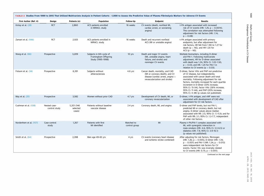
TABLE 2 Studies From 1999 to 2015 That Utilized Multivariate Analysis in Patient Cohorts >1,000 to Assess the Predictive Value of Plasma Fibrinolysis Markers for Adverse CV Events
First Author (Ref. #) Design Patients (n) Population Follow-Up Endpoint Results
Kinlay et al. (28) RCT 2,860 ACS patients enrolledin MIRACL study
16 weeks CV events (death, nonfatal MI,cardiac arrest, or worseningangina)
t-PA antigen associated with increasedrisk of CV events (HR: 1.25; p ¼ 0.0014).his correlation was attenuated followingdjustment for risk factors (HR: 1.14;¼ 0.08)
Zamani et al. (106) RCT 2,925 ACS patients enrolled inMIRACL study
16 weeks Death and recurrent nonfatalACS (MI or unstable angina)
t- antigen associated with primaryndpoints, but after adjustment forisk factors, HR fell from 1.90 to 1.27 foreath (p ¼ NS), and HR 1.20 forCS (p ¼ NS).
Wang et al. (66) Prospective 3,209 Subjects in 6th cycle ofFramingham OffspringStudy (1995–1998)
10 yrs Death and major CV events(MI, unstable angina, heartfailure, and stroke) andnonmajor CV events
M tiple biomarkers, including D-dimernd PAI-1. Following multivariatedjustment, HR for D-dimer associatedith death was 1.24; 95% CI: 1.02–1.50;¼ 0.03; and HR: 1.24 for PAI-1 in
elation to CV events (p ¼ 0.03).
Folsom et al. (34) Prospective 6,391 Subjects withoutatherosclerosis
4.6 yrs Cancer death, mortality, and CAD(MI or coronary death), and CVdisease (cardiac arrest, angina �revascularization and stroke)
D imer, factor VIIIc and PAP not predictivef CV disease, but independentlyssociated with cancer death and totalortality. Following adjustment for riskactors, mortality increased for each quartilencrement in D-dimer (33% increase;5% CI: 15–54), factor VIIIc (26% increase;5% CI: 11–44), and PAP (20% increase;5% CI: 4–38) (p values not published).
May et al. (25) Prospective 3,582 Women without prior CAD 4.7 yrs Development of CV death, MI, orcoronary revascularization
D imer, t-PA antigen, and vWF were notssociated with development of CAD afterdjustment for CV risk factors.
Cushman et al. (108) Nested case-control study
5,201 (146selectedcases)
Patients without baselinevascular disease
2.4 yrs Coronary death, MI, and angina. D imer and PAP levels, but not PAI-1,redicted MI or coronary death, but notngina. D-dimer values above medianssociated with RR: 2.5; 95% CI: 1.1–5.9; and forAP with RR: 3.1; 95% CI: 1.3–7.7, independentf other risk factors.
Nordenhem et al. (107) Case-controlstudy
1,267 Patients with firstMI identified
Matched tocontrol group
MI P ma t-PA/PAI-1 complex associated withI, with synergistic interaction inale smokers (OR: 4.6; 95% CI: 3.3–6.5) oriabetics (OR: 7.9; 95% CI: 3.9–16.1)p values not published).
Smith et al. (64) Prospective 2,398 Men age 49–65 yrs 4 yrs CV events (coronary heart diseaseand ischemic stroke combined)
A r adjusting for risk factors, fibrinogenHR: 1.26; p ¼ 0.005), D-dimer (HR: 1.34;¼ 0.001) and PAI-1 (HR: 1.24; p ¼ 0.013)ere independent risk factors for CVvents. Factor VIIc was inversely relatedo CV events (HR: 0.75; p ¼ 0.001).
Continued on the next page
Okafor
andGorog
JACC
VOL.65,NO.16,2015
EndogenousThrom
bolysisin
CVDisease
APRIL
28,2015:1
683–99
1688
Tap
PAerdA
ulaawpr
-doamfi999
-daa
-dpaaPo
lasMmd(
fte(pwet
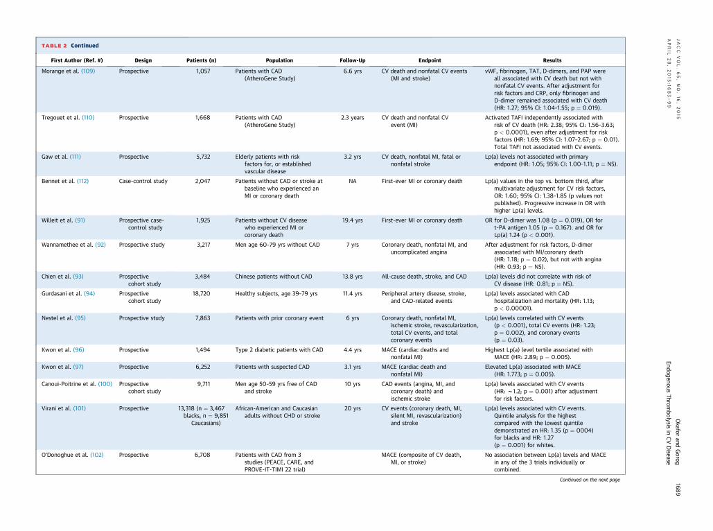
TABLE 2 Continued
First Author (Ref. #) Design Patients (n) Population Follow-Up Endpoint Results
Morange et al. (109) Prospective 1,057 Patients with CAD(AtheroGene Study)
6.6 yrs CV death and nonfatal CV events(MI and stroke)
vWF, fibrinogen, TAT, D-dimers, and PAP wereall associated with CV death but not withnonfatal CV events. After adjustment forrisk factors and CRP, only fibrinogen andD-dimer remained associated with CV death(HR: 1.27; 95% CI: 1.04–1.55; p ¼ 0.019).
Tregouet et al. (110) Prospective 1,668 Patients with CAD(AtheroGene Study)
2.3 years CV death and nonfatal CVevent (MI)
Activated TAFI independently associated withrisk of CV death (HR: 2.38; 95% CI: 1.56–3.63;p < 0.0001), even after adjustment for riskfactors (HR: 1.69; 95% CI: 1.07–2.67; p ¼ 0.01).Total TAFI not associated with CV events.
Gaw et al. (111) Prospective 5,732 Elderly patients with riskfactors for, or establishedvascular disease
3.2 yrs CV death, nonfatal MI, fatal ornonfatal stroke
Lp(a) levels not associated with primaryendpoint (HR: 1.05; 95% CI: 1.00–1.11; p ¼ NS).
Bennet et al. (112) Case-control study 2,047 Patients without CAD or stroke atbaseline who experienced anMI or coronary death
NA First-ever MI or coronary death Lp(a) values in the top vs. bottom third, aftermultivariate adjustment for CV risk factors,OR: 1.60; 95% CI: 1.38–1.85 (p values notpublished). Progressive increase in OR withhigher Lp(a) levels.
Willeit et al. (91) Prospective case-control study
1,925 Patients without CV diseasewho experienced MI orcoronary death
19.4 yrs First-ever MI or coronary death OR for D-dimer was 1.08 (p ¼ 0.019), OR fort-PA antigen 1.05 (p ¼ 0.167). and OR forLp(a) 1.24 (p < 0.001).
Wannamethee et al. (92) Prospective study 3,217 Men age 60–79 yrs without CAD 7 yrs Coronary death, nonfatal MI, anduncomplicated angina
After adjustment for risk factors, D-dimerassociated with MI/coronary death(HR: 1.18; p ¼ 0.02), but not with angina(HR: 0.93; p ¼ NS).
Chien et al. (93) Prospectivecohort study
3,484 Chinese patients without CAD 13.8 yrs All-cause death, stroke, and CAD Lp(a) levels did not correlate with risk ofCV disease (HR: 0.81; p ¼ NS).
Gurdasani et al. (94) Prospectivecohort study
18,720 Healthy subjects, age 39–79 yrs 11.4 yrs Peripheral artery disease, stroke,and CAD-related events
Lp(a) levels associated with CADhospitalization and mortality (HR: 1.13;p < 0.00001).
Nestel et al. (95) Prospective study 7,863 Patients with prior coronary event 6 yrs Coronary death, nonfatal MI,ischemic stroke, revascularization,total CV events, and totalcoronary events
Lp(a) levels correlated with CV events(p < 0.001), total CV events (HR: 1.23;p ¼ 0.002), and coronary events(p ¼ 0.03).
Kwon et al. (96) Prospective 1,494 Type 2 diabetic patients with CAD 4.4 yrs MACE (cardiac deaths andnonfatal MI)
Highest Lp(a) level tertile associated withMACE (HR: 2.89; p ¼ 0.005).
Kwon et al. (97) Prospective 6,252 Patients with suspected CAD 3.1 yrs MACE (cardiac death andnonfatal MI)
Elevated Lp(a) associated with MACE(HR: 1.773; p ¼ 0.005).
Canoui-Poitrine et al. (100) Prospectivecohort study
9,711 Men age 50–59 yrs free of CADand stroke
10 yrs CAD events (angina, MI, andcoronary death) andischemic stroke
Lp(a) levels associated with CV events(HR: w1.2; p ¼ 0.001) after adjustmentfor risk factors.
Virani et al. (101) Prospective 13,318 (n ¼ 3,467blacks, n ¼ 9,851
Caucasians)
African-American and Caucasianadults without CHD or stroke
20 yrs CV events (coronary death, MI,silent MI, revascularization)and stroke
Lp(a) levels associated with CV events.Quintile analysis for the highestcompared with the lowest quintiledemonstrated an HR: 1.35 (p ¼ 0004)for blacks and HR: 1.27(p ¼ 0.001) for whites.
O’Donoghue et al. (102) Prospective 6,708 Patients with CAD from 3studies (PEACE, CARE, andPROVE-IT-TIMI 22 trial)
MACE (composite of CV death,MI, or stroke)
No association between Lp(a) levels and MACEin any of the 3 trials individually orcombined.
Continued on the next page
JACC
VOL.65,NO.16,2015
Okafor
andGorog
APRIL
28,2015:1
683–99
EndogenousThrom
bolysisin
CVDisease
1689

TABLE
2Co
ntinue
d
FirstAut
hor(R
ef.#
)Des
ign
Patien
ts(n
)Pop
ulation
Follow
-Up
Endp
oint
Res
ults
SukDan
iket
al.(103)
Prospe
ctive
27,791
Hea
lthy
wom
enag
e>45
yrs
10yrs
MACE
(non
fatalMI,no
nfatal
cerebrov
ascu
larev
ent,co
rona
ryreva
scularization,
orCV
death)
Lp(a)leve
lsin
thehigh
estvs.lowestqu
artiles
associated
withad
verseev
ents
(HR:1.35
;p<
0.001fortren
dacross
quartiles).
Kam
strupet
al.(104)
Prospe
ctive
9,330
Subjects
witho
utpriorCA
D10
yrs
CAD
(includ
ingMI)or
death
RaisedLp
(a)leve
lassociated
withHR:1.09
(95%
CI:1.06–1.12;p¼
0.93)
forMIa
nd1.06
(95%
CI:1.04–
1.08;p¼
0.86)forCA
D.
Shilp
aket
al.(105)
RCT
2,76
3Po
st-m
enop
ausalwom
enag
e<80
yrswithCA
D4.1yrs
CVev
ents
(non
fatalMIan
dCV
death)
Lp(a)leve
lsassociated
withCV
even
ts(H
R:1.54
;95%
CI:0.99–2.39
;p¼
0.03)
ACS
¼acuteco
rona
rysynd
romes;CA
D¼
corona
ryartery
disease;
CARE
¼Ch
olesterolAnd
Recurrent
Even
ts;CH
D¼
corona
ryhe
artdisease;
CI¼
confi
denc
einterval;CR
P¼
C-reactive
protein;
CV¼
cardiova
scular;HR¼
hazard
ratio;
Lp(a)¼
lipop
rotein
(a);
MACE
¼major
adve
rsecardiova
scular
even
ts;MI¼
myo
cardialinfarction
;MIRACL
¼Myo
cardialIsch
emia
Red
uction
withAgg
ressiveCh
olesterolLo
wering;
NA¼
notap
plicab
le;NS¼
notsign
ificant;OR¼
odds
ratio;
PAP¼
plasmin-alpha
2-an
tiplasmin
complex
;PE
ACE
¼Prev
ention
ofEv
ents
WithAng
iotensin-C
onve
rtingEn
zymeInhibitorTh
erap
y;PR
OVE-IT-TIM
I22¼
Prav
astatinor
AtorvastatinEv
alua
tion
andInfectionTh
erap
y-Th
rombo
lysisIn
Myo
cardialInfarction22
;RCT
¼rand
omized
controlle
dtrial;RR¼
relative
risk;
TAFI
¼thrombin-activa
tablefibrinolysisinhibitor;TA
T¼
thrombin-an
tithrombinco
mplex
;vW
F¼
vonWilleb
rand
factor;othe
rab
brev
iation
sas
inTa
ble1.
Okafor and Gorog J A C C V O L . 6 5 , N O . 1 6 , 2 0 1 5
Endogenous Thrombolysis in CV Disease A P R I L 2 8 , 2 0 1 5 : 1 6 8 3 – 9 9
1690
dynamics of clot formation. As blood clot formationoccurs around the pin, fibrin strands form betweenthe cylindrical cup and pin. With additional rotationof the cylindrical cup, this is transmitted to the pinand the resulting mechanical-electrical transductionis depicted in a numerical and graphical representa-tion. With the ROTEM technique, movement is in-stead generated from the oscillation of the pin/wiretransduction system, while the cup is held immobileand an optical detection system is utilized to trans-duce the signal. TEG can be modified to use a varietyof different activators and inhibitors to provide in-formation on specific components of the coagulationsystem, including platelet function testing and fibri-nolytic status (38–45). In fibrinolysis assessment, TEGis typically compared in the presence and absence ofthe fibrinolysis inhibitor aprotinin (37,46). Table 1shows the features of thromboelastography and howthese compare with the GTT.
There are a number of important limitations to theuse of TEG as a clinical tool to assess global throm-botic status. TEG was originally designed for native,nonanticoagulated blood (47), but subsequent modi-fications have included the use of activators of coag-ulation and additional reagents to evaluate specificcomponents of hemostasis (38,47). This has helpedstandardize the initiation of coagulation, but does notreflect a patient’s physiological state. Although TEG isa useful tool for assessing bleeding risk, for examplein cardiac surgery, its practical value in assessing the(spontaneous) thrombolytic status of patients or theeffect of medications is questionable. The shortcom-ings of this technique begin with the testing ofcitrated and recalcified blood. The effect of extracel-lular calcium concentration on coagulation indexesand thromboelastography results is significant (48).There are significant differences in TEG results be-tween fresh native whole blood and recalcified citra-ted whole blood (49,50) and the correlation betweenTEG results performed on kaolin- versus nonkaolin-activated native and citrated blood is poor (51).
In the absence of shear or any other platelet-activating stimuli, clot formation can be initiatedeither by intrinsic (kaolin, ellagic acid) or extrinsic(tissue factor) activators, and the test results varyaccordingly. However, the major limitation of TEG isthat it fails to assess the procoagulant (thrombin-generating) and fibrinolysis-inhibiting (PAI-1; TAFI)properties of platelets. Furthermore, the use of gentlerotation of a cylindrical cup more closely resemblesthe low shear stress environment encountered withvenous stasis and does not reflect the high-shearsituation in stenosed arteries. Furthermore, thismitigates the contribution of platelet activation and

J A C C V O L . 6 5 , N O . 1 6 , 2 0 1 5 Okafor and GorogA P R I L 2 8 , 2 0 1 5 : 1 6 8 3 – 9 9 Endogenous Thrombolysis in CV Disease
1691
subsequent thrombin generation, which play keyroles in arterial thrombogenesis.
TEG results have only demonstrated a weak cor-relation with standard tests of coagulation (52), withno formal validation or standardization (53) and sig-nificant interlaboratory variability; coefficients ofvariation range between 8% and 40% for TEG and upto 4% to 84% for ROTEM (54). Due to the problemswith standardization and determination of normalreference values, TEG is better utilized as a measureof the change in coagulation status over time, when apatient’s baseline results are already known (55).Despite these limitations, TEG benefits from itsavailability as a point-of-care test, providing rapidinformation on the coagulation profile of patients.
GLOBAL THROMBOSIS TEST
The GTT is a newer point-of-care test that simulta-neously assesses platelet reactivity, thrombosis,and thrombolytic activity, from a single, non-anticoagulated blood sample (56,57). This technique
TABLE 3 Clinical Studies Evaluating the GTT in the Prediction of Card
First Author (Ref. #) Population Patients (n)
Sharma et al. (60) End-stage renal diseasepatients on hemodialysis
216
Saraf et al. (61) ACS patients receiving dualantiplatelet therapy
300
Saraf et al. (61) ACS vs. healthycontrol subjects
300
Suehiro et al. (75) Healthy subjects of smokingand nonsmoking status
Smokers ¼ 76 vs.nonsmokers ¼ 63
Ikarugi et al. (76) Healthy young malesand elderly males
Young ¼ 30 vs.elderly ¼ 34
Suehiro et al. (77) Males with MetS vs.control subjects
MetS ¼ 30 vs.control ¼ 53
Rosser et al. (98) ACS or stable coronary diseaserandomized to vorapaxar vs.placebo, in addition tostandard of care
57
Taomoto et al. (99) Acute cerebrovasculardisease (CVA) vs.healthy control subjects
CVA ¼ 185control subjects ¼ 19
CVA ¼ cerebrovascular accident (stroke); LT ¼ lysis time; MetS ¼ metabolic syndrome;
utilizes native, nonanticoagulated blood that is freeof any external agonists (Table 1). Platelets becomeactivated by the high shear stress generated by thepassage of blood through a conical tube containingnarrow gaps. The predominant stimulus for plateletactivation in severely-narrowed atherosclerotic cor-onary arteries is pathologically high shear stress(>10,000 s�1), which leads to rapid platelet activa-tion. The GTT mimics this pathological environmentto provide high shear stress as the primary stimulusfor platelet aggregation, platelet microparticle, andthrombin generation, resulting in occlusive thrombusformation (58,59). The time taken for an occlusivethrombus to form in the space downstream, reflectingplatelet aggregation and initiation of coagulation, ismanifested in the arrest of flow as detected by anoptical sensor, and is termed the occlusion time(OT, s). The restart of blood flow, due to spontaneousdissolution of the formed thrombus, representsendogenous thrombolytic activity and is recordedagain by an optical sensor and termed the lysis time(LT, s).
iovascular Risk
Follow-Up Methods Primary Endpoint Results
276 � 166 days GTT MACE (CV death,nonfatal MI, CVA,and peripheralarterial thrombosis)
Impaired endogenous thrombolysis(LT >3,000 s) strongly associatedwith MACE (HR: 4.25; p ¼ 0.004),nonfatal MI, and CVA (HR: 14.28;p ¼ 0.0 ¼ 1) and peripheralthrombosis (HR: 9.08;p ¼ 0.003)
12 months GTT MACE (CV death,nonfatal MI,or CVA)
LT >3,000 s was an independentpredictor of MACE (HR: 2.52;p ¼ 0.004) and CV death (HR: 4.2;p ¼ 0.033).
N/A GTT MACE (CV death,nonfatalMI, or CVA)
OT prolonged in ACS (428 s vs. 378 s;p < 0.001) and LT shorter in ACS(1,053 s vs. 1,362 s; p < 0.001) thanin control subjects
3 months GTT Effect of smoking onthrombotic profile
LT was significantly longer in smokersthan in nonsmokers (1,794 s vs.1,530 s;p ¼ 0.029) with no significantdifference in OT
N/A GTT Effect of age, smoking,and exercise onthrombotic profile
LT was significantly longer in elderly vs.young (p < 0001), and prolonged inelderly smokers than nonsmokers(p < 0.001)
N/A GTT Comparison ofthrombotic profilebetween groups
LT significantly longer in MetS than incontrol subjects (1,494 s vs. 1,246 s).PAI-1 level correlated with LT(p < 0.01)
N/A GTT Thrombotic status,as shown by OTand LT of GTT
Vorapaxar treatment prolonged OT(561 s vs. 372 s; p ¼ 0.003) andshortened LT (1,158 s vs. 1,733 s;p ¼ 0.016)
5N/A GTT Thrombotic status,
as shown by OTand LT of GTT
In stroke patients, OT was shorter(p < 0.0001) and LT was longer(p < 0.0001) than in healthycontrol subjects
OT ¼ occlusion time; other abbreviations as in Tables 1 and 2.

TABLE 4 Studies Utilizing TEG in the Evaluation of Platelet Reactivity and its Correlation to the Risk of Ischemic Events in Nonsurgical Cardiovascular Patients
First Author (Ref. #) Population Patients (n) Follow-Up Method Primary Endpoint Results
Jeong et al. (113) PCI-treated patients receivingaspirin and clopidogrel
197 24 months MA-thrombin TEGmeasurements,conventionalaggregometry, andgenotyping
Relationship between MA-thrombin on high on-treatment platelet ctivity(HPR) and long-te ACE
HPR and high MA-thrombinwere both independentlyassociated with MACE (HR:3.09 and 2.24, respectively).The combination of bothincreased HR for MACE to5.56; p ¼ 0.0002. High MA-thrombin also predicted therisk for HPR (OR: 13.89;p < 0.001)
Gurbel et al. (114) Patients undergoing PCI andtaking aspirin and clopidogrel
225 36 months ADP-induced (MA-ADP)and thrombin-induced(MA-thrombin) TEGmeasurement and LTA
Prediction of long-ter ventoccurrence (ischem ndbleeding) followin enting
Patients with ischemic eventshad higher MA (ADP), MA(thrombin), and LTA (p <
0.0001 for all), which wereindependent predictors ofischemic events at 3 years(HR: 10.3, 3.8, and 4.8,respectively; all p < 0.0001)
Tang et al. (115) Patients undergoing PCI dividedinto 3 groups depending oninhibition rates to aspirin andclopidogrel (n ¼ 90): controlgroup (n ¼ 30) and resistancegroup (n ¼ 60), who were thenrandomized to 2 subgroups(R þ R and R þ L) to receivedifferent antiplateletcombinations
90 12 months TEG Occurrence of CV isch c events(including stent th bosis,recurrent unstable ina,and MI)
Patients resistant to antiplatelettherapy vs. nonresistantcontrol groups, had anincreased risk of stentthrombosis (20% vs. 3%),recurrent unstable angina(36% vs.10%), and (MI 17%vs. 1%; p < 0.01).Randomization to a loadingdose regimen improvedinhibition rates and reducedthe rates of CV events(p < 0.01)
Gurbel et al. (116) Patients undergoing PCI 84 24 months TEG and conventionalaggregometry.Biomarker evaluationwith fluorokinemultianalyte profiling
Thrombogenicity and arkersof inflammation acorrelation to the rrenceof ischemic events
Patients with high MA had anischemic event more oftenthan patients with low MA(48% vs. 13%; p ¼ 0.02).Those in the highest MAgroup demonstrated higherlevels of CRP, IL-8, andepidermal and vascularendothelial growth factors.
Gurbel et al. (117) Patients undergoingnonemergent PCI
192 6 months ADP-induced LTA andTEG
Platelet reactivity and tstrength and the r f post-discharge ischemic nts
Patients experiencing ischemicevents (n ¼ 38)demonstrated higherplatelet reactivity by LTA(63 � 12% vs. 56 � 15%;p ¼ 0.02), higher clotstrength (MA) (74 � 5 mmvs. 65 � 4 mm; p ¼ 0.001)and more rapid fibringeneration (4.3 � 1.3 min vs.5.9 � 1.5 min; p ¼ 0.001)
Continued on the next page
Okafor
andGorog
JACC
VOL.65,NO.16,2015
EndogenousThrom
bolysisin
CVDisease
APRIL
28,2015:1
683–99
1692
rearm M
m eic ag st
emiromang
biomndoccu
cloisk oeve

TABLE 4 Continued
First Author (Ref. #) Population Patients (n) Follow-Up Method Primary Endpoint Results
Bliden et al. (118) Patients receiving aspirin (325 mgqd) and clopidogrel (75 mg qd)undergoing nonemergent PCI
100 12 months Measurement of plateletaggregation bystandard LTA and TEG
Correlation between heightenedplatelet aggregation andoccurrence of ischemic events
High on-treatment plateletreactivity, as measured byaggregometry and TEG,were significantly related toischemic events (p ¼ 0.001for both assays).
Gurbel et al. (119) African-American and Caucasianpatients undergoingelective PCI
252 6 months TEG Assess race and sex difference inthrombogenicity and relatethis to adverse ischemic events
TEG-derived platelet clotstrength measurements (RR:2.52; p ¼ 0.017) and sex(RR: 2.56; p ¼ 0.009) asindependent predictors ofischemic events. African-American women exhibitedhigher thrombogenicity thanthe other race and sexgroups (p < 0.05)
Kreutz et al. (24) Patients with coronary arterydisease, treated with aspirinand clopidogrel
211 3 � 1.9 yrs Platelet aggregometryassessed by LTA andclot formation usingTEG. Genotyping ofVal34Leu usingTaqMan assay
Evaluate effects of Val34Leu onfibrin generation, plateletaggregation, and long-termclinical outcomes
Homozygous carriers of 34Leuvariant had the greatest riskof MI and CV death (p ¼0.002), associated withreduced fibrin clot formationtime (TEG K: 1.27 � 0.3 minvs. 1.68 � 1.1 min;p ¼ 0.011).
Tang et al. (120) Chinese patients undergoingPCI for ACS
577 12 months Detection of CYP2C19G681A and P2Y12C34T polymorphismsby ligase detectionreaction. Plateletreactivity assessed byTEG
Clopidogrel responsiveness andMACE (CV death, nonfatal MI,target vessel revascularization,and stent thrombosis)
118 patients with mutational Aallele of CYP2C19 andmutational T allele of P2Y12demonstrated lowest ADPinhibition (49.74 � 32.61%)and highest prevalence ofclopidogrel low response(29.7%), which correlatedwith the highest CV eventrates (8.5% vs. 1.5%).
Wu et al. (121) NSTEMI patientsundergoing PCI
233 24 h CYP2C19*2 and *3 LOFalleles were evaluatedusing DNA microarraymethod. Plateletreactivity assessedby TEG
CYP2C19 genotype on HPR andrisk of periprocedural MI
HPR more frequent in patientswith periprocedural MI andan independent risk factorfollowing multivariateanalysis (OR: 4.348; p ¼0.001). HPR also correlatedwith 2 CYP2C19 LOF allelecarriage, associated with a3-fold increased risk(p ¼ 0.037).
Continued on the next page
JACC
VOL.65,NO.16,2015
Okafor
andGorog
APRIL
28,2015:1
683–99
EndogenousThrom
bolysisin
CVDisease
1693

TABLE
4Co
ntinue
d
FirstAut
hor(R
ef.#
)Pop
ulation
Patien
ts(n
)Fo
llow
-Up
Metho
dPrimaryEn
dpoint
Res
ults
CaoJet
al.(122
)Elde
rlymen
withCV
disease
receivingda
ilyaspirin
therap
y(>
75mg)
304
1.8yrs
Platelet
aggreg
ation
mea
suredby
LTAan
dTE
G
MACE
(com
posite
ofde
ath,
MI,
unstab
lean
gina
,strok
e,an
dtran
sien
tisch
emic
attack)
Asp
irinresistan
ce(assessedby
TEG)no
tassociated
with
vascular
even
ts(17.7%
vs.
10.9%;p
¼0.452
),althou
ghaspirin
-resistanc
e(defi
ned
byLT
A)increa
sedris
kof
compo
site
outcom
e(18.3%
vs.9.8%;HR:1.864;
p¼
0.003)
Tang
etal.(123
)Ch
inesepa
tien
tsun
dergoing
PCI
670
12mon
ths
Antiplateleteffect
assessed
byTE
G,
CYP2
C19,A
BCB
1,an
dPO
N1ge
notype
sde
tected
bylig
ase
detectionreaction
Relationshipbe
twee
nge
notype
varia
ntson
clop
idog
rel
resp
onsive
ness
andco
rrelation
toMACE
(CVde
ath,
nonfatal
MI,target
vessel
reva
scularization,
andsten
tthrombo
sis)
CYP2
C19LO
Fallelesfoun
din
57.3%
ofpa
tien
tsan
dassociated
withage
nedo
se-
depe
nden
teffect
ontheris
kof
low
resp
onse
toclop
idog
relan
dad
verse
isch
emic
even
ts
Drid
ietal.(124)
Patien
tswithST
EMI
unde
rgoing
urge
ntPC
I23
312
mon
ths
Platelet
activity
mea
sured
withTE
G-M
A.
Relationshipbe
twee
nTE
Gan
dmyo
cardialda
mag
e(assessed
withCM
R)in
STEM
Ipatients
TEG-defi
nedhy
percoa
gulation
presen
tin
35.2%
not
correlated
withinfarctsize,
myo
cardialsalvag
einde
x,or
adve
rseev
ents.
ABCB
1¼
ATP
-binding
cassette,s
ub-fam
ilyB,m
embe
r1;
ADP¼
aden
osinediph
osph
ate;
CMR¼
cardiacmag
neticresona
nce;
CYP2
C19¼
cytoch
romeP4
502C
19;D
NA¼
deox
yribon
ucleic
acid;H
PR¼
high
platelet
reactivity;IL¼
interleu
kin;
LOF¼
loss-of-func
tion
;LT
A¼
light
tran
smittanc
eag
greg
ometry;MA
¼maxim
umam
plitud
e;NST
EMI¼
non–ST
-seg
men
telev
ationmyo
cardialinfarction
;PC
I¼
percutan
eous
corona
ryinterven
tion
;PO
N1¼
serum
paraox
onase1;
qd¼
daily
;ST
EMI¼
ST-seg
men
telev
ationmyo
cardial
infarction
;TE
G¼
thrombo
elastograp
hy;othe
rab
brev
iation
sas
inTa
bles
2an
d3.
Okafor and Gorog J A C C V O L . 6 5 , N O . 1 6 , 2 0 1 5
Endogenous Thrombolysis in CV Disease A P R I L 2 8 , 2 0 1 5 : 1 6 8 3 – 9 9
1694
Because the GTT assesses both thrombus formationand thrombus lysis in native blood, without externalagonists and using high shear, it is arguably the mostphysiological assessment of global thrombotic statuscurrently available.
This test has been studied in patient groups at highrisk of cardiovascular thrombosis, with early resultssuggesting that it may be useful in predicting clinicaloutcomes (60,61). It has not been compared withother platelet function tests or TEG. However, theresults would not be expected to correlate because ofthe different flows (high vs. low shear stress) and theuse of native versus anticoagulated blood (Table 1).
ENDOGENOUS FIBRINOLYSIS:
EVIDENCE FOR AN IMPORTANT ROLE
IN CARDIOVASCULAR DISEASE
PLASMA MARKERS OF FIBRINOLYSIS IN CARDIO-
VASCULAR DISEASE. A number of studies haveattempted to examine the relationship betweenplasma markers of fibrinolysis, signifying impairedfibrinolysis, and the occurrence of cardiovascularevents.
Genetic polymorphisms in key enzyme regulatorsof fibrinolysis may increase susceptibility to throm-botic events (62). The 4G4G phenotype of the 4G/5GPAI-1 gene polymorphism was found to be an inde-pendent predictor of AMI (odds ratio: 2.7, p ¼ 0.002)(63), and was observed more frequently in patientswith a previous history of AMI than in those withstable angina. A review of the prospective studiesundertaken between 1999 and 2009, encompassingsome 45 studies and nearly 50,000 patients, demon-strates the conflicting results regarding the useful-ness of these markers (31). Most of these were largeepidemiological studies assessing thousands of pa-tients. Table 2 summarizes publications between 1999and 2015 that utilized multivariate analysis in patientcohorts >1,000 to assess the predictive value ofplasma fibrinolysis markers for adverse cardiovascu-lar events.
In 1 of the larger prospective studies that evaluatedt-PA levels in 3,582 women, there was a weak corre-lation between t-PA and the development of coronaryartery disease (25). In patients with established cor-onary disease or AMI, t-PA levels were predictive offuture cardiovascular events. In the Caerphilly Studyof 2,398 men with 13 years of follow-up, baseline PAI-1 levels were significantly associated with cardiovas-cular risk (64), but after multivariate analysis, thecorrelation became nonsignificant (64). Other studiesevaluating PAI-1 demonstrated a significant associa-tion with coronary events (65), cardiogenic shock,

J A C C V O L . 6 5 , N O . 1 6 , 2 0 1 5 Okafor and GorogA P R I L 2 8 , 2 0 1 5 : 1 6 8 3 – 9 9 Endogenous Thrombolysis in CV Disease
1695
death (32), and major adverse cardiovascular events(MACE) (32). In the Framingham study involving3,209 participants, PAI-1 levels were not relatedto cardiovascular events (66). Other studies have alsofailed to demonstrate a prognostic role for base-line t-PA or PAI-1 levels (27,28). There is also con-flicting data from studies on the role of other plasmamarkers of fibrinolysis, including D-dimer assays(25,28,31,64), plasmin-alpha2-antiplasmin complexmeasurements (31), TAFI (31), and Lp(a) levels (31,32).
Even allowing for publication bias, in that negativestudies are less likely to be published, it is clear thatbiomarkers of fibrinolysis may, at best, allow a weakprediction of increased cardiovascular risk at a pop-ulation level only. It is difficult to ascertain globalfibrinolytic status on the basis of the plasma level of 1or even several biomarkers. Furthermore, there is stillcontroversy regarding the ideal laboratory techniqueto use. Determination of the total antigen levelsof plasma markers can be achieved using enzyme-linked immunosorbent assays; alternatively, mea-surement of specific biological activity levels ofplasma markers can be undertaken with immuno-functional chromogenic substrate kinetic assays.With some plasma markers, such as PAI-1, which has arelatively long half-life (approximately 1 h), there islikely to be a good correlation between PAI-1 antigenand PAI-1 activity levels. However, a poor correlationhas been demonstrated between measurements ofTAFI antigen and TAFI activity (67), which may be areflection of its short half-life (approximately 10 min).
These problems with plasma marker measure-ments are confounded by the additional role ofcomplementary pathways involved in mediatingendogenous fibrinolysis. Studies have demonstratedthe importance of plasma fibrin architecture in facil-itating effective endogenous fibrinolysis (68) (CentralIllustration). Additionally, the release of proteolyticenzymes from thrombus-associated neutrophils,namely elastase, has been shown to result in directdigestion of fibrin and inactivation of PAI-1 (69)(Central Illustration). Moreover, thrombus-adherentmonocytes have been demonstrated to enhanceTAFI activity, reducing fibrinolytic activity and pro-tecting against clot lysis (70).
Platelets represent an important source of PAI-1,containing up to 90% of the total PAI-1 content ofblood (71). During thrombus formation, activatedplatelets release high local concentrations of PAI-1,which serve to inhibit thrombolysis and stabilizeclot formation (Central Illustration). The most func-tionally important source of PAI-1 is, therefore,platelets, and this pool of PAI-1 varies independentlyof plasma PAI-1 levels (72–74).
These studies have highlighted that regulation ofthrombus formation is a dynamic, multifaceted phe-nomenon, and measurements of individual compo-nents of the pathway do not give an accuratereflection of this complex system.GTT IN CARDIOVASCULAR DISEASE. Because thebalance between prothrombotic factors and endoge-nous thrombolytic activity determines the propensityfor thrombus formation in ACS, an overall assessmentof thrombotic risk requires a global evaluation of apatient’s thrombotic profile, including platelet reac-tivity, activation of the coagulation system (thrombingeneration), and endogenous fibrinolysis.
Clinical studies evaluating the GTT are shown inTable 3. A study of 300 patients with ACS (61)revealed that although platelet reactivity wasreduced, endogenous thrombolysis was impaired inACS patients compared with healthy volunteers,despite taking dual antiplatelet medication. Therewas no correlation between OT and MACE. Some 23%of ACS patients had a markedly prolonged LT, afinding that was not demonstrated in normal sub-jects. Impaired endogenous thrombolysis was an in-dependent predictor of MACE. LT >3,000 s wasidentified as the optimal cutoff point to predictMACE; above this level, the hazard ratio for cardio-vascular events increased as the LT increased. LTremained a statistically-significant predictor forMACE, even after adjustments for a number of base-line cardiovascular risk factors.
The GTT has also been used to assess the thromboticprofile of patients with established cardiovascular riskfactors. LT was significantly prolonged in smokerscompared with nonsmokers, whereas no significantdifference in OT was observed (75). There was a directcorrelation between LT and daily cigarette consump-tion. Following 3 months of smoking cessation, LTvalues were found to be significantly shorter whencompared with baseline GTT measurements. Anotherstudy demonstrated impaired endogenous thrombo-lytic activity in elderly male patients and in those whosmoked, but showed no difference in OT (76), sug-gesting that the increased susceptibility of smokers tothrombosis may, in part, be related to decreasedfibrinolytic activity. In patients with metabolic syn-drome, LT was significantly prolonged compared withnormal volunteers, and was associated with signifi-cantly higher PAI-1 levels, although no difference wasobserved in OT (77).
Patients with end-stage renal disease (ESRD) are atmuch higher cardiovascular risk than the generalpopulation (60,78), and impairment of endogenousfibrinolytic activity has also been observed, withreduced t-PA secretion and elevated levels of

Okafor and Gorog J A C C V O L . 6 5 , N O . 1 6 , 2 0 1 5
Endogenous Thrombolysis in CV Disease A P R I L 2 8 , 2 0 1 5 : 1 6 8 3 – 9 9
1696
fibrinogen and PAI-1 (79). Patients with ESRDdemonstrated significantly prolonged OT and LTcompared with normal volunteers (60). Additionally,42% of patients demonstrated an LT >3,000 s and34% demonstrated markedly impaired fibrinolyticstatus with LT >6,000 s, compared with none of thecontrol subjects. LT was strongly predictive of thecomposite of cardiovascular death, nonfatal myocar-dial infarction, cerebrovascular events, and periph-eral thrombotic events, even after adjustment forbaseline variables. No relationship between OT andMACE was observed.
TEG IN CARDIOVASCULAR DISEASE. TEG has beenapplied to guide the use of blood and blood productsduring trauma resuscitation (80) and liver and cardiacsurgery (81,82), and, more recently, it has also beenevaluated in obstetric patients (83). It has now beenreliably demonstrated that TEG detects hyper-fibrinolysis in the perioperative and trauma setting(84,85), with increasing evidence that TEG-guidedalgorithms can help to optimize patient manage-ment (82,86). Its indications for use in the assessmentof cardiovascular patients are ever expanding,including the monitoring of patients on aspirin, clo-pidogrel, and glycoprotein IIb/IIIa antagonists(87,88). A number of studies have demonstrated thatTEG-derived measurements of platelet responsive-ness can be utilized as a prognostic marker to predictthe risk of long-term ischemic events (Table 4).
However, TEG has proven to be a less robust mea-sure of hypofibrinolysis, with unmodified TEG assaysin normal subjects exhibiting only a minor degree offibrinolysis. Indeed, in 1 study, the normal range ofROTEMmaximum lysis at 60min was demonstrated tobe <12% (range 0% to 12%) (89). The current limitationwith existing TEG techniques to evaluate hypofi-brinolysis has prompted the development of novelmethods to improve its sensitivity. These techniqueshave included the use of exogenous urokinase or t-PAin concentrations that allow for the assessment of clotformation, whilst simultaneously enhancing clot lysis,permitting more accurate assessment of hypofi-brinolysis (90). However, there has been no formalstandardization and very little published data on theseapproaches, and further work is required to improvethe sensitivity and standardization of TEG techniquesto evaluate hypofibrinolysis.
A large number of studies have evaluated theusefulness of TEG in assessing clot strength. In thisregard, TEG has been shown to be very useful inpredicting increased cardiovascular risk in patientswith established coronary disease and in thoseundergoing percutaneous coronary intervention(Table 4).
CONCLUSIONS
Although previously viewed as a secondary phe-nomenon in response to the formation of thrombi, alarge body of evidence now points to a muchmore prominent role for endogenous fibrinolysis inthrombus formation.
The technical limitations, difficulty in interpreta-tion, and conflicting data regarding prognostic use-fulness of plasma markers in patients with coronarydisease limit their adoption into clinical practice.Recognition of these limitations prompted the de-velopment of global assays of fibrinolytic status. TEGis a useful tool for assessing bleeding risk, and hasalso been used to assess clot strength, which hasbeen shown to predict future cardiovascular events.However, its practical value in assessing the (spon-taneous) thrombolytic status of patients or the effectof medications is questionable, due to its inability toassess the procoagulant and fibrinolysis-inhibitingproperties of platelets and its low shear-stress mi-lieu, which more closely resembles venous flow. TheGTT provides a physiological assessment of globalthrombotic status by assessing both thrombus for-mation and thrombus lysis in native blood in a high-shear setting that is relevant to arterial flow. Earlyclinical studies suggest that it may be useful inidentifying patients at risk of future cardiovascularevents. Endogenous fibrinolysis, until recently apoorly-understood area, represents an expandingand exciting area for identifying patients at in-creased cardiovascular risk and as a potentialtarget for pharmacological modulation to improveoutcomes.
REPRINT REQUESTS AND CORRESPONDENCE: Prof.Diana A. Gorog, Imperial College, Dovehouse Street,London SW3 6LY, United Kingdom. E-mail: [email protected].
RE F E RENCE S
1. Fuster V, Moreno PR, Fayad ZA, et al.Atherothrombosis and high-risk plaque: part I:evolving concepts. J Am Coll Cardiol 2005;46:937–54.
2. Penz S, Reininger AJ, Brandi R, et al. Humanatheromatous plaques stimulate thrombus for-mation by activating platelet glycoprotein VI.FASEB J 2005;19:898–909.
3. Reininger AJ, Bernlochner I, Penz SM, et al. A 2-step mechanism of arterial thrombus formationinduced by human atherosclerotic plaques. J AmColl Cardiol 2010;55:1147–58.
J A C C V O L . 6 5 , N O . 1 6 , 2 0 1 5 Okafor and GorogA P R I L 2 8 , 2 0 1 5 : 1 6 8 3 – 9 9 Endogenous Thrombolysis in CV Disease
1697
4. Cambruzzi E, Sebben JC, Budzyn R, et al. His-topathological evaluation of coronary thrombi inpatients with ST-segment elevation myocardialinfarction. Revista Brasileira de Cardiologia Inva-siva 2012;20:267–73.
5. Rittersma SZ, van der Wal AC, Koch KT, et al.Plaque instability frequently occurs days or weeksbefore occlusive coronary thrombosis: a patho-logical thrombectomy study in primary percuta-neous coronary intervention. Circulation 2005;111:1160–5.
6. Verouden NJ, Kramer MC, Li X, et al. Histopa-thology of aspirated thrombus and its associationwith ST-segment recovery in patients undergoingprimary percutaneous coronary intervention withroutine thrombus aspiration. Catheter CardiovascInterv 2011;77:35–42.
7. Kramer MC, van der Wal AC, Koch KT, et al.Histopathological features of aspirated thrombiafter percutaneous coronary intervention in pa-tients with ST-elevation myocardial infarction.PLoS One 2009;4:e5817.
8. Henriques de GR, van der Wal AC, van derLoos CM, et al. Sudden unexpected death in youngadults. Discrepancies between initiation of acuteplaque complications and the onset of acute cor-onary death. Eur Heart J 2002;23:1433–40.
9. Swan HJ. Thrombolysis in acute myocardialinfarction: treatment of the underlying coronaryartery disease. Circulation 1982;66:914–6.
10. Bainey KR, Fu Y, Wagner GS, et al. Sponta-neous reperfusion in ST-elevation myocardialinfarction: comparison of angiographic and elec-trocardiographic assessments. Am Heart J 2008;156:248–55.
11. Assessment of the Safety and Efficacy of aNew Treatment Strategy with Percutaneous Cor-onary Intervention (ASSENT-4 PCI) investigators.Primary versus tenecteplase-facilitated percu-taneous coronary intervention in patients withST-segment elevation acute myocardial infarction(ASSENT-4 PCI): randomised trial. Lancet 2006;367:569–78.
12. Fefer P, Hod H, Hammerman H, et al., for theAcute Coronary Syndrome Israeli Survey (ACSIS)2006 Study Group. Relation of clinically definedspontaneous reperfusion to outcome in ST-eleva-tion myocardial infarction. Am J Cardiol 2009;103:149–53.
13. Mann J, Davies MJ. Mechanisms of progressionin native coronary artery disease: role of healedplaque disruption. Heart 1999;82:265–8.
14. Burke AP, Kolodgie FD, Farb A, et al. Healedplaque ruptures and sudden coronary death: evi-dence that subclinical rupture has a role in plaqueprogression. Circulation 2001;103:934–40.
15. Varin R, Mirshahi S, Mirshahi P, et al. Wholeblood clots are more resistant to lysis than plasmaclots—greater efficacy of rivaroxaban. Thromb Res2013;131:e100–9.
16. Jang IK, Gold HK, Ziskind AA, et al. Differentialsensitivity of erythrocyte-rich and platelet-richarterial thrombi to lysis with recombinant tissue-type plasminogen activator: a possible explanationfor resistance to coronary thrombolysis. Circula-tion 1989;79:920–8.
17. Zhu Y, Carmeliet P, Fay WP. Plasminogenactivator inhibitor-1 is a major determinant ofarterial thrombolysis resistance. Circulation 1999;99:3050–5.
18. Undas A. Fibrin clot properties and theirmodulation in thrombotic disorders. ThrombHaemost 2014;112:32–42.
19. Dhall TZ, Shah GA, Ferguson IA, et al. Fibrinnetwork structure: modification by platelets.Thromb Haemost 1983;49:42–6.
20. Mitchell JL, Lionikiene AS, Fraser SR, et al.Functional factor XIII-A is exposed on stimulatedplatelet surface. Blood 2014;124:3982–90.
21. Dickneite G, Herwald H, Korte W, et al.Coagulation factor XIII: a multifunctional trans-glutaminase with clinical potential in a range ofconditions. Thromb Haemost 2015;113:686–97.
22. Hethershaw EL, Cilia La Corte AL, Duval C,et al. The effect of blood coagulation factor XIII onfibrin clot structure and fibrinolysis. J ThrombHaemost 2014;12:197–205.
23. Mutch NJ, Koikkalainen JS, Fraser SR, et al.Model thrombi formed under flow reveal the roleof factor-XIII- mediated cross-linking in resistanceto fibrinolysis. J Thromb Haemost 2010;8:2017–24.
24. Kreutz RP, Bitar A, Owens J, et al. Factor XIIIVal34Leu polymorphism and recurrent myocardialinfarction in patients with coronary artery disease.J Thromb Thrombolysis 2014;38:380–7.
25. May M, Lawlor DA, Patel R, et al. Associationsof von Willebrand factor, fibrin D-dimer and tissueplasminogen activator with incident coronaryheart disease: British Women’s Heart and Healthcohort study. Eur J Cardiovasc Prev Rehabil 2007;14:638–45.
26. Pradhan AD, LaCroix AZ, Langer RD, et al.Tissue plasminogen activator antigen and D-dimeras markers for atherothrombotic risk amonghealthy postmenopausal women. Circulation2004;110:292–300.
27. Smith FB, Fowkes FG, Rumley A, et al. Tissueplasminogen activator and leucocyte elastase aspredictors of cardiovascular events in subjectswith angina pectoris: Edinburgh Artery Study. EurHeart J 2000;21:1607–13.
28. Kinlay S, Schwartz GG, Olsson AG, et al.Endogenous tissue plasminogen activator and riskof recurrent cardiac events after an acute coronarysyndrome in the MIRACL study. Atherosclerosis2009;206:551–5.
29. Lee CW, Ahn JM, Park DW, et al. Tissueplasminogen activator on admission is an impor-tant predictor of 30-day mortality in patientswith acute myocardial infarction undergoingprimary angioplasty. Atherosclerosis 2008;196:327–32.
30. Soeki T, Tamura Y, Shinohara H, et al. Plasmaconcentrations of fibrinolytic factors in the sub-acute phase of myocardial infarction predictrecurrent myocardial infarction or sudden cardiacdeath. Int J Cardiol 2002;85:277–83.
31. Gorog DA. Prognostic value of plasma fibri-nolysis activation markers in cardiovascular dis-ease. J Am Coll Cardiol 2010;55:2701–9.
32. Marcucci R, Brogi D, Sofi F, et al. PAI-1 andhomocysteine, but not lipoprotein (a) and throm-bophilic polymorphisms, are independently asso-ciated with the occurrence of major adversecardiac events after successful coronary stenting.Heart 2006;92:377–81.
33. Lowe GD, Danesh J, Lewington S, et al. Tissueplasminogen activator antigen and coronary heartdisease: prospective study and meta-analysis. EurHeart J 2004;25:252–9.
34. Folsom AR, Aleksic N, Park E, et al. Prospec-tive study of fibrinolytic factors and incident cor-onary heart disease: the Atherosclerosis Risk inCommunities (ARIC) Study. Arterioscler ThrombVasc Biol 2001;21:611–7.
35. Kowalski E, Kope�c M, Niewiarowski S. Anevaluation of the euglobulin method for thedetermination of fibrinolysis. J Clin Pathol 1959;12:215–8.
36. Smith AA, Jacobson LJ, Miller BI, et al. A neweuglobulin clot lysis assay for global fibrinolysis.Thromb Res 2003;112:329–37.
37. Luddington RJ. Thrombelastography/throm-boelastometry. Clin Lab Haematol 2005;27:81–90.
38. Perry DJ, Fitzmaurice DA, Kitchen S, et al.Point-of-care testing in haemostasis. Br JHaematol 2010;150:501–14.
39. Bowbrick VA, Mikhailidis DP, Stansby G. Valueof thromboelastography in the assessment ofplatelet function. Clin Appl Thromb Hemost 2003;9:137–42.
40. Kettner SC, Panzer OP, Kozek SA, et al. Use ofabciximab-modified thrombelastography in pa-tients undergoing cardiac surgery. Anesth Analg1999;89:580–4.
41. Tuman KJ, McCarthy RJ, Djuric M, et al. Eval-uation of coagulation during cardiopulmonarybypass with a heparinase-modified thromboelas-tographic assay. J Cardiothorac Vasc Anesth 1994;8:144–9.
42. Levrat A, Gros A, Rugeri L, et al. Evaluation ofrotation thrombelastography for the diagnosis ofhyperfibrinolysis in trauma patients. Br J Anaesth2008;100:792–7.
43. Genet GF, Ostrowski SR, Sorensen AM, et al.Detection of tPA-induced hyperfibrinolysis inwhole blood by RapidTEG, KaolinTEG, and func-tional fibrinogenTEG in healthy individuals. ClinAppl Thromb Hemost 2012;18:638–44.
44. Kashuk JL, Moore EE, Sawyer M, et al. Primaryfibrinolysis is integral in the pathogenesis of theacute coagulopathy of trauma. Ann Surg 2010;252:434–42. discussion 443–4.
45. Dirkmann D, Radu-Berlemann J, Gorlinger K,et al. Recombinant tissue-type plasminogen acti-vator-evoked hyperfibrinolysis is enhanced byacidosis and inhibited by hypothermia but still canbe blocked by tranexamic acid. J Trauma AcuteCare Surg 2013;74:482–8.
46. Avidan MS, Da Fonseca J, Parmar K, et al. Theeffects of aprotinin on thromboelastography withthree different activators. Anesthesiology 2001;95:1169–74.
47. Zambruni A, Thalheimer U, Leandro G,et al. Thromboelastography with citrated blood:

Okafor and Gorog J A C C V O L . 6 5 , N O . 1 6 , 2 0 1 5
Endogenous Thrombolysis in CV Disease A P R I L 2 8 , 2 0 1 5 : 1 6 8 3 – 9 9
1698
comparability with native blood, stability of citratestorage and effect of repeated sampling. BloodCoagul Fibrinolysis 2004;15:103–7.
48. Pretorius E, Oberholzer HM, van der Spuy WJ,et al. Comparing techniques: the use of recalcifiedplasma in comparison with citrated plasma aloneand in combination with thrombin in ultrastruc-tural studies. Hematology 2011;16:337–40.
49. Rajwal S, Richards M, O’Meara M. The use ofrecalcified citrated whole blood–a pragmaticapproach for thromboelastography in children.Paediatr Anaesth 2004;14:656–60.
50. Wasowicz M, Srinivas C, Meineri M, et al.Technical report: analysis of citrated blood withthromboelastography: comparison with freshblood samples. Can J Anaesth 2008;55:284–9.
51. Thalheimer U, Triantos CK, Samonakis DN,et al. A comparison of kaolin-activated versusnonkaolin-activated thromboelastography innative and citrated blood. Blood Coagul Fibrino-lysis 2008;19:495–501.
52. Jeger V, Zimmermann H, Exadaktylos AK. CanRapidTEG accelerate the search for coagulopathiesin the patient with multiple injuries? J Trauma2009;66:1253–7.
53. Chitlur M, Sorensen B, Rivard GE, et al. Stan-dardization of thromboelastography: a reportfrom the TEG-ROTEM working group. Haemophilia2011;17:532–7.
54. Kitchen DP, Kitchen S, Jennings I, et al. Qualityassurance and quality control of thrombelastog-raphy and rotational thromboelastometry: the UKNEQAS for blood coagulation experience. SeminThromb Hemost 2010;36:757–63.
55. Dai Y, Lee A, Critchley LA, et al. Does throm-boelastography predict postoperative thrombo-embolic events? A systematic review of theliterature. Anesth Analg 2009;108:734–42.
56. Yamamoto J, Yamashita T, Ikarugi H, et al.Görög Thrombosis Test: a global in-vitro test ofplatelet function and thrombolysis. Blood CoagulFibrinolysis 2003;14:31–9.
57. Yamamoto J, Inoue N, Otsui K, et al.Global Thrombosis Test (GTT) can detect majordeterminants of haemostasis including plateletreactivity, endogenous fibrinolytic and thrombingenerating potential. Thromb Res 2014;133:919–26.
58. Bark DL Jr., Ku DN. Wall shear over highdegree stenoses pertinent to atherothrombosis. JBiomech 2010;43:2970–7.
59. Maxwell MJ, Westein E, Nesbitt WS, et al.Identification of a 2-stage platelet aggregationprocess mediating shear-dependent thrombusformation. Blood 2007;109:566–76.
60. Sharma S, Farrington K, Kozarski R, et al.Impaired thrombolysis: a novel cardiovascular riskfactor in end-stage renal disease. Euro Heart J2013;34:354–63.
61. Saraf S, Christopoulos C, Salha IB, et al.Impaired endogenous thrombolysis in acute coro-nary syndrome patients predicts cardiovasculardeath and nonfatal myocardial infarction. J AmColl Cardiol 2010;55:2107–15.
62. Isordia-Salas I, Leaños-Miranda A, Sainz IM,et al. Association of the plasminogen activator
inhibitor-1 gene 4G/5G polymorphism with STelevation acute myocardial infarction in youngpatients. Rev Esp Cardiol 2009;62:365–72.
63. Onalan O, Balta G, Oto A, et al. Plasminogenactivator inhibitor-1 4G4G genotype is associatedwith myocardial infarction but not with stablecoronary artery disease. J Thromb Thrombolysis2008;26:211–7.
64. Smith A, Patterson C, Yarnell J, et al. Whichhemostatic markers add to the predictive value ofconventional risk factors for coronary heart dis-ease and ischemic stroke? The Caerphilly Study.Circulation 2005;112:3080–7.
65. Takazoe K, Ogawa H, Yasue H, et al. Increasedplasminogen activator inhibitor activity and dia-betes predict subsequent coronary events in pa-tients with angina pectoris. Ann Med 2001;33:206–12.
66. Wang TJ, Gona P, Larson MG, et al. Multiplebiomarkers for the prediction of first major car-diovascular events and death. N Engl J Med 2006;355:2631–9.
67. Marx PF, Plug T, Havik SR, et al. The activationpeptide of thrombin-activatable fibrinolysis in-hibitor: a role in activity and stability of theenzyme? J Thromb Haemost 2009;7:445–52.
68. Collet JP, Allali Y, Lesty C, et al. Altered fibrinarchitecture is associated with hypofibrinolysisand premature coronary atherothrombosis. Arte-rioscler Thromb Vasc Biol 2006;26:2567–73.
69. Bach-Gansmo ET, Halvorsen S, Godal HC,et al. D-dimers are degraded by human neutrophilelastase. Thromb Res 1996;82:177–86.
70. Semeraro F, Ammollo CT, Semeraro N, et al.Tissue factor-expressing monocytes inhibit fibri-nolysis through a TAFI-mediated mechanism, andmake clots resistant to heparins. Haematologica2009;94:819–26.
71. Torr-Brown SR, Sobel BE. Attenuation ofthrombolysis by release of plasminogen activatorinhibitor type-1 from platelets. Thromb Res 1993;72:413–21.
72. Simpson AJ, Booth NA, Moore NR, et al. Theplatelet and plasma pools of plasminogen acti-vator inhibitor (PAI-1) vary independently in dis-ease. Br J Haematol 1990;75:543–8.
73. Soeki T, Tamura Y, Fukuda N, et al. Plasma andplatelet plasminogen activator inhibitor-1 in pa-tients with acute myocardial infarction. Jpn Circ J2000;64:547–53.
74. Katsaros KM, Kastl SP, Huber K, et al.Clopidogrel pretreatment abolishes increase ofPAI-1 after coronary stent implantation. ThrombRes 2008;123:79–84.
75. Suehiro A, Wakabayashi I, Yamashita T, et al.Attenuation of spontaneous thrombolytic activitymeasured by the global thrombosis test in malehabitual smokers. J Thromb Thrombolysis 2014;37:414–8.
76. Ikarugi H, Yamashita T, Aoki R, et al. Impairedspontaneous thrombolytic activity in elderly and inhabitual smokers, as measured by a new globalthrombosis test. Blood Coagul Fibrinolysis 2003;14:781–4.
77. Suehiro A, Wakabayashi I, Uchida K, et al.Impaired spontaneous thrombolytic activity
measured by global thrombosis test in males withmetabolic syndrome. Thromb Res 2012;129:499–501.
78. Levey AS, Beto JA, Coranado BE, et al. Con-trolling the epidemic of cardiovascular disease inchronic renal disease: what do we know? What dowe need to learn? Where do we go from here?National Kidney Foundation Task Force on Car-diovascular Disease. Am J Kidney Dis 1998;32:853–906.
79. Sagripanti A, Cupisti A, Baicchi U, et al. Plasmaparameters of the prothrombotic state in chronicuremia. Nephron 1993;63:273–8.
80. Davenport R, Khan S. Management of majortrauma haemorrhage: treatment priorities andcontroversies. Br J Haematol 2011;155:537–48.
81. Krenn CG, De Wolf AM. Current approach tointraoperative monitoring in liver transplantation.Curr Opin Organ Transplant 2008;13:285–90.
82. Shore-Lesserson L, Manspeizer HE, DePerio M,et al. Thromboelastography-guided transfusionalgorithm reduces transfusions in complex cardiacsurgery. Anesth Analg 1999;88:312–9.
83. Onwuemene O, Green D, Keith L. Postpartumhemorrhage management in 2012: predicting thefuture. Int J Gynaecol Obstet 2012;119:3–5.
84. MacIvor D, Rebel A, Hassan ZU. How do weintegrate thromboelastography with perioperativetransfusion management? Transfusion 2013;53:1386–92.
85. Davenport R. Pathogenesis of acute traumaticcoagulopathy. Transfusion 2013;53 Suppl 1:23S–7S.
86. Royston D, von Kier S. Reduced haemostaticfactor transfusion using heparinase-modifiedthromboelastography during cardiopulmonarybypass. Br J Anaesth 2001;86:575–8.
87. Khurana S, Mattson JC, Westley S, et al.Monitoring platelet glycoprotein IIb/IIIa-fibrininteraction with tissue factor-activated throm-boelastography. J Lab Clin Med 1997;130:401–11.
88. Hobson AR, Petley GW, Dawkins KD, et al. Anovel fifteen minute test for assessment of indi-vidual time-dependent clotting responses toaspirin and clopidogrel using modified thrombe-lastography. Platelets 2007;18:497–505.
89. Lang T, Bauters A, Braun SL, et al. Multi-centre investigation on reference ranges forROTEM thromboelastometry. Blood Coagul Fibri-nolysis 2005;16:301–10.
90. Kupesiz A, Rajpurkar M, Warrier I, et al.Tissue plasminogen activator induced fibrinolysis:standardization of method using thromboelas-tography. Blood Coagul Fibrinolysis 2010;21:320–4.
91. Willeit P, Thompson A, Aspelund T, et al. He-mostatic factors and risk of coronary heart diseasein general populations: new prospective study andupdated meta-analyses. PloS One 2013;8:e55175.
92. Wannamethee SG, Whincup PH, Shaper AG,et al. Circulating inflammatory and hemostaticbiomarkers are associated with risk of myocardialinfarction and coronary death, but not anginapectoris, in older men. J Thromb Haemost 2009;7:1605–11.

J A C C V O L . 6 5 , N O . 1 6 , 2 0 1 5 Okafor and GorogA P R I L 2 8 , 2 0 1 5 : 1 6 8 3 – 9 9 Endogenous Thrombolysis in CV Disease
1699
93. Chien KL, Hsu HC, Su TC, et al. Lipoprotein(a)and cardiovascular disease in ethnic Chinese: theChin-Shan Community Cardiovascular CohortStudy. Clin Chem 2008;54:285–91.
94. Gurdasani D, Sjouke B, Tsimikas S, et al. Lip-oprotein(a) and risk of coronary, cerebrovascular,and peripheral artery disease: the EPIC-Norfolkprospective population study. Arterioscler ThrombVasc Biol 2012;32:3058–65.
95. Nestel PJ, Barnes EH, Tonkin AM, et al. Plasmalipoprotein(a) concentration predicts future coro-nary and cardiovascular events in patients withstable coronary heart disease. Aterioscler ThrombVasc Biol 2013;33:2902–8.
96. Kwon SW, Kim JY, Sung JM, et al. Elevatedlipoprotein(a) has incremental prognostic value inType 2 Diabetic patients with symptomatic coro-nary artery disease. J Atheroscler Thrombo 2014Nov 29 [E-pub ahead of print].
97. Kwon SW, Lee BK, Hong BK, et al. Prognosticsignificance of elevated lipoprotein(a) in coronaryartery revascularization patients. Int J Cardiol2013;167:1990–4.
98. Rosser G, Tricoci P, Morrow D, et al. PAR-1antagonist vorapaxar favorably improves globalthrombotic status in patients with coronary dis-ease. J Thromb Thrombolysis 2014;38:423–9.
99. Taomoto K, Ohnishi H, Kuga Y, et al. Plateletfunction and spontaneous thrombolytic activity ofpatients with cerebral infarction assessed by theglobal thrombosis test. Pathophysiol HaemostThromb 2010;37:43–8.
100. Canoui-Poitrine F, Luc G, Bard JM, et al.Relative contribution of lipids and apolipoproteinsto incident coronary heart disease and ischemicstroke: the PRIME Study. Cerebrovasc Dis 2010;30:252–9.
101. Virani SS, Brautbar A, Davis BC, et al. Asso-ciations between lipoprotein (a) levels and car-diovascular outcomes in black and white subjects:the Atherosclerosis Risk in Communities (ARIC)Study. Circulation 2012;125:241–9.
102. O’Donoghue ML, Morrow DA, Tsimikas S,et al. Lipoprotein(a) for risk assessment in patientswith established coronary artery disease. J Am CollCardiol 2014;63:520–7.
103. Suk Danik J, Rifai N, Buring JE, et al. Lip-oprotein(a), measured with an assay independentof apolipoprotein(a) isoform size, and risk offuture cardiovascular events among initiallyhealthy women. JAMA 2006;296:1363–70.
104. Kamstrup PR, Benn M, Tybjaerg-Hansen A,et al. Extreme lipoprotein(a) levels and risk ofmyocardial infarction in the general population:
the Copenhagen City Heart Study. Circulation2008;117:176–84.
105. Shilpak MG, Simon JA, Vittinghoff E, et al.Estrogen and progestin, lipoprotein(a), and therisk of recurrent coronary heart disease eventsafter menopause. JAMA 2000;283:1845–52.
106. Zamani P, Schwartz CG, Olsson AG, et al., forthe Myocardial Ischemia Reduction with Aggres-sive Cholesterol Lowering (MIRACL) Study In-vestigators. Inflammatory biomarkers, death, andrecurrent nonfatal coronary events after an acutecoronary syndrome in the MIRACL study. J AmHeart Assoc 2013;2:e003103.
107. Nordenhem A, Leander K, Hallqvist J, et al.The complex between tPA and PAI-1: risk factorfor myocardial infarction as studied in the SHEEPproject. Thromb Res 2005;116:223–32.
108. Cushman M, Lemaitre RN, Kuller LH, et al.Fibrinolytic activation markers predict myocardialinfarction in the elderly. The Cardiovascular HealthStudy. Arterioscler Thromb Vasc Biol 1999;19:493–8.
109. Morange PE, Bickel C, Nicaud V, et al. Hae-mostatic factors and the risk of cardiovasculardeath in patients with coronary artery disease: theAtheroGene study. Arterioscler Thromb Vasc Biol2006;26:2793–9.
110. Tregouet DA, Schnabel R, Alessi MC, et al., forthe AtheroGene Investigators. Activated thrombinactivatable fibrinolysis inhibitor levels are associ-atedwith the riskof cardiovascular death inpatientswith coronary artery disease: the AtheroGenestudy. J Thromb Haemost 2009;7:49–57.
111. Gaw A, Murray HM, Brown EA, for the PROS-PER Study Group. Plasma lipoprotein(a) [Lp(a)]concentrations and cardiovascular events in theelderly: evidence from the prospective study ofpravastatin in the elderly at risk (PROSPER).Atherosclerosis 2005;180:381–8.
112. Bennet A, Di Angelantonio E, Ergou S, et al.Lipoprotein(a) levels and risk of future coronaryheart disease: large-scale prospective data. ArchIntern Med 2008;168:598–608.
113. Jeong YH, Bliden KP, Shuldiner AR, et al.Thrombin-induced platelet-fibrin clot strength: rela-tion to high on-clopidogrel platelet reactivity, geno-type, and post-percutaneous coronary interventionoutcomes. Thromb Haemost 2014;111:713–24.
114. Gurbel PA, Bliden KP, Navickas IA, et al.Adenosine diphosphate-induced platelet-fibrinclot strength: a new thrombelastographic indica-tor of long-term poststenting ischemic events. AmHeart J 2010;160:346–54.
115. Tang FK, Lin LJ, Hua N, et al. Earlier applica-tion of loading doses of aspirin and clopidogrel
decreases rate of recurrent cardiovascularischemic events for patients undergoing percuta-neous coronary intervention. Chin Med J (Engl)2012;125:631–8.
116. Gurbel PA, Bliden KP, Kreutz RP, et al. Thelink between heightened thrombogenicity andinflammation: pre-procedure characterization ofthe patient at high risk for recurrent events afterstenting. Platelets 2009;20:97–104.
117. Gurbel PA, Bliden KP, Guyer K, et al. Plateletreactivity in patients and recurrent events post-stenting: results of the PREPARE POST-STENTINGStudy. J Am Coll Cardiol 2005;46:1820–6.
118. Bliden KP, DiChiara J, Tantry US, et al.Increased risk in patients with high platelet ag-gregation receiving chronic clopidogrel therapyundergoing percutaneous coronary interventionisthe current antiplatelet therapy adequate? J AmColl Cardiol 2007;49:657–66.
119. Gurbel PA, Bliden KP, Cohen E, et al. Race andsex differences in thrombogenicity: risk ofischemic events following coronary stenting.Blood Coagul Fibrinolysis 2008;19:268–75.
120. Tang XF, Zhang JH, Wang J, et al. Effects ofcoexisting polymorphisms of CYP2C19 and P2Y12on clopidogrel responsiveness and clinicaloutcome in patients with acute coronary syn-dromes undergoing stent-based coronary inter-vention. Chin Med J (Engl) 2013;126:1069–75.
121. Wu H, Qian J, Sun A, et al. Association ofCYP2C19 genotype with periprocedural myocardialinfarction after uneventful stent implantation inChinese patients receiving clopidogrel pretreat-ment. Circ J 2012;76:2773–8.
122. Cao J, Liu L, Fan L, et al. The prevalence, riskfactors and prognosis of aspirin resistance inelderly male patients with cardiovascular disease.Aging Male 2012;15:140–7.
123. Tang XF, Wang J, Zhang JH, et al. Effect ofthe CYP2C19*2 and *3 genotypes, ABCB1 C3435Tand PON1 Q192R alleles on the pharmacodynamicsand adverse clinical events of clopidogrel in Chi-nese people after percutaneous coronary inter-vention. Eur J Clin Pharmacol 2013;69:1103–12.
124. Dridi NP, Lønborg JT, Radu MD, et al.Hypercoagulation assessed by thromboelastog-raphy is neither related to infarct size nor toclinical outcome after primary percutaneous cor-onary intervention. Clin Appl Thromb Hemost2013;20:825–31.
KEY WORDS atherosclerosis, plateletactivation, platelet aggregation, plateletfunction tests, thrombolysis, thrombosis