endodontic sealers in dentistry - in vitro and in vivo cytotoxicity studies · the classic filling...
TRANSCRIPT

Endodontic sealers in dentistry - in vitro and in vivo cytotoxicity studies
S. Pereira1, H. P. Cunha2, B. Oliveiros2, J. M. Santos3, D. Sequeira3, C. Brites3, P. Coimbra4, M. M. Figueiredo4, A. C. Santos2 1Dept. of Physics, Faculty of Sciences and Technology, University of Coimbra, Rua Larga, 3004-516 Coimbra, Portugal 2Biophysics and Biomathematics Institute, IBILI-Faculty of Medicine, University of Coimbra, Azinhaga de Sta Comba –
Celas, 3000-548 Coimbra, Portugal 3Department of Dentistry, Faculty of Medicine, University of Coimbra, Av. Bissaya Barreto, Bloco de Celas, 3000-075
Coimbra, Portugal 4Dept. of Chemical Engineering, Faculty of Sciences and Technology, University of Coimbra, Rua Sílvio Lima, Pólo II
3030-790 Coimbra, Portugal
Dentistry has currently registered an increasing relevance in the endodontic field with the development of new biocompatible endodontic cements, known as filling cements. The present work aims to assess the in vitro and in vivo cytotoxicity of three different endodontic cements: an epoxy-resin, AH Plus JetTM, and two based-silicone cements, GuttaFlow®2 and GuttaFlow® Bioseal. The in vitro cytotoxicity assay selected for this work was the 3-(4,5-dimethylthiazol-2-yl)-2,5-diphenyltetrazolium bromide (MTT) assay. The in vivo biocompatibility of these three endodontic materials was evaluated after subcutaneous implantation. The results obtained suggest GuttaFlow® Bioseal as the least cytotoxic of the three tested materials.
Keywords: endodontic treatment; endodontic cements; cytotoxicity.
1. Introduction
When pulp becomes inflamed or infected a root canal treatment, also known as endodontic treatment, is necessary. During this kind of treatment, the inflamed or infected pulp is removed and the inside of the root canals are carefully cleaned and disinfected. After cleaning of the pulp chamber and root canals, the latter are filled with a biocompatible material, with adequate physical, chemical and biological properties, to prevent any further infection [1;2]. An ideal root canal filling material should be biocompatible, seal the root canal system in three dimensions, should not be affected by tissue fluids and should be insoluble in the oral environment [3]. It must also be non-toxic, radiopaque and easily removed from the canal if necessary (to enable re-treatment). Furthermore, it should be easily manipulated, have ample working time, should not deteriorate [3], and must allow or induce bone repair [2;3]. Some of these materials also have the ability to flow adequately into the canal irregularities and ramifications of the root canal system, which, together with their antimicrobial efficacy, aid to eliminate microorganisms from the canal [1]. The classic filling technique associates a sealer with a solid core material. The core material acts as a nucleus for the sealer, and this sealer should fill in the blanks and adhere to the walls of the dentin. Root canal filling materials can be divided in two different categories: solid core materials (usually gutta-percha cones), and cements/sealers. The ability of a sealer to flow, during canal filling procedures, reflects its capacity to penetrate into small irregularities and ramifications of the root canal system [1]. It is universally accepted that sealers should be able to fill imperfections and increase adaptation of the root canal filling [4;5]. Moreover, sealers act as a lubricant during insertion of the gutta-percha cones into the radicular system, thus allowing the filling of spaces where gutta-percha was not able to adapt [4]. Nowadays, there are many types of endodontic sealers commercially available. To date, they can be divided into five different groups: zinc-oxide-eugenol-based cements, calcium hydroxide cements, glass-ionomers, epoxy resins, and silicone-based cements [6;7]. In this work, three different sealers were tested: AH Plus JetTM, which is an epoxy-resin composed of amines, and two silicone-based cements GuttaFlow®2 and the GuttaFlow® Bioseal, that combine gutta-percha in a powder form with a sealer (polydimethylsiloxane). These sealers are available in self mixing syringes for direct application. AH PlusTM is an epoxy-resin whose first formulation was developed more than 50 years ago in Switzerland. It has been shown to have low solubility and outstanding flow characteristics. AH PlusTM also shows low disintegration, good adhesion and excellent radiopacity [8]. It is normally used in combination with gutta-percha. However, although it has adequate long-term dimensional stability, it does not bond to gutta-percha, which is why controversy remains [9]. GuttaFlow®2 (Coltène/Whaledent, Langenau, Germany) is a novel silicone-based material for root canal filling that combines gutta-percha in a powder form with a particle size of less than 30 µm and a sealer (polydimethylsiloxane) into an auto-mix syringe [10;11]. It is the first sealer/gutta-percha combination sealer, fluid at room temperature, that can be used not only as a sealer, but also as an obturating paste without a solid master cone. This combination intends to solve the problems resulting from the formation of interfaces between sealer/gutta-percha cones and sealer/internal tooth structure. Upon setting, the sealer may shrink and thus voids are created resulting in the absence of a complete seal. The micro-silver in GuttaFlow®2 is a metallic silver that is uniformly distributed on the surface of the filling [10] and, being an antibacterial component, provides optimal protection against re-infection of the root canal. It does not cause
Polymer science: research advances, practical applications and educational aspects (A. Méndez-Vilas; A. Solano, Eds.) _______________________________________________________________________________________________
224

corrosion or colour changes in the GuttaFlow® [12]. It has some characteristics that makes it almost an ideal root filling material, such as good homogeneity and adaptation to the root canal walls [13], excellent physical and biological properties [11], good biocompatibility [10] and excellent flow properties, which ensure optimum distribution throughout the root canal. GuttaFlow® 2 also shows virtually no solubility, resulting in a dimensionally stable and impervious root canal filling with great adhesion to the dentine wall [14]. GuttaFlow® Bioseal (Coltène/Whaledent, Langenau, Germany) aims to be an improved formulation of GuttaFlow®2. It is an intelligent obturating material with the ability to seal and fill the root canal. Upon in contact with fluids, this material provides natural repair constituents, such as silicates and calcium, which contribute to the activation of biochemical processes, providing additional support to the root canal regeneration.
The previously referred materials were evaluated using the MTT assay, in order to determine which of these three endodontic cements displays the least cytotoxicity and best biocompatibility.
2. Materials and methods
2.1 Cell lines and cell cultures
The toxicity of materials is usually tested on cell cultures, in order to decrease the number of in vivo studies required. Indeed, tests using cell cultures are simple, fast and economical, and allow to test a wide variety of cells and co-culture under the same conditions. In case of root canal perforation, endodontic cements will be in direct contact with the periapical tissues. In this context it was decided to perform the in vitro studies with fibroblasts and macrophages. The fibroblasts used in the development of this work were human fibroblasts isolated from the dental pulp by the team. They were cultured in 75 cm2 flasks (Corning®, 4314640) with complete DMEM 5% [Dulbecco’s Modified Eagle’s Medium (DMEM, Sigma, D5648), 15% of Fetal Bovine Serum (FBS, Sigma, F7524), 1% L-Glutamine (Sigma, G7513), 1% antibiotic Penicillin/Streptomycin (Pen/Strep, Sigma, A5955)]. When reaching confluence, they had to be transferred into new flasks by an enzymatic method. Macrophages were used in the in vitro studies, since they are the “big eaters” of the immune system. The peritoneal macrophages used in this work were harvested by the team from Wistar rats, and then cultured in complete RPMI-1640 [Roswell Park Memorial Institute Medium (RPMI, Sigma, R8758), 10% of FBS, 1% L-Glutamine, 1% of antibiotic (Pen/Strep)]. Both cell lines, macrophages and fibroblasts, are adherent cell lines and both were maintained at 37ºC in a humidified atmosphere of 5% CO2 in a Binder incubator (06-10960, verified by Certilab).
2.2 Seeding cells onto endodontic cements
A cell suspension of 6 103 fibroblasts/mL and a cell suspension of 6 104 macrophages/mL were used. Regarding the co-culture, the proportion of cells used was ≈ 1 fibroblast per 10 macrophages (1:10). The concentration of each cell suspension used in co-culture was the same as the one used for each type of cell per itself. To study the influence of the volume of material in the interaction between the referred cell lines and the endodontic materials, different volumes of AH Plus JetTM, GuttaFlow®2 and GuttaFlow® Bioseal were used. The volume of materials was primarily placed in the wells of a 96-well microplate (Costar®, Corning Incorporation, 3596), using a 1 mL syringe with a 21G × 1½’’, 0.8 × 40 mm, needle (T Terumos® Neolus, NN-2138R), and only then 150 µL of each one of the three prepared cell suspensions were placed in the different wells of the microplate, being directly deposited over the materials. These volumes of material (0.01, 0.02 and 0.03 mL) were chosen trying to mimic the volumes used for the endodontic procedures. These volumes were also chosen since they are the smallest volumes that are possible to measure with a syringe with a certain degree of confidence, due to the fluidity of the endodontic materials. Since GuttaFlow®2 and the GuttaFlow® Bioseal have Gutta-percha in their composition, and AH Plus JetTM is always used in combination with a solid support in the clinic, tests were also performed with 0.01 and 0.02 mL of AH Plus JetTM in combination with small pieces of Gutta-percha cones, in the presence of macrophages, fibroblasts, and their co-culture.
The cytotoxicity of the three endodontic sealers tested was evaluated at 72 and 120 hours of incubation. These times were chosen because, as previously referred, macrophages do not divide in vitro, several toxic products are accumulated in the culture medium and cells start to die after 120 hours of incubation. Wells containing only fibroblasts, macrophages and their co-culture were used as control groups.
2.3 In vitro studies: cytotoxicity assay (MTT)
The MTT (3-(4,5-dimethylthiazol-2-yl)-2,5-diphenyltetrazolium bromide) assay is based on the ability of the dehydrogenase enzyme, present in metabolically active cells, to cleave the tetrazolium ring of MTT and consequently convert the yellow water-soluble tetrazolium salt into dark-blue/purple formazan crystals insoluble in aqueous solution
Polymer science: research advances, practical applications and educational aspects (A. Méndez-Vilas; A. Solano, Eds.) _______________________________________________________________________________________________
225

[15]. This reduction process is associated with the function of dehydrogenases, but may also be due to the action of molecules such as NADH and NADPH (reducing equivalents). The amount of formazan crystals formed is directly proportional to the mitochondrial enzyme activity, i.e. to the number of viable cells present [16;17]. After an adequate period of incubation of cells in the presence of the MTT solution, the MTT formazan reaction product is only partially soluble in the medium, hence an alcohol is used to dissolve the formazan and produce a homogenous solution, suitable for measurement of optical density [14]. Therefore, through ELISA spectrophotometry, it is possible to quantify the amount of formed crystals, which is a measure of metabolic activity. To assess cell proliferation, the culture medium was discarded and 135 µL of culture medium and 15 µL of MTT (0.5 mg/mL) were added to all wells of the assay. After four hours of incubation in the dark, at 37ºC, in an atmosphere containing 5% CO2 at 95% relative humidity, the MTT was removed and the purple bluish crystals were solubilized, respectively, in 150 µL per well of a 0.04 M solution of hydrochloric acid (HCl) in isopropanol. After 15 minutes at room temperature, the absorbance was measured with an ELISA spectrophotometer, using a wavelength of 570 nm and a reference standard of 620 nm [18].
2.4 In vivo studies with endodontic sealers: subcutaneous implantation
For the in vivo assays 20 Wistar rats, adult females, with weights ranging between 120 and 260 g, were used. The study comprised two different time periods: 8 and 30 days. The inflammatory response of the following materials was studied: AH Plus JetTM, GuttaFlow®2 and the GuttaFlow® Bioseal. The evaluation was assessed by routine histological study, using optical microscopy. For this purpose, the tested materials were inserted in Teflon® tubes (cut from sterile intravenous catheters 18 GA ), which later were implanted in loca after surgical dissection in the cellular subcutaneous tissue. A layer of proteins adsorbs to the implanted materials, resulting in a surface modification of the implant. The aggression that is caused to a vascularized tissue, due to the insertion of the material, triggers an inflammatory response with immediate release of active biological substances that induce blood cell migration (e.g. neutrophils) to the reaction site [19]. Toxic materials cause necrosis of the surrounding tissues. Biocompatible materials are not susceptible to phagocytosis by neutrophils. The macrophages, whose migration is slower, are the ones that engulf and digest the implant as a foreign body. Macrophages are unsuccessful, and, and trying to increase their effectiveness in the engulfment process, they will fuse, forming giant cells. Nonetheless, these cell are incapable of engulfing the implant. Thus, the giant cells send chemical signals in order to bring fibroblasts to the implant site. This process is often called frustrated phagocytosis. Fibroblasts encapsulate the implant in a thin, avascular collagenous bursa to isolate it from the body [19;20].
In this work, all the tested materials were implanted in each animal, in four distinct locations of subcutaneous cellular tissue as later described. The rats were anaesthetized using a combination of Ketamine 50 mg (Ketalar®, Cetamina Pfizer, 8276907) and chlorpromazine 5mg/mL (Largactil®, Laboratórios Vitória, 9977827) (3:10) i.m.. This combination was administrated, via intramuscular (i.m.), with a dosage of 0.3 mL per 100 g of body weight. The surgical procedure was performed inside a laminar flow chamber, and all surgical equipment (scalpel+blade 11, scissors, tweezers, needle holder, sutures) was sterile. First, the trichotomy of the involved areas in the intervention was done, over the 4 dorsum quadrants: right and left scapular areas and over the right and left biceps femoris (Fig.1 a). The animal was positioned in ventral decubitus. Then, the disinfection of the trichotomized areas was carried out with a dermal solution of iodine polyvidone (Egrema, Paracelsia, 0670). The skin incisions (4 per animal) were done at the selected points of each quadrant, and a locus was created by blunt dissection of the subcutaneous cellular tissue. The studied materials were placed inside the referred 8 mm tubes just prior to implantation. The interventions were finalized with closure of operatory wounds, using non-resorbable suture material, silk 4/0. All enclosures of used materials were previously disinfected with alcohol at 75% before going inside the laminar flow chamber. At the end of surgery, the animal was warmed up and monitored until awaken. Animals were kept in cages (one animal per cage), inside a temperature, light and in & out air flow controlled cabinet (Tecniplast, 9ARMI/4120). Animals were provided with proper food and water ad libitum, and sterile bedding was changed once or twice a week, as needed. Animals were checked daily and weighted (Seca, model 734, serie 1/1) once a week.
a) b) c)
Fig. 1 a) Anatomical localization of the implantation of the studied materials; b) chirurgic removal of the implant; c) expellant with a wide margin of surrounding tissue.
Polymer science: research advances, practical applications and educational aspects (A. Méndez-Vilas; A. Solano, Eds.) _______________________________________________________________________________________________
226

At the end of each period (8 and 30 days), animals were occised by cervical dislocation according to the procedure described in Annex II of the Portuguese Law Decree nº 113/2013, August, 7th. The weight of the animals was registered prior to occision. They were placed in ventral decubitus over an absorbent material. The location of the implants was searched by tactile sensitivity and trichotomy of the areas was re-done. Then the chirurgic removal of the implants was performed (Fig. 1 b), with a wide safety margin of surrounding tissue. The collected materials were photographed and immediately fixed in pre-identified laboratory cassettes (Fig. 1 c) for Anatomical Pathology routine processing by immersion in buffered formaldehyde at 10% (Sigma, 252549) (usually a volume 40 times larger than the samples was used). Containers were placed at 4ºC for at least 48 hours. Routine processing was performed [21] for inclusion in paraplast (Sigma, P3568) and further histologic studies.
2.5.1. Histologic studies
In what concerns the histological preparation of the implants with surrounding tissue, they were fixed by immersion in neutral buffered formaldehyde at 10% bb. Following fixation, each tissue sample was placed in alcohol, beginning with 60% alcohol and progressing gradually to 100% alcohol, in order to remove the water content. Then, the alcohol was replaced by xylene (Sigma-Aldrich, 534056), and in a next step xylene was replaced by paraplast (Sigma, P3568) (overnight at 56ºC). The samples were included in paraplast to make blocks, using a dedicated equipment. Finally, the tissue blocks were cut into thin sections (5 µm of thickness), using a microtome (Leica RM 2155, Leica, Portugal) with disposable knives. The sections were, then, mounted on individual microscope slides (previously coated with a liquid adhesive and dried), using a black background water bath at 37ºC. Afterwards, the microscope slides were placed in a storage tray, in order to dry in an incubator at 37ºC. Haematoxylin & eosin routine histologic coloring technique has been performed to enable optical microscopy observation. Since the dyes are aqueous, the paraplast was removed with several steps of xylene, followed by a grading sequence of alcohols (from absolute ethanol to 60%). The sections were washed with tap water and were finally stained with Aldrich hematoxylin (Sigma-Aldrich, MHS1). Dye excess was removed with tap water and counterstaining was done with eosin (Sigma-Aldrich, HT110280) [20]. In order to obtain long lasting preparations, the sections were protected with a glass coverslip. The mounting media are not water soluble, hence the removal of water with an upgrading sequence of alcohol baths and xylene. After drying over night at room temperature or in the incubator at 37ºC for a short period of time, sections were ready to be observed with an optical microscope and to obtain digital photos using a dedicated computer program.
3. Results and discussion
3.1. In vitro studies with endodontic cements
To study the influence that different volumes of material might have in the interaction between cells and the endodontic materials, specifically AH Plus JetTM, GuttaFlow®2 and the GuttaFlow® Bioseal, at 72 hours, three different volumes: 0.01, 0.02 and 0.03 mL of each one of the materials were tested. From the analysis of the obtained results (Table 1) for each one of the volumes it can be seen that for 0.01 mL, statistically significant differences (p<0.05) have been found between all the materials in study. For the volume of 0.02 mL, statistically significant differences have been found between GuttaFlow®2 and AH Plus JetTM, as well as between GuttaFlow®2 and the Guttaflow® Bioseal. For tests made with 0.03 mL, statistically significant differences have only been found between GuttaFlow®2 and the GuttaFlow® Bioseal. However, for each material no statistically significant differences occur between the different volumes used. Based on these results, it can be concluded that the material used has greater influence in the outcome of the experiment than the volume chosen. During this work, it was also decided to study a possible temporal influence. For this purpose, tests lasting 120 hours were also performed. Owing to the fact that no significant differences were seen between the different volumes of material tested at 72 h, only two different volumes were tested at 120 hours. The chosen volumes were 0.01 and 0.02 mL, since the previously obtained results for 0.02 and 0.03 mL were very similar. Moreover, the 0.03 mL corresponds to a higher quantity of material than what actually would be used in an endodontic treatment.
Polymer science: research advances, practical applications and educational aspects (A. Méndez-Vilas; A. Solano, Eds.) _______________________________________________________________________________________________
227

Table 1 Summary of the obtained “p” values for the comparison of the materials for each one of the used volumes. The significance level is 0.05.
Sample 1- Sample 2 Volumes of the material (mL)
0.01 0.02 0.03
GuttaFlow® 2 – AH Plus JetTM 0.029 0.001 0.051
GuttaFlow® 2 – “improved” GuttaFlow ® 0.000 0.000 0.001
AH Plus JetTM – “improved” GuttaFlow® 0.014 0.191 0.932
Comparing the obtained results at 120 hours with the ones obtained at 72 hours, independently of the cell type (Fig. 2), it is seen that the best results occur at 72 hours, which means that at 120 hours the materials appear to be more toxic to cells. The values decreased with time, and it is also possible to verify that the greatest (negative) variations occur for the highest volume of the material, in this case 0.02 mL (since tests with 0.03 mL were not made at 120 hours). This is supported by the literature, as longer times, with increased surface of contact between the root canal sealer and the medium, lead to a higher amount of molecules leaching within the medium and thus the cytotoxicity of the material is augmented [21].
Fig. 2 Results at 72 hours.
In all the assays conducted with GuttaFlow® Bioseal better MTT results were obtained than with AH Plus JetTM and GuttaFlow®2, both at 72 and 120 hours. As previously referred in this work the behavior of AH Plus JetTM combined with small pieces of Gutta-Percha cones, at 72 h, was also studied, and the results were compared with the ones obtained using only AH Plus JetTM, GuttaFlow®2 and the GuttaFlow® Bioseal. The obtained results are represented in Fig. 3 for each one of the used cell types. It is possible to see that the behavior of the different materials is similar for all tested cell types, with the single exception of GuttaFlow®2, which provides much better results for the case of co-culture. A hypothesis to explain this could be that GuttaFlow®2 has properties that stimulate the interaction between the material and connective tissue (e.g: fibroblasts). As it is also possible to see in Fig. 3, the obtained results for AH Plus JetTM are better than the ones obtained for the set AH Plus JetTM + small pieces of Gutta-percha. However, between all the tested materials the GuttaFlow® Bioseal continues to be the least cytotoxic of the tested materials. AH Plus JetTM has a complex chemical composition, which is why there are several substances that can be released during the polymerization and bring forward toxic effects. A possible explanation for the cytotoxic characteristics of AH Plus JetTM might be the release of the amine and epoxy resin components from the sealer [22]. Besides this, the amines present can also accelerate the polymerization of the material. Additionally, despite the manufacturer referring to this being a “formaldehyde free” material several studies demonstrate that AH Plus JetTM has a small percentage of this component in its composition [22;23]. This endodontic cement features antibacterial effect which could add up to the toxic effects [24]. GuttaFlow®2 appears to be more cytotoxic than AH Plus JetTM and GuttaFlow® Bioseal in these studies. This could be due to some extra-additives of GuttaFlow®2. It contains Gutta-percha powder and micro-silver as a preservative. The
Polymer science: research advances, practical applications and educational aspects (A. Méndez-Vilas; A. Solano, Eds.) _______________________________________________________________________________________________
228

Gutta-percha powder can have an irritating effect in tissues, and the silver according to reported studies has some toxicity [22-24].
Fig. 3 Representation of the behavior of the different materials for all the tested cell types, where GF2 corresponds to GuttaFlow®2, AH+ to AH Plus JetTM, GF3 to the GuttaFlow® Bioseal, AH+&GP, to the set AH Plus JetTM + small pieces of Gutta-percha cones and finally GP refers to Gutta-percha.
3.2. In vivo studies with endodontic cements
For the in vivo studies, the materials were implanted in subcutaneous tissue inside 8 mm tubes. Four different locations were chosen. The first one, corresponding to the control, consisted in the implantation of empty tubes in order to study the reaction of the tissue to their presence. The other three sites correspond to the implantation of tubes containing the materials to be tested: AH Plus JetTM, GuttaFlow®2 and the GuttaFlow® Bioseal, respectively. These materials were implanted in tubes in order to mimic what happens in the clinic, as only the tip of the material contained inside the tube can contact with the surrounding tissue. The four locations were then organized into four different groups. Concerning to the control group, on the 8th day, no formation of fibrous capsule was observed (Fig. 4 a). The presence of inflammatory infiltrate was noted. In sum, it can be concluded that no reaction to the tube has occurred. In Fig. 4 b an image of the skin without the presence of foreign body reaction can be seen.
a) b)
Fig. 4 a) Microscopic image of the site where the control tube has been implanted, being (A) a blood capillary, (B) areolar cells, (C) the subcutaneous tissue where the control tube was implanted and (D) adipose tissue cells; b) Microscopic image of rat skin without the presence of foreign body reaction, where (A) corresponds to hair follicles, (B) to sebaceous glands, (C) to the hair erector muscle, (D) to collagenous fibers and (E) to the cellular subcutaneous tissue. For AH Plus JetTM, 8 days post subcutaneous implantation of the tubes, the intensity of the inflammatory reaction is milder and the tissue was organized exhibiting the formation of connective fibers (Fig. 5 a and b). The microscopic analysis of the tissue-biomaterial interface showed that AH Plus JetTM was surrounded by a thin fibrous capsule and was also possible to observe some inflammatory infiltrate (Fig. 5 c).
Polymer science: research advances, practical applications and educational aspects (A. Méndez-Vilas; A. Solano, Eds.) _______________________________________________________________________________________________
229
a) b) c)
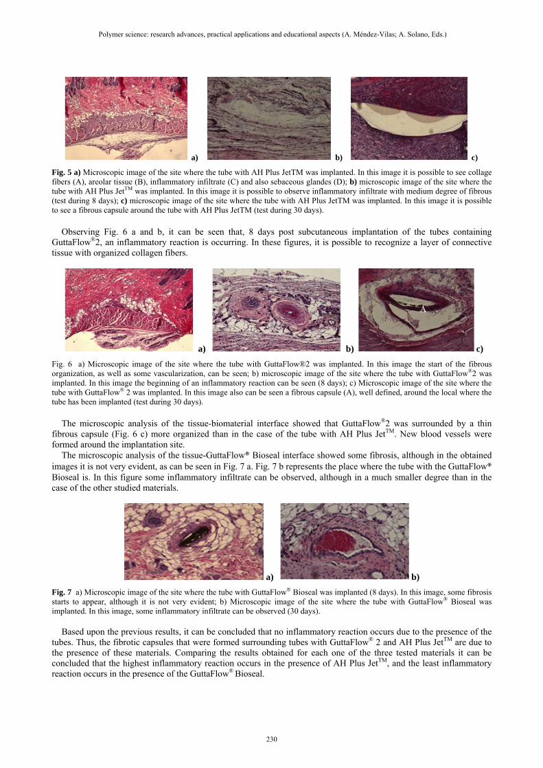
Fig. 5 a) Microscopic image of the site where the tube with AH Plus JetTM was implanted. In this image it is possible to see collage fibers (A), areolar tissue (B), inflammatory infiltrate (C) and also sebaceous glandes (D); b) microscopic image of the site where the tube with AH Plus JetTM was implanted. In this image it is possible to observe inflammatory infiltrate with medium degree of fibrous (test during 8 days); c) microscopic image of the site where the tube with AH Plus JetTM was implanted. In this image it is possible to see a fibrous capsule around the tube with AH Plus JetTM (test during 30 days). Observing Fig. 6 a and b, it can be seen that, 8 days post subcutaneous implantation of the tubes containing GuttaFlow®2, an inflammatory reaction is occurring. In these figures, it is possible to recognize a layer of connective tissue with organized collagen fibers.
a) b) c)
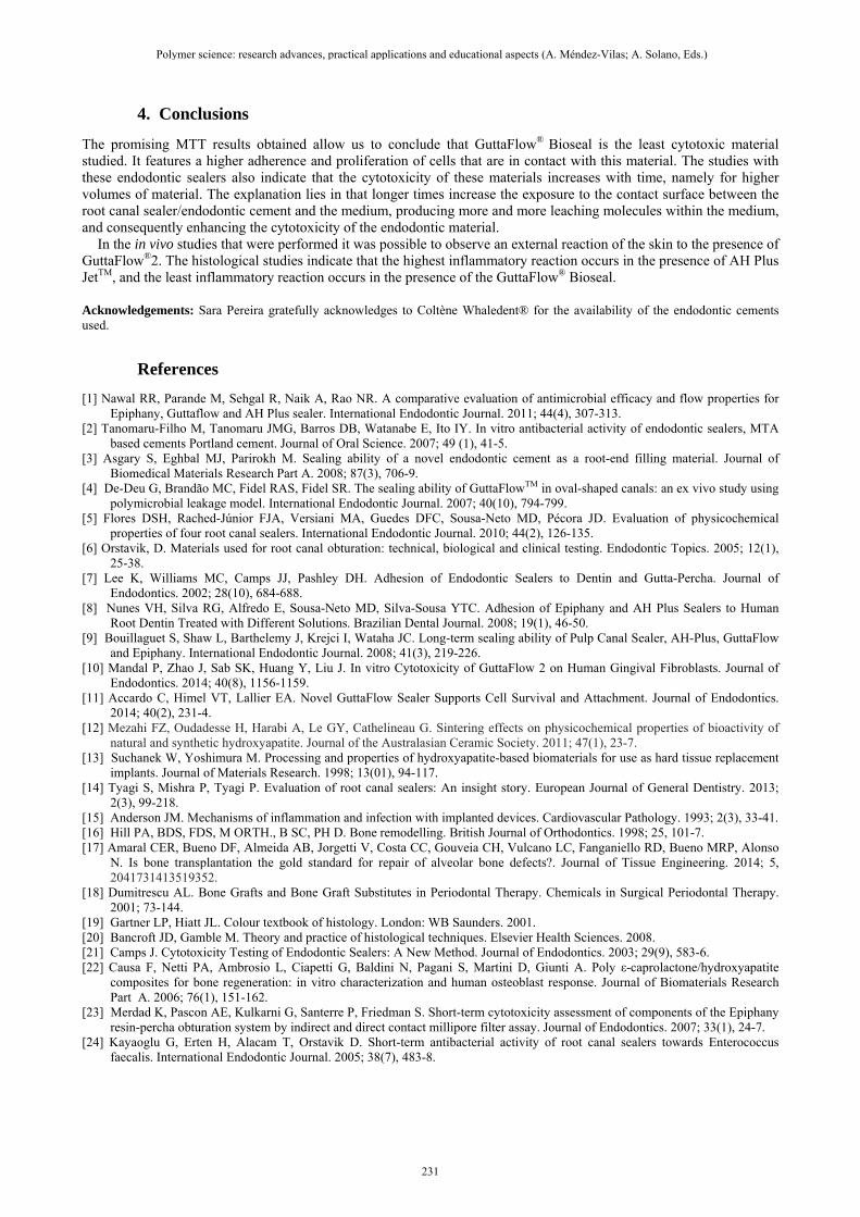
Fig. 6 a) Microscopic image of the site where the tube with GuttaFlow®2 was implanted. In this image the start of the fibrous organization, as well as some vascularization, can be seen; b) microscopic image of the site where the tube with GuttaFlow®2 was implanted. In this image the beginning of an inflammatory reaction can be seen (8 days); c) Microscopic image of the site where the tube with GuttaFlow® 2 was implanted. In this image also can be seen a fibrous capsule (A), well defined, around the local where the tube has been implanted (test during 30 days). The microscopic analysis of the tissue-biomaterial interface showed that GuttaFlow®2 was surrounded by a thin fibrous capsule (Fig. 6 c) more organized than in the case of the tube with AH Plus JetTM. New blood vessels were formed around the implantation site. The microscopic analysis of the tissue-GuttaFlow® Bioseal interface showed some fibrosis, although in the obtained images it is not very evident, as can be seen in Fig. 7 a. Fig. 7 b represents the place where the tube with the GuttaFlow® Bioseal is. In this figure some inflammatory infiltrate can be observed, although in a much smaller degree than in the case of the other studied materials.
a) b)
Fig. 7 a) Microscopic image of the site where the tube with GuttaFlow® Bioseal was implanted (8 days). In this image, some fibrosis starts to appear, although it is not very evident; b) Microscopic image of the site where the tube with GuttaFlow® Bioseal was implanted. In this image, some inflammatory infiltrate can be observed (30 days). Based upon the previous results, it can be concluded that no inflammatory reaction occurs due to the presence of the tubes. Thus, the fibrotic capsules that were formed surrounding tubes with GuttaFlow® 2 and AH Plus JetTM are due to the presence of these materials. Comparing the results obtained for each one of the three tested materials it can be concluded that the highest inflammatory reaction occurs in the presence of AH Plus JetTM, and the least inflammatory reaction occurs in the presence of the GuttaFlow® Bioseal.
A
Polymer science: research advances, practical applications and educational aspects (A. Méndez-Vilas; A. Solano, Eds.) _______________________________________________________________________________________________
230
4. Conclusions
The promising MTT results obtained allow us to conclude that GuttaFlow® Bioseal is the least cytotoxic material studied. It features a higher adherence and proliferation of cells that are in contact with this material. The studies with these endodontic sealers also indicate that the cytotoxicity of these materials increases with time, namely for higher volumes of material. The explanation lies in that longer times increase the exposure to the contact surface between the root canal sealer/endodontic cement and the medium, producing more and more leaching molecules within the medium, and consequently enhancing the cytotoxicity of the endodontic material. In the in vivo studies that were performed it was possible to observe an external reaction of the skin to the presence of GuttaFlow®2. The histological studies indicate that the highest inflammatory reaction occurs in the presence of AH Plus JetTM, and the least inflammatory reaction occurs in the presence of the GuttaFlow® Bioseal.
Acknowledgements: Sara Pereira gratefully acknowledges to Coltène Whaledent® for the availability of the endodontic cements used.
References
[1] Nawal RR, Parande M, Sehgal R, Naik A, Rao NR. A comparative evaluation of antimicrobial efficacy and flow properties for Epiphany, Guttaflow and AH Plus sealer. International Endodontic Journal. 2011; 44(4), 307-313.
[2] Tanomaru-Filho M, Tanomaru JMG, Barros DB, Watanabe E, Ito IY. In vitro antibacterial activity of endodontic sealers, MTA based cements Portland cement. Journal of Oral Science. 2007; 49 (1), 41-5.
[3] Asgary S, Eghbal MJ, Parirokh M. Sealing ability of a novel endodontic cement as a root-end filling material. Journal of Biomedical Materials Research Part A. 2008; 87(3), 706-9.
[4] De-Deu G, Brandão MC, Fidel RAS, Fidel SR. The sealing ability of GuttaFlowTM in oval-shaped canals: an ex vivo study using polymicrobial leakage model. International Endodontic Journal. 2007; 40(10), 794-799.
[5] Flores DSH, Rached-Júnior FJA, Versiani MA, Guedes DFC, Sousa-Neto MD, Pécora JD. Evaluation of physicochemical properties of four root canal sealers. International Endodontic Journal. 2010; 44(2), 126-135.
[6] Orstavik, D. Materials used for root canal obturation: technical, biological and clinical testing. Endodontic Topics. 2005; 12(1), 25-38.
[7] Lee K, Williams MC, Camps JJ, Pashley DH. Adhesion of Endodontic Sealers to Dentin and Gutta-Percha. Journal of Endodontics. 2002; 28(10), 684-688.
[8] Nunes VH, Silva RG, Alfredo E, Sousa-Neto MD, Silva-Sousa YTC. Adhesion of Epiphany and AH Plus Sealers to Human Root Dentin Treated with Different Solutions. Brazilian Dental Journal. 2008; 19(1), 46-50.
[9] Bouillaguet S, Shaw L, Barthelemy J, Krejci I, Wataha JC. Long-term sealing ability of Pulp Canal Sealer, AH-Plus, GuttaFlow and Epiphany. International Endodontic Journal. 2008; 41(3), 219-226.
[10] Mandal P, Zhao J, Sab SK, Huang Y, Liu J. In vitro Cytotoxicity of GuttaFlow 2 on Human Gingival Fibroblasts. Journal of Endodontics. 2014; 40(8), 1156-1159.
[11] Accardo C, Himel VT, Lallier EA. Novel GuttaFlow Sealer Supports Cell Survival and Attachment. Journal of Endodontics. 2014; 40(2), 231-4.
[12] Mezahi FZ, Oudadesse H, Harabi A, Le GY, Cathelineau G. Sintering effects on physicochemical properties of bioactivity of natural and synthetic hydroxyapatite. Journal of the Australasian Ceramic Society. 2011; 47(1), 23-7.
[13] Suchanek W, Yoshimura M. Processing and properties of hydroxyapatite-based biomaterials for use as hard tissue replacement implants. Journal of Materials Research. 1998; 13(01), 94-117.
[14] Tyagi S, Mishra P, Tyagi P. Evaluation of root canal sealers: An insight story. European Journal of General Dentistry. 2013; 2(3), 99-218.
[15] Anderson JM. Mechanisms of inflammation and infection with implanted devices. Cardiovascular Pathology. 1993; 2(3), 33-41. [16] Hill PA, BDS, FDS, M ORTH., B SC, PH D. Bone remodelling. British Journal of Orthodontics. 1998; 25, 101-7. [17] Amaral CER, Bueno DF, Almeida AB, Jorgetti V, Costa CC, Gouveia CH, Vulcano LC, Fanganiello RD, Bueno MRP, Alonso
N. Is bone transplantation the gold standard for repair of alveolar bone defects?. Journal of Tissue Engineering. 2014; 5, 2041731413519352.
[18] Dumitrescu AL. Bone Grafts and Bone Graft Substitutes in Periodontal Therapy. Chemicals in Surgical Periodontal Therapy. 2001; 73-144.
[19] Gartner LP, Hiatt JL. Colour textbook of histology. London: WB Saunders. 2001. [20] Bancroft JD, Gamble M. Theory and practice of histological techniques. Elsevier Health Sciences. 2008. [21] Camps J. Cytotoxicity Testing of Endodontic Sealers: A New Method. Journal of Endodontics. 2003; 29(9), 583-6. [22] Causa F, Netti PA, Ambrosio L, Ciapetti G, Baldini N, Pagani S, Martini D, Giunti A. Poly ε-caprolactone/hydroxyapatite
composites for bone regeneration: in vitro characterization and human osteoblast response. Journal of Biomaterials Research Part A. 2006; 76(1), 151-162.
[23] Merdad K, Pascon AE, Kulkarni G, Santerre P, Friedman S. Short-term cytotoxicity assessment of components of the Epiphany resin-percha obturation system by indirect and direct contact millipore filter assay. Journal of Endodontics. 2007; 33(1), 24-7.
[24] Kayaoglu G, Erten H, Alacam T, Orstavik D. Short-term antibacterial activity of root canal sealers towards Enterococcus faecalis. International Endodontic Journal. 2005; 38(7), 483-8.
Polymer science: research advances, practical applications and educational aspects (A. Méndez-Vilas; A. Solano, Eds.) _______________________________________________________________________________________________
231