endocrine hypothalamus and anterior...
TRANSCRIPT
Chapter 3
Anterior Pituitary &
The Endocrine Hypothalamus
Nam Deuk Kim, Ph.D.
1
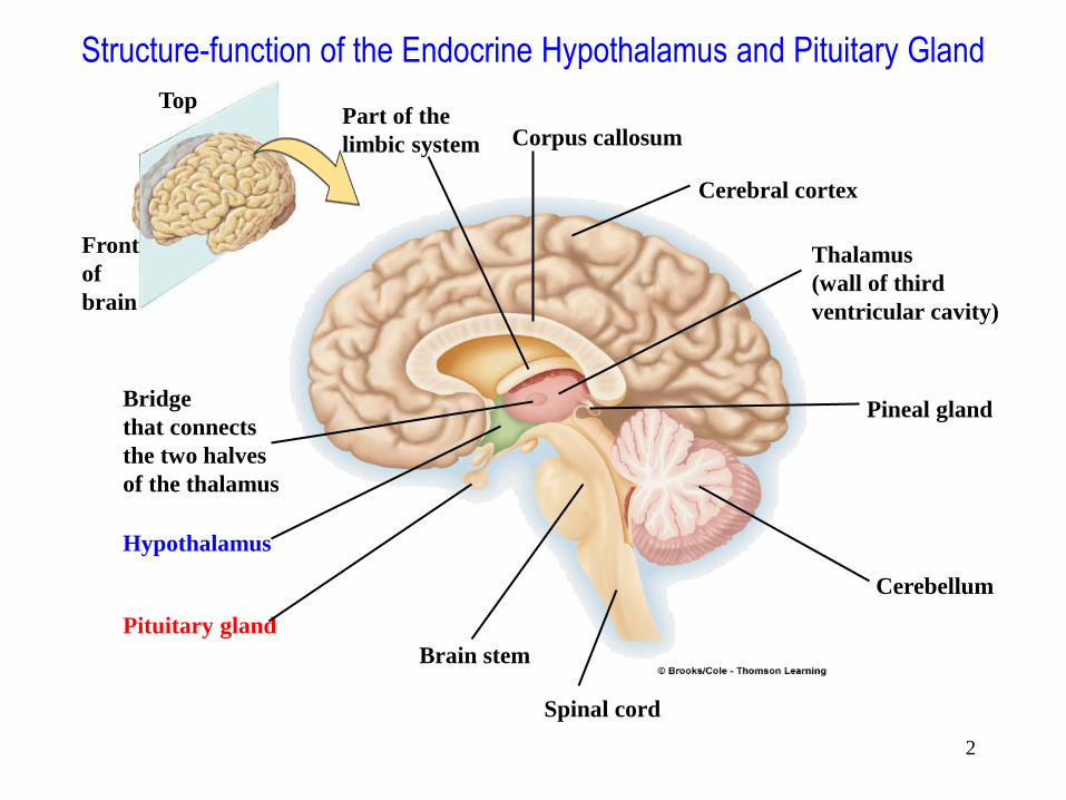
Top
Front
of
brain
Corpus callosum
Cerebral cortex
Thalamus
(wall of third
ventricular cavity)
Pineal gland
Cerebellum
Part of the
limbic system
Bridge
that connects
the two halves
of the thalamus
Hypothalamus
Pituitary gland
Brain stem
Spinal cord
Structure-function of the Endocrine Hypothalamus and Pituitary Gland
2

Fig. 6-1: Frontal section through the cerebral hemispheres of the human brain;
the principal hypothalamic nuclei present within the plane of transect are
indicated. 3

• Hypophysiotropic
• Median eminence to suprachiasmatic region
• Parvocellular neurosecretory system – CNS input via synaptic
contact
– Synthesis of hypophysiotropic factors
– Release hypophysial portal system
• Via neuronal impingement
• Secrete peptide hormones
Nonhypophysiotrophic
functions of
hypothalamus
• Thirst
• Hunger/satiety
• Thermo-regulation
• Regulatory systems of
emotions (behavior)
• Sex hormones associated
Medial Basal Hypothalamus
4

5
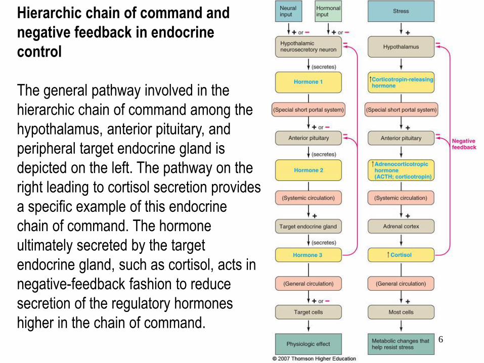
Hierarchic chain of command and
negative feedback in endocrine
control
The general pathway involved in the
hierarchic chain of command among the
hypothalamus, anterior pituitary, and
peripheral target endocrine gland is
depicted on the left. The pathway on the
right leading to cortisol secretion provides
a specific example of this endocrine
chain of command. The hormone
ultimately secreted by the target
endocrine gland, such as cortisol, acts in
negative-feedback fashion to reduce
secretion of the regulatory hormones
higher in the chain of command.
6

Anatomy of the Pituitary Gland
※Pituitary gland: smaller than the tip of the little finger; 0.5~1.0 g
7

8

Fig. 5.1. Anatomical components of the pituitary gland.
9

Pituitary Gland
• Hypophysis (Pituitary Gland)
• Small gland located in bony cavity just below
hypothalamus
– Thin stalk connects pituitary gland to hypothalamus
• Consists of two anatomically and functionally
distinct lobes
– Posterior pituitary (neurohypophysis)
• Composed of nervous tissue
– Anterior pituitary (adenohypophysis)
• Consists of glandular epithelial tissue
10

Pituitary Gland • Anterior pituitary
– Secretes six different peptide hormones that it produces itself • Tropic hormones
– Thyroid-stimulating hormone (TSH)
» Stimulates secretion of thyroid hormone
– Adrenocorticotropic hormone (ACTH)
» Stimulates secretion of cortisol by adrenal cortex
– Follicle-stimulating hormone (FSH)
» In females, stimulates growth and development of ovarian follicles; promotes secretion of estrogen by ovaries
» In males, required for sperm production
– Luteinizing hormone (LH)
» In females, responsible for ovulation and luteinization; regulates ovarian secretion of female sex hormones
» In males, stimulates testosterone secretion
– Growth hormone (GH)
» Primary hormone responsible for regulating overall body growth; important in intermediary metabolism
• Not a tropic hormone – Prolactin (PRL)
» Enhances breast development and milk production in females
11
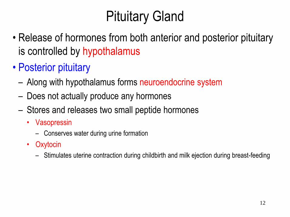
Pituitary Gland
• Release of hormones from both anterior and posterior pituitary
is controlled by hypothalamus
• Posterior pituitary
– Along with hypothalamus forms neuroendocrine system
– Does not actually produce any hormones
– Stores and releases two small peptide hormones
• Vasopressin
– Conserves water during urine formation
• Oxytocin
– Stimulates uterine contraction during childbirth and milk ejection during breast-feeding
12

Relationship of the Hypothalamus and Posterior Pituitary
1. The paraventricular and supraoptic nuclei
both contain neurons that produce
vasopressin and oxytocin. The hormone,
either vasopressin or oxytocin depending
on the neuron, is synthesized in the
neuronal cell body in the hypothalamus.
2. The hormone travels down the axon to be
stored in the neuronal terminals within the
posterior pituitary.
3. On excitation of the neuron, the stored
hormone is released from these terminals
into the systemic blood for distribution
throughout the body.
13

14

Vascular Link Between the Hypothalamus and Anterior Pituitary
15

Hypothalamus & Pituitary Gland
• Hypothalamic releasing and inhibiting hormones help
regulate anterior pituitary hormone secretion
• Two most important factors that regulate anterior pituitary
hormone secretion
– Hypothalamic hormones
– Feedback by target-gland hormones
16

17

18

• Hypophysiotropic hormones from hypothalamus
– GHRH, SST (GHIH), TRH, PIH, GnRH, CRH
• Three main families of anterior pituitary hormones
– Somatomammotropic (lactogenic): GH, PRL
– Glycoproteins sharing common a subunit: TSH, FSH, LH
– Proopiomelanocortin (POMC) ACTH, MSH
• Two neurohypophysial hormones (posterior pituitary
hormones): oxytocin & vasopressin
Pituitary Hormones
19

20

21

Functions of the anterior pituitary hormones
22
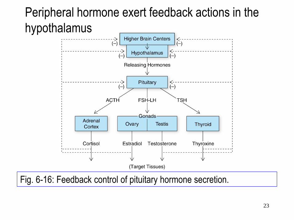
Fig. 6-16: Feedback control of pituitary hormone secretion.
Peripheral hormone exert feedback actions in the
hypothalamus
23

Fig. 6-17: Model of long-loop and short-loop mechanisms and autoregulation
of pituitary hormone secretion.
24

Regulation of
Thyroid
Hormone
Secretion
Regulation of
Glucocorticoid
Hormone
Secretion
25

1. Control of Growth
Hormone Secretion
26

• GHRH: 44 aa, simple peptide – First 29 residues highly conserved
– Expressed in arcuate and ventromedial nuclei
• Target cells: ant. pit. somatotrophs – Heptahelical G-protein coupled receptor
– Stimulating adenylyl cyclase • PKA through activating plasma Ca2+ channels
• PLC through IP3, increased intracell Ca2+
Synth. GH + discharge stored GH
– Cortisol, thyroid hormones required
Growth Hormone Releasing Hormone
27
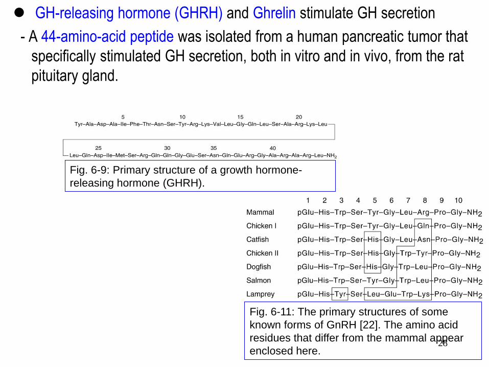
Fig. 6-9: Primary structure of a growth hormone-
releasing hormone (GHRH).
GH-releasing hormone (GHRH) and Ghrelin stimulate GH secretion
- A 44-amino-acid peptide was isolated from a human pancreatic tumor that
specifically stimulated GH secretion, both in vitro and in vivo, from the rat
pituitary gland.
Fig. 6-11: The primary structures of some
known forms of GnRH [22]. The amino acid
residues that differ from the mammal appear
enclosed here. 28

CREB=Cyclic AMP Response Element Binding Protein
CRE=Cyclic AMP Response Element
FOS=protein that regulates several genes
PIT1=Pituitary specific transcription factor
29

• Amount of GH discharged depends on:
– Amount of GHRH
– Somatotroph GHRH receptor status
– Amount stored GH
• Episodic GHRH episodic GH release
• Termination GH burst w/ negative feedback on brain neurons
– Neurons prod SST
– Somatotrophs have SST receptors
• Cortisol GHRH receptor expression + GH expression
30

• GH accounts for 4% to 10% of the wet weight of the ant. pit. in the human adult (5 mg to 10 mg per gland).
• GH is polypeptide of 191 a.a. made by somatotrophs and contains two disulfide bonds. It is very similar to Prl and is identical in 161 a.a.
• GH is made as a prohormone and is cleaved to GH by proteolysis.
• GH circulates in the plasma bound to one or more binding proteins.
• Circulating levels of GH decrease 2-3 wks after birth and reach basal levels characteristic of adulthood.
Growth Hormone (somatotropin)
31

• GH remains constant during accelerated growth in early childhood but there is an increase during the maximal growth period around puberty.
• In all mammals studied so far, spontaneous episodes of GH occur several times over a 24 h period and most during the first 90 minutes of nocturnal sleep.
• GHRH from the hypothalamus and somatostatin control GH secretion.
Growth Hormone
32

• GH is anabolic and enhances amino acid incorporation into muscle protein and stimulates collagen deposition and produces a concomitant decrease in blood urea nitrogen and amino acid levels.
• Effects of GH are mediated by somatomedins (insulin-like growth factors, IGF’s) released from the liver in response to GH.
• They stimulate cellular growth in a number of organs and tissues.
Growth Hormone
33

• Acromegalics may display increased BMR and lipolytic actions of GH combined with anti-insulin actions on other tissues, may result in hyperglycemia as a symptom of developing diabetes mellitus.
• In young animals the epiphyses of long bones are separated from the shaft of bones by an epiphyseal cartilaginous plate. Chondrogenesis is accelerated by GH which results in widening of the epiphyseal plates as more chondroitin sulfate is made and released by chondrocytes. This is also used as a bioassay for GH.
Growth Hormone
34

• Somatostatin (SST) [also known as
growth hormone-inhibiting hormone
(GHIH) or somatotropin release-
inhibiting factor (SRIF)]
• SST: inhibition of GH
• 14 aa’s; 2 cys intrachain disulfide
bond cyclization
• SRIF gene on chromosome 3 116
aa preproSRIF
• Convertase 14 aa brain peptide
– also pancreas, GI tract SST’s
Somatostatin (SST)
35
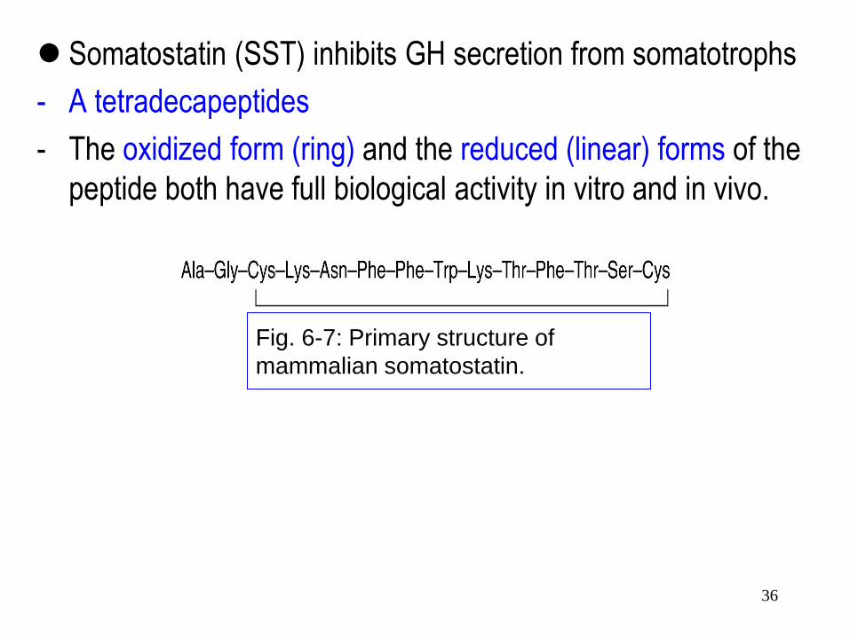
Somatostatin (SST) inhibits GH secretion from somatotrophs
- A tetradecapeptides
- The oxidized form (ring) and the reduced (linear) forms of the
peptide both have full biological activity in vitro and in vivo.
Fig. 6-7: Primary structure of
mammalian somatostatin.
36

• SRIF’s (somatotropin release-inhibiting factor)
– Heptahelical receptors
– G-protein coupled
– Inhibit adenylate cyclase
• Somatotrophs: turn off GH secretion
• Widespread effects
– Inhibits calcitonin, PTH, renin, gastric HCl, ACh,
adrenergic neurotransmitters
– Lowers serum [glucose]
– TRH antagonist
– Antagonizes messengers important to cell proliferation
37

2. Control of Prolactin
Secretion
Secretion Rates of Placental Hormones
Sucking Reflexes
38

• Prl is a single chain polypeptide ~ 23 kDa with 3 disulfide bonds.
• Structural variants of Prl arise from differential splicing and CHO moieties vary among species.
• Serum Prl increases at puberty but only modestly compared to the rise in FSH/LH.
• E2 stimulates Prl secretion.
• Prl increases during pregnancy and reaches maximal values at parturition.
Prolactin (Prl)
39

• Production and secretion of Prl by lactotrophs is stimulated by E2. E2 increases mitotic activity and cell number of pituitary lactotrophs.
• Prl levels decrease by 3 wks in mothers that do not nurse. Concentrations of Prl in amniotic fluid exceed that found in serum.
• Mammotrophic action of Prl requires participation of E2, P4, insulin, glucocorticoids and GH. In ovx (ovaryectomized) rabbits given E2 and P4 to induce lobuloalveolar growth, Prl was able to induce milk production.
Prolactin
40

Fig. 5.6. Human plasma levels of PRL during a 24-hour period. 41

• Prl is luteotrophic in some mammals and may act in concert with LH and FSH on the CL to stimulate P4 biosynthesis and secretion.
• Prl has profound effects on the growth, differentiation and function of hair, sebaceous glands, and brood patch feathers.
• In pigeons and doves Prl controls the production of so-called crop sac “milk”. Epithelium of the walls of the crop thicken and cells accumulate lipid and begin to degenerate and form crop milk.
Prolactin
42

• Basal PRL release unless interrupted w/ hypothalamus
signal
• PIH=dopamine
– Also produced by tuberoinfundibular, tuberohypophyseal
dopamine neurons
• Dopamine receptor (D2)
Prolactin Inhibiting Hormone (PIH)
43

• Estrogens stimulate PRL gene expression
– Inhibit tuberohypophyseal DA neurons
– Induce lactotroph hyperplasia
• TRH stimulates PRL secretion
– Not physiological release factor during lactation
• Oxytocin stimulates PRL secretion (?)
• Angiotensin II stimulates PRL secretion
Signals to Advance PRL Synthesis
44

• Prolactin Releasing Hormone??
– VIP (from GHRH family) stim’s PRL secr’n in vitro
• Acts through adenylate cyclase
• Produced by lactotrophs (autocrine?)
• Serotonin & adrenaline stimulate PRL secretion
• Histamine may inhibit PRL via H2 receptors
45

• Three hormones of the anterior pituitary are glycoproteins,
containing up to 33% carbohydrate by weight.
• LH, FSH, and TSH contain covalently bound carbohydrate
moieties at one or more positions within their structures.
• Composed of two chains, so called α and β subunits.
• α subunit: identical to each other; 92 aa’s.
• β subunit: 110-111 aa’s for LHs; 112-118 aa’s for TSHs, 117-
121 aa’s for LHs; 145 aa’s for human chorionic gonadotropin
(hCG)
Luteinizing Hormone, Follicle-Stimulating Hormone, and Thyrotropin are Glycoprotein Hormones
46

Fig. 5.7. Glycoprotein subunit hybridization studies. 47

3. Regulation of Thyroid
Hormone Secretion
48

• Gene for TRH on chromosome 3
– Inhibit expression by glucocorticoids
• Precursor protein; post-translational cleavage
– 255 aa’s
– TRH tripeptide duplicated 5 times in sequence
• Most important regulator of synthesis of TRH=thyroid hormones (T3 > T4)
– Long-loop negative feedback
Thyrotropin Releasing Hormone (TRH) Thyroid Stimulating Hormone (TSH)
49

Fig. 6-6: The cDNA sequence of the TRH precursor encodes a protein of 255
amino acids. The sequence Gln-His-Pro-Gly occurs five times and each
tetrapeptide is flanked by paired basic residues (Lys Arg or Arg Arg). Thus, the
prohormone generates five TRH molecules from each precursor protein.
50

Fig. 6-4: Six synthetic peptides
related to thyrotropin-releasing
hormone (TRH).
Thyrotropin-releasing hormone (TRH) stimulates synthesis and
secretion of TSH
- From about 25,000 ovine and porcine hypothalamic fragments,
about 1 mg of TRH was obtained.
51

Fig. 6-5: Synthesis of thyrotropin-releasing hormone (TRH).
52

• Episodic secretion portal system
Circadian rhythm of TSH secretion
– Stimulated by a adrenergic receptor agonists
– Inhibited by dopamine agonists, endorphins, SST
• Target cells: anterior pituitary thyrotrophs
– Heptahelical receptor
– Coupled to G protein
– Activates IP3 pathway
• PLC and PKC activity
Synthesis and secretion of TSH
• Gene for TRH receptor on chromosome 8
53

• Control of TRH secretion
- Noradrenergic neurons stimulate TSH secretion
by a stimulatory action on TRH-secreting
neurons.
- Glucocorticoid excess inhibits thyroid function at
a suprapituitary level
54

4. Regulation of FSH & LH
Hormone Secretion
Correlation between hormonal levels
and cyclic ovarian and uterine
changes
55

Feedback control of FSH and tonic LH
secretion during the follicular phase Control of the LH surge at ovulation
56

• Mammalian decapeptide; well conserved
– Chromosome 8p21-8p11.2
• GnRH neurons
– Human development in olfactory placode
• Migrate via olfactory bulb guided by anosmin (neural cell adhesion molecule)
• Anosmin also reg’s migration olfactory epithelium (including those from
vomeronasal organ)
• Anosmin-1 is a secreted, EM associated glycoprotein found in humans and other
organisms responsible for normal development, which is expressed in the brain,
spinal cord and kidney. Absence or damage to the protein results in Kallmann
syndrome in humans, which is characterized by loss of olfactory bulbs and GnRH
secretion leading to anosmia and hypothalamic hypogonadotropic hypogonadism.
Anosmin-1 is coded by the KAL-1 gene, which is found on the X chromosome; 100
kDa; and is expressed on the outside of cells.
Gonadotropin Releasing Hormone FSH, LH
57

Gonadotropin-releasing hormone (GnRH) stimulates LH and
FSH secretion from gonadotrophs
- A decapeptide hormone
Fig. 6-10: Primary structure of mammalian gonadotropin-releasing
hormone (GnRH).
58
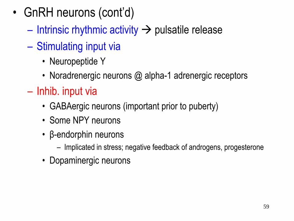
• GnRH neurons (cont’d)
– Intrinsic rhythmic activity pulsatile release
– Stimulating input via
• Neuropeptide Y
• Noradrenergic neurons @ alpha-1 adrenergic receptors
– Inhib. input via
• GABAergic neurons (important prior to puberty)
• Some NPY neurons
• β-endorphin neurons
– Implicated in stress; negative feedback of androgens, progesterone
• Dopaminergic neurons
59

• GnRH target cells: gonadotrophs
• GnRH receptors
– Encoded by chromosome 4q13.2-13.3
– Contains response elements that regulate its expression
• For glucocorticoid, progesterone, thyroid hormones, CREB
– G-protein linked heptahelical transmembrane
• Gαq PLC pathway
IP3-Ca2+, DAG-PKC-MAP kinase
expression of a subunit common to FSH/LH
60

• GnRH receptors (cont’d)
– Also opening of voltage gated Ca2+ channels
Ca2+ influx
• Important to release gonadotropins
selective expression LH-β subunit
– Downregulated when continuous (not pulsatile) GnRH
– Upregulated w/ high pulses GnRH
• FSH, LH
– Glycoprotein similar to TSH, hCG
– Share common a subunit
– Hormone specific β subunits
61

• FSH/LH α subunit
– Chromosome 6
– Required for receptor binding
– Expression controlled by several hormones
• Coordinated w/ expression β subunit genes
– In gonadotrophs, stimulated by
• GnRH via DAG-PKC-MAP kinase pathway
– Inhibited by
• Estrogens
– Unaffected by thyroid hormones
62

• FSH β subunit
– Chromosome 11
– Required for receptor specificity
– Highest when low freq GnRH pulses received by
gonadotrophs
• GnRH pulses @ higher frequency suppression FSH β subunit
• Continuous GnRH absolute inhibiting FSH β subunit
– Activins (similar to inhibins) increased FSH β mRNA
(autocrine)
• High GnRH pulses producing follistatin within pituitary gl.
• Follistatin binds activins prevent FSH-stimulating activity
63

• LH β subunit
– Chromosome 19 (homologous w/ hCG β subunit genes)
– Required for receptor specificity
– Stimulated w/
• GnRH applied @ higher frequency, amplification
– Suppressed w/
• Androgens (male), progesterone (female) inhib freq GnRH pulses
• Estrogens (directly @ pit)
• Testosterone estradiol in pit (via aromatase)
64

• FSH/LH β subunit reg’n
– Higher GnRH pulses upregulating GnRH receptors favors LH β prod’n
– Sertoli cell inhibin B inhibiting FSH β @ gonadotrophs • Inhibin B stimulated by FSH
– Testosterone/DHT suppress FSH β • Via gonadotroph androgen receptors
• FSH/LH secretion pulsatile
– LH high amplified fluctuations
– FSH relatively stable amplifications
65

66

67

Pro-opiomelanocortin (POMC)
ACTH α-MSH β-Endorphin
5. Control of Cortisol
Secretion
68

• 41 aa’s; highly conserved
• Synthesized in parvocellular region of
paraventricular nucleus in hypothalamus
– Sensitive to glucocorticoids (negative feedback)
– Co-express AVP
• Widely expressed in CNS
– Mediate stress-related psychological anxiety
– Overproducing or increased CRH
• In CSF correlates w/ major depression, anorexia nervosa
• Mediates sleep/appetite disturbances of depression
Corticotropin Releasing Hormone (CRH) ACTH
69

Corticotropin-releasing hormone (CRH) stimulates
ACTH secretion from corticotrophs
• 41-reside peptide
Fig. 6-12. Primary structure of a corticotropin-releasing hormone (CRH).
70

• CRH outside CNS
– May be proinflammatory signal • Stimulating synthesis of PG’s
– From placenta, parturition initiation
• Induction release w/ central catecholamines into portal cap. plexus
– Probably w/ stress
– Stress ACTH release (through CRH) • Emotional; hypoxia; hypercapnea
• Decreased blood pressure; depleting ECF volume
• Infections (w/ increased IL-1β)
• Environmental temperature changes; fever
• Ethanol consumption
71

• CRH has binding protein (CRH-BP)
– CRH “sink”
• Assoc’d w/ membr’s near CRH brain target cells
• Another form secreted by liver, placenta
• Target cells: corticotrophs in ant. pit. gland
• Two CRH receptors (CRH ligand of CRH1R)
– G-protein coupled heptahelical receptors
– Binding ad cyclase synth. & release ACTH
– Negative feedback controlled by cortisol
• Glucocorticoids suppress CRH1R mRNA
72

• ACTH (corticotropin)
– Synthesized in pars distalis
– Precursor = POMC (proopiomelanocortin) • 241 aa’s
• Holds ACTH, β-LPH (lipotropin), β-MSH (melanocyte-stimulating hormone) – β-MSH not physiologically active
– w/ β-LPH: β-endorphin, met enkephalin (neurohormones)
• Proprotein convertase 3 catalyzes POMC ACTH + β-LPH
– Cleaved α-MSH in pars distalis • Similar sequence
• ACTH melanotropic activity
– Circadian adrenocortical rhythm
73

74

• ACTH (cont’d)
– Smallest ant. pit. peptide hormone
• 39 aa chain; highly conserved
– Bio activity in invariant 1-24 aa N-terminal sequence
– Synthesis & secretion were stimulated
• CRH
• ADH (AVP)
• CCK (sometimes)
• ACh CNS cholinergic structures ACTH release
– Suppressed by
• Glucocorticoids
» Hypothalamus membrane receptor non-genomic mech, then @ ant. pit.
• CRH-BP
75

Fig. 5.8. Comparative primary structures of some corticotropins
76

• ACTH (cont’d)
– Target = adrenal steroidogenic tissue • Stimulates glucocorticoid biosynthesis
– Cortisol, corticosterone
– Important in carbohydrate metabolism
– Glucocorticoid feedback inhibit @ hypothal. & ant. pit.
• α-MSH produced in
– Human intermed. ant. pit. in fetal life
– 13 aa’s
– Skin keratinocytes w/ UVB eumelanin
– Monocytes – anti-inflammatory
– Some neurons – appetite suppressing, decrease body temp.
77

Pituitary Pathophysiology
78
Hyperthyroidism

Pathophysiology of Hypothalamic Dysfunction
• Related to defects in hypophysiotropic hormone synthesis and secretion
or to altered activity of the neuronal inputs to hypophysiotropin neurons
• Overproduction or underproduction of dopamine within CNS neurons, for
example, is believed responsible for the etiology of schizophrenia and
Parkinson’s disease, respectively.
• Some tumors may secrete hypothalamic hormones.
a. Ectopic secretion of a CRH-like peptide for certain cases of Cushing’s
disease
b. PRL-secreting tumor
79

80