emotional conflict 2-law
TRANSCRIPT

1
Emotional conflict processing in adolescent Chronic Fatigue Syndrome: a 1
pilot study using fMRI 2
3
Laura Anne Wortinger1, 2*, Tor Endestad2, Annika Melinder3, Merete 4
Glenne Øie2, 4, Dag Sulheim5, 6, Even Fagermoen7, and Vegard Bruun 5
Wyller1 6
71Department of Pediatrics, Akershus University Hospital, Nordbyhagen, Norway 82Department of Psychology, University of Oslo, Norway 93Cognitive Developmental Research Unit, Department of Psychology, University of Oslo, 10
Norway 114Research Department, Innlandet Hospital Trust, Lillehammer, Norway 125Department of Pediatrics, Oslo University Hospital, Norway 136Department of Pediatrics, Innlandet Hospital Trust, Lillehammer, Norway 147Department of Anesthesiology and Critical Care, Oslo University Hospital, Norway 15
16
17
18
19
20
21
22
23
24
25
26
27
28
* Address for correspondence: Laura Anne Wortinger, Department of Psychology, University of Oslo, PO Box 1094 Blindern, 0317 Oslo, Norway. Email: [email protected]

2
29
Emotional conflict processing in adolescent Chronic Fatigue Syndrome: a 30
pilot study using fMRI 31
Introduction: Studies of neurocognition suggest abnormalities in cognitive control contribute 32
to the pathophysiology of chronic fatigue syndrome (CFS) in adolescents, yet these 33
abnormalities remain poorly understood at the neurobiological level. Reports indicate that 34
adolescents with CFS are significantly impaired in conflict processing, a primary element of 35
cognitive control. Method: In this study, we examine whether emotional conflict processing is 36
altered on behavioral and neural levels in adolescents with CFS and a healthy comparison 37
group. Fifteen adolescent patients with CFS and 24 healthy adolescent participants underwent 38
functional magnetic resonance imaging (fMRI) while performing an emotional conflict task 39
that involved categorizing facial affect while ignoring overlaid affect labeled words. Results: 40
Adolescent CFS patients were less able to engage the left amygdala and left mid-posterior 41
insula (mpINS) in response to conflict compared to the healthy comparison group. An 42
association between accuracy interference and conflict-related reactivity in the amygdala was 43
observed in CFS patients. A relationship between response time interference and conflict-44
related reactivity in the mpINS was also reported. Neural responses in the amygdala and 45
mpINS were specific to fatigue severity. Conclusions: These data demonstrate that adolescent 46
CFS patients displayed deficits in emotional conflict processing. Our results suggest 47
abnormalities in affective and cognitive functioning of the salience network, which might 48
underlie the pathophysiology of adolescent CFS. 49
50
51
52
53
Key words: chronic fatigue syndrome; adolescents; functional MRI; cognitive control; 54
emotion; conflict 55
56
57
58
59
60
61

3
Introduction 62
Disabling physical and mental fatigue, which worsens from physical and mental exertion, 63
characterize chronic fatigue syndrome (CFS) (IOM, 2015). CFS constitutes one of the major 64
threats towards adolescent health (Royal College of Paediatrics and Child Health, 2004). 65
Neuropsychological studies have documented cognitive impairments in adolescent patients, 66
suggesting that a sustained stress response might be an important part of the pathophysiology 67
(Wyller, Eriksen, & Malterud, 2009). Recently, a deficit in cognitive inhibition was reported 68
in adolescents with CFS (Sulheim et al., 2015). In another study, the Eriksen Flanker test 69
revealed conflict processing impairment in adolescents with CFS (van de Putte et al., 2008). 70
Haig-Ferguson, Tucker, Eaton, Hunt, and Crawley (2009) and Kawatani et al. (2011) reported 71
attention impairments in adolescent CFS patients. An event-related potential (ERP) study 72
found working memory impairment was associated with frontal lobe alterations in adolescents 73
with CFS using KANA-Pick-out test (Tomoda et al., 2007). These studies point to a specific 74
impairment in cognitive control and warrant further investigation. 75
The human cognitive system has the remarkable ability to adapt efficiently and effectively to 76
a changing environment. This would entail an efficient salience network (SN) in the brain that 77
identifies and integrates salient events, both endogenously and externally cued, to help guide 78
behavior. The main regions of the brain that comprise the SN are the insula, dorsal anterior 79
cingulate cortex (dACC), and amygdala. The dynamic hub of the SN is the fronto-insular 80
cortex (FIC) that integrates other SN regions in the processing of sensory, emotional and 81
cognitive information (Menon, 2015). Importantly, the right FIC has been implicated as the 82
region of the SN, which mediates autonomic signaling with conscious awareness (Craig, 2002; 83
Critchley, 2004; Critchley, Melmed, Featherstone, Mathias, & Dolan, 2002). The worsening 84
of symptoms after physical and mental exertion (post-exertional malaise), as seen in CFS, 85

4
could suggest a malfunction of the SN, because inefficiencies might demand greater mental 86
effort and disrupt the interpretation of salient biological and cognitively important information. 87
SN theory suggests that deficiencies in the in filtering and registering of salient stimulus cues 88
into the SN influence the engagement of other brain networks, such as the lateral 89
frontoparietal network, which leads to impoverished cognition (Menon, 2015). Previous 90
neuroimaging studies on adults with CFS have shown functional and anatomical alterations in 91
brain areas with reduced activity in the dorsolateral and medial prefrontal cortex, anterior 92
cingulate gyrus, insula, and parietal cortices (Caseras et al., 2006; Caseras et al., 2008; de 93
Lange et al., 2004) during cognitive tasks, and decreased grey-matter volume in bilateral 94
prefrontal cortices (de Lange et al., 2008; Okada, Tanaka, Kuratsune, Watanabe, & Sadato, 95
2004). A recent fMRI study on childhood CFS found that patients exhibited less efficient 96
frontal activity during a dual verbal task, where increased mental effort afforded costly energy 97
requirements (Mizuno et al., 2015). Fatigue influences neural function and studies relating 98
these changes to the specificity of fatigue and not to confounding factors like anxiety and 99
depressive symptoms are currently missing in the literature. 100
In a recent review on predisposing, precipitating, and perpetuating factors in adolescent CFS, 101
an eminent finding was the higher rate of comorbidity with anxiety and depression disorders 102
compared to healthy controls or illness control groups (Lievesley, Rimes, & Chalder, 2014). 103
Anxiety and depression are indicators of altered emotional processing and also a negative 104
affect bias, in terms of impaired performance to faces expressing negative emotion (Bar-Haim, 105
Lamy, Pergamin, Bakermans-Kranenburg, & van, 2007; Gotlib, Krasnoperova, Yue, & 106
Joormann, 2004). However, studies focusing on how emotional processing interact with 107
underlying brain regions and their associated cognitive functions have not been undertaken 108
previously in adolescents with CFS. 109
5
The cognitive ability to detect a conflict and configure a response for successful conflict 110
resolution has been operationalized in the context of the Stroop task (Botvinick, Braver, Barch, 111
Carter, & Cohen, 2001; Kerns et al., 2004; MacDonald, Cohen, Stenger, & Carter, 2000). 112
Conflicting stimuli in a Stroop task produce an interference in cognitive processing that can 113
be measured behaviorally by increases in response time and decreases in accuracy. An 114
emotional Stroop task was designed to explore how emotion-laden stimuli interact with 115
cognitive control and might therefore provide important knowledge on adolescent CFS 116
pathophysiology. When this task was performed on healthy adults, the medial prefrontal 117
cortex (mPFC), anterior cingulate cortex (ACC), and amygdalae have been implicated in 118
conflict detection, stimuli appraisal, and regulation (Egner, Etkin, Gale, & Hirsch, 2008; Etkin, 119
Egner, Peraza, Kandel, & Hirsch, 2006). However, the detrimental effects of interpersonal 120
stress appeared to alter activity in the right FIC and left mid-posterior insula (mpINS) during 121
an emotional conflict task (Bruce et al., 2012; Marusak, Etkin, & Thomason, 2015). CFS 122
appears to be strongly associated with childhood adversities (Afari et al., 2014), and the 123
underlying pathophysiology is indicative of a sustained stress response (Wyller et al., 2009), 124
adding to the possible usefulness of applying the emotional Stroop task in an experimental 125
design. 126
The aim of this study was to explore and link emotional conflict processing to underlying 127
neural mechanisms in adolescent CFS. To gauge emotional conflict processing, we measured 128
the amount of interference, response time slowing and decrease in accuracy, observed on 129
behavioral measures. Firstly, we hypothesized adolescent CFS patients would show reduced 130
behavioral conflict interferences. Secondly, we hypothesized that adolescent CFS patients 131
would exhibit decreased responses in conflict detection regions of the SN: ACC, amygdalae, 132
and insula, and that these alterations would be related to behavioral interferences. Finally, we 133
explored associations between depressive and anxiety symptoms and neural function and 134

6
tested whether adolescent CFS patients would show a negative affect bias, a common marker 135
in mood and anxiety disorders. 136
Material and Methods 137
This study is part of the NorCAPITAL-project (The Norwegian Study of Chronic Fatigue 138
Syndrome in Adolescents: Pathophysiology and Intervention Trial) (Clinical Trials ID: 139
NCT01040429). It was conducted at the Department of Pediatrics, Oslo University Hospital, 140
Norway, which is a national referral center for young CFS patients. The current study is based 141
on cross-sectional data collected during the first clinical in-hospital day of NorCAPITAL, 142
from March 2010 to May 2012. All participants received a gift-card worth NOK 200. 143
Informed, written consent was obtained from all participants and from parents/next-of-kin if 144
required. The study was conducted in accordance with the Helsinki Declaration of the World 145
Medical Assembly and approved by the Norwegian National Committee for Ethics in Medical 146
Research. 147
Participants 148
All hospital pediatric departments in Norway (n=20), as well as primary care pediatricians 149
and general practitioners, were invited to refer CFS patients aged 12-18 years consecutively to 150
our department. 151
The referring units were equipped with written information for distribution to potential study 152
participants and their parents/next-of-kin. If consent was given, a standard form required the 153
referral unit to confirm the result of clinical investigations considered compulsory to diagnose 154
pediatric CFS (pediatric specialist assessment, comprehensive hematology and biochemistry 155
analyses, chest x-ray, abdominal ultrasound, and brain magnetic resonance imaging)(Royal 156
College of Paediatrics and Child Health, 2004). Also, the referring units were required to 157
confirm that the patient a) was unable to follow normal school routines due to fatigue; b) was 158
7
not permanently bedridden; c) did not have any concurrent medical or psychiatric disorder 159
that might explain the fatigue; d) did not experience any concurrent demanding life event 160
(such as parents’ divorce) that might explain the fatigue; e) did not use pharmaceuticals 161
(including hormone contraceptives) regularly. If medical history or current health status 162
indicated a psychiatric condition, physicians were required to refer patients to a psychiatrist 163
for evaluation. If a comorbid psychiatric disorder was found, those patients were removed 164
from the study (Sulheim et al., 2014). No patients received graded exercise therapy (GET) and 165
two patients (out of the 15 viable fMRI datasets) received cognitive behavioral therapy (CBT) 166
at baseline. Completed forms were consecutively conveyed to the study center and carefully 167
evaluated by either of two authors (DS or EF). Patients, considered eligible for this study, 168
were summoned to a clinical meeting at our study center, and after which, a final inclusion 169
decision was made. 170
In agreement with NICE clinical guidelines (Royal College of Paediatrics and Child Health, 171
2004; National Institute for Health and Clinical Excellence, 2007), we applied a ‘broad’ case 172
definition of CFS, requiring three months of unexplained, disabling chronic/relapsing fatigue 173
of new onset. We did not require that patients meet any other accompanying symptom criteria, 174
in contrast to the case definition from the International Chronic Fatigue Syndrome Study 175
Group at the Centers for Disease Control and Prevention (commonly referred to as the 176
Fukuda-definition), which appears to be most frequently used in the scientific community 177
(Fukuda et al., 1994). The Fukuda-definition requires at least six months of unexplained 178
chronic or relapsing fatigue of new onset, severely affecting daily activities, as well as four or 179
more of eight specific accompanying symptoms (headache, muscle pain, joint pain, sore 180
throat, tender lymph nodes, impaired memory or concentration, unrefreshing sleep, and 181
malaise after exertion). However, the validity of this definition has not been established 182
(Brurberg, Fonhus, Larun, Flottorp, & Malterud, 2014). In fact, several empirical findings 183

8
raise concerns about the validity, in particular among adolescents: A formal factor analysis of 184
symptoms in a broadly defined group of chronic fatigued patients did not show a strong 185
correspondence with the Fukuda accompanying symptoms (Nisenbaum, Reyes, Unger, & 186
Reeves, 2004). A study based upon the Swedish twin registry concluded that there was no 187
empirical support for the requirement of four out of eight Fukuda accompanying symptoms 188
(Sullivan, Pedersen, Jacks, & Evengard, 2005). A report on a broadly defined population of 189
adolescent CFS patients concluded that the subgroup adhering to the Fukuda criteria was not 190
characterized by a certain level of disability, nor was this subgroup specifically related to 191
characteristics of underlying pathophysiology (alteration of cardiovascular autonomic control) 192
(Wyller & Helland, 2013). Accordingly, subgrouping based upon the Fukuda criteria did not 193
influence the cross-sectional comparisons or the intervention effects in previously reported 194
results from the NorCAPITAL project (Sulheim et al., 2014). Thus, the inclusion criteria in 195
this study are wider than the Fukuda criteria. The main reason for not adhering to the Fukuda 196
case definition was too few accompanying symptoms. Disease duration and percentage of 197
patients fulfilling Fakuda and NICE criteria were reported. 198
In NorCAPITAL, a total of 120 CFS patients were included. This study was based upon a 199
subset of patients generated from a computer-based randomization procedure, where one 200
fourth of the patients were randomized to be included in the present study (Sulheim et al., 201
2014). Disease mechanism in CFS might change over time; in addition, disease duration 202
might be a marker of prognosis. Thus, 18 months disease duration (median disease duration in 203
the NorCAPITAL cohort) served as stratification criterion for the randomization procedure 204
(Sulheim et al., 2014). The randomization procedure allocated 30 patients to fMRI assessment: 205
of these, five patients did not want to participate in the study, four patients were excluded due 206
to orthodontic treatment, three patients were excluded due to excessive movement parameters > 207
3 mm in either of the three translation parameters or three rotation parameters, one was 208

9
excluded due to poor performance (<50% accuracy), and two were excluded due to scanning 209
error, resulting in a total fMRI dataset of n = 15 adolescent CFS patients for the final analyses. 210
A group of 24 healthy controls having a comparable distribution of gender and age were 211
recruited from local schools. No chronic disease and no regular use of pharmaceuticals were 212
allowed. All participants completed the Spielberger State-Trait Anxiety Inventory 213
(Spielberger, Gorsuch, & Lushene, 1973), Mood and Feelings Questionnaire for Depression 214
(Sund, Larsson, & Wichstrom, 2001),Wechsler Abbreviated Scale of Intelligence (WASI) 215
(Wechsler, 2007), and Chalder Fatigue Questionnaire (Chalder et al., 1993). Symptom data 216
were missing at random for two of the patients, and the group mean was used for their lost 217
data. 218
Clinical Measures 219
The Chalder Fatigue Questionnaire is a valid outcome measure in adult (Chalder et al., 1993) 220
and adolescent CFS (Tanaka et al., 2008) based on symptoms during the preceding month. 221
The sum across 11 items is scored on a 0-3 Likert scale, thus ranging from 0 (less severe 222
fatigue) to 33 (more severe fatigue). 223
The Mood and Feelings Questionnaire (MFQ) has been validated in children and adolescents 224
(Sund et al., 2001). MFQ consists of 34 items to be rated based on symptoms during the 225
preceding month. Each item is scored on a 0-2 Likert scale, and the total sum score is from 0 226
to 68. Higher scores imply more depressive symptoms. 227
The state anxiety measure from the Spielberger State-Trait Anxiety Inventory (Spielberger et 228
al., 1973) is a valid measure of 12 items that asks participants to indicate how they feel right 229
now on 4-point forced-choice Likert-type response scales. Scores range from 12 to 48, with 230
higher scores suggesting greater levels of anxiety. 231

10
Experimental Paradigm 232
The emotional conflict task was modified from the previously described paradigm (Egner et 233
al., 2008; Etkin et al., 2006). It consisted of 168 presentations of photographs of happy or 234
fearful facial expressions drawn from the Karolinska database (Lundqvist, 1998). Faces were 235
cropped and overlaid with the words “FRYKT” or “GLEDE,” (English: fear or happy, 236
respectively) and written in prominent red letters across the face, such that word and facial 237
expression were either congruent or incongruent trial types (Figure 1). Stimuli were presented 238
using E-Prime 2.0 software (Psychology Software Tools, Pittsburgh, PA) and MR-compatible 239
goggles with two LCD-displays (VisualSystems®; NordicNeuroLab, Bergen, Norway), while 240
responses were collected using an MR-compatible response grip with two response buttons 241
(ResponseGrip®; NordicNeuroLab, Bergen, Norway). Stimuli were presented for 1000 msec, 242
interstimulus interval with fixation + of 3000 msec, and jitter of 1250-2000 msec (mean ITI = 243
4000) in a pseudorandom order, and counterbalanced across trial types for expression, word, 244
gender and response button. Participants indicated facial affect with a button press response. 245
Participants were instructed to indicate the emotional expression in the face (target), where a 246
word was written across that was either semantically congruent or incongruent with the facial 247
affect. Congruent conditions have semantically compatible faces and words and usually result 248
in better performance, as there is no conflict. The congruent word thus represents a distractor 249
that facilitates cognition. However, an incongruent word stimulus would elicit incompatible 250
response tendencies, one of which is the overlearned prepotent response to read the word. The 251
incongruent word represents a distractor that interferes with cognition and initiates cognitive 252
conflict processing. 253
Behavioral data, accuracy and response times (excluding error trials), were analyzed in SPSS, 254
version 20, (SPSS, Inc., Chicago, IL). Interference in the response time (RT) data was 255

11
calculated by subtracting the mean response times of the congruent (C) trials from the mean 256
response times of the incongruent (I) trials. Interference in accuracy (AC) data was also 257
calculated by subtracting mean accuracy scores on congruent trials from mean accuracy 258
scores on incongruent trials. Emotional conflict (I-C) would be indicated on behavioral 259
measures by producing a slowdown in IC-RT and a reduction in IC-AC. Behavioral effects 260
were considered significant at a p ≤ 0.05 (two-tailed) threshold. 261
To test for a negative affect bias in both the RT and AC data, a 2 x 2 x 2 facial affect (fear vs. 262
happy) x congruency (congruent vs. incongruent) x group (CFS patients vs. comparison group) 263
repeated measures ANOVAs were used. 264
fMRI Data Acquisition 265
Scanning was conducted on a 3 T, Phillips Achieva whole-body scanner, with an 8 channel 266
Philips SENSE head coil (Philips Medical Systems). Functional images were obtained with a 267
single-shot T2* - weighted echo planar imaging sequence (repetition time (TR): 2000 msec; 268
slice echo time (TE): 30 msec; field of view (FOV): 240 x 240; imaging matrix: 80 x 80; flip 269
angle 80° 35 axial slices, interleaved at 3mm thickness, no gap, voxel size 3 x 3 x 3 mm. The 270
scanning session consisted of 510 volumes, synchronized to the onset of the experiment. A T1 271
- weighted anatomical image with a voxel size of 2 x 2 x 2 mm was recorded for registration 272
of the functional images (60 transverse slices; TR: 10462 msec; TE: 54 msec; FOV: 224 x 273
224; flip angle: 90°). 274
fMRI Data Analysis 275
Functional images were converted to 4D NIfTI files 276
(http://lcni.uoregon.edu/jolinda/MRIConvert/) and analyzed using SPM8 277
(http://www.fil.ion.ucl.ac.uk/spm/software/spm8). Images were preprocessed using the 278
standard SPM pipeline (Ashburner et al., 2012)- corrected for slice timing, realigned to 279

12
correct for residual head movement, coregistered to the segmented anatomical image, and 280
spatially transformed to the Montreal Neurologic Institute coordinate system (K. J. Friston et 281
al., 1995). Images were resampled every 3 mm, and smoothed with an 8 mm full-width - half-282
maximum (FWHM) Gaussian kernel. A 128-second temporal high-pass filter was applied to 283
the data. Separate regressors for the stimulus events (congruent and incongruent) were created 284
and convolved with a canonical hemodynamic response function. Error trials were modeled 285
separately. Additional regressors of no interest corresponding to the six motion parameters 286
were also included. This model was applied to normalized data in the context of a generalized 287
linear model (Karl J Friston et al., 1994) and submitted to a group level random-effects 288
analyses using two-sample t tests. 289
We report group differences from separate a priori region of interest analyses using both a 290
voxel family-wise error-correction (FWE) and a Bonferroni correction of p<0.01 for multiple 291
tests. Regions online during conflict detection, within the SN and commonly recruited in 292
conflict tasks: bilateral amygdala (left, x = -30, y = -6, z = -14; right, x = 32, y = 0, z = -12), 293
dACC (x = 2, y = 32, z = 31), right FIC (x = 40, y = 30, z = -7) were derived from prior work 294
(Egner et al., 2008; Etkin et al., 2006; Marusak, Etkin, et al., 2015). The left mid-posterior 295
insula region (x = -34, y = -10, z = 10) was included because of its association with stress in 296
previous studies (Bruce et al., 2012; Marusak, Etkin, et al., 2015). Regional masks (8 mm 297
radii spheres) were created around each coordinate using the WFU PickAtlas (Maldjian, 298
Laurienti, Kraft, & Burdette, 2003). The reported voxels correspond to standardized Montreal 299
Neurological Institute (MNI) coordinate space. For regions showing group differences, signal 300
was extracted using MarsBar (Brett, Anton, Valabregue, & Poline, 2002) and analyzed in 301
IBM SPSS v.22 to evaluate the relationship of brain activation, task performance, and 302
symptom severity. Effects were considered significant at a threshold of p ≤ 0.05 (two-tailed). 303

13
Two-way ANOVAs were conducted to examine group variance and activity in brain regions 304
of interest on interference variables. 305
Next, we explored associations between neural function and fatigue severity and tested for 306
influences of anxiety and depressive symptoms on that relationship in linear regression 307
analyses. 308
Exploratory whole-brain results for the between-group effects of I-C contrasts are also 309
reported at a threshold of p < 0.005, cluster minimum = 10 voxels. This threshold was derived 310
from suggested standards for whole-brain comparisons (Lieberman & Cunningham, 2009) 311
and presented for visualization. 312
Results 313
Adolescent CFS patient and comparison groups were well matched for age, gender, body 314
mass index (BMI) and IQ; however, patients scored higher on clinical symptom scales (Table 315
1). 316
Behavioral 317
In the analysis to determine a negative affect bias in the RT data, there was a significant main 318
effect of congruency (F[1, 37]=20.79, p<0.001). There were no effects for group, either 319
interactions or between-subjects. Since there was no evidence of groups responding 320
differently with the two emotions, further analyses combined emotions across congruency 321
conditions. There was very strong evidence of response time slowing for incongruent trials, 322
compared to congruent trials, in the comparison group (incongruent response time=785 msec 323
[SD=89], congruent response time=748 msec [SD=87]; t=5.63, df=23, p<0.001). This effect 324
was not observed in the patient group (incongruent response time=749 msec [SD=75], 325
congruent response time=730 msec [SD=77]; t=1.66, df=14, p=0.120; see Figure 2A). 326

14
In the analysis to determine a negative affect bias in the AC data, there was a significant main 327
effect of congruency (F[1, 37]=19.04, p<0.001). There was no between-subjects effect for 328
group, but there was evidence of an interaction for emotion and group (F [1, 37]=8.62, 329
p<0.01). Independent t-tests revealed a significant difference between patients and controls on 330
happy trials only (patients: happy congruent accuracy =90.0% [SD=8.6]; comparison: happy 331
congruent accuracy=95.8% [SD=4.5]; t=2.73, df=37, p=0.01; patients: happy incongruent 332
accuracy=83.5% [SD=11.8]; comparison: happy incongruent accuracy=90.9% [SD=8.9]; 333
t=2.22, df=37, p=0.03). There was no 3-way interaction effect for emotion, congruency and 334
group. A negative affect bias was not observed in the accuracy data, and further analyses 335
combined emotions across congruency conditions. There was good evidence of a decrease in 336
accuracy for incongruent trials, compared to congruent trials, in both the CFS group 337
(incongruent accuracy=86.8% [SD=10.0], congruent accuracy=91.1% [SD=7.2]; t=-3.10, 338
df=14, p=0.008), and comparison group (incongruent accuracy=91.6% [SD=8.7], congruent 339
accuracy=95.2% [SD=5.2]; t=-3.17, df=23, p=0.004; see Figure 2B). 340
Functional MRI 341
We examined the neural correlates of group differences on the emotional conflict contrast (I-342
C) in a priori region of interest analyses and found the left amygdala revealed a significant 343
decrease in conflict-related activity in CFS patients compared to the comparison group, x = -344
27, y = -4, z = -11, Z = 3.45, pFWE = 0.009 (Figure 3A). The right amygdala did not show 345
group differences in responses to conflict. CFS patients showed a significant decrease in 346
reactivity of the left mid-posterior insula (mpINS), x = -39, y = -7, z = 7, Z = 3.80, pFWE = 347
0.003 (Figure 3C), in response to conflict compared to the comparison group. Conflict-related 348
activity in the right FIC and dACC did not show group differences. 349

15
Left amygdala activity was significantly related to accuracy interference, F(1, 35) = 4.25, p < 350
0.05, ηp2 = .11 There was also good evidence of an interaction between the effects of group 351
and left amygdala reactivity on accuracy interference, F(1, 35) = 8.58, p < 0.01, ηp2 = .20; 352
Figure 3B). The relationship between conflict-related activity in the left amygdala and IC-AC 353
decreased in the CFS group, but increased in the comparison group. 354
Group variance was significantly related to response time interference, F(1, 35) = 4.28, p < 355
0.05, ηp2 = .11. There was also evidence of an interaction between the effects of group and 356
mpINS reactivity on response time interference, F(1, 35) = 4.53, p < 0.05, ηp2 = .12; Figure 357
3D). The relationship between conflict reactivity in the mpINS and IC-RT decreased in the 358
CFS group, but in the comparison group this relationship increased. 359
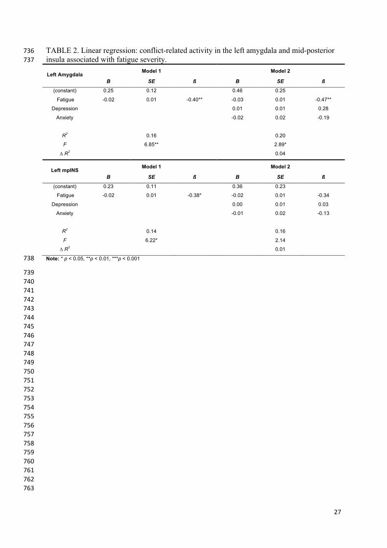
We found fatigue severity significantly predicted neural reactivity of the left amygdala and 360
mpINS, explaining 16% and 14% of the variance, respectively (Table 2). Adding anxiety and 361
depressive variables, did not improve the fit of the model. 362
Exploratory whole-brain effects of I-C are provided in Table 3. In short, adolescent CFS 363
patients showed decreased response to conflict in the insula, precentral gyrus, middle 364
temporal gyrus, cingulate, middle occipital gyrus, middle frontal gyrus, thalamus, precuneus 365
and cuneus. 366
Discussion 367
The main findings of this study were that adolescents with CFS were less able to engage the 368
left amygdala and left mid-posterior insula (mpINS) in response to conflict compared to the 369
healthy comparison group. Reactivity of the amygdala was associated with accuracy 370
interference during conflict. Conflict-related activity in the mpINS was related to response 371

16
time interference. There were no associations between brain activity and depressive and 372
anxiety symptoms. The CFS group did not produce a negative affect bias. 373
Within CFS group, we found inefficient response time interference performance; however, 374
this effect was not significantly different between-groups. This finding is in contrast to studies 375
on adult depression and anxiety disorder (Etkin, Prater, Hoeft, Menon, & Schatzberg, 2010; 376
Etkin & Schatzberg, 2011) and trauma-exposed youths, which do produce significant group 377
differences on interference performances (Marusak, Etkin, et al., 2015; Marusak, Martin, 378
Etkin, & Thomason, 2015). The response times for congruent and incongruent trials were not 379
significantly different between groups. However, the absence of a response time conflict 380
effect within patients indicates inefficient response time processing and suggests the 381
emotional information in the stimuli either did not aid in cognitive processing on congruent 382
trials and/or did not interfere in cognitive processing on incongruent trials, as observed in 383
healthy adolescents. During the congruent condition, the competing word stimulus is 384
understood to prime cognition, as there is no conflict in stimuli semantics. In the incongruent 385
condition, the competing word stimulus is understood to interfere with cognition, as there is 386
conflicting semantics in the stimuli. Both conditions represent a degree of cognitive inhibition 387
of the overlearned prepotent response to read the word. Consequently, the absence of a 388
response time conflict effect in adolescent CFS seems to be due to inefficient processing of 389
the word-distractor stimuli, which should either facilitate or interfere with cognition, 390
accordingly. 391
Decreases in neural reactivity to conflict in the left amygdala and left mpINS indicate deficits 392
in cognitive and affective functioning of the salience network (SN) (Marusak, Etkin, et al., 393
2015; Menon, 2015). Perceptive sensory areas send information to amygdala and FIC that is 394
further relayed to the mpINS (Cauda, Geminiani, & Vercelli, 2014). The lateralized left 395
amygdala and mpINS effects found in our study might be related to the different roles of 396

17
hemisphere function in emotional processing. In a systematic review, researchers found 397
predominate left amygdala activation across fMRI studies (Baas, Aleman, & Kahn, 2004). 398
Their study supports the theoretical view of functional brain asymmetry in the local and 399
global processing of information. Local processing of the left hemisphere is relatively biased 400
towards the processing fine-grained details of a stimulus or scene; whereas, global processing 401
of the right hemisphere is relatively biased towards the processing of holistic aspects of a 402
stimulus or scene (Hugdahl & Davidson, 2004). The decrease in left amygdala reactivity of 403
adolescent CFS patients could suggest inefficient local functioning in the detection of salient 404
stimuli cues that might influence further cognitive processing to the left mpINS. 405
Current SN theory suggests salience filtering occurs in the SN initiating cognitive control 406
signaling that influence how stimuli are processed (Menon, 2015). As such, neural activity in 407
regions responsible for detecting conflict should reflect the amount of behavioral interference, 408
resulting in higher activity in those regions. Even though the behavioral interference measures 409
did not reveal between-groups effects, we found the relationship between group differences 410
and interference measures varied as a function of conflict-related neural activity in the 411
amygdala and insula, which better illustrates group differences in brain behavior relations. 412
The negative association between accuracy interference and conflict-related reactivity in the 413
left amygdala, and the lack of association between response time interference and conflict-414
related reactivity in the mpINS in CFS patients are indicators of inefficient salience detection 415
and filtering function of the SN. In a previous study on childhood CFS, increasing poor task 416
performance in patients resulted in a less efficient use of neural resources in frontal regions, 417
where greater mental effort afforded costly energy requirements (Mizuno et al., 2015). 418
Exertion intolerance experienced by CFS patients might be a consequence of impoverished 419
cognitive control signaling from SN regions that results in inefficient use of neural resources 420
and energy consumption, a vicious cycle that increases fatigue. 421

18
Adolescent CFS group and comparison group did not differ on demographic factors, allowing 422
us to compare effects of fatigue in groups that had similar sociodemographic backgrounds. 423
Thus, the observed neural changes may represent specific fatigue related alterations, as 424
symptoms of depression and anxiety did not influence the relationship between fatigue 425
severity and neural function. Additionally, negative affect bias to faces expressing negative 426
emotion, a surrogate maker in anxiety and depression disorders (Bar-Haim et al., 2007; Gotlib 427
et al., 2004) and a developmental effect seemingly amplified during childhood (Augusti, 428
Torheim, & Melinder, 2014), was not observed in the CFS group of this study. Within the 429
adolescent CFS patients, psychiatric disorders were ruled out prior to inclusion in this study 430
(Sulheim et al., 2014). Even though they reported significantly more anxiety and depressive 431
symptoms than the healthy comparison group, they did not produce this bias effect. In fact 432
they were less accurate on both happy conditions suggesting trials that should be relatively 433
easy did not help cognition. The lack of neural associations with depressive and anxiety 434
symptoms and the absence of a negative affect bias further supports CFS as an important 435
clinical entity (Lamers, Hickie, & Merikangas, 2013), where the relationship between 436
prolonged fatigue and neural dysfunction potentially threatens normal development in 437
adolescents. 438
The decreased neural reactivity to conflict in adolescent CFS patients is not clear. Following 439
the Sustained Arousal model of disease mechanisms in CFS, a maladaptive stress response is 440
a central pathophysiological element (Wyller et al., 2009), eliciting autonomic and 441
neuroendocrine alterations that parallel the pathophysiology of chronic PTSD (Pervanidou & 442
Chrousos, 2010; Sulheim et al., 2014). Prolonged stress affects both the structure and function 443
of the PFC and stress related alterations occur in regions mediating the highest levels of 444
cognitive function (see McEwen and Morrison (2013) for review). Neuroimaging findings on 445
trauma-exposed youths at-risk for stress-related psychopathology showed a similar abnormal 446

19
activity pattern in regions of the SN (Marusak, Etkin, et al., 2015). Even though SN 447
dysfunction seems to parallel those that are stress-related, the neural responses were opposite 448
in CFS adolescents. 449
A supplemental frame for the findings of this study might be alexithymia, which literally 450
means - no words for emotions. Individuals suffering from alexithymia have difficulty in 451
verbally describing their own emotions and are known to be comorbid with a number of 452
psychiatric conditions (Taylor, Bagby, & Parker, 1999). Previous CFS research has reported 453
alexithymic correlates in adolescents and adults (Sepede et al., 2011; van de Putte, Engelbert, 454
Kuis, Kimpen, & Uiterwaal, 2007). Future research could focus on the connection between 455
the clinical phenomena of alexithymia and the apparent “washing out of emotions” in 456
adolescent CFS, suggested by the deficits in salience detection observed in this study. 457
Strengths and limitations 458
Using an adolescent patient population, it might be easier to identify real disease mechanisms 459
as opposed to secondary phenomena associated with years of chronicity. As the findings of 460
our study are only preliminary, future studies should confirm the results in a larger sample of 461
patients. Furthermore, a causal relationship cannot be inferred in a cross-sectional design. We 462
used liberal inclusion criteria, where not all patients adhere to the Fukuda-definition that is 463
most widely accepted. Of the CFS patients allocated to this study, there were extenuating 464
circumstances that reduced the number from 30 to 15 (i.e. not wanting to participate, having 465
orthodontic treatment, and movement). So far as can be determined, there was no reason to 466
suspect selection bias. 467
The emotional conflict task used in this study did not include a neutral condition, which 468
would have helped to dissociate interference related to emotion in patients. Specifically, a 469
neutral face condition would provide information to see if the lack of response time conflict 470
20
effect in patients was due to the inability to perceive emotion. The paradigm design did not 471
allow us to measure conflict regulation. Conflict regulation effects provide information to 472
dissociate neural regions active specifically in the regulation of emotional conflict. However, 473
future research might consider using these aspects to further understand alterations in 474
emotional conflict processing of adolescent CFS patients. 475
Conclusion 476
Our data highlight behavioral and neural deviations in emotional conflict processing of 477
adolescent CFS patients and deficits in these neural regions may contribute importantly to the 478
maintenance CFS pathology. Our findings of amygdala and insula dysfunction demonstrates a 479
disruption in cognitive and affective functioning of the salience network, a potential 480
biomarker in developmental psychopathology (Uddin, Supekar, Ryali, & Menon, 2011). 481
However, it is unknown whether impairments in salience detection during emotional conflict 482
processing in adolescent CFS reflect a disorder-specific abnormality or a more general 483
endophenotype of complex symptom disorders, such as chronic stress and pain. Nevertheless, 484
the group differences suggest that the inability of patients to efficiently and effectively resolve 485
emotional conflict is an important aspect of the pathophysiology of adolescent chronic fatigue 486
syndrome and potentially of other chronic disorders. This nature of research warrants further 487
investigation. 488
Conflict of Interest Statement 489
None. 490
Acknowledgements 491
The authors thank the participants and their families for their contribution in this study. We 492
thank Nils Inge Landrø for helpful comments on the manuscript. Also, we express gratitude to 493
Kari Gjersum for secretary assistance; Tommy Sinnes, Ellen Wessel, Berit Widerøe Njølstad, 494

21
and Pelle Rohdin for practical assistance; Berit Bjelkåsen for the development of the 495
computerized randomization procedure; and all referring hospital units. 496
This work was supported by the Norwegian Research Council (VBW, grant number 228874); 497
Health South–East Hospital Trust (VBW); and the University of Oslo (VBW). 498
Author Contributions 499
LW carried out data analyses and drafted the manuscript. TE and VBW conceptualized and 500
contributed to the study design. DS and EF collected clinical data and contributed to the study 501
design. AM and MØ contributed to the study design. All authors contributed to data 502
interpretation and drafting of the manuscript. 503
504

22
References 505
Afari, N., Ahumada, S. M., Wright, L. J., Mostoufi, S., Golnari, G., Reis, V., & Cuneo, J. G. 506(2014). Psychological trauma and functional somatic syndromes: a systematic review 507and meta-analysis. Psychosom Med, 76(1), 2. 508
Ashburner, J., Barnes, G., Chen, C., Daunizeau, J., Flandin, G., Friston, K., . . . Litvak, V. 509(2012). SPM8 manual. Functional Imaging Laboratory, Institute of Neurology. 510
Augusti, E. M., Torheim, H. K., & Melinder, A. (2014). The effect of emotional facial 511expressions on children's working memory: associations with age and behavior. Child 512Neuropsychology, 20(1), 86-105. doi: 10.1080/09297049.2012.749225 513
Baas, D., Aleman, A., & Kahn, R. S. (2004). Lateralization of amygdala activation: a 514systematic review of functional neuroimaging studies. Brain Res Brain Res Rev, 45(2), 51596-103. doi: 10.1016/j.brainresrev.2004.02.004 516
Bar-Haim, Y., Lamy, D., Pergamin, L., Bakermans-Kranenburg, M. J., & van, I. M. H. (2007). 517Threat-related attentional bias in anxious and nonanxious individuals: a meta-analytic 518study. Psychological Bulletin, 133(1), 1-24. doi: 10.1037/0033-2909.133.1.1 519
Botvinick, M. M., Braver, T. S., Barch, D. M., Carter, C. S., & Cohen, J. D. (2001). Conflict 520monitoring and cognitive control. Psychol Rev, 108(3), 624-652. 521
Brett, M., Anton, J.-L., Valabregue, R., & Poline, J.-B. (2002). Region of interest analysis 522using the MarsBar toolbox for SPM 99. Neuroimage, 16(2), S497. 523
Bruce, S. E., Buchholz, K. R., Brown, W. J., Yan, L., Durbin, A., & Sheline, Y. I. (2012). 524Altered emotional interference processing in the amygdala and insula in women with 525Post-Traumatic Stress Disorder. Neuroimage Clin, 2, 43-49. doi: 52610.1016/j.nicl.2012.11.003 527
Brurberg, K. G., Fonhus, M. S., Larun, L., Flottorp, S., & Malterud, K. (2014). Case 528definitions for chronic fatigue syndrome/myalgic encephalomyelitis (CFS/ME): a 529systematic review. British Medical Journal Open, 4(2), e003973. doi: 53010.1136/bmjopen-2013-003973 531
Caseras, X., Mataix-Cols, D., Giampietro, V., Rimes, K. A., Brammer, M., Zelaya, F., . . . 532Godfrey, E. L. (2006). Probing the working memory system in chronic fatigue 533syndrome: a functional magnetic resonance imaging study using the n-back task. 534Psychosom Med, 68(6), 947-955. doi: 10.1097/01.psy.0000242770.50979.5f 535
Caseras, X., Mataix-Cols, D., Rimes, K. A., Giampietro, V., Brammer, M., Zelaya, F., . . . 536Godfrey, E. (2008). The neural correlates of fatigue: an exploratory imaginal fatigue 537provocation study in chronic fatigue syndrome. Psychol Med, 38(7), 941-951. doi: 53810.1017/S0033291708003450 539
Cauda, F., Geminiani, G. C., & Vercelli, A. (2014). Evolutionary appearance of von 540Economo's neurons in the mammalian cerebral cortex. Front Hum Neurosci, 8, 104. 541doi: 10.3389/fnhum.2014.00104 542
Chalder, T., Berelowitz, G., Pawlikowska, T., Watts, L., Wessely, S., Wright, D., & Wallace, 543E. P. (1993). Development of a fatigue scale. J Psychosom Res, 37(2), 147-153. 544
Craig, A. D. (2002). How do you feel? Interoception: the sense of the physiological condition 545of the body. Nat Rev Neurosci, 3(8), 655-666. doi: 10.1038/nrn894 546
Critchley, H. D. (2004). The human cortex responds to an interoceptive challenge. Proc Natl 547Acad Sci U S A, 101(17), 6333-6334. doi: 10.1073/pnas.0401510101 548
Critchley, H. D., Melmed, R. N., Featherstone, E., Mathias, C. J., & Dolan, R. J. (2002). 549Volitional control of autonomic arousal: a functional magnetic resonance study. 550Neuroimage, 16(4), 909-919. 551

23
de Lange, F. P., Kalkman, J. S., Bleijenberg, G., Hagoort, P., van der Werf, S. P., van der 552Meer, J. W., & Toni, I. (2004). Neural correlates of the chronic fatigue syndrome--an 553fMRI study. Brain, 127(Pt 9), 1948-1957. doi: 10.1093/brain/awh225 554
de Lange, F. P., Koers, A., Kalkman, J. S., Bleijenberg, G., Hagoort, P., van der Meer, J. W., 555& Toni, I. (2008). Increase in prefrontal cortical volume following cognitive 556behavioural therapy in patients with chronic fatigue syndrome. Brain, 131(Pt 8), 2172-5572180. doi: 10.1093/brain/awn140 558
Egner, T., Etkin, A., Gale, S., & Hirsch, J. (2008). Dissociable neural systems resolve conflict 559from emotional versus nonemotional distracters. Cereb Cortex, 18(6), 1475-1484. doi: 56010.1093/cercor/bhm179 561
Etkin, A., Egner, T., Peraza, D. M., Kandel, E. R., & Hirsch, J. (2006). Resolving emotional 562conflict: a role for the rostral anterior cingulate cortex in modulating activity in the 563amygdala. Neuron, 51(6), 871-882. doi: 10.1016/j.neuron.2006.07.029 564
Etkin, A., Prater, K. E., Hoeft, F., Menon, V., & Schatzberg, A. F. (2010). Failure of anterior 565cingulate activation and connectivity with the amygdala during implicit regulation of 566emotional processing in generalized anxiety disorder. Am J Psychiatry, 167(5), 545-567554. doi: 10.1176/appi.ajp.2009.09070931 568
Etkin, A., & Schatzberg, A. F. (2011). Common abnormalities and disorder-specific 569compensation during implicit regulation of emotional processing in generalized 570anxiety and major depressive disorders. Am J Psychiatry, 168(9), 968-978. doi: 57110.1176/appi.ajp.2011.10091290 572
Friston, K. J., Ashburner, J., Frith, C. D., Poline, J. B., Heather, J. D., & Frackowiak, R. S. J. 573(1995). Spatial registration and normalization of images. Human Brain Mapping, 3(3), 574165-189. 575
Friston, K. J., Holmes, A. P., Worsley, K. J., Poline, J. P., Frith, C. D., & Frackowiak, R. S. 576(1994). Statistical parametric maps in functional imaging: a general linear approach. 577Human Brain Mapping, 2(4), 189-210. 578
Fukuda, K., Straus, S. E., Hickie, I., Sharpe, M. C., Dobbins, J. G., Komaroff, A., . . . Reeves, 579W. C. (1994). The Chronic Fatigue Syndrome - a Comprehensive Approach to Its 580Definition and Study. Annals of Internal Medicine, 121(12), 953-959. 581
Gotlib, I. H., Krasnoperova, E., Yue, D. N., & Joormann, J. (2004). Attentional biases for 582negative interpersonal stimuli in clinical depression. J Abnorm Psychol, 113(1), 121-583135. doi: 10.1037/0021-843X.113.1.121 584
Haig-Ferguson, A., Tucker, P., Eaton, N., Hunt, L., & Crawley, E. (2009). Memory and 585attention problems in children with chronic fatigue syndrome or myalgic 586encephalopathy. Arch Dis Child, 94(10), 757-762. doi: 10.1136/adc.2008.143032 587
Hugdahl, K., & Davidson, R. J. (2004). The asymmetrical brain: MIT Press. 588IOM. (2015). Beyond Myalgic Encephalomyelitis/Chronic Fatigue Syndrome: Redefining an 589
Illness. Beyond Myalgic Encephalomyelitis/Chronic Fatigue Syndrome: Redefining an 590Illness. Washington (DC): The National Academies Press. 591
Kawatani, J., Mizuno, K., Shiraishi, S., Takao, M., Joudoi, T., Fukuda, S., . . . Tomoda, A. 592(2011). Cognitive dysfunction and mental fatigue in childhood chronic fatigue 593syndrome--a 6-month follow-up study. Brain Dev, 33(10), 832-841. doi: 59410.1016/j.braindev.2010.12.009 595
Kerns, J. G., Cohen, J. D., MacDonald, A. W., 3rd, Cho, R. Y., Stenger, V. A., & Carter, C. S. 596(2004). Anterior cingulate conflict monitoring and adjustments in control. Science, 597303(5660), 1023-1026. doi: 10.1126/science.1089910 598
Lamers, F., Hickie, I., & Merikangas, K. R. (2013). Prevalence and correlates of prolonged 599fatigue in a U.S. sample of adolescents. Am J Psychiatry, 170(5), 502-510. doi: 60010.1176/appi.ajp.2012.12040454 601

24
Lieberman, M. D., & Cunningham, W. A. (2009). Type I and Type II error concerns in fMRI 602research: re-balancing the scale. Soc Cogn Affect Neurosci, 4(4), 423-428. doi: 60310.1093/scan/nsp052 604
Lievesley, K., Rimes, K. A., & Chalder, T. (2014). A review of the predisposing, precipitating 605and perpetuating factors in Chronic Fatigue Syndrome in children and adolescents. 606Clin Psychol Rev, 34(3), 233-248. doi: 10.1016/j.cpr.2014.02.002 607
Lundqvist, D. F., A.; Øhman, A. (1998). The Karolinska Directed Emotional Faces - KDEF, 608CD ROM. In D. o. C. Neuroscience (Ed.). Psychology Department: Karolinska 609Institute. 610
MacDonald, A. W., 3rd, Cohen, J. D., Stenger, V. A., & Carter, C. S. (2000). Dissociating the 611role of the dorsolateral prefrontal and anterior cingulate cortex in cognitive control. 612Science, 288(5472), 1835-1838. 613
Maldjian, J. A., Laurienti, P. J., Kraft, R. A., & Burdette, J. H. (2003). An automated method 614for neuroanatomic and cytoarchitectonic atlas-based interrogation of fMRI data sets. 615Neuroimage, 19(3), 1233-1239. doi: Doi 10.1016/S1053-8119(03)00169-1 616
Marusak, H. A., Etkin, A., & Thomason, M. E. (2015). Disrupted insula-based neural circuit 617organization and conflict interference in trauma-exposed youth. Neuroimage Clin, 8, 618516-525. doi: 10.1016/j.nicl.2015.04.007 619
Marusak, H. A., Martin, K. R., Etkin, A., & Thomason, M. E. (2015). Childhood trauma 620exposure disrupts the automatic regulation of emotional processing. 621Neuropsychopharmacology, 40(5), 1250-1258. doi: 10.1038/npp.2014.311 622
McEwen, B. S., & Morrison, J. H. (2013). The brain on stress: vulnerability and plasticity of 623the prefrontal cortex over the life course. Neuron, 79(1), 16-29. doi: 62410.1016/j.neuron.2013.06.028 625
Menon, V. (2015). Salience Network. In A. W. Toga (Ed.), Brain Mapping: An Encyclopedic 626Reference (Vol. 2, pp. 597-611): Academic Press: Elsevier. 627
Mizuno, K., Tanaka, M., Tanabe, H. C., Joudoi, T., Kawatani, J., Shigihara, Y., . . . Watanabe, 628Y. (2015). Less efficient and costly processes of frontal cortex in childhood chronic 629fatigue syndrome. Neuroimage Clin, 9, 355-368. doi: 10.1016/j.nicl.2015.09.001 630
NICE. (2007). Chronic fatigue syndrome/myalgic encephalomyelitis (or encephalopathy): 631Diagnosis and management of CFS/ME in adults and children:NICE clinical guideline 63253., from National Institute for Health and Clinical Excellence 633
Nisenbaum, R., Reyes, M., Unger, E. R., & Reeves, W. C. (2004). Factor analysis of 634symptoms among subjects with unexplained chronic fatigue: what can we learn about 635chronic fatigue syndrome? J Psychosom Res, 56(2), 171-178. doi: 10.1016/S0022-6363999(03)00039-4 637
Okada, T., Tanaka, M., Kuratsune, H., Watanabe, Y., & Sadato, N. (2004). Mechanisms 638underlying fatigue: a voxel-based morphometric study of chronic fatigue syndrome. 639BioMed Central Neurology, 4(1), 14. doi: 10.1186/1471-2377-4-14 640
Pervanidou, P., & Chrousos, G. P. (2010). Neuroendocrinology of post-traumatic stress 641disorder. Prog Brain Res, 182, 149-160. doi: 10.1016/S0079-6123(10)82005-9 642
Royal, C. o. P. a. C. H. (2004). Evidence based guidelines for the management of CFS/ME 643(Chronic Fatigue Syndrome/Myalgic Encephalopathy) in Children and Young Adults. 644http://www.rcpch.ac.uk/child-health/standards-care/clinical-guidelines-and-645standards/published-rcpch/clinical-guidelines-an, from Royal College of Paediatrics 646and Child Health 647
Sepede, G., Racciatti, D., Gorgoretti, V., Nacci, M., Pizzigallo, E., Onofrj, M., . . . Gambi, F. 648(2011). Psychophysical distress and alexithymic traits in chronic fatigue syndrome 649with and without comorbid depression. International Journal of Immunopathology 650and Pharmacology, 24(4), 1017-1025. 651

25
Spielberger, C. D., Gorsuch, R. L., & Lushene, R. E. (1973). STAI manual for the Stait-Trait 652Anxiety Inventory. Palo Alto, CA: Consulting Psychologists Press. 653
Sulheim, D., Fagermoen, E., Sivertsen, O. S., Winger, A., Wyller, V. B., & Oie, M. G. (2015). 654Cognitive dysfunction in adolescents with chronic fatigue: a cross-sectional study. 655Arch Dis Child. doi: 10.1136/archdischild-2014-306764 656
Sulheim, D., Fagermoen, E., Winger, A., Andersen, A. M., Godang, K., Muller, F., . . . Wyller, 657V. B. (2014). Disease mechanisms and clonidine treatment in adolescent chronic 658fatigue syndrome: a combined cross-sectional and randomized clinical trial. Journal of 659the American Medical Association Pediatrics, 168(4), 351-360. doi: 66010.1001/jamapediatrics.2013.4647 661
Sullivan, P. F., Pedersen, N. L., Jacks, A., & Evengard, B. (2005). Chronic fatigue in a 662population sample: definitions and heterogeneity. Psychol Med, 35(9), 1337-1348. doi: 66310.1017/S0033291705005210 664
Sund, A. M., Larsson, B., & Wichstrom, L. (2001). Depressive symptoms among young 665Norwegian adolescents as measured by the Mood and Feelings Questionnaire (MFQ). 666Eur Child Adolesc Psychiatry, 10(4), 222-229. 667
Tanaka, M., Fukuda, S., Mizuno, K., Imai-Matsumura, K., Jodoi, T., Kawatani, J., . . . 668Watanabe, Y. (2008). Reliability and validity of the Japanese version of the Chalder 669Fatigue Scale among youth in Japan. Psychol Rep, 103(3), 682-690. doi: 67010.2466/pr0.103.3.682-690 671
Taylor, G. J., Bagby, R. M., & Parker, J. D. (1999). Disorders of affect regulation: 672Alexithymia in medical and psychiatric illness: Cambridge University Press. 673
Tomoda, A., Mizuno, K., Murayama, N., Joudoi, T., Igasaki, T., Miyazaki, M., & Miike, T. 674(2007). Event-related potentials in Japanese childhood chronic fatigue syndrome. 675Journal of Pediatric Neurology, 5(3), 199-208. 676
Uddin, L. Q., Supekar, K. S., Ryali, S., & Menon, V. (2011). Dynamic reconfiguration of 677structural and functional connectivity across core neurocognitive brain networks with 678development. J Neurosci, 31(50), 18578-18589. doi: 10.1523/JNEUROSCI.4465-67911.2011 680
van de Putte, E. M., Böcker, K. B., Buitelaar, J., Kenemans, J. L., Engelbert, R. H., Kuis, 681W., . . . Uiterwaal, C. S. (2008). Deficits of Interference Control in Adolescents With 682Chronic Fatigue Syndrome. Archives of pediatrics & adolescent medicine, 162(12), 6831196-1197. 684
van de Putte, E. M., Engelbert, R. H., Kuis, W., Kimpen, J. L., & Uiterwaal, C. S. (2007). 685Alexithymia in adolescents with chronic fatigue syndrome. J Psychosom Res, 63(4), 686377-380. doi: 10.1016/j.jpsychores.2007.07.009 687
Wechsler, D. (2007). Wechsler Abbreviated Scale of Intelligence (WASI). Norwegian manual 688supplement. Stockholm, Sweden: Pearson. 689
Wyller, V. B., Eriksen, H. R., & Malterud, K. (2009). Can sustained arousal explain the 690Chronic Fatigue Syndrome? Behavioral and Brain Functions, 5, 10. doi: 69110.1186/1744-9081-5-10 692
Wyller, V. B., & Helland, I. B. (2013). Relationship between autonomic cardiovascular 693control, case definition, clinical symptoms, and functional disability in adolescent 694chronic fatigue syndrome: an exploratory study. Biopsychosoc Med, 7(1), 5. doi: 69510.1186/1751-0759-7-5 696
697
698

26
FIGURE 1. Emotional conflict task 699 700 701Figure 1 shows a sample task time course illustrating the contrasts made to examine 702emotional conflict effect using incongruent and congruent trials. Participants were asked to 703respond to the emotional face and ignore the word. 704 705 706 707TABLE 1. Demographic and clinical characteristics of adolescent Healthy Comparison Participants and Patients with Chronic Fatigue Syndrome in an fMRI Study
Characteristic
Patients with Chronic Fatigue Syndrome
(N=15) Healthy comparison group
(N=24) p
N % N %
Female 14 93 16 67 n.s. aFukuda criteria 10 67 bNICE criteria 12 80 Mean SD Mean SD Disease duration in months 19.5 10.7 Age 15.8 1.5 15.2 1.6 n.s. cBMI 22.5 3.6 20.2 2.9 n.s. dWASI IQ 107.9 13.3 115.3 17.1 n.s. Depression eMFQ 15.9 8.4 5.6 6.9 <0.001* Spielberger State Anxiety Inventory 20.3 4.8 16.0 3.5 <0.01* Chalder Fatigue Questionnaire 19.7 6.5 9.0 3.6 <0.001*
aParticipants fulfilling the Fukuda-definition of CFS (Fukuda et al., 1994) 708bParticipants fulfilling the National Institute for Health and Care Excellence (2007) definition of CFS 709cBody Mass Index 710dWechlser Abbreviated Scale of Intelligence-estimated full IQ 711eMood and Feelings Questionnaire for Depression 712*Indicates group comparison is significant at p ≤ 0.05. 713The χ2 test was used for sex; two-sample t-tests were used for continuous variables. 714Not significant (n.s.) 715 716 717 718FIGURE 2. Behavioral response interference to emotional conflict in adolescent patients with 719Chronic Fatigue Syndrome and Healthy Comparison participants 720 721 722Figure 2 shows response time and accuracy interference scores reflecting the emotional 723conflict effect (I-C). Panel A, adolescent CFS patients did not show response time 724interference for incongruent relative to congruent trials (I-C). Higher values indicate a loss in 725performance. ** One-sample t-test, p <0.001. Panel B, adolescent CFS patients show a 726similar loss of accuracy for incongruent relative to congruent trials (I-C). Negative values 727indicate a loss in performance. * One-sample t-test, p <0.01. Error bars represent standard 728error. 729 730 731 732 733 734 735
27
TABLE 2. Linear regression: conflict-related activity in the left amygdala and mid-posterior 736insula associated with fatigue severity. 737
Left Amygdala Model 1 Model 2
B SE ß B SE ß
(constant) 0.25 0.12 0.46 0.25
Fatigue -0.02 0.01 -0.40** -0.03 0.01 -0.47**
Depression 0.01 0.01 0.28
Anxiety -0.02 0.02 -0.19
R2 0.16 0.20 F 6.85** 2.89*
∆ R2 0.04
Left mpINS Model 1 Model 2
B SE ß B SE ß
(constant) 0.23 0.11 0.36 0.23 Fatigue -0.02 0.01 -0.38* -0.02 0.01 -0.34
Depression 0.00 0.01 0.03
Anxiety -0.01 0.02 -0.13
R2 0.14 0.16
F 6.22* 2.14
∆ R2 0.01
Note: * p < 0.05, **p < 0.01, ***p < 0.001 738
739 740 741 742 743 744 745 746 747 748 749 750 751 752 753 754 755 756 757 758 759 760 761 762 763

28
TABLE 3. Group differences in Whole-Brain activity during conflict (Incongruent minus 764Congruent) 765 Peak
Contrast Brain Area BA x y z # of
voxels T-Score comparison > CFS R Parahippocampus 36 30 -40 -8 17 4.35 L Insula 13 -39 -7 7 223 4.23 R Parietal Precuneus 7 15 -76 40 192 4.15 R Lentiform Nucleus 21 -10 4 14 3.83 L Mid Temporal gyrus 37 -48 -67 4 65 3.75 R Insula 13 39 5 16 12 3.65 R Precentral gyrus 44 57 8 10 82 3.58 R Mid Occipital gyrus 18 24 -91 -2 24 3.52 R Mid Frontal gyrus 6 21 -7 40 46 3.51 R Mid Occipital gyrus 37 39 -67 1 47 3.44 L Inferior Parietal 40 -42 -46 55 26 3.40 R Putamen 27 17 4 55 3.39 R Superior Frontal gyrus 6 15 -1 64 26 3.38 L Thalamus -15 -22 7 44 3.33 L Posterior Cingulate 30 -18 -61 4 13 3.24 R Inferior Parietal 40 45 -37 52 17 3.22 R Cuneus 17 18 -76 10 26 3.21 L Precentral gyrus 6 -27 -13 64 17 3.10 L Postcentral gyrus 2 -45 -25 37 12 2.97 L Cingulate gyrus 32 -9 8 40 10 2.96 CFS > comparison no voxels survived at this threshold
Exploratory whole-brain results provided at p < 0.005 uncorrected, cluster minimum = 10 766voxels. Abbreviations: BA, Brodmann’s Area; L, left; R, right. Coordinates are given in MNI 767convention. 768 769 770 771FIGURE 3. Left Amygdala (AMY) and left mid-posterior insula (mpINS) responses to 772emotional conflict 773 774 775Figure 3 shows decreased (A) left AMY and (C) left mpINS responses to emotional conflict 776in adolescent CFS patients compared to healthy comparison (HC) group. (B) The linear 777relationship between left AMY conflict-related activity and accuracy interference 778significantly differed in CFS patients than HC. (D) The linear relationship between left 779mpINS conflict-related activity and response time interference significantly differed in CFS 780patients than HC. Data used for plotting are extracted data from 8mm ROI spheres. *p ≤ 0.02 781two-sample t-test. Error bars represent standard error. 782