emergency quick reference guidehtml.esahq.org/patientsafetykit/.../emergency_cl/emergency... ·...
TRANSCRIPT
EMERGENCY QUICK REFERENCE GUIDE
Sven Staender (CH)Andrew Fairley-Smith (UK)Guttorm Bratteboe (Norway)David Whitaker (UK) andJannicke Mellin-Olsen (Norway; ESA board member) and David Borshoff (Australia; non Task Force member)
Preface
The ESA and EBA Helsinki Declaration on Patient Safety in Anaesthesiology1 requires to have protocols available for the management of perioperative complications. Based on the book “The Anaesthetic Crisis Manual“2 by David Borshoff and the previous ESA project “OLEH” (Online Electronic Help) by Azriel Perel, the Task Force Patient Safety has developed checklists for various emergencies that appear similar to reference guides used in aviation for in-flight emergencies.
The first draft has been made available on the webpage of the European Society of Anaesthesiology at the end of the year 2012 for open feedback by every ESA member. Taking all the different and very valuable feedbacks into account, these emergency quick reference guidelines have been produced with prudence. Nevertheless, each practitioner is advised to pay careful attention when using these checklists as we do not take responsibility for the consequences out of their application. Furthermore we continue to invite everyone to give us qualified feedback for any possible improvement of these checklists in the future.
These checklists are thought to be a service for the ESA and EBA members and every anaesthetist or national anaesthesia society should feel free to adopt them to their local or national practice.
Our special thanks goes to Anja Sandemose (Norway), Flavia Petrini (Italy), Janusz Andres (Poland), Helmut Trimmel (Austria), Julio Cortinas (Spain), Nick Toff (UK) and Filippo Bressan (Italy) for valuable feedback and suggestions for improvement. Furthermore, various colleagues from the Task Force members and Co-authors have read and corrected the first draft. Thank you very much to all of them.
Sven Staender (Chairman ESA/EBA Task Force Patient Safety)
1 Mellin-Olsen J, Staender S, Whitaker DK, Smith AF. The Helsinki Declaration on Patient Safety in Anaesthesiology. Eur J Anaesthesiol 2010 Jul;27(7):592-7.
2 Borshoff D. The Anaesthetic Crisis Manual. Cambridge University Press, Cambridge, UK 2011
EsA/EbA TAsK FoRCE PATIENT sAFETY
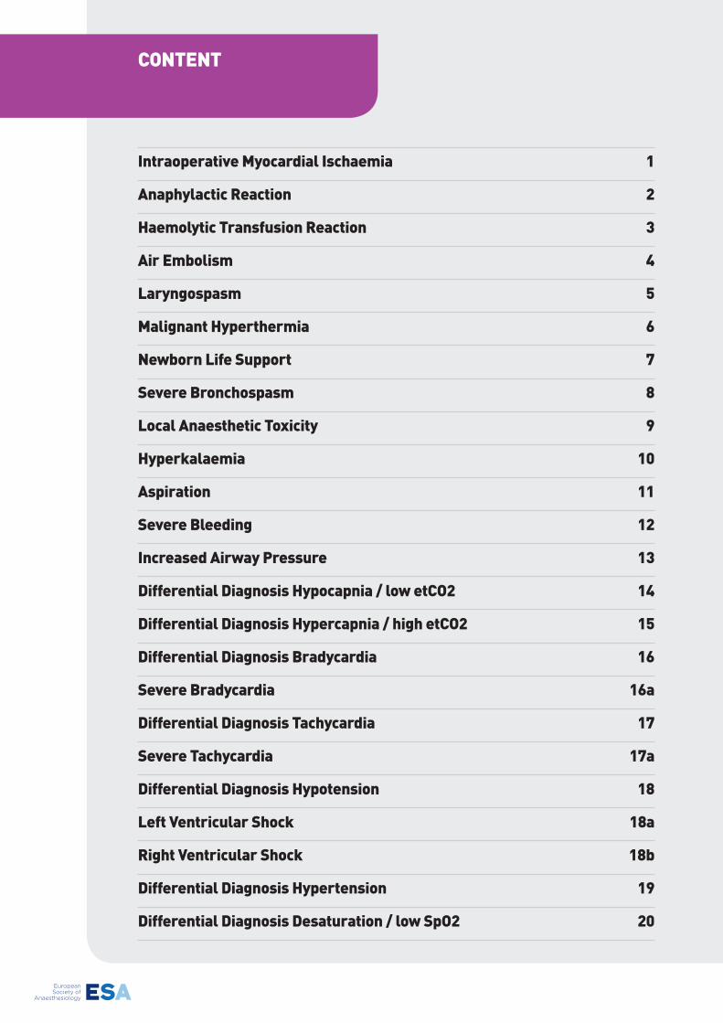
Intraoperative Myocardial Ischaemia 1
Anaphylactic Reaction 2
Haemolytic Transfusion Reaction 3
Air Embolism 4
Laryngospasm 5
Malignant Hyperthermia 6
Newborn Life support 7
severe bronchospasm 8
Local Anaesthetic Toxicity 9
Hyperkalaemia 10
Aspiration 11
severe bleeding 12
Increased Airway Pressure 13
Differential Diagnosis Hypocapnia / low etCo2 14
Differential Diagnosis Hypercapnia / high etCo2 15
Differential Diagnosis bradycardia 16
severe bradycardia 16a
Differential Diagnosis Tachycardia 17
severe Tachycardia 17a
Differential Diagnosis Hypotension 18
Left Ventricular shock 18a
Right Ventricular shock 18b
Differential Diagnosis Hypertension 19
Differential Diagnosis Desaturation / low spo2 20
CoNTENT
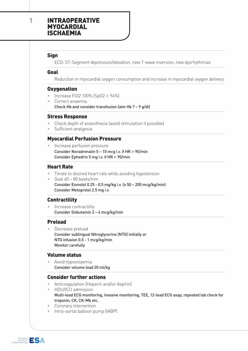
signECG: ST-Segment depression/elevation, new T-wave inversion, new dysrhythmias
Goal Reduction in myocardial oxygen consumption and increase in myocardial oxygen delivery
oxygenation • Increase FiO2 100% (SpO2 > 94%)• Correct anaemia. Check Hb and consider transfusion (aim Hb 7 – 9 g/dl)
stress Response • Check depth of anaesthesia (avoid stimulation if possible) • Sufficient analgesia
Myocardial Perfusion Pressure • Increase perfusion pressure Consider Noradrenalin 5 – 10 mcg i.v. if HR > 90/min Consider Ephedrin 5 mg i.v. if HR < 90/min
Heart Rate • Titrate to desired heart rate while avoiding hypotension• Goal 60 – 80 beats/min Consider Esmolol 0.25 - 0.5 mg/kg i.v. (± 50 – 200 mcg/kg/min) Consider Metoprolol 2.5 mg i.v.
Contractility • Increase contractility Consider Dobutamin 2 – 4 mcg/kg/min
Preload • Decrease preload Consider sublingual Nitroglycerine (NTG) initially or NTG infusion 0.5 - 1 mcg/kg/min Monitor carefully
Volume status • Avoid hypovolaemia Consider volume load 20 ml/kg
Consider further actions • Anticoagulation (Heparin and/or Aspirin)• HDU/ICU admission Multi-lead ECG monitoring, invasive monitoring, TEE, 12-lead ECG asap, repeated lab check for
troponin, CK, CK-Mb etc.• Coronary intervention• Intra-aortal balloon pump (IABP)
INTRAoPERATIVE MYoCARDIAL IsCHAEMIA
1
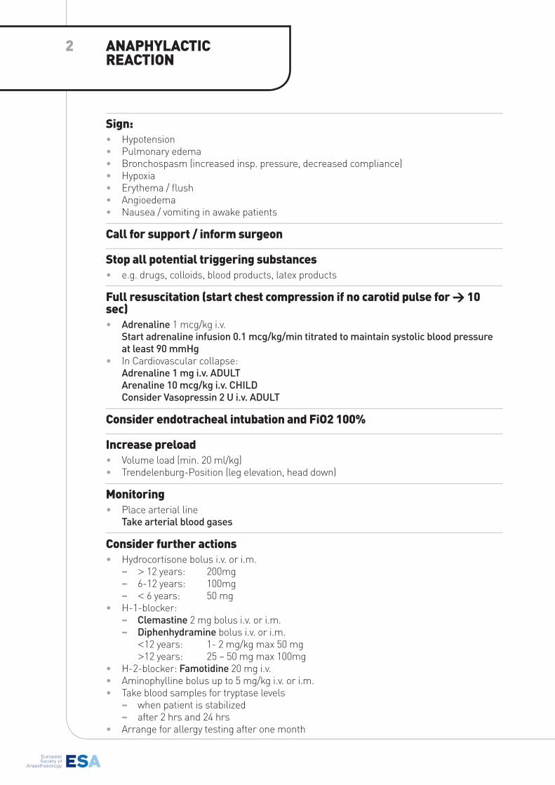
sign: • Hypotension• Pulmonary edema• Bronchospasm (increased insp. pressure, decreased compliance)• Hypoxia• Erythema / flush• Angioedema• Nausea / vomiting in awake patients
Call for support / inform surgeon
stop all potential triggering substances• e.g. drugs, colloids, blood products, latex products
Full resuscitation (start chest compression if no carotid pulse for > 10 sec) • Adrenaline 1 mcg/kg i.v.
Start adrenaline infusion 0.1 mcg/kg/min titrated to maintain systolic blood pressure at least 90 mmHg
• In Cardiovascular collapse:Adrenaline 1 mg i.v. ADULTArenaline 10 mcg/kg i.v. CHILD Consider Vasopressin 2 U i.v. ADULT
Consider endotracheal intubation and Fio2 100%
Increase preload • Volume load (min. 20 ml/kg)• Trendelenburg-Position (leg elevation, head down)
Monitoring• Place arterial line
Take arterial blood gases
Consider further actions • Hydrocortisone bolus i.v. or i.m.
− > 12 years: 200mg − 6-12 years: 100mg − < 6 years: 50 mg
• H-1-blocker: − Clemastine 2 mg bolus i.v. or i.m. − Diphenhydramine bolus i.v. or i.m.
<12 years: 1- 2 mg/kg max 50 mg>12 years: 25 – 50 mg max 100mg
• H-2-blocker: Famotidine 20 mg i.v.• Aminophylline bolus up to 5 mg/kg i.v. or i.m.• Take blood samples for tryptase levels
− when patient is stabilized − after 2 hrs and 24 hrs
• Arrange for allergy testing after one month
ANAPHYLACTIC REACTIoN
2
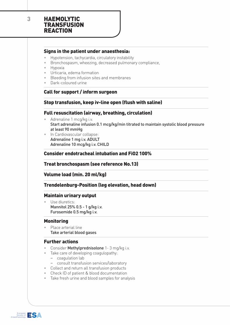
signs in the patient under anaesthesia:• Hypotension, tachycardia, circulatory instability• Bronchospasm, wheezing, decreased pulmonary compliance,• Hypoxia• Urticaria, edema formation• Bleeding from infusion sites and membranes• Dark-coloured urine
Call for support / inform surgeon
stop transfusion, keep iv-line open (flush with saline)
Full resuscitation (airway, breathing, circulation) • Adrenaline 1 mcg/kg i.v.
Start adrenaline infusion 0.1 mcg/kg/min titrated to maintain systolic blood pressure at least 90 mmHg
• In Cardiovascular collapse:Adrenaline 1 mg i.v. ADULTAdrenaline 10 mcg/kg i.v. CHILD
Consider endotracheal intubation and Fio2 100%
Treat bronchospasm (see reference No.13)
Volume load (min. 20 ml/kg)
Trendelenburg-Position (leg elevation, head down)
Maintain urinary output• Use diuretics:
Mannitol 25% 0.5 - 1 g/kg i.v.Furosemide 0.5 mg/kg i.v.
Monitoring• Place arterial line
Take arterial blood gases
Further actions • Consider Methylprednisolone 1- 3 mg/kg i.v.• Take care of developing coagulopathy:
− coagulation lab − consult transfusion services/laboratory
• Collect and return all transfusion products• Check ID of patient & blood documentation• Take fresh urine and blood samples for analysis
HAEMoLYTIC TRANsFUsIoN REACTIoN
3
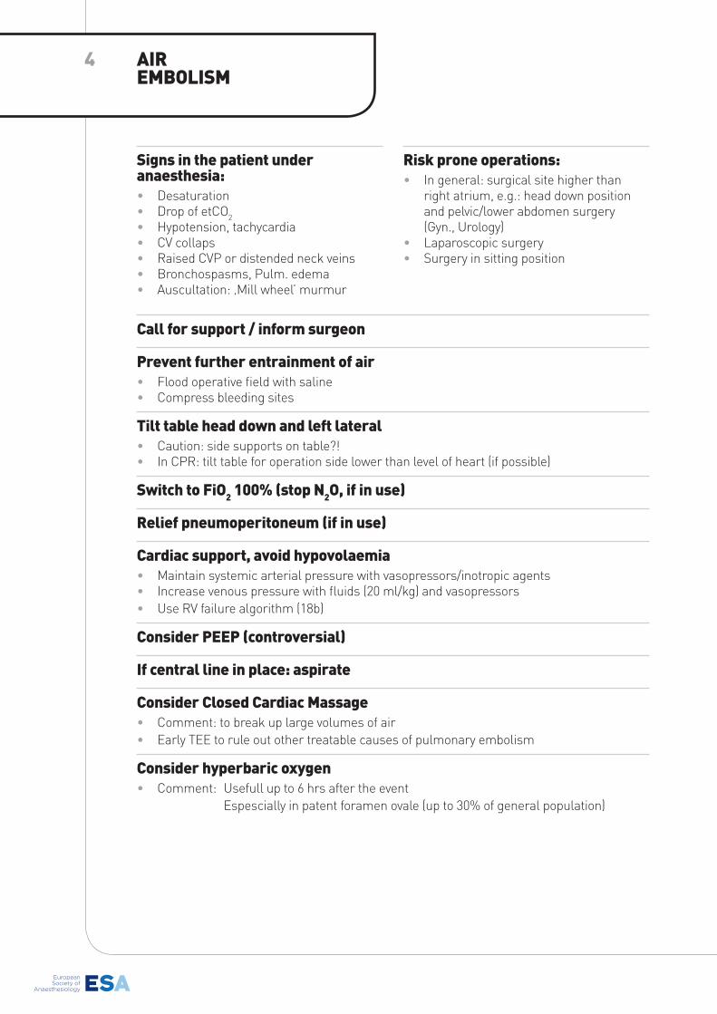
signs in the patient under anaesthesia: • Desaturation• Drop of etCO2• Hypotension, tachycardia• CV collaps• Raised CVP or distended neck veins• Bronchospasms, Pulm. edema• Auscultation: ‚Mill wheel’ murmur
Risk prone operations:• In general: surgical site higher than
right atrium, e.g.: head down position and pelvic/lower abdomen surgery (Gyn., Urology)
• Laparoscopic surgery• Surgery in sitting position
Call for support / inform surgeon
Prevent further entrainment of air • Flood operative field with saline• Compress bleeding sites
Tilt table head down and left lateral• Caution: side supports on table?!• In CPR: tilt table for operation side lower than level of heart (if possible)
switch to Fio2 100% (stop N2o, if in use)
Relief pneumoperitoneum (if in use)
Cardiac support, avoid hypovolaemia• Maintain systemic arterial pressure with vasopressors/inotropic agents• Increase venous pressure with fluids (20 ml/kg) and vasopressors• Use RV failure algorithm (18b)
Consider PEEP (controversial)
If central line in place: aspirate
Consider Closed Cardiac Massage • Comment: to break up large volumes of air • Early TEE to rule out other treatable causes of pulmonary embolism
Consider hyperbaric oxygen• Comment: Usefull up to 6 hrs after the event
Espescially in patent foramen ovale (up to 30% of general population)
AIR EMboLIsM
4
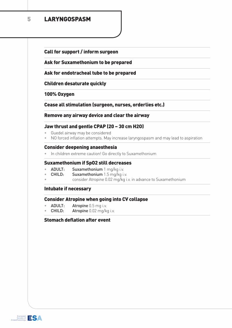
Call for support / inform surgeon
Ask for suxamethonium to be prepared
Ask for endotracheal tube to be prepared
Children desaturate quickly
100% oxygen
Cease all stimulation (surgeon, nurses, orderlies etc.)
Remove any airway device and clear the airway
Jaw thrust and gentle CPAP (20 – 30 cm H2o)• Guedel airway may be considered• NO forced inflation attempts. May increase laryngospasm and may lead to aspiration
Consider deepening anaesthesia• In children extreme caution! Go directly to Suxamethonium
suxamethonium if spo2 still decreases• ADULT: Suxamethonium 1 mg/kg i.v.• CHILD: Suxamethonium 1.5 mg/kg i.v. • consider Atropine 0.02 mg/kg i.v. in advance to Suxamethonium
Intubate if necessary
Consider Atropine when going into CV collapse • ADULT: Atropine 0.5 mg i.v.• CHILD: Atropine 0.02 mg/kg i.v.
stomach deflation after event
LARYNGosPAsM5
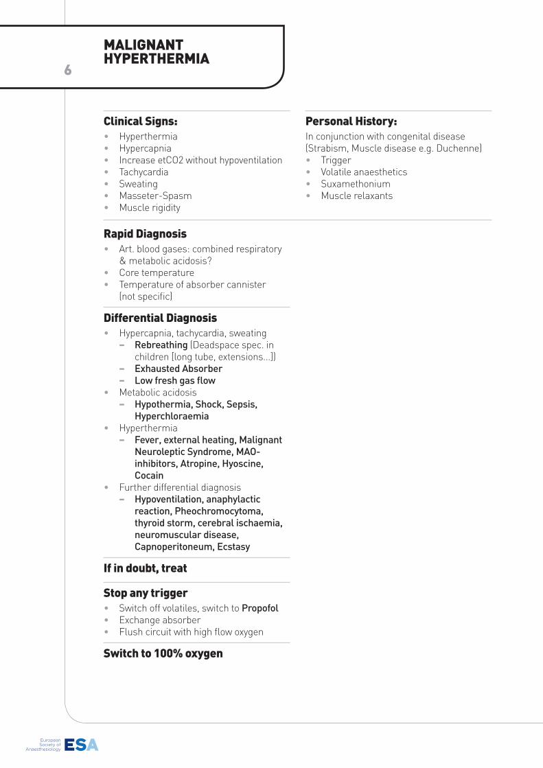
Rapid Diagnosis• Art. blood gases: combined respiratory
& metabolic acidosis?• Core temperature • Temperature of absorber cannister
(not specific)
Differential Diagnosis • Hypercapnia, tachycardia, sweating
− Rebreathing (Deadspace spec. in children [long tube, extensions...])
− Exhausted Absorber − Low fresh gas flow
• Metabolic acidosis − Hypothermia, Shock, Sepsis,
Hyperchloraemia• Hyperthermia
− Fever, external heating, Malignant Neuroleptic Syndrome, MAO-inhibitors, Atropine, Hyoscine, Cocain
• Further differential diagnosis − Hypoventilation, anaphylactic
reaction, Pheochromocytoma, thyroid storm, cerebral ischaemia, neuromuscular disease, Capnoperitoneum, Ecstasy
If in doubt, treat
stop any trigger• Switch off volatiles, switch to Propofol• Exchange absorber• Flush circuit with high flow oxygen
switch to 100% oxygen
Clinical signs:• Hyperthermia• Hypercapnia• Increase etCO2 without hypoventilation• Tachycardia• Sweating• Masseter-Spasm• Muscle rigidity
Personal History: In conjunction with congenital disease (Strabism, Muscle disease e.g. Duchenne)• Trigger• Volatile anaesthetics• Suxamethonium• Muscle relaxants
MALIGNANT HYPERTHERMIA
6
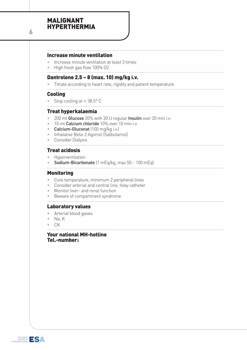
Increase minute ventilation• Increase minute ventilation at least 3 times• High fresh gas flow 100% O2
Dantrolene 2.5 – 8 (max. 10) mg/kg i.v.• Titrate according to heart rate, rigidity and patient temperature
Cooling• Stop cooling at < 38.5° C
Treat hyperkalaemia• 200 ml Glucose 20% with 20 U regular Insulin over 20 min i.v.• 10 ml Calcium chloride 10% over 10 min i.v.• Calcium-Gluconat (100 mg/kg i.v.)• Inhalative Beta-2 Agonist (Salbutamol)• Consider Dialysis
Treat acidosis• Hyperventilation• Sodium-Bicarbonate (1 mEq/kg, max 50 – 100 mEq)
Monitoring• Core temperature, minimum 2 peripheral lines• Consider arterial and central line, foley catheter• Monitor liver- and renal function• Beware of compartment syndrome
Laboratory values• Arterial blood gases• Na, K• CK
Your national MH-hotline Tel.-number:
MALIGNANT HYPERTHERMIA
6
* www.pedlatrlcs.org/cgl/dolItO.t542/pedS.2009·t5tO Copyright European Resuscitation Council - www.erc.edu - 2013/005
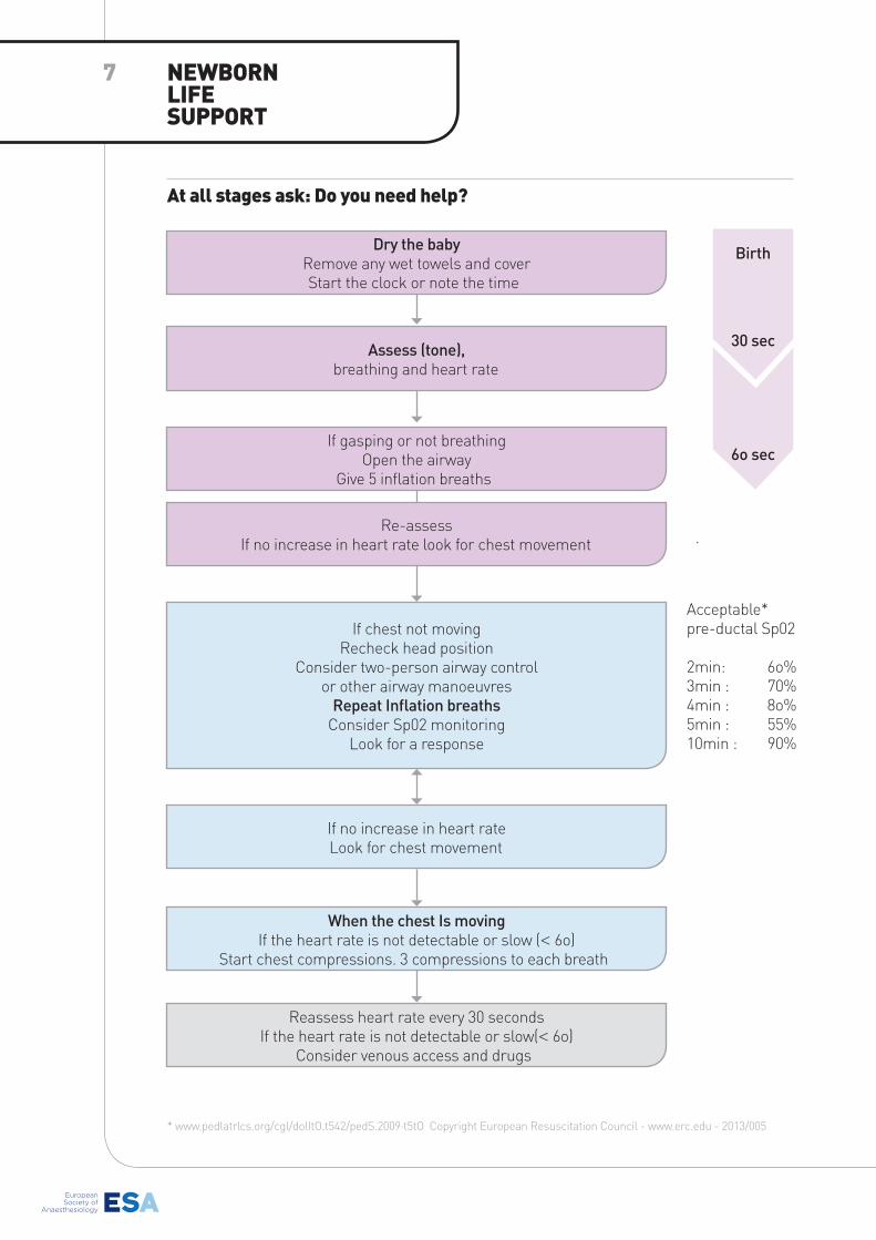
At all stages ask: Do you need help?
NEwboRN LIFE sUPPoRT
7
Dry the babyRemove any wet towels and coverStart the clock or note the time
Assess (tone), breathing and heart rate
If gasping or not breathingOpen the airway
Give 5 inflation breaths
Re-assessIf no increase in heart rate look for chest movement
If chest not movingRecheck head position
Consider two-person airway control or other airway manoeuvres
Repeat Inflation breaths Consider Sp02 monitoring
Look for a response
If no increase in heart rateLook for chest movement
When the chest Is movingIf the heart rate is not detectable or slow (< 6o)
Start chest compressions. 3 compressions to each breath
Reassess heart rate every 30 secondsIf the heart rate is not detectable or slow(< 6o)
Consider venous access and drugs
Birth
30 sec
6o sec
Acceptable*pre-ductal Sp02
2min: 6o%3min : 70%4min : 8o%5min : 55%10min : 90%
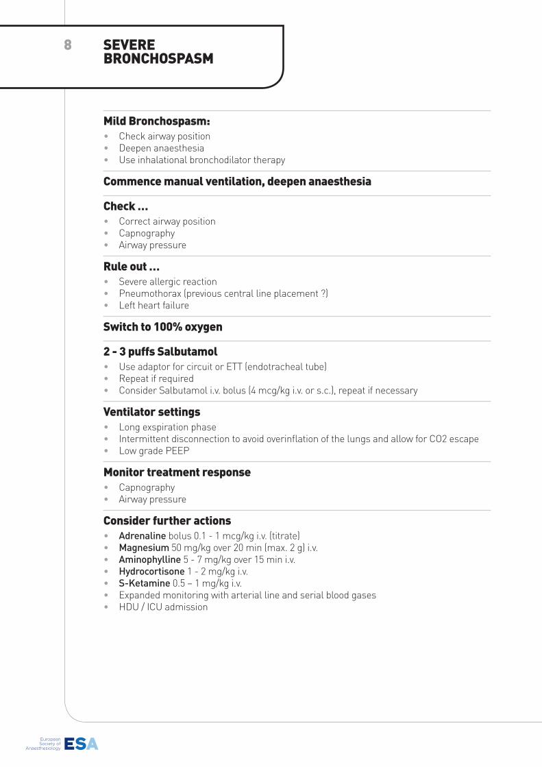
Mild bronchospasm:• Check airway position• Deepen anaesthesia• Use inhalational bronchodilator therapy
Commence manual ventilation, deepen anaesthesia
Check …• Correct airway position• Capnography• Airway pressure
Rule out …• Severe allergic reaction• Pneumothorax (previous central line placement ?)• Left heart failure
switch to 100% oxygen
2 - 3 puffs salbutamol• Use adaptor for circuit or ETT (endotracheal tube)• Repeat if required• Consider Salbutamol i.v. bolus (4 mcg/kg i.v. or s.c.), repeat if necessary
Ventilator settings• Long exspiration phase• Intermittent disconnection to avoid overinflation of the lungs and allow for CO2 escape • Low grade PEEP
Monitor treatment response • Capnography• Airway pressure
Consider further actions • Adrenaline bolus 0.1 - 1 mcg/kg i.v. (titrate)• Magnesium 50 mg/kg over 20 min (max. 2 g) i.v.• Aminophylline 5 - 7 mg/kg over 15 min i.v.• Hydrocortisone 1 - 2 mg/kg i.v.• S-Ketamine 0.5 – 1 mg/kg i.v.• Expanded monitoring with arterial line and serial blood gases• HDU / ICU admission
sEVERE bRoNCHosPAsM
8
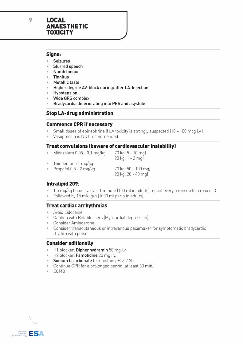
signs:• Seizures • Slurred speech • Numb tongue • Tinnitus• Metallic taste• Higher degree AV-block during/after LA-Injection• Hypotension • Wide QRS complex • Bradycardia deteriorating into PEA and asystole
stop LA-drug administration
Commence CPR if necessary• Small doses of epinephrine if LA toxicity is strongly suspected (10 – 100 mcg i.v.) • Vasopressin is NOT recommended
Treat convulsions (beware of cardiovascular instability)• Midazolam 0.05 - 0.1 mg/kg (70 kg: 5 - 10 mg)
(20 kg: 1 - 2 mg)• Thiopentone 1 mg/kg • Propofol 0.5 - 2 mg/kg (70 kg: 50 - 100 mg)
(20 kg: 20 - 40 mg)
Intralipid 20%• 1.5 mg/kg bolus i.v. over 1 minute (100 ml in adults) repeat every 5 min up to a max of 3• Followed by 15 ml/kg/h (1000 ml per h in adults)
Treat cardiac arrhythmias• Avoid Lidocaine• Caution with Betablockers (Myocardial depression)• Consider Amiodarone• Consider transcutaneous or intravenous pacemaker for symptomatic bradycardic
rhythm with pulse
Consider aditionally • H1 blocker: Diphenhydramin 50 mg i.v.• H2 blocker: Famotidine 20 mg i.v.• Sodium bicarbonate to maintain pH > 7.25• Continue CPR for a prolonged period (at least 60 min)• ECMO
LoCAL ANAEsTHETIC ToxICITY
9
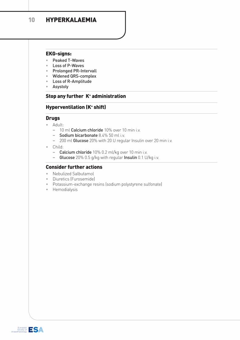
EKG-signs:• Peaked T-Waves • Loss of P-Waves• Prolonged PR-Intervall• Widened QRS-complex• Loss of R-Amplitude• Asystoly
stop any further K+ administration
Hyperventilation (K+ shift)
Drugs• Adult:
− 10 ml Calcium chloride 10% over 10 min i.v. − Sodium bicarbonate 8.4% 50 ml i.v. − 200 ml Glucose 20% with 20 U regular Insulin over 20 min i.v.
• Child: − Calcium chloride 10% 0.2 ml/kg over 10 min i.v. − Glucose 20% 0.5 g/kg with regular Insulin 0.1 U/kg i.v.
Consider further actions • Nebulized Salbutamol• Diuretics (Furosemide)• Potassium-exchange resins (sodium polystyrene sulfonate)• Hemodialysis
HYPERKALAEMIA10
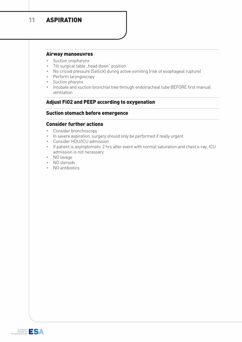
Airway manoeuvres• Suction oropharynx• Tilt surgical table „head down“ position• No cricoid pressure (Sellick) during active vomiting (risk of esophageal rupture)• Perform laryngoscopy• Suction pharynx• Intubate and suction bronchial tree through endotracheal tube BEFORE first manual
ventilation
Adjust Fio2 and PEEP according to oxygenation
suction stomach before emergence
Consider further actions • Consider bronchoscopy• In severe aspiration, surgery should only be performed if really urgent• Consider HDU/ICU admission• If patient is asymptomatic 2 hrs after event with normal saturation and chest x-ray, ICU
admission is not necessary• NO lavage• NO steroids• NO antibiotics
AsPIRATIoN11
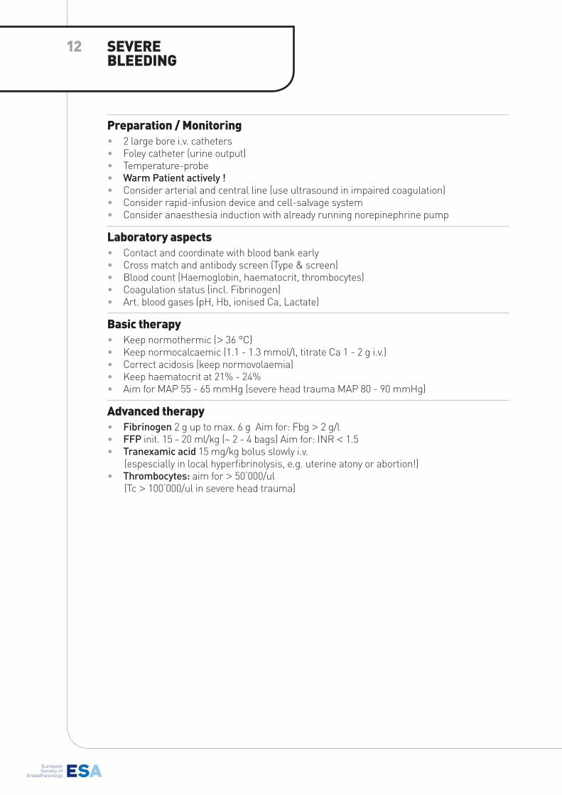
Preparation / Monitoring• 2 large bore i.v. catheters• Foley catheter (urine output)• Temperature-probe• Warm Patient actively !• Consider arterial and central line (use ultrasound in impaired coagulation) • Consider rapid-infusion device and cell-salvage system• Consider anaesthesia induction with already running norepinephrine pump
Laboratory aspects• Contact and coordinate with blood bank early• Cross match and antibody screen (Type & screen)• Blood count (Haemoglobin, haematocrit, thrombocytes)• Coagulation status (incl. Fibrinogen)• Art. blood gases (pH, Hb, ionised Ca, Lactate)
basic therapy• Keep normothermic (> 36 °C)• Keep normocalcaemic (1.1 - 1.3 mmol/l, titrate Ca 1 - 2 g i.v.)• Correct acidosis (keep normovolaemia)• Keep haematocrit at 21% - 24%• Aim for MAP 55 - 65 mmHg (severe head trauma MAP 80 - 90 mmHg)
Advanced therapy• Fibrinogen 2 g up to max. 6 g Aim for: Fbg > 2 g/l• FFP init. 15 - 20 ml/kg (~ 2 - 4 bags) Aim for: INR < 1.5• Tranexamic acid 15 mg/kg bolus slowly i.v. (espescially in local hyperfibrinolysis, e.g. uterine atony or abortion!)• Thrombocytes: aim for > 50‘000/ul
(Tc > 100‘000/ul in severe head trauma)
sEVERE bLEEDING
12
1. DISTINGUISH
Circuit• Respirator settings• Kinked tubing• Valve failure• Failure of high pressure valve• Failure of O2-flush
Airway• Laryngospasm (if not intubated)• Tube position• Tube size• Blocked or kinked tube (patient biting
on tube)
Patient• Bronchospasm• Laryngospasm (if not intubated)• Pneumothorax• Pneumoperitoneum• Tracheal pathology
− Foreign body (e.g. chewing gum) − Secretions − Tumor
• Chest wall rigidity• Obesity• Alveolar pathology
− Oedema − Infection − ARDS − Contusion − Fibrosis
Most likely• Insufficient relaxation• endotracheal tube position• Laryngospasm (if not intubated)• Respirator settings
2. ACTIONS
Check• Muscle relaxation• Depth of anaesthesia• Capnogram
− Bronchospasm ? − Kinked endotracheal tube ?
• Spirometry − Endobronchial intubation ? − Kinked endotracheal tube ?
• Tubing circuit − Kinked tubing ? − Obstructed tubing ?
Do• Auscultate• Manually ventilate• Suction bronchial tree• Flexible bronchoscopic exam• If LMA in place consider endotracheal
tube
If problems persist• Review possible patient causes• Call for assistance• Repeat checklist together
INCREAsED AIRwAY PREssURE
13
No etCo2• No etCO2 - NO VENTILATION, NO PATENT AIRWAY !!! • Oesophageal intubation?• Disconnection of tubing, complete failure of respirator• Apnea• Cardiac arrest
Diminished production of Co2• Hypothermia• Deep anaesthesia• Hypothyroidism
Enhanced excretion of Co2• (Spontaneous) hyperventilation• Inappropriate ventilator setting
Reduced transport of Co2 in blood• Severe hypotension• Anaphylaxis• Cardiac arrest• Pulmonary embolus
Reduced transport of Co2 in lung• Endotracheal tube obstruction• Incorrect airway placement (endobronchial intubation)• Laryngospasm• Severe bronchospasm
sampling dilution• Disconnection of respirator• Dilution of sampling gas with room-air• Gas sampler placed wrong• High fresh gas flow in circuit
Most likely• Rule out MALPLACED AIRWAY (OESOPHAGEAL)• Hyperventilation (too high minute ventilation)• Bronchospasm• Laryngospasm• Hypotension
DIFFERENTIAL DIAGNosIs HYPoCAPNIA / Low etCo2
14
Increased production of Co2
a. Exogenous: − CO2 insufflation (e.g. laparoscopy) − Bicarbonate administration − Re-breathing (valves, Soda lime, fresh gas-flow)
b. Endogenous: − Painful stimulus − Increased body temperature − Reperfusion after Tourniquet − Sepsis, Malignant Hyperthermia − Thyroid storm, Malignant Neuroleptic Syndrome
Reduced excretion of Co2
a. Lungs: − Hypoventilation (spont. or respirator settings) − Bronchospasm, asthma − COPD (chronic airway disease)
b. Breathing circuit: − Increased dead space − Inadequate fresh gas flow − Valve malfunction − Incorrect respirator settings
Most likely• Hypoventilation (spontaneous or respirator settings)• Exhausted soda lime• Fresh gas flow setting
DIFFERENTIAL DIAGNosIs HYPERCAPNIA / HIGHetCo2
15
Primary causes• Atrioventricular block• Pacemaker malfunction• Cardiomyopathy• Sick sinus syndrome• Myocarditis• Pericarditis• Valvular heart disease• Pumonary Hypertension
secondary causes• Elektrolyte abnormalities• Antiarrhytmic medication• Hypothyroidism• Hypothermia• Hypervagal• Increased intracranial pressure• Temponade• Tension pneumothorax
Anaesthetic causes• Hypoxia• Volatile agent side effects• Muscle relaxant side effects• Narcotic• Anticholinesterase drugs• High spinal/ epidural anaesthesia• Local anaesthetic toxicity• Hyper- Hypokalaemia• Vasopressor reflex• Auto-PEEP• Malignant Hyperthermia
Most likely• Drug related• Hypervagal• Spinal anaesthesia• Fitness
DIFFERENTIAL DIAGNosIs bRADYCARDIA
16
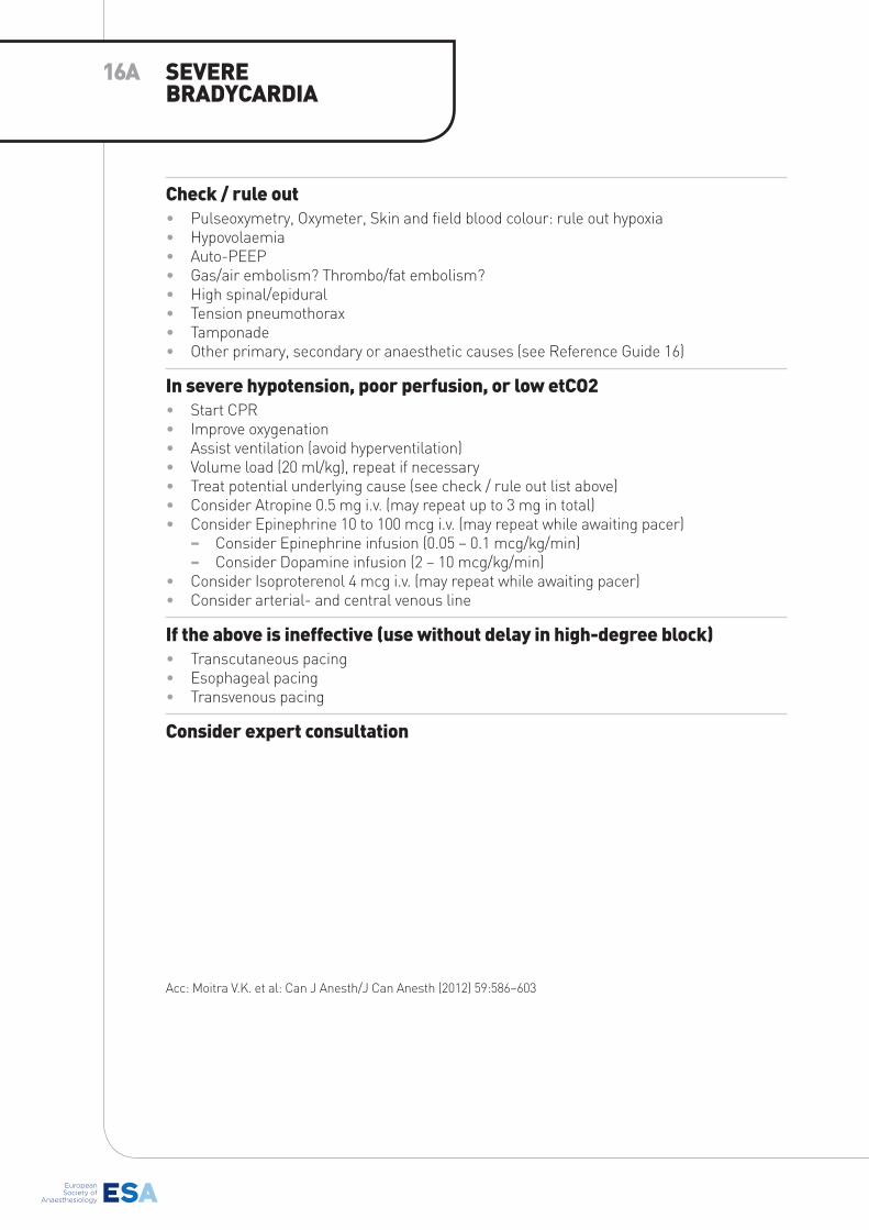
Check / rule out• Pulseoxymetry, Oxymeter, Skin and field blood colour: rule out hypoxia• Hypovolaemia• Auto-PEEP• Gas/air embolism? Thrombo/fat embolism?• High spinal/epidural• Tension pneumothorax• Tamponade• Other primary, secondary or anaesthetic causes (see Reference Guide 16)
In severe hypotension, poor perfusion, or low etCo2• Start CPR• Improve oxygenation• Assist ventilation (avoid hyperventilation)• Volume load (20 ml/kg), repeat if necessary• Treat potential underlying cause (see check / rule out list above)• Consider Atropine 0.5 mg i.v. (may repeat up to 3 mg in total)• Consider Epinephrine 10 to 100 mcg i.v. (may repeat while awaiting pacer)
− Consider Epinephrine infusion (0.05 – 0.1 mcg/kg/min) − Consider Dopamine infusion (2 – 10 mcg/kg/min)
• Consider Isoproterenol 4 mcg i.v. (may repeat while awaiting pacer)• Consider arterial- and central venous line
If the above is ineffective (use without delay in high-degree block)• Transcutaneous pacing• Esophageal pacing• Transvenous pacing
Consider expert consultation
Acc: Moitra V.K. et al: Can J Anesth/J Can Anesth (2012) 59:586–603
sEVERE bRADYCARDIA
16A
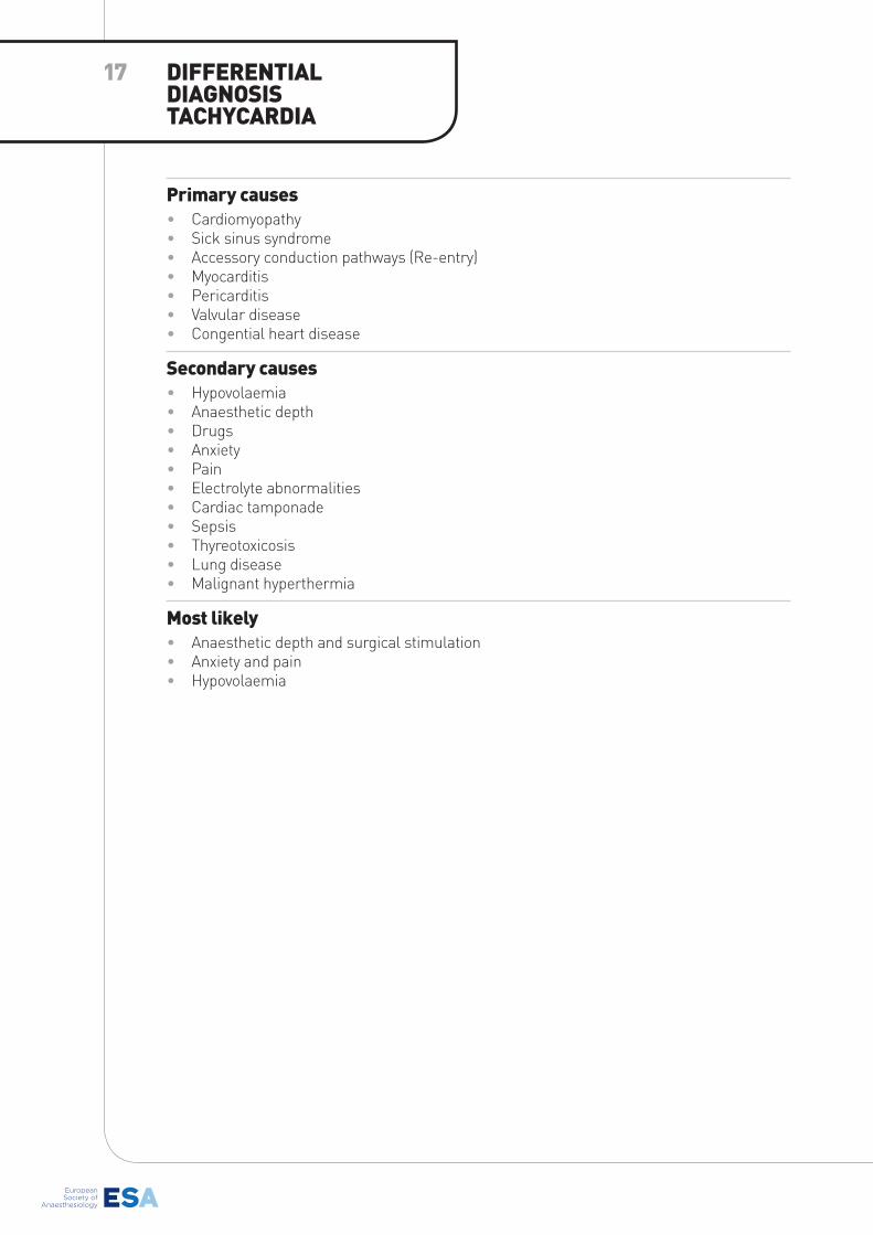
Primary causes• Cardiomyopathy • Sick sinus syndrome• Accessory conduction pathways (Re-entry)• Myocarditis• Pericarditis• Valvular disease• Congential heart disease
secondary causes• Hypovolaemia• Anaesthetic depth• Drugs• Anxiety• Pain• Electrolyte abnormalities• Cardiac tamponade• Sepsis• Thyreotoxicosis• Lung disease• Malignant hyperthermia
Most likely• Anaesthetic depth and surgical stimulation• Anxiety and pain• Hypovolaemia
DIFFERENTIAL DIAGNosIs TACHYCARDIA
17
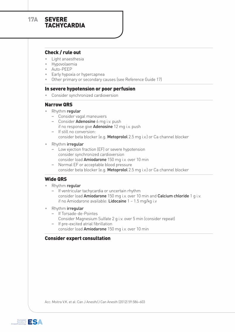
Check / rule out• Light anaesthesia• Hypovolaemia• Auto-PEEP• Early hypoxia or hypercapnea• Other primary or secondary causes (see Reference Guide 17)
In severe hypotension or poor perfusion• Consider synchronized cardioversion
Narrow QRs• Rhythm regular
− Consider vagal maneuvers − Consider Adenosine 6 mg i.v. push
if no response give Adenosine 12 mg i.v. push − If still no conversion:
consider beta blocker (e.g. Metoprolol 2.5 mg i.v.) or Ca channel blocker
• Rhythm irregular − Low ejection fraction (EF) or severe hypotension
consider synchronized cardioversionconsider load Amiodarone 150 mg i.v. over 10 min
− Normal EF or acceptable blood pressureconsider beta blocker (e.g. Metoprolol 2.5 mg i.v.) or Ca channel blocker
wide QRs• Rhythm regular
− If ventricular tachycardia or uncertain rhythmconsider load Amiodarone 150 mg i.v. over 10 min and Calcium chloride 1 g i.v.if no Amiodarone available: Lidocaine 1 – 1.5 mg/kg i.v
• Rhythm irregular − If Torsade-de-Pointes
Consider Magnesium Sulfate 2 g i.v. over 5 min (consider repeat) − If pre-excited atrial fibrillation
consider load Amiodarone 150 mg i.v. over 10 min
Consider expert consultation
Acc: Moitra V.K. et al: Can J Anesth/J Can Anesth (2012) 59:586–603
sEVERE TACHYCARDIA
17A

Preload Reduction• Blood loss• Hypovolaemia• Decreased venous return (caval vein?)• Elevated intrathoracic pressure• Cardiac Tamponade • Embolism
Reduced Contractility• Drugs (including volatile agents)• Ischaemic heart disease• Cardiomyopathy• Myocarditis• Arrhythmia• Valvular heart disease
Reduced systemic Vascular Resistance• Volatile anaesthetics• Narcotics• Vasodilators• Antihypertensive drugs• Regional blockade (spinal/epidural)• Sepsis• Release of tourniquet• Anaphylaxis• Addison’s disease• Thyroid disease
Most likely• Depth of anaesthesia and volatile anaesthetics• Narcotics• Regional blockade (spinal/epidural)• Hypovolaemia• Transducer height (invasive monitoring)
DIFFERENTIAL DIAGNosIs HYPoTENsIoN
18
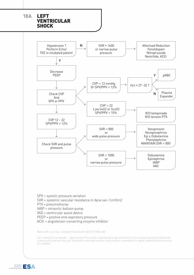
SPV = systolic pressure variation SVR = systemic vascular resistance in dyne sec-1cm5m2PTX = pneumothoraxIABP = intraortic balloon pump VAD = ventricular assist device PEEP = positive end-expiratory pressureACEi = angiotensin-converting enzyme inhibitor
Moitra V.K. et al: Can J Anesth/J Can Anesth (2012) 59:586–603
Can J Anesth/J Can Anesth: „Open Access This article is distributed under the terms of the Creative Commons Attribution License which permits any use, distribution, and reproduction in any medium, provided the original author(s) and the source are credited.“
LEFT VENTRICULAR sHoCK
18A
Hypotensive ?Perform Echo/
TEE in intubated patient
SVR > 1600or narrow pulse
pressure
Afterload ReductionFenoldopam
NitroprussideNesiritide, ACEi
DobutamineEpinephrine
IABPVAD
DecreasePEEP
CVP < 12 mmHgOr SPV/PPV > 12% Hct < 27 -32 ?
pRBC
PlasmaExpander
Check CVPAnd
SPV or PPVCVP > 22
Low SvO2 or ScvO2SPV/PPV > 15% R/O tamponade
R/O tension PTXCVP 12 – 22
SPV/PPV < 12%
SVR < 800or
wide pulse pressure
Check SVR and pulse pressure
SVR > 1000or
narrow pulse pressure
VasopressinNorepinephrine
Epi ± DobutaminePhenylephrine
MAINTAIN SVR < 800
N
N
Y
Y
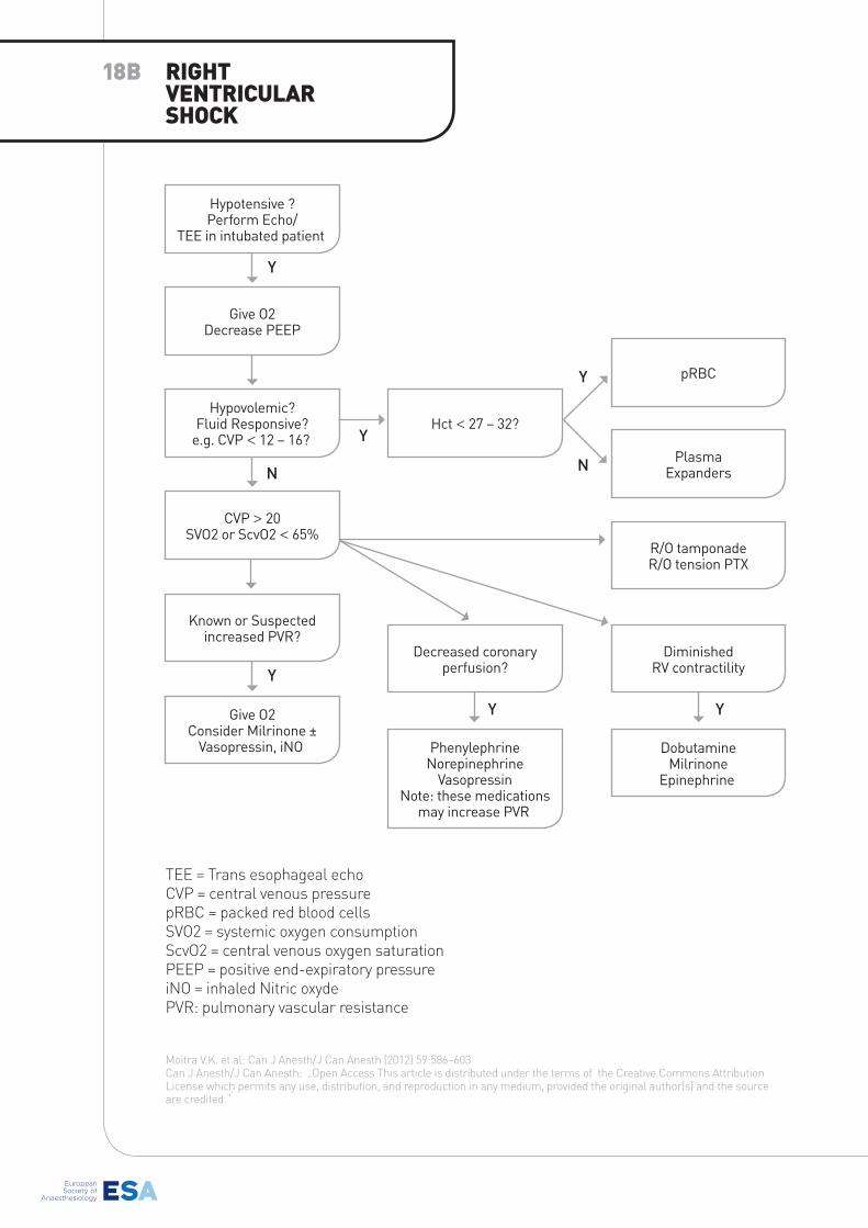
TEE = Trans esophageal echo CVP = central venous pressurepRBC = packed red blood cellsSVO2 = systemic oxygen consumptionScvO2 = central venous oxygen saturation PEEP = positive end-expiratory pressureiNO = inhaled Nitric oxydePVR: pulmonary vascular resistance
Moitra V.K. et al: Can J Anesth/J Can Anesth (2012) 59:586–603Can J Anesth/J Can Anesth: „Open Access This article is distributed under the terms of the Creative Commons Attribution License which permits any use, distribution, and reproduction in any medium, provided the original author(s) and the source are credited.“
RIGHT VENTRICULAR sHoCK
18b
PhenylephrineNorepinephrine
VasopressinNote: these medications
may increase PVR
Hypotensive ?Perform Echo/
TEE in intubated patient
CVP > 20SVO2 or ScvO2 < 65%
Give O2Decrease PEEP
Known or Suspectedincreased PVR?
Hypovolemic?Fluid Responsive?
e.g. CVP < 12 – 16?
Give O2Consider Milrinone ±
Vasopressin, iNO DobutamineMilrinone
Epinephrine
DiminishedRV contractility
Decreased coronary perfusion?
Hct < 27 – 32?
R/O tamponadeR/O tension PTX
PlasmaExpanders
pRBC
Y Y
N N
Y
Y
Y
Y
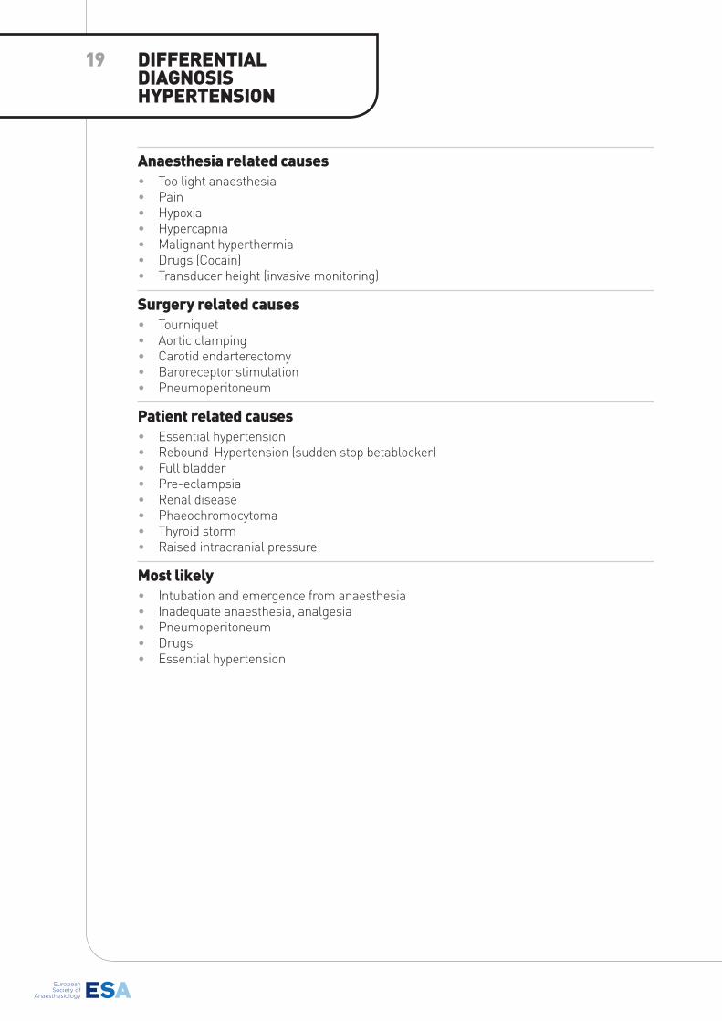
Anaesthesia related causes• Too light anaesthesia• Pain• Hypoxia• Hypercapnia• Malignant hyperthermia• Drugs (Cocain)• Transducer height (invasive monitoring)
surgery related causes• Tourniquet• Aortic clamping • Carotid endarterectomy• Baroreceptor stimulation• Pneumoperitoneum
Patient related causes• Essential hypertension• Rebound-Hypertension (sudden stop betablocker)• Full bladder• Pre-eclampsia• Renal disease• Phaeochromocytoma• Thyroid storm• Raised intracranial pressure
Most likely• Intubation and emergence from anaesthesia• Inadequate anaesthesia, analgesia• Pneumoperitoneum• Drugs• Essential hypertension
DIFFERENTIAL DIAGNosIs HYPERTENsIoN
19
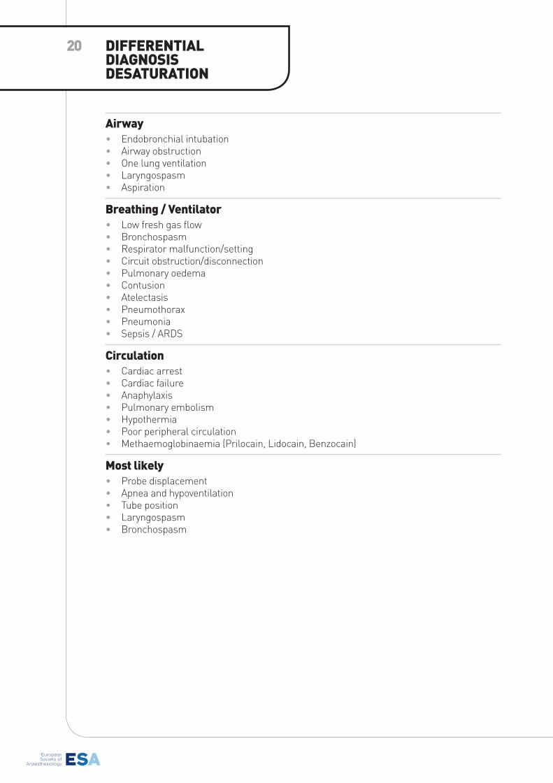
20
Airway• Endobronchial intubation• Airway obstruction• One lung ventilation• Laryngospasm• Aspiration
breathing / Ventilator• Low fresh gas flow• Bronchospasm• Respirator malfunction/setting• Circuit obstruction/disconnection• Pulmonary oedema• Contusion• Atelectasis• Pneumothorax• Pneumonia• Sepsis / ARDS
Circulation• Cardiac arrest• Cardiac failure• Anaphylaxis• Pulmonary embolism• Hypothermia• Poor peripheral circulation• Methaemoglobinaemia (Prilocain, Lidocain, Benzocain)
Most likely• Probe displacement• Apnea and hypoventilation• Tube position• Laryngospasm• Bronchospasm
DIFFERENTIAL DIAGNosIsDEsATURATIoN