elsevier editorial system(tm) for pharmacological research...
TRANSCRIPT

Elsevier Editorial System(tm) for Pharmacological Research Manuscript Draft Manuscript Number: YPHRS-D-16-00011R2 Title: mTOR pathway inhibition as a new therapeutic strategy in epilepsy and epileptogenesis. Article Type: VSI: New Drug for Epilepsy Keywords: mTOR; mTOR inhibitors; Epilepsy; Epileptogenesis; Tuberous Sclerosis Complex; Animal Epilepsy Models Corresponding Author: Prof. Giovambattista De Sarro, M.D. Corresponding Author's Institution: University of Catanzaro First Author: Rita Citraro, Phd Order of Authors: Rita Citraro, Phd; Antonio Leo, Phd; Andrew Costanti, Phd; Emilio Russo, Phd; Giovambattista De Sarro, M.D. Abstract: Several preclinical and some clinical studies have revealed that the mammalian target of rapamycin (mTOR) signaling pathway is involved in both genetic and acquired epilepsy syndromes. Excessive activation of mTOR signaling, as a consequence of loss-of-function of genes encoding for tuberous sclerosis complex (TSC) 1 and 2, is linked to the development of cortical malformations and epilepsy. This mTOR hyperactivation is associated with different epileptogenic conditions under the term of 'mTORopathies' such as tuberous sclerosis, focal cortical dysplasia, hemimegalencephaly and ganglioglioma. mTOR overactivation produces brain abnormalities that include dysplastic neurons, abnormal cortical organization and astrogliosis. mTOR inhibitors (e.g. rapamycin) have consistent protective effects in various genetic (e.g. TSC models and WAG/Rij rats) and acquired (e.g. kainate or pilocarpine post-status epilepticus) epilepsy animal models. Furthermore, clinical studies in patients with TSC and cortical dysplasia (CD) have confirmed the effectiveness of mTOR inhibitors also in epileptic patients. Therefore, mTOR is currently a very good candidate as a target for epilepsy and epileptogenesis. This review describes the relevance of the mTOR pathway to epileptogenesis and its potential as a therapeutic target in epilepsy treatment by presenting the most recent findings on mTOR inhibitors.

1
mTOR pathway inhibition as a new therapeutic strategy in epilepsy and epileptogenesis
Rita Citraroa, Antonio Leoa, Andrew Constantib, Emilio Russoa, Giovambattista De Sarroa
aDepartment of Science of Health, School of Medicine and Surgery, University of Catanzaro, Italy;
bDepartment of Pharmacology, UCL School of Pharmacy, 29/39 Brunswick Square, London,
United Kingdom
Running Title: mTOR in epilepsy and epileptogenesis
* Author for correspondence:
Prof. Giovambattista De Sarro, MD
Chair of Pharmacology,
Department of Science of Health, School of Medicine and Surgery,
University of Catanzaro;
Viale Europa – Germaneto 88100 Catanzaro, ITALY.
Phone +39 0961 3694191/Fax: +39 0961 3694192
e-mail: [email protected]
*ManuscriptClick here to view linked References
2
Abstract
Several preclinical and some clinical studies have revealed that the mammalian target of rapamycin
(mTOR) signaling pathway is involved in both genetic and acquired epilepsy syndromes.
Excessive activation of mTOR signaling, as a consequence of loss-of-function of genes encoding
for tuberous sclerosis complex (TSC) 1 and 2, is linked to the development of cortical
malformations and epilepsy. This mTOR hyperactivation is associated with different epileptogenic
conditions under the term of 'mTORopathies' such as tuberous sclerosis, focal cortical dysplasia,
hemimegalencephaly and ganglioglioma. mTOR overactivation produces brain abnormalities that
include dysplastic neurons, abnormal cortical organization and astrogliosis. mTOR inhibitors (e.g.
rapamycin) have consistent protective effects in various genetic (e.g. TSC models and WAG/Rij
rats) and acquired (e.g. kainate or pilocarpine post-status epilepticus) epilepsy animal models.
Furthermore, clinical studies in patients with TSC and cortical dysplasia (CD) have confirmed the
effectiveness of mTOR inhibitors also in epileptic patients. Therefore, mTOR is currently a very
good candidate as a target for epilepsy and epileptogenesis. This review describes the relevance of
the mTOR pathway to epileptogenesis and its potential as a therapeutic target in epilepsy treatment
by presenting the most recent findings on mTOR inhibitors.
Keywords: mTOR; mTOR inhibitors; Epilepsy; Epileptogenesis; Tuberous Sclerosis Complex;
Animal Epilepsy Models;

3
1. Introduction
Epilepsy is a chronic neurological disorder characterized by recurrent seizures and caused by a
large variety of genetic and acquired etiologies. Although many epilepsy patients are seizure-free
when treated with drugs, about a third of patients remain still drug-resistant [1-3]. In addition, even
when seizures are well controlled with antiepileptic drugs (AEDs), currently available drugs are
only a symptomatic therapy in suppressing seizures (antiseizure or anticonvulsant) but do not have
disease-modifying properties for preventing or reducing the development of epilepsy
(antiepileptogenic) [4-6]. Therefore, novel treatments need to be searched to address both the
problem of drugs-resistant epilepsy and the lack of disease modifying therapies.
The mammalian target of rapamycin (mTOR) pathway regulates a number of important
physiological functions and in the brain it is clearly involved in cell proliferation, growth and
survival, protein synthesis, neuronal morphology and cortical development [7,8]; more recently, it
has also been involved in the pathophysiology of several neurological diseases with particular
attention to the epileptogenic process being indicated as a potential novel target for epilepsy
treatments [9-12]. Dysregulation of the mTOR pathway has been involved in the development of
different brain disorders that include focal cortical dysplasia (FCD), tuberous sclerosis complex
(TSC), ganglioglioma and hemimegalencephaly, all potentially or certainly leading to epilepsy
[9,13].
Hyperactived mTOR seem to play a pivotal role in the pathogenesis of different animal models of
acquired epilepsy, such as infantile spasms (IS), temporal lobe epilepsy (TLE), status epilepticus
(SE), absence epilepsy, traumatic brain injury (TBI) and neonatal hypoxia–ischemia [11,14].
Accordingly, different studies have demonstrated that mTOR inhibitors, such as rapamycin and its
analogues, decrease the development of seizures preventing epileptogenesis related mechanisms in
many animal models and in some cases also some anticonvulsant activity has been evidenced
[11,15,16]. From a clinical point of view, small trials have already indicated some kind of activity
while larger controlled studies are ongoing using mTOR inhibitors (e.g. everolimus) in patients with
4
tuberous sclerosis complex and intractable epilepsy [17,18]. The importance of the mTOR pathway
in epileptogenesis associated with tuberous sclerosis has been well demonstrated [19], while its role
in epileptogenesis occurring in other forms of epilepsy remains to be better clarified and its
potential as a target to be confirmed [20-22]
Here, we review the most recent advances concerning the possible role of the mTOR signaling
pathway in epilepsy and epileptogenesis, the preclinical studies of mTOR inhibitors treatment in
different models of epilepsy, and the available clinical studies in patients with epilepsy.
2. mTOR Pathways in Neurological Diseases
The physiological regulation of the mTOR pathway is essential for normal cellular function; while,
its dysregulation may promote the development/progression of disease under pathological
conditions such as type 2 diabetes, inflammation, cancer, and cardiovascular disease [8,23,24].
Furthermore, abnormal mTOR signaling has been implicated in a variety of neurological disease
[12,25].
In the brain, mTOR mediates several processes involved in CNS development including
neurogenesis, cell survival and migration, but it is involved in some other specific processes such as
axonal sprouting, axonal regeneration and myelination, dendritic development and microtubule
dynamics. A direct role of mTOR in the modulation of glial functions has also been demonstrated
[12,26,27]. The mTOR pathway is a key regulator during brain development, in fact, it participates
in the control of protein expression and other cellular mechanisms including neuronal and glial
differentiation, axon growth, navigation and synaptogenesis, all playing a role in neuronal
excitability [28-30]. The mTOR pathway can influence neuronal excitability indirectly through
mechanisms controlling synaptic structure and plasticity. In fact, Ras-PI3K-Akt-mTOR and Ras-
MAPK signaling pathways play an important role in the regulation of dendrite arborisation and
spine formation, which are critical for the functioning of neurons and neuronal networks [28,29,31].

5
Accordingly, mTOR pathway also affects neuronal excitability by modulating the expression of ion
channels and receptors [32-35].
Considering the relevant role of this pathway in the brain, its dysregulation, such as loss-of-function
gene mutations encoding for mTOR inhibitor proteins (e.g. TSC1, TSC2, PTEN), has been involved
in neurological diseases such as epilepsy, Parkinson’s disease (PD), Alzheimer’s disease (AD),
Huntington’s disease (HD) and brain traumatisms [11,12,36] but also psychiatric diseases such as
depression, mental retardation, schizophrenia and cognitive impairment [37].
Recently, particular attention has been given to the role of mTORC1 in major depressive disorder
(MDD) [38]. A post-mortem analysis of the prefrontal cortex (PFC) of subjects with MDD revealed
deficits in mTOR signaling [39]. Furthermore, Chandran, et al. [40] showed that a chronic
unpredictable stress (CUS) exposure produces deficits in mTOR signaling pathway in the
amygdala. In agreement, chronic stress associated with depression induced by long-term
corticosterone treatment causes an inhibition of the PI3K-Akt-TORC1 pathway [41]. These studies
show an association between deficits in synaptic proteins and dysregulation of mTOR signaling in
MDD and annulment of these abnormalities may underlie antidepressant activity. For example fast
antidepressant response to ketamine, a NMDA receptor antagonist, seems be to mediate by
activation of the mTOR pathway in the PFC of rats [42] and depressed patients [43].
Interestingly, abnormal mTOR signaling has also been implicated in diseases as fragile X syndrome
[44], Down syndrome, [45] and Rett syndrome [46]. The potential involvement of dysregulated
mTOR signaling in these neurological disorders characterized by cognitive deficits is likely linked
to its role in physiological mechanisms of learning and memory [47]. In particular, mTOR plays an
important role in the consolidation of memory through long-term potentiation (LTP) [33,48].
Furthermore, changes in dendritic morphology may represent a structural substrate for memory
persistence and can be regulated by the mTOR pathway [28,29] whereas abnormalities in dendritic
morphology have been demonstrated in many neurogenetic syndromes, including TSC, fragile X,
Down and Rett [44,49].

6
In contrast, other studies reported a relationship between enhanced mTORC1 activity and memory
improvement; rapamycin seems to disrupt this process in several behavioral models including
auditory fear conditioning and Morris water maze task [50,51]. Moreover, rapamycin
administration decreased both spatial hippocampus memory and consolidation memory in many
brain regions such as amygdala and hippocampus [50,52-54]. However, mTORC1 hyperactivity has
been associated with memory deficits in human patients and experimental models of TSC [55]. In a
mouse model of TSC, rapamycin rescued memory performance, which could be, at least in part,
related to an abnormal mTOR activity [56]. The importance of mTOR signaling in CNS
physiology is underscored by the several disorders in which mTOR pathway disruption is
implicated, such as tumors, autism, mood disorders, neurodegenerative diseases as well as epilepsy
[36,57,58].
Finally, another link between mTOR and neurodegenerative diseases could be autophagy, which
represents a catabolic process. It has been demonstrated how mTOR is a crucial regulator of
autophagy [59]. A typical hallmark of neurodegenerative diseases, such as Alzheimer’s,
Parkinson’s and Huntington’s diseases, is the aberrant accumulation of protein aggregates in the
brain [60,61] and associated neuronal death. The clearance of these proteins would seem to be
increased by mTOR inhibition [62,63]. Pharmacological manipulation of mTOR signaling is thus
proving to be a promising therapeutic branch for the treatment of several neurological disorders
[57].
3. Role of mTOR pathway in epilepsy and epileptogenesis
Considering mTOR involvement in cellular functions influencing neuronal excitability, it is not
surprising that this signaling pathway can be responsible for or participate to the development of
spontaneous seizures, and that this pathway could represent an important target for both
epileptogenesis and seizure pharmacotherapy [11,20,64]. Since early 2000, many preclinical and
some clinical data have underscored the importance of mTOR pathway in both genetic and acquired

7
epilepsy syndromes [65]. Excessive activation of mTOR signaling, as a consequence of loss-of-
function mutations of genes encoding for natural mTOR inhibitors such as TSC1 and TSC2 (coding
for the proteins hamartin and tuberin, respectively), phosphatase and tensin homolog (PTEN) and
STE20-related kinase adaptor alpha (STRADalpha), are linked both to the development of cortical
malformations and epilepsy. These malformations or "mTORopathies"-related epilepsies include:
hemimegalencephaly, ganglioglioma, focal cortical dysplasia (FCD), and tuberous sclerosis
complex (TSC) [13,66].
The term "mTORpathies" describes neurological disorders characterized by altered cortical
architecture, abnormal neuronal morphology and intractable epilepsy as a consequence of excessive
mTOR signaling, providing a likely histopathological substrate for epileptogenesis [14,67]. On the
other hand, seizures themselves, in the absence of any other associated pathology, may directly
cause activation of mTORC1 activity [20]. Many experimental models of genetic and acquired
epilepsy, in which mTOR hyperactivation was present, are responsive to mTOR inhibitors [15,16].
Rapamycin and other mTOR inhibitors decrease seizures, delay seizure development, or prevent
epileptogenesis in many experimental models [65]. This evidence supports the hypothesis that
dysregulation of the mTOR pathway seems to be a key condition for the development of
epileptogenesis and epilepsy. To date, the mechanisms by which mTOR inhibition gives rise to the
inhibition of seizure activity in several experimental models is still unclear. Nevertheless, the use of
selective mTOR inhibitors can represent an important new therapeutic strategy for managing or
eventually preventing epilepsy due to these disorders [9].
3.1 Preclinical Studies
Dysfunction of mTOR signaling pathway is involved in the pathophysiology of Tuberous sclerosis
complex (TSC) [68]. However, this dysfunction also plays an important role during the latent phase
of epileptogenesis of some acquired forms of epilepsy, such as temporal lobe epilepsy (TLE),
traumatic brain injury (TBI) [69], infantile spams (IS) [70] and neonatal hypoxia–ischemia [71].

8
3.1.1 Tuberous sclerosis complex models
Among the genetic epilepsy syndromes, TSC has drawn particular attention, since it is strongly
linked with the dysregulation of the mTOR pathway [10]. TSC is an inherited autosomal disorder
resulting from a mutation of one of two tumor suppressor genes: TSC1 and TSC2 [72]. In TSC,
benign tumors may develop in multiple organs such as skin, liver, heart, kidney, lung and the brain,
in which it is often associated with the development of subependymal giant cell astrocytoma
(SEGA) among other tumors [73]. Unfortunately, many TSC patients suffer of drug-resistant
epilepsy [74-76]. mTOR dysregulation in TSC directly affects many downstream mechanisms,
including alteration of neurotransmitter receptors and ion channel expression, and synaptic and
neuronal organization, leading to epileptogenesis process [15,77]. In the context of TSC-associated
epilepsy, aberrant mTOR signaling has been repeatedly demonstrated, and conditional knockout of
TSC1 or TSC2 in various brain cell populations has been associated with elevated levels of mTOR
signaling and seizures in several transgenic mouse models [78,79]. Animal models of TSC are
crucial to study the link between mTOR, TSC and epilepsy [80,81]. Mice with TSC1 or TSC2
deleted, in specific neural populations (astrocytes or neurons), show neuropathological phenotypes
(i.e. astrogliosis, neuronal autophagy, macrocephaly, seizures and premature death) similar to those
found in human TSC [78,82].
Clinical and pre-clinical studies, using mTOR inhibitors (i.e. rapamycin and everolimus)
demonstrated the role of mTOR in TSC-associated epilepsy [83,84]. Inhibition of mTOR signaling
by rapamycin in a mouse model of TSC with conditional inactivation of the Tsc1 gene primarily in
glia (Tsc1GFAPCKO mice) can prevent astrogliosis, neuronal disorganization and seizures early in
the course of the disease, suggesting that the aberrant mTOR activation interferes with normal brain
function and leads to epilepsy [85]. Similarly, using a knock-out mouse model of TSC in which
Tsc1 was ablated in most neurons during cortical development, rapamycin treatment and its derivate
everolimus were able to reverse the animal phenotype, rescuing the mutants from epilepsy [86].

9
Early treatment with rapamycin in Tsc2GFAP1CKO mice, also rescued the mutants from epilepsy and
increased their survival [87,88]. Rapamycin seems effective not only reducing seizures once they
start but also in preventing seizures from ever developing as well as many of the pathological and
molecular changes (as progressive astrogliosis, inflammatory mechanisms, hippocampal
neurodegeneration, brain hypercellularity) in the brain that likely promote epileptogenesis in these
mice; this indicates that mTOR may have an anti-epileptogenic effect in these genetic models
[85,86,88]. However, following discontinuation of rapamycin therapy, these phenotypes at least
partially return and are accompanied by progressive development of severe seizures and early
death. Inflammatory signaling mechanisms, particularly the cytokine IL-1β and chemokine
CXCL10, are abnormally activated in Tsc1GFAPCKO mice; these inflammatory mediators were
reversed by rapamycin treatment, indicating that cytokine and chemokine signaling is downstream
from mTORC1 and occurred in astrocyte culture in vitro and before epilepsy onset in vivo [89].
Recently, it has been reported a primary role for TORC1 signaling in epileptogenesis, using mice
with biallelic Tsc1 deletion; this latter resulted in activation of TORC1, enhanced neuronal
excitability and epilepsy development without any obvious histological changes. Increased TORC1
activation appears sufficient for the development of epilepsy, even in the absence of changes in
brain pathology. Rapamycin treatment reduced TORC1 activity, seizure frequency and increased
survival indicating an important role of mTOR for managing seizures not only in the presence of
major brain pathology but also in other type of epilepsies that result from increased mTOR
hyperactivation [90]. Among current AEDs, vigabatrin (VGB) has been shown to have unique
efficacy in partial seizures related to TSC and infantile spasms [91] and early treatment, even before
seizure onset, can improve the long-term outcome of epilepsy in patients with TSC [92,93]. To date,
the exact mechanism by which VGB is effective in TSC remains unclear. In addition to its already
proven mechanism of action to increase brain γ-aminobutyric acid (GABA) levels by inhibition of
γ-aminobutyrate transaminase (GABA-T) [94], VGB also seems to inhibit mTOR pathway in the
neocortex and hippocampus of TSC1GFAPconditional knockout mice, providing a possible

10
explanation for the unique effectiveness of this drug in TSC [95]. Therefore, VGB seems to also
directly act on the mTOR pathway; however, an indirect action cannot yet be excluded.
Furthermore, it was reported that a prophylactic antiepileptic treatment of TSC patients (and at
high risk of epilepsy) with VGB, but also levetiracetam, valproic acid and topiramate, markedly
improved their risk of developing mental retardation and reduced the incidence of drug-resistant
seizures [92,93] (Table 1 and 2).
3.1.2 Cortical dysplasia models
Similar to TSC, several other, relatively rare genetic disorders entangle a dysregulation of the
mTOR pathway and an increased risk for tumors and epilepsy. Brain-specific deletion of the mouse
homolog PTEN, an upstream activator of the mTOR pathway [96], mimics several features of
human cortical dysplasia (CD), including neuronal hypertrophy, cortical and hippocampal
disorganization, aberrant mossy fiber sprouting and epilepsy [19]. CD (also known as malformation
of cortical development) is another recognized type of "mTORpathies" characterized by intractable
epilepsy in which mTOR dysregulation plays a key role in determining epilepsy phenotype [97].
Since CD has been linked to mutations of genes encoding for mTOR regulators [68], mTOR
inhibitors through their antiepileptogenic mechanisms might be useful for the treatment of the CD-
related epilepsy [98,99]. Recently, a link in CD between the up-regulated miRNAs (i.e.hsa-miR-21
and hsa-miR-155) and mTOR pathway has been evidenced [100]. Furthermore, PTEN deficiency is
linked with the excessive growth, migration and proliferation of dysplastic cells in CD [101]. To
better understand the role of mTOR in CD and epilepsy, Ljungberg, et al. [102] have characterized
neuron subset-specific Pten knockout (NS-Pten KO) mice as an experimental model of CD. Pten
knock-out mice exhibit neuronal hypertrophy, megalencephaly and seizures as a consequence of
enhanced mTOR activity, and both early and later treatment with rapamycin decreases pathological
abnormalities, suppresses the development of seizures and reduces established late-stage epilepsy
[102]. Rapamycin treatment, at late stages of the pathology, decreased mTORC signaling,
astrogliosis and microgliosis that were found in NS-Pten KO mice of CD [103]. Treatment with
11
mTOR inhibitors also reverses the neuronal hypertrophy and megalencephaly in PTEN knock-out
mice [104,105]. Similar to the TSC models, seizures return with the cessation of rapamycin
treatment, although intermittent rapamycin treatment is able to maintain a long‑term antiseizure
effect [104] (Table 2).
3.1.3 Temporal lobe epilepsy models
PTEN inactivation, in human and animals hippocampal dentate granule cells, induces the
development of abnormal granule cells and spontaneous seizures similar to temporal lobe epilepsy
(TLE) [31]. During epileptogenesis, adult-generated dentate granule cells (DGCs) form aberrant
neuronal connections with neighboring DGCs increase neuronal excitability in the hippocampus.
Sutula and Dudek [106] demonstrated that PTEN deletion among hippocampal granule cells was
sufficient to develop spontaneous seizures in a few weeks, and that mTOR signaling played a
fundamental role in this process. Therefore, hyperactivation of the mTOR pathway as a result of
PTEN deletion is a possible mechanism of epileptogenesis also in TLE. Moreover, rapamycin
administration was effective in inhibiting epileptogenesis and presence of abnormal granule cells in
this PTEN animal model [31]. mTOR inhibitors can decrease pathological abnormalities that are
associated with epileptogenesis, in particular mossy fiber sprouting [20,107,108]. Accordingly, it
was demonstrated that mTOR inhibitors rescued fiber sprouting by promoting the survival of the
somatostatin/green fluorescent protein (GFP)-positive interneurons after pilocarpine-induced status
epilepticus (SE) in mice [109]. Some studies report that rapamycin is also able to decrease both
epileptiform activity and mossy fiber sprouting in mouse models of TLE such as pilocarpine and
kainate-post SE spontaneous seizures [107]. Furthermore, rapamycin acts on axonal sprouting and
is able to revert abnormal cell growth [19]. However, other studies have shown that rapamycin
treatment induced reduction of mossy fiber sprouting but not the frequency of pilocarpine-induced

12
spontaneous seizures in mice [107,110]. In addition to mossy fiber sprouting, rapamycin treatment
reversed neuronal death and neurogenesis that contribute to epileptogenesis, but these data are
controversial [20]. Different studies demonstrated that the mTOR pathway is markedly enhanced, in
a biphasic manner, after kainate-induced SE in both hippocampus and cortex. The exact mechanism
by which kainate induces this mTOR enhancement is unclear. However, it was hypothesized that
excessive release of glutamate could be involved in this process [111,112]. Administration of
rapamycin, prior or after kainate-induced SE, blocked cell death, neurogenesis, mossy fiber
sprouting, and the reduced spontaneous epilepsy in this mouse model of TLE [20,108]. Similar
results were obtained in the pilocarpine-SE model [113]. Accordingly, the authors claimed that the
mTOR pathway mediates mechanisms of epileptogenesis in kainate and pilocarpine rat models and
rapamycin could have anti-epileptogenic effects in these models [20,106,114,115]. However,
paradoxical effects of mTOR inhibition have also been reported. In fact, mTOR activation can have
both pro-apoptotic and anti-apoptotic effects, depending on different phases of the cell cycle [116].
Rapamycin administration within 1 hour of kainate injection in rats, induced enhancement of the
mTOR pathway, higher than with kainate alone, whereas when rapamycin was administered after
this time period, the expected inhibition was observed [117]. Therefore, mTOR would seem to act
as a master switch that regulates, under different situations, neuronal death and epileptogenesis. It
was reported that post-treatment with rapamycin after amygdala electrical stimulation-induced SE,
did not stop the epileptogenic process and did not decrease disease severity. These data suggest that
the antiepileptogenic effects of mTOR inhibition are not universal within animal models and may
depend on several variables [22,110]. In agreement, rapamycin treatment started after electrically
induced SE, reduced the development of recurrent spontaneous seizures. Likewise, rapamycin
reduced other potential features related to epilepsy and epileptogenesis, such SE-induced neuronal
cell loss, mossy fiber sprouting, and blood–brain barrier (BBB) albumin leakage; however, it did
not reduce hippocampal microglia or astrocyte activation indicating only partial effects [118].

13
Very recently, it has been reported that rapamycin treatment after kainic acid–induced SE
influences BBB leakage. Moreover, rapamycin is not able to reduce the seizure onset through an
improvement of the BBB during the early phase of epileptogenesis. At odds, it is able to reduce
BBB leakage during the chronic phase decreasing: gliosis, brain inflammation and angiogenesis.
These effects could be related to the inhibitory properties of rapamycin on the development of
epilepsy [119,120] (Table 1 and 2). Finally, mTOR activation in astrocytes contributes to TLE and
may be targeted to suppress astrogliosis and spontaneous seizure [121].
3.1.4 Other models of epileptogenesis
Recently, mTOR overactivation has been also shown in an animal model of absence epilepsy (the
WAG/Rij rat), suggesting that this mechanism might be a very common pathological component for
epileptogenesis in different models of epilepsy [122]. It was also established that WAG/Rij rats, a
well-validated genetic model of absence epilepsy, epileptogenesis and mild-depression comorbidity
[123-125], have higher levels of total mTOR in several brain areas, including the cortex,
hippocampus and thalamus in comparison to Wistar rats, [126]. Inhibition of mTOR by rapamycin
(started before seizure onset; i.e. at postnatal day 45) permanently reduces the development of
spontaneous absence seizures in this model. In addition, WAG/Rij rats in comparison to Wistar rats
also showed an age-related decline in hippocampal neural progenitor cell proliferation rate,
suggesting that mTORC1 overexpression might be one of the triggers of epileptogenesis [122,126].
Rapamycin effects in this model have been linked to a modulation of inflammatory
responses/alterations following its administration; rapamycin would block the lipopolysaccharide
dependent increase in pro-inflammatory cytokines in the brain [127].
The mTOR pathway is also activated by hypoxia or toxin-related insults that acutely induce
neonatal seizures or infantile spasms [70,128]. In hypoxia-induced neonatal seizures in rodents,
activation of mTORC1 pathway was associated with the induction of seizures in the immature rat
brain; rapamycin treatment immediately before and after seizures reversed early increases in
14
glutamate neurotransmission and seizure susceptibility attenuating later life epilepsy and autistic-
like behavior [128]. Rapamycin treatment suppresses infantile spasms (IS) permanently and
improve cognitive outcome; the suppression of spasms was correlated with the ability of rapamycin
to normalize TORC1 activity in perilesional cortical neurons [70].
In a rat model of cryptogenic infantile spasms, in which seizures were triggered by N-mehtyl-D-
aspartate (NMDA), pretreatment with VGB, but not rapamycin in low doses, suppressed IS [129].
While genetic epilepsies affecting the mTOR pathway are relatively rare, there is increasing interest
as to whether the mTOR pathway may be involved in other, more common types of epilepsy, such
as following acquired brain injury. Traumatic brain injury (TBI) is a major cause of death, mental
diseases and disability. Among the consequences, TBI post-traumatic epilepsy is very common and
is a cause of significant morbidity and mortality in TBI patients [130]. Akt and mTORC1 activation
have been highlighted in a number of traumatic brain injury (TBI) models [131,132]. Studies have
reported that mTOR inhibitors might have antiepileptogenic effects in the development of post-
traumatic epilepsy in an animal model of TBI [69]. Rapamycin injection 4h following closed head
injury significantly improved functional recovery. In rodent models of TBI, mTOR inhibition
reduced neuronal death and mossy fibers sprouting and, as a result, improved cognitive outcome
[133]. Rapamycin was tested in an experimental model of controlled cortical impact (CCI) injury, a
well-validated model of TBI, in which has also been proven that an aberrant activation of mTORC1
occurs. Rapamycin administration, started after CCI, had no effect on acute symptomatic seizures,
but significantly prevented the development of chronic post-traumatic epilepsy [134]. mTOR
involvement in TBI has also been demonstrated in a rat hippocampal organotypic culture model of
post-traumatic epilepsy. Ictal activity was measured both by lactate production and by multiple
electrode array (MEA) recordings (Table 1 and 2).
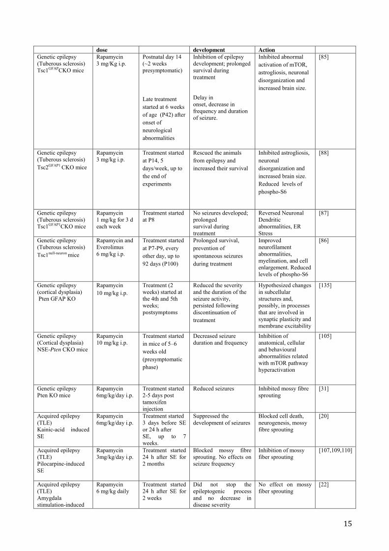
Table 1. Preclinical evidence on the role of mTOR inhibitors in preventing epileptogenesis
Epilepsy Type/ AnimalModel
mTOR inhibitor and
Protocol of Administration
Effect of mTOR inhibitor on epilepsy
Hypothesized Mechanism(s) of
References
15
dose development Action Genetic epilepsy (Tuberous sclerosis) Tsc1GFAPCKO mice
Rapamycin 3 mg/Kg i.p.
Postnatal day 14 (~2 weeks presymptomatic)
Late treatment started at 6 weeks of age (P42) after onset of neurological abnormalities
Inhibition of epilepsy development; prolonged survival during treatment Delay in onset, decrease in frequency and duration of seizure.
Inhibited abnormal activation of mTOR, astrogliosis, neuronal disorganization and increased brain size.
[85]
Genetic epilepsy (Tuberous sclerosis) Tsc2GFAP1 CKO mice
Rapamycin 3 mg/kg i.p.
Treatment started at P14, 5 days/week, up to the end of experiments
Rescued the animals from epilepsy and increased their survival
Inhibited astrogliosis, neuronal disorganization and increased brain size. Reduced levels of phospho-S6
[88]
Genetic epilepsy (Tuberous sclerosis) Tsc1GFAP1CKO mice
Rapamycin 1 mg/kg for 3 d each week
Treatment started at P8
No seizures developed; prolonged survival during treatment
Reversed Neuronal Dendritic abnormalities, ER Stress
[87]
Genetic epilepsy (Tuberous sclerosis) Tsc1null-neuron mice
Rapamycin and Everolimus 6 mg/kg i.p.
Treatment started at P7-P9, every other day, up to 92 days (P100)
Prolonged survival, prevention of spontaneous seizures during treatment
Improved neurofilament abnormalities, myelination, and cell enlargement. Reduced levels of phospho-S6
[86]
Genetic epilepsy (cortical dysplasia) Pten GFAP KO
Rapamycin 10 mg/kg i.p.
Treatment (2 weeks) started at the 4th and 5th weeks; postsymptoms
Reduced the severity and the duration of the seizure activity, persisted following discontinuation of treatment
Hypothesized changes in subcellular structures and, possibly, in processes that are involved in synaptic plasticity and membrane excitability
[135]
Genetic epilepsy (Cortical dysplasia) NSE-Pten CKO mice
Rapamycin 10 mg/kg i.p.
Treatment started in mice of 5–6 weeks old (presymptomatic phase)
Decreased seizure duration and frequency
Inhibition of anatomical, cellular and behavioural abnormalities related with mTOR pathway hyperactivation
[105]
Genetic epilepsy Pten KO mice
Rapamycin 6mg/kg/day i.p.
Treatment started 2-5 days post tamoxifen injection
Reduced seizures Inhibited mossy fibre sprouting
[31]
Acquired epilepsy (TLE) Kainic-acid induced SE
Rapamycin 6mg/kg/day i.p.
Treatment started 3 days before SE or 24 h after SE, up to 7 weeks.
Suppressed the development of seizures
Blocked cell death, neurogenesis, mossy fibre sprouting
[20]
Acquired epilepsy (TLE) Pilocarpine-induced SE
Rapamycin 3mg/kg/day i.p.
Treatment started 24 h after SE for 2 months
Blocked mossy fibre sprouting. No effects on seizure frequency
Inhibition of mossy fiber sprouting
[107,109,110]
Acquired epilepsy (TLE) Amygdala stimulation-induced
Rapamycin 6 mg/kg daily
Treatment started 24 h after SE for 2 weeks
Did not stop the epileptogenic process and no decrease in disease severity
No effect on mossy fiber sprouting
[22]
16
SE
Acquired epilepsy (TLE) Pilocarpine induced SE in mice
Rapamycin 6 mg/kg daily
Treatment started 24 h after the onset of SE for 6 consecutive days
Suppressed epileptiform activity
inhibited mTOR pathway and repressing mossy fiber sprouting
[114]
Acquired epilepsy (TLE) Electrical stimulation of the angular bundle (SE)
Rapamycin 6 mg/kg/day i.p.
Treatment for 7 days, started 4 hours after the induction of SE, and continued until rats were sacrificed, 6 weeks after SE.
Reduced the development of epilepsy
Inhibition of mossy fiber sprouting, reduction in neuronal death, decreased BBB leakage; no reduction of inflammatory response after SE induction
[118]
Acquired epilepsy (TLE) kainic acid–induced SE
Rapamycin 6 mg/kg/day
Treatment started 4 h after SE, once daily for 7 days, and continued until rats were killed 7 weeks post-SE.
reduction or prevention of recurrent seizures at a later stage.
Reduced BBB leakage during the chronic phase, via reduction of gliosis, brain inflammation and angiogenesis.
[119,120]
Acquired epilepsy TBI
Rapamycin 6 mg/kg/day i.p.
Treatment started 1 hour after injury and continued for 1 month
Prevented the development of post-traumatic epilepsy
Decreased neuronal degeneration and mossy fibre sprouting, although this effect did not directly correlate with inhibition of epileptogenesis
[134,136]
Acquired epilepsy Hypoxia-induced seizures in rats
Rapamycin 3 mg/kg i.p.
Treatment started 24 h before and 1 h after exposure to hypoxia
No effect on acute seizures; decreased chronic seizures
inhibited mTORC1 pathway, and subsequent increased glutamatergic neurotransmission.
[128]
Absence epilepsy model WAG/Rij rats
Rapamycin 1 mg/kg os
Treatment started at P45 and continued for 17 weeks
Decreased the development of absence seizures
inhibition of the release of inflammatory cytokines
[122,126]
SE = Status Epilepticus; TLE = Temporal Lobe Epilepsy; BBB = Blood Brain Barrier; CKO = Conditional Knockout; GFAP = Glial Fibrillary Acid Protein; i.p. = intraperitoneally; KO = Knockout; NS-Pten KO = Neurone Subset-Specific Pten knockout; NSE = Neuron-Specific Enolase; P = Postnatal day; PTEN = Phosphatase and Tensin homolog; TBI = Traumatic Brain Injury; TSC 1 = Tuberous Sclerosis Complex 1; TSC 2 = Tuberous Sclerosis Complex 2; WAG/Rij rat = Wistar Albino Glaxo/Rij-rat.
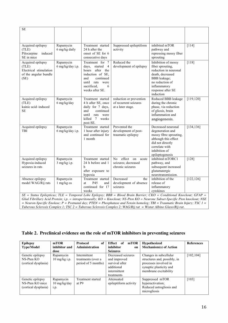
Table 2. Preclinical evidence on the role of mTOR inhibitors in preventing seizures
Epilepsy Type/Model
mTOR inhibitor and dose
Protocol of Administration
Effect of mTOR inhibitor on Seizures
Hypothesized Mechanism(s) of Action
References
Genetic epilepsy NS-Pten KO (cortical dysplasia)
Rapamycin 10 mg/kg i.p.
Intermittent treatments (over a period of 5 months)
Decreased seizures and improved survival after additional intermittent treatments
Changes in subcellular structures and, possibly, in processes involved in synaptic plasticity and membrane excitability
[102,104]
Genetic epilepsy NS-Pten KO mice (cortical dysplasia)
Rapamycin 10 mg/kg/day i.p.
Treatment started at P9
Attenuated epileptiform activity
Suppressed mTOR hyperactivation; Reduced astrogliosis and microgliosis
[103]

17
Genetic epilepsy Pten GFAP KO
Temsirolimus 7.5 mg/kg
Treatment started from 6 to 16 weeks, when mutant mice were symptomatic
Decreased seizures and mortality
Decreased megalencephaly, cell size
[101]
Genetic epilepsy Acute biallelic deletion of Tsc1 in adult mice
Rapamycin 5 and 10 mg/kg/day i.p.
Treatment started 2–4 months post-natal
Prolonged survival; No seizures developed during treatment
Effectively reduced pS6 levels
[90]
Epileptic encephalopathy Multiple-hit rat model of infantile spasms
Rapamycin different doses
After the onset of spasms.
Reduction of acutely-induced spasms in a dose-related way
Unclear
[70]
TLE following Pilocarpine- induced SE in adult rats
Rapamycin 5mg/kg/day i.p.
Pre-treatment for 3 days before the induction of seizures with pilocarpine
Reduction seizure activity during treatment, gradually returning after discontinuing treatment
Suppressed mossy fiber sprouting
[108]
Acquired epilepsy Acute seizure models in Sprague-Dawley rats
Rapamycin 5 mg/kg i.p.
Treatment started, in immature and mature rats, prior to induction of seizures by PTZ, pilocarpine or kainate
Increased severity of seizures; decreased seizure threshold after 3 daily doses of rapamycin in 3–4 weeks old, but not adult, rats
Treatment down-regulates KCC2 expression in CNS, which could increase susceptibility to pilocarpine-induce seizures in immature rats
[32]
Epileptic encephalopathy Infantile spasms in rats prenatally treated with betamethasone, triggered with NMDA
Rapamycin 3 mg/kg i.p.
Pre-treatment 24h prior to induction of spasms
No effects in this model
[129]
Acquired epilepsy Acute seizure models in Sprague-Dawley rats acute seizure tests
Rapamycin 3 or 6 mg/kg i.p.
Used different treatment paradigm in rat pups (P15) and juvenile (P55–60) rats
Variable efficacy on acute seizures which are age, time, treatment paradigm and model dependent.
The lack of effects, above all in immature rats, has been correlated with decreased NPY expression in the cortex and hippocampus
[115]
Acquired epilepsy Multiple Acute seizure (6Hz, PTZ or kainate) tests in NIH Swiss mice
Rapamycin 4.5 mg/kg i.p.
Short-term treatment (single dose) 3h before seizure onset and long-term treatment (3 daily doses) before seizure onset
Variable efficacy on acute seizures, which are age, time, treatment paradigm and model- dependent
Reduction in neuronal excitability and/or neurotransmitter release may occur with rapamycin
[137]
CNS = Central Nervous System; GFAP = Glial Fibrillary Acid Protein; i.p. = intraperitoneally; KCC2 = Potassium Chloride Cotransporter 2; KO = Knockout; mTOR = mammalian Target Of Rapamycin; NPY = Neuropeptide Y; NS-Pten KO = Neurone Subset-Specific Pten knockout; P = Postnatal day; pS6 = Phospho-S6; PTEN = Phosphatase and Tensin homolog; PTZ = Pentylenetetrazole; TSC 1 = Tuberous Sclerosis Complex 1; TSC 2 = Tuberous Sclerosis Complex 2;
3.2 Clinical studies
Despite the current pharmacological and non-pharmacological treatment options, about a third of
the epileptic patients remain drug-resistant [138,139]. Indeed, TSC is also characterized by

18
pharmacologically uncontrolled seizures. The United States Food and Drug Administration (FDA)
approved everolimus, a rapamycin analogue, for the treatment of patients with TSC associated with
inoperable SEGAs [17,140]. Beneficial effects of mTOR inhibition with everolimus have been
reported previously in patients with TSC and epilepsy [74,141-143]. Therefore mTOR inhibitors
provide a potential therapy based on the pathophysiology of TSC [77].
In 2009, it has been described for the first time that rapamycin treatment (10 months) induced a
reduction in seizure frequency and severity in a 10-year-old girl with difficult-to-treat seizures in
TSC, although pre- and post-treatment magnetic resonance imaging did not reveal any change in the
cortical tubers [144]. In another study, a child treated with everolimus for a regrowing SEGA, a
complete cessation of previously intractable seizures was reported at 12 months follow-up [18].
Everolimus treatment improved seizure control in prospective phase I/II studies in patients with
TSC [141] and in patients with TSC and associated SEGA [74]. In one of these studies (prospective
open label phase I/II study), Krueger, et al. [74] showed that everolimus used in TSC to limit SEGA
(associated with TSC) (primary end point), decreases seizure frequency in approximately 60% of
patients studied (secondary end point), but 1 patient experienced increased seizure frequency with
the drug [74,145]. In particular, 9 out of 16 patients with TSC showed a decrease in seizure
frequency, 6 did not show significant reduction, whereas in 1 an increased seizure frequency was
noted. The effect of everolimus, after 12 weeks of treatment in the management of
pharmacoresistant epilepsy in patients > 2 years affected by SEGAs, was also investigated in a
prospective, multicentre, open-label, phase I/II clinical trial [141]. The study compared seizure data
during the last 4 weeks of everolimus treatment (weeks 13–16) with the 4-weeks period before
everolimus initiation (baseline, weeks 1–4).
Everolimus treatment (12 weeks) in pediatric patients with TSC and refractory epilepsy reduced
seizure frequency by a median reduction of 73% in 17 of 20 patients examined and a median 70%
decrease in cumulative seizure duration. Four of these patients were seizure-free at 12 weeks, and
seven had a 90% reduction in seizure frequency [141]. Everolimus was well tolerated; all adverse
19
events were mostly grade 1 or 2 in severity and usually transient, never requiring everolimus
withdrawal. Upper respiratory infections, stomatitis and mucositis were the most common adverse
events [141]. Therefore, these findings strongly suggest that everolimus might be an effective
treatment for SEGA-related epilepsy. The different animal studies and limited clinical data have led
to further current clinical trials for inhibition of mTOR in genetic epilepsies. A compassionate use
trial for seven TSC patients with drug resistant epilepsy has demonstrated the efficacy of
everolimus; one patient discontinued treatment because of rash, four of six patients exhibited a
reduction of seizure frequency of 25-100%, two of six 6 patients, did not show alteration of seizure
frequency. Everolimus treatment seemed to be well-tolerated with adverse effects similar to those
reported in previous studies [143].
Recently, an open-label case series in seven patients (median age 6 years), with TSC and refractory
epilepsy, described the efficacy mTOR inhibitors (six with rapamycin and one with everolimus), in
seizures improvement, which were reported to have only minimal adverse effects. Of the intractable
seizure group (7 patients), 1 patient had >90% reduction, 4 had 50%-90% reduction, and 2 had
<50% reduction. Moreover, this treatment was reported to improve other characteristics of TSC,
such as facial angiofibromas and cognition; three reported subjective improvements in learning
[146]. Recently, another single case of seizures aggravation after everolimus treatment for SEGA
has been reported [147]. Cessation of seizures or a reduction in seizure frequency was also reported
in a small number of pediatric patients with TSC who were treated with long-term everolimus. In 6
out of 8 children, at least a 50% reduction in SEGA volume was observed; everolimus resulted in
permanent seizure cessation in one child with severe drug-resistant epilepsy and in at least a 50%
reduction in the number of seizures in two other [142].
Moreover, in a prospective study of 5 neonatal patients with TSC, it was demonstrated that EEG
changes occurring in TSC patients before clinical seizures reflect the process of epileptogenesis in
these patients. Therefore, EEG recording could have predictive value during infancy in patients
with TSC [148].

20
More recently, the case of a 13-year-old girl with TSC-associated with refractory generalized
seizures who initiated treatment with everolimus experiencing subsequent improvement in several
TSC manifestations, including a reduction in seizure frequency from clusters of two or three daily
to one every 2 to 4 weeks after 1.5 years of treatment was described [149]. All these clinical studies
confirm the potential benefits of mTOR inhibitors on epilepsy associated with TSC, but the open-
label design of the studies as well as the heterogeneity of enrolled patients and the small number of
cases still does not allow making definite conclusions.
A randomized, blinded, placebo controlled, phase III trial is currently in progress to determine the
efficacy of everolimus on seizures in patients with TSC; patients between the ages of 2 and 65
years, with a clinically definite diagnosis of TSC and uncontrolled partial-onset seizures, are
currently being enrolled for this study and results are expected during the next year
(clinicaltrials.gov identifier NCT01713946) (Table 3).
Recently, a genomics study of infantile spasms and Lennox-Gastaut Syndrome has found a de novo
mutation in mTOR gene without associated brain malformations suggesting a possible role of
mTOR in these disorders and mTOR inhibitors could be a possible treatment [150]. Furthermore, de
novo somatic mutations of PI3K, AKT3 or mTOR genes in patients with hemimegalencephaly
(HME) a condition associated with resistant epilepsy have been described [31,151].
Table 3. Clinical Studies with mTOR Inhibitors in TSC-Associated Epilepsy
Type of Study Disease Drug and Dose
Number and Age of Patient(s)
Duration of Treatment
Clinical Result References
Case report TSC and refractory epilepsy
Rapamycin 0.15 mg/kg/day
1 patient; 9-year-old girl
10 months Reduction in seizure frequency. 1 to 5 brief seizures (< 2 minutes) continued daily
[144]
Case report TSC and refractory epilepsy
Everolimus 4.5 mg/m2/day
1 patient; 10- year-old boy
12 months Complete cessation of epileptic seizures
[18]
Prospective, open-label, phase I–II study
TSC and related epilepsy
Everolimus 4.7–5.6 mg/m2/day
16 patients; 3 year-old or older
Median duration 21.5 months (range: 4.7-34.4 months)
Reduction in seizure frequency in 9/16 patients, did not change in 6, and increased in 1
[74]

21
Prospective, multicenter, open-label, phase I/II clinical trial
TSC and refractory epilepsy
Everolimus 5 mg/m2/day, then titrated to a serum trough level of 5–15 ng/ml
20 patients; median age:8 years (age range:2–21)
12 weeks Reduction in seizure frequency in 17/20 patients (median reduction of 73%). 4 of these patients were seizure-free at 12 weeks, and 7 had a 90% reduction in seizure frequency
[17,141]
Case study series
TSC and refractory epilepsy
Everolimus 5-7 mg/day
6 patients; median age:5 years (age range: 2-12).
36 weeks Reduction in seizure frequency in 4/6 patients (of 25% -100%). The percentage of seizure-free days increased in 3/4 of these patients. In 2/6 patients, no lteration of seizure frequency
[143]
Open-label, single-center case series
TSC and refractory epilepsy
Sirolimus 1 mg/m2/d. then adjusted to trough blood levels of 4-10 ng/mL
7 patients; median age: 6 years (age range: 3-17).
median duration: 18 months (range: 6-36 months)
1 patient had >90% reduction, 4 had 50%-90% reduction, and 2 had <50% reduction
[146]
Case study SEGAs associated with TSC
Everolimus 5 mg/m2/day
1 patient; 13.5-year-old girl
12 days Seizure aggravation [147]
Prospective, double-blind, parallel-group, placebo-controlled, multicenter phase 3 (EXIST-1)
SEGAs associated with TSC
Everolimus 4.5 mg/m2/day then adjusted to attain a blood concentration of 5-15 ng/mL.
8 patients; children under the age of 3
35 months (range:33-38 months)
Cessation of seizures in 1 patient, significant (at least a 50% ) reduction in the number of seizures in 2 patient
[142]
Case report TSC-associated epilepsy with refractory generalized seizures
Everolimus 5 mg/day
1 patient; 13-year-old girl
1.5 year
Reduction in seizure frequency from clusters of two or three daily to one every 2 to 4 weeks
[149]
Randomized, blinded, lacebo controlled, phase III trial
TSC associated refractory seizures
Everolimus titrated from 3 to 7 ng/mL and also from 9 to 15 ng/mL and placebo
Male or female between the ages of 2 and 65 years
This study is currently recruiting participants
Novartis Pharmaceuticals
EXIST-1 = EXamining everolimus In a Study of TSC; SEGA = Subependymal Giant cell Astrocytoma; TSC = Tuberous Sclerosis Complex
4. Conclusions
Epilepsy represents one of the oldest and most prevalent neurological disorders. Currently available
treatments are effective but provide only symptomatic management of the disease, and an unmet
need exists for a rational therapy that targets the etiology of epilepsy [2,139]. Understanding
epileptogenesis, the process by which a normal brain becomes epileptic, may help identify
22
molecular targets for drugs that could prevent epilepsy. To date, the mTOR pathway represents
perhaps, the most promising molecular target for producing a better understanding and treatment of
this disease. The first favorable point in this regard, is the availability of a number of already
marketed drugs acting on this target; such drugs (i.e. rapamycin, everolimus etc) are already used
clinically for other conditions (e.g. as immunosuppressants in kidney transplantation, in treatment
of advanced renal carcinoma and breast cancer).
Different preclinical data confirm that mTOR dysregulation may play a pathogenetic role in the
epileptogenesis of different forms of epilepsy, and that mTOR inhibition may prevent epilepsy
development, especially in genetic mTORopathies. mTOR inhibitors show efficacy in treating some
genetic forms of epilepsy (e.g. TSC and WAG/Rij rats) but also possess antiepileptogenic effects in
preclinical models. The potential inhibition of mTOR as a therapeutic strategy for preventing
epileptogenesis is therefore promising and may be applicable not only to TSC, but also to other
forms of epilepsy. These findings are starting to be translated into the clinical arena; successful
anticonvulsant effects of both rapamycin and everolimus have been reported in individuals with
focal-onset seizures in the context of TSC. While several studies with TSC mouse models suggest
that mTOR inhibitors have antiepileptogenic properties for preventing epilepsy, clinical
antiepileptogenic drug trials are difficult to conduct and have not yet been attempted with mTOR
inhibitors. Some controversial results with mTOR inhibitors also exist and while it is clear that the
mTOR pathway might be a good target candidate, some drawbacks of ‘general’ mTOR inhibition
must still be considered and more studies are warranted (ideally with more selective drugs), before
full clinical translation of mTOR targeting can result. In particular, the time-window, duration and
dosages to be used in prevention of epileptogenesis are still far from being clearly identified. On the
other hand, mTOR inhibitors possess only a very low efficacy in preclinical animal models of
seizures/epilepsy and therefore, their effectiveness in stopping seizures is probably very limited
and/or in any case, it might require time before some kind of effectiveness can be observed;
generally, a few days of treatment are necessary to observe an antiseizure effect if any. As most of

23
the beneficial effects of mTOR inhibition might cease after drug discontinuation, a life-long
treatment might be necessary. The effect of long-term mTOR inhibition on developing brain
structures and functions is still not fully understood. Despite several drugs acting on mTOR that are
already currently available (both considering marketed and experimental drugs), none of these
agents has selectivity for the CNS, which obviously underlies the appearance of peripheral side
effects that might limit dosages and therefore efficacy. The search for more selective agonists
targeting specific effectors of the mTOR pathway can lead to the discovery of better drugs to treat
epilepsy. As mentioned above, clinical trials are currently undergoing for both everolimus and
rapamycin in epilepsy. The results of these trials will indeed shed light on the possibility to use such
molecules in TSC patients; however, this will only be an initial step, which will need further
clinical trials to understand the possible use of mTOR inhibitors in other clinical situations with
patients at risk of epilepsy development (e.g. traumatic brain injury).
Interactions between mTOR inhibitors and available antiepileptic drugs are not fully known and
will have to be analyzed. The role of mTOR inhibitors in epilepsy treatment still needs further
research and an extension of clinical trials to non-TSC epilepsy syndromes. Further clinical studies
could help to clarify the clinical efficacy, dosage and safety profile of mTOR inhibitors in epilepsy
and epileptogenesis. Despite these limitations, recent developments in mTOR pathway modulation
open up new perspectives for future therapy of epilepsy and mTOR inhibitors represent a promising
therapeutic option for the treatment of epilepsy.
In conclusion, as in the case of many other diseases, we feel that the mTOR pathway represents a
great opportunity for future novel drug development in epilepsy therapeutics; the actual available
data with mTOR inhibitors, both clinical and pre-clinical are intriguing and highly support future
experiments. Finally, a very important task will be that of developing new mTOR inhibitors with a
higher selectivity for the central nervous system (CNS) and a better safety profile.

24
References
1 P. Kwan, M.J. Brodie, Refractory epilepsy: Mechanisms and solutions, Expert Rev Neurother. 6
(2006) 397-406.
2 C. Palleria, A. Coppola, R. Citraro, L. Del Gaudio, S. Striano, G. De Sarro, E. Russo,
Perspectives on treatment options for mesial temporal lobe epilepsy with hippocampal sclerosis,
Expert Opin Pharmacother. 16 (2015) 2355-2371.
3 H. Potschka, M.J. Brodie, Pharmacoresistance, Handb Clin Neurol. 108 (2012) 741-757.
4 A. Pitkanen, J. Engel, Jr., Past and present definitions of epileptogenesis and its biomarkers,
Neurotherapeutics. 11 (2014) 231-241.
5 H.S. White, W. Loscher, Searching for the ideal antiepileptogenic agent in experimental models:
Single treatment versus combinatorial treatment strategies, Neurotherapeutics. 11 (2014) 373-384.
6 E. Trinka, F. Brigo, Antiepileptogenesis in humans: Disappointing clinical evidence and ways to
move forward, Curr Opin Neurol. 27 (2014) 227-235.
7 N. Takei, H. Nawa, Mtor signaling and its roles in normal and abnormal brain development, Front
Mol Neurosci. 7 (2014) 28.
8 J. Bockaert, P. Marin, Mtor in brain physiology and pathologies, Physiol Rev. 95 (2015) 1157-
1187.
9 A.P. Ostendorf, M. Wong, Mtor inhibition in epilepsy: Rationale and clinical perspectives, CNS
Drugs. 29 (2015) 91-99.
10 J. Liu, C. Reeves, Z. Michalak, A. Coppola, B. Diehl, S.M. Sisodiya, M. Thom, Evidence for
mtor pathway activation in a spectrum of epilepsy-associated pathologies, Acta Neuropathol
Commun. 2 (2014) 71.
11 E. Russo, R. Citraro, A. Constanti, G. De Sarro, The mtor signaling pathway in the brain: Focus
on epilepsy and epileptogenesis, Mol Neurobiol. 46 (2012) 662-681.
12 M. Wong, Mammalian target of rapamycin (mtor) pathways in neurological diseases, Biomed J.
36 (2013) 40-50.

25
13 P.B. Crino, Mtor: A pathogenic signaling pathway in developmental brain malformations,
Trends Mol Med. 17 (2011) 734-742.
14 P.B. Crino, Mtor signaling in epilepsy: Insights from malformations of cortical development,
Cold Spring Harb Perspect Med. 5 (2015)
15 P. Curatolo, R. Moavero, Mtor inhibitors as a new therapeutic option for epilepsy, Expert Rev
Neurother. 13 (2013) 627-638.
16 K. Sadowski, K. Kotulska-Jozwiak, S. Jozwiak, Role of mtor inhibitors in epilepsy treatment,
Pharmacol Rep. 67 (2015) 636-646.
17 D.N. Franz, E. Belousova, S. Sparagana, E.M. Bebin, M. Frost, R. Kuperman, O. Witt, M.H.
Kohrman, J.R. Flamini, J.Y. Wu, P. Curatolo, P.J. de Vries, V.H. Whittemore, E.A. Thiele, J.P.
Ford, G. Shah, H. Cauwel, D. Lebwohl, T. Sahmoud, S. Jozwiak, Efficacy and safety of everolimus
for subependymal giant cell astrocytomas associated with tuberous sclerosis complex (exist-1): A
multicentre, randomised, placebo-controlled phase 3 trial, Lancet. 381 (2013) 125-132.
18 M. Perek-Polnik, S. Jozwiak, E. Jurkiewicz, D. Perek, K. Kotulska, Effective everolimus
treatment of inoperable, life-threatening subependymal giant cell astrocytoma and intractable
epilepsy in a patient with tuberous sclerosis complex, Eur J Paediatr Neurol. 16 (2012) 83-85.
19 M. Wong, Mammalian target of rapamycin (mtor) activation in focal cortical dysplasia and
related focal cortical malformations, Exp Neurol. 244 (2013) 22-26.
20 L.H. Zeng, N.R. Rensing, M. Wong, The mammalian target of rapamycin signaling pathway
mediates epileptogenesis in a model of temporal lobe epilepsy, J Neurosci. 29 (2009) 6964-6972.
21 P.S. Buckmaster, F.H. Lew, Rapamycin suppresses mossy fiber sprouting but not seizure
frequency in a mouse model of temporal lobe epilepsy, J Neurosci. 31 (2011) 2337-2347.
22 A. Sliwa, G. Plucinska, J. Bednarczyk, K. Lukasiuk, Post-treatment with rapamycin does not
prevent epileptogenesis in the amygdala stimulation model of temporal lobe epilepsy, Neurosci
Lett. 509 (2012) 105-109.

26
23 R. Zoncu, A. Efeyan, D.M. Sabatini, Mtor: From growth signal integration to cancer, diabetes
and ageing, Nat Rev Mol Cell Biol. 12 (2011) 21-35.
24 C.K. Tsang, H. Qi, L.F. Liu, X.F. Zheng, Targeting mammalian target of rapamycin (mtor) for
health and diseases, Drug Discov Today. 12 (2007) 112-124.
25 Z.Z. Chong, Q. Yao, H.H. Li, The rationale of targeting mammalian target of rapamycin for
ischemic stroke, Cell Signal. 25 (2013) 1598-1607.
26 D.K. Sandsmark, C. Pelletier, J.D. Weber, D.H. Gutmann, Mammalian target of rapamycin:
Master regulator of cell growth in the nervous system, Histol Histopathol. 22 (2007) 895-903.
27 C. Dello Russo, L. Lisi, D.L. Feinstein, P. Navarra, Mtor kinase, a key player in the regulation of
glial functions: Relevance for the therapy of multiple sclerosis, Glia. 61 (2013) 301-311.
28 J. Jaworski, S. Spangler, D.P. Seeburg, C.C. Hoogenraad, M. Sheng, Control of dendritic
arborization by the phosphoinositide-3'-kinase-akt-mammalian target of rapamycin pathway, J
Neurosci. 25 (2005) 11300-11312.
29 V. Kumar, M.X. Zhang, M.W. Swank, J. Kunz, G.Y. Wu, Regulation of dendritic
morphogenesis by ras-pi3k-akt-mtor and ras-mapk signaling pathways, J Neurosci. 25 (2005)
11288-11299.
30 Y. Lee da, Roles of mtor signaling in brain development, Exp Neurobiol. 24 (2015) 177-185.
31 R.Y. Pun, I.J. Rolle, C.L. Lasarge, B.E. Hosford, J.M. Rosen, J.D. Uhl, S.N. Schmeltzer, C.
Faulkner, S.L. Bronson, B.L. Murphy, D.A. Richards, K.D. Holland, S.C. Danzer, Excessive
activation of mtor in postnatally generated granule cells is sufficient to cause epilepsy, Neuron. 75
(2012) 1022-1034.
32 X. Huang, J. McMahon, J. Yang, D. Shin, Y. Huang, Rapamycin down-regulates kcc2
expression and increases seizure susceptibility to convulsants in immature rats, Neuroscience. 219
(2012) 33-47.
33 K.F. Raab-Graham, P.C. Haddick, Y.N. Jan, L.Y. Jan, Activity- and mtor-dependent suppression
of kv1.1 channel mrna translation in dendrites, Science. 314 (2006) 144-148.

27
34 M. Wong, K.C. Ess, E.J. Uhlmann, L.A. Jansen, W. Li, P.B. Crino, S. Mennerick, K.A. Yamada,
D.H. Gutmann, Impaired glial glutamate transport in a mouse tuberous sclerosis epilepsy model,
Ann Neurol. 54 (2003) 251-256.
35 N. Lozovaya, S. Gataullina, T. Tsintsadze, V. Tsintsadze, E. Pallesi-Pocachard, M. Minlebaev,
N.A. Goriounova, E. Buhler, F. Watrin, S. Shityakov, A.J. Becker, A. Bordey, M. Milh, D.
Scavarda, C. Bulteau, G. Dorfmuller, O. Delalande, A. Represa, C. Cardoso, O. Dulac, Y. Ben-Ari,
N. Burnashev, Selective suppression of excessive glun2c expression rescues early epilepsy in a
tuberous sclerosis murine model, Nat Commun. 5 (2014) 4563.
36 K. Maiese, Targeting molecules to medicine with mtor, autophagy, and neurodegenerative
disorders, Br J Clin Pharmacol. (2015)
37 M. Costa-Mattioli, L.M. Monteggia, Mtor complexes in neurodevelopmental and
neuropsychiatric disorders, Nat Neurosci. 16 (2013) 1537-1543.
38 H.M. Abelaira, G.Z. Reus, M.V. Neotti, J. Quevedo, The role of mtor in depression and
antidepressant responses, Life Sci. 101 (2014) 10-14.
39 C.S. Jernigan, D.B. Goswami, M.C. Austin, A.H. Iyo, A. Chandran, C.A. Stockmeier, B.
Karolewicz, The mtor signaling pathway in the prefrontal cortex is compromised in major
depressive disorder, Prog Neuropsychopharmacol Biol Psychiatry. 35 (2011) 1774-1779.
40 A. Chandran, A.H. Iyo, C.S. Jernigan, B. Legutko, M.C. Austin, B. Karolewicz, Reduced
phosphorylation of the mtor signaling pathway components in the amygdala of rats exposed to
chronic stress, Prog Neuropsychopharmacol Biol Psychiatry. 40 (2013) 240-245.
41 K.R. Howell, A. Kutiyanawalla, A. Pillai, Long-term continuous corticosterone treatment
decreases vegf receptor-2 expression in frontal cortex, PLoS One. 6 (2011) e20198.
42 N. Li, B. Lee, R.J. Liu, M. Banasr, J.M. Dwyer, M. Iwata, X.Y. Li, G. Aghajanian, R.S. Duman,
Mtor-dependent synapse formation underlies the rapid antidepressant effects of nmda antagonists,
Science. 329 (2010) 959-964.
28
43 C. Yang, J. Shen, T. Hong, T.T. Hu, Z.J. Li, H.T. Zhang, Y.J. Zhang, Z.Q. Zhou, J.J. Yang,
Effects of ketamine on lipopolysaccharide-induced depressive-like behavior and the expression of
inflammatory cytokines in the rat prefrontal cortex, Mol Med Rep. 8 (2013) 887-890.
44 A. Sharma, C.A. Hoeffer, Y. Takayasu, T. Miyawaki, S.M. McBride, E. Klann, R.S. Zukin,
Dysregulation of mtor signaling in fragile x syndrome, J Neurosci. 30 (2010) 694-702.
45 A.M. Iyer, J. van Scheppingen, I. Milenkovic, J.J. Anink, H. Adle-Biassette, G.G. Kovacs, E.
Aronica, Mtor hyperactivation in down syndrome hippocampus appears early during development,
J Neuropathol Exp Neurol. 73 (2014) 671-683.
46 S. Ricciardi, E.M. Boggio, S. Grosso, G. Lonetti, G. Forlani, G. Stefanelli, E. Calcagno, N.
Morello, N. Landsberger, S. Biffo, T. Pizzorusso, M. Giustetto, V. Broccoli, Reduced akt/mtor
signaling and protein synthesis dysregulation in a rett syndrome animal model, Hum Mol Genet. 20
(2011) 1182-1196.
47 T.E. Graber, P.K. McCamphill, W.S. Sossin, A recollection of mtor signaling in learning and
memory, Learn Mem. 20 (2013) 518-530.
48 C.A. Hoeffer, E. Klann, Mtor signaling: At the crossroads of plasticity, memory and disease,
Trends Neurosci. 33 (2010) 67-75.
49 M. Dierssen, G.J. Ramakers, Dendritic pathology in mental retardation: From molecular genetics
to neurobiology, Genes Brain Behav. 5 Suppl 2 (2006) 48-60.
50 R.G. Parsons, G.M. Gafford, F.J. Helmstetter, Translational control via the mammalian target of
rapamycin pathway is critical for the formation and stability of long-term fear memory in amygdala
neurons, J Neurosci. 26 (2006) 12977-12983.
51 L. Stoica, P.J. Zhu, W. Huang, H. Zhou, S.C. Kozma, M. Costa-Mattioli, Selective
pharmacogenetic inhibition of mammalian target of rapamycin complex i (mtorc1) blocks long-term
synaptic plasticity and memory storage, Proc Natl Acad Sci U S A. 108 (2011) 3791-3796.

29
52 P.K. Dash, S.A. Orsi, A.N. Moore, Spatial memory formation and memory-enhancing effect of
glucose involves activation of the tuberous sclerosis complex-mammalian target of rapamycin
pathway, J Neurosci. 26 (2006) 8048-8056.
53 C.A. Hoeffer, W. Tang, H. Wong, A. Santillan, R.J. Patterson, L.A. Martinez, M.V. Tejada-
Simon, R. Paylor, S.L. Hamilton, E. Klann, Removal of fkbp12 enhances mtor-raptor interactions,
ltp, memory, and perseverative/repetitive behavior, Neuron. 60 (2008) 832-845.
54 W. Tischmeyer, H. Schicknick, M. Kraus, C.I. Seidenbecher, S. Staak, H. Scheich, E.D.
Gundelfinger, Rapamycin-sensitive signalling in long-term consolidation of auditory cortex-
dependent memory, Eur J Neurosci. 18 (2003) 942-950.
55 D. Ehninger, P.J. de Vries, A.J. Silva, From mtor to cognition: Molecular and cellular
mechanisms of cognitive impairments in tuberous sclerosis, J Intellect Disabil Res. 53 (2009) 838-
851.
56 D. Ehninger, S. Han, C. Shilyansky, Y. Zhou, W. Li, D.J. Kwiatkowski, V. Ramesh, A.J. Silva,
Reversal of learning deficits in a tsc2+/- mouse model of tuberous sclerosis, Nat Med. 14 (2008)
843-848.
57 J.O. Lipton, M. Sahin, The neurology of mtor, Neuron. 84 (2014) 275-291.
58 Z.M. Ignacio, G.Z. Reus, C.O. Arent, H.M. Abelaira, M.R. Pitcher, J. Quevedo, New
perspectives on the involvement of mtor in depression as well as in the action of antidepressant
drugs, Br J Clin Pharmacol. (2015)
59 B. Nyfeler, P. Bergman, C.J. Wilson, L.O. Murphy, Quantitative visualization of autophagy
induction by mtor inhibitors, Methods Mol Biol. 821 (2012) 239-250.
60 M. Arrasate, S. Finkbeiner, Protein aggregates in huntington's disease, Exp Neurol. 238 (2012)
1-11.
61 G.B. Irvine, O.M. El-Agnaf, G.M. Shankar, D.M. Walsh, Protein aggregation in the brain: The
molecular basis for alzheimer's and parkinson's diseases, Mol Med. 14 (2008) 451-464.

30
62 B. Ravikumar, C. Vacher, Z. Berger, J.E. Davies, S. Luo, L.G. Oroz, F. Scaravilli, D.F. Easton,
R. Duden, C.J. O'Kane, D.C. Rubinsztein, Inhibition of mtor induces autophagy and reduces
toxicity of polyglutamine expansions in fly and mouse models of huntington disease, Nat Genet. 36
(2004) 585-595.
63 Z. Berger, B. Ravikumar, F.M. Menzies, L.G. Oroz, B.R. Underwood, M.N. Pangalos, I.
Schmitt, U. Wullner, B.O. Evert, C.J. O'Kane, D.C. Rubinsztein, Rapamycin alleviates toxicity of
different aggregate-prone proteins, Hum Mol Genet. 15 (2006) 433-442.
64 X.F. Meng, J.T. Yu, J.H. Song, S. Chi, L. Tan, Role of the mtor signaling pathway in epilepsy, J
Neurol Sci. 332 (2013) 4-15.
65 S.S. McDaniel, M. Wong, Therapeutic role of mammalian target of rapamycin (mtor) inhibition
in preventing epileptogenesis, Neurosci Lett. 497 (2011) 231-239.
66 M. Wong, P.B. Crino. Mtor and epileptogenesis in developmental brain malformations. In:
Noebels JL, Avoli M, Rogawski MA, Olsen RW, Delgado-Escueta AV, eds. Jasper's basic
mechanisms of the epilepsies. Bethesda (MD), 2012.
67 C.L. Lasarge, S.C. Danzer, Mechanisms regulating neuronal excitability and seizure
development following mtor pathway hyperactivation, Front Mol Neurosci. 7 (2014) 18.
68 A.S. Galanopoulou, J.A. Gorter, C. Cepeda, Finding a better drug for epilepsy: The mtor
pathway as an antiepileptogenic target, Epilepsia. 53 (2012) 1119-1130.
69 S. Erlich, A. Alexandrovich, E. Shohami, R. Pinkas-Kramarski, Rapamycin is a neuroprotective
treatment for traumatic brain injury, Neurobiol Dis. 26 (2007) 86-93.
70 E. Raffo, A. Coppola, T. Ono, S.W. Briggs, A.S. Galanopoulou, A pulse rapamycin therapy for
infantile spasms and associated cognitive decline, Neurobiol Dis. 43 (2011) 322-329.
71 S. Carloni, S. Girelli, C. Scopa, G. Buonocore, M. Longini, W. Balduini, Activation of
autophagy and akt/creb signaling play an equivalent role in the neuroprotective effect of rapamycin
in neonatal hypoxia-ischemia, Autophagy. 6 (2010) 366-377.

31
72 G.L. Holmes, C.E. Stafstrom, Tuberous sclerosis complex and epilepsy: Recent developments
and future challenges, Epilepsia. 48 (2007) 617-630.
73 P. Curatolo, B.L. Maria, Tuberous sclerosis, Handb Clin Neurol. 111 (2013) 323-331.
74 D.A. Krueger, M.M. Care, K. Holland, K. Agricola, C. Tudor, P. Mangeshkar, K.A. Wilson, A.
Byars, T. Sahmoud, D.N. Franz, Everolimus for subependymal giant-cell astrocytomas in tuberous
sclerosis, N Engl J Med. 363 (2010) 1801-1811.
75 P. Curatolo, Mechanistic target of rapamycin (mtor) in tuberous sclerosis complex-associated
epilepsy, Pediatr Neurol. 52 (2015) 281-289.
76 D.A. Krueger, Management of cns-related disease manifestations in patients with tuberous
sclerosis complex, Curr Treat Options Neurol. 15 (2013) 618-633.
77 M. Wong, Mammalian target of rapamycin (mtor) inhibition as a potential antiepileptogenic
therapy: From tuberous sclerosis to common acquired epilepsies, Epilepsia. 51 (2010) 27-36.
78 L. Meikle, D.M. Talos, H. Onda, K. Pollizzi, A. Rotenberg, M. Sahin, F.E. Jensen, D.J.
Kwiatkowski, A mouse model of tuberous sclerosis: Neuronal loss of tsc1 causes dysplastic and
ectopic neurons, reduced myelination, seizure activity, and limited survival, J Neurosci. 27 (2007)
5546-5558.
79 D.M. Feliciano, T. Su, J. Lopez, J.C. Platel, A. Bordey, Single-cell tsc1 knockout during
corticogenesis generates tuber-like lesions and reduces seizure threshold in mice, J Clin Invest. 121
(2011) 1596-1607.
80 D.M. Feliciano, T.V. Lin, N.W. Hartman, C.M. Bartley, C. Kubera, L. Hsieh, C. Lafourcade,
R.A. O'Keefe, A. Bordey, A circuitry and biochemical basis for tuberous sclerosis symptoms: From
epilepsy to neurocognitive deficits, Int J Dev Neurosci. 31 (2013) 667-678.
81 M. Wong, A tuber-ful animal model of tuberous sclerosis at last? , Epilepsy Currents. 1 (2012)
82 E.J. Uhlmann, W. Li, D.K. Scheidenhelm, C.L. Gau, F. Tamanoi, D.H. Gutmann, Loss of
tuberous sclerosis complex 1 (tsc1) expression results in increased rheb/s6k pathway signaling
important for astrocyte cell size regulation, Glia. 47 (2004) 180-188.
32
83 J.P. MacKeigan, D.A. Krueger, Differentiating the mtor inhibitors everolimus and sirolimus in
the treatment of tuberous sclerosis complex, Neuro Oncol. 17 (2015) 1550-1559.
84 K. Sadowski, K. Kotulska, R.A. Schwartz, S. Jozwiak, Systemic effects of treatment with mtor
inhibitors in tuberous sclerosis complex: A comprehensive review, J Eur Acad Dermatol Venereol.
(2015)
85 L.H. Zeng, L. Xu, D.H. Gutmann, M. Wong, Rapamycin prevents epilepsy in a mouse model of
tuberous sclerosis complex, Ann Neurol. 63 (2008) 444-453.
86 L. Meikle, K. Pollizzi, A. Egnor, I. Kramvis, H. Lane, M. Sahin, D.J. Kwiatkowski, Response of
a neuronal model of tuberous sclerosis to mammalian target of rapamycin (mtor) inhibitors: Effects
on mtorc1 and akt signaling lead to improved survival and function, J Neurosci. 28 (2008) 5422-
5432.
87 J. Goto, D.M. Talos, P. Klein, W. Qin, Y.I. Chekaluk, S. Anderl, I.A. Malinowska, A. Di Nardo,
R.T. Bronson, J.A. Chan, H.V. Vinters, S.G. Kernie, F.E. Jensen, M. Sahin, D.J. Kwiatkowski,
Regulable neural progenitor-specific tsc1 loss yields giant cells with organellar dysfunction in a
model of tuberous sclerosis complex, Proc Natl Acad Sci U S A. 108 (2011) E1070-1079.
88 L.H. Zeng, N.R. Rensing, B. Zhang, D.H. Gutmann, M.J. Gambello, M. Wong, Tsc2 gene
inactivation causes a more severe epilepsy phenotype than tsc1 inactivation in a mouse model of
tuberous sclerosis complex, Hum Mol Genet. 20 (2011) 445-454.
89 B. Zhang, J. Zou, N.R. Rensing, M. Yang, M. Wong, Inflammatory mechanisms contribute to
the neurological manifestations of tuberous sclerosis complex, Neurobiol Dis. 80 (2015) 70-79.
90 E. Abs, S.M. Goorden, J. Schreiber, I.E. Overwater, M. Hoogeveen-Westerveld, C.F. Bruinsma,
E. Aganovic, N.Z. Borgesius, M. Nellist, Y. Elgersma, Torc1-dependent epilepsy caused by acute
biallelic tsc1 deletion in adult mice, Ann Neurol. 74 (2013) 569-579.
91 D. Friedman, M. Bogner, K. Parker-Menzer, O. Devinsky, Vigabatrin for partial-onset seizure
treatment in patients with tuberous sclerosis complex, Epilepsy Behav. 27 (2013) 118-120.

33
92 S. Jozwiak, K. Kotulska, D. Domanska-Pakiela, B. Lojszczyk, M. Syczewska, D. Chmielewski,
D. Dunin-Wasowicz, T. Kmiec, J. Szymkiewicz-Dangel, M. Kornacka, W. Kawalec, D. Kuczynski,
J. Borkowska, K. Tomaszek, E. Jurkiewicz, M. Respondek-Liberska, Antiepileptic treatment before
the onset of seizures reduces epilepsy severity and risk of mental retardation in infants with
tuberous sclerosis complex, Eur J Paediatr Neurol. 15 (2011) 424-431.
93 R. Bombardieri, M. Pinci, R. Moavero, C. Cerminara, P. Curatolo, Early control of seizures
improves long-term outcome in children with tuberous sclerosis complex, Eur J Paediatr Neurol. 14
(2010) 146-149.
94 E. Ben-Menachem, Mechanism of action of vigabatrin: Correcting misperceptions, Acta Neurol
Scand Suppl. (2011) 5-15.
95 B. Zhang, S.S. McDaniel, N.R. Rensing, M. Wong, Vigabatrin inhibits seizures and mtor
pathway activation in a mouse model of tuberous sclerosis complex, PLoS One. 8 (2013) e57445.
96 S.A. Backman, V. Stambolic, A. Suzuki, J. Haight, A. Elia, J. Pretorius, M.S. Tsao, P. Shannon,
B. Bolon, G.O. Ivy, T.W. Mak, Deletion of pten in mouse brain causes seizures, ataxia and defects
in soma size resembling lhermitte-duclos disease, Nat Genet. 29 (2001) 396-403.
97 R. Spreafico, I. Blumcke, Focal cortical dysplasias: Clinical implication of neuropathological
classification systems, Acta Neuropathol. 120 (2010) 359-367.
98 S. Fauser, C. Essang, D.M. Altenmuller, A.M. Staack, B.J. Steinhoff, K. Strobl, T. Bast, S.
Schubert-Bast, U. Stephani, G. Wiegand, M. Prinz, A. Brandt, J. Zentner, A. Schulze-Bonhage,
Long-term seizure outcome in 211 patients with focal cortical dysplasia, Epilepsia. 56 (2015) 66-
76.
99 J.S. Lim, W.I. Kim, H.C. Kang, S.H. Kim, A.H. Park, E.K. Park, Y.W. Cho, S. Kim, H.M. Kim,
J.A. Kim, J. Kim, H. Rhee, S.G. Kang, H.D. Kim, D. Kim, D.S. Kim, J.H. Lee, Brain somatic
mutations in mtor cause focal cortical dysplasia type ii leading to intractable epilepsy, Nat Med.
(2015)
34
100 J.Y. Lee, A.K. Park, E.S. Lee, W.Y. Park, S.H. Park, J.W. Choi, J.H. Phi, K.C. Wang, S.K.
Kim, Mirna expression analysis in cortical dysplasia: Regulation of mtor and lis1 pathway, Epilepsy
Res. 108 (2014) 433-441.
101 C.H. Kwon, X. Zhu, J. Zhang, S.J. Baker, Mtor is required for hypertrophy of pten-deficient
neuronal soma in vivo, Proc Natl Acad Sci U S A. 100 (2003) 12923-12928.
102 M.C. Ljungberg, C.N. Sunnen, J.N. Lugo, A.E. Anderson, G. D'Arcangelo, Rapamycin
suppresses seizures and neuronal hypertrophy in a mouse model of cortical dysplasia, Dis Model
Mech. 2 (2009) 389-398.
103 L.H. Nguyen, A.L. Brewster, M.E. Clark, A. Regnier-Golanov, C.N. Sunnen, V.V. Patil, G.
D'Arcangelo, A.E. Anderson, Mtor inhibition suppresses established epilepsy in a mouse model of
cortical dysplasia, Epilepsia. 56 (2015) 636-646.
104 C.N. Sunnen, A.L. Brewster, J.N. Lugo, F. Vanegas, E. Turcios, S. Mukhi, D. Parghi, G.
D'Arcangelo, A.E. Anderson, Inhibition of the mammalian target of rapamycin blocks epilepsy
progression in ns-pten conditional knockout mice, Epilepsia. 52 (2011) 2065-2075.
105 J. Zhou, J. Blundell, S. Ogawa, C.H. Kwon, W. Zhang, C. Sinton, C.M. Powell, L.F. Parada,
Pharmacological inhibition of mtorc1 suppresses anatomical, cellular, and behavioral abnormalities
in neural-specific pten knock-out mice, J Neurosci. 29 (2009) 1773-1783.
106 T.P. Sutula, F.E. Dudek, Unmasking recurrent excitation generated by mossy fiber sprouting in
the epileptic dentate gyrus: An emergent property of a complex system, Prog Brain Res. 163 (2007)
541-563.
107 P.S. Buckmaster, E.A. Ingram, X. Wen, Inhibition of the mammalian target of rapamycin
signaling pathway suppresses dentate granule cell axon sprouting in a rodent model of temporal
lobe epilepsy, J Neurosci. 29 (2009) 8259-8269.
108 X. Huang, H. Zhang, J. Yang, J. Wu, J. McMahon, Y. Lin, Z. Cao, M. Gruenthal, Y. Huang,
Pharmacological inhibition of the mammalian target of rapamycin pathway suppresses acquired
epilepsy, Neurobiol Dis. 40 (2010) 193-199.

35
109 P.S. Buckmaster, X. Wen, Rapamycin suppresses axon sprouting by somatostatin interneurons
in a mouse model of temporal lobe epilepsy, Epilepsia. 52 (2011) 2057-2064.
110 K. Heng, M.M. Haney, P.S. Buckmaster, High-dose rapamycin blocks mossy fiber sprouting
but not seizures in a mouse model of temporal lobe epilepsy, Epilepsia. 54 (2013) 1535-1541.
111 M. Macias, M. Blazejczyk, P. Kazmierska, B. Caban, A. Skalecka, B. Tarkowski, A. Rodo, J.
Konopacki, J. Jaworski, Spatiotemporal characterization of mtor kinase activity following kainic
acid induced status epilepticus and analysis of rat brain response to chronic rapamycin treatment,
PLoS One. 8 (2013) e64455.
112 L.H. Zeng, N.R. Rensing, M. Wong, Developing antiepileptogenic drugs for acquired epilepsy:
Targeting the mammalian target of rapamycin (mtor) pathway, Mol Cell Pharmacol. 1 (2009) 124-
129.
113 M.S. Hester, B.E. Hosford, V.R. Santos, S.P. Singh, I. Rolle, C.L. LaSarge, J.P. Liska, N.
Garcia-Cairasco, S.C. Danzer, Impact of rapamycin on status epilepticus induced hippocampal
pathology and weight gain, Exp Neurol. (2016)
114 H. Tang, H. Long, C. Zeng, Y. Li, F. Bi, J. Wang, H. Qian, B. Xiao, Rapamycin suppresses the
recurrent excitatory circuits of dentate gyrus in a mouse model of temporal lobe epilepsy, Biochem
Biophys Res Commun. 420 (2012) 199-204.
115 T. Chachua, K.L. Poon, M.S. Yum, L. Nesheiwat, K. DeSantis, J. Veliskova, L. Velisek,
Rapamycin has age-, treatment paradigm-, and model-specific anticonvulsant effects and modulates
neuropeptide y expression in rats, Epilepsia. 53 (2012) 2015-2025.
116 L. Asnaghi, P. Bruno, M. Priulla, A. Nicolin, Mtor: A protein kinase switching between life
and death, Pharmacol Res. 50 (2004) 545-549.
117 L.H. Zeng, S. McDaniel, N.R. Rensing, M. Wong, Regulation of cell death and epileptogenesis
by the mammalian target of rapamycin (mtor): A double-edged sword?, Cell Cycle. 9 (2010) 2281-
2285.

36
118 E.A. van Vliet, G. Forte, L. Holtman, J.C. den Burger, A. Sinjewel, H.E. de Vries, E. Aronica,
J.A. Gorter, Inhibition of mammalian target of rapamycin reduces epileptogenesis and blood-brain
barrier leakage but not microglia activation, Epilepsia. 53 (2012) 1254-1263.
119 E.A. van Vliet, W.M. Otte, W.J. Wadman, E. Aronica, G. Kooij, H.E. de Vries, R.M.
Dijkhuizen, J.A. Gorter, Blood-brain barrier leakage after status epilepticus in rapamycin-treated
rats i: Magnetic resonance imaging, Epilepsia. (2015)
120 E.A. van Vliet, W.M. Otte, W.J. Wadman, E. Aronica, G. Kooij, H.E. de Vries, R.M.
Dijkhuizen, J.A. Gorter, Blood-brain barrier leakage after status epilepticus in rapamycin-treated
rats ii: Potential mechanisms, Epilepsia. (2015)
121 X. Wang, L. Sha, N. Sun, Y. Shen, Q. Xu, Deletion of mtor in reactive astrocytes suppresses
chronic seizures in a mouse model of temporal lobe epilepsy, Mol Neurobiol. (2016)
122 E. Russo, R. Citraro, G. Donato, C. Camastra, R. Iuliano, S. Cuzzocrea, A. Constanti, G. De
Sarro, Mtor inhibition modulates epileptogenesis, seizures and depressive behavior in a genetic rat
model of absence epilepsy, Neuropharmacology. 69 (2013) 25-36.
123 A. Coppola, S.L. Moshe, Animal models, Handb Clin Neurol. 107 (2012) 63-98.
124 R. Citraro, A. Leo, P. De Fazio, G. De Sarro, E. Russo, Antidepressants but not antipsychotics
have antiepileptogenic effects with limited effects on comorbid depressive-like behaviour in the
wag/rij rat model of absence epilepsy, Br J Pharmacol. 172 (2015) 3177-3188.
125 E. Russo, R. Citraro, F. Scicchitano, A. Urzino, R. Marra, V. Rispoli, G. De Sarro, Vigabatrin
has antiepileptogenic and antidepressant effects in an animal model of epilepsy and depression
comorbidity, Behav Brain Res. 225 (2011) 373-376.
126 E. Russo, P. Follesa, R. Citraro, C. Camastra, A. Donato, D. Isola, A. Constanti, G. De Sarro,
G. Donato, The mtor signaling pathway and neuronal stem/progenitor cell proliferation in the
hippocampus are altered during the development of absence epilepsy in a genetic animal model,
Neurol Sci. 35 (2014) 1793-1799.

37
127 E. Russo, F. Andreozzi, R. Iuliano, V. Dattilo, T. Procopio, G. Fiume, S. Mimmi, N. Perrotti,
R. Citraro, G. Sesti, A. Constanti, G. De Sarro, Early molecular and behavioral response to
lipopolysaccharide in the wag/rij rat model of absence epilepsy and depressive-like behavior,
involves interplay between ampk, akt/mtor pathways and neuroinflammatory cytokine release,
Brain Behav Immun. (2014)
128 D.M. Talos, H. Sun, X. Zhou, E.C. Fitzgerald, M.C. Jackson, P.M. Klein, V.J. Lan, A. Joseph,
F.E. Jensen, The interaction between early life epilepsy and autistic-like behavioral consequences:
A role for the mammalian target of rapamycin (mtor) pathway, PLoS One. 7 (2012) e35885.
129 T. Chachua, M.S. Yum, J. Veliskova, L. Velisek, Validation of the rat model of cryptogenic
infantile spasms, Epilepsia. 52 (2011) 1666-1677.
130 A. Pitkanen, T. Bolkvadze, R. Immonen, Anti-epileptogenesis in rodent post-traumatic epilepsy
models, Neurosci Lett. 497 (2011) 163-171.
131 A.S. Don, C.K. Tsang, T.M. Kazdoba, G. D'Arcangelo, W. Young, X.F. Zheng, Targeting mtor
as a novel therapeutic strategy for traumatic cns injuries, Drug Discov Today. 17 (2012) 861-868.
132 X. Zhu, J. Park, J. Golinski, J. Qiu, J. Khuman, C.C. Lee, E.H. Lo, A. Degterev, M.J. Whalen,
Role of akt and mammalian target of rapamycin in functional outcome after concussive brain injury
in mice, J Cereb Blood Flow Metab. 34 (2014) 1531-1539.
133 J. Park, J. Zhang, J. Qiu, X. Zhu, A. Degterev, E.H. Lo, M.J. Whalen, Combination therapy
targeting akt and mammalian target of rapamycin improves functional outcome after controlled
cortical impact in mice, J Cereb Blood Flow Metab. 32 (2012) 330-340.
134 D. Guo, L. Zeng, D.L. Brody, M. Wong, Rapamycin attenuates the development of
posttraumatic epilepsy in a mouse model of traumatic brain injury, PLoS One. 8 (2013) e64078.
135 M.C. Ljungberg, M.B. Bhattacharjee, Y. Lu, D.L. Armstrong, D. Yoshor, J.W. Swann, M.
Sheldon, G. D'Arcangelo, Activation of mammalian target of rapamycin in cytomegalic neurons of
human cortical dysplasia, Ann Neurol. 60 (2006) 420-429.

38
136 Y. Berdichevsky, A.M. Dryer, Y. Saponjian, M.M. Mahoney, C.A. Pimentel, C.A. Lucini, M.
Usenovic, K.J. Staley, Pi3k-akt signaling activates mtor-mediated epileptogenesis in organotypic
hippocampal culture model of post-traumatic epilepsy, J Neurosci. 33 (2013) 9056-9067.
137 A.L. Hartman, P. Santos, A. Dolce, J.M. Hardwick, The mtor inhibitor rapamycin has limited
acute anticonvulsant effects in mice, PLoS One. 7 (2012) e45156.
138 E. Ferlazzo, C. Sueri, S. Gasparini, U. Aguglia, Challenges in the pharmacological
management of epilepsy and its causes in the elderly, Pharmacological Research. (2016)
139 L. Santulli, A. Coppola, S. Balestrini, S. Striano, The challenges of treating epilepsy with 25
antiepileptic drugs, Pharmacological Research. (2016)
140 T.T. Gipson, H. Jennett, L. Wachtel, M. Gregory, A. Poretti, M.V. Johnston, Everolimus and
intensive behavioral therapy in an adolescent with tuberous sclerosis complex and severe behavior,
Epilepsy Behav Case Rep. 1 (2013) 122-125.
141 D.A. Krueger, A.A. Wilfong, K. Holland-Bouley, A.E. Anderson, K. Agricola, C. Tudor, M.
Mays, C.M. Lopez, M.O. Kim, D.N. Franz, Everolimus treatment of refractory epilepsy in tuberous
sclerosis complex, Ann Neurol. 74 (2013) 679-687.
142 K. Kotulska, D. Chmielewski, J. Borkowska, E. Jurkiewicz, D. Kuczynski, T. Kmiec, B.
Lojszczyk, D. Dunin-Wasowicz, S. Jozwiak, Long-term effect of everolimus on epilepsy and
growth in children under 3 years of age treated for subependymal giant cell astrocytoma associated
with tuberous sclerosis complex, Eur J Paediatr Neurol. 17 (2013) 479-485.
143 G. Wiegand, T.W. May, P. Ostertag, R. Boor, U. Stephani, D.N. Franz, Everolimus in tuberous
sclerosis patients with intractable epilepsy: A treatment option?, Eur J Paediatr Neurol. 17 (2013)
631-638.
144 J. Muncy, I.J. Butler, M.K. Koenig, Rapamycin reduces seizure frequency in tuberous sclerosis
complex, J Child Neurol. 24 (2009) 477.
145 M.P. Curran, Everolimus: In patients with subependymal giant cell astrocytoma associated with
tuberous sclerosis complex, Paediatr Drugs. 14 (2012) 51-60.

39
146 M. Cardamone, D. Flanagan, D. Mowat, S.E. Kennedy, M. Chopra, J.A. Lawson, Mammalian
target of rapamycin inhibitors for intractable epilepsy and subependymal giant cell astrocytomas in
tuberous sclerosis complex, J Pediatr. 164 (2014) 1195-1200.
147 A. Wiemer-Kruel, H. Woerle, K. Strobl, T. Bast, Everolimus for the treatment of
subependymal giant cell astrocytoma probably causing seizure aggravation in a child with tuberous
sclerosis complex: A case report, Neuropediatrics. 45 (2014) 129-131.
148 D. Domanska-Pakiela, M. Kaczorowska, E. Jurkiewicz, K. Kotulska, D. Dunin-Wasowicz, S.
Jozwiak, Eeg abnormalities preceding the epilepsy onset in tuberous sclerosis complex patients - a
prospective study of 5 patients, Eur J Paediatr Neurol. 18 (2014) 458-468.
149 J.W. Wheless, Use of the mtor inhibitor everolimus in a patient with multiple manifestations of
tuberous sclerosis complex including epilepsy, Epilepsy Behav Case Rep. 4 (2015) 63-66.
150 A.S. Allen, S.F. Berkovic, P. Cossette, N. Delanty, D. Dlugos, E.E. Eichler, M.P. Epstein, T.
Glauser, D.B. Goldstein, Y. Han, E.L. Heinzen, Y. Hitomi, K.B. Howell, M.R. Johnson, R.
Kuzniecky, D.H. Lowenstein, Y.F. Lu, M.R. Madou, A.G. Marson, H.C. Mefford, S. Esmaeeli
Nieh, T.J. O'Brien, R. Ottman, S. Petrovski, A. Poduri, E.K. Ruzzo, I.E. Scheffer, E.H. Sherr, C.J.
Yuskaitis, B. Abou-Khalil, B.K. Alldredge, J.F. Bautista, A. Boro, G.D. Cascino, D. Consalvo, P.
Crumrine, O. Devinsky, M. Fiol, N.B. Fountain, J. French, D. Friedman, E.B. Geller, S. Glynn, S.R.
Haut, J. Hayward, S.L. Helmers, S. Joshi, A. Kanner, H.E. Kirsch, R.C. Knowlton, E.H. Kossoff, R.
Kuperman, S.M. McGuire, P.V. Motika, E.J. Novotny, J.M. Paolicchi, J.M. Parent, K. Park, R.A.
Shellhaas, J.J. Shih, R. Singh, J. Sirven, M.C. Smith, J. Sullivan, L. Lin Thio, A. Venkat, E.P.
Vining, G.K. Von Allmen, J.L. Weisenberg, P. Widdess-Walsh, M.R. Winawer, De novo mutations
in epileptic encephalopathies, Nature. 501 (2013) 217-221.
151 J.H. Lee, M. Huynh, J.L. Silhavy, S. Kim, T. Dixon-Salazar, A. Heiberg, E. Scott, V. Bafna,
K.J. Hill, A. Collazo, V. Funari, C. Russ, S.B. Gabriel, G.W. Mathern, J.G. Gleeson, De novo
somatic mutations in components of the pi3k-akt3-mtor pathway cause hemimegalencephaly, Nat
Genet. 44 (2012) 941-945.

40

Supplemental Material
The mTOR Complexes and Signaling Pathways
The mammalian target of rapamycin (mTOR) is a protein serine-threonine kinase that belongs to
the phosphoinositide 3-kinase (PI3K)-related kinase family [1,2]. Rapamycin (also known as
sirolimus) is a macrolide antibiotic molecule extracted from the mycelium of the Easter Island
(Rapa-Nui) soil-dwelling bacterium Streptomyces hygroscopicus. This macrolide, through its
specific ability to inhibit mTOR, is mainly used as an antifungal, immunosuppressant, and
anticancer agent [3]. mTOR is activated by phosphorylation in response to several modulators such
as growth factors, mitogens and hormones. The molecular factors and downstream target signaling
molecules associated with the mTOR pathway are numerous and complex. The signal transduction
mechanisms linked to mTOR have been studied extensively and have been related to a huge
spectrum of fundamental biochemical and physiological processes, such as metabolism, cell
growth, proliferation, differentiation, longevity, apoptosis, and autophagy [4]. In mammalian cells,
the functions of mTOR are mediated through two mTOR heteromeric and functionally distinct
protein complexes: mTORC1 and mTORC2. These complexes are formed by mTOR and several
proteins, two of which are in common to both mTORC1 and 2: one is the mammalian lethal with
Sec13 protein 8 (mLST8, also known as GβL), a positive regulator and the other named DEPTOR
(DEP-domain containing mTOR-interacting protein) is a physiological negative regulator.
mTORC1 has two other associated proteins, the positive regulator named RAPTOR (regulatory-
associated protein of mTOR) and PRAS40 (proline-rich AKT substrate of 40 kDa) or AKTS1
(AKT1 substrate), which shows a suppressive action. mTORC2 shows three components in
common with mTORC1 and three specific proteins: RICTOR (rapamycin-insensitive companion of
mTOR), which plays an important role also for the interaction between mTORC2 and tuberous
sclerosis complex 2 (TSC2), a direct activator of this complex [5]; mSIN-1 (mammalian stress-
activated protein kinase interacting protein) fundamental for its ability to phosphorylate AKT [6],
Supplementary Material

and PROTOR-1 (protein observed with RICTOR-1), which is required for efficient mTORC2-
mediated activation of SGK-1. Moreover, PROTOR-1 is able to bind RICTOR [7].
In particular, mTORC1 rapamycin-sensitive complex, the better characterized of the two
complexes, is fundamental in controlling a wide variety of cellular processes including protein
synthesis, autophagy, cell cycle and microtubule dynamics. Conversely, mTORC2, a rapamycin-
insensitive complex, regulates both the development of cytoskeleton and cell survival.
Recent findings demonstrated that mTORC2 plays a role in genome stability maintenance under
oxidative and replicative stress [8] showing a different sensitivity to rapamycin, which inhibits
mTOR through the binding to FKBP12 (FK 506-binding protein of 12 KDa). In particular, it has
been demonstrated that although mTORC2 is insensitive to acute rapamycin exposure, it can be
modulated by prolonged rapamycin treatment [9].
Upstream of mTOR Signaling
mTOR pathway is activated by mitogens, brain-derived neurotrophic factor (like BDNF) or
hormones (such as insulin and insulin-like growth factor 1 (IGF1), vascular endothelial growth
factor (VEGF) and ciliary neurotrophic factor (CNTF), glutamate, and guidance molecules [10-12].
All these input signals that positively or negatively control mTORC1 activity are mediated by the
activation of receptor tyrosine kinase (RTKs) and converge on tuberous sclerosis complex,
comprised of TSC1 (harmatin) and TSC2 (tuberin). TSC1/2 is a GTPase activating protein (GAP)
for the small G protein Rheb (Ras homolog enriched in brain) and Rhes (a form of Rheb mainly
expressed in the brain) [13,14]. The phosphorylation of TSC1/2 occurs through activation of
mTORC1 by channel receptors, GPCRs, RTKs and cytokine receptors via protein kinases (e.g. Akt
and ribosomal S6 kinase (RSK) [15]. As a result, TSC1/2 complex results inactivated and induces
an increase of Rheb-GTP, that is an important activator of mTORC1 [16]. Rheb-GTP activates
mTORC1 through interacting with mTOR [17]. One of the most important upstream modulators of
mTOR activity is the heterodimer TSC1/2, whose activity can inhibit mTOR. Only TSC2 has

GTPase-activating properties; however, both TSC1 and TSC2 are required for the function of this
heterodimer. A small G protein Rheb (Ras homolog enriched in brain) is a direct downstream target
of TSC1/2 and a positive regulator of mTOR function, which induces a conformational change of
mTOR that results in activation and phosphorylation of protein effectors [18]. More recently, a third
component (TBC1D7) collaborating with TSC1/2 in the activation of Rheb has been identified
[19,20]. Rheb-GTP activates the mTORC1 complex interacting with Raptor. In addition, Rheb
regulates 4EBP1 binding to mTORC1 reducing mTOR activity through the control of mTOR
association with mTORC1 by FKBP38. The latter is structurally linked to an endogenous inhibitor
of mTOR named FKBP12.
The TSC1/2 complex receives inputs from different signal-transduction pathways [21], predictably,
many of the molecules that bind or regulate the TSC1/2 complex can also regulate mTOR, among
them there is the serine/threonine kinase Akt. This kinase is activated by growth factors such as
insulin or epidermal growth factor (EGF), through both phosphorylation and the lipid products of
phosphatidylinositol 3-kinase (PI3K). Once activated, Akt then directly phosphorylates and reduces
the activity of TSC2 and, therefore, increases signaling through Rheb and mTOR [22-24].
Mutations (including nonsense, missense, insertion, and deletion) of TSC1 or TSC2, causing loss of
TSCs function produce an overactivation of mTORC1 and brain diseases. TSC2, and its
phosphorylation, induces both activation and inactivation of mTORC1 depending on the phospho-
acceptor amino acid residues. Phosphorylation of TSC2 (in particular at Thr1462) by Akt and by
MAPK (at Ser664) (akaErk) inhibits TSC1/2 activity thus activating mTORC1, whereas
phosphorylation of TSC2 (at Ser1345 and Thr1227) by AMP-activated protein kinase (AMPK)
enhances TSC1/2 activity leading to the suppression of mTORC1 activity [23,25]. Hence, AMPK
and Akt exert opposite regulations of TSC2 activity. AMPK is able to inhibit mTORC1 through the
phosphorylation of Raptor [26].
In neurons, mTOR activity is also modulated by some neurotransmitters such as glutamate and
dopamine receptors [27,28]. In particular, G-protein coupled receptors (GPCRs) such as,

metabotropic glutamate, μ-opioid and cannabinoid receptors, activate mTORC1 in neurons [29-31].
Because GPCRs are known to transduce signals to Akt and/or MAPK, mTORC1activation triggered
by these ligands seems to inhibit TSC2. In particular, group I mGluRs are coupled to mTOR-
p70S6K and ERK1/2-p70S6K pathways in striatal and hippocampal synaptoneurosomes [30] and
hippocampal CB1 cannabinoid receptor (CB1R) activation modulated the mammalian target of
rapamycin (mTOR)/p70S6K pathway inducing amnesic effects [31]
Downstream of mTOR Signaling
Many substrates have been identified as mTOR downstream effectors mediating several cellular
responses. p70 ribosomal S6 protein kinases 1 and 2 (p70S6K1/2) and eukaryotic initiation factor
4E (eIF4E)-binding proteins (4E-BPs) are the best-characterized substrates for mTORC1 regulating
translation [16,32].
The binding of 4E-BPs to elongation factor 4E binding protein 1 (eIF4E) inhibits the formation of
the eIF4F complex, required for the initiation of translation [33]. mTOR phosphorylates and
activates S6K and 4EBPs, suppressing this inhibition and promoting ribosomal biogenesis and
protein translation [34,35]. Consequently, the protein synthesis is induced by the inhibition of the
elongation factor 4E binding protein 1 (4EBP1) [32].
mTORC1 pathway also controls autophagy as a consequence of the inhibition of ULK1 complex
(Unc51-like kinase 1)/Atg13 (autophagy-related genes 13)/FIP200 (focal adhesion kinase family-
interacting protein of 200 kDa) [36]. mTOR controls the cellular response to hypoxia regulating the
transcription/translation of hypoxia inducible factor 1α (HIF1α) [37]. HIF1α-mediated mechanisms
are important for angiogenesis following hypoxia and have been involved in different
neurodegenerative disease but also in stroke and neonatal hypoxic-ischemic injury [38]. Among the
transcription factors regulated by mTORC1 are the sterol-response binding proteins (SREBPs),
which regulate lipogenesis [39,40].

References 1 E. Russo, R. Citraro, A. Constanti, G. De Sarro, The mtor signaling pathway in the brain: Focus on epilepsy and epileptogenesis, Mol Neurobiol. 46 (2012) 662-681. 2 L. Asnaghi, P. Bruno, M. Priulla, A. Nicolin, Mtor: A protein kinase switching between life and death, Pharmacol Res. 50 (2004) 545-549. 3 S. Sofroniadou, D. Goldsmith, Mammalian target of rapamycin (mtor) inhibitors: Potential uses and a review of haematological adverse effects, Drug Saf. 34 (2011) 97-115. 4 C.K. Tsang, H. Qi, L.F. Liu, X.F. Zheng, Targeting mammalian target of rapamycin (mtor) for health and diseases, Drug Discov Today. 12 (2007) 112-124. 5 J. Huang, S. Wu, C.L. Wu, B.D. Manning, Signaling events downstream of mammalian target of rapamycin complex 2 are attenuated in cells and tumors deficient for the tuberous sclerosis complex tumor suppressors, Cancer Res. 69 (2009) 6107-6114. 6 M.A. Frias, C.C. Thoreen, J.D. Jaffe, W. Schroder, T. Sculley, S.A. Carr, D.M. Sabatini, Msin1 is necessary for akt/pkb phosphorylation, and its isoforms define three distinct mtorc2s, Curr Biol. 16 (2006) 1865-1870. 7 L.R. Pearce, X. Huang, J. Boudeau, R. Pawlowski, S. Wullschleger, M. Deak, A.F. Ibrahim, R. Gourlay, M.A. Magnuson, D.R. Alessi, Identification of protor as a novel rictor-binding component of mtor complex-2, Biochem J. 405 (2007) 513-522. 8 K. Shimada, I. Filipuzzi, M. Stahl, S.B. Helliwell, C. Studer, D. Hoepfner, A. Seeber, R. Loewith, N.R. Movva, S.M. Gasser, Torc2 signaling pathway guarantees genome stability in the face of DNA strand breaks, Mol Cell. 51 (2013) 829-839. 9 D.D. Sarbassov, S.M. Ali, S. Sengupta, J.H. Sheen, P.P. Hsu, A.F. Bagley, A.L. Markhard, D.M. Sabatini, Prolonged rapamycin treatment inhibits mtorc2 assembly and akt/pkb, Mol Cell. 22 (2006) 159-168. 10 G. Lenz, J. Avruch, Glutamatergic regulation of the p70s6 kinase in primary mouse neurons, J Biol Chem. 280 (2005) 38121-38124. 11 N. Takei, N. Inamura, M. Kawamura, H. Namba, K. Hara, K. Yonezawa, H. Nawa, Brain-derived neurotrophic factor induces mammalian target of rapamycin-dependent local activation of translation machinery and protein synthesis in neuronal dendrites, J Neurosci. 24 (2004) 9760-9769. 12 C. Quevedo, M. Salinas, A. Alcazar, Regulation of cap-dependent translation by insulin-like growth factor-1 in neuronal cells, Biochem Biophys Res Commun. 291 (2002) 560-566. 13 K. Inoki, Y. Li, T. Xu, K.L. Guan, Rheb gtpase is a direct target of tsc2 gap activity and regulates mtor signaling, Genes Dev. 17 (2003) 1829-1834. 14 T. Nobukini, G. Thomas, The mtor/s6k signalling pathway: The role of the tsc1/2 tumour suppressor complex and the proto-oncogene rheb, Novartis Found Symp. 262 (2004) 148-154; discussion 154-149, 265-148. 15 M. Wataya-Kaneda, Mammalian target of rapamycin and tuberous sclerosis complex, J Dermatol Sci. 79 (2015) 93-100. 16 M. Laplante, D.M. Sabatini, Mtor signaling in growth control and disease, Cell. 149 (2012) 274-293. 17 X. Long, Y. Lin, S. Ortiz-Vega, K. Yonezawa, J. Avruch, Rheb binds and regulates the mtor kinase, Curr Biol. 15 (2005) 702-713. 18 K. Inoki, M.N. Corradetti, K.L. Guan, Dysregulation of the tsc-mtor pathway in human disease, Nat Genet. 37 (2005) 19-24. 19 A. Nakashima, K. Yoshino, T. Miyamoto, S. Eguchi, N. Oshiro, U. Kikkawa, K. Yonezawa, Identification of tbc7 having tbc domain as a novel binding protein to tsc1-tsc2 complex, Biochem Biophys Res Commun. 361 (2007) 218-223.

20 C.C. Dibble, W. Elis, S. Menon, W. Qin, J. Klekota, J.M. Asara, P.M. Finan, D.J. Kwiatkowski, L.O. Murphy, B.D. Manning, Tbc1d7 is a third subunit of the tsc1-tsc2 complex upstream of mtorc1, Mol Cell. 47 (2012) 535-546. 21 M. Rosner, A. Freilinger, M. Hengstschlager, Proteins interacting with the tuberous sclerosis gene products, Amino Acids. 27 (2004) 119-128. 22 X. Gao, Y. Zhang, P. Arrazola, O. Hino, T. Kobayashi, R.S. Yeung, B. Ru, D. Pan, Tsc tumour suppressor proteins antagonize amino-acid-tor signalling, Nat Cell Biol. 4 (2002) 699-704. 23 K. Inoki, Y. Li, T. Zhu, J. Wu, K.L. Guan, Tsc2 is phosphorylated and inhibited by akt and suppresses mtor signalling, Nat Cell Biol. 4 (2002) 648-657. 24 C.J. Potter, L.G. Pedraza, T. Xu, Akt regulates growth by directly phosphorylating tsc2, Nat Cell Biol. 4 (2002) 658-665. 25 K. Inoki, T. Zhu, K.L. Guan, Tsc2 mediates cellular energy response to control cell growth and survival, Cell. 115 (2003) 577-590. 26 D.M. Gwinn, D.B. Shackelford, D.F. Egan, M.M. Mihaylova, A. Mery, D.S. Vasquez, B.E. Turk, R.J. Shaw, Ampk phosphorylation of raptor mediates a metabolic checkpoint, Mol Cell. 30 (2008) 214-226. 27 N. Takei, H. Nawa, Mtor signaling and its roles in normal and abnormal brain development, Front Mol Neurosci. 7 (2014) 28. 28 J.L. Banko, L. Hou, F. Poulin, N. Sonenberg, E. Klann, Regulation of eukaryotic initiation factor 4e by converging signaling pathways during metabotropic glutamate receptor-dependent long-term depression, J Neurosci. 26 (2006) 2167-2173. 29 R.D. Polakiewicz, S.M. Schieferl, A.C. Gingras, N. Sonenberg, M.J. Comb, Mu-opioid receptor activates signaling pathways implicated in cell survival and translational control, J Biol Chem. 273 (1998) 23534-23541. 30 G. Page, F.A. Khidir, S. Pain, L. Barrier, B. Fauconneau, O. Guillard, A. Piriou, J. Hugon, Group i metabotropic glutamate receptors activate the p70s6 kinase via both mammalian target of rapamycin (mtor) and extracellular signal-regulated kinase (erk 1/2) signaling pathways in rat striatal and hippocampal synaptoneurosomes, Neurochem Int. 49 (2006) 413-421. 31 E. Puighermanal, G. Marsicano, A. Busquets-Garcia, B. Lutz, R. Maldonado, A. Ozaita, Cannabinoid modulation of hippocampal long-term memory is mediated by mtor signaling, Nat Neurosci. 12 (2009) 1152-1158. 32 D.C. Fingar, S. Salama, C. Tsou, E. Harlow, J. Blenis, Mammalian cell size is controlled by mtor and its downstream targets s6k1 and 4ebp1/eif4e, Genes Dev. 16 (2002) 1472-1487. 33 X.M. Ma, J. Blenis, Molecular mechanisms of mtor-mediated translational control, Nat Rev Mol Cell Biol. 10 (2009) 307-318. 34 C.C. Thoreen, L. Chantranupong, H.R. Keys, T. Wang, N.S. Gray, D.M. Sabatini, A unifying model for mtorc1-mediated regulation of mrna translation, Nature. 485 (2012) 109-113. 35 J.D. Richter, N. Sonenberg, Regulation of cap-dependent translation by eif4e inhibitory proteins, Nature. 433 (2005) 477-480. 36 E.A. Dunlop, A.R. Tee, Mtor and autophagy: A dynamic relationship governed by nutrients and energy, Semin Cell Dev Biol. 36 (2014) 121-129. 37 C.C. Hudson, M. Liu, G.G. Chiang, D.M. Otterness, D.C. Loomis, F. Kaper, A.J. Giaccia, R.T. Abraham, Regulation of hypoxia-inducible factor 1alpha expression and function by the mammalian target of rapamycin, Mol Cell Biol. 22 (2002) 7004-7014. 38 F.R. Sharp, M. Bernaudin, Hif1 and oxygen sensing in the brain, Nat Rev Neurosci. 5 (2004) 437-448. 39 T.R. Peterson, S.S. Sengupta, T.E. Harris, A.E. Carmack, S.A. Kang, E. Balderas, D.A. Guertin, K.L. Madden, A.E. Carpenter, B.N. Finck, D.M. Sabatini, Mtor complex 1 regulates lipin 1 localization to control the srebp pathway, Cell. 146 (2011) 408-420.

40 C.A. Lewis, B. Griffiths, C.R. Santos, M. Pende, A. Schulze, Regulation of the srebp transcription factors by mtorc1, Biochem Soc Trans. 39 (2011) 495-499.

Rapamicyn Everolimus
mTOR Complex
mTORC1 mTORC2
Hyperactivation of mTOR
Abnormal cell proliferation and differentiation Change in expression of ion channels/neurotrasmitter receptors
Abnormal dentrite morphology and axon outgrowth Abnormal autophagy
Abnormal neuronal polarization and connectivity Hyperexcitability
Epilepsy Epileptogenesis
• Antiseizure Effects in Animal Models and Clinical Studies
• Antiepileptogenic Effects in Animal Models
*Graphical Abstract