electrophoretic mobility shift assay'' by kate, wisdom deebeke
TRANSCRIPT
BGM3009 PresentationOn
Presenters:Kate Wisdom, Songo Lolomari, Nicholas Leach & Abhay Jethwani
Aims and Objectives:• To describe the basic principles behind EMSA• To highlight methods used in EMSA• To discuss the applications of EMSA including
– Kd evaluation &– Conformation changes in DNA upon protein binding.
Electrophoretic Mobility Shift Assay (EMSA):
A Method for Analysing Protein-DNA Interactions

Definition and Basic Principle
Fig. Source: http://universe-review.ca/R11-16-DNAsequencing.htm
•Technique used to study interactions between proteins and DNA.•Simple, efficient and sensitive technique
•DNA moves through the gel faster when not bound to protein•A reduction in electrophoretic mobility shows that a complex is formed between DNA and protein
•Can be used to identify DNA-binding proteins present in a nuclear cell extract. For example, transcription factors.

Methods5 basic steps are in conventional EMSA protocol
–Preparation of purified or crude protein sample
–Preparation of nucleic acid
–Binding reactions
–Non-denaturing gel electrophoresis
–Detection of the outcome
EMSA Variant: Supershiftassay
Yang, V.W. (1998), JN, 128(11), pp. 2045-2051

EMSA Applications (I)
DNA
ProteinComplex
Kd EVALUATION
• Kd can be calculated as the concentration when 50% of receptors are occupied
• For a protein-DNA reaction, the receptor (binding site) is on the DNA
• Hence, Kd is gotten when 50% of DNA is bound
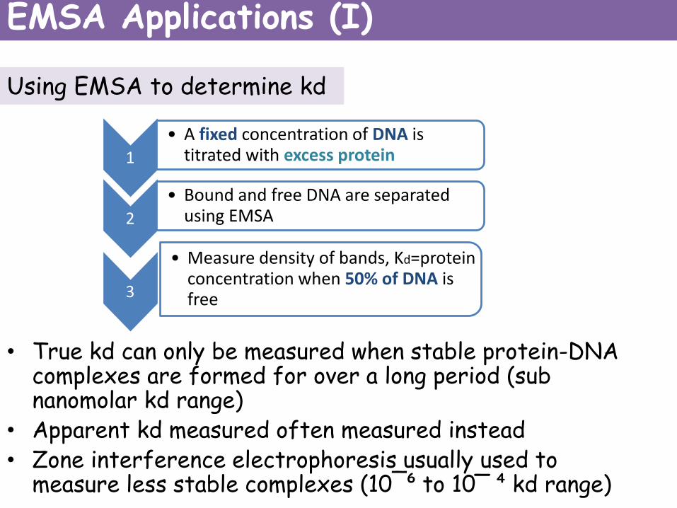
Using EMSA to determine kd
• True kd can only be measured when stable protein-DNA complexes are formed for over a long period (sub nanomolar kd range)
• Apparent kd measured often measured instead• Zone interference electrophoresis usually used to
measure less stable complexes (10¯⁶ to 10‾ ⁴ kd range)
1
• A fixed concentration of DNA is titrated with excess protein
2
• Bound and free DNA are separated using EMSA
3
• Measure density of bands, Kd=protein concentration when 50% of DNA is free
EMSA Applications (I)

Kd evaluation example
• Study of the kd for binding between BV 04-01 and different ssDNAhomopolymers
• BV 04-01= anti-DNA antibody implicated in SLE in mice
• EMSA done to confirm results obtained with other methods
• Showed oligo (T) had best affinity with a kd of ~4.4nM, similar to that obtained using other methods
• In addition, showed that antibody also had a high affinity for oligo (dG) (140nM).
• Helped in the understanding of affinity of the anti-DNA antibody for ssDNA.
Graph of % free oligo (dG) against protein concentration, used to measure Kd. Each point on plot is an average of 3 binding titrations(Stevens et al, 1994).
EMSA Applications (I)
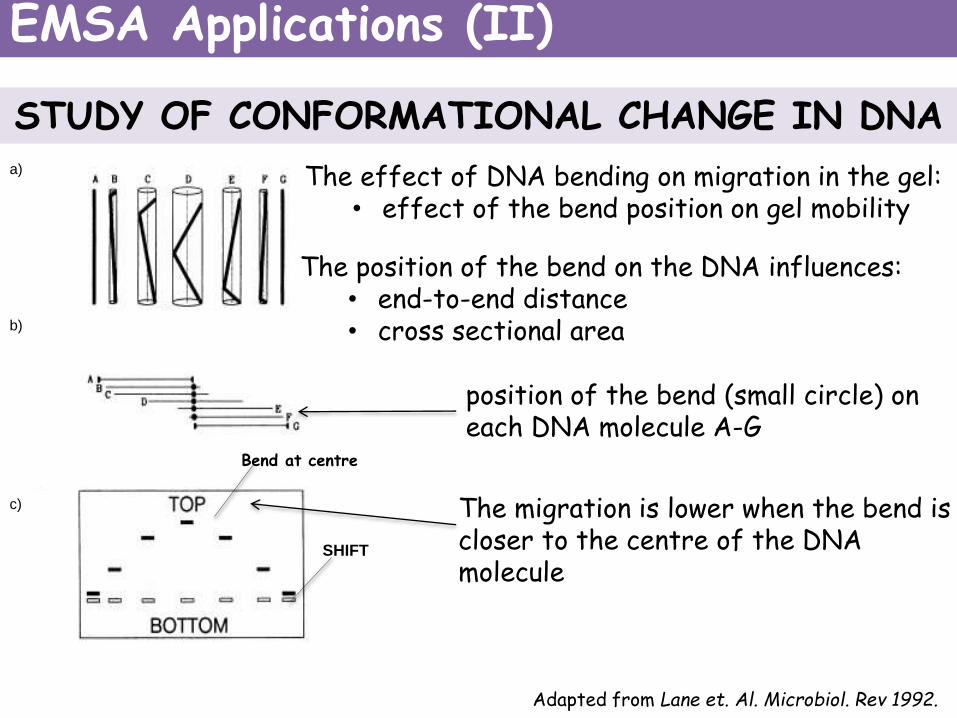
Adapted from Lane et. Al. Microbiol. Rev 1992.
SHIFT
a)
b)
c)
Bend at centre
STUDY OF CONFORMATIONAL CHANGE IN DNA
The effect of DNA bending on migration in the gel:• effect of the bend position on gel mobility
The position of the bend on the DNA influences: • end-to-end distance• cross sectional area
The migration is lower when the bend is closer to the centre of the DNA molecule
position of the bend (small circle) on each DNA molecule A-G
EMSA Applications (II)

Example of a circular permutation polyacrylamide gel retardation assay (1)
• Studies the bending of DNA by p53DBD protein (human p53) by binding to several natural occurring response elements
• Oligonucleotides used contain a 20bp response element within a 30mer oligonucleotide.
-cloned into DNA vector pBend3 using XbaI and SalI restriction sites
• Recombinant plasmids are digested by various restriction enzymes
-p53 response element at increasing distances across the DNA molecule.
• Fragments are 32P-radiolabelled.Adapted from Nagaich et al. JBC, 1997
EMSA Applications (II)

• EMSA is used for the p53DBD-DNA complexes.
• Electrophoretic mobility of the complex is dependent upon the position of p53DBD on the DNA
Example of a circular permutation polyacrylamide gel retardation assay (2)
c) d)
Adapted from Nagaich et al. JBC, 1997
Restriction enzyme Restriction enzyme
• Bending angles can be determined:
μM= mobility of complex when binding site is at the middle of DNA
μE=mobility of complex when binding site is at the end of DNA
α=angle of induced bend
k= coeffecient
EMSA Applications (II)
Advantages and Limitations
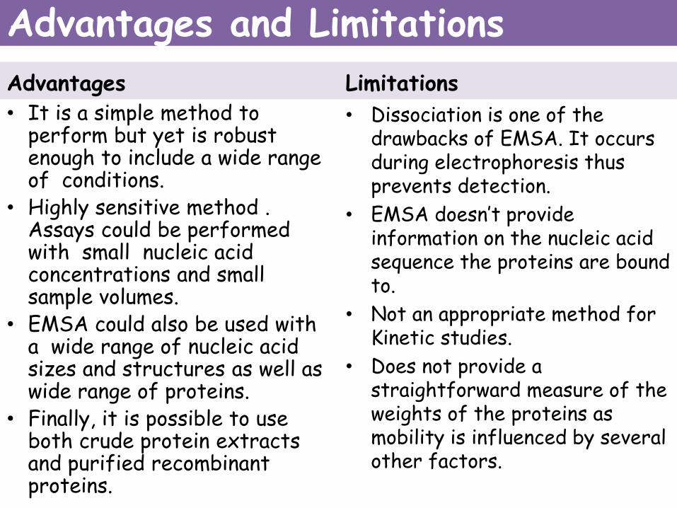
Advantages
• It is a simple method to perform but yet is robust enough to include a wide range of conditions.
• Highly sensitive method . Assays could be performed with small nucleic acid concentrations and small sample volumes.
• EMSA could also be used with a wide range of nucleic acid sizes and structures as well as wide range of proteins.
• Finally, it is possible to use both crude protein extracts and purified recombinant proteins.
Limitations
• Dissociation is one of the drawbacks of EMSA. It occurs during electrophoresis thus prevents detection.
• EMSA doesn’t provide information on the nucleic acid sequence the proteins are bound to.
• Not an appropriate method for Kinetic studies.
• Does not provide a straightforward measure of the weights of the proteins as mobility is influenced by several other factors.
Conclusion
• Electrophoretic Mobility Shift Assay (EMSA) is the most widely used method for the detection of protein-DNA interactions.
• Works on the observation that protein-bound DNA migrate slowly as compared to free DNA when subjected through electrophoresis through a non-denaturing gel.
• Used for various purposes such as quantifying interactions between proteins and DNA, determination of binding affinities but most importantly in the characterization of Transcription Factors.
• There are several alternatives to EMSA which include Foot printing, Yeast hybrid systems, etc.

• Alves, C. and C. Cunha, Gel Electrophoresis - Advanced Techniques. Electrophoretic Mobility Shift Assay: Analyzing Protein - Nucleic Acid Interactions, ed. M. Sameh. 2012: InTech. 500.
• Hellman, L.M. and M.G. Fried, Electrophoretic mobility shift assay (EMSA) for detecting protein-nucleic acid interactions. Nat. Protocols, 2007. 2(8): p. 1849-1861.
• Lane, D., Prentki, P. and Chandler, M. (1992) 'Use of gel retardation to analyze protein-nucleic acid interactions', Microbiological Reviews, 56(4), pp. 509-528.
• Nagaich, A.K., Appella, E. and Harrington, R.E. (1997) 'DNA Bending Is Essential for the Site-specific Recognition of DNA Response Elements by the DNA Binding Domain of the Tumor Suppressor Protein p53', Journal of Biological Chemistry, 272(23), pp. 14842-14849.
References