electron-microscope observations on the effects of localized crush
TRANSCRIPT

J. Anat. (1968), 103, 2, pp. 233-243 233With 1O figuresPrinted in Great Britain
Electron-microscope observations on the effects of localized crushinjuries on the connective tissues of peripheral nerve
J. HAFTEK AND P. K. THOMAS
Institute of Neurology, Queen Square, London, W.C. 1
Peripheral nerve injuries have been classified (Seddon, 1943) into those that pro-duce a localized conduction block (neurapraxia), those that give rise to interruptionof the axons without severance of the nerve (axonotmesis), and those in which there istransection of the nerve trunk (neurotmesis). A convenient way of producing axonot-mesis experimentally is by crushing a nerve with smooth-tipped forceps. Functionalrecovery after injuries of this type is considerably better than after suture followingtransection of the nerve and this is also true of the degree to which restitution of thediameter of the fibres in the distal stump takes place (Gutmann & Sanders, 1943).Moreover, the latent period before which axons appear in the distal stump is alsoless (Gutmann, Guttmann, Medawar & Young, 1942). The explanation of these differ-ences is presumably to be found in the conditions existing at the site of injury. Follow-ing section and suture, considerable interlacing of the regenerating axons occurs atthe suture line so that many fibres fail to enter the appropriate endoneurial tubes inthe distal stump. After a crush injury it has been found that the endoneurial sheathspersist in the crushed region, and it was assumed that they provide the pathwaysthat guide the regenerating axons through the injured portion of nerve so that theyreach their former peripheral connexions (Young, 1949).
Electron microscopists in recent years have examined the nature of the connectivetissue sheaths that surround peripheral nerve fibres (Thomas, 1963; Gamble 1964;Gamble & Eames, 1964). The Schwann cells of myelinated nerve fibres are ensheathedby basement membrane that is continuous across the nodes of Ranvier from oneinternodal segment to the next, surrounding which there is a condensation of theendoneurial collagen fibrils. Around the larger fibres this collagen layer can be sub-divided into a narrow inner endoneurial sheath (sheath of Plenk and Laidlaw) withcircularly and obliquely orientated fibrils that are inflected at the nodes of Ranvier,and an outer endoneurial sheath (sheath of Key and Retzius) of longitudinal fibrilsthat are continuous across the nodes without being inflected. Around the smallerfibres, no separation into inner and outer sheaths is discernible, the surroundingcollagen fibrils displaying a predominantly longitudinal orientation. Thomas (1964)found that during Wallerian degeneration the basement membranes that surroundthe Schwann cells persist and it was suggested that they could conveniently betermed 'Schwann tubes'. After breakdown and removal of the degenerating myelinand axons, the tubes become filled by columns of proliferated Schwann cells (bands ofBungner). Shrinkage of the tubes occurs and a zone of collagen fibrils is depositedaround the tubes between the inner endoneurial sheaths and the persisting Schwanncell basement membranes.

234 J. HAFTEK AND P. K. THOMAS
Localized crush injuries oJ peripheral nerveIn the light of these observations, it was considered of interest to re-examine by
electron microscopy the effects of crush injury to peripheral nerve, with particularattention to the alterations in the connective tissue framework of the nerve. A pre-liminary report of the findings has already appeared (Haftek & Thomas, 1967).
METHODS
The observations were made on the sural and peroneal nerves of 10 adult hoodedrats. The lesions were produced by compression for 10 s with smooth-tipped watch-makers' forceps. In six animals the nerves were fixed in situ immediately followingcompression, and after 1 and 2 h respectively in two further animals. Observationswere also made in two animals allowed to survive for 3 d after injury. In order tolocalize the crushed region, immediately following the injury a loop of fine blacksilk (0000000) was inserted through the epineurium alongside the crush using anatraumatic needle. At biopsy, the nerves were fixed by immersion for 3 h at 4°C in1% osmium tetroxide in mammalian Ringer solution, buffered to pH 7-4 withveronal-acetate. After dehydration in graded concentrations of ethanol, some speci-mens were stained with 1% phosphotungstic acid in absolute ethanol for 3 h. Thespecimens were then embedded in Araldite and sections cut with a Porter-Blummicrotome. After collection on carbon-coated grids, the sections from the material nottreated with phosphotungstic acid were stained with 30 aqueous uranyl acetate for5-10 min followed by lead citrate (Venable & Coggeshall, 1965) for 5-10 min. Thematerial was examined with a Siemens Elmiskop I.
RESULTS
Appearances immediately following injuryIn the material fixed immediately after removal of the forceps, the region of the
crush was found to consist mainly of densely packed collagen fibrils and basementmembranes, very little protoplasmic material being present. No intact nerve fibresremained. Figure 1 shows the peripheral portion of the compressed region. A flattenedbut partially expanded 'Schwann tube', defined by the basement membrane (bm)that had surrounded a Schwann cell in the intact nerve, contains only a narrow zoneof cytoplasmic material apparently adherent to its inner surface. Other tubes con-tained small amounts of myelin debris. The endoneurial collagen fibrils between theSchwann tubes were disorganized, being irregularly orientated instead of displayingthe predominantly longitudinal arrangement seen in the normal nerve. Figure 1 alsoshows the perineurium (pn), which is seen as a narrow zone of compacted basementmembranes and collagen fibrils. Epineurial collagen fibrils (ep), recognized by theirlarger diameter, are visible external to the perineurium.The alterations in the portions of nerve on either side of the crushed region were
Fig. 1. Transverse section of sural nerve in region of crush immediately following injury, showinga flattened Schwann tube (S.t.). ep., Epineurium; pn., perineurium; en., endoneurium; b.m.,basement membrane. Phosphotungstic acid stain.Fig. 2. Transverse section of sural nerve adjacent to region of crush immediately followinginjury, showing Schwann tube containing disorganized myelin (my.) and a portion of the axon(ax.). Uranyl acetate and lead citrate stain.
235

J. HAFTEK AND P. K. THOMAS
A. : rT
N N . . =5~~~~~~~~~~~~~~~0
't ~~~ ~~~~~~~i
''f ;' .............. ...w
1 ;, m
236
U .
.,4" 41:;: :.: 'E. N.
"L M"',-r".. ': :..!.-
, ;z.

Localized crush injuries of peripheral nerve 237also examined. The Schwann tubes adjacent to the injured region contained dis-organized myelin and irregular portions of membrane-bounded axoplasm in varyingproportions (Figs. 2, 3). This type of alteration was observed for distances of approxi-mately 2 mm from the lesion. At times Schwann tubes were seen with an axon ofrelatively normal appearance but associated with disrupted myelin. Tubes close tothe injured region tended to contain predominantly myelin debris. The changes weremuch more prominent for the fibres of larger diameter, the smaller myelinatedfibres, especially at the longer intervals from the crush, often appearing normal.The diameter of the Schwann tubes close to the site of the crush was considerablyincreased with a corresponding diminution in the size of the endoneurial spaces,these sometimes being reduced to narrow clefts (Fig. 4). The basement membranes ofthe tubes usually remained intact, although occasionally myelin fragments wereobserved free in the endoneurial spaces indicating that rupture had occurred. Anappearance more commonly encountered is seen in Fig. 4, where the basementmembranes of the expanded Schwann tubes have ruptured in a number of places,adjacent tubes having become confluent. Non-myelinated axons frequently displayeda greatly increased diameter without rupture of the axolemma (Fig. 5). No differenceswere detected between the portions of nerve proximal and distal to the crush.
Appearances at 1-2 h after injuryThe changes in the crushed region at 1 and 2 h after injury were indistinguishable.
The endoneurial spaces had become oedematous, with wide separation of the bundlesof collagen fibrils. In Fig. 6 the Schwann tubes are flattened and contain myelinremnants and other cellular debris. Other fibres, as shown in Fig. 7, had expanded toa circular form. Although often of very irregular contour, no instances were observedin which the basement membranes comprising the Schwann tubes had becomeruptured.
Figure 8 displays the edge of the fascicle, showing the basement membranes ofthe perineurial cells of the fascicle, between which collagen fibrils and cellular debrisare visible. No intact perineurial cells remained in the crushed region.
Appearances at 3 days after injuryAt this stage, the epineurium in the region of the crush showed some infiltration
with leucocytes, but no other conspicuous change was apparent. An ensheathmentwith perineurial cells had been restored. These cells tended to display a greaterdevelopment of the endoplasmic reticulum and of other cytoplasmic organelles thanin the normal nerve. The cells were often not closely apposed to the basement mem-branes, but tight junctions between adjacent cells were evident. Occasional leuco-cytes were also seen within the perineurium.
Fig. 3. Transverse section of sural nerve adjacent to region of crush immediately followinginjury, showing Schwann tube containing portions of axon (ax.) and disorganized myelin(my.). Uranyl acetate and lead citrate stain.Fig. 4. Transverse section of sural nerve closely adjacent to region of crush immediatelyfollowing injury, showing confluence between adjacent Schwann tubes. Phosphotungstic acidstain.

238 J. HAFTEK AND P. K. THOMAS,. /:. { : , *." s s - -...................................................................... .r^<
-sSl'' .,.s<., XX
r eee w ZS '<SI #;
* '..V twP >4fie eXiNk
.:: .. .:.
.: W.i :e
eS;
a x.ax.
I $w0 ivi5''t'''t' ~~~IX't- k '' X '
5 ,_", 0 +"f12 ,F 0' M.i''','~~~~~~~~~~~~~~~~~~~~~~~~~~~~~~~~~~~~~~Z
li

Localized crush injuries ofperipheral nerveThe endoneurial spaces were oedematous, particularly immediately internal to
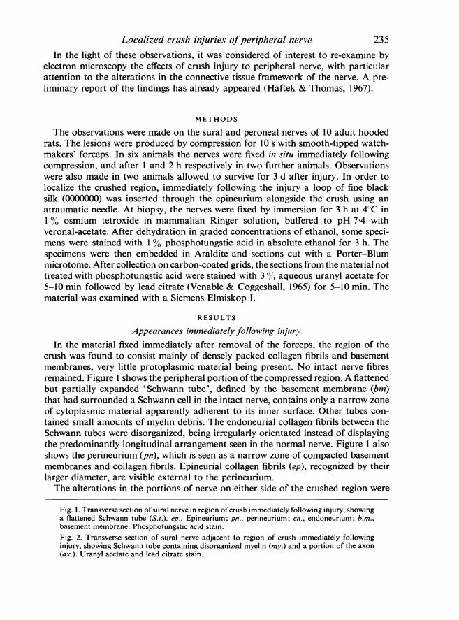
the perineurium and contained leucocytes, macrophages and fibroblasts. The base-ment membranes of the Schwann tubes had persisted. Many of the tubes containeddegenerating axons and myelin, and proliferating Schwann cells. In others (Fig. 9),regenerating axon sprouts were seen in relation to the Schwann cells, either lyingbetween Schwann-cell processes or embedded in Schwann cell cytoplasm but con-nected to the cell surface by mesaxons.The axon sprouts showed appearances similar to those described by Wettstein &
Sotelo (1963), possessing numerous small vesicles, mitochondria and multivesicularbodies, and also densely osmiophilic lamellar bodies as observed in transected axonsadjacent to the site of injury by Bluimcke, Niedorf & Rode (1966).
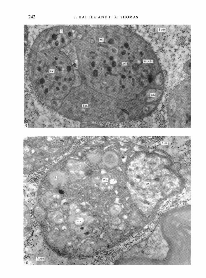
In the portion of the nerve immediately central (1-2 mm) to the crush, the Schwanntubes also contained degenerating myelin, proliferating Schwann cells and axonsprouts. In some tubes, intact axons were present, as shown in Fig. 10. Here thebasement membrane (bm) encloses an axon (ax) and a cell, probably a Schwann cell,within which vacuoles containing myelin figures (my) and lipid (1) are visible. Thenature of the three small cell processes also enclosed within the basement membraneand themselves surrounded by basement membrane is uncertain; thev are probablySchwann-cell processes. Confluence between adjacent Schwann tubes was not ob-served and the basement membranes were everywhere intact, although reduplica-tion was sometimes seen.
Sections taken distal to the site of the crush showed the changes of early Walleriandegeneration as described by a number of previous investigators (for references,see Thomas, 1966).
DISCUSSION
The early changes produced by a localized crush injury to peripheral nerve werestudied in detail using light microscopy by Causey & Palmer (1952), who examinedcrush lesions of rabbit nerve produced by compression with forceps in the samemanner as in the present investigation. They described displacement of myelin andaxonal material from the crushed region into the adjacent portions of the nervewhere they became mixed. The displaced material remained within the 'neurilemmaltubes', which became expanded but remained undamaged. After the pressure on thenerve was released, the mixed myelin and axonal material rapidly flowed back intothe crushed region, re-expanding the neurilemmal tubes.The present investigation has shown that continuity across the compressed region
is maintained by the collagen fibrils of the epineurium, perineurium and endoneurium,and the basement membranes of the perineurial and Schwann cells. The myelin andaxonal material that is displaced out of the crushed region is retained within thebasement membranes that constitute the Schwann tubes. These membranes must
Fig. 5. Transverse section of sural nerve adjacent to region of crush immediately followinginjury, showing expanded non-myelinated axons (ax.). Uranyl acetate and lead citrate stain.Fig. 6. Transverse section of sural nerve in region of crush 2 h after injury, showing flattenedSchwann tubes (S.t.). b.m., Basement membrane; en., endoneurium. Phosphotungstic acidstain.
239

240 J. HAFTEK AND P. K. THOMAS

Localized crush injuries of peripheral niervepossess definite elastic properties as they are capable of withstanding considerabledistension, although rupture sometimes occurs. In the compressed region, theSchwann tubes remain intact and rapidly become filled with the mixed myelin andaxonal material that flows back after the pressure of the forceps is released.
In the nerves examined at 3 days after the crush, Schwann cells had grown into theSchwann tubes in the crushed region and were associated with regenerating axonsprouts. It therefore seems clear that it is these basement membranes that definethe pathways across the damaged portion of nerve, presumably supported by thecollagenous framework of the nerve. The situation is therefore comparable withthat existing in the distal stump of a nerve during Wallerian degeneration, where theSchwann-cell basement membranes define the channels within which the Schwanncells proliferate to give rise to the longitudinally continuous columns of cells pre-paratory to regeneration.A point of some interest in relation to the increasing use of nerve biopsy for
diagnostic purposes in clinical neurology is the fact that adjacent to the injuredregion in the nerves examined in the present investigation, Schwann tubes weresometimes seen with an axon of relatively normal appearance but with disorganiza-tion of the myelin. Such changes may resemble those of a demyelinating neuropathyand emphasize the need for careful removal of biopsy specimens to avoid misinterpret-ing the results of trauma as being indicative of a true pathological change.
SUMMARY
Localized lesions of rat peroneal and sural nerves produced by compression withforceps were examined by electron microscopy. The basement membranes thatsurround the Schwann cells and which constitute the 'Schwann tubes' persist in thecrushed region and remain intact. They become re-expanded when the myelin andaxonal material that is displaced into the adjacent portions of the tubes flows backinto the crushed zone after the compression is released. Schwann cells and axonslater grow into these tubes, which therefore appear to provide the pathways thatguide the regenerating axons across the damaged region.
We wish to thank Miss Ann Armstrong and Miss Theresa Tilley for technicalassistance, Professor Sir Herbert Seddon for helpful discussion, and Professor W. H.McMenemy for laboratory facilities at Maida Vale Hospital. A personal grant toone of us (P. K. T.) from the National Fund for Research into Poliomyelitis andother Crippling Diseases is gratefully acknowledged. Dr J. Haftek held a BritishCouncil Scholarship while on leave from the Medical Academy, Warsaw.
Fig. 7. Transverse section of sural nerve in region of crush 2 h after injury. The Schwann tubes(S.t.) are expanded and contain myelin and other cellular debris. b.m., Basement membrane.Phosphotungstic acid stain.Fig. 8. Transverse section through periphery of sural nerve 2 h after injury. ep., Epineurium;pn., perineurium; en., endoneurium; S.t., Schwann tube; b.m., basement membrane. Phospho-tungstic acid stain.
Anat. 103
241
242 J. HAFTEK AND P. K. THOMAS
i *'m I.,
I~~~~~ ~~mb
ax.

Localized crush injuries ofperipheral nerve 243
REFERENCES
BLUMCKE, S., NIEDORF, H. R. & RODE, J. (1966). Axoplasmic alterations in the proximal and distal stumpsof transected nerves. Acta Neuropath. 7, 44 61.
CAUSEY, G. & PALMER, E. (1952). Early changes in degenerating mammalian nerves. Proc. R. Soc. B139, 597-609.
GAMBLE, H. J. (1964). Comparative electron-microscopic observations on the connective tissue of aperipheral nerve and a spinal nerve root in the rat. J. Anat. 98, 17-25.
GAMBLE, H. J. & EAMES, R. A. (1964). An electron microscopic study of the connective tissues of humanperipheral nerve. J. Anat. 98, 655-663.
GUTMANN, E., GUTEMAN, L., MEDAWAR, P. B. & YOUNG, J. Z. (1942). The rate of regeneration of nerve.J. exp. Biol. 19, 14 44.
GUTMANN, E. & SANDERS, F. K. (1943). Recovery of fibre numbers and diameters in the regeneration ofperipheral nerves. J. Physiol., Lond. 101, 429-518.
HAFTEK, J. & THOMAS, P. K. (1967). An electron microscope study of crush injuries to peripheral nerve.J. Anat. (in the Press).
SEDDON, H. J. (1943). Three types of nerve injury. Brain 66, 237-288.THOMAS, P. K. (1963). The connective tissue of peripheral nerve: an electron microscope study. J. Anat.
97, 35-44.THOMAS, P. K. (1964). Changes in the endoneurial sheaths of peripheral myelinated nerve fibres during
Wallerian degeneration. J. Anat. 98, 175-182.THOMAS, P. K. (1966). The cellular response to nerve injury. 1. The cellular outgrowth from the distalstump of transected nerve. J. Anat. 100, 287-303.
VENABLE, J. H. & COGGESHALL, R. (1965). A simplified lead citrate stain for use in electron microscopy.J. Cell Biol. 25, 407-408.
WETTSTEIN, R. & SOTELO, J. R. (1963). Electron microscope study on the regenerative process ofperipheral nerves of mice. Z. Zellforsch. mikrosk. Anat. 59, 708-740.
YOUNG, J. Z. (1949). Factors influencing the regeneration of nerves. Adv. Surg. 1, 165-220.
Fig. 9. Transverse section through peroneal nerve in region of crush 3 days after injury, showinga Schwann tube containing Schwann-cell processes (S.p.) together with regenerating axonsprouts (ax.) within which there are mitochondria (m.), lamellar bodies (I.) and multivesicularbodies (m.v.b.). Phosphotungstic acid stain.Fig. 10. Transverse section through peroneal nerve just above level of crush 3 days after injury,showing a Schwann tube within which there is a demyelinated axon (ax.) and part of a cell thatcontains degenerating myelin (my.) and lipid (1.). b.m., Basement membrane. Phosphotungsticacid stain.
I6-2