eie article - cardioldepartamentos.cardiol.br/dic/publicacoes/revistadic/revista/2015/... · eie...
TRANSCRIPT

80
Review Article
Morphological and Functional Echocardiographic Evaluation of Patient with Pulmonary HypertensionGeórgia Macário Rocha1, José Maria Del Castillo2
Instituto Politécnico de Lisboa, Escola Superior de Tecnologia da Saúde de Lisboa1, Lisboa - Portugal; Pronto Socorro Cardiológico Universitário de Pernambuco Professor Luiz Tavares (PROCAPE) - Universidade de Pernambuco (UPE)2, Recife, PE - Brazil
AbstractPulmonary hypertension is a serious multifactorial and
multidisciplinary clinical syndrome with great damage to the quality of life of patients and high morbidity and mortality. Echocardiography is the main test used for screening pulmonary hypertension due to its easy access and for being non-invasive. This work aims to analyze the application of echocardiography in the morphological and functional evaluation of the heart of patients with pulmonary hypertension.
IntroductionPulmonary hypertension (PH) is the increase in mean
pulmonary arterial pressure (MPAP) ≥ 25 mmHg at rest, measured by right heart catheterization (RHC). Normal MPAP was estimated at 14 ± 3 mmHg, 20 mmHg1 being its maximum value.
PH is characterized by hemodynamic and pathophysiological abnormalities, becoming a complex syndrome of difficult diagnosis and treatment and often poor prognosis2-5.
Patients with MPAP ≥ 25 mmHg are diagnosed with PH and, after that determination, it must be defined whether PH is pre-capillary or post-capillary.
If the pulmonary capillary wedge pressure (PCWP) is ≤ 15 mmHg, PH is said to be pre-capillary, including pulmonary arterial hypertension in the absence of other causes (group 1), PH of pulmonary origin (group 3), chronic pulmonary thromboembolism (group 4) and PH of unclear cause or due to multifactorial factors (group 5).
If the PCWP is > 15 mmHg, PH is the post-capillary type, originating from left heart diseases (group 2), where the Transpulmonary Pressure Gradient (TPG) must be determined by the following formula6:
TPG = MPAP – average PCWP
When this difference is ≤12 mm Hg, the increased pulmonary artery pressure (PAP) is said to be passive, that is, it is caused exclusively by cardiac involvement. If the TPG is > 12 mmHg, increase in PAP is disproportionate to the increase of pressure in the left ventricle (LV), therefore, there is pulmonary vascular remodeling or other causes related to increased PAP.
The definition of HP in exercise as MPAP > 30 mmHg, measured by RHC, has not been validated by data published2.
In patients with congenital heart diseases, several factors should be evaluated: defect type, size, magnitude of flow, risk factors, previous surgeries and extracardiac abnormalities associated7.
All of these factors significantly influence the development of PH and the evolution to an extreme condition called the Eisenmenger’s syndrome, in which the pulmonary vascular resistance becomes greater than the systemic resistance and the flow through defect is inverted irreversibly. This condition has a high morbidity due to hypoxemia and various secondary hematological alterations4,5.
DiagnosisPatients with dyspnea on exertion, chest pain, dizziness
and/or syncope and signs of right heart failure without an evident cause should be evaluated for PH investigation8. Symptoms at rest only occur in advanced cases.
Several additional tests (with high sensitivity and specificity spectrum) can be used for the initial evaluation of these patients, including electrocardiogram (ECG), echocardiogram (ECHO), pulmonary function testing, high resolution computed tomography (HRCT) and pulmonary angiography. If the result of the tests is not compatible with PH, other causes should be looked for. If this data is compatible with PH, it should be determined whether the origin is cardiac or pulmonary and if the HP is proportional to the severity of the disease.
The ECG shows signs of right ventricular hypertrophy in 87% of cases, but the absence of these signs does not exclude PH. Its sensitivity (55%) and specificity (70%) are considered low for the evaluation of patients suspected of having HP2.
Chest radiology is abnormal in 90% of patients with PH, showing dilatation of the pulmonary artery and decreased peripheral pulmonary vasculature, but in general, this data does not correlate with the degree of PH8.
Pulmonary function tests and arterial blood gas analysis can identify parenchymal lung diseases that cause PH, such as chronic obstructive pulmonary disease.
KeywordsPulmonary hypertension; Echocardiography/use;
Diagnost ic techniques and procedures; Cardiac Catheterization; Heart Ventricles.
Mailing address: José Maria Del Castillo •Rua Jorge de Lima, 245, apto. 303, Postal Code 51160-070, Salute, Imbiribeira, Recife, PE - Brazil E-mail: [email protected] received on December 3, 2014; revised on February 16, 2014; accepted on March 1, 2015.
DOI: 10.5935/2318-8219.20150016
81
Review Article
Del Castillo et al.Echocardiographic Assessment of Pulmonary Hypertension
Arq Bras Cardiol: Imagem cardiovasc. 2015;28(2):80-88
Pulmonary scintigraphy of ventilation/perfusion can be performed with patients with PH suspected of having chronic pulmonary thromboembolism, detecting this disease with high sensitivity and specificity9.
HRCT provides detailed images of the lung parenchyma and helps diagnosing interstitial pulmonary disease and emphysema and is very useful when venous occlusive disease is suspected10. Computed tomography with contrast is recommended to investigate PH by chronic pulmonary thromboembolism.
Traditional lung angiography with contrast can be used to diagnose patients with pulmonary embolism susceptible to pulmonary endarterectomy11. RHC is important to determine the pulmonary artery pressures and pulmonary wedge pressure, allowing differentiating precapillary from post-capillary PH. Pulmonary vasoreactivity can be tested with inhalation of nitric oxide or infusion of adenosine or epoprostenol12.
Echocardiography in the morphological and functional assessment of the heart of patients with pulmonary hypertension
Echocardiography is the main test used for screening PH because it is easily accessible, noninvasive, and allows the diagnosis of heart defects and diseases.
The main limitations are the fact that this method is dependent and technically inadequate in some patients.
Methodology of echocardiographic studyThe echocardiographic study can be systematized in three
sequential steps5:
A) Study of the right heart
Evaluation of the size of the right cavities, determination of right ventricular function (RVF), calculation of pulmonary artery systolic pressure (PASP) and MPAP.
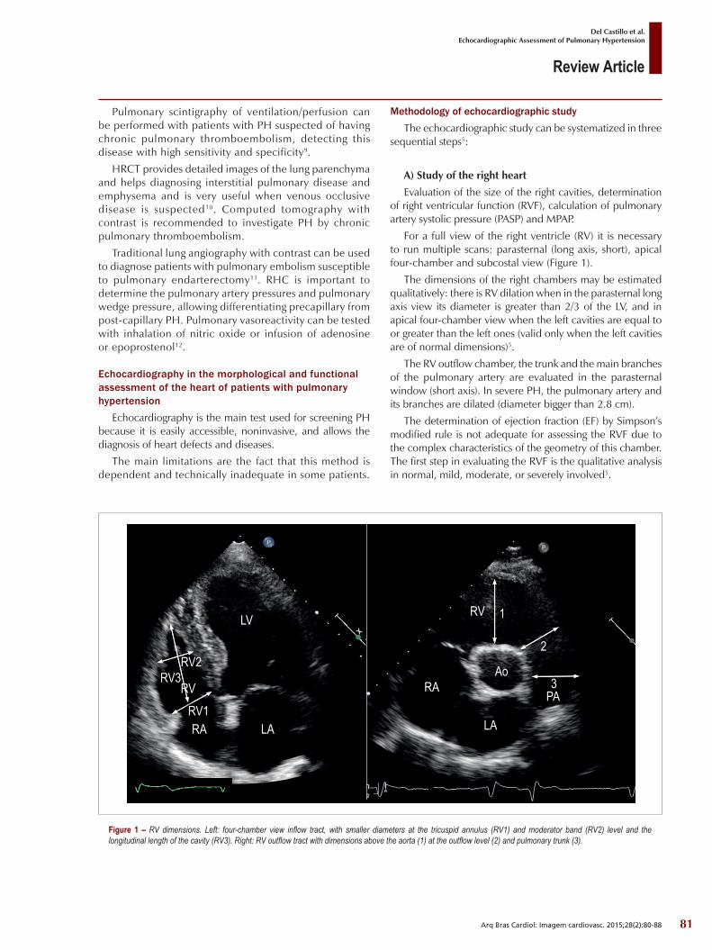
For a full view of the right ventricle (RV) it is necessary to run multiple scans: parasternal (long axis, short), apical four-chamber and subcostal view (Figure 1).
The dimensions of the right chambers may be estimated qualitatively: there is RV dilation when in the parasternal long axis view its diameter is greater than 2/3 of the LV, and in apical four-chamber view when the left cavities are equal to or greater than the left ones (valid only when the left cavities are of normal dimensions)5.
The RV outflow chamber, the trunk and the main branches of the pulmonary artery are evaluated in the parasternal window (short axis). In severe PH, the pulmonary artery and its branches are dilated (diameter bigger than 2.8 cm).
The determination of ejection fraction (EF) by Simpson’s modified rule is not adequate for assessing the RVF due to the complex characteristics of the geometry of this chamber. The first step in evaluating the RVF is the qualitative analysis in normal, mild, moderate, or severely involved5.
Figure 1 – RV dimensions. Left: four-chamber view inflow tract, with smaller diameters at the tricuspid annulus (RV1) and moderator band (RV2) level and the longitudinal length of the cavity (RV3). Right: RV outflow tract with dimensions above the aorta (1) at the outflow level (2) and pulmonary trunk (3).
LV
LA LA
Ao
PA
1
2
3
RA
RA
RV1
RV2RV3
RV
RV
82
Review Article
Del Castillo et al.Echocardiographic Assessment of Pulmonary Hypertension
Arq Bras Cardiol: Imagem cardiovasc. 2015;28(2):80-88
Pulmonary circulation with chronically high pressures leads to dilation of the right cavities followed by right ventricular hypertrophy and, subsequently, chamber dysfunction.
The interventr icular septum may be f lattened, with abnormal movement or hypertrophied, with an interventricular septum/posterior wall ratio > 1.
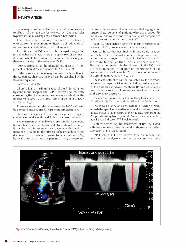
The estimated PASP depends on the tricuspid regurgitation jet and right atrial pressure (RAP). In up to 10% of the cases, it is not possible to measure the tricuspid insufficiency jet, therefore preventing the estimate of PASP3.
PASP is estimated by the tricuspid insufficiency (TI) jet, present in about 80% of patients with PH (Figure 2).
In the absence of pulmonary stenosis or obstruction in the RV outflow chamber, the PASP can be calculated by the Bernoulli equation:
PASP = 4 . V² + RAPwhere V is the maximum speed of the TI jet obtained
by continuous Doppler and RAP is determined indirectly considering the diameter and respiratory variability of the inferior vena cava (IVC)13. The normal upper limit of PASP is 37.2 mmHg5.
There is a strong correlation between the PASP estimated by echocardiography and by right heart catheterization.
However, the significant number of false positives requires confirmation of diagnosis by right heart catheterization1-5.
The measurement of pulmonary pressure during exercise has not been validated by clinical observations2, although it may be used in asymptomatic patients with functional mitral regurgitation for the purposes of taking a therapeutic decision. PH is unusual in asymptomatic patients (9%), but was observed in 58% of patients during exercise and
is a major determinant of events after mitral regurgitation surgery. Sixty percent of patients who experienced PH during exercise were event-free in five years compared to 88% of patients who did not have PH14.
As the RV function has a significant role in the prognosis of patients with PH, proper evaluation is necessary.
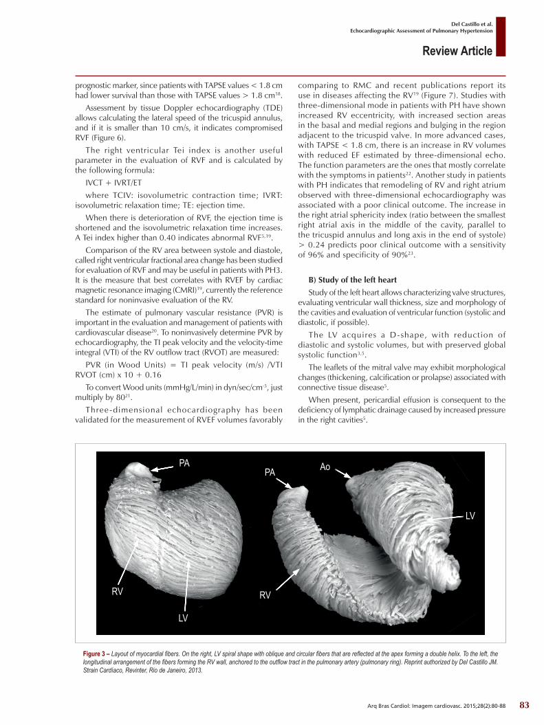
Unlike the LV that has thick walls and conical shape, the RV has thin walls and semilunar shape (or crescent moon shape). Its myocardial mass is significantly smaller and more trabecular than the LV myocardial mass. The contraction pattern is also different: in the RV, there is a predominance of longitudinal contraction of the myocardial fibers, while in the LV, there is a predominance of a spiraling movement15 (Figure 3).
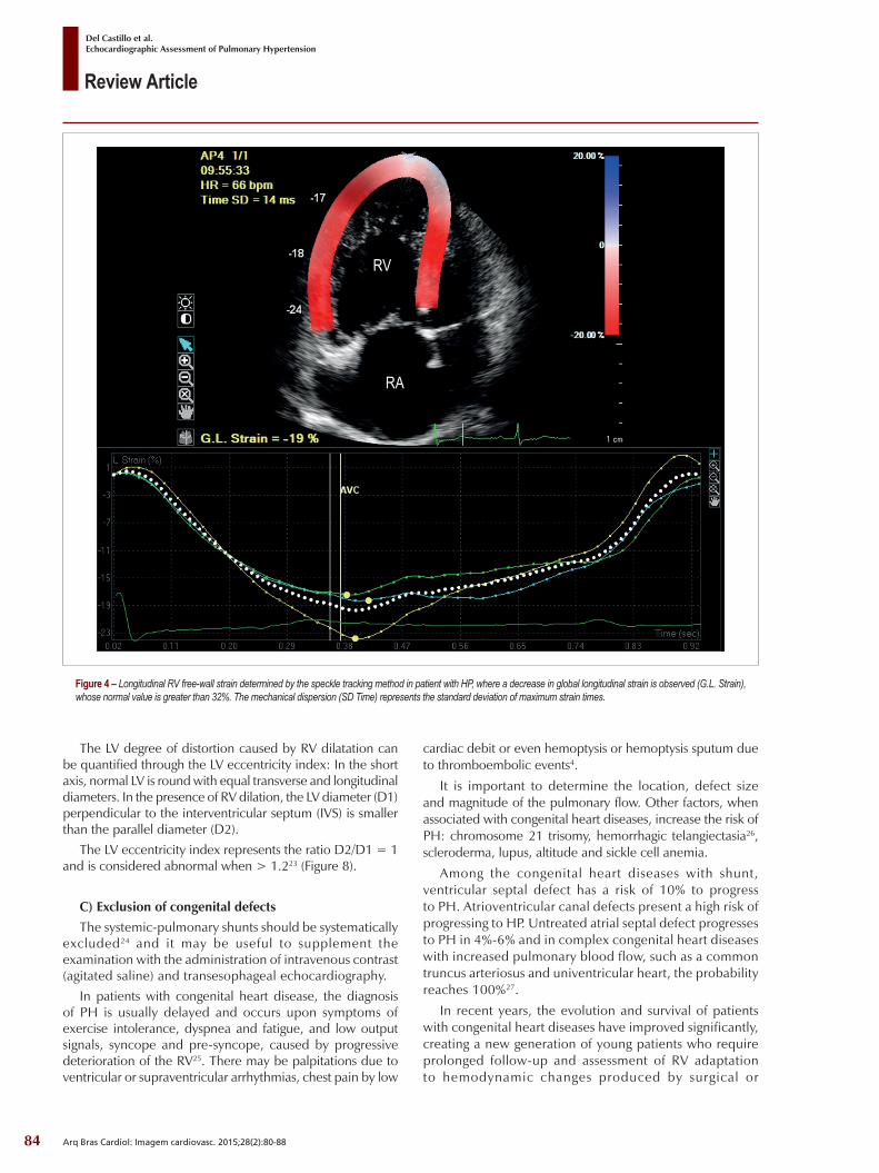
These characteristics can be evaluated by the methods that measure myocardial strain, including cardiac strain16. For the purposes of measurement, the RV free wall strain is used, since the septal wall presents strain values influenced by the LV strain (Figure 4).
The reference values for LV free wall longitudinal strain are -32.5% ± 5.1% for males and -34.6% ± 5.3% for females17.
The tricuspid annular plane systolic excursion (TAPSE) towards the apex has proved to be a good technique to assess the RV. TAPSE is the measure of the ring excursion from the RV apex during systole (Figure 5). An excursion smaller less than 1.5 cm indicates RVF involvement5.
A study comparing the assessment of RVF by TAPSE with measurements taken on the RHC showed an excellent correlation of the values found.
TAPSE values < 1.8 cm showed good accuracy for the detection of RV dysfunction and were considered as a
Tricuspid valve regurgitation
Jet velocity (V)
PASP = 4. V2 + RAP
75mm/s ***bpm
-6.0-5.0-4.0
-3.0-2.0
-1.0
1.0m/s
cm/s
RA
RV
Figure 2 – Determination of Pulmonary Artery Systolic Pressure (PASP) by the tricuspid regurgitation jet velocity.
83
Review Article
Del Castillo et al.Echocardiographic Assessment of Pulmonary Hypertension
Arq Bras Cardiol: Imagem cardiovasc. 2015;28(2):80-88
prognostic marker, since patients with TAPSE values < 1.8 cm had lower survival than those with TAPSE values > 1.8 cm18.
Assessment by tissue Doppler echocardiography (TDE) allows calculating the lateral speed of the tricuspid annulus, and if it is smaller than 10 cm/s, it indicates compromised RVF (Figure 6).
The right ventricular Tei index is another useful parameter in the evaluation of RVF and is calculated by the following formula:
IVCT + IVRT/ETwhere TCIV: isovolumetric contraction time; IVRT:
isovolumetric relaxation time; TE: ejection time.When there is deterioration of RVF, the ejection time is
shortened and the isovolumetric relaxation time increases. A Tei index higher than 0.40 indicates abnormal RVF5,19.
Comparison of the RV area between systole and diastole, called right ventricular fractional area change has been studied for evaluation of RVF and may be useful in patients with PH3. It is the measure that best correlates with RVEF by cardiac magnetic resonance imaging (CMRI)19, currently the reference standard for noninvasive evaluation of the RV.
The estimate of pulmonary vascular resistance (PVR) is important in the evaluation and management of patients with cardiovascular disease20. To noninvasively determine PVR by echocardiography, the TI peak velocity and the velocity-time integral (VTI) of the RV outflow tract (RVOT) are measured:
PVR (in Wood Units) = TI peak velocity (m/s) /VTI RVOT (cm) x 10 + 0.16
To convert Wood units (mmHg/L/min) in dyn/sec/cm-5, just multiply by 8021.
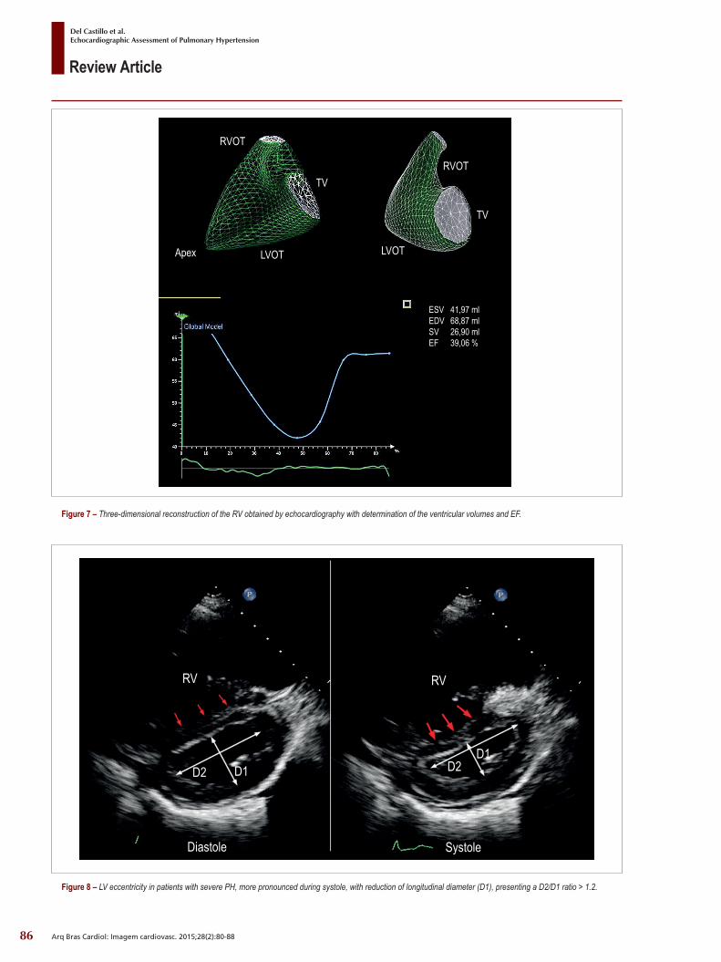
Three-dimensional echocardiography has been validated for the measurement of RVEF volumes favorably
comparing to RMC and recent publications report its use in diseases affecting the RV19 (Figure 7). Studies with three-dimensional mode in patients with PH have shown increased RV eccentricity, with increased section areas in the basal and medial regions and bulging in the region adjacent to the tricuspid valve. In more advanced cases, with TAPSE < 1.8 cm, there is an increase in RV volumes with reduced EF estimated by three-dimensional echo. The function parameters are the ones that mostly correlate with the symptoms in patients22. Another study in patients with PH indicates that remodeling of RV and right atrium observed with three-dimensional echocardiography was associated with a poor clinical outcome. The increase in the right atrial sphericity index (ratio between the smallest right atrial axis in the middle of the cavity, parallel to the tricuspid annulus and long axis in the end of systole) > 0.24 predicts poor clinical outcome with a sensitivity of 96% and specificity of 90%23.
B) Study of the left heartStudy of the left heart allows characterizing valve structures,
evaluating ventricular wall thickness, size and morphology of the cavities and evaluation of ventricular function (systolic and diastolic, if possible).
The LV acquires a D-shape, with reduction of diastolic and systolic volumes, but with preserved global systolic function3,5.
The leaflets of the mitral valve may exhibit morphological changes (thickening, calcification or prolapse) associated with connective tissue disease5.
When present, pericardial effusion is consequent to the deficiency of lymphatic drainage caused by increased pressure in the right cavities5.
Figure 3 – Layout of myocardial fibers. On the right, LV spiral shape with oblique and circular fibers that are reflected at the apex forming a double helix. To the left, the longitudinal arrangement of the fibers forming the RV wall, anchored to the outflow tract in the pulmonary artery (pulmonary ring). Reprint authorized by Del Castillo JM. Strain Cardíaco, Revinter, Rio de Janeiro, 2013.
RV RV
LV
LV
PAPA Ao
84
Review Article
Del Castillo et al.Echocardiographic Assessment of Pulmonary Hypertension
Arq Bras Cardiol: Imagem cardiovasc. 2015;28(2):80-88
Figure 4 – Longitudinal RV free-wall strain determined by the speckle tracking method in patient with HP, where a decrease in global longitudinal strain is observed (G.L. Strain), whose normal value is greater than 32%. The mechanical dispersion (SD Time) represents the standard deviation of maximum strain times.
RV
RA
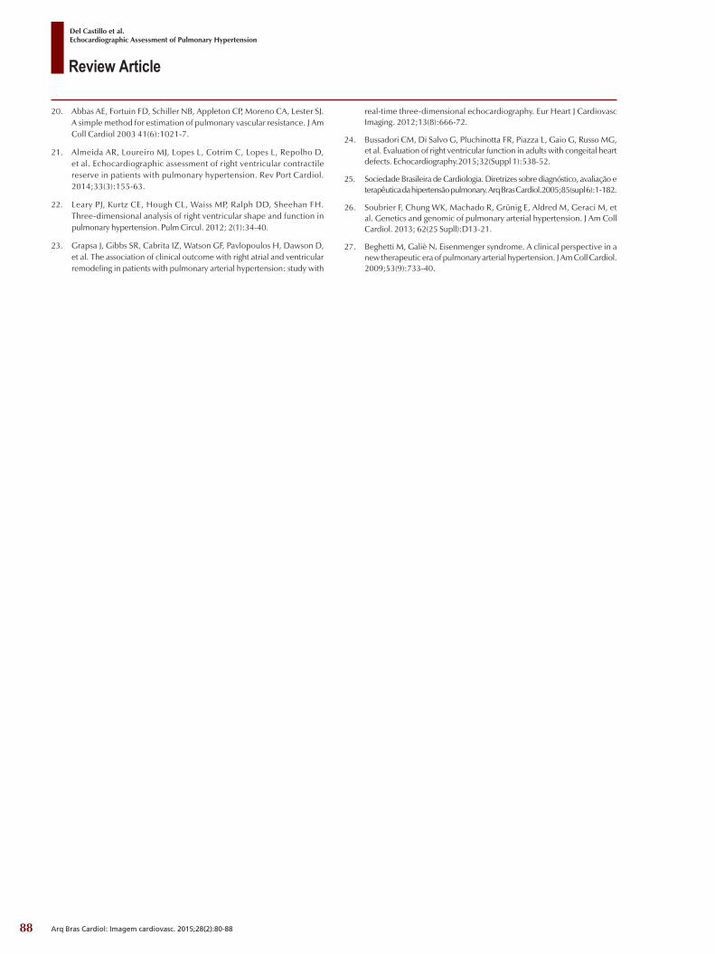
The LV degree of distortion caused by RV dilatation can be quantified through the LV eccentricity index: In the short axis, normal LV is round with equal transverse and longitudinal diameters. In the presence of RV dilation, the LV diameter (D1) perpendicular to the interventricular septum (IVS) is smaller than the parallel diameter (D2).
The LV eccentricity index represents the ratio D2/D1 = 1 and is considered abnormal when > 1.223 (Figure 8).
C) Exclusion of congenital defectsThe systemic-pulmonary shunts should be systematically
excluded24 and it may be useful to supplement the examination with the administration of intravenous contrast (agitated saline) and transesophageal echocardiography.
In patients with congenital heart disease, the diagnosis of PH is usually delayed and occurs upon symptoms of exercise intolerance, dyspnea and fatigue, and low output signals, syncope and pre-syncope, caused by progressive deterioration of the RV25. There may be palpitations due to ventricular or supraventricular arrhythmias, chest pain by low
cardiac debit or even hemoptysis or hemoptysis sputum due to thromboembolic events4.
It is important to determine the location, defect size and magnitude of the pulmonary flow. Other factors, when associated with congenital heart diseases, increase the risk of PH: chromosome 21 trisomy, hemorrhagic telangiectasia26, scleroderma, lupus, altitude and sickle cell anemia.
Among the congenital heart diseases with shunt, ventricular septal defect has a risk of 10% to progress to PH. Atrioventricular canal defects present a high risk of progressing to HP. Untreated atrial septal defect progresses to PH in 4%-6% and in complex congenital heart diseases with increased pulmonary blood flow, such as a common truncus arteriosus and univentricular heart, the probability reaches 100%27.
In recent years, the evolution and survival of patients with congenital heart diseases have improved significantly, creating a new generation of young patients who require prolonged follow-up and assessment of RV adaptation to hemodynamic changes produced by surgical or

85
Review Article
Del Castillo et al.Echocardiographic Assessment of Pulmonary Hypertension
Arq Bras Cardiol: Imagem cardiovasc. 2015;28(2):80-88
Figure 5 – Determination of the tricuspid annular plane systolic excursion (TAPSE) using M-mode echocardiography.
M-mode line
TAPSE
Figure 6 – Tissue Doppler (TD) showing the lateral velocity of the tricuspid ring. S’: systolic velocity; E’: initial diastolic velocity; A’: end-diastolic velocity.
Sample volume
115 bpm
86
Review Article
Del Castillo et al.Echocardiographic Assessment of Pulmonary Hypertension
Arq Bras Cardiol: Imagem cardiovasc. 2015;28(2):80-88
Figure 8 – LV eccentricity in patients with severe PH, more pronounced during systole, with reduction of longitudinal diameter (D1), presenting a D2/D1 ratio > 1.2.
RV RV
D2
Diastole Systole
D2D1D1
Figure 7 – Three-dimensional reconstruction of the RV obtained by echocardiography with determination of the ventricular volumes and EF.
RVOT
RVOT
TV
TV
Apex
ESV 41,97 mlEDV 68,87 mlSV 26,90 mlEF 39,06 %
LVOT LVOT

87
Review Article
Del Castillo et al.Echocardiographic Assessment of Pulmonary Hypertension
Arq Bras Cardiol: Imagem cardiovasc. 2015;28(2):80-88
interventional procedures24. Operated patients with congenital heart diseases have reduced the risk of severe PH and Eisenmenger syndrome, but some cases, especially when the heart disease presented a large pulmonary blood flow, should be carefully evaluated as they may evolve to obstructive pulmonary vascular disease.
ConclusionPulmonary hypertension is a severe progressive clinical
syndrome that may complicate many systemic, valvular or congenital diseases, or can be idiopathic, causing high morbidity and mortality. Diagnosis of PH must be done through a number of tests and examinations, invasive or not, among which echocardiography plays an important role, being the main method used for screening. Its confirmation diagnosis requires, however, right heart catheterization.
Recently, with the introduction of tissue Doppler, cardiac strain and three-dimensional echocardiography, there have been significant advances in morphological and functional
assessment of the RV, making echocardiography an important tool for the exclusion of pulmonary hypertension.
Authors’ contributionsResearch creation and design: Rocha GM; Data acquisition:
Rocha GM; Data analysis and interpretation: Rocha GM, Del Castillo JM; Manuscript drafting: Rocha GM, Del Castillo JM; Critical revision of the manuscript as for important intellectual content: Del Castillo JM.
Potential Conflicts of InterestNo relevant potential conflicts of interest.
Sources of FundingThis study had no external funding sources.
Academic AssociationThis study is not associated with any graduate program.
1. Badesch BD, Champion HC, Gomez-Sanchez MA, Hoeper M, Loyd J, Manes A, et al. Diagnosis and assessment of pulmonary arterial hypertension. J Am Coll Cardiol 2009; 54(1 Suppl):S55-S56.
2. Galiè N, Hoeper MM, Humbert M, Torbicki A, Vachiéry JL, Barbera J,et al. Guidelines for the diagnosis and treatment of pulmonary hypertension. The Task force for diagnosis and treatment of pulmonary hypertension of European Society of Cardiology (ESC) and the European Respiratory Society (ERS), endorsed by the International Society of Heart and Lung Transplantation (ISHLT). Eur Heart J. 2009;30(20):2493-537.
3. Hoette S, Jardim C, Souza R. Diagnóstico e tratamento da hipertensão pulmonar: uma atualização. J Bras Pneumol. 2010; 36(6):795-811.
4. Pfeiffer ME. Hipertensão arterial pulmonar: abordagem clínica, diagnóstica e avaliação funcional. Rev DERC. 2014; 20(1):10-1.
5. Reis A, Rocha N, Barros R, Martins A, Oliveira F, Diogo AN, et al. Recomendações para a abordagem clinica do doente com hipertensão pulmonar. Rev Port Pneumol. 2010;16(4):S7-S85.
6. Naeije R, Vachiery JL, Yerly P, Vanderpool R. The transpulmonary pressure gradient for the diagnosis of pulmonary vascular disease. Eur Respir J. 2013; 41(1):217-23.
7. Galiè N, Manes A, Palazzini M, Negro L, Marinelli A, Gambetti S, et al. Management of pulmonary arterial hypertension associated with congenital systemic-to-pulmonary shunts and Eisenmenger’s syndrome. Drugs. 2008;68(8):1049-66.
8. Rich S, Dantzker DR, Ayres SM, Bergofsky EH, Brundage BH, Detre KM, et al. Primary pulmonary hypertension. A national prospective study. Ann Intern Med 1987; 107(2):216-23.
9. Tunariu N, Gibbs SJR, Win Z, Gin-Sing W, Graham A, Gishen P, et al. Ventilation-perfusion scintigraphy is more sensitive than multidetector CTPA in detecting chronic thromboembolic pulmonary disease as a treatable cause of pulmonary hypertension. J Nucl Med. 2007;48(5):680-4.
10. Resten A, Maitre S, Humbert M, Rabiller A, Sitbon O, Capron F, et al. Pulmonary hypertension: CT of the chest in pulmonary venoocclusive disease. Am J Roentgenol. 2004;183(1):65-70.
11. Fedullo PF, Auger WR, Kerr KM, Rubin LJ. Chronic thromboembolic pulmonary hypertension. N Engl J Med. 2001; 345(20):1465-72.
12. Galiè N, Ussia G, Passarelli P, Parlangeli R, Branzi A, Magnani B. Role of pharmacologic tests in the treatment of primary pulmonary hypertension. Am J Cardiol. 1995;75(3):55A-6A.
13. Rudski LG, La i WW, Af i la lo J, Hua L, Handschumacher LD, Chandrasekaran K, et al. Guidelines for the Echocardiographic Assessment of the Right Heart in Adults: A Report from the American Society of Echocardiography Endorsed by the European Association of Echocardiography, a registered branch of the European Society of Cardiology, and the Canadian Society of Echocardiography. J Am Soc Echocardiogr. 2010;23(7):685-713.
14. Magne J, Donal E, Mahjoub H, Miltner B, Dulgheru R, Thebault C, et al. Impact of exercise pulmonary hypertension on postoperative outcome in primary mitral regurgitation. Heart.2015;101(5):391-6.
15. Stoeck CT, Kalinowska A, von Deuster C, Harmer J, Chan RW, Niemann M, et al. Dual-phase cardiac diffusion tensor imaging with strain correction. PLoS One.2014;9(9):e107-59.
16. Del Castillo JM, Albuquerque ES, Laranjeiras V, Bandeira A, Gondim P, Cavalcante C, et al. Right ventricular strain in patients with pulmonary artery hypertension: In 17 World Congress of Ecocardiography and Allied Techniques [Poster Session] São Paulo(BR);2012 March 9th.
17. Ermacora D, Badano LP, Muraru D, Gentian D, Dal Bianco L, Casablanca S,et al. Reference values of right ventricular longitudinal strain by speckle tracking echocardiography in 219 healthy volunteers. Eur Heart J. 2012;33(Suppl 1):319-38.
18. Forfia PR, Fisher MR, Mathai SC, Housten-Harris T, Hemnes AR, Borlaug BA, et al. Tricuspid annular displacement predicts survival in pulmonary hypertension. Am J Respir Crit Care Med. 2006 174(9):1034-41.
19. Anavekar NS, Gerson D, Skali H, Kwong RY, Yucerl K, Solomon SD. Two-dimensional assessment of right ventricular function: an echocardiographic-MRI correlative study. Echocardiography 2007 24: 452-6
References
88
Review Article
Del Castillo et al.Echocardiographic Assessment of Pulmonary Hypertension
Arq Bras Cardiol: Imagem cardiovasc. 2015;28(2):80-88
20. Abbas AE, Fortuin FD, Schiller NB, Appleton CP, Moreno CA, Lester SJ. A simple method for estimation of pulmonary vascular resistance. J Am Coll Cardiol 2003 41(6):1021-7.
21. Almeida AR, Loureiro MJ, Lopes L, Cotrim C, Lopes L, Repolho D, et al. Echocardiographic assessment of right ventricular contractile reserve in patients with pulmonary hypertension. Rev Port Cardiol. 2014;33(3):155-63.
22. Leary PJ, Kurtz CE, Hough CL, Waiss MP, Ralph DD, Sheehan FH. Three-dimensional analysis of right ventricular shape and function in pulmonary hypertension. Pulm Circul. 2012; 2(1):34-40.
23. Grapsa J, Gibbs SR, Cabrita IZ, Watson GF, Pavlopoulos H, Dawson D, et al. The association of clinical outcome with right atrial and ventricular remodeling in patients with pulmonary arterial hypertension: study with
real-time three-dimensional echocardiography. Eur Heart J Cardiovasc Imaging. 2012;13(8):666-72.
24. Bussadori CM, Di Salvo G, Pluchinotta FR, Piazza L, Gaio G, Russo MG, et al. Evaluation of right ventricular function in adults with congeital heart defects. Echocardiography.2015;32(Suppl 1):538-52.
25. Sociedade Brasileira de Cardiologia. Diretrizes sobre diagnóstico, avaliação e terapêutica da hipertensão pulmonary. Arq Bras Cardiol.2005;85(supl 6):1-182.
26. Soubrier F, Chung WK, Machado R, Grünig E, Aldred M, Geraci M, et al. Genetics and genomic of pulmonary arterial hypertension. J Am Coll Cardiol. 2013; 62(25 Supll):D13-21.
27. Beghetti M, Galiè N. Eisenmenger syndrome. A clinical perspective in a new therapeutic era of pulmonary arterial hypertension. J Am Coll Cardiol. 2009;53(9):733-40.