efficacy of stem cell in improvement of left ventricular ... · efficacy of stem cell in...
TRANSCRIPT
efficacy of stem cell in improvement of left ventricular function in acute myocardial infarction - mi3 trial
Velu Nair1, Hemant Madan1, Sunil Sofat1, Prosenjit Ganguli1, M.J. Jacob1, Rajat Datta2, Prashant Bharadwaj2, R.S. Sarkar2, A.J. Pandit2, Soniya Nityanand3, Pravin K. Goel3, Naveen Garg3, Sanjay Gambhir3, Paul V. George4, Sunil Chandy4, Vikram Mathews4, Oomen K. George4, K.K. Talwar5, Ajay Bahl5, Neelam Marwah5, Anish Bhatacharya5, Balram Bhargava6, Balram Airan6, Sujata Mohanty6, Chetan D. Patel6, Alka Sharma7, Shinjini Bhatnagar6, A. Mondal8, Jacob Jose4 & A. Srivastava4, for MI3 Trial
1Army Hospital (Research and Referral), New Delhi, 2Military Hospital, Cardio Thoracic Centre, Pune, 3Sanjay Gandhi Postgraduate Institute of Medical Sciences, Lucknow, 4Christian Medical College, Vellore, 5Postgraduate Institute of Medical Education & Research, Chandhigarh, 6All India Institute of Medical Sciences, New Delhi, 7Department of Biotechnology, Government of India, New Delhi & 8Institute of Nuclear Medicine & Allied Sciences, Delhi, India
Received July 8, 2014
Background & objectives: Acute myocardial infarction (AMI) is characterized by irreparable and irreversible loss of cardiac myocytes. Despite major advances in the management of AMI, a large number of patients are left with reduced left ventricular ejection fraction (LVEF), which is a major determinant of short and long term morbidity and mortality. A review of 33 randomized control trials has shown varying improvement in left ventricular (LV) function in patients receiving stem cells compared to standard medical therapy. Most trials had small sample size and were underpowered. This phase III prospective, open labelled, randomized multicenteric trial was undertaken to evaluate the efficacy in improving the LVEF over a period of six months, after injecting a predefined dose of 5-10 × 108 autologous mononuclear
Indian J Med Res 142, August 2015, pp 165-174DOI:10.4103/0971-5916.164245
165
The following investigators and committee members participated in the MI3 study (figures in parentheses are the numbers):Army Hospital (Research and Referral), New Delhi (87) - Velu Nair, Hemant Madan, Vinay Jetley, Sunil Sofat, M.S. Sandhu, S.K. Malani, O.P. Matthew, Y.K. Arora, Ajay Sharma, S. Das, Sanjeevan Sharma, D.K. Mishra, Jyoti Kotwal, P. Ganguli, M. Jacob, Harkirat Singh.Military Hospital, Cardio Thoracic Centre (MHCTC), Pune (58) - Rajat Datta, Prashant Bharadwaj, A. Banerji, R.S. Sarkar, A.J. Pandit.Sanjay Gandhi Postgraduate Institute of Medical Sciences (SGPGIMS), Lucknow (36) - Soniya Nityanand, P.K. Goel, Aditya Kapoor, Sanjay Gambhir, Naveen Garg, Naresh Tripathi. Postgraduate Institute of Medical Education and Research (PGIMER), Chandigarh (23) - K.K. Talwar, Ajay Bahl, Shreenivas Reddy, Neelam Marwah, Anish Bhatacharya.Christian Medical College (CMC), Vellore (34) - Jacob Jose, Sunil Chandy, Paul V. George, Oomen K. George, Regi Oomen, Vikram Matthew, Alok Srivastava.All India Institute of Medical Sciences (AIIMS), New Delhi (12) - Balram Bhargava, Balram Airan, Sandeep Seth, Sandeep Singh, Sujata Mohanty, Ambuj Roy, Rajeev Narang, S. Ramakrishnan, R.M. Pandey, Chetan D. Patel, Chandini Raj.
Acute myocardial infarction (AMI) is characterized by irreparable and irreversible loss of cardiac myocytes pursuant to occlusion of the infarct related coronary artery1. Despite major advances in the management of AMI, a large number of patients are left with reduced left ventricular ejection fraction (LVEF), which is a major determinant of short and long term morbidity and mortality2. This is especially true in cases of AMI that present late and thus do not receive the benefits of early reperfusion therapy3,4, a scenario often encountered in developing countries like India5.
Interest in the clinical application of stem cells as a regenerative strategy for treatment of AMI is based on the premise that transplanted exogenous stem cells have a paracrine effect and can engraft and integrate with host myocardium for cardiac regeneration6. However, studies suggest that multiple additional mechanisms, such as remodelling of extracellular matrix, enhancement of neovascularization and recruitment of endogenous stem cells are also likely to contribute to the beneficial effects of stem cell therapy (SCT)6,7. Bone marrow-derived cells and skeletal myoblasts have been among the types of cells tested in various clinical trials7.
A review of 33 randomized control trials (1765 participants) on this subject showed sustained and significant improvement in left ventricular function in patients receiving SCT as compared to those treated with standard of care medical therapy8. However, a
high degree of heterogeneity was noted with respect to study design, standardization of methodology, cell product formulation, cell dosing, time of intervention and method of evaluation of LV function among the included trials. Most trials had small sample size and were underpowered. The present clinical trial was a Phase III prospective, open labelled, randomized, multicentric trial to assess the efficacy of autologous bone marrow derived mononuclear cells (MNC) on LV function of patients with post ST elevation AMI.
Material & Methods
Period and place of study: Patients of AMI from five premier centres, namely, Army Hospital (Research and Referral), New Delhi; Military Hospital, Cardio Thoracic Centre (MH, CTC), Pune; Sanjay Gandhi Postgraduate Institute of Medical Sciences (SGPGI), Lucknow; Post Graduate Institute of Medical Education and Research (PGIMER), Chandigarh; Christian Medical College (CMC), Vellore; and All India Institute of Medical Sciences (AIIMS), New Delhi, were included in the study conducted from July 7, 2007 to July 8, 2010.
Study design: The study was a randomized, multicentric, phase III trial to evaluate the efficacy of stem cell in improvement of LV function in patients with ST elevation AMI. Patients aged 20-65 yr presenting with first acute ST elevation AMI who underwent coronary angiography (CAG), between 1 - 3 wk, were included
cells (MNC) by intra-coronary route, in patients, one to three weeks post ST elevation AMI, in addition to the standard medical therapy. Methods: In this phase III prospective, multicentric trial 250 patients with AMI were included and randomized into stem cell therapy (SCT) and non SCT groups. All patients were followed up for six months. Patients with AMI having left ventricular ejection fraction (LVEF) of 20-50 per cent were included and were randomized to receive intracoronary stem cell infusion after successfully completing percutaneous coronary intervention (PCI).Results: On intention-to-treat analysis the infusion of MNCs had no positive impact on LVEF improvement of ≥ 5 per cent. The improvement in LVEF after six months was 5.17 ± 8.90 per cent in non SCT group and 4.82 ± 10.32 per cent in SCT group. The adverse effects were comparable in both the groups. On post hoc analysis it was noted that the cell dose had a positive impact when infused in the dose of ≥ 5 X 10⁸ (n=71). This benefit was noted upto three weeks post AMI. There were 38 trial deviates in the SCT group which was a limitation of the study. Interpretation & conclusions: Infusion of stem cells was found to have no benefit in ST elevation AMI. However, the procedure was safe. A possible benefit was seen when the predefined cell dose was administered which was noted upto three weeks post AMI, but this was not significant and needs confirmation by larger trials.
key words Acute myocardial infarction - autologous bone marrow derived mononuclear cells - left ventricular ejection fraction - MI3: mononuclear infusion in myocardial infarction - multicentrial-trial in India - stem cell therapy
166 INDIAN J MED RES, AuGuST 2015
in the study if they fulfilled the following: (i) Killip Class I - III at admission; (ii) Proximal and/or mid left anterior descending (LAD) artery involvement on CAG; and (iii) LVEF of 20-50 per cent by multigated graphical analysis (MuGA) scan.
Patients with multi-vessel coronary artery disease (CAD), pulmonary oedema, Killip class IV, advanced renal or hepatic dysfunction, associated mechanical complications like ventricular septal rupture, previous history of angioplasty or significant circumflex and right coronary artery (RCA) involvement, LVEF < 20 per cent by echocardiography, percutaneous coronary intervention (PCI) done within two hours of AMI, and pregnant women were excluded from the study.
Echocardiography: Echocardiography was done as a screening procedure in all patients at baseline and repeated at six months. Patients with baseline LVEF between 20 - 50 per cent were eligible for MuGA study.
MUGA: LVEF was measured by MuGA, a modality using radionuclide 99mTc-pertechnitate (Tc-99m) that provides cine image of the beating heart8 to study regional and global LV function. The test was done at baseline and after six months. An independent external observer, not involved in the study, reviewed MuGA scans at all centers. In case of intraobserver variation, the P value by the independent observer was taken as the final value for analysis. The nuclear medicine specialists in all centers and the independent external observer were blinded to each other and patient assignment.
Study oversight: The ethics committee of the Department of Biotechnology, New Delhi, and the respective ethics committees of all the participating centres approved the study protocol. The trial was registered with the Clinical Trial Registry- India (CTRI- PROVCTRI/2008/091/000232)]. All patients or their legally authorized representatives gave written informed consent. An independent Data and Safety Monitoring Board (DSMB) was responsible for safety and data integrity. An external data management centre was responsible for random allocation of patients, data entry and data management. An independent Contract Research Organization (CRO) was responsible for gathering and monitoring data.
Baseline assessment: History of past events and co-morbidities, family history of CAD, smoking, relevant drug history, clinical examination and laboratory tests [blood sugar, blood urea nitrogen (BuN), creatinine,
cholesterol, electrolytes and haemogram] were recorded.
Bone marrow recovery and cell processing: Bone marrow was aseptically aspirated under local anaesthesia from one or both iliac crests using adult marrow sternal aspiration needles in the SCT group. A total of 100-150 ml of bone marrow was collected. The predefined cell dose was >5 × 10⁸ MNCs, whereas doses >2 and <5 × 10⁸ MNCs were labelled as trial deviates. Any cell dose ≥ 2 × 10⁸ MNCs was infused while cell dose of < 2 × 10⁸ MNCs was discarded.
Coronary angiography: All patients included in the study underwent PCI and achieved TIMI 3 flow. This was followed by standard of care medical therapy, which was given to both groups (SCT and non SCT). The patients randomized to SCT group received intracoronary stem cell infusion after successful completion of PCI and stenting. The processed MNCs were infused immediately without storage into the infarct related LAD artery. The total time from bone marrow collection to stem cell infusion was £ four hours. The post procedure care of these patients was similar to any PCI procedure.
Clinical monitoring post MNC infusion and follow up: Patients were monitored in the intensive care setting for 48 h, post infusion of MNCs, for blood pressure, heart rate, pulse oximetry, ECG and any event including fever, chills, chest pain, rigors and urticaria. The primary follow up was scheduled six months after intervention when repeat echocardiography and MuGA were performed to assess the improvement in LVEF.
Outcome measures
Measurement of primary outcome: The primary outcome was defined as an absolute improvement in LVEF by ≥5 per cent at six months when compared to the baseline as measured by MuGA.
Measurement of secondary outcome and serious adverse events: The following parameters were recorded: (i) subjects dying in either group, (ii) episodes of repeat AMI, cerebral infarctions and need for target vessel revascularization, (iii) patients requiring hospitalization for treatment of chest pain, breathing difficulty, syncope, heart failure or arrhythmias, and (iv) safety of the intervention was evaluated.
Statistical analysis: The following assumptions were made for sample size calculation. A standard deviation of 10, alpha error 5 per cent, power 90 per cent and approximate dropout of 10 per cent which
NAIR et al: EFFICACY OF STEM CELL THERAPY IN ACuTE MYOCARDIAL INFRACTION 167
added up to a sample size of 115-120 in each group. The sample size of 125 was taken in each group making a total sample of 250.
The randomization list and numbered packing of the intervention, allocating patient in 1:1 ratio to either SCT or non SCT groups, were prepared off site by central data coordinator, for all centres. The random numbers were generated by a computer programme using permuted blocks of variable length.
Baseline characteristics were recorded for both groups and compared using Student’s t-test for continuous variables and chi square test for categorical variables. Analysis of primary outcome was performed both by intention to treat (ITT) principle as well as per-protocol analysis. Intervention related factors like stem cell dose and timing of intervention were also evaluated for their impact on the primary outcome. Post hoc univariate analysis was done for variables likely to affect primary outcome, including age, sex, history of smoking, presence of diabetes, hypertension, raised serum cholesterol and baseline LVEF. A significant number of patients did not receive the predefined cell dose and were designated as trial deviates. Hence, a stratified analysis of patients who received the intervention with the predefined cell dose was compared with a nested cohort. All the tests were two-sided, and P<0.05 was considered significant. Analysis was performed with SPSS software 17.0 version (SPSS, Inc., Chicago, uSA).
results
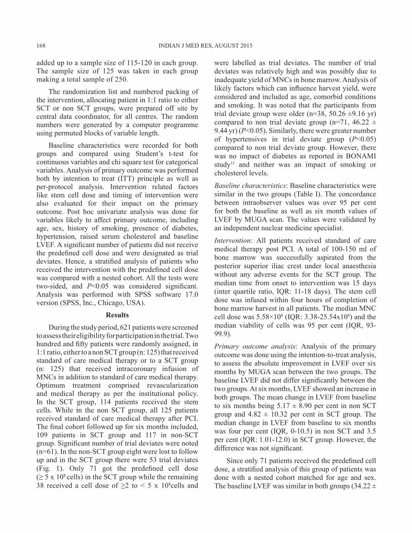
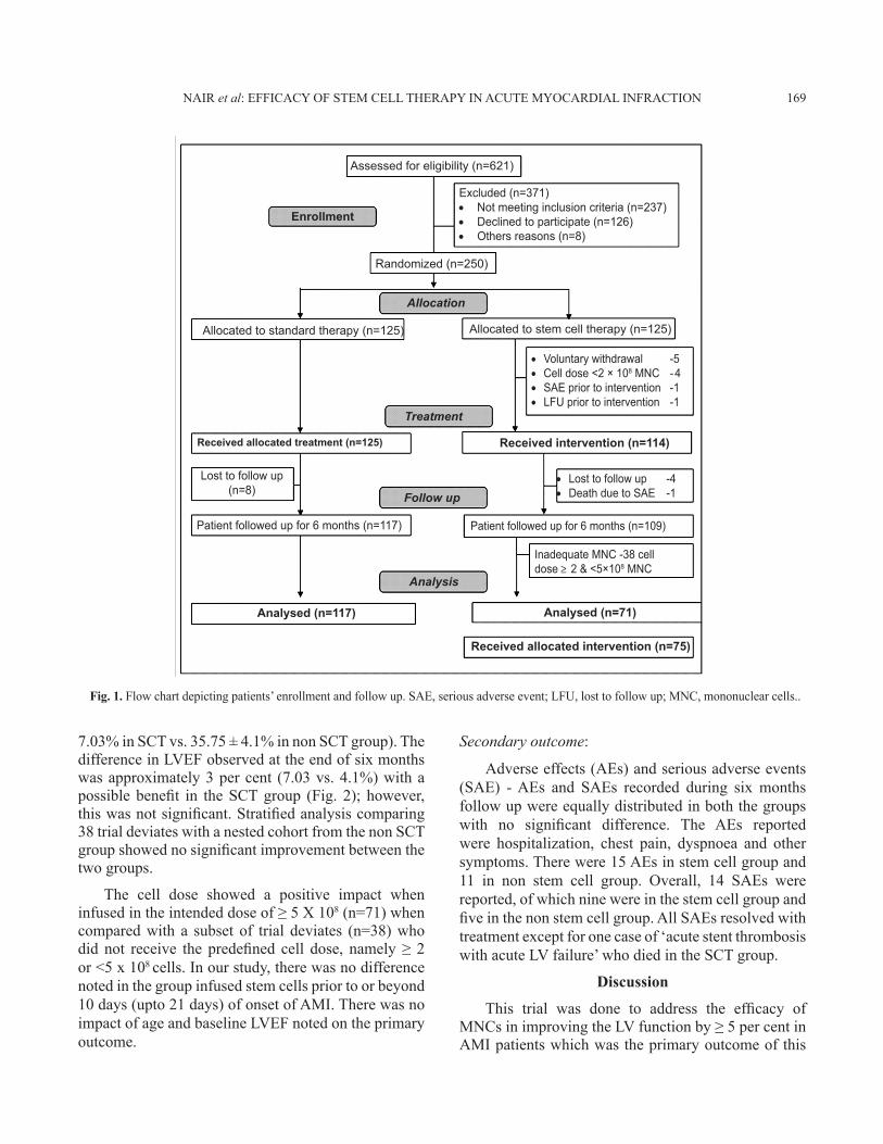
During the study period, 621 patients were screened to assess their eligibility for participation in the trial. Two hundred and fifty patients were randomly assigned, in 1:1 ratio, either to a non SCT group (n: 125) that received standard of care medical therapy or to a SCT group (n: 125) that received intracoronary infusion of MNCs in addition to standard of care medical therapy. Optimum treatment comprised revascularization and medical therapy as per the institutional policy. In the SCT group, 114 patients received the stem cells. While in the non SCT group, all 125 patients received standard of care medical therapy after PCI. The final cohort followed up for six months included, 109 patients in SCT group and 117 in non-SCT group. Significant number of trial deviates were noted (n=61). In the non-SCT group eight were lost to follow up and in the SCT group there were 53 trial deviates (Fig. 1). Only 71 got the predefined cell dose (≥ 5 x 10⁸ cells) in the SCT group while the remaining 38 received a cell dose of ≥2 to < 5 x 10⁸cells and
were labelled as trial deviates. The number of trial deviates was relatively high and was possibly due to inadequate yield of MNCs in bone marrow. Analysis of likely factors which can influence harvest yield, were considered and included as age, comorbid conditions and smoking. It was noted that the participants from trial deviate group were older (n=38, 50.26 ±9.16 yr) compared to non trial deviate group (n=71, 46.22 ± 9.44 yr) (P<0.05). Similarly, there were greater number of hypertensives in trial deviate group (P<0.05) compared to non trial deviate group. However, there was no impact of diabetes as reported in BONAMI study11 and neither was an impact of smoking or cholesterol levels.
Baseline characteristics: Baseline characteristics were similar in the two groups (Table I). The concordance between intraobserver values was over 95 per cent for both the baseline as well as six month values of LVEF by MuGA scan. The values were validated by an independent nuclear medicine specialist.
Intervention: All patients received standard of care medical therapy post PCI. A total of 100-150 ml of bone marrow was successfully aspirated from the posterior superior iliac crest under local anaesthesia without any adverse events for the SCT group. The median time from onset to intervention was 15 days (inter quartile ratio, IQR: 11-18 days). The stem cell dose was infused within four hours of completion of bone marrow harvest in all patients. The median MNC cell dose was 5.58×10⁸ (IQR: 3.38-25.54x10⁸) and the median viability of cells was 95 per cent (IQR, 93-99.9).
Primary outcome analysis: Analysis of the primary outcome was done using the intention-to-treat analysis, to assess the absolute improvement in LVEF over six months by MuGA scan between the two groups. The baseline LVEF did not differ significantly between the two groups. At six months, LVEF showed an increase in both groups. The mean change in LVEF from baseline to six months being 5.17 ± 8.90 per cent in non SCT group and 4.82 ± 10.32 per cent in SCT group. The median change in LVEF from baseline to six months was four per cent (IQR, 0-10.5) in non SCT and 3.5 per cent (IQR: 1.01-12.0) in SCT group. However, the difference was not significant.
Since only 71 patients received the predefined cell dose, a stratified analysis of this group of patients was done with a nested cohort matched for age and sex. The baseline LVEF was similar in both groups (34.22 ±
168 INDIAN J MED RES, AuGuST 2015
Enrollment
Assessed for eligibility (n=621)
Randomized (n=250)
Excluded (n=371)Not meeting inclusion criteria (n=237) •Declined to participate (n=126) •Others reasons (n=8) •
Voluntary withdrawal -5 •Cell dose <2 × 10 • 8 MNC - 4SAE prior to intervention -1 •LFU prior to intervention -1 •
Lost to follow up -4 •Death due to SAE -1 •
Inadequate MNC -38 celldose 2 & <5×10⁸ MNC
Patient followed up for 6 months (n=109)
Allocated to standard therapy (n=125)
Received allocated treatment (n=125) Received intervention (n=114)
Follow up
Treatment
Allocation
Analysis
Analysed (n=117) Analysed (n=71)
Received allocated intervention (n=75)
Patient followed up for 6 months (n=117)
Lost to follow up(n=8)
Allocated to stem cell therapy (n=125)
Fig. 1. Flow chart depicting patients’ enrollment and follow up. SAE, serious adverse event; LFu, lost to follow up; MNC, mononuclear cells..
NAIR et al: EFFICACY OF STEM CELL THERAPY IN ACuTE MYOCARDIAL INFRACTION 169
7.03% in SCT vs. 35.75 ± 4.1% in non SCT group). The difference in LVEF observed at the end of six months was approximately 3 per cent (7.03 vs. 4.1%) with a possible benefit in the SCT group (Fig. 2); however, this was not significant. Stratified analysis comparing 38 trial deviates with a nested cohort from the non SCT group showed no significant improvement between the two groups.
The cell dose showed a positive impact when infused in the intended dose of ≥ 5 X 10⁸ (n=71) when compared with a subset of trial deviates (n=38) who did not receive the predefined cell dose, namely ≥ 2 or <5 x 10⁸ cells. In our study, there was no difference noted in the group infused stem cells prior to or beyond 10 days (upto 21 days) of onset of AMI. There was no impact of age and baseline LVEF noted on the primary outcome.
Secondary outcome:
Adverse effects (AEs) and serious adverse events (SAE) - AEs and SAEs recorded during six months follow up were equally distributed in both the groups with no significant difference. The AEs reported were hospitalization, chest pain, dyspnoea and other symptoms. There were 15 AEs in stem cell group and 11 in non stem cell group. Overall, 14 SAEs were reported, of which nine were in the stem cell group and five in the non stem cell group. All SAEs resolved with treatment except for one case of ‘acute stent thrombosis with acute LV failure’ who died in the SCT group.
Discussion
This trial was done to address the efficacy of MNCs in improving the LV function by ≥ 5 per cent in AMI patients which was the primary outcome of this
170 INDIAN J MED RES, AuGuST 2015
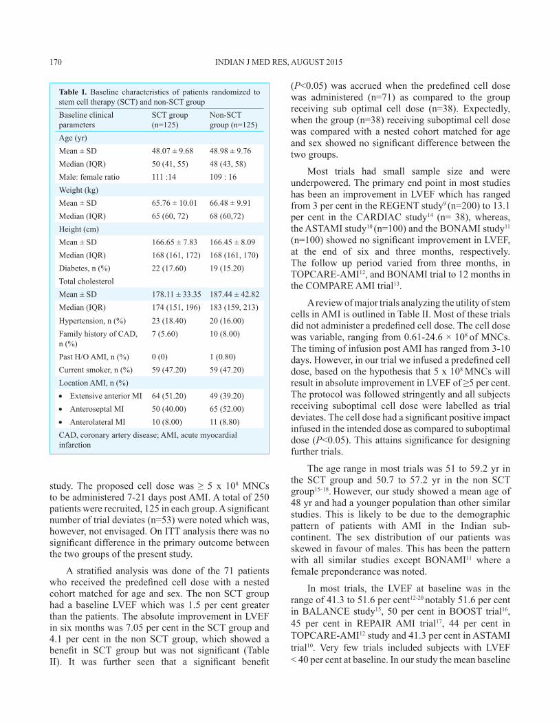
Table I. Baseline characteristics of patients randomized to stem cell therapy (SCT) and non-SCT group Baseline clinical parameters
SCT group(n=125)
Non-SCT group (n=125)
Age (yr)Mean ± SD 48.07 ± 9.68 48.98 ± 9.76Median (IQR) 50 (41, 55) 48 (43, 58)Male: female ratio 111 :14 109 : 16Weight (kg)Mean ± SD 65.76 ± 10.01 66.48 ± 9.91Median (IQR) 65 (60, 72) 68 (60,72)Height (cm)Mean ± SD 166.65 ± 7.83 166.45 ± 8.09Median (IQR) 168 (161, 172) 168 (161, 170)Diabetes, n (%) 22 (17.60) 19 (15.20)Total cholesterolMean ± SD 178.11 ± 33.35 187.44 ± 42.82Median (IQR) 174 (151, 196) 183 (159, 213)Hypertension, n (%) 23 (18.40) 20 (16.00)Family history of CAD, n (%)
7 (5.60) 10 (8.00)
Past H/O AMI, n (%) 0 (0) 1 (0.80)Current smoker, n (%) 59 (47.20) 59 (47.20)Location AMI, n (%)
Extensive anterior MI • 64 (51.20) 49 (39.20)Anteroseptal MI • 50 (40.00) 65 (52.00)Anterolateral MI • 10 (8.00) 11 (8.80)
CAD, coronary artery disease; AMI, acute myocardial infarction
study. The proposed cell dose was ≥ 5 x 10⁸ MNCs to be administered 7-21 days post AMI. A total of 250 patients were recruited, 125 in each group. A significant number of trial deviates (n=53) were noted which was, however, not envisaged. On ITT analysis there was no significant difference in the primary outcome between the two groups of the present study.
A stratified analysis was done of the 71 patients who received the predefined cell dose with a nested cohort matched for age and sex. The non SCT group had a baseline LVEF which was 1.5 per cent greater than the patients. The absolute improvement in LVEF in six months was 7.05 per cent in the SCT group and 4.1 per cent in the non SCT group, which showed a benefit in SCT group but was not significant (Table II). It was further seen that a significant benefit
(P<0.05) was accrued when the predefined cell dose was administered (n=71) as compared to the group receiving sub optimal cell dose (n=38). Expectedly, when the group (n=38) receiving suboptimal cell dose was compared with a nested cohort matched for age and sex showed no significant difference between the two groups.
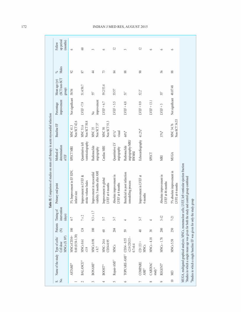
Most trials had small sample size and were underpowered. The primary end point in most studies has been an improvement in LVEF which has ranged from 3 per cent in the REGENT study9 (n=200) to 13.1 per cent in the CARDIAC study14 (n= 38), whereas, the ASTAMI study10 (n=100) and the BONAMI study11
(n=100) showed no significant improvement in LVEF, at the end of six and three months, respectively. The follow up period varied from three months, in TOPCARE-AMI12, and BONAMI trial to 12 months in the COMPARE AMI trial13.
A review of major trials analyzing the utility of stem cells in AMI is outlined in Table II. Most of these trials did not administer a predefined cell dose. The cell dose was variable, ranging from 0.61-24.6 × 10⁸ of MNCs. The timing of infusion post AMI has ranged from 3-10 days. However, in our trial we infused a predefined cell dose, based on the hypothesis that 5 x 10⁸ MNCs will result in absolute improvement in LVEF of ≥5 per cent. The protocol was followed stringently and all subjects receiving suboptimal cell dose were labelled as trial deviates. the cell dose had a significant positive impact infused in the intended dose as compared to suboptimal dose (P<0.05). This attains significance for designing further trials.
The age range in most trials was 51 to 59.2 yr in the SCT group and 50.7 to 57.2 yr in the non SCT group15- 18. However, our study showed a mean age of 48 yr and had a younger population than other similar studies. This is likely to be due to the demographic pattern of patients with AMI in the Indian sub-continent. The sex distribution of our patients was skewed in favour of males. This has been the pattern with all similar studies except BONAMI11 where a female preponderance was noted.
In most trials, the LVEF at baseline was in the range of 41.3 to 51.6 per cent12-20 notably 51.6 per cent in BALANCE study15, 50 per cent in BOOST trial16, 45 per cent in REPAIR AMI trial17, 44 per cent in TOPCARE-AMI12 study and 41.3 per cent in ASTAMI trial10. Very few trials included subjects with LVEF < 40 per cent at baseline. In our study the mean baseline
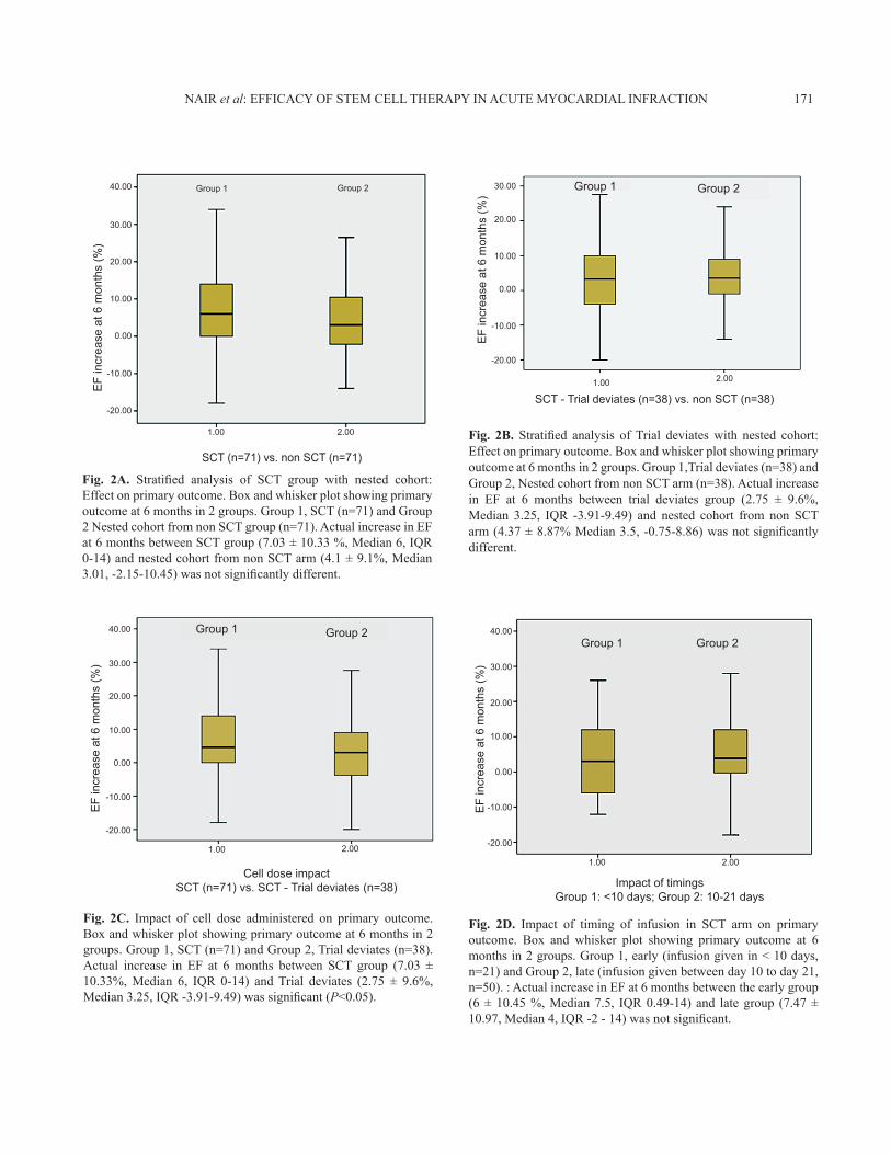
Fig. 2A. Stratified analysis of SCT group with nested cohort: Effect on primary outcome. Box and whisker plot showing primary outcome at 6 months in 2 groups. Group 1, SCT (n=71) and Group 2 Nested cohort from non SCT group (n=71). Actual increase in EF at 6 months between SCT group (7.03 ± 10.33 %, Median 6, IQR 0-14) and nested cohort from non SCT arm (4.1 ± 9.1%, Median 3.01, -2.15-10.45) was not significantly different.
NAIR et al: EFFICACY OF STEM CELL THERAPY IN ACuTE MYOCARDIAL INFRACTION 171
40.00 Group 1 Group 2
SCT (n=71) vs. non SCT (n=71)
EF
incr
ease
at 6
mon
ths
(%)
30.00
20.00
10.00
0.00
-10.00
-20.00
1.00 2.00 Fig. 2B. Stratified analysis of Trial deviates with nested cohort: Effect on primary outcome. Box and whisker plot showing primary outcome at 6 months in 2 groups. Group 1,Trial deviates (n=38) and Group 2, Nested cohort from non SCT arm (n=38). Actual increase in EF at 6 months between trial deviates group (2.75 ± 9.6%, Median 3.25, IQR -3.91-9.49) and nested cohort from non SCT arm (4.37 ± 8.87% Median 3.5, -0.75-8.86) was not significantly different.
-20.00
1.00 2.00
-10.00
0.00
10.00
20.00
30.00 Group 1 Group 2
SCT - Trial deviates (n=38) vs. non SCT (n=38)
EF
incr
ease
at 6
mon
ths
(%)
Fig. 2C. Impact of cell dose administered on primary outcome. Box and whisker plot showing primary outcome at 6 months in 2 groups. Group 1, SCT (n=71) and Group 2, Trial deviates (n=38). Actual increase in EF at 6 months between SCT group (7.03 ± 10.33%, Median 6, IQR 0-14) and Trial deviates (2.75 ± 9.6%, Median 3.25, IQR -3.91-9.49) was significant (P<0.05).
-20.00
1.00 2.00
-10.00
0.00
10.00
20.00
30.00
40.00
Cell dose impactSCT (n=71) vs. SCT - Trial deviates (n=38)
EF
incr
ease
at 6
mon
ths
(%)
Group 1 Group 2
Fig. 2D. Impact of timing of infusion in SCT arm on primary outcome. Box and whisker plot showing primary outcome at 6 months in 2 groups. Group 1, early (infusion given in < 10 days, n=21) and Group 2, late (infusion given between day 10 to day 21, n=50). : Actual increase in EF at 6 months between the early group (6 ± 10.45 %, Median 7.5, IQR 0.49-14) and late group (7.47 ± 10.97, Median 4, IQR -2 - 14) was not significant.
-20.00
1.00 2.00
-10.00
0.00
10.00
20.00
30.00
40.00Group 1 Group 2
Impact of timings Group 1: <10 days; Group 2: 10-21 days
EF
incr
ease
at 6
mon
ths
(%)
Tabl
e II.
Comp
ariso
n of s
tudies
on st
em ce
ll the
rapy i
n acu
te my
ocard
ial in
farcti
on
S.
No.
Name
of th
e stud
yTy
pe of
cells
/ Ce
ll dos
e M
NCs (
X 10
8 )
Patie
nts
(N)
Timi
ng of
int
erven
tion
(day
s)
Prim
ary en
d poin
tM
ethod
of
deter
mina
tion
of E
F
Base
line E
FPe
rcenta
ge
impr
ovem
ent
Mea
n age
(yr)
(SCT
/non-
SCT
grou
p)
% Male
sFo
llow
up pe
riod
(mon
ths)
1AS
TAM
I10M
NCs/C
D34+
0.6
8 (0.5
4-1.30
)10
04-
75%
Impr
ovem
ent in
EF E
DV,
infarc
t size
SPEC
T M
RIM
NC 41
.3 No
n SCT
42.6
Not s
ignifi
cant
58/56
926
2BA
LANC
E15M
NCs 0
.61
±3.9
124
7 ± 2
Impr
ovem
ent in
LVEF
&
strok
e volu
me In
dex
Quan
titati
ve le
ft ve
ntricu
lograp
hyM
NC 51
.6
Non S
CT 50
.8LV
EF +7
.951
.4/50
.787
60
3BO
NAM
I11M
NCs 0
.98
± 8.7
100
9.3 ±
1.7Im
prov
emen
t in m
yoca
rdial
via
bility
3 mo
nths a
fter A
MI
Radio
nucli
de
angio
grap
hyM
NC 35
No
n SCT
37No
im
prov
emen
t55
*44
3
4BO
OST16
MNC
- 24.6
CD
34-0
.9560
5-7
Impr
ovem
ent in
glob
al
LVEF
at 6
month
sCa
rdiac
MRI
MNC
50
Non S
CT 51
.3LV
EF +
6.759
.2/53
.473
6
5Re
pair-
AMI17
MNC
s20
43-
7Ab
solut
e imp
rove
ment
in
LVEF
at 4
month
sQu
antit
ative
LV
angio
grap
hy45
%#
visua
lLV
EF +
5.555
/5784
12
6TO
PCAR
E-AM
I12CD
34 -
0.55
±2.8
CD13
3 -
0.7±0
.4
594
Bene
ficial
in po
st inf
arctio
n rem
odell
ing pr
oces
sRa
dionu
clide
an
giogr
aphy
/MRI
/ RW
MSI
44%
#LV
EF +
4.851
*88
3
7CO
MPA
RE
AMI13
CD 13
3 +
MNC
s40
3-7
Impr
ovem
ent in
LVEF
at
4 mon
thsEc
hoca
rdiog
raphy
41.2%
#LV
EF +
9.952
.2*90
12
8CA
RDIA
C stu
dy14
MNC
s - 4.
1838
4SP
ECT
LVEF
+ 13
.16
9RE
GENT
9M
NCs -
1.78
200
3-12
Abso
lute i
mpro
veme
nt in
LV
EF at
46 m
onths
MRI
37%
#LV
EF +
3 55
*56
6
10m
i3M
NCs 5
.5825
07-
215%
abso
lute i
mpro
veme
nt in
LV
FE at
6 mo
nths
MuG
AM
NC 34
.76
Non-
SCT
34.53
Not s
ignifi
cant
48.07
/4888
6
MuG
A, m
ultiga
ted gr
aphic
al an
alysis
; MNC
s, mo
nonu
clear
cells
; LVE
F, lef
t ven
ticula
r ejec
tion f
ractio
n* St
udies
in w
hich a
sing
le me
an ag
e was
give
n for
both
the st
udy a
nd co
ntrol
grou
p com
bined
# Stud
ies in
whic
h a si
ngle
base
line E
F was
give
n for
only
the st
udy g
roup
172 INDIAN J MED RES, AuGuST 2015
LVEF was 34.22 per cent in the SCT group and 35.75 per cent in the non SCT group which was also noted in the BONAMI trial (33 and 37%, respectively). It is well known that even modest gains in LV function with pharmacotherapy result in significant benefits in long-term morbidity21.
SAEs were noted in the form of acute, sub-acute and delayed stent thrombosis, which were comparable in the two groups of the study. There was one death due to acute stent thrombosis with acute LV failure in the SCT group. Another important observation pertains to the timing of stem cell infusion. Most previous trials have infused stem cells between 3-10 days. However, in our study there was no difference noted in the group infused stem cells prior to or beyond 10 days (upto 21 days) of onset of AMI. This observation could lead to an increase in the therapeutic window for stem cell infusion.
The strengths of this study included a large sample size, administration of a predefined cell dose; a wider time window for stem cell infusion (7-21 days post AMI) and 90.4 per cent of total patients completed a six month follow up. The limitations included a large number of trial deviates (n=53) which reduced the number of patients receiving the intended intervention from 125 to 71 patients. The follow up period of six months was relatively short and preferably should be more than one year.
In conclusion, our study demonstrates that autologous MNCs can be safely administered in patients with AMI. On ITT analysis there was no significant difference in the primary outcome between the two groups. However, a stratified analysis of 71 patients who received the predefined cell dose compared to a nested cohort from the non SCT group showed a possible benefit in the SCT group. A benefit was also seen when the predefined cell dose was administered. This benefit was noted upto three weeks post AMI in contrast to other trials that demonstrated the same mostly within 10 days post AMI. In future, larger randomized trials need to be done to specifically validate and address issues regarding cell dose and timing of infusion.
Acknowledgment Authors acknowledge the Department of Biotechnology, Ministry of Science & Technology, Government of India, for financial support and R.M. Pandey and Shankar Subramanian, for statistical analysis. Authors thank Shankar Subramanium, Vineet Behera, J.P. Arya, Pankaj Kumar, for editorial assistance.
Data Safety and Monitoring Board: R. Aggarwal (SGPGI), L. Jeyaseelan (CMC, Vellore), V. Puri, u. Kaul, D. Prabhakaran, R.K. Saran, S. Kumar
Central Data Management office: Department of Pediatrics, All India Institute of Medical Sciences, New Delhi - Shinjini Bhatnagar.
Contract Research Organization: Manipal AcuNova, Bengaluru, India
conflicts of interest: None.
referencesSu1. tton MG, Sharpe N. Left ventricular remodeling after myocardial infarction: pathophysiology and therapy. Circulation 2000; 101 : 2981-8.Hellermann JP, Jacobsen SJ, Redfield MM, Reeder GS, Weston 2. SA, Roger VL. Heart failure after myocardial infarction: clinical presentation and survival. Eur J Heart Fail 2005; 7 : 119-25.De Luca G, Suryapranata H, Ottervanger JP, 3. Antman Em. Time delay to treatment and mortality in primary angioplasty for acute myocardial infarction: every minute of delay counts. Circulation 2004; 109 : 1223-5Boersma E, Maas AC, Deckers JW, 4. Simoons ML. Early thrombolytic treatment in acute myocardial infarction: reappraisal of the golden hour. Lancet 1996; 348 : 771-5.Xavier D, Pais P, Devereaux PJ, Xie C, Prabhakaran D, Reddy 5. KS, et al; CREATE registry investigators. Treatment and outcomes of acute coronary syndromes in India (CREATE): a prospective analysis of registry data. Lancet 2008; 371 : 1435-42.Abdel-Latif A, Bolli R, Tleyjeh IM, Montori VM, Perin EC, 6. Hornung CA, et al. Adult bone marrow-derived cells for cardiac repair: a systematic review and meta-analysis. Arch Intern Med 2007; 167 : 989-97.Burt RK, Loh Y, Pearce W, Beohar N, Barr WG, Craig R, 7. et al. Clinical applications of blood-derived and marrow-derived stem cells for nonmalignant diseases. JAMA 2008; 299 : 925-36.Godkar 8. D, Bachu K, Dave B, Megna R, Niranjan S, Khanna A. Comparison and co-relation of invasive and noninvasive methods of ejection fraction measurement. J Natl Med Assoc 2007; 99 : 1227-8, 1231-4.Tendera 9. m, Wojakowski W, Ruzyłło W, Chojnowska L, Kepka C, Tracz W, et al; REGENT Investigators. Intracoronary infusion of bone marrow-derived selected CD34+CXCR4+ cells and non-selected mononuclear cells in patients with acute STEMI and reduced left ventricular ejection fraction: results of randomized, multicentre myocardial regeneration by intracoronary infusion of selected population of stem cells in acute myocardial infarction (REGENT) Trial. Eur Heart J 2009; 30 : 1313-21.Beitnes J10. O, Gjesdal O, Lunde K, Solheim S, Edvardsen t, Arnesen H, et al. Left ventricular systolic and diastolic function improve after acute myocardial infarction treated with acute percutaneous coronary intervention, but are not influenced
NAIR et al: EFFICACY OF STEM CELL THERAPY IN ACuTE MYOCARDIAL INFRACTION 173
by intracoronary injection of autologous mononuclear bone marrow cells: a 3 year serial echocardiographic sub-study of the randomized-controlled ASTAMI study. Eur J Echocardiogr 2011; 12 : 98-106. Roncalli J, Mouquet F, Piot C, Trochu JN, Le Corvoisier P, 11. Neuder Y, et al. Intracoronary autologous mononucleated bone marrow cell infusion for acute myocardial infarction: results of the randomized multicenter BONAMI trial. Eur Heart J 2011; 32 : 1748-57.Britten MB, Abolmaali ND, Assmus B, Lehmann R, Honold 12. J, Schmitt J, et al. Infarct remodeling after intracoronary progenitor cell treatment in patients with acute myocardial infarction (TOPCARE-AMI): Mechanistic insights from serial contrast-enhanced magnetic resonance imaging. Circulation 2003; 108 : 2212-8.Mansour S, Roy DC, Bouchard V, Nguyen BK, Stevens 13. LM, Gobeil F, et al. COMPARE-AMI trial: Comparison of intracoronary injection of CD133+ bone marrow stem cells to placebo in patients after acute myocardial infarction and left ventricular dysfunction: study rationale and design. J Cardiovasc Transl Res 2010; 3 : 153-9. Piepoli MF, Vallisa D, Arbasi M, Cavanna L, Cerri L, Mori 14. M, et al. Bone marrow cell transplantation improves cardiac, autonomic and functional indexes in acute anterior myocardial infarction patients (CARDIAC STuDY). Eur J Heart Fail 2010; 12 : 172-80.Yousef M, Schannwell CM, Köstering M, Zeus T, Brehm M, 15. Strauer BE, et al. The BALANCE Study: Clinical benefit
and long-term outcome after intracoronary autologous bone marrow cell transplantation in patients with acute myocardial Infarction. J Am Coll Cariol 2009; 53 : 2262-9.Wollert KC, Meyer GP, Lotz J, Ringes-Lichtenberg S, Lippolt 16. P, Breidenbach C, et al. Intracoronary autologous bone-marrow cell transfer after myocardial infarction: the BOOST randomised controlled clinical trial. Lancet 2004; 364 : 141-8.Schachinger V, Erbs S, Elsasser A, Haberbosch W, Hambrecht 17. R, Holschermann H, et al; REPAIR-AMI Investigators. Improved clinical outcome after intracoronary administration of bone-marrow-derived progenitor cells in acute myocardial infarction: final1-year results of the REPAIR-AMI trial. Eur Heart J 2006; 27 : 2775-83.Lunde K, Solheim S, Aakhus S, Arnesen H, Abdelnoor M, 18. Egeland T, et al. Intracoronary injection of mononuclear bone marrow cells in acute myocardial infarction. N Engl J Med 2006; 355 : 1199-209.Pawani 19. H, Bhartiya D. Pluripotent stem cells for cardiac regeneration: overview of recent advances & emerging trends. Indian J Med Res 2013; 137 : 270-82.Shah V20. K, Shalia KK. Stem cell therapy in acute myocardial infarction: A pot of gold or pandora’s box. Stem Cells Int 2011; 2011 : 536758.Clifford DM, Fisher SA, Brunskill SJ, Doree C, 21. Mathur A, Watt S, et al. Stem cell treatment for acute myocardial infarction. Cochrane Database Syst Rev 2012; 2 : CD006536.
174 INDIAN J MED RES, AuGuST 2015
Reprint requests: Dr Velu Nair, Department of Haematology & Bone Marrow Transplantation, Army Research and Referral Hospital, Dhaula Kuan, Delhi Cantt 110 010, India e-mail: [email protected]