assessment of safety and functional efficacy of stem cell
TRANSCRIPT

Review ArticleAssessment of Safety and Functional Efficacy of StemCell-Based Therapeutic Approaches Using Retinal DegenerativeAnimal Models
Tai-Chi Lin,1,2,3,4 Magdalene J. Seiler,5,6 Danhong Zhu,1,7 Paulo Falabella,1
David R. Hinton,1,7 Dennis O. Clegg,8 Mark S. Humayun,1,2 and Biju B. Thomas1,2
1Department of Ophthalmology, USC Roski Eye Institute, University of Southern California, Los Angeles, CA, USA2USC Institute for Biomedical Therapeutics, University of Southern California, Los Angeles, CA, USA3Department of Ophthalmology, Taipei Veterans General Hospital, Taipei, Taiwan4Institute of Clinical Medicine, National Yang-Ming University, Taipei, Taiwan5Stem Cell Research Center, University of California-Irvine, Irvine, CA, USA6Department of Physical Medicine & Rehabilitation, University of California-Irvine, Irvine, CA, USA7Department of Pathology, Keck School of Medicine, University of Southern California, Los Angeles, CA, USA8Center for Stem Cell Biology and Engineering, University of California, Santa Barbara, CA, USA
Correspondence should be addressed to Biju B. Thomas; [email protected]
Received 15 April 2017; Accepted 19 June 2017; Published 27 August 2017
Academic Editor: Tilo Kunath
Copyright © 2017 Tai-Chi Lin et al. This is an open access article distributed under the Creative Commons AttributionLicense, which permits unrestricted use, distribution, and reproduction in any medium, provided the original work isproperly cited.
Dysfunction and death of retinal pigment epithelium (RPE) and or photoreceptors can lead to irreversible vision loss. The eyerepresents an ideal microenvironment for stem cell-based therapy. It is considered an “immune privileged” site, and thenumber of cells needed for therapy is relatively low for the area of focused vision (macula). Further, surgical placement ofstem cell-derived grafts (RPE, retinal progenitors, and photoreceptor precursors) into the vitreous cavity or subretinal spacehas been well established. For preclinical tests, assessments of stem cell-derived graft survival and functionality are conductedin animal models by various noninvasive approaches and imaging modalities. In vivo experiments conducted in animalmodels based on replacing photoreceptors and/or RPE cells have shown survival and functionality of the transplanted cells,rescue of the host retina, and improvement of visual function. Based on the positive results obtained from these animalexperiments, human clinical trials are being initiated. Despite such progress in stem cell research, ethical, regulatory, safety,and technical difficulties still remain a challenge for the transformation of this technique into a standard clinical approach. Inthis review, the current status of preclinical safety and efficacy studies for retinal cell replacement therapies conducted inanimal models will be discussed.
1. Introduction
Stem cell-based therapies have shown to restore or rescuevisual function in preclinical models of retinal degenerativediseases [1–5] which are built on previous data with trans-plantation of fetal retinal tissue sheets. This has set a standardwhat these optimal cells can do [6–9]. Although retinal
degenerative diseases such as retinitis pigmentosa (RP),age-related macular degeneration (AMD), and Stargardt’sdisease differ in their causes and demographics, all of themcause RPE and/or photoreceptor destruction which can leadto blindness [1–5]. Currently, there is no clinically acceptedcure for irreversible dysfunction or death of photoreceptorsand RPE. Since the retina, like other central nervous system
HindawiStem Cells InternationalVolume 2017, Article ID 9428176, 19 pageshttps://doi.org/10.1155/2017/9428176

tissue, has little regenerative potential [4, 10], stem cell-basedtherapies that aimed to replace the dysfunctional or deadcells remain a major hope.
In 1959, a rat fetal retina was transplanted into the ante-rior chamber of a pregnant rat’s eye [11]. Several decadeslater, dissociated retinal cells or cell aggregates were trans-planted into the subretinal space of rats [12–17]. In the 80s,Dr. Gouras demonstrated transplantation of cultured humanretinal pigment epithelial cells into the monkey retina. Thetransplanted cells were identified on the Bruch’s membraneby autoradiography [18]. Turner and Blair reported high sur-vival (90–100%) and development of lamination for newbornrat retinal aggregates grafted into a lesion site of an adult ratretina [19]. Silverman and Hughes were the first one to iso-late stripes of photoreceptor sheets from the postnatal andadult retina [20], and this method was modified later on byother researchers by transplanting photoreceptor sheets[21], full thickness fetal [6, 7, 22–24] or adult retina [25].These earlier transplantation studies helped to establish“proof of concept” for future cell replacement therapies inthe eye. Although the initial transplantation studies did notshow any safety issues, ethical restrictions and absence ofsuitable animal models for preclinical evaluations delayedfurther progress of this approach [3]. In 2009, human embry-onic stem cell- (hESC-) derived RPE cells were transplantedinto Royal College of Surgeon (RCS) rats in preclinical stud-ies [26] that eventually lead to clinical trials. Although thelong-term outcomes of the preclinical investigations are notyet concluded [27–31], recent advancement in the area ofinduced pluripotent stem cell- (iPSC-) derived products pro-vided a new source for transplantation. This method usesmature cells that return to a pluripotent state similar to thatseen in embryonic stem cells [32–35]. Preclinical testing ofiPSC-derived RPE (iPSC-RPE) cells has been established[36, 37], and human clinical trials based on iPSC-RPE havebeen initiated [38]. These studies indicate survival of thetransplanted RPE with signs of visual functional improve-ment and no signs of adverse events. However, one of the firsthuman clinical trials using autologous iPSC-RPE cells lead byMasayo Takahashi was halted for a period of time after unex-pected chromosomal abnormalities were found in the secondpatient [39, 40]. In a different incident, severe vision loss wasobserved in three AMD patients after intravitreal injection ofautologous adipose tissue-derived “stem cells” (https://blog.cirm.ca.gov/2017/03/15/three-people-left-blind-by-florida-clinics-unproven-stem-cell-therapy/comment-page-1/). Theabove report raises some concerns regarding the existingsafety requirements and regulations of the use of unregu-lated stem cell trials [41].
In this review, current progress in stem cell-based thera-pies will be discussed based on safety assessments and func-tional evaluations conducted in various animal models ofhuman retinal degenerative diseases.
2. Stem Cell Sources and Their Applications inthe Eye
Stem cell-based therapy for RPE replacement has been initi-ated at various centers. Since Klimanskaya et al. developed
the original protocol for hESC-derived RPE-like cells [42],various groups have used several strategies to derive RPEcells from stem cells. In earlier studies, subretinal transplan-tation of hESC-derived RPE (hESC-RPE) cells based on cellsuspension injection was shown to rescue degenerating pho-toreceptors and improve vision in immunosuppressed RCSrats [26, 43]. In a more recent technique, a pregeneratedRPE monolayer grown on a scaffold and transplanted inimmunosuppressed RCS rats showed improved survival ofhESC-RPE and better clinical outcomes [44, 45] suggestingthat RPE function is dependent on polarization of the trans-planted RPE cells and the monolayer morphology [44–46].iPSCs are considered to have several advantages over hESCsincluding protection from immune rejection, wide varietyof potential sources, and reduced ethical concerns [47].Transplantation of iPSC-RPE [37, 48] and iPSC-derived pho-toreceptor precursor cells [49] has demonstrated success indifferent animal models. The iPSC-RPE cells were shown tohave morphological and functional similarities to developingand mature RPE cells in vitro and in vivo [37, 50–52].Although it will be advantageous to use patient-derivedRPE (autologous transplants), the time requirements andproduction cost make allograft transplantation a desirableoption [47].
Patients need to have a sufficient number of surviving,functional photoreceptor cells; otherwise, replacement ofonly RPE will not help to rescue vision. Therefore, stem cell-derived photoreceptors [53–56] or retinal progenitor cells(RPCs) have been used with or without RPE for transplanta-tion experiments [3, 57–60]. Previously, several types of scaf-folds made of materials having different architectures,biocompatibility, size, and stiffness have been used to enhancecell survival, migration from the scaffold pores, integrationinto the host retina, and in vivo differentiation [61–63].
Studies have shown that the beneficial effect of RPCtransplantation is likely achieved by their differentiation intofunctional retinal cells and subsequent replacement of lost ordysfunctional elements [64, 65]. Other investigations sug-gested that success of RPC transplantation is achievedthrough trophic factor release rather than direct replacementof the lost cells [59, 66–68]. A major challenge in incorporat-ing photoreceptors and other neuronal cell types is the estab-lishment of synaptic connections with the proximal neuronalelements of the recipient retina [2, 69–71]. Using transsynap-tic tracing techniques and donor cell label, synaptic connec-tions between fetal retinal sheet transplants and the hostretina have been previously reported [72–74]. Replacementtherapies involving the whole retina are also in progress usingretinal organoids (3D retina) [70, 71, 75, 76]. Recently, hESCsand iPSCs were differentiated into optic cups and storablestratified neural retina [77, 78]. Such 3D retinal tissue derivedfrom iPSCs or hESCs when transplanted in rd1 mice [70, 71]and immunosuppressed retinal degeneration (RD) monkeys[75] developed a structured outer nuclear layer and showedsigns of synaptic formations [70, 71, 75]. The above mile-stone studies highlight the new concepts of regenerativemedicine in retinal therapeutics emphasizing the possibilityof establishing functional connections between the transplantand the host tissue.
2 Stem Cells International
3. AnimalModels for Stem Cell-Based Therapies
Retinal degenerate rodent models have been extensively usedfor biomedical research, but because of the key differencesbetween the rodent and human eye, rodent models do notcompletely replicate the human disease conditions. Mostimportantly, the rodents do not have a fovea, and in mostof the rodents, the photoreceptors are mainly rod cells.Among rodent models, there are both naturally occurring[79–83] and transgenic animal models [84–87]. Light dam-age [88], laser-induced choroidal neovascularization [89],and retinotoxic agents such as sodium iodoacetate [90] andN-methyl-N-nitrosourea [91, 92] have been also used toinduce retinal degenerative conditions. Among this, sodiumiodate (SI) has been widely used to induce outer retinaldegeneration in otherwise normal animals [93–96].
Rabbits, cats, dogs, pigs, and nonhuman primates have aneye diameter more or less similar to the human eye whichallows easy testing of surgical tools and procedures developedfor human patients. However, in these large animal models,inducing a disease condition similar to human patients ischallenging mostly because the etiology of human diseasesis multifactorial, involving both genetic and environmentalcontributions [1, 2]. The following section summarizes thesmall and large animal models that are currently used in stemcell-based research.
3.1. Mouse Models. The advantage of using mouse models istheir ability to express gene mutations mimicking thoseidentified in humans. However, dissimilarities in life spanand rate of disease progression between mice and humanslimit the interpretation of the disease conditions. A varietyof mutations in mice can cause loss of photoreceptors andreduced rod function and hence were used as AMDmodels [97–103]. In humans, mutations in the Abca4 generesult in Stargardt’s disease, RP, cone-rod dystrophy, andthe accumulation of lipofuscin granules in RPE, a charac-teristic of AMD [104, 105]. Therefore, Abca4 knockoutmice which also show lipofuscin accumulation in RPEare considered a model for macular dystrophy conditions[106–110]. Mutations of the RPE65 gene in humans causemost frequently Leber congenital amaurosis, with a smallpercentage of severe early childhood onset retinal dystrophy[111]. Hence, Rpe65 knockout mice are a model for studyingRPE65-mediated retinal dystrophy [112, 113]. Transgenicmice with a rhodopsin Pro23His (P23H) mutation thatcauses photoreceptor degeneration are highly comparableto human RP disease [99, 114, 115]. In humans, a generesponsible for the autosomal dominant form of Stargardt’sdisease was identified recently [116, 117]. Transgenic miceharboring this defective gene (Elovl4) are considered a goodmodel for macular degeneration diseases because of the accu-mulation of high levels of lipofuscin in the RPE and subse-quent photoreceptor degeneration in the central retina.This disease pattern closely resembles human Stargardt’s dis-ease and AMD [118]. Finally, there are naturally occurringmutations in mice that are used as models of RP diseaseinheritance [79–83]. Many of the mouse models discussedhere are tested for stem cell therapies using cell suspension
injections [89, 119]. In conclusion, the wide variety of genemanipulated mouse models provides a valuable tool for stud-ies on therapeutic intervention of various forms of humanRD. However, because of their small eye size, implantationof laminated sheets is found to be difficult in mice [44, 120].
3.2. Rat Models. Rats’ eyes are twice the size of mouse eyes[121] which makes it easier to perform surgical procedures[121] and transplant both fetal retinal sheets [3, 9] and RPEcells grown as a monolayer [44, 45]. The RCS rat is an animalmodel widely used for investigating therapeutic applicationsin the eye [122, 123]. The dystrophic RCS rats are character-ized by RPE dysfunction due to the deletion in the Mer tyro-sine kinase (Mertk) receptor that abolishes internalization ofshed photoreceptor outer segments by RPE cells [124]. Accu-mulation of debris in the subretinal space can lead to drasticphotoreceptor degeneration and rapid loss of vision. In RCSrats, the degeneration progresses slowly. At one month ofage, the retinal thickness remains close to the normal level[125] and near complete photoreceptor layer thickness ispresent [126]. Subretinal transplantation of RPE cells derivedfrom both iPSC [127] and hESC [44, 128–130] into 21 to 28-day old RCS rats showed photoreceptor preservation and res-cue of declining vision. Certain other rat models mimic thepathology and progression of RD such as the OXYS rat whichspontaneously develops a phenotype similar to human agingand AMD-like pathology [131, 132]. The transgenic P23Hrat (available in 3 lines with different degeneration rates),similar to P23H mouse, is frequently used as a model ofstudying RP diseases [85, 133]. S334ter rats carry a mutantmouse rhodopsin which leads to photoreceptor degeneration[134–136]. The five lines of this model have different charac-teristic rates of RD, in which S334ter line-3 and S334ter line-5 represent fast and intermediate slow degenerating models,respectively [87]. Several studies have been performed inthe above rat models to assess the feasibility of retinal cellreplacement therapies [3, 73, 137–141].
The advantage of using slow degeneration models is thatthey mimic the generally slow progression of human diseaseconditions. With the inner retina relatively well preserved,there is better opportunity for rescue or restoration of visionfollowing various treatment strategies [138, 142]. However,challenges like immunological reactions and the presence ofresidual host photoreceptors can make it difficult to detectthe transplant effects. To overcome the immunologicalissues, recently immunodeficient rat models (more detailsare provided in Section 4) are developed for testing cell-based therapies [143, 144]. In summary, rodent models arecurrently the leading in vivo tool for testing retinal cell ther-apies due to their affordable cost and easy availability [145].
3.3. Rabbit Models. Rabbits have an eye size comparable tohumans and are considered a desirable model to examinetherapeutic effects. However, the rabbit retina differs fromthat of human because it is rod-dominated and contains thevisual streak, a horizontal band lying inferior to the opticnerve absent in humans [146, 147]. The densities of rodsand cones in the visual streak are higher than elsewhere inthe entire retina [146, 147]. Despite this difference, full-field
3Stem Cells International

electroretinography (ERG) developed for the human eye canbe used in the rabbit with reproducible results [148]. Thetransgenic TgP347L rabbit closely tracks human cone-sparing RP disease [86, 149]. Histopathological study inTgP347L rabbits reported that the retinal degeneration devel-oped earlier in the visual streak than in other areas [86] alongwith some ERG abnormalities [150].
Previously, a dose-dependent correlation between theintravitreal injection of sodium iodate (SI) and retinal degen-eration (RD) has been reported in rabbits [151]. According tothe investigators, since injected SI may not be evenly distrib-uted in the vitreous due to its uneven liquefaction character-istics, uneven retinal degeneration is caused. Anotherphotoreceptor degeneration rabbit model produced by intra-vitreal injection of N-methyl-N-nitrosourea showed selectivebut inconsistent photoreceptor degeneration [152]. Subretin-ally injected hESC-RPE in immunosuppressed SI-inducedRD rabbits failed to integrate into the areas that showed geo-graphic atrophy-like symptoms [153]. This shortcoming wasprobably due to the unique features of the rabbit eye with ahigher degree of immune rejection [153]. In summary, rab-bits serve as a useful midsized animal model to study humandiseases and therapeutics because they have large eyes com-pared to rodents.
3.4. Cat Models. Abyssinian cats with inherited rod-conedegeneration (rdAc model) are used as a model for studyingretinal therapeutics [154, 155]. The genetic defect causativeof retinal degeneration in Abyssinian cats has been identifiedas a single base pair change in intron 50 of the centrosomalprotein 290 (CEP290) gene (IVS50+ 9T>G). This results inabnormalities in the transport and distribution of photo-transduction and/or structural proteins through the connect-ing cilia resulting in photoreceptor degeneration [156]. Ahigh prevalence of affected and carrier cats (45% and 44%,resp.) in the population was first observed in Sweden in1983 [155]. The cause is speculated to be inbreeding [157].In addition to have tapetum lucidum (discussed in the nextsection) which is different from the human eye, a majorshortcoming of this model is that it does not entirely resem-ble the human RP diseases where the peripheral retina isstrongly abnormal compared to the central area that remainrelatively less damaged. No such distinction is observed inAbyssinian cats. In this model, the degeneration is evenly dis-tributed during the early stages where normal and diseasedphotoreceptors are often found side-by-side [158]. In addi-tion, this cat model manifests a very slow progression ofdegeneration, taking from 12 months up to four years [154,155, 157]. Cat breeds with faster RD disease conditions arenow available [159, 160]. An early onset autosomal recessiveRD disease in Persian cats was virtually completed at 16weeks of age [160]. Another cat model, the CrxRdy cats,develops retinal thinning that initially takes place in thecentral retina [159]. An acute, reliable, and complete pho-toreceptor degeneration model in cats can be achieved byear vein injection of high-dose iodoacetic acid [161]. Reti-nal sheet transplantation studies conducted in Abyssiniancats showed good signs of transplant integration with thehost retina and lamination of transplant photoreceptors.
However, no considerable functional improvement wasnoticed [162].
3.5. Dog Models. A major difference in dog eyes from that ofhumans is the tapetum lucidum, which is a multilayeredreflective tissue of the choroid. The tapetum lucidum is inter-posed between the branching vessels in the choroid and thesingle layer of the choriocapillaris beneath the retina. TheRPE cells over the tapetum lucidum are normally unpigmen-ted. The tapetum lucidum acts to amplify and reflect lightback through the photoreceptor layer again in dim lightconditions [163]. Tapetum degeneration called toxic tapeto-pathy has been described in association with the administra-tion of several drugs in beagle dogs [164–168]. Toxictapetopathy is the characteristic of an altered tapetal colorwith degeneration or necrosis of the tapetum lucidum [164,168]. Tapetum degeneration is not observed in the eyes ofanimals without a tapetum lucidum (rodents, monkeys)[164, 168] and most importantly not in humans [168].
A naturally occurring canine model of autosomal dom-inant RP caused by a RHO mutation was found tostrongly resemble the human RP phenotype [169]. Highsimilarity in eye size and preretinal light transmissioncharacteristics between dogs and humans made this modelsuitable for examining the genetic and environmental causesof RD diseases. Previous studies demonstrated acute retinalinjury in RHO mutation dogs after exposure to strong light[170, 171]. By varying the dose of light exposure, its long-term consequences including fast or slow disease progressionand injury repair have been examined [170]. Although therehave been no reports of stem cell-based studies conducted inthese animals, this dog model can be considered a suitablecandidate for future preclinical studies.
3.6. Pig Models. Several transgenic pig models have beendeveloped for RP diseases, including the Pro347Leu trans-genic pig with a rhodopsin mutation [84, 172, 173], theP347S transgenic pig [174, 175], and the P23H transgenicpig, which is considered a model for autosomal dominantRP [176]. Disease progression in most of the above modelsis slow, making it difficult to assess therapeutic benefits.
Several reports of cell transplantation experiments con-ducted in pigs are available. Rhodopsin transgenic pigshave been used for transplantation of full-thickness retina[177] and retinal progenitor cell (RPC) transplantationeffects [178]. A feasibility and safety study of subretinalimplantation of an hESC-RPE monolayer has been reportedin immunosuppressed Yucatán minipigs [179]. This studydemonstrated preservation of the outer nuclear layer andphotoreceptor outer segment overlying the implant. In non-immunosuppressed pigs, adaptive immune responses wereactivated following allogenic iPSC-RPE transplantation[180]. The above finding suggests that immunologicallymatched and autologous donor cells should be consideredfor RPE cell replacement therapies to obviate chronicimmune suppression [180].
A major advantage of using pig models is that surgicaltools can be developed without much adaptation from thehuman parameters [1]. Previous RPE cell replacement
4 Stem Cells International

therapy studies conducted in pigs were focused on testingsurgical feasibility of the approach rather than testingfunctional improvements [1, 181]. This can be also dueto the absence of a suitable pig RPE dysfunction model.Although an RPE debridement model can be developed inpigs [182, 183], it is not preferred for testing RPE cell replace-ment therapies presumably due to both the difficulty in creat-ing a consistent disease pattern and the severity of the traumathat could adversely affect the study outcome.
3.7. Nonhuman Primate Models. The macula is a structure ofthe eye unique to humans, apes, and monkeys that plays arole as the zone of greatest visual acuity. Therefore, nonhu-man primates are a potentially valuable animal model forinvestigating macular diseases of humans [163]. AMD-likeappearance could be found in rhesus monkey (Macacamulatta), cynomolgus macaque (Macaca fascicularis), andthe Japanese macaque (Macaca fuscata). This suggests thatthe pathogenic mechanisms and associated gene variationsare common between human and nonhuman primates[184–190]. Induced RD monkey models have been reportedbased on systemic injection of iodoacetic acid [191] andcobalt chloride [75], fiber optic light-induced retinal damage[192, 193], and focal damage by severe light exposure [193].However, these models hold one or more adverse featuresincluding ethical issues and inability to produce adequatelysized lesions [75]. Housing, maintenance, costs, and ethicalconcerns due to a close evolutionary relationship to humansfurther make the nonhuman primate models less appealingfor stem cell researchers [194, 195].
Immune rejection of allogeneic iPSC-RPE transplantswas studied in cynomolgus monkeys (Macaca fascicularis)[196]. In a recent investigation, researchers used a cobaltchloride-induced retinal degeneration RP monkey model todemonstrate possible integration of hESC-derived retinalsheets with the host bipolar cells [75]. The above findingdemonstrated clinical feasibility of retina sheet transplanta-tion approach and suggests the need for developing newstrategies for future clinical applications [75].
4. Tools and Approaches for In Vivo Assessmentof the Transplants and Their Functionality
The eyes are one of the few paired organs in the bodywhere it is possible that one eye is treated while the con-tralateral eye will serve as control. The transparent natureof the eye makes the evaluations possible through noninva-sive imaging modalities.
4.1. In Vivo Imaging of Retinal Transplants and Assessment ofDisease Status. Fundus imaging and fluorescein angiographyare used to record baseline and follow-up examinations afterstem cell therapies. Optical coherence tomography (OCT) isa noncontact, noninvasive imaging technique widely used inthe clinic. The advancement of OCT technology providedrapid assessment of transplant morphology and placementlocation in the eye [9, 52, 197, 198]. The use of OCTimaging to assess changes in the retinal thickness post-transplantation has been established [9, 197–201]. Theabove studies conducted in rat models suggested that OCTis a reliable tool for in vivo screening and evaluation of retinaltransplants. In our rat experiments, we observed that OCTwas helpful using a novel OCT-based screening techniquedeveloped by our team. Using OCT software (HeidelbergSpectralis’s macular thickness feature), distance between theinternal limiting membrane (ILM) and top of the implantwas measured (Figure 1). The maximum and minimumvalues are recorded to determine the delta value. The deltavalue is obtained by subtracting the “maximum value−minimum value.” Based on the delta value, it is possibleto predict whether the implant is placed flat or tilted relativeto the retinal surface.
Recently, Seiler et al. [9] used a Bioptigen Envisu R2200Spectral Domain Ophthalmic Imaging System (Bioptigen,Research Triangle Park, NC, USA) to obtain SD-OCT imagesof the rat retina that showed similarity between OCT andhistology in the lamination pattern and thickness of thetransplants (Figure 2). Other techniques like scanning laserophthalmoscopy (SLO) can generate images from retinal
200 �휇m
(a)
200 �휇m
(b)
Figure 1: OCT imaging to assess surgical placement of hESC-RPE implantation in rats. (a) Fundus image showing hESC-RPE implantsplaced inside the rat eye (arrowhead). (b) Optical coherence tomography software was used to measure the distance between the internallimiting membrane (ILM, arrowhead) and the top of the implant (arrow). The maximum and minimum values were recorded. The deltavalue obtained by subtracting “maximum value−minimum value” can be used to determine if the implant is placed flat or tilted relativelyto the retinal surface.
5Stem Cells International

reflectance, autofluorescence, and extrinsic fluorescence.With the confocal arrangement, the SLO is capable of reject-ing scattered light, thereby improving image contrast andachieving moderate depth sectioning [202]. Confocal near-infrared SLO imaging was used for in vivo detection of sub-retinally placed hESC-RPE implants in rats [203]. Althoughthe lateral resolution achieved with SLO systems is compara-ble to that obtained with OCT, the depth resolution was rel-atively poor. But the advantage of SLO is the ability to detect
the presence of pigments on the hESC-RPE in vivo [203].Hence, the survival and potential functionality of an RPEgraft can be established. Moreover, SLO is useful when theOCT images are difficult to interpret due to the loss of retinalarchitecture, as in the case of advanced AMD.
4.2. Electrophysiological Assessments. Visually evoked poten-tials (VEPs) have been used to determine whether the photo-receptor sheet transplants to RD rats can activate the
90 d Y: 0.050X: 0.100
(a)
62 d Y: 0.050X: 0.100
(b)
170 d
Host (H)
Transplant (T)
GCLIPL
IPL
INL
INL
ONL
RPE100 �휇m
(c)
70 d
Transplant (T)
Host (H)
GCL
IPL
INL
ONL
⁎
⁎
⁎⁎
⁎
ONLINL INL
100 �휇m
IPLRPE
(d)
DAPIDonorrho
H
OS
ONL
T INL
INL
IPL
RPE
(e)
GCL
INLH
DAPI
Donor
rho
OSOSONL
ONLT
INL
IPL
IPL
(f)
Figure 2: Correlation of live SD-OCT imaging with histology. Fetal retinas (embryonic day 19) derived from rats expressing human placentalalkaline phosphatase (hPAP) in the cytoplasm of all cells were transplanted to the subretinal space of immunodeficient retinal degenerate rhoS334ter-3 rats. Transplanted rats’ eyes were imaged in vivo by SD-OCT. Two transplant examples are shown. (a, b) Stretched cross-sectionalB-scans of laminated (a) and rosetted (b) transplant to better distinguish different retinal layers. Rosettes are indicated by yellow arrows (b)and seen as hyperreflective orbs. (c, d) Transplant-specific histochemistry for hPAP using BCIP (purple). hPAP is expressed in the cytoplasm(not the nuclei) of donor cells. Transplant number 5 (a, c, e) has a large area of lamination parallel retinal layers with photoreceptor outersegments, indicated by yellow diamonds (c) and strong rhodopsin expression (e) in the donor outer retina. Transplant number 1 (b, d, f)is more disorganized with photoreceptors in rosettes [rosette lumens indicated by yellow asterisks in (d)]. The rhodopsin-positive outersegments face inward (f). This transplant (d) was partially placed upside down in the subretinal space. (a, b) Scale bars: vertical bar:50μm; horizontal bar: 200μm; (c, d): 100 μm; (e, f) bars: 20μm. Modified after Figure 3 of Seiler et al. vision recovery and connectivity byfetal retinal sheet transplantation in an immunodeficient retinal degenerate rat model, IOVS 2017;58:614–630. DOI:10.1167/iovs.15-19028;licensed under the Creative Commons attribution license.
6 Stem Cells International
central visual system [204]. VEPs were elicited by usingstrobe flash stimuli, and responses were recorded contra-lateral to the stimulated eye. The results showed that thereconstructed retina can produce characteristic light-evoked responses in the visual cortex [204]. Electrophysio-logical analysis was used to demonstrate that corticalvisual function could be preserved by subretinal RPE cellgrafting in RCS rats [205]. This was also established usingoptical imaging techniques [206]. Morphological assess-ments confirmed good correlation between photoreceptorsurvival and the extent of cortical functional preservation[206]. However, the degree of visual acuity achieved bytransplants cannot be completely addressed using visuallyevoked cortical responses.
Electroretinography (ERG) is employed to access thediffuse electrical response of the retina. Response to flashERG has been used to evaluate the visual functional changesin retinal degenerative animal models [43, 207, 208]. ERGassessments have revealed improved photoreceptor functionin RCS rats after hESC-RPE injection [43]. A major limita-tion in using full-field flash ERG is that it may fail to detectsignals from the comparatively small transplant area. Thisis because the ERG response is the cumulative effect of signalsfrom the entire retina, whereas signal output from thetransplant area may not be sufficient to generate considerabledifference in the ERG wave form [9].
Focal electroretinography (fERG) is used to study a dis-crete region of the retina and determine if there is signifi-cantly more electrical activity in that area compared to thesurrounding retina. This technique has been successfullyemployed in RCS rats to show photoreceptor rescue afteriPSC-RPE injection [209]. Although multifocal ERG(mfERG) is also considered an equally efficient tool to ana-lyze focal retinal changes, its application in stem cell researchis still not well established. Previously, the technique has beenproven to be useful in primate recordings [210] and effectivein rats to show focal retinal defects [211]. However, its appli-cation in small animal studies is not very popular, presum-ably due to the inconsistency in the recording patternwhich causes difficulties during data interpretation (unpub-lished observations).
Transplant functionality may be reliably assessed bymeans of electrophysiological mapping of the superior col-liculus (SC). The SC receives direct retinal input whichcorresponds to the areas of the retina that are being stim-ulated by light [138, 212] and can provide point to pointestimates of the retinal function [2]. Our previous studieshave demonstrated improved SC responses in rodentmodels of the RD following cell-based therapies [9, 43,137, 138, 213]. The SC mapping data can demonstrate thatthe quality of fetal retinal sheet transplants corresponds tothe quality of the SC response [9, 214]. The transplantswith more lamination shown in OCT images were later con-firmed by SC electrophysiology as having better restorationof visual responses compared to those transplants that wererosetted [9] (Figure 3).
4.3. Visual Behavioral Testing.Optokinetic (OKN) testing is anoninvasive visual behavioral testing method widely used for
the assessment of spatial visual acuity in rodents [3, 67, 68,215]. The OKN response is a compensatory eye movementthat reduces movement of images across the retina. Factorswhich affect the OKN responses are the population and dis-tribution of surviving photoreceptors, the inner retina plas-ticity status, and the morphological status of subcorticalvisual areas of the brain like the SC [2]. The outcome of stemcell-based therapies can be assessed based on OKN responsesby varying the stimulus parameters, such as grating spatialfrequency and contrast sensitivity [26, 67, 68, 216]. A majoradvantage of the OKN testing is the ability to assess visualfunction without prior training of the animals. It can alsoenable testing of the left and right eyes independently byusing a special apparatus [217] or by changing the directionof the rotation of the stimuli [218]. Previous studies demon-strated that eyes that received stem cell therapies elicit higherlevels of optokinetic response compared to the controlgroups [43, 213, 217, 219, 220]. However, as in the case offull-field ERG, the OKN responses may be inadequate fordetecting visual function from a small area showing trans-plant function [144]. Since the animal could see only aspot-like light from a small area in the visual field, it may failto evoke head-tracking responses [71]. Another drawback ofthe OKN testing is its inability to measure higher visual pro-cessing since these responses are elicited mostly by subcorti-cal centers. According to McGill et al. [219], since OKNresponses in RD animals show conflicting results, it shouldbe used with caution because of the subjective nature of thetests. Other techniques developed for visual behavioral test-ing include water maze [221, 222] and visual discriminationapparatus [223]. Although the above techniques requireextensive training for the animals, they provide the opportu-nity to test wide variety of visual stimuli that require highervisual processing. However, these tests remain unpopulardue to the training requirements, time constraints, and gen-eral concerns regarding the accuracy of two-choice tests.
5. Safety Studies for Stem CellTransplantation Approaches
Based on some of the recent reports, the occurrence ofadverse events following ocular cell replacement therapiescannot be ruled out. The first study that used autologousiPSC-RPE cells for therapy of AMD in Japan was halted afterunexpected mutations were noticed in the iPSCs derivedfrom the second patient. To overcome this issue, cord bloodand samples from cord blood banks were targeted as a mainsource of the cells for reprogramming and a human leukocyteantigen (HLA) homozygous iPSC bank was also established[39]. The major purpose of developing this iPSC bank is tosolve the issue of high cost and time consumption in process-ing autogenic iPSC-RPE cells [224]. There are some reportsof human clinical trials based on RPE allografts that failedto survive because of immune rejection [225–227]. Thedegree of allografts that undergo rejection depends partlyon the degree of similarity or histocompatibility betweenthe donor and the recipient. HLA matching has great clinicalimpact in kidney and bone marrow transplantation but isless of a consideration in heart and lung transplantation
7Stem Cells International

1.0
1.4
1.8
2.2
Late
ral f
rom
Bre
gma (
mm
)
1.8
2.2
2.6
3.0
Late
ral f
rom
Bre
gma (
mm
)
1.6
2.0
2.4
2.8
Late
ral f
rom
Bre
gma (
mm
)
1.01.5
2.0
3.0
2.5
3.0
Late
ral f
rom
Bre
gma (
mm
)
1.6
2.0
2.4
2.8La
tera
l fro
m B
regm
a (m
m)
1.01.4
1.8
2.2
2.6
Late
ral f
rom
Bre
gma (
mm
)
1.0
1.4
1.8
2.2
2.6
Late
ral f
rom
Bre
gma (
mm
)
1.4
1.8
2.2
3.0
2.6
Late
ral f
rom
Bre
gma (
mm
)
1.01.6
2.0
2.4
2.8
Late
ral f
rom
Bre
gma (
mm
)
5.0 5.5 6.0 6.5Posterior from Bregma (mm)
5.0 5.5 6.0 6.5 7.0Posterior from Bregma (mm)
4.8 5.2 5.6 6.0Posterior from Bregma (mm)
5.0 5.5 6.0 6.5 7.0 7.5Posterior from Bregma (mm)
4.8 5.2 5.6 6.0 6.4
120
100
80
60
40
20
0
Posterior from Bregma (mm)5.0 5.5 6.0 6.5 7.0Posterior from Bregma (mm)
5.0 5.5 6.0 6.5 7.0Posterior from Bregma (mm)
5.2 5.6 6.0 6.4Posterior from Bregma (mm)
5.0 5.4 5.8 6.2Posterior from Bregma (mm)
L
L
L
D
R
L R
R
D8 10 4
923
75 1
1510
Spik
es
50‒100 ‒50 0
Time (ms)50 100 150 200
1510
Spik
es
50‒100 ‒50 0
Time (ms)50 100 150 200
1510
Spik
es
50‒100 ‒50 0
Time (ms)50 100 150 200
2030
Am
plitu
de (m
V)
100 200Time (ms)
300 400 500
203010
0‒10
Am
plitu
de (m
V)
‒20‒30
100
‒10‒20‒30
‒100 0
‒100 0
100 200Time (ms)
300 400 500
203010
0‒10
Am
plitu
de (m
V)
‒20‒30‒100 0 100 200
Time (ms)300 400 500
2010
0‒10
Am
plitu
de (m
V)
‒20‒100 0 100 200
Time (ms)300 400 500
201510
Spik
es
50‒100 ‒50 0
Time (ms)50 100 150 200
201510
Spik
es
50‒100 ‒50 0
Time (ms)50 100 150 200
201510
Spik
es
50‒100 ‒50 0
Time (ms)(b) (c)
50 100 150 200
2010
0‒10
Am
plitu
de (m
V)
‒20‒100 0 100 200
Time (ms)300 400 500
2010
0‒10
Am
plitu
de (m
V)
‒20‒100 0 100 200
Time (ms)300 400 500
(a)
Figure 3: SC recordings from RD nude rats with retinal sheet transplants. (a) Spike count totals (heat maps) over the entirety of the regionrecorded in SC for all transplanted rats. L: laminated transplant; R: rosetted transplant; D: disorganized transplant. Responses were observedonly in certain areas in the SC and were centered on a peak. Sample traces from areas (marked with X) with robust, intermediate, and noresponse for (b) transplant number 5 with strong responses and (c) transplant number 1 with weak responses. Arrows and black barsindicate the light stimulus. Taken from Figure 7 of Seiler et al., vision recovery and connectivity by fetal retinal sheet transplantation in animmunodeficient retinal degenerate rat model; IOVS 2017;58:614–630. DOI:10.1167/iovs.15-19028; licensed under the Creative Commonsattribution license.
8 Stem Cells International
[228, 229]. Matching for HLA-B plus HLA-DR resulted in asignificant correlation with graft outcome in kidney trans-plant patients. Grafts with no HLA-B,-DR incompatibilities
had approximately 20% higher success rates at one year thangrafts with 4 mismatches [230]. Sugita et al. tested all six HLAgenotypes (A, B, C, DRB1, DQB1, and DPB1) and reported
(a) (b)
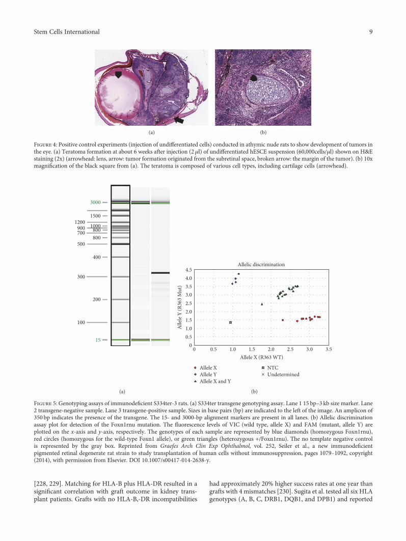
Figure 4: Positive control experiments (injection of undifferentiated cells) conducted in athymic nude rats to show development of tumors inthe eye. (a) Teratoma formation at about 6 weeks after injection (2 μl) of undifferentiated hESCE suspension (60,000cells/μl) shown on H&Estaining (2x) (arrowhead: lens, arrow: tumor formation originated from the subretinal space, broken arrow: the margin of the tumor). (b) 10xmagnification of the black square from (a). The teratoma is composed of various cell types, including cartilage cells (arrowhead).
3000
1500
1000900 800800
500
400
300
200
100
15
700
1200
(a)
Allelic discrimination
Alle
le Y
(R36
3 M
ut)
00
0.5
0.5
1.0
1.0
1.5 3.0 3.5
3.54.04.5
1.5
2.0
2.0
2.5
2.5
Allele XAllele YAllele X and Y
NTCUndetermined
Allele X (R363 WT)
3.0
(b)
Figure 5: Genotyping assays of immunodeficient S334ter-3 rats. (a) S334ter transgene genotyping assay. Lane 1 15 bp–3 kb size marker. Lane2 transgene-negative sample. Lane 3 transgene-positive sample. Sizes in base pairs (bp) are indicated to the left of the image. An amplicon of350 bp indicates the presence of the transgene. The 15- and 3000-bp alignment markers are present in all lanes. (b) Allelic discriminationassay plot for detection of the Foxn1rnu mutation. The fluorescence levels of VIC (wild type, allele X) and FAM (mutant, allele Y) areplotted on the x-axis and y-axis, respectively. The genotypes of each sample are represented by blue diamonds (homozygous Foxn1rnu),red circles (homozygous for the wild-type Foxn1 allele), or green triangles (heterozygous +/Foxn1rnu). The no template negative controlis represented by the gray box. Reprinted from Graefes Arch Clin Exp Ophthalmol, vol. 252, Seiler et al., a new immunodeficientpigmented retinal degenerate rat strain to study transplantation of human cells without immunosuppression, pages 1079–1092, copyright(2014), with permission from Elsevier. DOI 10.1007/s00417-014-2638-y.
9Stem Cells International
that the effector T cells can recognize MHC molecules on theallogeneic iPSC-RPE cells, but the immune reaction causedby the T cells can be prevented after HLA blood tests [224].Therefore, the future clinical trials can make use of alloge-neic RPE cells derived from iPSC lines procured from theHLA-homozygous iPSC bank [39, 224]. Nevertheless, furtherdetailed analysis is needed using larger sample size and long-term follow-up [224].
In a recent report, three AMD patients in Florida sufferedsevere vision loss after receiving injection of autologousadipose tissue-derived stem cells. In this study, adipose-derived stem cells were injected into the eye based on mini-mal clinical evidence of safety or efficacy. The injectioncaused ocular hypertension, hemorrhagic retinopathy, vitre-ous hemorrhage, combined traction and rhegmatogenousretinal detachment, and lens dislocation [41].
The major concern of optimum safety and purity of thecells is that the products should be free of undifferentiated
cells and should demonstrate the genetic and functional sig-nature of the desired stem cell-derived tissue. Undifferenti-ated pluripotent stem cells have the capacity to differentiateinto all cell types of the three germ layers and may causetumor formation. Therefore, extensive testing for the absenceof tumor formation and cell migration before implantationis crucial [2]. Differentiation into nondesired cell types is apotential threat to the success of stem cell-derived celltherapies. Confirming the purity of stem cell derivationsbefore transplantation is mandatory [231]. In one study,subcutaneous transplantation of iPSCs into immunosup-pressed mice resulted in tumor formation, demonstratingthe pluripotency of the injected iPSCs and its capability toevade immune detection [232]. The ability of tumor forma-tion is often assessed using tumorigenicity studies in animalmodels. According to Nazari et al. [2], assessing tumorigenic-ity potential in immunocompetent animal models can bemisleading since the absence of tumor formation might be
RCS-p+/ RCS-p+
Retinal degeneration,normal immunity, hairy
Foxn1rnu/ Foxn1rnu
Normal eyes, immunodeficient,hairless
Founder breeders
+/ RCS-p+ and +/ Foxn1rnu
Normal eyes, normalimmunity, hairy
+/ RCS-p+ and +/ Foxn1rnu
Normal eyes, normalimmunity, hairy
First generation
RCS-p+/ RCS-p+
and +/ Foxn1rnu
Retinal degeneration,normal immunity, hairy
RCS-p+/ RCS-p+
and Foxn1rnu/ Foxn1rnu
Retinal degeneration,immunodeficient, hairless
Second generation
RCS-p+/ RCS-p+
and Foxn1rnu/ Foxn1rnu
Retinal degeneration,immunodeficient, hairless
Third generation(Target animal model)
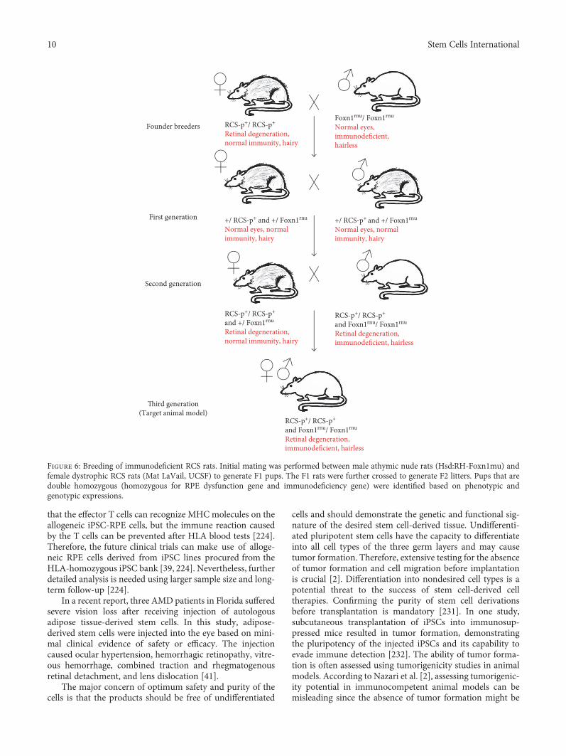
Figure 6: Breeding of immunodeficient RCS rats. Initial mating was performed between male athymic nude rats (Hsd:RH-Foxn1mu) andfemale dystrophic RCS rats (Mat LaVail, UCSF) to generate F1 pups. The F1 rats were further crossed to generate F2 litters. Pups that aredouble homozygous (homozygous for RPE dysfunction gene and immunodeficiency gene) were identified based on phenotypic andgenotypic expressions.
10 Stem Cells International

related to the ability of the host to reject tumorigenic cellsbefore tumors form. However, this can be overcome by usingpositive controls (injection of undifferentiated cells) that areexpected to develop tumors in the target area (Figure 4).
Although the eye is to a large extent regarded as animmune privileged organ, there is strong evidence forimmune response to xenografts [199, 233–235]. When dis-ease models are used for assessing functional efficacy,immunosuppressant drugs are administered to avoid immu-nological rejection. Most of the preclinical studies involvinghuman-derived cells used animal models that are exposedto severe immunosuppression regimes [26, 196]. Adminis-tration of immunosuppressants in rodents is labor intensiveandmay cause additional pain and discomfort to the animals.A recent study demonstrated more adverse effects of immu-nosuppression in animal models. Cyclosporine A plusdexamethasone-administered RCS rats showed depressedscores on visual behavioral and electrophysiological testing[236]. To overcome the above issues, we have developednew immunodeficient rat models. This was accomplishedby crossing between nondystrophic immunodeficient ani-mals (NIH nude rats) and RD disease models. The doublehomozygous pups (immunodeficient RD) can be determinedby genotyping [143, 144]. Based on this, an immunodeficientS334ter-line-3 rat colony has been established which elimi-nated the need for immunosuppression when transplantingxenografts [143] (Figure 5). More recently, a new immuno-deficient RCS rat model has been also created [144] and iscurrently being tested for various stem cell-based products(Figure 6). By employing these models, it is possible to justifyethical concerns by reducing animal use and the overall studycost can be considerably lowered.
6. Conclusion
Stem cell-based therapies provide a new treatment option forretinal degenerative diseases that were previously consideredincurable. Preclinical experiments conducted in animal dis-ease models demonstrated functional efficacy and safety ofocular cell replacement therapies. Studies conducted in largeanimal models helped to establish the surgical techniquesrequired for clinical trials. The above animal studies havepaved the way for several clinical trials based on cell-basedtherapies currently in progress.
Conflicts of Interest
Magdalene J. Seiler has proprietary interest in the instrumentand method for transplanting retinal sheets (OcularTransplantation LLC). Mark S. Humayun, David R. Hinton,and Dennis O. Clegg are cofounders and consultants toRegenerative Patch Technologies (RPT). The authors declarethat there is no conflict of interest regarding the publicationof this paper.
Acknowledgments
This study was supported by the CIRM (Magdalene J. Seiler,Mark S. Humayun, David R. Hinton, and Dennis O. Clegg),
Bright Focus Foundation (Biju B. Thomas), and Research toPrevent Blindness (USC Roski Eye Institute). The authorsthank Dr. Robert Aramant (UCI, Stem Cell Research Center;Ocular Transplantation LLC, Crestwood, KY) for his helpfulcomments to the article and his contributions to retinal sheettransplant research. Work shown in the figures was madepossible by Xiaopeng Wang (USC), Bin Lin, Ph.D., BryceMcLelland, B.S., and Anu Mathur M.S. (UC Irvine, Stem CellResearch Center).
References
[1] M. K. Jones, B. Lu, S. Girman, and S. Wang, “Cell-based ther-apeutic strategies for replacement and preservation in retinaldegenerative diseases,” Progress in Retinal and Eye Research,vol. 58, pp. 1–27, 2017.
[2] H. Nazari, L. Zhang, D. Zhu et al., “Stem cell based therapiesfor age-related macular degeneration: the promises and thechallenges,” Progress in Retinal and Eye Research, vol. 48,pp. 1–39, 2015.
[3] M. J. Seiler and R. B. Aramant, “Cell replacement and visualrestoration by retinal sheet transplants,” Progress in Retinaland Eye Research, vol. 31, no. 6, pp. 661–687, 2012.
[4] J. D. Sengillo, S. Justus, Y. T. Tsai, T. Cabral, and S. H. Tsang,“Gene and cell-based therapies for inherited retinal disorders:an update,” American Journal of Medical Genetics Part C,Seminars in Medical Genetics, vol. 172, no. 4, pp. 349–366,2016.
[5] M. Zarbin, “Cell-based therapy for degenerative retinaldisease,” Trends in Molecular Medicine, vol. 22, no. 2,pp. 115–134, 2016.
[6] M. J. Seiler and R. B. Aramant, “Intact sheets of fetal retinatransplanted to restore damaged rat retinas,” InvestigativeOphthalmology & Visual Science, vol. 39, no. 11, pp. 2121–2131, 1998.
[7] R. B. Aramant, M. J. Seiler, and S. L. Ball, “Successful cotrans-plantation of intact sheets of fetal retina with retinal pigmentepithelium,” Investigative Ophthalmology & Visual Science,vol. 40, no. 7, pp. 1557–1564, 1999.
[8] N. D. Radtke, R. B. Aramant, H. M. Petry, P. T. Green, D. J.Pidwell, and M. J. Seiler, “Vision improvement in retinaldegeneration patients by implantation of retina together withretinal pigment epithelium,” American Journal of Ophthal-mology, vol. 146, no. 2, pp. 172–182, 2008.
[9] M. J. Seiler, R. E. Lin, B. T. McLelland et al., “Vision recoveryand connectivity by fetal retinal sheet transplantation in animmunodeficient retinal degenerate rat model,” InvestigativeOphthalmology & Visual Science, vol. 58, no. 1, pp. 614–630,2017.
[10] A. Dahlmann-Noor, S. Vijay, H. Jayaram, A. Limb, and P. T.Khaw, “Current approaches and future prospects for stemcell rescue and regeneration of the retina and optic nerve,”Canadian Journal of Ophthalmology, vol. 45, no. 4, pp. 333–341, 2010.
[11] P. E. Royo and W. B. Quay, “Retinal transplantationfrom fetal to maternal mammalian eye,” Growth, vol. 23,pp. 313–336, 1959.
[12] M. del Cerro, H. H. Yeh, A. Marrero-Rodriguez, E. Lazar, andC. del Cerro, “Intraocular transplantation of cell layersderived from neonatal rat retina,” Brain Research, vol. 535,no. 1, pp. 25–32, 1990.
11Stem Cells International

[13] R. Aramant, M. Seiler, B. Ehinger et al., “Transplantation ofhuman embryonic retina to adult rat retina,” RestorativeNeurology and Neuroscience, vol. 2, no. 1, pp. 9–22, 1990.
[14] M. del Cerro, J. R. Ison, G. P. Bowen, E. Lazar, and C. delCerro, “Intraretinal grafting restores visual function inlight-blinded rats,” Neuroreport, vol. 2, no. 9, pp. 529–532,1991.
[15] P. Gouras, J. Du, M. Gelanze, R. Kwun, H. Kjeldbye, andR. Lopez, “Transplantation of photoreceptors labeled withtritiated thymidine into RCS rats,” Investigative Ophthal-mology & Visual Science, vol. 32, no. 5, pp. 1704–1707,1991.
[16] R. Aramant and M. Seiler, “Cryopreservation and transplan-tation of immature rat retina into adult rat retina,” BrainResearch Developmental Brain Research, vol. 61, no. 2,pp. 151–159, 1991.
[17] R. B. Aramant and M. J. Seiler, “Human embryonic retinalcell transplants in athymic immunodeficient rat hosts,” CellTransplantation, vol. 3, no. 6, pp. 461–474, 1994.
[18] P. Gouras, M. T. Flood, and H. Kjeldbye, “Transplantationof cultured human retinal cells to monkey retina,” Anaisda Academia Brasileira de Ciencias, vol. 56, no. 4, pp. 431–443, 1984.
[19] J. E. Turner and J. R. Blair, “Newborn rat retinal cells trans-planted into a retinal lesion site in adult host eyes,” BrainResearch, vol. 391, no. 1, pp. 91–104, 1986.
[20] M. S. Silverman and S. E. Hughes, “Transplantation of photo-receptors to light-damaged retina,” Investigative Ophthalmol-ogy & Visual Science, vol. 30, no. 8, pp. 1684–1690, 1989.
[21] S. Mohand-Said, D. Hicks, M. Simonutti et al., “Photorecep-tor transplants increase host cone survival in the retinaldegeneration (rd) mouse,” Ophthalmic Research, vol. 29,no. 5, pp. 290–297, 1997.
[22] R. B. Aramant and M. J. Seiler, “Transplanted sheets ofhuman retina and retinal pigment epithelium develop nor-mally in nude rats,” Experimental Eye Research, vol. 75,no. 2, pp. 115–125, 2002.
[23] F. Ghosh, K. Arner, and B. Ehinger, “Transplant of full-thickness embryonic rabbit retina using pars plana vitrec-tomy,” Retina (Philadelphia, Pennsylvania), vol. 18, no. 2,pp. 136–142, 1998.
[24] F. Ghosh, B. Juliusson, K. Arner, and B. Ehinger, “Partial andfull-thickness neuroretinal transplants,” Experimental EyeResearch, vol. 68, no. 1, pp. 67–74, 1999.
[25] J. Wasselius and F. Ghosh, “Adult rabbit retinal transplants,”Investigative Ophthalmology & Visual Science, vol. 42, no. 11,pp. 2632–2638, 2001.
[26] B. Lu, C. Malcuit, S. Wang et al., “Long-term safety and func-tion of RPE from human embryonic stem cells in preclinicalmodels of macular degeneration,” Stem Cells (Dayton, Ohio),vol. 27, no. 9, pp. 2126–2135, 2009.
[27] E. Lee and R. E. MacLaren, “Sources of retinal pigment epi-thelium (RPE) for replacement therapy,” The British Journalof Ophthalmology, vol. 95, no. 4, pp. 445–449, 2011.
[28] B. Lu, D. Zhu, D. Hinton, M. S. Humayun, and Y. C. Tai,“Mesh-supported submicron parylene-C membranes for cul-turing retinal pigment epithelial cells,” Biomedical Microde-vices, vol. 14, no. 4, pp. 659–667, 2012.
[29] S. D. Schwartz, J. P. Hubschman, G. Heilwell et al., “Embry-onic stem cell trials for macular degeneration: a preliminaryreport,” Lancet, vol. 379, no. 9817, pp. 713–720, 2012.
[30] S. D. Schwartz, C. D. Regillo, B. L. Lam et al., “Human embry-onic stem cell-derived retinal pigment epithelium in patientswith age-related macular degeneration and Stargardt’s macu-lar dystrophy: follow-up of two open-label phase 1/2 studies,”Lancet, vol. 385, no. 9967, pp. 509–516, 2015.
[31] S. D. Schwartz, G. Tan, H. Hosseini, and A. Nagiel,“Subretinal transplantation of embryonic stem cell-derivedretinal pigment epithelium for the treatment of maculardegeneration: an assessment at 4 years,” Investigative Oph-thalmology & Visual Science, vol. 57, no. 5, pp. ORSFc1–ORSFc9, 2016.
[32] T. Aoi, K. Yae, M. Nakagawa et al., “Generation of plurip-otent stem cells from adult mouse liver and stomach cells,”Science, vol. 321, no. 5889, pp. 699–702, 2008.
[33] J. Hanna, S. Markoulaki, P. Schorderet et al., “Direct repro-gramming of terminally differentiated mature B lymphocytesto pluripotency,” Cell, vol. 133, no. 2, pp. 250–264, 2008.
[34] K. Takahashi, K. Okita, M. Nakagawa, and S. Yamanaka,“Induction of pluripotent stem cells from fibroblast cultures,”Nature Protocols, vol. 2, no. 12, pp. 3081–3089, 2007.
[35] K. Takahashi, K. Tanabe, M. Ohnuki et al., “Induction of plu-ripotent stem cells from adult human fibroblasts by definedfactors,” Cell, vol. 131, no. 5, pp. 861–872, 2007.
[36] S. Okamoto and M. Takahashi, “Induction of retinal pig-ment epithelial cells from monkey iPS cells,” InvestigativeOphthalmology & Visual Science, vol. 52, no. 12,pp. 8785–8790, 2011.
[37] Y. Li, Y. T. Tsai, C. W. Hsu et al., “Long-term safety and effi-cacy of human-induced pluripotent stem cell (iPS) grafts in apreclinical model of retinitis pigmentosa,” Molecular Medi-cine, vol. 18, pp. 1312–1319, 2012.
[38] A. Garg, J. Yang,W. Lee, and S. H. Tsang, “Stem cell therapiesin retinal disorders,” Cell, vol. 6, no. 1, 2017.
[39] D. Ilic, “iPSC in the past decade: the Japanese dominance,”Regenerative Medicine, vol. 11, no. 8, pp. 747–749, 2016.
[40] N. Wu and M. Doorenbos, “Induced pluripotent stem cells:development in the ophthalmologic field,” Stem Cells Inter-national, vol. 2016, Article ID 2361763, 7 pages, 2016.
[41] A. E. Kuriyan, T. A. Albini, J. H. Townsend et al., “Vision lossafter intravitreal injection of autologous “stem cells” forAMD,” The New England Journal of Medicine, vol. 376,no. 11, pp. 1047–1053, 2017.
[42] I. Klimanskaya, J. Hipp, K. A. Rezai, M.West, A. Atala, and R.Lanza, “Derivation and comparative assessment of retinalpigment epithelium from human embryonic stem cells usingtranscriptomics,” Cloning and Stem Cells, vol. 6, no. 3,pp. 217–245, 2004.
[43] R. D. Lund, S. Wang, I. Klimanskaya et al., “Human embry-onic stem cell-derived cells rescue visual function in dystro-phic RCS rats,” Cloning Stem Cells, vol. 8, no. 3, pp. 189–199, 2006.
[44] B. Diniz, P. Thomas, B. Thomas et al., “Subretinal implanta-tion of retinal pigment epithelial cells derived from humanembryonic stem cells: improved survival when implanted asa monolayer,” Investigative Ophthalmology & Visual Science,vol. 54, no. 7, pp. 5087–5096, 2013.
[45] B. B. Thomas, D. Zhu, L. Zhang et al., “Survival and function-ality of hESC-derived retinal pigment epithelium cells cul-tured as a monolayer on polymer substrates transplanted inRCS rats,” Investigative Ophthalmology & Visual Science,vol. 57, no. 6, pp. 2877–2887, 2016.
12 Stem Cells International

[46] S. R. Hynes and E. B. Lavik, “A tissue-engineered approachtowards retinal repair: scaffolds for cell transplantation tothe subretinal space,” Graefe's Archive for Clinical and Exper-imental Ophthalmology, vol. 248, no. 6, pp. 763–778, 2010.
[47] K. Bharti, M. Rao, S. C. Hull et al., “Developing cellular ther-apies for retinal degenerative diseases,” Investigative Ophthal-mology & Visual Science, vol. 55, no. 2, pp. 1191–1202, 2014.
[48] T. Maeda, M. J. Lee, G. Palczewska et al., “Retinal pigmentedepithelial cells obtained from human induced pluripotentstem cells possess functional visual cycle enzymes in vitroand in vivo,” The Journal of Biological Chemistry, vol. 288,no. 48, pp. 34484–34493, 2013.
[49] B. A. Tucker, R. F. Mullins, L. M. Streb et al., “Patient-specificiPSC-derived photoreceptor precursor cells as a means toinvestigate retinitis pigmentosa,” eLife, vol. 2, article e00824,2013.
[50] D. E. Buchholz, S. T. Hikita, T. J. Rowland et al., “Derivationof functional retinal pigmented epithelisum from inducedpluripotent stem cells,” Stem Cells (Dayton, Ohio), vol. 27,no. 10, pp. 2427–2434, 2009.
[51] A. J. Carr, A. Vugler, J. Lawrence et al., “Molecular charac-terization and functional analysis of phagocytosis byhuman embryonic stem cell-derived RPE cells using a novelhuman retinal assay,” Molecular Vision, vol. 15, pp. 283–295, 2009.
[52] Y. Hu, L. Liu, B. Lu et al., “A novel approach for subretinalimplantation of ultrathin substrates containing stem cell-derived retinal pigment epithelium monolayer,” OphthalmicResearch, vol. 48, no. 4, pp. 186–191, 2012.
[53] D. A. Lamba, J. Gust, and T. A. Reh, “Transplantation ofhuman embryonic stem cell-derived photoreceptors restoressome visual function in Crx-deficient mice,” Cell Stem Cell,vol. 4, no. 1, pp. 73–79, 2009.
[54] D. A. Lamba, A. McUsic, R. K. Hirata, P. R. Wang, D. Russell,and T. A. Reh, “Generation, purification and transplantationof photoreceptors derived from human induced pluripotentstem cells,” PLoS One, vol. 5, no. 1, article e8763, 2010.
[55] B. A. Tucker, I. H. Park, S. D. Qi et al., “Transplantation ofadult mouse iPS cell-derived photoreceptor precursorsrestores retinal structure and function in degenerative mice,”PLoS One, vol. 6, no. 4, article e18992, 2011.
[56] D. M. Gamm and L. S. Wright, “From embryonic stem cellsto mature photoreceptors,” Nature Biotechnology, vol. 31,no. 8, pp. 712-713, 2013.
[57] B. L. Coles, B. Angenieux, T. Inoue et al., “Facile isolation andthe characterization of human retinal stem cells,” Proceedingsof the National Academy of Sciences of the United States ofAmerica, vol. 101, no. 44, pp. 15772–15777, 2004.
[58] H. J. Klassen, T. F. Ng, Y. Kurimoto et al., “Multipotent reti-nal progenitors express developmental markers, differentiateinto retinal neurons, and preserve light-mediated behavior,”Investigative Ophthalmology & Visual Science, vol. 45,no. 11, pp. 4167–4173, 2004.
[59] J. Luo, P. Baranov, S. Patel et al., “Human retinal progenitorcell transplantation preserves vision,” The Journal of Biologi-cal Chemistry, vol. 289, no. 10, pp. 6362–6371, 2014.
[60] D. A. Lamba, M. O. Karl, C. B. Ware, and T. A. Reh, “Efficientgeneration of retinal progenitor cells from human embryonicstem cells,” Proceedings of the National Academy of Sciencesof the United States of America, vol. 103, no. 34, pp. 12769–12774, 2006.
[61] B. A. Tucker, S. M. Redenti, C. Jiang et al., “The use of pro-genitor cell/biodegradable MMP2-PLGA polymer constructsto enhance cellular integration and retinal repopulation,”Biomaterials, vol. 31, no. 1, pp. 9–19, 2010.
[62] S. Tao, C. Young, S. Redenti et al., “Survival, migrationand differentiation of retinal progenitor cells transplantedon micro-machined poly(methyl methacrylate) scaffoldsto the subretinal space,” Lab on a Chip, vol. 7, no. 6,pp. 695–701, 2007.
[63] K. E. Kador and J. L. Goldberg, “Scaffolds and stem cells:delivery of cell transplants for retinal degenerations,” ExpertReview of Ophthalmology, vol. 7, no. 5, pp. 459–470, 2012.
[64] H. Klassen, J. F. Kiilgaard, T. Zahir et al., “Progenitor cellsfrom the porcine neural retina express photoreceptormarkers after transplantation to the subretinal space ofallorecipients,” Stem Cells (Dayton, Ohio), vol. 25, no. 5,pp. 1222–1230, 2007.
[65] K. Warfvinge, J. F. Kiilgaard, E. B. Lavik et al., “Retinalprogenitor cell xenografts to the pig retina: morphologicintegration and cytochemical differentiation,” Archives ofOphthalmology, vol. 123, no. 10, pp. 1385–1393, 2005.
[66] R. D. Lund, S. Wang, B. Lu et al., “Cells isolated from umbil-ical cord tissue rescue photoreceptors and visual functions ina rodent model of retinal disease,” Stem Cells (Dayton, Ohio),vol. 25, no. 3, pp. 602–611, 2007.
[67] R. D. Lund, P. Adamson, Y. Sauve et al., “Subretinal trans-plantation of genetically modified human cell lines attenuatesloss of visual function in dystrophic rats,” Proceedings of theNational Academy of Sciences of the United States of America,vol. 98, no. 17, pp. 9942–9947, 2001.
[68] R. D. Lund, A. S. Kwan, D. J. Keegan, Y. Sauve, P. J. Coffey,and J. M. Lawrence, “Cell transplantation as a treatment forretinal disease,” Progress in Retinal and Eye Research,vol. 20, no. 4, pp. 415–449, 2001.
[69] A. Gonzalez-Cordero, E. L. West, R. A. Pearson et al., “Pho-toreceptor precursors derived from three-dimensionalembryonic stem cell cultures integrate and mature withinadult degenerate retina,” Nature Biotechnology, vol. 31,no. 8, pp. 741–747, 2013.
[70] J. Assawachananont, M. Mandai, S. Okamoto et al., “Trans-plantation of embryonic and induced pluripotent stem cell-derived 3D retinal sheets into retinal degenerative mice,”Stem Cell Reports, vol. 2, no. 5, pp. 662–674, 2014.
[71] M. Mandai, M. Fujii, T. Hashiguchi et al., “iPSC-derivedretina transplants improve vision in rd1 end-stageretinal-degeneration mice,” Stem Cell Reports, vol. 8, no. 1,pp. 69–83, 2017.
[72] M. J. Seiler, B. T. Sagdullaev, G. Woch, B. B. Thomas, and R.B. Aramant, “Transsynaptic virus tracing from host brain tosubretinal transplants,” The European Journal of Neurosci-ence, vol. 21, no. 1, pp. 161–172, 2005.
[73] M. J. Seiler, B. B. Thomas, Z. Chen, R. Wu, S. R. Sadda,and R. B. Aramant, “Retinal transplants restore visualresponses: trans-synaptic tracing from visually responsivesites labels transplant neurons,” The European Journal ofNeuroscience, vol. 28, no. 1, pp. 208–220, 2008.
[74] M. J. Seiler, R. B. Aramant, B. B. Thomas, Q. Peng, S. R.Sadda, and H. S. Keirstead, “Visual restoration and transplantconnectivity in degenerate rats implanted with retinal pro-genitor sheets,” The European Journal of Neuroscience,vol. 31, no. 3, pp. 508–520, 2010.
13Stem Cells International

[75] H. Shirai, M. Mandai, K. Matsushita et al., “Transplantationof human embryonic stem cell-derived retinal tissue in twoprimate models of retinal degeneration,” Proceedings of theNational Academy of Sciences of the United States of America,vol. 113, no. 1, pp. E81–E90, 2016.
[76] D. M. Gamm, M. J. Phillips, and R. Singh, “Modeling ret-inal degenerative diseases with human iPS-derived cells:current status and future implications,” vol. 8, no. 3,pp. 213–216, 2013.
[77] T. Nakano, S. Ando, N. Takata et al., “Self-formation of opticcups and storable stratified neural retina from human ESCs,”Cell Stem Cell, vol. 10, no. 6, pp. 771–785, 2012.
[78] X. Zhong, C. Gutierrez, T. Xue et al., “Generation of three-dimensional retinal tissue with functional photoreceptorsfrom human iPSCs,” Nature Communications, vol. 5,p. 4047, 2014.
[79] M. E. McLaughlin, M. A. Sandberg, E. L. Berson, and T. P.Dryja, “Recessive mutations in the gene encoding the beta-subunit of rod phosphodiesterase in patients with retinitispigmentosa,” Nature Genetics, vol. 4, no. 2, pp. 130–134,1993.
[80] C. H. Sung, C. Makino, D. Baylor, and J. Nathans, “Arhodopsin gene mutation responsible for autosomal domi-nant retinitis pigmentosa results in a protein that is defec-tive in localization to the photoreceptor outer segment,”The Journal of Neuroscience, vol. 14, no. 10, pp. 5818–5833, 1994.
[81] M. Frasson, J. A. Sahel, M. Fabre, M. Simonutti, H. Dreyfus,and S. Picaud, “Retinitis pigmentosa: rod photoreceptor res-cue by a calcium-channel blocker in the rd mouse,” NatureMedicine, vol. 5, no. 10, pp. 1183–1187, 1999.
[82] A. J. Jimenez, J. M. Garcia-Fernandez, B. Gonzalez, and R. G.Foster, “The spatio-temporal pattern of photoreceptor degen-eration in the aged rd/rd mouse retina,” Cell and TissueResearch, vol. 284, no. 2, pp. 193–202, 1996.
[83] B. Chang, N. L. Hawes, M. T. Pardue et al., “Two mouse ret-inal degenerations caused by missense mutations in the beta-subunit of rod cGMP phosphodiesterase gene,” VisionResearch, vol. 47, no. 5, pp. 624–633, 2007.
[84] Z. Y. Li, F. Wong, J. H. Chang et al., “Rhodopsin transgenicpigs as a model for human retinitis pigmentosa,” InvestigativeOphthalmology & Visual Science, vol. 39, no. 5, pp. 808–819,1998.
[85] S. Machida, M. Kondo, J. A. Jamison et al., “P23H rhodopsintransgenic rat: correlation of retinal function with histopa-thology,” Investigative Ophthalmology & Visual Science,vol. 41, no. 10, pp. 3200–3209, 2000.
[86] M. Kondo, T. Sakai, K. Komeima et al., “Generation of atransgenic rabbit model of retinal degeneration,” Investiga-tive Ophthalmology & Visual Science, vol. 50, no. 3,pp. 1371–1377, 2009.
[87] G.Martinez-Navarrete, M. J. Seiler, R. B. Aramant, L. Fernan-dez-Sanchez, I. Pinilla, and N. Cuenca, “Retinal degenerationin two lines of transgenic S334ter rats,” Experimental EyeResearch, vol. 92, no. 3, pp. 227–237, 2011.
[88] D. T. Organisciak and D. K. Vaughan, “Retinal light damage:mechanisms and protection,” Progress in Retinal and EyeResearch, vol. 29, no. 2, pp. 113–134, 2010.
[89] R. S. Shah, B. T. Soetikno,M. Lajko, andA.A. Fawzi, “Amousemodel for laser-induced choroidal neovascularization,” Jour-nal of Visualized Experiments, vol. 106, article e53502, 2015.
[90] C. Graymore and K. Tansley, “Iodoacetate poisoning of therat retina. I. Production of retinal degeneration,” The BritishJournal of Ophthalmology, vol. 43, no. 3, pp. 177–185, 1959.
[91] Y. Y. Chen, S. L. Liu, D. P. Hu, Y. Q. Xing, and Y. Shen, “N-methyl- N -nitrosourea-induced retinal degeneration inmice,”Experimental Eye Research, vol. 121, pp. 102–113, 2014.
[92] A. Tsubura, K. Yoshizawa, M. Kuwata, and N. Uehara,“Animal models for retinitis pigmentosa induced byMNU; disease progression, mechanisms and therapeutictrials,” Histology and Histopathology, vol. 25, no. 7,pp. 933–944, 2010.
[93] A. Sorsby, “Experimental pigmentary degeneration of the ret-ina by sodium iodate,” The British Journal of Ophthalmology,vol. 25, no. 2, pp. 58–62, 1941.
[94] K. Kiuchi, K. Yoshizawa, N. Shikata, K. Moriguchi, and A.Tsubura, “Morphologic characteristics of retinal degenera-tion induced by sodium iodate in mice,” Current EyeResearch, vol. 25, no. 6, pp. 373–379, 2002.
[95] V. Enzmann, B. W. Row, Y. Yamauchi et al., “Behavioral andanatomical abnormalities in a sodium iodate-induced modelof retinal pigment epithelium degeneration,” ExperimentalEye Research, vol. 82, no. 3, pp. 441–448, 2006.
[96] M. Carido, Y. Zhu, K. Postel et al., “Characterization of amouse model with complete RPE loss and its use forRPE cell transplantation,” Investigative Ophthalmology &Visual Science, vol. 55, no. 8, pp. 5431–5444, 2014.
[97] S. Dithmar, C. A. Curcio, Le NA, S. Brown, and H. E.Grossniklaus, “Ultrastructural changes in Bruch’s membraneof apolipoprotein E-deficient mice,” Investigative Ophthal-mology & Visual Science, vol. 41, no. 8, pp. 2035–2042, 2000.
[98] J. Ambati, A. Anand, S. Fernandez et al., “An animalmodel of age-related macular degeneration in senescentCcl-2- or Ccr-2-deficient mice,” Nature Medicine, vol. 9,no. 11, pp. 1390–1397, 2003.
[99] M. Rudolf, B. Winkler, Z. Aherrahou, L. C. Doehring, P.Kaczmarek, and U. Schmidt-Erfurth, “Increased expressionof vascular endothelial growth factor associated with accu-mulation of lipids in Bruch’s membrane of LDL receptorknockout mice,” The British Journal of Ophthalmology,vol. 89, no. 12, pp. 1627–1630, 2005.
[100] Y. Imamura, S. Noda, K. Hashizume et al., “Drusen, choroi-dal neovascularization, and retinal pigment epithelium dys-function in SOD1-deficient mice: a model of age-relatedmacular degeneration,” Proceedings of the National Academyof Sciences of the United States of America, vol. 103, no. 30,pp. 11282–11287, 2006.
[101] J. Tuo, C. M. Bojanowski, M. Zhou et al., “Murine ccl2/cx3cr1deficiency results in retinal lesions mimicking human age-related macular degeneration,” Investigative Ophthalmology& Visual Science, vol. 48, no. 8, pp. 3827–3836, 2007.
[102] U. F. Luhmann, S. Robbie, P. M. Munro et al., “The drusen-like phenotype in aging Ccl2-knockout mice is caused by anaccelerated accumulation of swollen autofluorescent subret-inal macrophages,” Investigative Ophthalmology & VisualScience, vol. 50, no. 12, pp. 5934–5943, 2009.
[103] Z. Zhao, Y. Chen, J. Wang et al., “Age-related retinopathyin NRF2-deficient mice,” PLoS One, vol. 6, no. 4, articlee19456, 2011.
[104] R. Allikmets, “Simple and complex ABCR: genetic predis-position to retinal disease,” American Journal of HumanGenetics, vol. 67, no. 4, pp. 793–799, 2000.
14 Stem Cells International

[105] F. P. Cremers, D. J. van de Pol, M. van Driel et al., “Autoso-mal recessive retinitis pigmentosa and cone-rod dystrophycaused by splice site mutations in the Stargardt’s disease geneABCR,” Human Molecular Genetics, vol. 7, no. 3, pp. 355–362, 1998.
[106] R. Allikmets, N. Singh, H. Sun et al., “A photoreceptor cell-specific ATP-binding transporter gene (ABCR) is mutatedin recessive Stargardt macular dystrophy,” Nature Genetics,vol. 15, no. 3, pp. 236–246, 1997.
[107] J. Weng, N. L. Mata, S. M. Azarian, R. T. Tzekov, D. G. Birch,and G. H. Travis, “Insights into the function of Rim proteinin photoreceptors and etiology of Stargardt’s disease fromthe phenotype in abcr knockout mice,” Cell, vol. 98, no. 1,pp. 13–23, 1999.
[108] N. L. Mata, J. Weng, and G. H. Travis, “Biosynthesis of amajor lipofuscin fluorophore in mice and humans withABCR-mediated retinal and macular degeneration,” Proceed-ings of the National Academy of Sciences of the United Statesof America, vol. 97, no. 13, pp. 7154–7159, 2000.
[109] N. F. Shroyer, R. A. Lewis, A. N. Yatsenko, T. G. Wensel,and J. R. Lupski, “Cosegregation and functional analysis ofmutant ABCR (ABCA4) alleles in families that manifestboth Stargardt disease and age-related macular degenera-tion,” Human Molecular Genetics, vol. 10, no. 23, pp. 2671–2678, 2001.
[110] P. Charbel Issa and A. R. Barnard, “Rescue of the Stargardtphenotype in Abca4 knockout mice through inhibition ofvitamin A dimerization,” Proceedings of the National Acad-emy of Sciences of the United States of America, vol. 112,no. 27, pp. 8415–8420, 2015.
[111] R. G. Weleber, M. Michaelides, K. M. Trzupek, N. B. Stover,and E. M. Stone, “The phenotype of severe early childhoodonset retinal dystrophy (SECORD) from mutation ofRPE65 and differentiation from Leber congenital amaurosis,”Investigative Ophthalmology & Visual Science, vol. 52, no. 1,pp. 292–302, 2011.
[112] B. Rohrer, P. Goletz, S. Znoiko et al., “Correlation of regener-able opsin with rod ERG signal in Rpe65−/− mice duringdevelopment and aging,” Investigative Ophthalmology &Visual Science, vol. 44, no. 1, pp. 310–315, 2003.
[113] S. L. Znoiko, B. Rohrer, K. Lu, H. R. Lohr, R. K. Crouch, and J.X. Ma, “Downregulation of cone-specific gene expression anddegeneration of cone photoreceptors in the Rpe65−/−mouseat early ages,” Investigative Ophthalmology & Visual Science,vol. 46, no. 4, pp. 1473–1479, 2005.
[114] J. E. Olsson, J. W. Gordon, B. S. Pawlyk et al., “Transgenicmice with a rhodopsin mutation (Pro23His): a mouse modelof autosomal dominant retinitis pigmentosa,” Neuron, vol. 9,no. 5, pp. 815–830, 1992.
[115] A. S. Lewin, B. Rossmiller, and H. Mao, “Gene augmentationfor adRP mutations in RHO,” Cold Spring Harbor Perspec-tives in Medicine, vol. 4, no. 9, article a017400, 2014.
[116] K. Zhang, M. Kniazeva, M. Han et al., “A 5-bp deletion inELOVL4 is associated with two related forms of autosomaldominant macular dystrophy,” Nature Genetics, vol. 27,no. 1, pp. 89–93, 2001.
[117] A. O. Edwards, L. A. Donoso, and R. Ritter 3rd, “A novel genefor autosomal dominant Stargardt-like macular dystrophywith homology to the SUR4 protein family,” InvestigativeOphthalmology & Visual Science, vol. 42, no. 11, pp. 2652–2663, 2001.
[118] G. Karan, C. Lillo, Z. Yang et al., “Lipofuscin accumulation,abnormal electrophysiology, and photoreceptor degenerationin mutant ELOVL4 transgenic mice: a model for maculardegeneration,” Proceedings of the National Academy of Sci-ences of the United States of America, vol. 102, no. 11,pp. 4164–4169, 2005.
[119] P. D. Westenskow, T. Kurihara, S. Bravo et al., “Performingsubretinal injections in rodents to deliver retinal pigment epi-thelium cells in suspension,” Journal of Visualized Experi-ments, vol. 95, article 52247, 2015.
[120] S. Arai, B. B. Thomas, M. J. Seiler et al., “Restoration of visualresponses following transplantation of intact retinal sheets inrd mice,” Experimental Eye Research, vol. 79, no. 3, pp. 331–341, 2004.
[121] S. Remtulla and P. E. Hallett, “A schematic eye for the mouse,and comparisons with the rat,” Vision Research, vol. 25, no. 1,pp. 21–31, 1985.
[122] P. M. D'Cruz, D. Yasumura, J. Weir et al., “Mutation of thereceptor tyrosine kinase gene Mertk in the retinal dystrophicRCS rat,” Human Molecular Genetics, vol. 9, no. 4, pp. 645–651, 2000.
[123] D. Vollrath, W. Feng, J. L. Duncan et al., “Correction of theretinal dystrophy phenotype of the RCS rat by viral genetransfer of Mertk,” Proceedings of the National Academy ofSciences of the United States of America, vol. 98, no. 22,pp. 12584–12589, 2001.
[124] E. F. Nandrot and E. M. Dufour, “Mertk in daily retinalphagocytosis: a history in the making,” Advances in Experi-mental Medicine and Biology, vol. 664, pp. 133–140, 2010.
[125] G. Li, B. De La Garza, Y. Y. Shih, E. R. Muir, and T. Q. Duong,“Layer-specific blood-flowMRI of retinitis pigmentosa inRCSrats,” Experimental Eye Research, vol. 101, pp. 90–96, 2012.
[126] T. J. McGill, R. M. Douglas, R. D. Lund, and G. T. Prusky,“Quantification of spatial vision in the Royal College of Sur-geons rat,” Investigative Ophthalmology & Visual Science,vol. 45, no. 3, pp. 932–936, 2004.
[127] A. J. Carr, A. A. Vugler, S. T. Hikita et al., “Protective effectsof human iPS-derived retinal pigment epithelium cell trans-plantation in the retinal dystrophic rat,” PLoS One, vol. 4,no. 12, article e8152, 2009.
[128] I. Pinilla, N. Cuenca, Y. Sauve, S. Wang, and R. D. Lund,“Preservation of outer retina and its synaptic connectivity fol-lowing subretinal injections of human RPE cells in the RoyalCollege of Surgeons rat,” Experimental Eye Research, vol. 85,no. 3, pp. 381–392, 2007.
[129] S. Wang, B. Lu, S. Girman, T. Holmes, N. Bischoff, and R. D.Lund, “Morphological and functional rescue in RCS rats afterRPE cell line transplantation at a later stage of degeneration,”Investigative Ophthalmology & Visual Science, vol. 49, no. 1,pp. 416–421, 2008.
[130] U. C. Park, M. S. Cho, J. H. Park et al., “Subretinal transplan-tation of putative retinal pigment epithelial cells derived fromhuman embryonic stem cells in rat retinal degenerationmodel,” Clinical and Experimental Reproductive Medicine,vol. 38, no. 4, pp. 216–221, 2011.
[131] O. S. Kozhevnikova, E. E. Korbolina, N. I. Ershov, and N. G.Kolosova, “Rat retinal transcriptome: effects of aging andAMD-like retinopathy,” Cell Cycle, vol. 12, no. 11,pp. 1745–1761, 2013.
[132] A. M. Markovets, V. B. Saprunova, A. A. Zhdankina, A.Fursova, L. E. Bakeeva, and N. G. Kolosova, “Alterations
15Stem Cells International

of retinal pigment epithelium cause AMD-like retinopathyin senescence-accelerated OXYS rats,” Aging (Albany, NewYork), vol. 3, no. 1, pp. 44–54, 2011.
[133] N. Cuenca, I. Pinilla, Y. Sauve, B. Lu, S. Wang, and R. D.Lund, “Regressive and reactive changes in the connectivitypatterns of rod and cone pathways of P23H transgenicrat retina,” Neuroscience, vol. 127, no. 2, pp. 301–317, 2004.
[134] C. Liu, Y. Li, M. Peng, A. M. Laties, and R. Wen, “Activationof caspase-3 in the retina of transgenic rats with the rhodop-sin mutation s334ter during photoreceptor degeneration,”The Journal of Neuroscience, vol. 19, no. 12, pp. 4778–4785,1999.
[135] A. Ray, G. J. Sun, L. Chan, N. M. Grzywacz, J. Weiland, and E.J. Lee, “Morphological alterations in retinal neurons in theS334ter-line3 transgenic rat,” Cell and Tissue Research,vol. 339, no. 3, pp. 481–491, 2010.
[136] C. L. Zhu, Y. Ji, E. J. Lee, and N. M. Grzywacz, “Spatiotempo-ral pattern of rod degeneration in the S334ter-line-3 ratmodel of retinitis pigmentosa,” Cell and Tissue Research,vol. 351, no. 1, pp. 29–40, 2013.
[137] B. T. Sagdullaev, R. B. Aramant, M. J. Seiler, G. Woch, andM. A. McCall, “Retinal transplantation-induced recovery ofretinotectal visual function in a rodent model of retinitispigmentosa,” Investigative Ophthalmology & Visual Science,vol. 44, no. 4, pp. 1686–1695, 2003.
[138] B. B. Thomas, M. J. Seiler, S. R. Sadda, and R. B. Aramant,“Superior colliculus responses to light - preserved by trans-plantation in a slow degeneration rat model,” ExperimentalEye Research, vol. 79, no. 1, pp. 29–39, 2004.
[139] Q. Peng, B. B. Thomas, R. B. Aramant, Z. Chen, S. R. Sadda,and M. J. Seiler, “Structure and function of embryonic rat ret-inal sheet transplants,” Current Eye Research, vol. 32, no. 9,pp. 781–789, 2007.
[140] K. Gregory-Evans, F. Chang, M. D. Hodges, and C. Y. Greg-ory-Evans, “Ex vivo gene therapy using intravitreal injectionof GDNF-secreting mouse embryonic stem cells in a ratmodel of retinal degeneration,” Molecular Vision, vol. 15,pp. 962–973, 2009.
[141] A. Yanai, U. O. Hafeli, A. L. Metcalfe et al., “Focused mag-netic stem cell targeting to the retina using superparamag-netic iron oxide nanoparticles,” Cell Transplantation,vol. 21, no. 6, pp. 1137–1148, 2012.
[142] A. Santos, M. S. Humayun, E. de Juan Jr. et al., “Preservationof the inner retina in retinitis pigmentosa. A morphometricanalysis,” Archives of Ophthalmology, vol. 115, no. 4,pp. 511–515, 1997.
[143] M. J. Seiler, R. B. Aramant, M. K. Jones, D. L. Ferguson, E. C.Bryda, and H. S. Keirstead, “A new immunodeficient pig-mented retinal degenerate rat strain to study transplantationof human cells without immunosuppression,” Graefe'sArchive for Clinical and Experimental Ophthalmology,vol. 252, no. 7, pp. 1079–1092, 2014.
[144] B. Thomas, C. Shih, D. Zhu, J. Martinez, D. Hinton, and M.Humayun, A new Immunodeficient Dystrophic RCS rat Modelfor Transplantation Studies Using Human Derived CellsAssosciation for Research in Vision and Ophthalmology;2016, Seattle, WA, USA, 2016.
[145] S. Veleri, C. H. Lazar, B. Chang, P. A. Sieving, E. Banin, andA. Swaroop, “Biology and therapy of inherited retinal degen-erative disease: insights from mouse models,” Disease Models& Mechanisms, vol. 8, no. 2, pp. 109–129, 2015.
[146] E. V. Famiglietti and S. J. Sharpe, “Regional topography ofrod and immunocytochemically characterized “blue” and“green” cone photoreceptors in rabbit retina,” Visual Neuro-science, vol. 12, no. 6, pp. 1151–1175, 1995.
[147] B. Juliusson, A. Bergstrom, P. Rohlich, B. Ehinger, T. vanVeen, and A. Szel, “Complementary cone fields of the rabbitretina,” Investigative Ophthalmology & Visual Science,vol. 35, no. 3, pp. 811–818, 1994.
[148] K. W. Gjorloff, F. Ghosh, S. Andréasson, and B. Ehinger,“Standardized full-field and multifocal electroretinographyin rabbits,” Investigative Ophthalmology & Visual Science,vol. 44, no. 13, p. 2695, 2003.
[149] B. W. Jones, M. Kondo, H. Terasaki et al., “Retinal remodel-ing in the Tg P347L rabbit, a large-eye model of retinal degen-eration,” The Journal of Comparative Neurology, vol. 519,no. 14, pp. 2713–2733, 2011.
[150] S. Ueno, T. Koyasu, T. Kominami et al., “Focal cone ERGs ofrhodopsin Pro347Leu transgenic rabbits,” Vision Research,vol. 91, pp. 118–123, 2013.
[151] B. J. Cho, J. M. Seo, H. G. Yu, and H. Chung, “Monocular ret-inal degeneration induced by intravitreal injection of sodiumiodate in rabbit eyes,” Japanese Journal of Ophthalmology,vol. 60, no. 3, pp. 226–237, 2016.
[152] S. Rosch, C. Werner, F. Muller, and P. Walter, “Photorecep-tor degeneration by intravitreal injection of N-methyl-N-nitrosourea (MNU) in rabbits: a pilot study,” Graefe's Archivefor Clinical and Experimental Ophthalmology, vol. 255, no. 2,pp. 317–331, 2017.
[153] S. Petrus-Reurer, H. Bartuma, M. Aronsson et al., “Integra-tion of subretinal suspension transplants of human embry-onic stem cell-derived retinal pigment epithelial cells in alarge-eyed model of geographic atrophy,” Investigative Oph-thalmology & Visual Science, vol. 58, no. 2, pp. 1314–1322,2017.
[154] R. Curtis, K. C. Barnett, and A. Leon, “An early-onset retinaldystrophy with dominant inheritance in the Abyssinian cat.Clinical and pathological findings,” Investigative Ophthal-mology & Visual Science, vol. 28, no. 1, pp. 131–139, 1987.
[155] K. Narfstrom, “Hereditary progressive retinal atrophy in theAbyssinian cat,” The Journal of Heredity, vol. 74, no. 4,pp. 273–276, 1983.
[156] M. Menotti-Raymond, V. A. David, A. A. Schaffer et al.,“Mutation in CEP290 discovered for cat model of human ret-inal degeneration,” The Journal of Heredity, vol. 98, no. 3,pp. 211–220, 2007.
[157] K. Narfstrom, V. David, O. Jarret et al., “Retinal degenerationin the Abyssinian and Somali cat (rdAc): correlation betweengenotype and phenotype and rdAc allele frequency in twocontinents,” Veterinary Ophthalmology, vol. 12, no. 5,pp. 285–291, 2009.
[158] M. W. Seeliger and K. Narfstrom, “Functional assessment ofthe regional distribution of disease in a cat model of heredi-tary retinal degeneration,” Investigative Ophthalmology &Visual Science, vol. 41, no. 7, pp. 1998–2005, 2000.
[159] L. M. Occelli, N. M. Tran, K. Narfstrom, S. Chen, andS. M. Petersen-Jones, “CrxRdy cat: a large animal modelfor CRX-associated Leber congenital amaurosis,” Investiga-tive Ophthalmology & Visual Science, vol. 57, no. 8,pp. 3780–3792, 2016.
[160] H. Rah, D. J. Maggs, T. N. Blankenship, K. Narfstrom,and L. A. Lyons, “Early-onset, autosomal recessive,
16 Stem Cells International

progressive retinal atrophy in Persian cats,” InvestigativeOphthalmology & Visual Science, vol. 46, no. 5, pp. 1742–1747, 2005.
[161] Y. Nan, Q. Zhang, C. Ren et al., “Functional evaluation ofiodoacetic acid induced photoreceptor degeneration in thecat,” Science China Life Sciences, vol. 56, no. 6, pp. 524–530,2013.
[162] M. J. Seiler, R. B. Aramant, M. W. Seeliger, R. Bragadottir, M.Mahoney, and K. Narfstrom, “Functional and structuralassessment of retinal sheet allograft transplantation in felinehereditary retinal degeneration,” Veterinary Ophthalmology,vol. 12, no. 3, pp. 158–169, 2009.
[163] K. Shibuya, M. Tomohiro, S. Sasaki, and S. Otake, “Charac-teristics of structures and lesions of the eye in laboratoryanimals used in toxicity studies,” Journal of ToxicologicPathology, vol. 28, no. 4, pp. 181–188, 2015.
[164] G. G. Cloyd, M. Wyman, J. A. Shadduck, M. J. Winrow, andG. R. Johnson, “Ocular toxicity studies with zinc pyridi-nethione,” Toxicology and Applied Pharmacology, vol. 45,no. 3, pp. 771–782, 1978.
[165] D. M. Schiavo, D. P. Sinha, H. E. Black et al., “Tapetal changesin beagle dogs. I. Ocular changes after oral administration ofa beta-adrenergic blocking agent—SCH 19927,” Toxicologyand Applied Pharmacology, vol. 72, no. 2, pp. 187–194, 1984.
[166] T. Massa, G. J. Davis, D. Schiavo et al., “Tapetal changes inbeagle dogs. II. Ocular changes after intravenous administra-tion of a macrolide antibiotic—rosaramicin,” Toxicology andApplied Pharmacology, vol. 72, no. 2, pp. 195–200, 1984.
[167] J. H. Fortner, W. B. Milisen, G. R. Lundeen, A. B. Jakowski,and P. M. Marsh, “Tapetal effect of an azalide antibiotic fol-lowing oral administration in beagle dogs,” Fundamentaland applied toxicology: Official Journal of the Society of Toxi-cology, vol. 21, no. 2, pp. 164–173, 1993.
[168] J. E. Dillberger, R. L. Peiffer, M. J. Dykstra, M. O'Mara, and D.K. Patel, “The experimental antipsychotic agent 1192U90 tar-gets tapetum lucidum in canine eyes,” Toxicologic Pathology,vol. 24, no. 5, pp. 595–601, 1996.
[169] J. W. Kijas, A. V. Cideciyan, T. S. Aleman et al., “Naturallyoccurring rhodopsin mutation in the dog causes retinaldysfunction and degeneration mimicking human dominantretinitis pigmentosa,” Proceedings of the National Academyof Sciences of the United States of America, vol. 99, no. 9,pp. 6328–6333, 2002.
[170] A. V. Cideciyan, S. G. Jacobson, T. S. Aleman et al., “In vivodynamics of retinal injury and repair in the rhodopsinmutant dog model of human retinitis pigmentosa,” Proceed-ings of the National Academy of Sciences of the United Statesof America, vol. 102, no. 14, pp. 5233–5238, 2005.
[171] S. Iwabe, G. S. Ying, G. D. Aguirre, and W. A. Beltran,“Assessment of visual function and retinal structure follow-ing acute light exposure in the light sensitive T4R rhodopsinmutant dog,” Experimental Eye Research, vol. 146, pp. 341–353, 2016.
[172] R. M. Petters, C. A. Alexander, K. D. Wells et al., “Geneticallyengineered large animal model for studying cone photorecep-tor survival and degeneration in retinitis pigmentosa,”NatureBiotechnology, vol. 15, no. 10, pp. 965–970, 1997.
[173] M. O. Tso, W. W. Li, C. Zhang et al., “A pathologic study ofdegeneration of the rod and cone populations of the rhodop-sin Pro347Leu transgenic pigs,” Transactions of the AmericanOphthalmological Society, vol. 95, pp. 467–483, 1997.
[174] L. C. Shaw, A. Skold, F. Wong, R. Petters, W. W. Hauswirth,and A. S. Lewin, “An allele-specific hammerhead ribozymegene therapy for a porcine model of autosomal dominant ret-initis pigmentosa,” Molecular Vision, vol. 7, pp. 6–13, 2001.
[175] T. W. Kraft, D. Allen, R. M. Petters, Y. Hao, Y. W. Peng, andF. Wong, “Altered light responses of single rod photorecep-tors in transgenic pigs expressing P347L or P347S rhodop-sin,” Molecular Vision, vol. 11, pp. 1246–1256, 2005.
[176] J. W. Ross, J. P. Fernandez de Castro, J. Zhao et al., “Genera-tion of an inbred miniature pig model of retinitis pigmen-tosa,” Investigative Ophthalmology & Visual Science, vol. 53,no. 1, pp. 501–507, 2012.
[177] F. Ghosh, F.Wong, K. Johansson, A. Bruun, and R.M. Petters,“Transplantation of full-thickness retina in the rhodopsintransgenic pig,” Retina (Philadelphia, Pennsylvania), vol. 24,no. 1, pp. 98–109, 2004.
[178] H. Klassen, J. F. Kiilgaard, K. Warfvinge et al., “Photoreceptordifferentiation following transplantation of allogeneic retinalprogenitor cells to the dystrophic rhodopsin Pro347Leutransgenic pig,” Stem Cells International, vol. 2012, ArticleID 939801, 9 pages, 2012.
[179] M. J. Koss, P. Falabella, F. R. Stefanini et al., “Subretinalimplantation of a monolayer of human embryonic stemcell-derived retinal pigment epithelium: a feasibility andsafety study in Yucatan minipigs,” Graefe's Archive for Clini-cal and Experimental Ophthalmology, vol. 254, no. 8,pp. 1553–1565, 2016.
[180] E. H. Sohn, C. Jiao, E. Kaalberg et al., “Allogenic iPSC-derivedRPE cell transplants induce immune response in pigs: a pilotstudy,” Scientific Reports, vol. 5, article 11791, 2015.
[181] C. Lane, M. Boulton, and J. Marshall, “Transplantation ofretinal pigment epithelium using a pars plana approach,”Eye (London, England), vol. 3, Part 1, pp. 27–32, 1989.
[182] L. V. Del Priore, R. Hornbeck, H. J. Kaplan et al., “Debride-ment of the pig retinal pigment epithelium in vivo,” Archivesof Ophthalmology, vol. 113, no. 7, pp. 939–944, 1995.
[183] N. B. Sorensen, N. Lassota, M. V. Kyhn et al., “Functionalrecovery after experimental RPE debridement, mfERG stud-ies in a porcine model,” Graefe's Archive for Clinical andExperimental Ophthalmology, vol. 251, no. 10, pp. 2319–2325, 2013.
[184] M. G. Nicolas, K. Fujiki, K. Murayama et al., “Studies on themechanism of early onset macular degeneration in cynomol-gus (Macaca fascicularis) monkeys. I. Abnormal concentra-tions of two proteins in the retina,” Experimental EyeResearch, vol. 62, no. 3, pp. 211–219, 1996.
[185] M. T. Suzuki, K. Terao, and Y. Yoshikawa, “Familial earlyonset macular degeneration in cynomolgus monkeys(Macaca fascicularis),” Primates, vol. 44, no. 3, pp. 291–294,2003.
[186] S. Umeda, R. Ayyagari, R. Allikmets et al., “Early-onsetmacular degeneration with drusen in a cynomolgus monkey(Macaca fascicularis) pedigree: exclusion of 13 candidategenes and loci,” Investigative Ophthalmology & VisualScience, vol. 46, no. 2, pp. 683–691, 2005.
[187] S. Umeda, M. T. Suzuki, H. Okamoto et al., “Molecularcomposition of drusen and possible involvement of anti-retinal autoimmunity in two different forms of maculardegeneration in cynomolgus monkey (Macaca fascicu-laris),” The FASEB Journal, vol. 19, no. 12, pp. 1683–1685, 2005.
17Stem Cells International

[188] P. J. Francis, S. Wang, Y. Zhang et al., “Subretinal transplan-tation of forebrain progenitor cells in nonhuman primates:survival and intact retinal function,” Investigative Ophthal-mology & Visual Science, vol. 50, no. 7, pp. 3425–3431, 2009.
[189] K. K. Singh, M. Krawczak, W. W. Dawson, and J. Schmidtke,“Association of HTRA1 and ARMS2 gene variation withdrusen formation in rhesus macaques,” Experimental EyeResearch, vol. 88, no. 3, pp. 479–482, 2009.
[190] C. J. Zeiss, “Animals as models of age-related maculardegeneration: an imperfect measure of the truth,” VeterinaryPathology, vol. 47, no. 3, pp. 396–413, 2010.
[191] W. K. Noell, “The impairment of visual cell structure byiodoacetate,” Journal of Cellular and Comparative Physiology,vol. 40, no. 1, pp. 25–55, 1952.
[192] D. Fuller, R. Machemer, and R. W. Knighton, “Retinaldamage produced by intraocular fiber optic light,” AmericanJournal of Ophthalmology, vol. 85, no. 4, pp. 519–537, 1978.
[193] J. M. Strazzeri, J. J. Hunter, B. D. Masella et al., “Focal damageto macaque photoreceptors produces persistent visual loss,”Experimental Eye Research, vol. 119, pp. 88–96, 2014.
[194] E. W. Lankau, P. V. Turner, R. J. Mullan, and G. G. Galland,“Use of nonhuman primates in research in North America,”Journal of the American Association for Laboratory AnimalScience, vol. 53, no. 3, pp. 278–282, 2014.
[195] A. Varki and T. K. Altheide, “Comparing the human andchimpanzee genomes: searching for needles in a haystack,”Genome Research, vol. 15, no. 12, pp. 1746–1758, 2005.
[196] H. Kamao, M. Mandai, S. Okamoto et al., “Characterizationof human induced pluripotent stem cell-derived retinal pig-ment epithelium cell sheets aiming for clinical application,”Stem Cell Reports, vol. 2, no. 2, pp. 205–218, 2014.
[197] B. B. Thomas, S. Arai, Y. Ikai et al., “Retinal transplantsevaluated by optical coherence tomography in photoreceptordegenerate rats,” Journal of Neuroscience Methods, vol. 151,no. 2, pp. 186–193, 2006.
[198] M. J. Seiler, B. Rao, R. B. Aramant et al., “Three-dimensionaloptical coherence tomography imaging of retinal sheetimplants in live rats,” Journal of Neuroscience Methods,vol. 188, no. 2, pp. 250–257, 2010.
[199] T. Ilmarinen, H. Hiidenmaa, P. Koobi et al., “Ultrathin poly-imide membrane as cell carrier for subretinal transplantationof human embryonic stem cell derived retinal pigment epi-thelium,” PLoS One, vol. 10, no. 11, article e0143669, 2015.
[200] C.R.Laver,A.L.Metcalfe, L. Szczygiel,A.Yanai,M.V. Sarunic,and K. Gregory-Evans, “Bimodal in vivo imaging providesearly assessment of stem-cell-based photoreceptor engraft-ment,” Eye (London, England), vol. 29, no. 5, pp. 681–690,2015.
[201] S. Al-Nawaiseh, F. Thieltges, Z. Liu et al., “A step by stepprotocol for subretinal surgery in rabbits,” Journal of Visual-ized Experiments, vol. 115, 2016.
[202] P. Zhang, A. Zam, Y. Jian et al., “In vivo wide-field multispec-tral scanning laser ophthalmoscopy-optical coherencetomography mouse retinal imager: longitudinal imaging ofganglion cells, microglia, and Muller glia, and mapping ofthe mouse retinal and choroidal vasculature,” Journal ofBiomedical Optics, vol. 20, no. 12, article 126005, 2015.
[203] R. M. Ribeiro, A. Oregon, B. Diniz et al., “In vivo detectionof hESC-RPE cells via confocal near-infrared fundus reflec-tance,” Ophthalmic Surgery, Lasers and Imaging Retina,vol. 44, no. 4, pp. 380–384, 2013.
[204] M. S. Silverman, S. E. Hughes, T. L. Valentino, and Y. Liu,“Photoreceptor transplantation: anatomic, electrophysio-logic, and behavioral evidence for the functional reconstruc-tion of retinas lacking photoreceptors,” ExperimentalNeurology, vol. 115, no. 1, pp. 87–94, 1992.
[205] S. V. Girman, S. Wang, and R. D. Lund, “Cortical visualfunctions can be preserved by subretinal RPE cell graftingin RCS rats,” Vision Research, vol. 43, no. 17, pp. 1817–1827, 2003.
[206] C. Gias, M. Jones, D. Keegan et al., “Preservation of visualcortical function following retinal pigment epithelium trans-plantation in the RCS rat using optical imaging techniques,”The European Journal of Neuroscience, vol. 25, no. 7,pp. 1940–1948, 2007.
[207] L. Q. Jiang and D. Hamasaki, “Corneal electroretinographicfunction rescued by normal retinal pigment epithelial graftsin retinal degenerative Royal College of Surgeons rats,” Inves-tigative Ophthalmology & Visual Science, vol. 35, no. 13,pp. 4300–4309, 1994.
[208] Y. Sauve, I. Pinilla, and R. D. Lund, “Partial preservation ofrod and cone ERG function following subretinal injection ofARPE-19 cells in RCS rats,” Vision Research, vol. 46, no. 8-9, pp. 1459–1472, 2006.
[209] T. U. Krohne, P. D. Westenskow, T. Kurihara et al., “Gener-ation of retinal pigment epithelial cells from small moleculesand OCT4 reprogrammed human induced pluripotent stemcells,” Stem Cells Translational Medicine, vol. 1, no. 2,pp. 96–109, 2012.
[210] D. C. Hood, L. J. Frishman, S. Saszik, and S. Viswanathan,“Retinal origins of the primate multifocal ERG: implicationsfor the human response,” Investigative Ophthalmology &Visual Science, vol. 43, no. 5, pp. 1673–1685, 2002.
[211] S. L. Ball and H. M. Petry, “Noninvasive assessment of retinalfunction in rats using multifocal electroretinography,” Inves-tigative Ophthalmology & Visual Science, vol. 41, no. 2,pp. 610–617, 2000.
[212] R. Siminoff, H. O. Schwassmann, and L. Kruger, “An electro-physiological study of the visual projection to the superiorcolliculus of the rat,” The Journal of Comparative Neurology,vol. 127, no. 4, pp. 435–444, 1966.
[213] T. J. McGill, G. T. Prusky, R. M. Douglas et al., “Discordantanatomical, electrophysiological, and visual behavioral pro-files of retinal degeneration in rat models of retinal degener-ative disease,” Investigative Ophthalmology & Visual Science,vol. 53, no. 10, pp. 6232–6244, 2012.
[214] P. B. Yang, M. J. Seiler, R. B. Aramant et al., “Trophic factorsGDNF and BDNF improve function of retinal sheet trans-plants,” Experimental Eye Research, vol. 91, no. 5, pp. 727–738, 2010.
[215] A. Cowey and C. Franzini, “The retinal origin of uncrossedoptic nerve fibres in rats and their role in visual discrimina-tion,” Experimental Brain Research, vol. 35, no. 3, pp. 443–455, 1979.
[216] B. B. Thomas, D. Shi, K. Khine, L. A. Kim, and S. R. Sadda,“Modulatory influence of stimulus parameters on optokinetichead-tracking response,” Neuroscience Letters, vol. 479, no. 2,pp. 92–96, 2010.
[217] B. B. Thomas, M. J. Seiler, S. R. Sadda, P. J. Coffey, and R. B.Aramant, “Optokinetic test to evaluate visual acuity of eacheye independently,” Journal of Neuroscience Methods,vol. 138, no. 1-2, pp. 7–13, 2004.
18 Stem Cells International

[218] R. M. Douglas, N. M. Alam, B. D. Silver, T. J. McGill, W. W.Tschetter, and G. T. Prusky, “Independent visual thresholdmeasurements in the two eyes of freely moving rats and miceusing a virtual-reality optokinetic system,” Visual Neurosci-ence, vol. 22, no. 5, pp. 677–684, 2005.
[219] T. J. McGill, B. Cottam, B. Lu et al., “Transplantation ofhuman central nervous system stem cells - neuroprotectionin retinal degeneration,” The European Journal of Neurosci-ence, vol. 35, no. 3, pp. 468–477, 2012.
[220] S. Wang, B. Lu, S. Girman et al., “Non-invasive stem celltherapy in a rat model for retinal degeneration and vascularpathology,” PLoS One, vol. 5, no. 2, article e9200, 2010.
[221] G. T. Prusky, P. W. West, and R. M. Douglas, “Behavioralassessment of visual acuity in mice and rats,” Vision Research,vol. 40, no. 16, pp. 2201–2209, 2000.
[222] G. T. Prusky, K. T. Harker, R. M. Douglas, and I. Q.Whishaw, “Variation in visual acuity within pigmented, andbetween pigmented and albino rat strains,” BehaviouralBrain Research, vol. 136, no. 2, pp. 339–348, 2002.
[223] B. B. Thomas, D. M. Samant, M. J. Seiler et al., “Behavioralevaluation of visual function of rats using a visual discrimina-tion apparatus,” Journal of Neuroscience Methods, vol. 162,no. 1-2, pp. 84–90, 2007.
[224] S. Sugita, Y. Iwasaki, K. Makabe et al., “Lack of T cell responseto iPSC-derived retinal pigment epithelial cells from HLAhomozygous donors,” Stem Cell Reports, vol. 7, no. 4,pp. 619–634, 2016.
[225] P. V. Algvere, “Clinical possibilities in retinal pigment epithe-lial transplantations,” Acta Ophthalmologica Scandinavica,vol. 75, no. 1, p. 1, 1997.
[226] J. M. Weisz, M. S. Humayun, E. De Juan Jr. et al., “Allogenicfetal retinal pigment epithelial cell transplant in a patient withgeographic atrophy,” Retina (Philadelphia, Pennsylvania),vol. 19, no. 6, pp. 540–545, 1999.
[227] G. A. Peyman, K. J. Blinder, C. L. Paris, W. Alturki, N. C.Nelson Jr., and U. Desai, “A technique for retinal pigmentepithelium transplantation for age-related macular degen-eration secondary to extensive subfoveal scarring,” Oph-thalmic Surgery, vol. 22, no. 2, pp. 102–108, 1991.
[228] S. Sheldon and K. Poulton, “HLA typing and its influenceon organ transplantation,” Methods in Molecular Biology(Clifton, New Jersey), vol. 333, pp. 157–174, 2006.
[229] G. Opelz, T. Wujciak, B. Dohler, S. Scherer, and J. Mytilineos,“HLA compatibility and organ transplant survival. Collabo-rative transplant study,” Reviews in Immunogenetics, vol. 1,no. 3, pp. 334–342, 1999.
[230] G. Opelz, “Correlation of HLA matching with kidney graftsurvival in patients with or without cyclosporine treatment,”Transplantation, vol. 40, no. 3, pp. 240–243, 1985.
[231] T. Kuroda, S. Yasuda, S. Kusakawa et al., “Highly sensitivein vitro methods for detection of residual undifferentiatedcells in retinal pigment epithelial cells derived from humaniPS cells,” PLoS One, vol. 7, no. 5, article e37342, 2012.
[232] K. Takahashi and S. Yamanaka, “Induction of pluripotentstem cells from mouse embryonic and adult fibroblastcultures by defined factors,” Cell, vol. 126, no. 4,pp. 663–676, 2006.
[233] D. DiLoreto Jr., C. del Cerro, andM. del Cerro, “Cyclosporinetreatment promotes survival of human fetal neural retinatransplanted to the subretinal space of the light-damaged
Fischer 344 rat,” Experimental Neurology, vol. 140, no. 1,pp. 37–42, 1996.
[234] L. Berglin, P. Gouras, Y. Sheng et al., “Tolerance of humanfetal retinal pigment epithelium xenografts in monkeyretina,” Graefe's Archive for Clinical and ExperimentalOphthalmology, vol. 235, no. 2, pp. 103–110, 1997.
[235] K. Warfvinge, J. F. Kiilgaard, H. Klassen et al., “Retinalprogenitor cell xenografts to the pig retina: immunologicalreactions,” Cell Transplantation, vol. 15, no. 7, pp. 603–612,2006.
[236] A. E. Cooper, J. H. Cho, S. Menges et al., “Immunosuppres-sive treatment can alter visual performance in the RoyalCollege of Surgeons rat,” Journal of Ocular Pharmacologyand Therapeutics, vol. 32, no. 5, pp. 296–303, 2016.
19Stem Cells International

Submit your manuscripts athttps://www.hindawi.com
Hindawi Publishing Corporationhttp://www.hindawi.com Volume 2014
Anatomy Research International
PeptidesInternational Journal of
Hindawi Publishing Corporationhttp://www.hindawi.com Volume 2014
Hindawi Publishing Corporation http://www.hindawi.com
International Journal of
Volume 201
Hindawi Publishing Corporationhttp://www.hindawi.com Volume 2014
Molecular Biology International
GenomicsInternational Journal of
Hindawi Publishing Corporationhttp://www.hindawi.com Volume 2014
The Scientific World JournalHindawi Publishing Corporation http://www.hindawi.com Volume 2014
Hindawi Publishing Corporationhttp://www.hindawi.com Volume 2014
BioinformaticsAdvances in
Marine BiologyJournal of
Hindawi Publishing Corporationhttp://www.hindawi.com Volume 2014
Hindawi Publishing Corporationhttp://www.hindawi.com Volume 2014
Signal TransductionJournal of
Hindawi Publishing Corporationhttp://www.hindawi.com Volume 2014
BioMed Research International
Evolutionary BiologyInternational Journal of
Hindawi Publishing Corporationhttp://www.hindawi.com Volume 2014
Hindawi Publishing Corporationhttp://www.hindawi.com Volume 2014
Biochemistry Research International
ArchaeaHindawi Publishing Corporationhttp://www.hindawi.com Volume 2014
Hindawi Publishing Corporationhttp://www.hindawi.com Volume 2014
Genetics Research International
Hindawi Publishing Corporationhttp://www.hindawi.com Volume 2014
Advances in
Virolog y
Hindawi Publishing Corporationhttp://www.hindawi.com
Nucleic AcidsJournal of
Volume 2014
Stem CellsInternational
Hindawi Publishing Corporationhttp://www.hindawi.com Volume 2014
Hindawi Publishing Corporationhttp://www.hindawi.com Volume 2014
Enzyme Research
Hindawi Publishing Corporationhttp://www.hindawi.com Volume 2014
International Journal of
Microbiology