effects of orexins, guanylins and feeding on duodenal
TRANSCRIPT
ACTA
UNIVERSITATIS
UPSALIENSIS
UPPSALA
2008
Digital Comprehensive Summaries of Uppsala Dissertationsfrom the Faculty of Medicine 337
Effects of Orexins, Guanylins andFeeding on Duodenal BicarbonateSecretion and EnterocyteIntracellular Signaling
MAGNUS WILHELM BENGTSSON
ISSN 1651-6206ISBN 978-91-554-7173-6urn:nbn:se:uu:diva-8664
���������� �������� �� ������ �������� � �� �������� ������� � ���� ����������������� ������ ������ ���� � !�� ������� "#������� ��� �$� �%%& �� �'(�$ )��#� ��!��� ) ���� ) *#����#� �+������ ) ��������, "#� �������� -��� �� �������� .-����#,
��������
��!���� � /, �%%&, 0))���� ) 1������ 2������ �� +����! ������ ���������.������ �� 0������� 3����������� .�!���!, 4��� ����������� ���������, ���������� � ���� ����� � � ������� ���� ������� �� �� ������� � � ����� ''5, 5%��, ������, 3.�� 65&76�7$$875�5'79,
"#� ������ ����#����� �������� ��������� �� �� �#�� �� ��!����� �� �#� ������� ��)������#���� �!���� �#� ���� ����#��!�� )�� �#� �����#, +� � �))����� �������� �#������� ���� ��� )���� �� � ����� �!� �����)��! ������ )�����, 0�������������� ��� -���7��������#�� �������� :�����; ����� �#�� ������� ��!���! �������� ���# ������� �� !������, <����� )����� �))��� �#� ������� ) �#��� ��������� -#��# ���������� �#� �������� ) ����� ����#����� �� ����� �����,
"#� ������ �#���� ������� �#� �))���� ) ����� �� !������ ������ ����������������, "#� ������ �������� ������ � �#� �������� -�� ������� � �����#������ ������ ���� �� �#� �))���� ) ����74 ������������ ������� ��!���! �� #��� �� -��� �� ��������� ��������� -��� ������� �� ����,
1����74� !����� �� ��!����� -��� ��� ��������� ) ��������� �������, "#����������� �))��� ) ����74 -�� �#������ �� �#� 1=
�7������� ��������� ���!���
.�7''8&95, "#� ��������� ���!��� ������ �#� �#�� #��� ��� � �))��� �#�����747������ �������� �������! �������� ) ��������� ��������, 1����74������ ������� ��!���! � ������� ��������� ��!!����! � ������ �))��� �� �#��� �����,3�������!��� ����7������ ������� �� ������� ��!���! �� -��� �� ����������7������� �>�4 �� 1=
�7������� ����� ������ -��� ��� ������������ �-��!������
� ����!#� )����� ���� ������� -��# ������ -��# ������ ������ � )�, +���#���������� ������ �� 1����74 -�� �#- � �� ������� � ������� ����� ) !����������!,
"#� ��!�����7������ ��������� ������� -�� ������� �� ������ ��!!����!�������� ) ��������� ��������, "#� ������ ������� ���!��� ��?���� ����������#� �������� ������ � ����7���������� ����������� !������ ��� #�� �))��� �������-#� �#� !������ -��� !��� ��������,
3 ������� �#� ������� ��!!��� �#�� ����74 �� -��� �� !������ ��� ����������� � �#���!����� ) ������ ��������� �������, +���#��� �#� ������ ���� ������ ��������� �#� )����! ������ ) �#� ������,
� ������ ������� �������� ����#�������� ������ ����� ������� �#����!�� ����������������� ������ ����� ������� �����#���))� ����� )����!� )����!� !������ !������������� �� #����� #�������� ����7��������� � ����� ��������������������� ������ �������?����� ����7�� .�7''8&95
������ ���� � � ������ � ���� �� � ! ����� �� �" #$% ������� ���� ����� �&'(#)*+ ������� �� � �
@ ��!�� /��#��� ��!��� �%%&
3..� �9$�79�%93.�� 65&76�7$$875�5'79��(�(��(��(����7&998 �#���(AA��,��,��A������B��C��(�(��(��(����7&998�

Potius sero quam numquam from Ab Urbe Condita Titus Livius (59 BC – AD 17)


List of Papers
This thesis is based on the papers listed below, which are referred to in the text by their Roman numerals. I Food-induced expression of orexin receptors in rat duodenal mucosa
regulates the bicarbonate secretory response to orexin-A. Bengtsson MW, Mäkelä K, Sjöblom M, Uotila S, Åkerman KE, Herzig KH and Flemström G. Am J Physiol Gastrointest Liver Physiol. 2007; 293:G501-9
II Short food deprivation inhibits orexin receptor 1 expression and
orexin-A induced intracellular calcium signaling in acutely isolated rat duodenal enterocytes. Bengtsson MW, Mäkelä K, Herzig KH, and Flemström G MANUSCRIPT
III Presence of glucose/carbohydrates but not other nutrients in the
duodenal lumen influences duodenal secretory response to orexin-A. Bengtsson MW and Flemström G. MANUSCRIPT
IV Duodenal bicarbonate secretion in rats: stimulation by intra-arterial
and luminal guanylin and uroguanylin. Bengtsson MW, Jedstedt G and Flemström G. Acta Physiol (Oxf). 2007; 191:309-17
Reprints are published with kind permission of The American Physiological Society (Paper I) and Blackwell Publishing (Paper IV)

Contents
Introduction...................................................................................................11 Morphology of duodenum........................................................................11 Enteric nervous system.............................................................................12 Duodenal mucosal protection...................................................................13
Pre-epithelial defence ..........................................................................14 Epithelial defence ................................................................................14 Sub-epithelial defence .........................................................................15
Duodenal alkaline secretion .....................................................................15 Regulation of duodenal bicarbonate secretion .........................................16
Peripheral neurohumoral control of bicarbonate secretion..................16 Central nervous control of bicarbonate secretion ................................17
The orexin system ....................................................................................17 General.................................................................................................17 Distribution of orexins and orexin receptors in the intestine...............18 Orexins in gastrointestinal physiology ................................................19
The guanylin and uroguanylin system......................................................20 General.................................................................................................20 Distribution of guanylins and guanylin receptors in the intestine .......20 Guanylins in gastrointestinal physiology.............................................21
Aims of the Thesis ........................................................................................23
Materials and Methods..................................................................................24 Animals ....................................................................................................24 Human Biopsies .......................................................................................24 Chemicals and drugs ................................................................................24 Data analyses............................................................................................25 In vivo experiments ..................................................................................26
Surgical preparations and experimental setup .....................................26 Intra-arterial infusion to the duodenum ...............................................26 Intracerebroventricular infusion ..........................................................27 Enteral feeding.....................................................................................27 Experimental protocols for in vivo experiments. .................................27
In vitro experiments .................................................................................29 Isolation of duodenal enterocyte clusters.............................................29 Preparation of duodenal tissue samples ...............................................30

Quantitative Real-Time PCR...............................................................30 Western blotting ..................................................................................31 Calcium imaging..................................................................................31 Experimental protocols for in vitro experiments. ................................32
Results...........................................................................................................34 Study I ......................................................................................................34
Effects of duodenal orexin receptor ligands ........................................34 Effects of duodenal muscarinic receptor ligands.................................35 Centrally administered orexin-A .........................................................35 Luminally administered orexin-A........................................................35 mRNA and protein expression of OXRs .............................................36
Study II.....................................................................................................36 Enterocytes from fed animals ..............................................................36 Enterocytes from fasted animals..........................................................37 Controls with atropine .........................................................................37 Enterocytes from human biopsies........................................................38 Orexin receptor antagonist and agonist ...............................................38 mRNA expression of OXRs ................................................................39
Study III ...................................................................................................39 Luminal nutrients by enteral feeding ...................................................39 Luminal glucose by perfusion .............................................................40 Luminal acid challange........................................................................40
Study IV ...................................................................................................41 Close intra-arterial infusion .................................................................41 Luminal administration........................................................................41 Fasted animals .....................................................................................42
Discussion .....................................................................................................43 Secretory response to orexin-A ................................................................43 Enterocyte calcium signaling ...................................................................45 Secretory response to guanylins ...............................................................47 Effects of feeding status ...........................................................................49
Conclusions...................................................................................................53
Svensk populärvetenskaplig sammanfattning...............................................54
Acknowledgements.......................................................................................57
References.....................................................................................................59
Abbreviations
5-HT 5-hydroxitryptamine (serotonin) [Ca2+]i Intracellular calcium concentration CCK Colecystokinin CHO cells Chinese hamster ovary cells CFTR Cystic fibrosis transmembrane conductance regulator CNS Central nervous system GC-C Guanylyl cyclase C EC cell Enterochromaffin cell ENS Enteric nervous system HCl Hydrochloric acid HCO3� Bicarbonate IPANs Intrinsic primary afferent neurons i.v. Intravenous MAP Mean arterial pressure NO Nitric oxide OX1R Orexin receptor 1 OX2R Orexin receptor 2 OXRs Orexin receptors PGs Prostaglandins PGE2 Prostaglandin E2 PPO Preproorexin STa Heat-stable enterotoxin of Escherichia coli TRH Thyrotropin-releasing hormone VIP Vasoactive intestinal peptide


11
Introduction
Morphology of duodenum A common feature of the entire intestinal tract is that the four separate lay-ers, from the outer surface the serosa, the muscularis externa, the submucosa and finally the mucosa, form the wall. The serosa is a layer of connective tissue that envelops the other three layers. The muscularis externa can be divided into two separate layers of smooth muscles, an outer longitudinal layer and an inner circular layer. These muscles are responsible for the peri-stalsis of the intestine. The submucosa contains loose connective tissue and larger blood vessels. In the proximal duodenum, the submuccosa also con-tain secreting glands facing the intestinal lumen. The lumen-facing layer is the mucosa, which in duodenum is evaginated to form villi and invaginated to form crypt, approximately 6-10 crypts for each villi (Potten and Loeffler 1990; Madara and Tier 1994). In general, villi are considered to contain en-terocytes with primarily absorptive function while crypt enterocytes primar-ily are secretory (Chang and Rao 1994). This traditional classification is to some extent a simplification and evidence has been presented that also villus enterocytes may possess secretory functions (Jodal and Lundgren 1996; Tuo et al. 2006).
The mucosa can be further subdivided into three layers. The deepest layer is the muscularis mucosa, a three to ten cells thick sheet of smooth muscle cells. The function of this sheet is not completely understood but it is sug-gested to be in part responsible for movements of the villi. These movements modulate the unstirred layer adjacent to the absorptive epithelium and are involved in emptying crypt luminal contents (Madara and Tier 1994). The middle layer of the mucosa is the lamina propria. This layer forms the con-nective tissue core of the villi and surrounds the crypt epithelium. It also contains immunologically important mononuclear cells, smaller nerve fibers, blood and lymph vessels and extracellular connective tissue elements. The innermost and lumen-facing layer is the epithelium, which is a one-cell thick continuous layer of columnar epithelial cells. The epithelium contains sev-eral types of epithelial cells with a wide variety of functions. In the crypts there are undifferentiated and proliferating progenitor cells, mucus-secreting goblet cells, enteroendocrine cells and Paneth cells. The villus epithelium is formed primarily by absorptive enterocytes and mucus-secreting goblet cells but also contains enteroendocrine cells. At the apical surface, microvilli fur-
12
ther increase the area of contact between luminal contents and the duodenal epithelium (Madara and Tier 1994).
There are several different types of enteroendocrine cells and these are classified according to immunohistochemical and ultrastructural features. Initially, it was considered that each type of enteroendocrine cells expresses only one signaling substance but a more complex picture is emerging with enteroendocrine cells co-expressing several types of peptides and amines (Solicia et al. 1987). Further, products of enteroendocrine cells exert not only true endocrine actions but also act in paracrine, neurocrine and exo-crine/luminocrine manners (Wade and Westfall 1985).
Of special interest in the present thesis are the enterochromaffin (EC) cells that are located in the crypts and lower parts of the villi (Nilsson et al. 1987; Cetin et al. 1994). These cells mainly release serotonin (5-HT) (Racké et al. 1996) but also produce several other signaling molecules, including orexin-A (Kirchgessner and Liu 1999; Näslund et al. 2002) and guanylins (Cetin et al. 1994; Perkins et al. 1997). Most EC cells extend from the basal lamina of the epithelium to the lumen and typically, they have a narrow api-cal pole covered by microvilli. Further, some cells seem to have basal cyto-plasmic extensions crossing the basal lamina (Wade and Westfall 1985) and secretory granules are located apically as well as basolaterally (Lundberg et al. 1978; Nilsson et al. 1987). Fenestrated capillaries as well as unmyeli-nated nerve fibres are frequently located in close proximity to the basolateral side of EC cells. No typical synaptic arrangements have been found but the distance to nerve fibres are within the functional range, i.e., 150-250 nm (Lundberg et al. 1978; Wade and Westfall 1985).
Enteric nervous system The enteric nervous system (ENS) in humans consists of approximately 100 million neurons, thus exceeding the number of neurons in the spinal cord and ENS is regarded as a separate part of the autonomic nervous system. The ENS is primarily arranged in two major ganglionated plexa. The myenteric (Auerbach’s) plexus is positioned between the longitudinal and circular muscle layers in the intestinal wall and the submucosal (Meissner’s) plexus is localized between the submucosa and the circular muscle layer. In general, the myenteric plexus regulates gastrointestinal motility and the submucosal plexus controls mucosal electrolyte transport and blood flow (Gershon et al. 1994; Hansen 2003).
The neurons of the ENS are, based on functionality, divided into three fun-damental classes, sensory neurons, interneurons and motorneurons. There is a multitude of different sensory neurons in the intestine. Some of them are vagal and spinal afferents with cell bodies outside the intestinal wall (extrinsic primary afferent neurons) but most have their cell bodies within the intestinal wall and

13
are hence called intrinsic primary afferent neurons (IPANs) (Holzer 2002; Furness et al. 2004). The sensory neurons possess mechano-, chemo- and ther-moreceptors and continuously monitor chemical changes in the intestinal lumen, distensions and contractions of the intestinal wall and mechanical distortion of the mucosa (Costa et al. 2000). The IPANs detect changes in luminal contents, probably indirectly by sensing release of signaling substances from enteroendo-crine cells (Furness et al. 1999). The information gathered by IPANS is relayed via the complex network of interneurons between the two plexa and is also processed in this interneuronal network, leading to activation of efferent motor neurons and initiation of the proper responses in intestinal effector systems. Via these intramural reflex arcs, ENS has the possibility to regulate, independently of the central nervous system (CNS), intestinal motility, electrolyte transport and release of humoral factors from enteroendocrine cells (Hansen 2003). Though ENS in principal is fully capable of sustaining normal functionality of the gas-trointestinal tract without input from the central nervous system (Gershon et al. 1994) it receives vagal (parasympathic) (Berthoud et al. 1991) as well as sympa-thetic efferents (Sjövall et al. 1983). This allows the central nervous system to modulate gastrointestinal function and to integrate the gut in whole body ho-meostasis and is often referred to as the brain-gut axis (Hansen 2003).
The range of neurotransmitters in the enteric nervous system is profuse. The primary excitatory neurotransmitter of motor neurons is acetylcholine acting at muscarinic receptors located on smooth muscle cells or in the intestinal epithe-lium. Among peptide transmitters, vasoactive intestinal peptide (VIP) plays an important role in the regulation of intestinal motility and also in stimulation of intestinal electrolyte and fluid secretion. Acetylcholine is also a primary neuro-transmitter of extrinsic efferents innervating the ENS (Gershon et al. 1994; Han-sen 2003; Furness et al. 2004).
Duodenal mucosal protection The intestinal mucosa is frequently exposed to a wide range of challenges. Several of them are exogenous and include bacteria as well as ingested nox-ious and aggressive agents. Other challenges such as digestive enzymes and hydrochloric acid (HCl) are endogenous. Throughout the gastrointestinal tract the mucosa provides a protective barrier to these potentially harmful factors. Being the first part of the small intestine, the duodenum is exposed to intermittent discharges of acidic chyme from the stomach and the duode-nal mucosa must be able to resist this challenge. Several mechanisms are involved in preserving the integrity of the duodenal mucosa and these mechanisms are often considered pre-epithelial, epithelial and sub-epithelial (Flemström and Isenberg 2001; Montrose et al. 2006).
14
Pre-epithelial defence Pre-epithelial factors are often referred to as first line of mucosal defence. The latter combines the presence of a continuous layer of mucus gel at the epithelial apical surface and duodenal mucosal secretion of bicarbonate. The viscoelastic mucus gel is formed by approximately 5% mucins and > 90 % water (Flemström and Isenberg 2001; Allen and Flemström 2005). Mucins are secreted mainly by goblet cells and are suggested to form two distin-guishable layers, one loosely adherent layer facing the intestinal lumen and one firm unstirred layer at the apical cell surface (Atuma et al. 2001). Thir-teen different mucins have so far been identified and some of these mucins are secreted while others are membrane associated (Corfield et al. 2001). The firmly adherent layer of mucus provides a relatively stable protection of the mucosa whereas the loosely adherent mucus gel provides lubrication and probably protects against mechanical injury (Atuma et al. 2001). A protec-tive role of mucus per se has been fully clarified but presence of acid in the duodenal lumen has been reported to upregulate mucus secretion (Ramirez et al. 2004). Further, the duodenal mucus layer has a low permeability to mac-romolecules (Flemström et al. 1999) and may also delay back diffusion of hydrogen ions (Livingston et al. 1995; Tanaka et al. 2002).
The role of the bicarbonate secretion by the duodenal mucosa is better known and will be described in a later section. In short, bicarbonate (HCO3�) secreted into the mucus layer creates a pH gradient on top of the duodenal epithelium and even if pH in the luminal bulk solution is as low as 2, pH remains close to neutral at the apical cell surface (Flemström 1994; Allen and Flemström 2005).
Epithelial defence The intestinal epithelium has low permeability to macromolecules and thus provides a “protective barrier” against aggressive factors in the lumen. Epithelial cells are linked by tight junctions closing the apical spaces be-tween enterocytes. Compared with the gastric mucosa, which is regarded as “moderate tight”, the duodenal epithelium is “leaky” meaning that this epi-thelium has relatively high paracellular permeability to electrolytes and wa-ter. This property of the duodenal epithelium allows rapid transepithelial transport of water and ions in response to changes in luminal osmolality (Nylander and Sjöblom 2007). Further, it is well established that the tight junctions can be physiologically regulated (Turner et al. 1997) and the mu-cosal permeability to HCO3� is elevated after long-lasting exposure to lu-minal acid (Flemström 1994).
A characteristic property of the intestinal epithelium is that the turnover rate of cells is very high. The average rodent enterocyte has a lifespan of only two to five days. The proliferative zone of the duodenal epithelium is in

15
the crypt region. New cells are produced by stem cells at a rate of 300 new cells per day and crypt, and these cells migrate towards the villus top. During this migration the duodenal enterocytes differentiate and acquire the func-tional characteristics of a villus cell (Lipkin 1987; Potten and Loeffler 1990). Despite this high turn-over rate (3-5 days) of the intestinal cells and that each villus loses approximately 1400 cells each day (Potten and Loeffler 1990), the barrier function of the epithelium is maintained. The mechanism respon-sible for the continuous restoration of epithelial continuity is not completely understood. It seems to involve cell migration, cellular shape changes and mechanical forces as well as a recently detected mechanism involving tem-porary filling of the cell-free gaps by an impermeable unidentified substance secreted from neighbouring cells (Watson et al. 2005). One additional mechanism of epithelial defence has been proposed from studies of intracel-lular pH, namely intracellular neutralization of acid (Kaunitz and Akiba 2002; Montrose et al. 2006). Luminal acid causes a slightly decrease in in-tracellular pH but upon removal of the acid challenge, the decrease is fol-lowed by a super-normal intracellular pH. A subsequent acid challenge causes a smaller declines in intracellular pH (Kaunitz and Akiba 2002).
Sub-epithelial defence The duodenal sub-epithelial defence to acid is, in principal the duodenal mucosal blood flow. The arteries supplying the proximal duodenum origi-nate from the celiac trunk and divide into the gastroduodenal and pancreati-coduodenal arteries. More distal parts of the duodenum receive blood from the superior mesenteric artery. The arteries of the gastrointestinal tract have a large number of collaterals assuring a rich supply of blood (Murakami et al. 1999). The blood flow to the mucosa is regulated at the level of the arte-rioles and the mechanisms for this regulation include neural, humoral, meta-bolic as well as myogenic factors. The duodenum has a high blood flow (Jönson et al. 1990) indicating that this intestinal segment has a high meta-bolic activity. A persistent blood flow is essential in providing the mucosa with O2 and HCO3� as well as in removal of hydrogen ions (Allen et al. 1993). Mucosal blood flow is stimulated by luminal acid (Ramirez et al. 2004) possibly through activation of acid sensitive vanilloid receptors (Montrose et al. 2006). However, there is one study reporting that luminal acid is without effect on duodenal blood flow (Nylander et al. 1994).
Duodenal alkaline secretion The secretion of HCO3� by the duodenal mucosa is currently accepted as an important mechanism in protection against acid discharged from the stomach and the duodenal epithelium has a high rate of HCO3� secretion. Duodenal
16
enterocytes have several mechanisms for acid-base transport, forming a complex system. Acid is extruded at the apical as well as the basolateral membrane by amiloride-sensitive Na+/H+ exchangers. HCO3� reaches the enterocytes via the blood and is imported basolaterally by Na+(n)-HCO3� cotransport. CO2 diffuses into the enterocytes from either the blood or the intestinal lumen and is converted to HCO3� by intracellular carbonic anhy-drases. The enterocyte export HCO3� at the apical membrane by Cl�/HCO3� exchange as well as via an anion conducting pathway (Allen and Flemström 2005). The cystic fibrosis transmembrane conductance regulator (CFTR) is considered the main anion conductive pathway and transports Cl� as well as HCO3� (Hogan et al. 1997; Seidler et al. 1997; Clarke and Harline 1998). Even so, it has recently been proposed that other types of anion channels complement CFTR function, specifically in the duodenum. A range of secre-tagogues thus retain some of their stimulatory actions on anion secretion even in CFTR-/- mice (Joo et al. 1998; Tuo et al. 2006). Further, the chloride conductance created by CFTR may also by supply of Cl�, facilitate HCO3� apical export by Cl�/HCO3� exchange (Simpson et al. 2005). Several elec-troneutral anion exchangers from the SLC26 (Jacob et al. 2002; Wang et al. 2002) and SLC4 (Xu et al. 2003) families are involved in duodenal entero-cyte HCO3� export.
Regulation of duodenal bicarbonate secretion Peripheral neurohumoral control of bicarbonate secretion The most important physiological stimulant of duodenal mucosal HCO3� secretion is the presence of acid in the duodenal lumen. Luminal acid in-duces release of prostaglandins (PGs) and other locally produced mediators from the duodenal mucosa and also elicits reflexes within the enteric nervous system (ENS). Several transmitters, including VIP, acetylcholine and mela-tonin are mediators in the efferent neural control (Allen and Flemström 2005). The rise in duodenal HCO3� secretion in response to luminal acid is dependent of an intact ENS, and chemical deafferentation inhibits the HCO3� secretory response (Takeuchi et al. 1991). Ablation of afferent neu-rons, in contrast does, in contrast, not affect the response to exogenous pros-taglandin E2 (PGE2) indicating that PGE2 acts directly at enterocyte EP3 and EP4 receptors (Takeuchi et al. 1999; Larsen et al. 2005). Nitric oxid (NO) is another mediator that seems involved in mediating the duodenal secretory response to luminal acid and the expression of inducible NO synthase is stimulated by luminal acid (Holm et al. 1997; Holm et al. 1998). The effects of inhibition of nitric oxide synthase are complex and conflicting results has
17
been published indicating stimulatory as well as inhibitory actions (Bilski and Konturek 1994; Hällgren et al. 1995; Sababi et al. 1996).
In addition to PGE2 and VIP a variety of peripherally acting substances has been found to affect duodenal HCO3� secretion, i.e. melatonin, orexin-A, guanylins, dopamine and �-endorphin. The primary sites of action of these secretagogues include the enteric nervous system as well as enterocyte membrane receptors. Three intracellular mediators have been identified in stimulation of duodenocyte HCO3� export, cyclic AMP, cyclic GMP and Ca2+ (Allen and Flemström 2005).
Central nervous control of bicarbonate secretion The influence of the central nervous system on gastrointestinal function was established already by Pavlov (Pavlov 1898) and sham feeding increases the duodenal alkaline secretion in human volunteers (Ballesteros et al. 1991) as well as in conscious dogs with duodenal pouches (Konturek and Thor 1986; Konturek et al. 1987). In humans, stimulation amounts to approximately 40% of that seen after stimulation with luminal acid (Ballesteros et al. 1991). Further, activation of vagal efferents by electrical stimulation increases the duodenal HCO3� secretion, an effect attenuated by atropine (Nylander et al. 1987; Glad et al. 1997). Intracerebroventricular infusion of several neu-ropeptides and catecholamines causes centrally elicited stimulation of the HCO3� secretion. These centrally acting stimuli include thyrotropin-releasing hormone, corticotropine-releasing hormone, bombesin and the alfa-adrenergic receptor agonist phenylephrine (Flemström and Jedstedt 1989; Lenz et al. 1989; Lenz et al. 1989; Larson et al. 1996). The influence of the central nervous system may be mediated by neurally transmitted actions at enterocyte receptors, by actions at receptors in the ENS or via release of transmitters from mucosal enteroendocrine cells.
The orexin system General Orexin-A and orexin-B are recently discovered orexogenic neuropeptides, initially thought to be located exclusively in hypothalamic neurons and in-volved in central nervous control of arousal and appetite (de Lecea et al. 1998; Sakurai et al. 1998; Kukkonen et al. 2002). However, since the origi-nal discovery there have been an increasing number of reports of orexin syn-thesis and presence of orexin receptor 1 (OX1R) and orexin receptor 2 (OX2R) in peripheral tissues, including the gastrointestinal tract, adrenal
18
gland, testis, kidneys, thyroid and lungs (Kirchgessner and Liu 1999; Kukk-onen et al. 2002; Heinonen et al. 2008). Both peptides are derived from the same precursor, preproorexin (PPO) (de Lecea et al. 1998; Sakurai et al. 1998) but orexin-A is more stable than orexin-B in the physiological envi-ronment (Kastin and Åkerström 1999). In the central nervous system the levels of orexin-B is 2-5 times higher than orexin-A levels. Orexin-A dis-plays much higher lipid solubility than orexin-B, probably making orexin-A, in contrast to orexin-B, blood-brain-barrier permeable (Kastin et al. 1999). The orexin receptors are G-protein-coupled receptors of class A and the re-spective receptor show a high sequence identity between different mammal-ian species (Kukkonen et al. 2002).
When expressed in Chinese hamster ovary (CHO) cells the OX1R have 10 times higher affinity for orexin-A than for orexin-B, whereas OX2R display the same affinity for both peptides. The same is valid for measurements of potency, orexin-A displays a 10 times higher potency at OX1R than orexin-B (Sakurai et al. 1998; Smart et al. 1999; Okumura et al. 2001). There are some selective antagonists for OX1R, most notably SB-334867, which dis-plays a 100-fold higher affinity for OX1R compared to OX2R (Porter et al. 2001; Smart et al. 2001).
Expression of orexin related peptide and receptor levels display an intrigu-ing relationship to feeding status even though the results from different studies seem organ-depedent and somewhat conflicting. In rats, fasting for 20 hours increase OX1R and OX2R expression in some nuclei in the hypothalamus (Lu et al. 2000). Food deprivation for a longer period of time (two days) do not affect PPO gene expression or hypothalamic orexin-A peptide levels in lactat-ing animals (Cai et al. 2001). However, there was a significant increase in hypothalamic orexin-B levels in these animals. Fasting for three days activates orexin-A containing intestinal submucosal neurons as measured by immuno-reactivity and pCREB expression (Kirchgessner and Liu 1999). Interestingly, a shorter period of food deprivation profoundly modulates the small intestinal duodenal secretory response to orexin-A. Stimulation of duodenal alkaline secretion is thus abolished by overnight fasting (Flemström et al. 2003; Bengtsson et al. 2007).
Distribution of orexins and orexin receptors in the intestine The expression of PPO, OX1R and OX2R receptor mRNA in small intestine has been established by RT-PCR (Kirchgessner and Liu 1999). Immunohis-tochemistry has been used to demonstrate presence of orexin-A in the entire gastrointestinal tract in mouse, rat and man. Orexin-A is also found in nerve fibres in both submucosal and myenteric plexuses and in the interneurones connecting both ganglia (Kirchgessner and Liu 1999; Näslund et al. 2002; Nakabayashi et al. 2003; Ehrström et al. 2005). The neuronal fibres express-ing orexin-A are highly varicose and extend to longitudal and circular mus-

19
cle layers as well as to the mucosa (Kirchgessner and Liu 1999; Näslund et al. 2002). In the mucosa the fibres encircle the crypts and travel within the lamina propria to the tips of the villi. The densest innervation is found in the proximal part of the small intestine (Kirchgessner and Liu 1999). In both enteric plexuses orexin-A is co-expressed with VIP and choline acetyltrans-ferase suggesting that these neurons are excitatory secromotorneurons, in-terneurones or secretory/inhibitory motorneurons (Näslund et al. 2002). In addition, a majority of the orexinergic neurons also display leptin receptor immunoreactivity (Kirchgessner and Liu 1999).
Epithelial cells of both crypts and villi in small intestine are sporadically immuno-positive for orexin-A. Based on morphological data (de Miguel and Burrell 2002) these cells are proposed to be enteroendocrine cells and they have positive staining for chromogranin-A (Nakabayashi et al. 2003). A subset of the enteroendocrine cells that express orexin-A also displays im-munoreactivity for 5-HT suggesting that they are enterochromaffin cells (Kirchgessner and Liu 1999; de Miguel and Burrell 2002; Näslund et al. 2002; Ehrström et al. 2005). The orexin-A like immunoreactivity in the en-docrine cells is localized to granules close to the basolateral membrane (Kirchgessner and Liu 1999; Näslund et al. 2002).
OX1R-like immunoreactivity is found in cells in close proximity to orexin-containing nerve fibers as well as in many submucosal and myenteric neurones. Further, OX1R-like immunorectivity is present also in nerve fibres surrounding the mucosal crypts and extends to the tip of the villi. Some of these fibers have typical morphology of vagal afferents (flower spray end-ings) (Kirchgessner and Liu 1999; Näslund et al. 2002). Results concerning intestinal OX2R are conflicting. In one study OX2R is found in both neurons and endocrine cells (Kirchgessner and Liu 1999) whereas other studies re-port OX2R expression only in enteroendocrine cells (Näslund et al. 2002; Ehrström et al. 2005).
Orexins in gastrointestinal physiology The expression of orexin-A in the ENS as well as in enterochromaffin cells has led to several suggestions about the physiological functions of this pep-tide in the gastrointestinal tract. Orexin-A increases the excitability of ileal neurons in vitro (Kirchgessner and Liu 1999; Katayama et al. 2005) and stimulates both colonic (Kirchgessner and Liu 1999) and ileal (Matsuo et al. 2002) motility in vitro in a tetradotoxin sensitive mode in guinea pig. The stimulation of ileal segments was also inhibited by atropine and the OX1R selective orexin antagonist SB-334867. In contrast, i.v. infusion of orexin-A as well as orexin-B inhibits small intestinal motility in fasted rats increasing the cycle length of the myoelectric motor complex (Näslund et al. 2002; Ehrström et al. 2003). Orexin-A infused i.v. also slows the gastric emptying rate in healthy human subjects (Ehrström et al. 2005). Close intra-arterial

20
infusion of orexin-A has recently been demonstrated to stimulate duodenal HCO3� secretion by a proposed peripheral action. Interestingly, the secretory response was only observed in animals with continuous access to food and not in animals fasted over night (Flemström et al. 2003).
Centrally administered orexin-A has been reported to influence gastroin-testinal secretions. Intracerebroventricular infusion of the peptide stimulates gastric acid secretion (Takahashi et al. 1999) as well as pancreatic exocrine secretion (Miyasaka et al. 2002).
The guanylin and uroguanylin system General Guanylin and uroguanylin are endogenous ligands for membrane-bound guanylyl cyclase C (GC-C). For a long period, GC-C was an orphan receptor with heat stable enterotoxin of Escherichia coli (STa) as the only known activa-tor (Field et al. 1978). Currie et al. designed a bioassay to elucidate the endoge-nous ligand for GC-C and these authors were the first to isolate guanylin from rat intestinal mucosa (Currie et al. 1992). Subsequently uroguanylin was isolated from opossum urine (Hamra et al. 1993) and it has become established that homologues of both peptides are present in all species investigated (Beltowski 2001) including man (Kita et al. 1994; Beltowski 2001).
Both uroguanylin and guanylin is primarily expressed in intestinal tissue but has also been found in the pancreas and in small amounts in the stomach, gallbladder, lungs, kidneys and the adrenal gland (Schulz et al. 1992; Mägert et al. 1998; Kulaksiz and Cetin 2001). Further, circulating guanylin and uroguanylin have been identified in human (Hess et al. 1995; Kuhn et al. 1995) and opossum plasma (Fan et al. 1996).
Guanylins are produced as precursor polypeptides that seem to be released into intestinal lumen or blood as inactive forms. Final activation occurs by cleavage of guanylin and uroguanylin from the COOH-terminal end of the pro-polypeptides (de Sauvage et al. 1992; Fan et al. 1996; Hamra et al. 1996). At neutral pH, uroguanylin is 10-fold more potent than guanylin in raising intracel-lular cyclic GMP (Hamra et al. 1993). Further, the potency of uroguanylin is increased 10-fold by acidification whereas that of guanylin decreases 100-fold when pH is decreased from pH 8 to pH 5 (Hamra et al. 1997).
Distribution of guanylins and guanylin receptors in the intestine Guanylin mRNA expression and peptide concentration are highest in the distal intestine (Wiegand et al. 1992; Wiegand et al. 1992; London et al. 1997; Whitaker et al. 1997) and are primarily localized in goblet cells (Li

21
and Goy 1993; Cohen et al. 1995; Li et al. 1995). In contrast, expression of uroguanylin is higher in the proximal segment of the small intestine (Fan et al. 1997; London et al. 1997; Whitaker et al. 1997; Nakazato et al. 1998) and concentrated to enterochromaffin cells (Perkins et al. 1997; Nakazato et al. 1998). In addition to the segment-specific differences in expression of the peptides, there is also a heterogenic expression along the crypt to villus axis. Expression of both peptides is found in the villi as well as the upper crypts in the small intestine. However, guanylin positive cells are found in equal amounts in villi and crypts (Lewis et al. 1993; Li and Goy 1993; Li et al. 1995; Whitaker et al. 1997) whereas uroguanylin expression is more sub-stantial in the villi (Perkins et al. 1997; Whitaker et al. 1997).
Expression of GC-C receptors is reported in all segments of the intestinal tract and the receptor is primarily expressed in the intestinal apical mem-brane (Vaandrager 2002). This has been found in humans, rats as well as in several other species. Along the vertical axis of the small intestine the recep-tor density is greatest in intestinal cells located along the basal part of the villi and in the neck and upper part of the crypts (Cohen et al. 1992; Krause et al. 1994; Joo et al. 1998).
Guanylins in gastrointestinal physiology Uroguanylin and guanylin are suggested to be released bidirectionally from the intestinal mucosa with a larger apical than basolateral release (Martin et al. 1999; Moro et al. 2000; Rudolph et al. 2002). Chymotrypsine, a pancre-atic protease secreted into the duodenal lumen, is proposed to activate lu-minal uroguanylin and rapidly inactivates guanylin. The latter peptide con-tains an aromatic residue which makes it susceptible for proteolytic cleavage (Hamra et al. 1996).
The guanylins are implicated to be regulators of intestinal fluid secretion. Both peptides as well as STa stimulate anion (Cl� and HCO3�) secretion and thus providing driving force for transport of Na+ and water into the lumen of the intestine (Guba et al. 1996; Joo et al. 1998; Ieda et al. 1999). Intracellu-lar cyclic GMP mediates phosphorylation of apical membrane-localized CFTR by cyclic GMP-dependent protein kinase II (Forte 1999).
In isolated rat duodenal mucosa mounted in Ussing chambers, luminal guanylin and STa stimulate electrogenic bicarbonate secretion and the in-duced secretion is additive to that caused by glucagons and carbachol (Guba et al. 1996). Uroguanylin stimulates the HCO3� secretion when applied to apical side of isolated sheets of small intestinal mucosal, but not when added to the serosal side (Joo et al. 1998). Further, uroguanylin and STa increase luminal pH by stimulating HCO3� secretion in closed jejunal loops from rats (Ieda et al. 1999) as well as in duodenal loops from mice (Sellers et al. 2005). Knockout animals have been used to further evaluate the involvement of the guanylins in intestinal HCO3� secretion. GC-C knockout mice have a
22
significantly lower basal duodenal HCO3� secretion as well as a severely impaired secretory response to luminal acid (Rao et al. 2004). Duodenum in CFTR-/- mice has similarly decreased rates of basal HCO3� secretion and do not respond to uroguanylin with a rise in secretion. However, STa does still induce an increase in duodenal alkaline secretion in these animals. The CFTR independent transport occurs by electroneutral and tyrosinkinase de-pendent Cl�/HCO3� exchange (Sellers et al. 2005). One additional role in the intestine has been proposed for guanylin. When administered i.v. the peptide increases the secretion of mucus from crypt goblet cells (Furuya et al. 1998).
Guanylin and uroguanylin as well as GC-C are also expressed in the pan-creatic duct cells (Kulaksiz and Cetin 2001; Kulaksiz et al. 2001) and the peptides appear to regulate transport of fluid, HCO3� and other electrolytes in a way similar to that proposed for the duodenum (Shumaker et al. 1999; Kulaksiz et al. 2001; Kulaksiz and Cetin 2002).

23
Aims of the Thesis
Duodenal secretion of bicarbonate is not only an important mechanism for mucosal protection but also an interesting model of epithelial anion transport in general. The comprehensive aims of this thesis were to further investigate the cellular and neurohumoral regulation of intestinal anion secretion in gen-eral and specifically the mucosa-protective bicarbonate secretion by duode-nal mucosa. The investigation was concentrated to the effects of the peptides and secretagogues, orexin-A, uroguanylin and guanylin. The specific aims of the thesis were:
Study I & IV
� To investigate the effect of feeding status on duodenal bicarbonate secretory response to orexin-A and guanylins and duodenal OXR expression.
� To elucidate possible sites of action by comparing effects of differ-ent routes of administration.
� To examine potential cholinergic and other neurohumoral interac-tions in secretory stimulation induced by orexin-A and the guanylins.
Study II
� To establish effects of orexin-A on intracellular calcium signaling in isolated duodenal enterocytes.
� To investigate effects of fasting and feeding on enterocyte mRNA expression of orexin receptors and enterocyte calcium signaling.
� To study receptor specificity of orexin-A induced calcium signal-ing.
Study III
� To investigate actions of different nutrients on orexin-A stimulation of mucosal bicarbonate secretion
� To examine the potential involvement of orexin-A in mediating acid-stimulated duodenal mucosal bicarbonate secretion.
24
Materials and Methods
Animals Male F1-hybrids of Lewis x Dark Agouti rats (230-280 g; Animal Depart-ment, Biomedical Center, Uppsala, Sweden) were used in all animal experi-ments. The animals were housed in standard macrolon cages (59 x 38 x 20 cm) containing wood-chip bedding, always in groups of at least two animals. Diurnal rhythm was regulated with a 12 hours light/dark cycle and tempera-ture and humidity conditions were standardized (20 ± 1ºC and 50 ± 10%, respectively). All animals had free access to water and pelleted food, unless experimental protocol included over night food deprivation. In these experi-ments the animals were fasted for 16 hours before surgical preparation. All experiments were approved by the Uppsala Ethics Committee for Experi-ments with Animals.
Human Biopsies In study II, human duodenal biopsies were used. These were obtained from patients undergoing upper endoscopy at the Gastroenterology Unit, Uppsala University Hospital and found to have endoscopically normal duodenal and gastric mucosa. Food intake had been avoided the morning before endo-scopy. The project was approved by the Ethics Committee of the Medical Faculty at Uppsala University, and all subjects provided written informed consent. Biopsies were taken between 9 am and 10 am with a radial jaw single-use biopsy forceps and immediately transported to the laboratory at the Biomedical Centre, Uppsala, Sweden for tissue preparation and isolation of enterocyte clusters.
Chemicals and drugs Orexin-A (human, bovine, rat), guanylin (rat, mouse), uroguanylin (human), prostaglandin E2, atropine sulphate, atropine methyl nitrate, bethanechol (carbamyl-methylcholine chloride), thyrotropin-releasing hormone (TRH),

25
D-(+)-glucose, lactalbumin hydrolysate, corn oil, hydrochloric acid, car-bachol (carbamylcholine chloride), collagenase type H, Dulbecco’s modified Eagle medium with Ham’s nutrient mixture F-12 (DME-F12), gentamicin and the anesthetic 5-ethyl-5-(1'-methyl-propyl)-2-thiobarbiturate (Inactin®) were all purchased from Sigma-Aldrich (St. Louis, MO, USA). The OX1R antagonist, SB-334867 (1-(2-methylbenzoxanzol-6-yl)-3-[1,5]naphthyridin-4-yl-urea hydrochloride), and the OX2R agonist, [Ala11, D-Leu15]-orexin-B, was supplied from Tocris Bioscience (Ellisville, MO, USA) and the mela-tonin receptor antagonist luzindole from Tocris Cookson Ltd. (Bristol, UK). Membrane filters (polycarbonate, diameter 25 mm, thickness 25 μm, 3x10⁶ pores/cm2, pore diameter 3 μm) were obtained from Osmonics, Livermore, CA, USA. Dispase II was purchased from Boehringer-Mannheim (Mann-heim, Germany) and Pluronic F-127, and the fura-2 calibration imaging kit (F-6774) were obtained from Molecular Probes (Eugene, OR, USA). The fura-2 acetoxymethyl ester (AM) was delivered from Calbiochem (La Jolla, CA, USA) and fetal calf serum (Svanocolone, FBS Super) was from The National Veterinary Institute (Håtunaholm, Sweden).
Atropine sulphate and atropine methyl nitrate were dissolved in cold iso-tonic saline and stored in stock solution protected from light at +4 °C for a maximum of four days. All peptides, orexin-A and both guanylins, were stored at -20 °C in stock solution, containing 0.5 mg/ml bovine serum albu-min. Final concentration was achieved by dilution with Ringer-bicarbonate solution (Na+ 145, Cl� 124, K+ 2.5, Ca2+ 0.75, and HCO3� 25 mM). Pros-taglandin E2 was added as a small amount from an ethanol stock solution stored at -20 °C.
Data analyses Descriptive statistics of parametric data are expressed as means ± SEM. Rates of alkaline secretion by the duodenum are expressed as microequiva-lents of base (HCO3�) per centimeter of intestine per hour. The secretion and the mean arterial blood pressure were recorded continuously and registered at 10-min intervals. The statistical significance of secretion and mean arterial blood pressure data were tested by repeated-measures ANOVA. To test the difference within a group, a one-factor repeated-measure ANOVA was used followed by Fisher’s protected least significant difference (PLSD) post hoc test in study I and by a Tukey-Kramer post hoc test in study II. Between groups, comparison was made by two-factor repeated-measures ANOVA followed by a one-way ANOVA at each time point. If the ANOVA was sig-nificant at a given time point, a post hoc analysis was used as specified above. Orexin expression data between groups were compared using the Student's non-paired t-test.

26
For non-parametric data used in study II, descriptive statistics are ex-pressed as RelativeRisk and 95% confidence interval. When appropriate, the statistical significance of data was calculated and tested by Fisher exact test. Linear trends between doses was tested by the chi-square test for trend.
All statistical analyses were performed on an IBM compatible computer using Prism 4.0 (GraphPad Software, San Diego, CA). P values of less than 0.05 were considered significant.
In vivo experiments Surgical preparations and experimental setup Animals were anesthetized at 9 am with Inactin®, 120 mg/kg, intraperito-neally. To minimize preoperative stress, the anesthesia was performed within the Animal Department at Biomedical Centre, Uppsala University. At the laboratory the body temperature was maintained at 37-38 °C throughout experiments by a heating pad controlled by a rectal thermistor probe. A femoral artery and vein were catheterized with PE-50 polyethylene catheters (Becton, Dickinson & Co., Franklin Lakes, NJ, USA). For continuous re-cordings of the mean arterial blood pressure the arterial catheter, containing 20 IU/ml heparin isotonic saline, was connected to a transducer operating a PowerLab system (AD Instruments, Hastings, UK). The vein was used for infusion of Ringer-bicarbonate solution at a rate of 1.0 ml/h to compensate for fluid loss and to minimize changes in body acid-base status during ex-periments. The vein was also used for the administration of substances dur-ing experiments.
The abdomen was opened by a midline incision, and to avoid bile and pancreatic secretions entering the intestine, the common bile duct was cathe-terized close to its entrance to the duodenum with a PE-10 polyethylene tub-ing (Becton, Dickinson & Co., Franklin Lakes, NJ, USA). For measurement of duodenal mucosal HCO3� secretion, a 12-mm segment of proximal duo-denum with its blood supply intact was cannulated in situ between two glass tubes connected to a reservoir. Minimal damage and preserved vagal inner-vations to the duodenal segment (Sjöblom et al. 2001) was secured by mak-ing minimal and precise incisions for the cannulation with an electrical high temperature cautery equipped with a fine loop tip (Bovie/Aaron Medical, St. Petersburg, FL, USA). Fluid (10 ml of isotonic saline), maintained at 37 °C by a water jacket, was rapidly circulated by a gas lift of 100% nitrogen.
Intra-arterial infusion to the duodenum To study effects elicited within duodenum the potential secretagogues and in some experiments receptor antagonists were administered by close intra-
27
arterial infusion. The hepatic artery was cannulated 3-4 mm proximal to its entrance into the liver. The artery was then perfused in the retrograde direc-tion at rate of 17 μl/min which results in distribution of the perfusate mainly to the duodenum (via the cranial pancreatico-duodenal artery) and pancreas. The distribution was checked visually at the start of experiments by intra-arterial injection of a small amount (<0.1 ml) of Ringer solution which tem-porarily changed the brightness of a properly cannulated duodenal segment. Close intra-arterial infusion allows local administration of small amounts of test substances, thus minimizing any possible central nervous action.
Intracerebroventricular infusion A stereotaxic instrument (Model 900, Kopf Instruments, Tujunga, CA) was used to insert a metal cannula into the right cerebral ventricle. A skin inci-sion was made over the right parietal bone and a 1.0 mm hole was drilled through the bone, 0.8 mm posterior to the bregma and 1.5 mm lateral to the midsagittal suture. The cannula was fixated by cementing it to the skull (Fuji type II, GC Corp., Tokyo, Japan). Artificial cerebrospinal fluid (Na+ 151.5, K+ 3.0, Ca2+ 1.2, Mg2+ 0.8, Cl� 132.8, HCO3� 25 and phosphate 0.5 mM; pH 7.4) was infused through this cannula at a rate of 30 μl/h. All drugs in-fused intracerebroventricularly had been dissolved in the artificial cerebro-spinal fluid. The presence of the cannula within the intracerebroventricular space was controlled after the end of experiments by addition of Evans blue solution to the infusate followed by dissection of the brain.
Enteral feeding Different nutrients were given to the fasted (unanesthetized) animals by en-teral feeding at 7 am. The animals were briefly immobilized and a PVC en-teral feeding tube (L.40 cm, 04Fr; Vygon, Ecouen, France) introduced through the mouth, passed down the esophagus and into the stomach. One of four different liquid diets or tap water as control (0.8 ml/100 g b.w.) was slowly infused via the tube without any visible discomfort to the animals. After the feeding, the animals were left undisturbed for 2 hours to allow proper discharge of the nutrients to the duodenum before anesthesia.
Experimental protocols for in vivo experiments. After completion of the operative setup, the abdomen was closed with su-tures and the animal was left undisturbed for 60 minutes for stabilization of cardiovascular, respiratory, and gastrointestinal functions. Basal rate of HCO3� secretion was then titrated for 30-50 minutes in all experiments. In all studies control experiments were performed by close intra-arterial infu-sion of Ringer-bicarbonate solution, intracerebroventricular infusion of arti-

28
ficial cerebrospinal fluid, or addition of isotonic saline to the luminal per-fusate. HCO3� secretion into the luminal perfusate was continuously titrated with 50 mM HCl at pH 7.4 under automatic control of a pH stat system (Ra-diometer, Copenhagen, Denmark). Further, acid-base balance (40 μl arterial blood samples, AVL Compact 3, Graz, Austria) and blood glucose levels (Precision Extra, Abbott Laboratories, Bedford, MA, USA) were controlled at the start and end of all experiments.
Study I Orexin-A (60, 240 and 600 pmol/kg,h), SB-334867 (0.6, 2.4 and 6.0 nmol/kg,h) and bethanechol (50, 500 and 5000 nmol/kg,h) respectively were administered by close intra-arterial infusion. The interval for dose increases was 50 minutes. In some experiments with close intra-arterial infusion of orexin-A and bethanechol the effects of pretreatment of SB-334867 (6 nmol/kg, intra-arterial bolus dose) or parallel infusion of atropine (0.75 μmol/kg + 0.15 μmol/kg,h both i.v.) were examined. Further experiments were performed with luminal administration of orexin-A in consecutively increasing doses (1, 10 and 100 nM). In these experiments each concentra-tion was allowed to infuse during 40 minutes to stimulate secretion. In ex-periments with intracerebroventricular infusion of orexin-A (2 and 20 nmol/kg,h) or TRH (8 nmol/kg,h) each dose was tested in individual experi-ments and the infusion lasted for 80 minutes.
Study III Four different isocalorimetric (8.5 kJ/ml) liquid nutrients were tested in a randomized schedule in overnight fasted rats. The nutrients used were car-bohydrate (glucose solution), protein (lactalbumin suspension), fat (corn oil emulsion) and a mixed nutrient (74% carbohydrate, 21% protein and 5% fat). The latter had the same energy distribution as in pelleted food regularly supplied to the rats. The nutrients had been added to water to receive iso-volymetric doses. Effects of pre-administration of glucose directly into the duodenal lumen were tested in another group of overnight fasted animals. Isoosmolar glucose solution (50 g/l) was introduced as luminal perfusate after completion of the surgical preparation and the time period of exposure was 60 minutes. Orexin-A (60 and 240 pmol/kg,h) was administered by close intra-arterial infusion in two individual 50 min periods separated by an equally long period with infusion of Ringer-bicarbonate solution alone.
Presence of acid in the duodenal lumen is one major stimulus of the bi-carbonate secretion by the duodenal mucosa and was tested in continuously fed animals. Hydrochloric acid (10 mM, pH 2.0, NaCl to isotonicity) was added to the luminal perfusate 20 min after close intra-arterial administration of SB-334867 (6 nmol/kg, bolus dose). The acid was rapidly recirculated for 5 min. The reservoir was then emptied and thoroughly rinsed twice with
29
isotonic saline (pH 7.0). The duodenum was allowed to stabilize for another 5 min before restart of measurement of HCO3� secretion.
Study IV Guanylin or uroguanylin were administered in consecutively increasing doses (50, 250 and 1000 pmol/kg h) in intervals of 50 minutes. In one set of experiments, we studied effects of atropine on the secretory response to in-tra-arterial uroguanylin. Atropine was administered i.v. as a bolus dose of 0.75 μmol/kg, followed by continuous infusion at a rate of 0.15 μmol/kg,h. In experiments with luminal administration of guanylin or uroguanylin, the peptides (0.05 μM and 0.5 μM) were added to the perfusate and times of exposure were 90 minutes. Effects of pretreatment with the melatonin recep-tor antagonist luzindole were assessed in animals exposed to luminal as well as intra-arterial guanylins. A bolus dose of luzindole (600 nmol/kg, i.v.) was injected 20 minutes before administration of guanylins.
In vitro experiments Isolation of duodenal enterocyte clusters Experiments begun at about 9.30 am and the animals were sacrificed by decapitation. An approximately 3 cm long segment of the duodenum, start-ing 2-3 mm distal to pylorus was excised via an abdominal midline incision. The segment was freed from mesentery tissue, opened along the antemes-enterial axis and the luminal surface was rinsed with a normal respiratory medium (NRM) solution containing (in mM) 114.4 Na+, 5.4 K+, 1.0 Ca+, 1.2Mg2+, 121.8 Cl�, 1.2 SO4
2-, 6.0 phosphate, 15.0 HEPES, 1.0 pyruvate and 10.0 glucose plus 10 mg/l phenol red, 0.1 mg/ml gentamicin and 2.0% fetal calf serum. The pH was adjusted to 7.40 immediately before use and tem-perature were maintained at 37 °C. The sheet of duodenal wall was then mounted on a precleaned glass slide (luminal side facing up) and the mucosa was gently scraped off.
The scraped-off rat mucosa sample or the human biopsy was cut into 0.3-0.8 mm diameter pieces that were dispersed and briefly shaken in NRM so-lution with an addition of 0.5 mM dithiothreitol (DTT). After allowing 2-3 minutes for sedimentation, the supernatant was removed, and the tissue fragments remaining in the sediment was washed three times in DDT free NRM solution. Following brief gassing with 100% O2, the tissue fragments (15-20 μl) were exposed to mild digestion for 3 minutes by inoculation in 10 ml NRM solution containing 0.1 mg/ml collagenase type H and 0.1 mg/ml dispase II. Digestion was performed at 37 °C in a horizontal shaking water bath, and it was stopped by adding DTT to a final concentration of 0.3 mM. The solution was then allowed to centrifuge for 3 minutes at 1,000 g. The
30
resulting pellet was washed three times by suspension in 10 ml DMEM/F-12 (with 15 mM HEPES and 2.5 mM glutamine) followed by centrifugation (3 minutes at 1,000 g). HCO3� (1 mM), Gentamicin (0.01 mg/ml) and fetal calf serum (2.0%) were always added to the DMEM/F-12, and pH adjusted to 7.40. The final pellet was yet again suspended in approximately 1 ml of DMEM/F-12 and immediately put on ice. The viability of the cells after preparation was tested by trypan blue exclusion (> 95% after the isolation procedure).
Preparation of duodenal tissue samples Continuously fed and overnight fasted rats were sacrificed by decapitation between 9 am and 10 am. A 10-15 mm segment of duodenum, starting ~5 mm distal to the pylorus was promptly excised via an abdominal midline incision and freed from mesentery. The segment was then opened along the antemesenterial, mounted as a sheet and cut into ~2 mm thick slices. Slices were treated with RNAlater (Qiagen, Hilden, Germany) and the samples were saved for later analysis.
Quantitative Real-Time PCR Levels of OX1R and OX2R mRNA were evaluated from tissue samples of duodenal mucosa (study I) as well as isolated clusters of duodenal entero-cytes (study II) from fed and overnight fasted rats by quantitative Real-Time PCR. Enterocytes were prepared as described above, then promptly frozen in liquid nitrogen after isolation and kept at -78 °C until analyses.
Total RNA was extracted with RNeasy (Qiagen, Hilden, Germany) ac-cording to manufacturer’s instruction. Genomic DNA was digested by thor-ough treatment with Dnase I (Qiagen, Hilden, Germany). cDNA was synthe-sized from 1μg RNA using the TaqMan® Reverse Transcription Reagents (Applied Biosystems, Foster City, CA, USA) with random hexamers as primers. Specific primers for rat OX1R and OX2R were used and all of the samples were normalized to rat �-actin mRNA in study I and rat 18s rRNA in study II. PCR reactions were performed with ABI-PRISM 7700 sequence detection system (Applied Biosystems, Foster City, CA, USA) in total a volume of 30 μl. Reactions contained 2 μl sample (400 ng), 1x SYBR Green master mix (Applied Biosystems) and forward and reverse primers (15 pmol forward and 30 pmol reverse for OX1R, 15 pmol of both OX2R primers and 6 pmol of both rat �-actin primers (study I) or 5 pmol of both rat 18s primers (study II)). All samples were investigated as duplicates according to the fol-lowing protocol: 2 min at 50 °C and 10 min at 95 °C followed by 42 cycles of 15 s at 95 °C and 1 min at 62 °C. Each assay included a relative standard curve of three serial dilutions of cDNA from fasted rat and no template con-trols.
31
Western blotting Tissue samples of duodenal mucosa from fed and overnight fasted rats were homogenized in 60 μl of solubilizing solution (25 mM tris pH 7.4, 0.1 mM EDTA, 1 mM DTT, protease inhibitor cocktail (Sigma-Aldrich, St. Louis, MO, USA) by sonication at +4 °C for 15 min. After centrifugation (12,500 rpm, 15 min) total protein concentrations were measured by the Bradford method (Bio-Rad protein assay, Bio-Rad Laboratories GmbH, Munich, Germany). Equal amounts of protein (100 μg protein/lane) were electropho-resed on a 12.5 % SDS-polyacrylamide gel, and then transferred to a nitro-cellulose membrane (Bio-Rad, Trans-Blot® Transfer Medium, Pure Nitro-cellulose Membrane (0.2 μm), Bio-Rad Laboratories, CA, USA). Blocking was done with 5% non-fat milkpowder in TBS containing 0.01% Tween 20 (TBST) at room temperature for 60 minutes. Incubation with OX1R antibody (1:1000, Anti-Rat/Human Orexin1, Cat # OX1R11-A, Alpha Diagnostic Intl. Inc., San Antonio, TX, USA) was performed overnight at +4 °C. �-actin antibody (1:2000) #4967, Cell Signaling Technology Inc., Danvers, MA) was used as a loading control. Membranes were then washed and incubated with a secondary antibody (1:10,000, ZymaxTM Goat anti-Rabbit IgG(H+L) HRP Conjugate, Zymed, San Francisco, CA, USA) for 60 minutes. After-wards, blots were washed and chemiluminescence detected with an ECL Plus (GE Healthcare, Amersham, UK) according to the manufacturer’s in-structions. Detection was made with Typhoon 9400 Imager (GE Healthcare) and band densities analyzed with ImageQuant™ TL program (GE Health-care).
Calcium imaging For measurements of intracellular calcium concentration ([Ca2+]i), 70 μl of the cell cluster suspension was loaded at 37 �C with fura-2 AM (2 μM) for 20-30 minutes in an electrolyte solution (in mM): 141.2 Na+; 5.4 K+; 1.0 Ca2+; 1.2Mg2+; 146.4 Cl�; 0.4 phosphate; 20.0 TES and 10.0 glucose (pH 7.40) found appropriate for studies of cell aggregates [27, 31]. Probenesid (1 mM), pleuronic F-127 (0.02%) and fetal calf serum (2.0%) were present during the loading procedure. The suspension with fura-2 AM-loaded cell aggregates were gently centrifuged and the concentrated suspension of clus-ters were placed on an uncoated and precleaned circular glass coverslip (di-ameter 25 mm; Knittel, Braunschweig, Germany) at the bottom of a tem-perature controlled (37 °C) perfusion chamber and fixed on the top of the coverslip by a uniformly sized pore polycarbonate membrane filter. The covering filter and the cell preparation were superfused (1 ml/min) with the electrolyte solution and receptor ligands to be tested by inclusion in the per-fusate. Calibration of the fluorescence data was performed in vitro according to the method presented by Grynkiewicz et al. (Grynkiewicz et al. 1985).

32
The fluorescence ratio calibration curve was made by use of commercially available fura-2 calcium imaging calibration kit (F-6774; Molecular Probes, Eugene, OR, USA). Changes in [Ca2+]i in the fura-2 AM loaded cells were measured by dual-wavelength excitation ratio technique by exposure to al-ternating 340 and 380 nm light with the use of a filter changer under the control of an InCytIM-2 system (Intacellular Imaging, Cincinatti, OH, USA) and a dichronic mirror (DM430; Nikon, Tokyo, Japan). Emission was meas-ured through a 510 nM barrier filter with an integrating charge-coupled de-vice camera (Cohu, San Diego, CA, USA) and measurements were per-formed 30 times per minute. The relative increase in [Ca2+]i was expressed as the ratio of the fluorescence intensity at 340 nm to that at 380 nm (340 nm/380 nm)
Experimental protocols for in vitro experiments.
Study II Calcium imaging experiments were divided into 16 groups with respect to treatment, concentrations, feeding status, and species (R = rat; H = human) according to table 1. The experiments were run on preparations taken from the final pellet up to 6 hours after the isolation procedure and caged entero-cytes were studied for up to 25 minutes in separate experiments in the tem-perature regulated perfusion chamber. Enterocytes were selected primarily from clusters of 10-30 interconnected enterocytes and the clusters were composed predominantly of cells with morphological characteristics of crypt cells. As tested by trypan blue exclusion, enterocyte viability was >95% after preparation and remained >80% after a 6 h period. Most caged cells dis-played a stable [Ca2+]i of approximately 100 nM during the initial pre-peptide period of the experiments. Control experiments were performed by perfusion with ligand-free electrolyte solution. The protocols for the experiments are summarized in table 1.

33
Species Feeding
status Time Treatment
Electrolyte perfusion solution mixed with
R Fed 180 sec Orexin-A 1 and 10 nM R Fed 180 sec Orexin-A 10 and 100 nM R Fed 180 sec Orexin-A 10 and 100 nM in Ca2+-free solution R Fed 180 sec Orexin-A 1 and 10 nM + Atropin 1μM R Fasted 180 sec Orexin-A 1 and 10 nM R Fasted 180 sec Orexin-A 1 and 10 nM R Fasted 600 sec Orexin-A 1 nM, Carbachol 10μM R Fasted 600 sec Orexin-A 10 nM, Carbachol 10μM R Fasted 600 sec Orexin-A 100 nM, Carbachol 10μM H Food restricted 600 sec Orexin-A 10 nM H Food restricted 600 sec Orexin-A 100 nM R Fed 180 sec SB-334867 1 and 10 nM R Fed 180 sec SB-334867 10 and 100 nM R Fed 180 sec SB-334867 1000 nM R Fed 180 sec Orexin-A 1 and 10 nM + SB334867 10 nM R Fed 180 sec (Ala11, D-Leu15)-Orexin-B
Table 1. Treatment regiments for calcium imaging experiments in study II.

34
Results
Study I
Effects of duodenal orexin receptor ligands The effects of orexin-A on duodenal mucosal HCO3� secretion in the fasted and fed stat were investigated in the anesthetized rat in vivo. Close intra-arterial infusion of orexin-A caused a dose dependent increase in mucosal HCO3� secretion only in the animals that were allowed continuous access to food. Pretreatment with a low bolus dose of the OX1R selective antagonist SB-334867 abolished the secretory response to orexin-A (fig. 1). Higher bolus doses of SB-334867 resulted in an increased HCO3� secretion and partial agonistic action was confirmed by continuous close intra-arterial in-fusion experiments with SB-334867. As with orexin-A, no secretory re-sponse could be seen in food deprived animals. Neither orexin-A nor SB-334867 significantly affected the mean arterial pressure (MAP).
Figure 1. Duodenal mucosal bicarbonate secretion increased significantly after close intra-arterial administration of orexin-A. Pretreatment with a bolus dose of the OX1 receptor selective antagonist SB-334867 (SB) abolished the secretory response. In contrast, parallel i.v. infusion of the muscarinic antagonist atropine (Atr) did not affect bicarbonate secretion induced by orexin-A.

35
Effects of duodenal muscarinic receptor ligands In a previous study it had been found that the mucosal secretory response not only to orexin-A but also to the muscarinic agonist bethanechol was affected by the feeding status of animals in the present model (Flemström et al. 2003). This indicates a possible interaction between muscarinic signaling and the effects of orexin-A. Thus, the HCO3� secretion induced by the mus-carinic agonist bethanechol was studied in further experiments. SB-334867, as an intra-arterial bolus dose at the same concentration inhibiting stimula-tion by orexin-A, did not prevent the rise in secretion induced by bethan-echol. In line with this finding, parallel i.v. infusion of the muscarinic an-tagonist atropine did not affect the orexin-A induced rise in mucosal HCO3� secretion (fig. 1) but totally abolished the secretory response to bethanechol.
Centrally administered orexin-A Intracerebroventricular infusion of orexin-A caused a small continuous rise in duodenal HCO3� secretion, and this rise continued after cessation of orexin-A infusion. However, there was a similar increase in secretion in control animals infused with vehicle (artificial cerebrospinal fluid) alone, and the rise in HCO3� secretion in the orexin-infused animals was not sig-nificantly different from that in the control group. To verify that efferent vagal innervations to the duodenum were intact the effect of TRH was also studied. It is well accepted that TRH elicit duodenal alkaline secretion medi-ated by vagal nerves (Lenz et al. 1989) and intracerebroventricular infusion TRH caused a rapid rise in HCO3� secretion confirming a functional vagal supply to the duodenum.
Luminally administered orexin-A Some agents, including the peptides glucagon, uroguanylin and heat stable enterotoxin (STa) (Joo et al. 1998; Flemström et al. 1999) as well as mela-tonin (Sjöblom and Flemström 2003) are potent stimuli of the duodenal se-cretion when added to the luminal perfusate. In addition, it has previously been shown that orexin releases cholecystokinin, a duodenal secretagogue, from a neuroendocrine cell line STC-1 (Larsson et al. 2003). However, pres-ence of orexin-A in the luminal perfusate did not affect the HCO3� secretion by the duodenal mucosa, not even at high concentrations. In contrast PGE2 added to the luminal perfusate at the end of all experiments, to test the vi-ability of the preparation, caused a significant rise in the HCO3� secretion.

36
mRNA and protein expression of OXRs The mRNA expression of OX1R as well as OX2R in duodenal mucosa sam-ples from fed and overnight fasted rats was evaluated by RT-PCR. The mRNA levels were found to be significantly down-regulated in fasted ani-mals compared with fed animals. The expression levels of OX1R and OX2R in fasted animals were reduced by 44.5 % and 70.1 % respectively.
Protein levels of OX1R were determined by Western blotting and com-pared between fed and fasted animals. Quantitative analysis revealed a 49.3 % reduction of OX1R protein levels in fasted animals (fig. 2).
Figure 2. Western blot analysis revealed OX1 receptors with a molecular mass of 50 kDa in duodenal mucosa samples from both fed and fasted animals (panel A). The protein expression levels were significantly reduced in fasted animals (panel B).
Study II
Enterocytes from fed animals The effects of orexin-A on duodenal enterocyte calcium signaling was evaluated by fluorescence imaging. The experiments were primarily per-formed on clusters of 10-30 interconnected crypt-like enterocytes identified by light microscopy. The isolation process provides cell preparations devoid of enteric neurons. Orexin-A increased [Ca2+]i at all concentrations tested in enterocytes harvested from continuously fed animals and the proportion of responding cells increased in a dose dependent way.
In a majority of the responding cells the [Ca2+]i increased without a clear initial peak response reaching a sustained plateau that remained stable within the time period of the experiment. Cessation of exposure to orexin-A did not result in any immediate decline in [Ca2+]i. In cells responding to both tested concentrations used in one protocol, the magnitude of [Ca2+]i increase to of orexin-A was concentration-dependent (fig. 3). The cholinergic agonist car-

37
bachol was added to the perfusate in some experiments as a test of the viabil-ity of the enterocytes from fed animals and induced calcium signaling in a majority of the caged enterocytes. The viability of the cells were also tested by trypan blue and was >80% up to 6 hours after preparation.
Orexin-A increased [Ca2+]i also in the absence of Ca2+ in the perfusate, however the signaling pattern was distinctly different from that observed in cells with access to extracellular calcium. The rise in [Ca2+]i under Ca2+-free conditions thus displayed an initial peak response followed by a rapid de-cline.
Figure 3. Orexin-A increased intracellular calcium concentration in isolated clusters of rat duodenal enterocytes.
Enterocytes from fasted animals Fasted animals were deprived of food overnight before the isolation of en-terocytes. This period of fasting inhibited the duodenal HCO3� secretory response to exogenous orexin-A in rats in vivo in a previous study (Flemström et al. 2003) as well as in study I. No significant responses to orexin-A were observed in these experiments. In contrast, the cholinergic agonist carbachol that was added to the perfusate at the end of experiments as a test of the viability of the enterocytes induced intracellular calcium sig-naling in a majority of enterocytes also from fasted animals.
Controls with atropine Expression of orexin receptor mRNA in enteric neurons has been demon-strated in some studies (Kirchgessner and Liu 1999; Näslund et al. 2002; Nakabayashi et al. 2003). It could thus not be excluded that enterocyte re-

38
sponses to orexin-A reflects an action primarily at acetylecholine releasing enteric neurons. However, only preparations microscopically devoid of en-teric neurons were used in the present study. Further, pretreatment of cell preparations (fed animals) with the muscarinergic antagonist atropine did not affect basal [Ca2+]i nor the proportion of cells responding to orexin-A. Atro-pine pretreatment did not affect the shape or magnitude of the orexin-induced [Ca2+]i responses either.
Enterocytes from human biopsies Food intake is avoided the morning before the procedure in patients under-going endoscopy. This is not directly comparable to overnight fasting but we used the same protocol for enterocytes from human biopsies as for those from fasted rats. Results from experiments with a lower concentration of orexin-A were inconclusive but at a higher concentration the peptide induced intracellular calcium signaling in a small but significant portion of human enterocytes. The signaling pattern was similar to that observed in rat entero-cytes, but in general, the magnitude of the response was smaller.
Orexin receptor antagonist and agonist In study I, evidence was provided that the OX1R selective antagonist SB-334867 at higher doses acts as a partial agonist in vivo. We thus performed experiments with addition of only SB-334867 to the perfusate. Low concen-trations of SB-334867 did not elicit [Ca2+]i response in the duodenal entero-cytes but in contrast, a very high concentration induced a robust increase in [Ca2+]i. The proportion of responding cells and calcium signaling pattern were similar to that induced by orexin-A, thus strongly suggesting that SB-334867, at higher concentrations, acts as a partial agonist also in vitro. A low concentration of SB-334867 was therefore selected to test whether the antagonist inhibits the [Ca2+]i response to orexin-A. The clusters were pre-perfused with SB-334867 and the antagonist was then present in the per-fusate throughout the experimental period. SB-334867 completely abolished the [Ca2+]i response to orexin-A (fig. 4).
In further experiments we examined effects of (Ala11, D-Leu15)-orexin-B, an agonist that displays a 400-fold selectivity for OX2R over OX1R (Asahi et al. 2003). The agonist did not induce intracellular calcium signaling at any of the tested doses.

39
Figure 4. The OX1 receptor selective antagonist SB-334867 abolished the intracellu-lar calcium response to orexin-A (**P < 0.01; P < 0.001).
mRNA expression of OXRs The mRNA expression of OX1R as well as OX2R in isolated duodenal en-terocytes from fed and overnight fasted rats was evaluated by RT-PCR. The mRNA levels were found to be significantly down-regulated in cells from fasted animals compared with those from fed animals. The expression levels of OX1R and OX2R mRNA in fasted animals were reduced by 54.5 and 60.4 %, respectively.
Study III Luminal nutrients by enteral feeding The influences of different liquid nutrients (carbohydrates, protein, fat and a mixed nutrient corresponding to standard pelleted food) on orexin-A stimu-lated duodenal mucosal HCO3� secretion were investigated. The nutrients were introduced to the gastrointestinal tract by gavage to eliminate the pos-sible influences from oral taste receptors or the process of eating per se. The total time from the introduction of the nutrients into the stomach to the actual start of the experimental measurements was approximately 4 hours (2 hours pre-anesthesia, 1 hour surgical preparation and 1 hour stabilization). Only animals given enteral glucose (carbohydrates) responded to orexin-A with a HCO3� secretory response (fig. 5).
Further, we made the interesting observation that different nutrients af-fected the rate of basal HCO3� secretion differently. Basal secretory rates were almost two times larger in animals that had received glucose or protein than the rates observed in controls. Gavage of the mixed diet resulted in rates

40
of HCO3� secretion that were higher than those in controls and fat-exposed animals but lower than those in protein and glucose-exposed animals.
Figure 5. Pretreatment with luminal glucose was a prerequisite for orexin-A to stimulate duodenal mucosal alkaline secretion. In contrast, orexin-A had no effect in animals pretreated with fat or protein. Further, enteral feeding with glucose or pro-tein per se increased basal bicarbonate secretion in the duodenum.
Luminal glucose by perfusion The downregulation of the duodenal orexin receptors at mRNA as well as protein levels by fasting seen in study I and study II, suggests that reactiva-tion of the mucosal orexin system requires time for protein synthesis. We thus investigated the influence of shorter interval of time between glucose exposure and orexin-A administration. The control perfusate (isotonic saline) normally used during a 60 min period immediately before the start of secre-tory measurements was replaced with isotonic glucose solution. Duodenum was thus allowed only 1 hour for priming of orexin receptors, compared to 4 hours in the enteral feeding protocol. The animals that had been exposed to glucose for only one hour did not display any secretory response to orexin-A but yet the basal HCO3� secretory rate was similar to that seen after glucose priming for the longer period of time.
Luminal acid challange Our earlier results showed that exogenous orexin-A has the ability to stimu-late duodenal HCO3� secretion in continuously fed animals and also that the stimulation most likely is mediated by the OX1R. It was thus interesting to elucidate whether endogenous orexin-A is involved in mediating the duode-
41
nal secretory response to luminal acid. Animals were thus pretreated with a close intra-arterial bolus dose of the OX1R selective antagonist SB-334867 before acid challenge. Control animals were given an equal volume of Ringer-bicarbonate solution alone. Both groups responded to the acid chal-lenge with a marked increase in mucosal HCO3� secretion. No significant difference in the magnitude of the secretory response between SB-334867-treated and control animals could be observed.
Study IV Close intra-arterial infusion Close intra-arterial infusion of both guanylin and uroguanylin caused a steady dose dependent increase in duodenal HCO3� secretion. Interestingly, the non-selective muscarinic receptor antagonist atropine completely abol-ished the stimulatory effects of close intra-arterial uroguanylin on alkaline secretion. PGE2 was added to the luminal perfusate at the end of all experi-ments to confirm the viability of the preparation and caused a significant rise in the HCO3� secretion.
In further experiments we tested the effect of the predominantly MT2 se-lective melatonin antagonist luzindole on secretory responses to guanylin and uroguanylin. Pretreatment with luzindole has previously been shown to not affect basal mucosal HCO3� but completely abolish the secretory re-sponse to melatonin administered intra-arterially as well as luminally (Sjöblom et al. 2001). In the present study, the same dose of luzindole did significantly attenuate the mucosal secretory response to both of the guanylin peptides. Even though an increased secretion was observed in ani-mals pretreated with luzindole the increase attained significance only at the highest dose tested (1000 pmol/kg,h).
Luminal administration Both guanylin and uroguanylin are assumed to be released from their cellular sources into the intestinal lumen as well as into blood (de Sauvage et al. 1992; Schulz et al. 1992; Wiegand et al. 1992; Hamra et al. 1996). It was thus of interest to study duodenal secretory responses to luminal guanylin and uroguanylin. Perfusing the duodenal lumen with either of the peptides caused a dose-dependent increase in luminal alkaline secretion. The increase at the lowest concentration was significant but modest. The higher concen-tration caused a rapid and marked increase in the alkaline secretion (fig. 6). The effect of uroguanylin was larger and more distinct than that of guanylin. Interestingly, and in contrast to the effect on intra-arterial administration of the guanylins, luzindole had no effect on the secretory response to luminal

42
guanylins. Further, atropine caused only a partial suppression of the response to luminal uroguanylin.
Figure 6. Perfusion of the duodenal lumen with guanylin (panel A) or uroguanylin (panel B) increased duodenal bicarbonate secretion. This secretory response was unaffected by pretratment with i.v. luzindole (Luz) but slightly suppressed by the muscarinic antagonist atropine (Atr).
Fasted animals The feeding status markedly influences the duodenal mucosal secretory re-sponse to orexin-A and bethanechol (Flemström et al. 2003). In contrast, no differences in mean secretory rates of HCO3� in response to uroguanylin between fed and overnight fasted rats. Alkaline secretion stimulated by uroguanylin thus seems unaffected by feeding status.
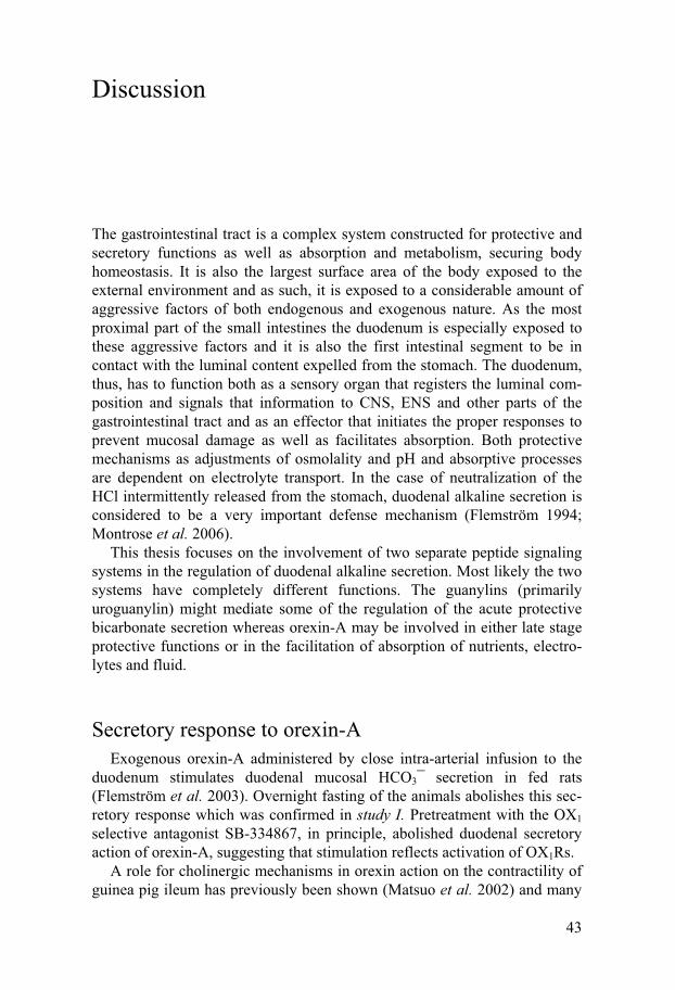
43
Discussion
The gastrointestinal tract is a complex system constructed for protective and secretory functions as well as absorption and metabolism, securing body homeostasis. It is also the largest surface area of the body exposed to the external environment and as such, it is exposed to a considerable amount of aggressive factors of both endogenous and exogenous nature. As the most proximal part of the small intestines the duodenum is especially exposed to these aggressive factors and it is also the first intestinal segment to be in contact with the luminal content expelled from the stomach. The duodenum, thus, has to function both as a sensory organ that registers the luminal com-position and signals that information to CNS, ENS and other parts of the gastrointestinal tract and as an effector that initiates the proper responses to prevent mucosal damage as well as facilitates absorption. Both protective mechanisms as adjustments of osmolality and pH and absorptive processes are dependent on electrolyte transport. In the case of neutralization of the HCl intermittently released from the stomach, duodenal alkaline secretion is considered to be a very important defense mechanism (Flemström 1994; Montrose et al. 2006).
This thesis focuses on the involvement of two separate peptide signaling systems in the regulation of duodenal alkaline secretion. Most likely the two systems have completely different functions. The guanylins (primarily uroguanylin) might mediate some of the regulation of the acute protective bicarbonate secretion whereas orexin-A may be involved in either late stage protective functions or in the facilitation of absorption of nutrients, electro-lytes and fluid.
Secretory response to orexin-A Exogenous orexin-A administered by close intra-arterial infusion to the
duodenum stimulates duodenal mucosal HCO3� secretion in fed rats (Flemström et al. 2003). Overnight fasting of the animals abolishes this sec-retory response which was confirmed in study I. Pretreatment with the OX1 selective antagonist SB-334867, in principle, abolished duodenal secretory action of orexin-A, suggesting that stimulation reflects activation of OX1Rs.
A role for cholinergic mechanisms in orexin action on the contractility of guinea pig ileum has previously been shown (Matsuo et al. 2002) and many
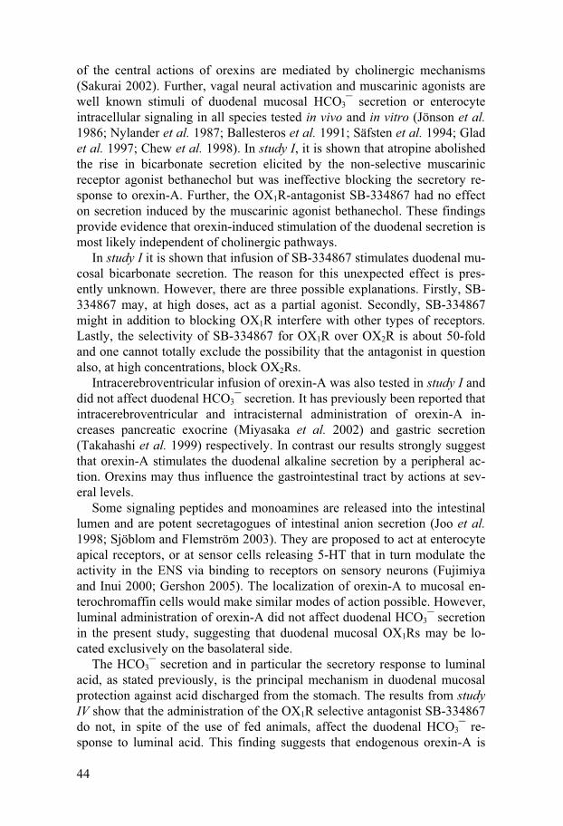
44
of the central actions of orexins are mediated by cholinergic mechanisms (Sakurai 2002). Further, vagal neural activation and muscarinic agonists are well known stimuli of duodenal mucosal HCO3� secretion or enterocyte intracellular signaling in all species tested in vivo and in vitro (Jönson et al. 1986; Nylander et al. 1987; Ballesteros et al. 1991; Säfsten et al. 1994; Glad et al. 1997; Chew et al. 1998). In study I, it is shown that atropine abolished the rise in bicarbonate secretion elicited by the non-selective muscarinic receptor agonist bethanechol but was ineffective blocking the secretory re-sponse to orexin-A. Further, the OX1R-antagonist SB-334867 had no effect on secretion induced by the muscarinic agonist bethanechol. These findings provide evidence that orexin-induced stimulation of the duodenal secretion is most likely independent of cholinergic pathways.
In study I it is shown that infusion of SB-334867 stimulates duodenal mu-cosal bicarbonate secretion. The reason for this unexpected effect is pres-ently unknown. However, there are three possible explanations. Firstly, SB-334867 may, at high doses, act as a partial agonist. Secondly, SB-334867 might in addition to blocking OX1R interfere with other types of receptors. Lastly, the selectivity of SB-334867 for OX1R over OX2R is about 50-fold and one cannot totally exclude the possibility that the antagonist in question also, at high concentrations, block OX2Rs.
Intracerebroventricular infusion of orexin-A was also tested in study I and did not affect duodenal HCO3� secretion. It has previously been reported that intracerebroventricular and intracisternal administration of orexin-A in-creases pancreatic exocrine (Miyasaka et al. 2002) and gastric secretion (Takahashi et al. 1999) respectively. In contrast our results strongly suggest that orexin-A stimulates the duodenal alkaline secretion by a peripheral ac-tion. Orexins may thus influence the gastrointestinal tract by actions at sev-eral levels.
Some signaling peptides and monoamines are released into the intestinal lumen and are potent secretagogues of intestinal anion secretion (Joo et al. 1998; Sjöblom and Flemström 2003). They are proposed to act at enterocyte apical receptors, or at sensor cells releasing 5-HT that in turn modulate the activity in the ENS via binding to receptors on sensory neurons (Fujimiya and Inui 2000; Gershon 2005). The localization of orexin-A to mucosal en-terochromaffin cells would make similar modes of action possible. However, luminal administration of orexin-A did not affect duodenal HCO3� secretion in the present study, suggesting that duodenal mucosal OX1Rs may be lo-cated exclusively on the basolateral side.
The HCO3� secretion and in particular the secretory response to luminal acid, as stated previously, is the principal mechanism in duodenal mucosal protection against acid discharged from the stomach. The results from study IV show that the administration of the OX1R selective antagonist SB-334867 do not, in spite of the use of fed animals, affect the duodenal HCO3� re-sponse to luminal acid. This finding suggests that endogenous orexin-A is

45
not involved in mediating duodenal protective alkaline secretion against gastric discharge of acid. The relatively long time interval required for in-duction of sensitivity to exogenous orexin-A in pre-starved animals, a phe-nomenon discussed in detail later on, is further evidence that other mecha-nisms including neural reflexes and mucosal prostaglandins (Flemström et al. 1982; Takeuchi et al. 1997), are more important mediating the rise in mucosal bicarbonate secretion in response to luminal acid.
Enterocyte calcium signaling Calcium signaling is critical for all cells and changes in [Ca2+]i is a highly versatile intracellular second messenger system linking many physiological stimuli to their intracellular effectors. A wide variety of cellular processes can be regulated ranging from events on the microsecond scale as exocytosis to gene transcription or cell proliferation that might require hours (Berridge et al. 2003). Normally cells keep their [Ca2+]i at a constant resting level of approximately 100 nM but at stimulation extracellular influx or the release of calcium from intracellular stores can rapidly increase the [Ca2+]i (Berridge et al. 2000). Much of the versatility of intracellular calcium signaling lies in the variability of speed, amplitude and spatiotemporal patterning by com-partmentalization. Further variations can be achieved by crosstalk with other types of signaling pathways (Friel and Chiel 2008).
As in other cells and tissues, intracellular calcium signaling is vital in regulation of enterocyte function in general and of special interest in this thesis with regard to anion and bicarbonate transport. In the last few years, several duodenal HCO3� secretagogues have been demonstrated to induce transient rises in [Ca2+]i. Cholecystokinin (CCK) and muscarinic agonist (Chew et al. 1998) as well as melatonin (Sjöblom et al. 2003) induce cal-cium signaling in both human and rat enterocytes. 5-HT induces duodenal HCO3� secretion in mouse (Tuo et al. 2004) and rat (Säfsten et al. 2006), via intracellular calcium signaling in a 5-HT4 receptor dependent pathway. Hence for some agents there is quite a strong association between stimula-tion of duodenal mucosal bicarbonate secretion and rises in [Ca2+]i. As an example of a connection between alkaline secretion and intracellular calcium signaling, CFTR is activated by increases in [Ca2+]i leading to a Cl� depend-ent HCO3� secretion in pancreatic duct cells (Namkung et al. 2003).
The results from study II demonstrate that orexin-A induces intracellular calcium signaling in isolated duodenal enterocytes from rats as well as hu-mans most probably by a direct action on the enterocytes. Cessation of expo-sure to orexin-A did not result in any immediate decline in [Ca2+]i. This sig-naling pattern is similar to that induced by 5-HT in acutely isolated entero-cytes (Säfsten et al. 2006) and that induced by orexin-A in subpopulations of neurons in the dorsal raphe and laterodorsal tegmentum (Kohlmeier et al.

46
2004). In contrast, if the signaling pattern in the majority of responding cells is compared to that induced by carbachol and CCK-8 in enterocytes in pri-mary culture (Chew et al. 1998) or that induced by melatonin in cluster preparations (Sjöblom et al. 2003), the rise in [Ca2+]i in response to orexin-A appears slower and clear-cut initial peak responses were not observed.
The initial rise in [Ca2+]i in response to orexin-A remained in spite of re-moval of extracellular Ca2+. This is in line with previous findings in duode-nal enterocytes demonstrating that absence of Ca2+ in the extracellular fluid abolishes the plateau phase but not the initial rise in [Ca2+]i induced by car-bachol (Chew et al. 1998) or melatonin (Sjöblom et al. 2003). Thus, it ap-pears that all three secretagogues cause release of Ca2+ from intracellular stores as well as influx of extracellular Ca2+. An explicit dependence of ex-tracellular Ca2+ for elevations in [Ca2+]i, in response to orexin-A stimulation, in neuronal cells (van den Pol et al. 1998; Uramura et al. 2001) but not in nonexcitable cell lines has been demonstrated. In the latter cells, intracellular calcium signaling induced by orexin-A displays an interesting dose depend-ence with low concentrations eliciting activation of extracellular Ca2+ de-pendent ROCs (Smart et al. 1999; Kukkonen and Åkerman 2001; Holmqvist et al. 2002; Larsson et al. 2003; Larsson et al. 2005) and high concentrations inducing intracellular Ca2+ release (Lund et al. 2000). The ability of the caged duodenal enterocytes to display [Ca2+]i transients under Ca2+-free con-ditions suggest presence of at least the former type of signaling.
In study II we also tested effects of the OX1R specific antagonist SB-334867 on orexin-A-induced intracellular calcium signaling and the OX2R specific agonist [Ala11, D-Leu15]-orexin-B. The combined results strongly suggest that the orexin induced [Ca2+]i response is mediated by OX1R.
Orexin-A and OXRs in nerve fibers has been detected by immunohisto-chemistry and are found in both submucosal and myenteric plexuses and in interconnecting neurons (Kirchgessner and Liu 1999; Näslund et al. 2002; Nakabayashi et al. 2003; Ehrström et al. 2005). Orexin-A has been sug-gested to induce ileal motility by release of acetylcholine from enteric cho-linergic neurons (Matsuo et al. 2002). However, orexins and their receptors are also present in epithelial cells in crypts and villi in the small intestine (Kirchgessner and Liu 1999; Näslund et al. 2002). As already discussed, the muscarinic antagonist, atropine, did not inhibit orexin-A induced duodenal HCO3� secretion, indicating independence of enteric cholinergic pathways. Interestingly, neither did atropine affect the orexin-A-induced intracellular calcium signaling in the clusters of enterocytes, microscopically devoid of neurons. These findings provide further strong evidence for a direct action of orexin-A at duodenal enterocytes.
The percentage of enterocytes in clusters responding to orexin-A never exceeded 20% and was lower than the percentage (~ 35%) responding to melatonin in cluster preparations (Sjöblom et al. 2003), but there was a re-sponse to carbachol in cells not responding to orexin-A. The lack of a re-

47
sponse in some enterocytes may reflect absence or loss of receptor expres-sion and/or a decline of cell-to-cell communication in some preparations. Further, it cannot be excluded that feeding induces expression of orexin re-ceptors only in a limited number of the enterocytes.
Secretory response to guanylins The fact that both guanylin peptides have the ability to regulate CFTR through GC-C and that they are potent stimulants of duodenal mucosal bi-carbonate secretion makes them interesting candidates as defenders of muco-sal integrity against the acid discharged from the stomach. Guanylin in-creases HCO3� secretion in rat duodenal mucosa in vitro (Guba et al. 1996) and uroguanylin increases luminal pH by stimulating HCO3� secretion in isolated mouse duodenal mucosa (Joo et al. 1998). Further, uroguanylin increase luminal HCO3� concentration in rat closed jejunal loops (Ieda et al. 1999) and, importantly, GC-C knockout mice have a significantly reduced rise in HCO3� secretion in response to luminal acid (Rao et al. 2004). As most of previous work has been performed with duodenal tissue in vitro or in other segments of the small intestine we examined, in study IV, the ability of the guanylins to induce duodenal mucosal HCO3� secretion in situ.
Intra-arterial as well as luminal administration of both guanylin and uroguanylin markedly increased duodenal mucosal bicarbonate secretion, which seems in line with the hypothesis that guanylins are released bidirec-tionally in the intestine (Martin et al. 1999; Moro et al. 2000; Rudolph et al. 2002). Luminal administration of the peptides induced a larger increase in bicarbonate secretion than did intra-arterial administration. Furthermore, the response to luminal guanylins also appeared more rapid suggesting an action directly on apical receptors. It has been demonstrated that luminal release of the peptides is considerably larger than that seen basolaterally (Martin et al. 1999; Moro et al. 2000). Further, the primary site of action is most likely apical GC-C receptors (Krause et al. 1994; Swenson et al. 1996) and evi-dence has been provided that uroguanylin accumulates in secretory vesicles close to the apical cell membrane (Nakazato et al. 1998). The secretory re-sponse to intra-arterial guanylins seen in study IV is thus noteworthy. By this route of administration the peptides displayed equal potency in stimulating HCO3� secretion. It is possible that there are additional, as yet not discov-ered GC-C like receptors located basolaterally on enterocytes or on neural elements in the enteric nervous system. Specific binding of 125I-STa in intes-tinal membrane homogenates from GC-C null mice is low but reproducible, amounting to approximately 10 % of the binding sites for radiolabelled STa (Mann et al. 1997). Recently the guanylins has been demonstrated to be ligands for the G-protein-coupled urotensin II receptor, GPR14 (Lehner et al. 2007) and even though this receptor has not yet been identified in the
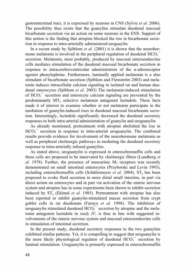
48
gastrointestinal tract, it is expressed by neurons in CNS (Sylvie et al. 2006). The possibility thus exists that the guanylins stimulate duodenal mucosal bicarbonate secretion via an action on some neurons in the ENS. Support of this notion is the finding that atropine blocked the rise in bicarbonate secre-tion in response to intra-arterially administered uroguaylin.
In a recent study by Sjöblom et al. (2001) it is shown that the neurohor-mone melatonin is involved in the peripheral regulation of duodenal HCO3� secretion. Melatonin, most probably, produced by mucosal enteroendocrine cells mediates stimulation of the duodenal mucosal bicarbonate secretion in response to intracerebroventricular administration of the �-adrenoceptor agonist phenylephrine. Furthermore, luminally applied melatonin is a also stimulant of bicarbonate secretion (Sjöblom and Flemström 2003) and mela-tonin induces intracellular calcium signaling in isolated rat and human duo-denal enterocytes (Sjöblom et al. 2003) The melatonin-induced stimulation of HCO3� secretion and enterocyte calcium signaling are prevented by the predominantly MT2 selective melatonin antagonist luzindole. These facts made it of interest to examine whether or not melatonin participate in the mediation of guanylin-induced rises in duodenal mucosal bicarbonate secre-tion. Interestingly, luzindole significantly decreased the duodenal secretory responses to both intra-arterial administration of guanylin and uroguanylin
As already mentioned, pretreatment with atropine abolished the rise in HCO3� secretion in response to intra-arterial uroguanylin. The combined results provide evidence for involvement of the neurohormone melatonin as well as peripheral cholinergic pathways in mediating the duodenal secretory response to intra-arterially infused guanylins.
As stated above, uroguanylin is expressed in enterochromaffin cells and these cells are proposed to be innervated by cholinergic fibres (Lundberg et al. 1978). Further, the presence of muscarinic M3 receptors was recently demonstrated on small intestinal enterocytes (Przyborski and Levin 1993), including enterochromaffin cells (Schäfermeyer et al. 2004). STa has been proposed to evoke fluid secretion in more distal small intestine, in part via direct action on enterocytes and in part via activation of the enteric nervous system and atropine has in some experiments been shown to inhibit secretion induced by STa (Eklund et al. 1985). Pretreatment with atropine has also been reported to inhibit guanylin-stimulated mucus secretion from crypt goblet cells in rat duodenum (Furuya et al. 1998). The inhibition of uroguanylin stimulated duodenal HCO3� secretion by atropine and the mela-tonin antagonist luzindole in study IV, is thus in line with suggested in-volvements of the enteric nervous system and mucosal enteroendocrine cells in stimulation of intestinal secretion.
In the present study, duodenal secretory responses to the two guanylins exhibited similar patterns. Yet, it is compelling to suggest that uroguanylin is the more likely physiological regulator of duodenal HCO3� secretion by luminal stimulation. Uroguanylin is primarily expressed in enterochromaffin
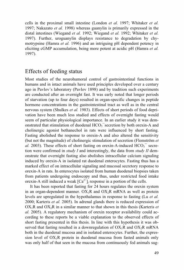
49
cells in the proximal small intestine (London et al. 1997; Whitaker et al. 1997; Nakazato et al. 1998) whereas guanylin is primarily expressed in the distal intestines (Wiegand et al. 1992; Wiegand et al. 1992; Whitaker et al. 1997). Further, uroguanylin displays resistance to degradation by chy-motrypsine (Hamra et al. 1996) and an intriguing pH dependent potency in eliciting cGMP accumulation, being more potent at acidic pH (Hamra et al. 1997).
Effects of feeding status Most studies of the neurohumoral control of gastrointestinal functions in humans and in intact animals have used principles developed over a century ago in Pavlov’s laboratory (Pavlov 1898) and by tradition such experiments are conducted after an overnight fast. It was early noted that longer periods of starvation (up to four days) resulted in organ-specific changes in peptide hormone concentrations in the gastrointestinal tract as well as in the central nervous system (Shulkes et al. 1983). Effects of short periods of food depri-vation have been much less studied and effects of overnight fasting would seem of particular physiological importance. In an earlier study it was dem-onstrated that stimulation of duodenal HCO3�secretion by both orexin-A and cholinergic agonist bethanechol in rats were influenced by short fasting. Fasting abolished the response to orexin-A and also altered the sensitivity (but not the magnitude) of cholinergic stimulation of secretion (Flemström et al. 2003). These effects of short fasting on orexin-A-induced HCO3� secre-tion were confirmed in study I and interestingly, the data from study II dem-onstrate that overnight fasting also abolishes intracellular calcium signaling induced by orexin-A in isolated rat duodenal enterocytes. Fasting thus has a marked effect of on intracellular signaling and mucosal secretory response to orexin-A in rats. In enterocytes isolated from human duodenal biopsies taken from patients undergoing endoscopy and thus, under restricted food intake orexin-A still induced a weak [Ca2+]i response in a portion of the cells.
It has been reported that fasting for 24 hours regulates the orexin system in an organ-dependent manner. OX1R and OX2R mRNA as well as protein levels are upregulated in the hypothalamus in response to fasting (Lu et al. 2000; Karteris et al. 2005). In adrenal glands there is reduced expression of OX1R and OX2R in a similar manner to that shown in this thesis (Karteris et al. 2005). A regulatory mechanism of orexin receptor availability could ac-cording to these reports be a viable explanation to the observed effects of short fasting presented in this thesis. In line with this hypothesis it was ob-served that fasting resulted in a downregulation of OX1R and OX2R mRNA both in the duodenal mucosa and in isolated enterocytes. Further, the expres-sion level of OX1R protein in duodenal mucosa from fasted animals only was only half of that seen in the mucosa from continuously fed animals sug-

50
gesting that the decline in mRNA levels really affects the levels of functional receptors available for stimulation. It would seem that the decline in expres-sion of OXRs is a likely mechanism for the absence of a duodenal secretory response and intracellular calcium signaling to exogenous orexin-A in fasted animals.
The mechanisms by which feeding upregulates the intestinal responsive-ness to orexin could depend on stimulation of food intake per se. Sham feed-ing has been shown to affect duodenal motility and induce alkaline secretion in dogs by a vagal mechanism (Konturek and Thor 1986). Even though the absence of muscarinic involvement in orexin-A induced luminal alkaliniza-tion and enterocyte calcium signaling had been shown in study I and II re-spectively an influence from the central nervous system cannot be com-pletely excluded in the regulation of OXR density. Yet a more likely expla-nation seemed to be that luminal nutrients by them self had a stimulatory action on orexin receptor expression.
The lining of the gastrointestinal tube contains a number of neuroendo-crine cells and neurons which may respond to feeding and allow the intestine to undergo an integrated response to changes in its luminal contents (Furness et al. 1999) and orexins are expressed both in enterochromaffin cells and in nerve endings of enteric neurons (Kirchgessner and Liu 1999; Näslund et al. 2002). It would thus seem reasonable that such influences are responsible for upregulation of orexin receptors with a consequent paracrine/endocrine orexin mediated response. As shown by the result in study III, priming of the gastrointestinal tract with carbohydrates (glucose) is a prerequisite for orexin-A induced stimulation of duodenal HCO3� secretion.
In contrast to the situation in the intestine, fasting does not decrease but increases expression of OX1R and OX2R mRNA as well as protein levels in the hypothalamus (Cai et al. 1999; Lu et al. 2000; Karteris et al. 2005). Fur-ther, both hypoglycemia and intracellular glucopenia increases gene tran-scription activity in neurons of lateral hypothalamus measured as elevated levels of Fos-immunoreactivity (Briski and Sylvester 2001; Paranjape et al. 2006) resulting in elevated levels of PPO mRNA (Griffond et al. 1999; Paranjape et al. 2006). The combined findings thus suggest a marked differ-ence between intestinal and central nervous actions of fasting and fasting-related conditions on expressions of the orexin system. Changes in extracel-lular glucose concentration have been shown to affect orexin related mRNA expression in some other peripheral tissues as well and organ-dependent differences appear to be considerable. There is an increased level of OX1R but a decreased level of OX2R mRNA in the adrenal glands of hyperglyce-mic, diabetic rats (Jöhren et al. 2001) and an elevation of glucose concentra-tion induce downregulation of PPO mRNA in pancreatic islet cells in vitro (Zhao et al. 2005).
It is well established that presence of carbohydrate, protein and fat in the intestinal lumen markedly affect intestinal motility and secretion as well as

51
pancreatic secretions (Buchan 1999; Furness et al. 1999; Raybould 1999). Some hormones, including CCK and secretin, mediate the responses. How-ever, it has also been proposed that presence of nutrients acts through neural pathways controlling electrolyte and fluid transport (Sjövall et al. 1983) and glucoresponsive neurons in the enteric nervous system react to changes in extracellular concentrations of glucose via KATP channel activity (Liu et al. 1999). The EC-cell-like line BON releases 5-HT in response to glucose (Kim et al. 2001) and luminal glucose stimulates vagal afferent discharge as well as 5-HT release in vivo (Freeman et al. 2006). Both these latter studies further suggest that the stimulation by glucose is dependent on the uptake of glucose by Na+-glucose cotransporters most probably SGLT-3.
In study III, glucose did not induce sensitivity to orexin-A when the time interval between duodenal exposure to glucose and administration of orexin-A was made shorter than 4 hours. The long activation time for induction of secretory responses to orexin-A may indicate requirement of time for gene transcription. It is interesting to note that all studies demonstrating signifi-cant changes in orexin expression levels in response to changes in glucose concentration in hypothalamic (Griffond et al. 1999; Briski and Sylvester 2001; Cai et al. 2001; Zhao et al. 2005; Paranjape et al. 2006) as well as in peripheral (Jöhren et al. 2001; Zhao et al. 2005) tissues are conducted with extended periods of hypo- or hyperglycemia. When Fos-immunoreactivity in the lateral hypothalamus is evaluated after 90 minutes of insulin induced hypoglycemia, no significant induction is observed (Kiss et al. 2004). Fur-ther, 2-deoxy-D-glucose induced glukopenia does not affect PPO expression during the initial two hours (Sergeyev et al. 2000) and Bayer et al. demon-strated that the effect of the intracellular glucopenia attains significance only after 3 hours (Bayer et al. 2000). These data would seem in line with our findings of a need for four hours for induction of orexin sensitivity.
Thus, the duodenal mucosal alkaline secretory response, induced by orexin-A, seems to depend on increased OX1R density in a process initiated by glucose availability. In contrast, the secretory response to close intra-arterial guanylins in study IV was not affected by short fasting. This is in line with other stipulated modifiers of protective alkaline mucosal secretion. Rises in HCO3� secretion in response to serotonin (Säfsten et al. 2006), me-latonin or vasoactive intestinal polypeptide (Flemström et al. 2003) are, in contrast to orexin-A not altered by overnight food deprivation. This means that certain pathways for stimulation of intestinal secretion are selectively downregulated during a fasted state.
It is interesting to note that basal alkaline secretion increases in response to 1-h priming of the duodenal mucosa to glucose or proteins. We have pre-viously observed that basal HCO3� secretion in continuously fed animals tends to be somewhat higher than that in overnight fasted animals. The mechanisms causing this stimulated basal alkaline secretory rate remain unanswered in his thesis. However, a mediation by orexins seem unlikely

52
since animals exposed to short time pretreatment with glucose showed the same increase in basal secretory rate but were insensitive to exogenous orexin-A. Since food is a potent stimulant of gastric acid secretion it seems physiologically relevant to up-regulate also duodenal mucosal bicarbonate secretion to assure a more effective mucosal protection against the acid dis-charged from the stomach. Furthermore, a higher turnover rate of epithelial ion exchangers in response to feeding may significantly contribute to a more efficient absorption of nutrients, electrolytes and water. Further studies are certainly required to elucidate this interesting finding of feeding and nutrient dependent changes in rates of duodenal basal secretion.
53
Conclusions
The results from the studies included in the present thesis provide new in-formation about the regulation of duodenal mucosal bicarbonate secretion and the main conclusions that can be drawn are:
I Orexin-A induced stimulation of duodenal mucosal bicarbonate
secretion in vivo is mediated by OX1 receptors but is independent of cholinergic pathways. The lack of effect of orexin-A on bicar-bonate secretion in fasted animals is due to downregulation of OX1 receptors in the duodenal mucosa. A prerequisite for orexin-A to stimulate duodenal mucosal bicarbonate secretion in vivo is the presence of glucose in the gastrointestinal lumen more than one hour before orexin-A administration.
II Orexin-A increases the intracellular calcium concentration in both human and rat isolated duodenal enterocytes in clusters strongly suggesting a direct effect of the peptide on these cells. This rise in intracellular calcium concentration is probably due to release of calcium from intracellular stores as well as extracellu-lar influx. Fasting downregulates the expression of OX1 receptors on isolated duodenocytes possibly explaining the lack of effect or the low responsivness of orexin-A on calcium signaling.
III Endogenous orexin-A does probably not participate in the media-
tion of the hydrochloric acid-induced rise in duodenal mucosal bicarbonate secretion.
IV Intra-arterial as well as luminal administration of guanylins in-crease the bicarbonate secretion in duodenum. Uroguanylin is a more potent duodenal secretagogue than guanylin. The rise in duodenal mucosal HCO3� secretion induced by intra-arterial uroguanylin is mediated by melatonin and muscarinic receptors.

54
Svensk populärvetenskaplig sammanfattning
Magen och tarmarna är ett komplicerat system med många uppgifter. De ska bryta ner födan i lämpliga delar för att kunna absorbera näringsämnen, elekt-rolyter och vatten och sedan göra sig av med restprodukter. Dessutom är mag- tarmkanalen kroppens största yta mot omgivningen och måste därför kunna skydda både sig själv och resten av kroppen från många olika skadliga ämnen, tex magsyra, etanol, läkemedel och bakterier. Alla dessa funktioner är väsentliga för att kroppen ska vara i balans, för att homeostasen ska beva-ras.
Tolvfingertarmen (duodenum) är det första tarmsegmentet som följer efter magsäcken och är därför först att komma i kontakt med näringsämnen men även olika skadliga faktorer. På grund av detta så måste tolvfingertarmen kunna fungera både som ett sensoriskt organ som identifierar innehållet i det material som passerar och som ett organ som kan vidta de rätta åtgärderna för att skydda resten av mag- tarmkanalen samt underlätta absorptionen av näringsämnen.
Tolvfingertarmens utsöndring (sekretion) av bikarbonatjoner (HCO3�) är en viktig fysiologisk försvarsmekanism mot den syra som utsöndras av ma-gen och töms ut i tarmarna. Tidigare observationer har visat att bikarbonat-sekretionen är kraftigt sänkt hos patienter med sår i tolvfingertarmen. Stör-ningar i regleringen av bikarbonatsekretionen kan alltså leda till att skador uppkommer i tolvfingertarmens slemhinna. Man kan också använda bikar-bonatsekretionen som en vetenskaplig modell för hur andra negativt laddade joner transporteras i tarmarna. Man har t.ex. sett att tarmarna hos personer som lider av cystisk fibros, en sjukdom där man inte kan transportera just negativa joner på ett normalt sätt, ofta inte tar upp näring, elektrolyter och vatten på ett normalt sätt vilket kan leda till undernäring och andra problem. Bättre förståelse för duodenums bikarbonatsekretion skulle alltså kunna hjälpa oss att komma närmare ett svar på hur vi skulle kunna hjälpa personer med den här typen av problem. Därför har jag i mitt avhandlingsarbete valt att arbeta med två peptider, orexiner och guanyliner, som eventuellt skulle kunna vara inblandade i styrningen av tolvfingertarmens bikarbonatsekre-tion.
I avhandlingsarbetet undersöktes om ovan nämnda peptider verkar lokalt i tolvfingertarmen eller via det centrala nervsystemet eller möjligen i mag-tarmkanalens eget nervsystem, det s.k. enteriska nervsystemet. Dessutom studerade jag om dessa peptider verkar via kopplingar med andra redan kän-

55
da hormoner och nervsignalsubstanser som deltar i regleringen av duode-nums bikarbonatsekretion. En annan intressant fråga som jag sökt svar på är om orexinernas och guanylinerna effekt på duodenums bikarbonatsekretion beror på om man har ätit eller inte.
Jag har i mina studier använt sövda råttor. För att kunna studera tolvfing-ertarmens bikarbonatsekretion så har jag använt en speciell kammare med två skänklar mellan vilka en del av duodenum monteras. I kammaren recir-kulerar fysiologisk koksaltlösning som kontinuerligt titreras med saltsyra. Åtgången av saltsyra motsvarar duodenums bikarbonatsekretion. Dessutom har jag studerat hur cellernas interna kommunikation, kalcium-signalering, påverkas av orexinet, orexin-A. Slutligen har jag också tittat på hur svält påverkar mängden mRNA och receptorer för orexin-A i duodenum genom att använda metoderna PCR och Western blot.
I korthet visar resultaten att orexin-A ökar duodenums bikarbonatsekre-tion, men endast i djur som inte har utsatts för kort svält (16 timmar). Den orexin-A stimulerade ökningen av bikarbonatsekretionen som ses hos de djur som har ätit, förmedlas genom en av de två receptorer som binder orexin, d.v.s. OX1-receptorn (OX1R). Resultaten visar också att effekten av orexin-A inte förmedlas via det kolinerga nervsystemets muskarinerga receptorer. Orexin-A ökar också tarmcellernas interna signalering och i celler från både människor och råttor ökar mängden intracellulärt kalcium som svar på orex-in-A. Detta visar att orexin-A kan ha effekt direkt på tarmceller. Ett intres-sant fynd är att svälta råttors duodenum innehåller färre OX1-receptorer än de från djur som haft fri tillgång till mat och detta är med största sannolikhet orsaken till den uteblivna effekten av orexin-A hos dessa svälta djur.
Anledningen till att antalet OX1-receptorer ökar som svar på födointag är inte känd men jag kan visa att närvaron av glukos (kolhydrater/socker) i tarmen minst en timme innan tillförseln av orexin-A är nödvändig för att bikarbonatsekretion ska öka. Det tar alltså lång tid för duodenums orexin-system att aktiveras. Det är känt sedan tidigare att om duodenum exponeras för saltsyra ökar slemhinnans bikarbonatsekretion och så skedde även i detta avhandlingsarbete. Däremot verkar inte orexin-A medverka i denna syra-stimulerade ökning av bikarbonatsekretionen. Vilken fysiologisk funktion orexin-A har i duodenum är i dagsläget osäkert men orexin-systemet skulle mycket väl kunna medverka i styrningen av de förändringar av de fysiolo-giska funktionerna som troligtvis sker i samband med födointag och svält.
Guanylin och uroguanylin är två snarlika peptider som båda bildas i tarm-slemhinnan. Resultaten visar att både guanylin och uroguanylin ökar tolv-fingertarmens bikarbonatsekretion. Ingen skillnad i sekretionsökning kan ses mellan djur som har fastat och de som har haft fri tillgång till mat. Båda pep-tiderna ökar sekretionen oavsett om de ges från blodsidan eller luminalt d.v.s. inuti tarmen. Däremot svarar tarmen mycket starkare på uroguanylin än på guanylin om peptiderna ges luminalt. Det verkar också som om guany-
56
linerna samverkar med både det kolinerga systemet och neurohormonet me-latonin i stimuleringen av duodenums bikarbonatsekretion.
Avslutningsvis kan man säga att båda peptidsystemen kan reglera tolv-fingertarmens bikarbonatsekretion men att de förmodligen har helt olika funktioner. Guanylinerna, särskilt uroguanylin kan med god sannolikhet antas delta i regleringen av duodenums skyddade sekretion medan orexin-A förmodligen reglerar bikarbonatsekretionen av något annat syfte.

57
Acknowledgements
This work was carried out at the Department of Neuroscience, Division of Physiology at Uppsala University and is the final result of the combined efforts of several people. I would like to express my sincere gratitude and appreciation to all of them. In particular, I would like to thank: Professor Gunnar Flemström, my supervisor, for introducing me to the fas-cinating area of gastrointestinal physiology, for giving me invaluable scien-tific training and constructive criticism. Thank you for believing in me. Professor Olof Nylander, my co-supervisor, for your willingness to discuss and your ability to answer every possible question. You have been a great support and an inspiration with your scientific enthusiasm. Professor Karl Åkerman, my co-supervisor, for your support and competent guidance on cell experiments. Gunilla Jedsted, for your devotion, patience and skilful help in the lab. Without you this work had never been possible. I’m forever in your debt! Markus Sjöblom, for your ability to always spare a minute for discussion and assistance and for always having some advice to share. Liselotte Pihl, for being a great travel companion, an exemplary roomie and a very good friend. I have enjoyed ever minute of barley audible Danish music and of course all the “bitter” laughs. Tackur! Annika Jägare, for your encouragement and for good times in the lab and for the invaluable advice on enteral feeding. Professor Karl-Heinz Herzig and Kari Mäkelä, Oulo University / Kuopio University, Finland, for interesting and rewarding co-operation. Emma Andersson, Lena Karlsson and Ulla Jonson, for all the help with uni-versity red tape and for always making shure my mailbox is full.

58
Gunno Nilsson and Birgitta Klang, for smoothing out the administrative bumps in teaching. Without you teaching would not be very much fun at all. The cell people in A:2, Jyrki Kukkonen, Karin Nygren, Marie Ekholm and Lisa Johansson, for interesting discussions on science, teaching but also ot-her more important things… All the colleagues, former and present, at the Division for Comperative Medi-cine, Hans-Erik Carlsson, Jann Hau, Mahinda Kommalage, Renée Goldkuhl, Gabriella Persdotter Hedlund, Anna-Linnéa Fernström and Klas Abelsson, for making the entire corridor a more enjoyable place. A special thanks to Anna-Linnéa for late nights at BMC cursing over late nights at BMC. Susanne, Sussie, Eva and Teres at the Animal Department for help with an-esthesia and for taking very good care of my animals. I know you have shed some of your blod for me and I appreciate it. All other persons connected to the divisions of physiology and comparative medicine for creating a creative and inspiring workplace. All my lovely friends for not loosing patience with me during this five year long roller-coaster ride. Tobbe, Jessica, Erika, Johan, Rebecka, Kajsa, Olle, N:son Olsson, Elin, Emma slemma, Sunkan, Robban, Thomas, Emma and Mathias, your friendship has really contributed to the completion of my the-sis. Thanks for everything and for keeping me sane… kind of, at least. All the friends and people at Göteborgs Nation, my home away from home! My caring, wonderful family. My sister Camilla for being the ultimate sup-port, you’re the best! Her family, Micke, Magdalena and Ellen for all the nice times when I every now and then find my way back to Skåne and the encouragements and great conversations on MSN; –“Vad gör du?”; –“Jobbar! Vad gör du då?”; –“Pratar med dig.”. Tack! My parents Bylle & Berne, for always supporting me, no matter what troubles I cause and for all the important notes on practical things. Mormor och Farmor, för att ni alltid har tagit hand om mig. För att ni är så underbara och dessutom såg till att jag blev så nyfiken och kunskapstörstig som jag ändå är. Utan er så hade jag inte varit här. Tack! _____________________________________________________________ This work was financially supported by the Swedish Medical Research Council and the Wallenberg foundation. Travel grants from the Swedish Pharmaceutical Society and the Scandinavian Physiological Society are gratefully acknowledged.

59
References
Allen, A. and G. Flemström (2005). "Gastroduodenal mucus bicarbonate barrier: protection against acid and pepsin." Am J Physiol Cell Physiol 288(1): C1-C19.
Allen, A., G. Flemström, A. Garner and E. Kivilaakso (1993). "Gastroduodenal mucosal protection." Physiol Rev 73(4): 823-57.
Asahi, S., S. Egashira, M. Matsuda, H. Iwaasa, A. Kanatani, M. Ohkubo, M. Ihara and H. Morishima (2003). "Development of an orexin-2 receptor selective ago-nist, [Ala(11), D-Leu(15)]orexin-B." Bioorg Med Chem Lett 13(1): 111-3.
Atuma, C., V. Strugala, A. Allen and L. Holm (2001). "The adherent gastrointestinal mucus gel layer: thickness and physical state in vivo." Am J Physiol Gastro-intest Liver Physiol 280(5): G922-9.
Ballesteros, M. A., J. D. Wolosin, D. L. Hogan, M. A. Koss and J. I. Isenberg (1991). "Cholinergic regulation of human proximal duodenal mucosal bicarbon-ate secretion." Am J Physiol Gastrointest Liver Physiol 261(2 Pt 1): G327-G331.
Bayer, L., C. Colard, N. U. Nguyen, P. Y. Risold, D. Fellmann and B. Griffond (2000). "Alteration of the expression of the hypocretin (orexin) gene by 2-deoxyglucose in the rat lateral hypothalamic area." Neuroreport 11(3): 531-3.
Beltowski, J. (2001). "Guanylin and related peptides." J Physiol Pharmacol 52(3): 351-75.
Bengtsson, M. W., K. Mäkelä, M. Sjöblom, S. Uotila, K. E. Åkerman, K. H. Herzig and G. Flemström (2007). "Food induced expression of orexin-receptors in rat duodenal mucosa regulates the bicarbonate secretory response to orexin-A." Am J Physiol Gastrointest Liver Physiol.
Berridge, M. J., M. D. Bootman and H. L. Roderick (2003). "Calcium signalling: dynamics, homeostasis and remodelling." Nat Rev Mol Cell Biol 4(7): 517-29.
Berridge, M. J., P. Lipp and M. D. Bootman (2000). "The versatility and universality of calcium signalling." Nat Rev Mol Cell Biol 1(1): 11-21.
Berthoud, H. R., N. R. Carlson and T. L. Powley (1991). "Topography of efferent vagal innervation of the rat gastrointestinal tract." Am J Physiol Renal Physiol 260(1 Pt 2): R200-7.
Bilski, J. and S. J. Konturek (1994). "Role of nitric oxide in gastroduodenal alkaline secretion." J Physiol Pharmacol 45(4): 541-53.
Briski, K. P. and P. W. Sylvester (2001). "Hypothalamic orexin-A-immunpositive neurons express Fos in response to central glucopenia." Neuroreport 12(3): 531-4.
Buchan, A. M. (1999). "Nutrient Tasting and Signaling Mechanisms in the Gut III. Endocrine cell recognition of luminal nutrients." Am J Physiol 277(6 Pt 1): G1103-7.
Cai, X. J., R. Denis, R. G. Vernon, J. C. Clapham, S. Wilson, J. R. Arch and G. Williams (2001). "Food restriction selectively increases hypothalamic orexin-B levels in lactating rats." Regul Pept 97(2-3): 163-8.
Cai, X. J., M. L. Evans, C. A. Lister, R. A. Leslie, J. R. Arch, S. Wilson and G. Williams (2001). "Hypoglycemia activates orexin neurons and selectively in-

60
creases hypothalamic orexin-B levels: responses inhibited by feeding and possi-bly mediated by the nucleus of the solitary tract." Diabetes 50(1): 105-12.
Cai, X. J., P. S. Widdowson, J. Harrold, S. Wilson, R. E. Buckingham, J. R. Arch, M. Tadayyon, J. C. Clapham, J. Wilding and G. Williams (1999). "Hypotha-lamic orexin expression: modulation by blood glucose and feeding." Diabetes 48(11): 2132-7.
Cetin, Y., M. Kuhn, H. Kulaksiz, K. Adermann, G. Bargsten, D. Grube and W. G. Forssmann (1994). "Enterochromaffin cells of the digestive system: cellular source of guanylin, a guanylate cyclase-activating peptide." Proc Natl Acad Sci U S A 91(8): 2935-9.
Chang, E. B. and M. C. Rao (1994). Intestinal water and electrolyte transport. Mechanisms of physiological and adaptive responses. Physiology of the Gastro-intestinal Tract. L. R. Johnson. New York, Raven Press. 2: 2027-2081.
Chew, C. S., B. Säfsten and G. Flemström (1998). "Calcium signaling in cultured human and rat duodenal enterocytes." Am J Physiol Gastrointest Liver Physiol 275(2 Pt 1): G296-G304.
Clarke, L. L. and M. C. Harline (1998). "Dual role of CFTR in cAMP-stimulated HCO3� secretion across murine duodenum." Am J Physiol Gastrointest Liver Physiol 274(4 Pt 1): G718-26.
Cohen, M. B., E. A. Mann, C. Lau, S. J. Henning and R. A. Giannella (1992). "A gradient in expression of the Escherichia coli heat-stable enterotoxin receptor exists along the villus-to-crypt axis of rat small intestine." Biochem Biophys Res Commun 186(1): 483-90.
Cohen, M. B., D. P. Witte, J. A. Hawkins and M. G. Currie (1995). "Immunohisto-chemical localization of guanylin in the rat small intestine and colon." Biochem Biophys Res Commun 209(3): 803-8.
Corfield, A. P., D. Carroll, N. Myerscough and C. S. Probert (2001). "Mucins in the gastrointestinal tract in health and disease." Front Biosci 6: D1321-57.
Costa, M., S. J. Brookes and G. W. Hennig (2000). "Anatomy and physiology of the enteric nervous system." Gut 47 Suppl 4: iv15-9; discussion iv26.
Currie, M. G., K. F. Fok, J. Kato, R. J. Moore, F. K. Hamra, K. L. Duffin and C. E. Smith (1992). "Guanylin: an endogenous activator of intestinal guanylate cy-clase." Proc Natl Acad Sci U S A 89(3): 947-51.
de Lecea, L., T. S. Kilduff, C. Peyron, X. Gao, P. E. Foye, P. E. Danielson, C. Fu-kuhara, E. L. Battenberg, V. T. Gautvik, F. S. Bartlett, 2nd, W. N. Frankel, A. N. van den Pol, F. E. Bloom, K. M. Gautvik and J. G. Sutcliffe (1998). "The hy-pocretins: hypothalamus-specific peptides with neuroexcitatory activity." Proc Natl Acad Sci U S A 95(1): 322-7.
de Miguel, M. J. and M. A. Burrell (2002). "Immunocytochemical detection of orexin A in endocrine cells of the developing mouse gut." J Histochem Cyto-chem 50(1): 63-9.
de Sauvage, F. J., S. Keshav, W. J. Kuang, N. Gillett, W. Henzel and D. V. Goeddel (1992). "Precursor structure, expression, and tissue distribution of human guanylin." Proc Natl Acad Sci U S A 89(19): 9089-93.
Ehrström, M., T. Gustafsson, A. Finn, A. Kirchgessner, P. Grybäck, H. Jacobsson, P. M. Hellström and E. Näslund (2005). "Inhibitory effect of exogenous orexin a on gastric emptying, plasma leptin, and the distribution of orexin and orexin re-ceptors in the gut and pancreas in man." J Clin Endocrinol Metab 90(4): 2370-7.
Ehrström, M., E. Näslund, J. Ma, A. L. Kirchgessner and P. M. Hellström (2003). "Physiological regulation and NO-dependent inhibition of migrating myoelectric complex in the rat small bowel by OXA." Am J Physiol Gastrointest Liver Physiol 285(4): G688-G695.

61
Eklund, S., M. Jodal and O. Lundgren (1985). "The enteric nervous system partici-pates in the secretory response to the heat stable enterotoxins of Escherichia coli in rats and cats." Neuroscience 14(2): 673-81.
Fan, X., F. K. Hamra, R. H. Freeman, S. L. Eber, W. J. Krause, R. W. Lim, V. M. Pace, M. G. Currie and L. R. Forte (1996). "Uroguanylin: cloning of preprouroguanylin cDNA, mRNA expression in the intestine and heart and iso-lation of uroguanylin and prouroguanylin from plasma." Biochem Biophys Res Commun 219(2): 457-62.
Fan, X., F. K. Hamra, R. M. London, S. L. Eber, W. J. Krause, R. H. Freeman, C. E. Smith, M. G. Currie and L. R. Forte (1997). "Structure and activity of uroguanylin and guanylin from the intestine and urine of rats." Am J Physiol Endocrinol Metab 273(5 Pt 1): E957-64.
Field, M., L. H. Graf, Jr., W. J. Laird and P. L. Smith (1978). "Heat-stable entero-toxin of Escherichia coli: in vitro effects on guanylate cyclase activity, cyclic GMP concentration, and ion transport in small intestine." Proc Natl Acad Sci U S A 75(6): 2800-4.
Flemström, G. (1994). Gastric and duodenal mucosal secretion of bicarbonate. Physiology of the gastrointestinal tract. L. R. Johnson. New York, Raven Press. 2: 1285-1309.
Flemström, G., A. Garner, O. Nylander, B. C. Hurst and J. R. Heylings (1982). "Sur-face epithelial HCO3� transport by mammalian duodenum in vivo." Am J Physiol Gastrointest Liver Physiol 243(5): G348-G358.
Flemström, G., A. Hällgren, O. Nylander, L. Engstrand, E. Wilander and A. Allen (1999). "Adherent surface mucus gel restricts diffusion of macromolecules in rat duodenum in vivo." Am J Physiol Gastrointest Liver Physiol 277(2 Pt 1): G375-G382.
Flemström, G. and J. I. Isenberg (2001). "Gastroduodenal mucosal alkaline secretion and mucosal protection." News Physiol Sci 16: 23-8.
Flemström, G. and G. Jedstedt (1989). "Stimulation of duodenal mucosal bicarbon-ate secretion in the rat by brain peptides." Gastroenterology 97(2): 412-20.
Flemström, G., M. Sjöblom, G. Jedstedt and K. E. Åkerman (2003). "Short fasting dramatically decreases rat duodenal secretory responsiveness to orexin A but not to VIP or melatonin." Am J Physiol Gastrointest Liver Physiol 285(6): G1091-G1096.
Forte, L. R. (1999). "Guanylin regulatory peptides: structures, biological activities mediated by cyclic GMP and pathobiology." Regul Pept 81(1-3): 25-39.
Freeman, S. L., D. Bohan, N. Darcel and H. E. Raybould (2006). "Luminal glucose sensing in the rat intestine has characteristics of a sodium-glucose cotrans-porter." Am J Physiol Gastrointest Liver Physiol 291(3): G439-45.
Friel, D. D. and H. J. Chiel (2008). "Calcium dynamics: analyzing the Ca2+ regula-tory network in intact cells." Trends Neurosci 31(1): 8-19.
Fujimiya, M. and A. Inui (2000). "Peptidergic regulation of gastrointestinal motility in rodents." Peptides 21(10): 1565-82.
Furness, J. B., C. Jones, K. Nurgali and N. Clerc (2004). "Intrinsic primary afferent neurons and nerve circuits within the intestine." Prog Neurobiol 72(2): 143-64.
Furness, J. B., W. A. Kunze and N. Clerc (1999). "Nutrient tasting and signaling mechanisms in the gut. II. The intestine as a sensory organ: neural, endocrine, and immune responses." Am J Physiol Gastrointest Liver Physiol 277(5 Pt 1): G922-G928.
Furuya, S., S. Naruse and T. Hayakawa (1998). "Intravenous injection of guanylin induces mucus secretion from goblet cells in rat duodenal crypts." Anat Embryol (Berl) 197(5): 359-67.
62
Gershon, M. D. (2005). "Nerves, reflexes, and the enteric nervous system: patho-genesis of the irritable bowel syndrome." J Clin Gastroenterol 39(4 Suppl 3): S184-93.
Gershon, M. D., A. L. Kirchgessner and P. R. Wade (1994). Functional anatomy of the enteric nervous system. Physiology of the Gastrointestinal Tract. L. R. John-son. New York, Raven Press. 1: 381-422.
Glad, H., P. Svendsen, O. Olsen and O. B. Schaffalitzky de Muckadell (1997). "Im-portance of vagus nerves in duodenal acid neutralization in anesthetized pigs." Am J Physiol Gastrointest Liver Physiol 272(1 Pt 1): G154-G160.
Griffond, B., P. Y. Risold, C. Jacquemard, C. Colard and D. Fellmann (1999). "Insu-lin-induced hypoglycemia increases preprohypocretin (orexin) mRNA in the rat lateral hypothalamic area." Neurosci Lett 262(2): 77-80.
Grynkiewicz, G., M. Poenie and R. Y. Tsien (1985). "A new generation of Ca2+ indicators with greatly improved fluorescence properties." J Biol Chem 260(6): 3440-50.
Guba, M., M. Kuhn, W. G. Forssmann, M. Classen, M. Gregor and U. Seidler (1996). "Guanylin strongly stimulates rat duodenal HCO3� secretion: proposed mechanism and comparison with other secretagogues." Gastroenterology 111(6): 1558-68.
Hamra, F. K., S. L. Eber, D. T. Chin, M. G. Currie and L. R. Forte (1997). "Regula-tion of intestinal uroguanylin/guanylin receptor-mediated responses by mucosal acidity." Proc Natl Acad Sci U S A 94(6): 2705-10.
Hamra, F. K., X. Fan, W. J. Krause, R. H. Freeman, D. T. Chin, C. E. Smith, M. G. Currie and L. R. Forte (1996). "Prouroguanylin and proguanylin: purification from colon, structure, and modulation of bioactivity by proteases." Endocrinol-ogy 137(1): 257-65.
Hamra, F. K., L. R. Forte, S. L. Eber, N. V. Pidhorodeckyj, W. J. Krause, R. H. Freeman, D. T. Chin, J. A. Tompkins, K. F. Fok, C. E. Smith and et al. (1993). "Uroguanylin: structure and activity of a second endogenous peptide that stimu-lates intestinal guanylate cyclase." Proc Natl Acad Sci U S A 90(22): 10464-8.
Hansen, M. B. (2003). "The enteric nervous system I: organisation and classifica-tion." Pharmacol Toxicol 92(3): 105-13.
Hansen, M. B. (2003). "The enteric nervous system II: gastrointestinal functions." Pharmacol Toxicol 92(6): 249-57.
Heinonen, M. V., A. K. Purhonen, K. A. Mäkelä and K. H. Herzig (2008). "Func-tions of orexins in peripheral tissues." Acta Physiol (Oxf) 192(4): 471-85.
Hess, R., M. Kuhn, P. Schulz-Knappe, M. Raida, M. Fuchs, J. Klodt, K. Adermann, V. Kaever, Y. Cetin and W. G. Forssmann (1995). "GCAP-II: isolation and characterization of the circulating form of human uroguanylin." FEBS Lett 374(1): 34-8.
Hogan, D. L., D. L. Crombie, J. I. Isenberg, P. Svendsen, O. B. Schaffalitzky de Muckadell and M. A. Ainsworth (1997). "CFTR mediates cAMP- and Ca2+-activated duodenal epithelial HCO3� secretion." Am J Physiol Gastrointest Liver Physiol 272(4 Pt 1): G872-8.
Holm, M., B. Johansson, A. Pettersson and L. Fändriks (1998). "Acid-induced duo-denal mucosal nitric oxide output parallels bicarbonate secretion in the anaes-thetized pig." Acta Physiol Scand 162(4): 461-8.
Holm, M., B. Johansson, C. von Bothmer, C. Jönson, A. Pettersson and L. Fändriks (1997). "Acid-induced increase in duodenal mucosal alkaline secretion in the rat involves the L-arginine/NO pathway." Acta Physiol Scand 161(4): 527-32.
Holmqvist, T., K. E. Åkerman and J. P. Kukkonen (2002). "Orexin signaling in recombinant neuron-like cells." FEBS Lett 526(1-3): 11-4.

63
Holzer, P. (2002). "Sensory neurone responses to mucosal noxae in the upper gut: relevance to mucosal integrity and gastrointestinal pain." Neurogastroenterol Motil 14(5): 459-75.
Hällgren, A., G. Flemström, M. Sababi and O. Nylander (1995). "Effects of nitric oxide inhibition on duodenal function in rat: involvement of neural mecha-nisms." Am J Physiol 269(2 Pt 1): G246-54.
Ieda, H., S. Naruse, M. Kitagawa, H. Ishiguro and T. Hayakawa (1999). "Effects of guanylin and uroguanylin on rat jejunal fluid and electrolyte transport: compari-son with heat-stable enterotoxin." Regul Pept 79(2-3): 165-71.
Jacob, P., H. Rossmann, G. Lamprecht, A. Kretz, C. Neff, E. Lin-Wu, M. Gregor, D. A. Groneberg, J. Kere and U. Seidler (2002). "Down-regulated in adenoma me-diates apical Cl�/HCO3� exchange in rabbit, rat, and human duodenum." Gas-troenterology 122(3): 709-24.
Jodal, M. and O. Lundgren (1996). "Do intestinal villi secrete?" Acta Physiol Scand 158(2): 115-8.
Joo, N. S., R. M. London, H. D. Kim, L. R. Forte and L. L. Clarke (1998). "Regula-tion of intestinal Cl� and HCO3
� secretion by uroguanylin." Am J Physiol Gas-trointest Liver Physiol 274(4 Pt 1): G633-G644.
Jöhren, O., S. J. Neidert, M. Kummer, A. Dendorfer and P. Dominiak (2001). "Pre-pro-orexin and orexin receptor mRNAs are differentially expressed in peripheral tissues of male and female rats." Endocrinology 142(8): 3324-31.
Jönson, C., L. Holm, T. Jansson and L. Fändriks (1990). "Effects of hypovolemia on blood flow, arterial [HCO3�], and HCO3� output in the rat duodenum." Am J Physiol 259(2 Pt 1): G179-83.
Jönson, C., O. Nylander, G. Flemström and L. Fändriks (1986). "Vagal stimulation of duodenal HCO3�-secretion in anaesthetized rats." Acta Physiol Scand 128(1): 65-70.
Karteris, E., R. J. Machado, J. Chen, S. Zervou, E. W. Hillhouse and H. S. Randeva (2005). "Food deprivation differentially modulates orexin receptor expression and signaling in rat hypothalamus and adrenal cortex." Am J Physiol Endocrinol Metab 288(6): E1089-E1100.
Kastin, A. J., W. Pan, L. M. Maness and W. A. Banks (1999). "Peptides crossing the blood-brain barrier: some unusual observations." Brain Res 848(1-2): 96-100.
Kastin, A. J. and V. Åkerström (1999). "Orexin A but not orexin B rapidly enters brain from blood by simple diffusion." J Pharmacol Exp Ther 289(1): 219-23.
Katayama, Y., K. Hirai, T. Homma, Y. Noda and K. Honda (2005). "Actions of orexins on individual myenteric neurons of the guinea-pig ileum: orexin A or B?" Neuroreport 16(7): 745-9.
Kaunitz, J. D. and Y. Akiba (2002). "Luminal acid elicits a protective duodenal mucosal response." Keio J Med 51(1): 29-35.
Kim, M., H. J. Cooke, N. H. Javed, H. V. Carey, F. Christofi and H. E. Raybould (2001). "D-glucose releases 5-hydroxytryptamine from human BON cells as a model of enterochromaffin cells." Gastroenterology 121(6): 1400-6.
Kirchgessner, A. L. and M. Liu (1999). "Orexin synthesis and response in the gut." Neuron 24(4): 941-51.
Kiss, A., D. Jezova and J. D. Mikkelsen (2004). "Activation of FOS in hypocretin neurons of the rat by insulin-induced hypoglycemia." Endocr Regul 38(3): 97-102.
Kita, T., C. E. Smith, K. F. Fok, K. L. Duffin, W. M. Moore, P. J. Karabatsos, J. F. Kachur, F. K. Hamra, N. V. Pidhorodeckyj, L. R. Forte and et al. (1994). "Char-acterization of human uroguanylin: a member of the guanylin peptide family." Am J Physiol Renal Physiol 266(2 Pt 2): F342-8.
64
Kohlmeier, K. A., T. Inoue and C. S. Leonard (2004). "Hypocretin/orexin peptide signaling in the ascending arousal system: elevation of intracellular calcium in the mouse dorsal raphe and laterodorsal tegmentum." J Neurophysiol 92(1): 221-35.
Konturek, S. J. and P. Thor (1986). "Relation between duodenal alkaline secretion and motility in fasted and sham-fed dogs." Am J Physiol 251(5 Pt 1): G591-6.
Konturek, S. J., P. Thor, J. Bilski, J. Tasler and M. Cieszkowski (1987). "Cephalic phase of gastroduodenal alkaline secretion." Am J Physiol 252(6 Pt 1): G742-7.
Krause, W. J., G. L. Cullingford, R. H. Freeman, S. L. Eber, K. C. Richardson, K. F. Fok, M. G. Currie and L. R. Forte (1994). "Distribution of heat-stable entero-toxin/guanylin receptors in the intestinal tract of man and other mammals." J Anat 184 ( Pt 2): 407-17.
Kuhn, M., H. Kulaksiz, Y. Cetin, M. Frank, R. Nold, R. Arnold, K. Boker, S. C. Bischoff, M. P. Manns and W. G. Forssmann (1995). "Circulating and tissue guanylin immunoreactivity in intestinal secretory diarrhoea." Eur J Clin Invest 25(12): 899-905.
Kukkonen, J. P., T. Holmqvist, S. Ammoun and K. E. Åkerman (2002). "Functions of the orexinergic/hypocretinergic system." Am J Physiol Cell Physiol 283(6): C1567-C1591.
Kukkonen, J. P. and K. E. Åkerman (2001). "Orexin receptors couple to Ca2+ chan-nels different from store-operated Ca2+ channels." Neuroreport 12(9): 2017-20.
Kulaksiz, H. and Y. Cetin (2001). "Uroguanylin and guanylate cyclase C in the human pancreas: expression and mutuality of ligand/receptor localization as in-dicators of intercellular paracrine signaling pathways." J Endocrinol 170(1): 267-75.
Kulaksiz, H. and Y. Cetin (2002). "The electrolyte/fluid secretion stimulatory pep-tides guanylin and uroguanylin and their common functional coupling proteins in the rat pancreas: a correlative study of expression and cell-specific localiza-tion." Pancreas 25(2): 170-5.
Kulaksiz, H., A. Schmid, M. Honscheid, R. Eissele, J. Klempnauer and Y. Cetin (2001). "Guanylin in the human pancreas: a novel luminocrine regulatory path-way of electrolyte secretion via cGMP and CFTR in the ductal system." Histo-chem Cell Biol 115(2): 131-45.
Larsen, R., M. B. Hansen and N. Bindslev (2005). "Duodenal secretion in humans mediated by the EP4 receptor subtype." Acta Physiol Scand 185(2): 133-40.
Larson, G. M., G. Jedstedt, O. Nylander and G. Flemström (1996). "Intracerebral adrenoceptor agonists influence rat duodenal mucosal bicarbonate secretion." Am J Physiol Gastrointest Liver Physiol 271(5 Pt 1): G831-G840.
Larsson, K. P., H. M. Peltonen, G. Bart, L. M. Louhivuori, A. Penttonen, M. Antika-inen, J. P. Kukkonen and K. E. Akerman (2005). "Orexin-A-induced Ca2+ entry: evidence for involvement of trpc channels and protein kinase C regulation." J Biol Chem 280(3): 1771-81.
Larsson, K. P., K. E. Åkerman, J. Magga, S. Uotila, J. P. Kukkonen, J. Näsman and K. H. Herzig (2003). "The STC-1 cells express functional orexin-A receptors coupled to CCK release." Biochem Biophys Res Commun 309(1): 209-16.
Lehner, U., A. Velic, R. Schroter, E. Schlatter and A. Sindic (2007). "Ligands and signaling of the G-protein-coupled receptor GPR14, expressed in human kidney cells." Cell Physiol Biochem 20(1-4): 181-92.
Lenz, H. J., I. Forquignon, G. Drüge and H. Greten (1989). "Effects of neuropep-tides on gastric acid and duodenal bicarbonate secretions in freely moving rats." Regul Pept 24(3): 293-300.

65
Lenz, H. J., W. W. Vale and J. E. Rivier (1989). "TRH-induced vagal stimulation of duodenal HCO3� mediated by VIP and muscarinic pathways." Am J Physiol Gastrointest Liver Physiol 257(5 Pt 1): G677-82.
Lewis, L. G., D. P. Witte, D. W. Laney, M. G. Currie and M. B. Cohen (1993). "Guanylin mRNA is expressed in villous enterocytes of the rat small intestine and superficial epithelia of the rat colon." Biochem Biophys Res Commun 196(2): 553-60.
Li, Z. and M. F. Goy (1993). "Peptide-regulated guanylate cyclase pathways in rat colon: in situ localization of GCA, GCC, and guanylin mRNA." Am J Physiol Gastrointest Liver Physiol 265(2 Pt 1): G394-402.
Li, Z., B. Taylor-Blake, A. R. Light and M. F. Goy (1995). "Guanylin, an endoge-nous ligand for C-type guanylate cyclase, is produced by goblet cells in the rat intestine." Gastroenterology 109(6): 1863-75.
Lipkin, M. (1987). Proliferation and differentiation of normal and diseased gastroin-testinal cells. Physiology of the gastrointestinal tract. L. R. Johnson. New York, Raven Press. 1: 255-284.
Liu, M., S. Seino and A. L. Kirchgessner (1999). "Identification and characterization of glucoresponsive neurons in the enteric nervous system." J Neurosci 19(23): 10305-17.
Livingston, E. H., J. Miller and E. Engel (1995). "Bicarbonate diffusion through mucus." Am J Physiol 269(3 Pt 1): G453-7.
London, R. M., W. J. Krause, X. Fan, S. L. Eber and L. R. Forte (1997). "Signal transduction pathways via guanylin and uroguanylin in stomach and intestine." Am J Physiol Gastrointest Liver Physiol 273(1 Pt 1): G93-105.
Lu, X. Y., D. Bagnol, S. Burke, H. Akil and S. J. Watson (2000). "Differential dis-tribution and regulation of OX1 and OX2 orexin/hypocretin receptor messenger RNA in the brain upon fasting." Horm Behav 37(4): 335-44.
Lund, P. E., R. Shariatmadari, A. Uustare, M. Detheux, M. Parmentier, J. P. Kukko-nen and K. E. Åkerman (2000). "The orexin OX1 receptor activates a novel Ca2+ influx pathway necessary for coupling to phospholipase C." J Biol Chem 275(40): 30806-12.
Lundberg, J. M., A. Dahlström, A. Bylock, H. Ahlman, G. Pettersson, I. Larsson, H. A. Hansson and J. Kewenter (1978). "Ultrastructural evidence for an innervation of epithelial enterochromaffine cells in the guinea pig duodenum." Acta Physiol Scand 104(1): 3-12.
Madara, J. L. and J. S. Tier (1994). The functional morphology of the mucosa of the small intestine. Physiology of the gastrointestinal tract. L. R. Johnson. New York, Raven Press. 2: 1577-1622.
Mann, E. A., M. L. Jump, J. Wu, E. Yee and R. A. Giannella (1997). "Mice lacking the guanylyl cyclase C receptor are resistant to STa-induced intestinal secre-tion." Biochem Biophys Res Commun 239(2): 463-6.
Martin, S., K. Adermann, W. G. Forssmann and M. Kuhn (1999). "Regulated, side-directed secretion of proguanylin from isolated rat colonic mucosa." Endocri-nology 140(11): 5022-9.
Matsuo, K., M. Kaibara, Y. Uezono, H. Hayashi, K. Taniyama and Y. Nakane (2002). "Involvement of cholinergic neurons in orexin-induced contraction of guinea pig ileum." Eur J Pharmacol 452(1): 105-9.
Miyasaka, K., M. Masuda, S. Kanai, N. Sato, M. Kurosawa and A. Funakoshi (2002). "Central Orexin-A stimulates pancreatic exocrine secretion via the vagus." Pancreas 25(4): 400-4.

66
Montrose, M. H., Y. Akiba, K. Takeuchi and J. D. Kaunitz (2006). Gatroduodenal Mucosal Defense. Physiology of the gastrointestinal tract. L. R. Johnson. New York, Academic Press. 2: 1259-1292.
Moro, F., F. Levenez, E. Nemoz-Gaillard, S. Pellissier, P. Plaisancie and J. C. Cuber (2000). "Release of guanylin immunoreactivity from the isolated vascularly per-fused rat colon." Endocrinology 141(7): 2594-9.
Murakami, G., K. Hirata, T. Takamuro, M. Mukaiya, F. Hata and S. Kitagawa (1999). "Vascular anatomy of the pancreaticoduodenal region: A review." J Hepatobiliary Pancreat Surg 6(1): 55-68.
Mägert, H. J., M. Reinecke, I. David, H. R. Raab, K. Adermann, H. D. Zucht, O. Hill, R. Hess and W. G. Forssmann (1998). "Uroguanylin: gene structure, ex-pression, processing as a peptide hormone, and co-storage with somatostatin in gastrointestinal D-cells." Regul Pept 73(3): 165-76.
Nakabayashi, M., T. Suzuki, K. Takahashi, K. Totsune, Y. Muramatsu, C. Kaneko, F. Date, J. Takeyama, A. D. Darnel, T. Moriya and H. Sasano (2003). "Orexin-A expression in human peripheral tissues." Mol Cell Endocrinol 205(1-2): 43-50.
Nakazato, M., H. Yamaguchi, Y. Date, M. Miyazato, K. Kangawa, M. F. Goy, N. Chino and S. Matsukura (1998). "Tissue distribution, cellular source, and struc-tural analysis of rat immunoreactive uroguanylin." Endocrinology 139(12): 5247-54.
Namkung, W., J. A. Lee, W. Ahn, W. Han, S. W. Kwon, D. S. Ahn, K. H. Kim and M. G. Lee (2003). "Ca2+ activates cystic fibrosis transmembrane conductance regulator- and Cl- -dependent HCO3� transport in pancreatic duct cells." J Biol Chem 278(1): 200-7.
Nilsson, O., H. Ahlman, M. Geffard, A. Dahlström and L. E. Ericson (1987). "Bipo-larity of duodenal enterochromaffin cells in the rat." Cell Tissue Res 248(1): 49-54.
Nylander, O., G. Flemström, D. Delbro and L. Fändriks (1987). "Vagal influence on gastroduodenal HCO3� secretion in the cat in vivo." Am J Physiol Gastrointest Liver Physiol 252(4 Pt 1): G522-8.
Nylander, O., L. Holm, E. Wilander and A. Hällgren (1994). "Exposure of the duo-denum to high concentrations of hydrochloric acid. Effects on mucosal perme-ability, alkaline secretion, and blood flow." Scand J Gastroenterol 29(5): 437-44.
Nylander, O. and M. Sjöblom (2007). "Modulation of mucosal permeability by vasoactive intestinal peptide or lidocaine affects the adjustment of luminal hy-potonicity in rat duodenum." Acta Physiol (Oxf) 189(4): 325-35.
Näslund, E., M. Ehrström, J. Ma, P. M. Hellström and A. L. Kirchgessner (2002). "Localization and effects of orexin on fasting motility in the rat duodenum." Am J Physiol Gastrointest Liver Physiol 282(3): G470-G9.
Okumura, T., S. Takeuchi, W. Motomura, H. Yamada, S. Egashira Si, S. Asahi, A. Kanatani, M. Ihara and Y. Kohgo (2001). "Requirement of intact disulfide bonds in orexin-A-induced stimulation of gastric acid secretion that is mediated by OX1 receptor activation." Biochem Biophys Res Commun 280(4): 976-81.
Paranjape, S. A., K. K. Vavaiya, A. Y. Kale and K. P. Briski (2006). "Habituation of insulin-induced hypoglycemic transcription activation of lateral hypothalamic orexin-A-containing neurons to recurring exposure." Regul Pept 135(1-2): 1-6.
Pavlov, J. P. (1898). Die Arbeit der Verdaungsdrüsen. Wiesbaden, JF Bergman Verlag.
Perkins, A., M. F. Goy and Z. Li (1997). "Uroguanylin is expressed by enterochro-maffin cells in the rat gastrointestinal tract." Gastroenterology 113(3): 1007-14.

67
Porter, R. A., W. N. Chan, S. Coulton, A. Johns, M. S. Hadley, K. Widdowson, J. C. Jerman, S. J. Brough, M. Coldwell, D. Smart, F. Jewitt, P. Jeffrey and N. Austin (2001). "1,3-Biarylureas as selective non-peptide antagonists of the orexin-1 re-ceptor." Bioorg Med Chem Lett 11(14): 1907-10.
Potten, C. S. and M. Loeffler (1990). "Stem cells: attributes, cycles, spirals, pitfalls and uncertainties. Lessons for and from the crypt." Development 110(4): 1001-20.
Przyborski, S. A. and R. J. Levin (1993). "Enterocytes on rat jejunal villi but not in the crypts posses m3 mRNA for the M3 muscarinic receptor localized by in situ hybridization." Exp Physiol 78(1): 109-12.
Racké, K., A. Reimann, H. Schworer and H. Kilbinger (1996). "Regulation of 5-HT release from enterochromaffin cells." Behav Brain Res 73(1-2): 83-7.
Ramirez, F. C., J. F. Holland, J. Harker and F. W. Leung (2004). "Effect of acid on duodenal blood flow and mucus secretion measured by reflectance spectropho-tometry: a prospective, randomized-controlled study." Aliment Pharmacol Ther 20(5): 517-25.
Rao, S. P., Z. Sellers, D. L. Crombie, D. L. Hogan, E. A. Mann, D. Childs, S. Keely, M. Sheil-Puopolo, R. A. Giannella, K. E. Barrett, J. I. Isenberg and V. S. Pratha (2004). "A role for guanylate cyclase C in acid-stimulated duodenal mucosal bi-carbonate secretion." Am J Physiol Gastrointest Liver Physiol 286(1): G95-G101.
Raybould, H. E. (1999). "Nutrient tasting and signaling mechanisms in the gut. I. Sensing of lipid by the intestinal mucosa." Am J Physiol Gastrointest Liver Physiol 277(4 Pt 1): G751-G5.
Rudolph, J. A., J. A. Hawkins and M. B. Cohen (2002). "Proguanylin secretion and the role of negative-feedback inhibition in a villous epithelial cell line." Am J Physiol Gastrointest Liver Physiol 283(3): G695-702.
Sababi, M., A. Hällgren and O. Nylander (1996). "Interaction between prostanoids, NO, and VIP in modulation of duodenal alkaline secretion and motility." Am J Physiol 271(4 Pt 1): G582-90.
Sakurai, T. (2002). "Roles of orexins in regulation of feeding and wakefulness." Neuroreport 13(8): 987-95.
Sakurai, T., A. Amemiya, M. Ishii, I. Matsuzaki, R. M. Chemelli, H. Tanaka, S. C. Williams, J. A. Richardson, G. P. Kozlowski, S. Wilson, J. R. Arch, R. E. Buck-ingham, A. C. Haynes, S. A. Carr, R. S. Annan, D. E. McNulty, W. S. Liu, J. A. Terrett, N. A. Elshourbagy, D. J. Bergsma and M. Yanagisawa (1998). "Orexins and orexin receptors: a family of hypothalamic neuropeptides and G protein-coupled receptors that regulate feeding behavior." Cell 92(4): 573-85.
Schulz, S., T. D. Chrisman and D. L. Garbers (1992). "Cloning and expression of guanylin. Its existence in various mammalian tissues." J Biol Chem 267(23): 16019-21.
Schäfermeyer, A., M. Gratzl, R. Rad, A. Dossumbekova, G. Sachs and C. Prinz (2004). "Isolation and receptor profiling of ileal enterochromaffin cells." Acta Physiol Scand 182(1): 53-62.
Seidler, U., I. Blumenstein, A. Kretz, D. Viellard-Baron, H. Rossmann, W. H. Colledge, M. Evans, R. Ratcliff and M. Gregor (1997). "A functional CFTR protein is required for mouse intestinal cAMP-, cGMP- and Ca2+-dependent HCO3� secretion." J Physiol 505 ( Pt 2): 411-23.
Sellers, Z. M., D. Childs, J. Y. Chow, A. J. Smith, D. L. Hogan, J. I. Isenberg, H. Dong, K. E. Barrett and V. S. Pratha (2005). "Heat-stable enterotoxin of Es-cherichia coli stimulates a non-CFTR-mediated duodenal bicarbonate secretory pathway." Am J Physiol Gastrointest Liver Physiol 288(4): G654-63.
68
Sergeyev, V., C. Broberger, O. Gorbatyuk and T. Hokfelt (2000). "Effect of 2-mercaptoacetate and 2-deoxy-D-glucose administration on the expression of NPY, AGRP, POMC, MCH and hypocretin/orexin in the rat hypothalamus." Neuroreport 11(1): 117-21.
Shulkes, A., Y. Caussignac, C. B. Lamers, T. E. Solomon, T. Yamada and J. H. Walsh (1983). "Starvation in the rat: effect on peptides of the gut and brain." Aust J Exp Biol Med Sci 61(( Pt 5)): 581-7.
Shumaker, H., H. Amlal, R. Frizzell, C. D. Ulrich, 2nd and M. Soleimani (1999). "CFTR drives Na+-nHCO�3 cotransport in pancreatic duct cells: a basis for de-fective HCO�3 secretion in CF." Am J Physiol Cell Physiol 276(1 Pt 1): C16-25.
Simpson, J. E., L. R. Gawenis, N. M. Walker, K. T. Boyle and L. L. Clarke (2005). "Chloride conductance of CFTR facilitates basal Cl�/HCO3� exchange in the villous epithelium of intact murine duodenum." Am J Physiol Gastrointest Liver Physiol 288(6): G1241-51.
Sjöblom, M. and G. Flemström (2001). "Central nervous stimuli increase duodenal bicarbonate secretion by release of mucosal melatonin." J Physiol Pharmacol 52(4 Pt 1): 671-8.
Sjöblom, M. and G. Flemström (2003). "Melatonin in the duodenal lumen is a potent stimulant of mucosal bicarbonate secretion." J Pineal Res 34(4): 288-93.
Sjöblom, M., G. Jedstedt and G. Flemström (2001). "Peripheral melatonin mediates neural stimulation of duodenal mucosal bicarbonate secretion." J Clin Invest 108(4): 625-33.
Sjöblom, M., B. Säfsten and G. Flemström (2003). "Melatonin-induced calcium signaling in clusters of human and rat duodenal enterocytes." Am J Physiol Gas-trointest Liver Physiol 284(6): G1034-G44.
Sjövall, H., S. Redfors, M. Jodal and O. Lundgren (1983). "On the mode of action of the sympathetic fibres on intestinal fluid transport: evidence for the existence of a glucose-stimulated secretory nervous pathway in the intestinal wall." Acta Physiol Scand 119(1): 39-48.
Smart, D., J. C. Jerman, S. J. Brough, S. L. Rushton, P. R. Murdock, F. Jewitt, N. A. Elshourbagy, C. E. Ellis, D. N. Middlemiss and F. Brown (1999). "Characteriza-tion of recombinant human orexin receptor pharmacology in a Chinese hamster ovary cell-line using FLIPR." Br J Pharmacol 128(1): 1-3.
Smart, D., C. Sabido-David, S. J. Brough, F. Jewitt, A. Johns, R. A. Porter and J. C. Jerman (2001). "SB-334867-A: the first selective orexin-1 receptor antagonist." Br J Pharmacol 132(6): 1179-82.
Solicia, E., C. Capella, R. Buffa, L. Usellini, R. Fiocca and F. Sessa (1987). Endo-crine cells of the digestive system. Physiology of the gastrointestinal tract. L. R. Johnson. New York, Raven Press. 1: 111-130.
Swenson, E. S., E. A. Mann, M. L. Jump, D. P. Witte and R. A. Giannella (1996). "The guanylin/STa receptor is expressed in crypts and apical epithelium throughout the mouse intestine." Biochem Biophys Res Commun 225(3): 1009-14.
Sylvie J, D. Cartier, C. Dubessy, B. J. Gonzales, D. Chatenet, H. Tostivint, E. Scal-bert, J. Leprince, H. Vaudry, and I. Lihrmann (2006). "Localization of the uro-tensin II receptor in the rat central nervous system." The Journal of Comparative Neurology 495(1): 21-36.
Säfsten, B., G. Jedstedt and G. Flemström (1994). "Cholinergic influence on duode-nal mucosal bicarbonate secretion in the anesthetized rat." Am J Physiol Gastro-intest Liver Physiol 267(1 Pt 1): G10-G7.

69
Säfsten, B., M. Sjöblom and G. Flemström (2006). "Serotonin increases protective duodenal bicarbonate secretion via enteric ganglia and a 5-HT4-dependent pathway." Scand J Gastroenterol 41: 1279-1289.
Takahashi, N., T. Okumura, H. Yamada and Y. Kohgo (1999). "Stimulation of gas-tric acid secretion by centrally administered orexin-A in conscious rats." Bio-chem Biophys Res Commun 254(3): 623-7.
Takeuchi, K., J. Matsumoto, K. Ueshima and S. Okabe (1991). "Role of capsaicin-sensitive afferent neurons in alkaline secretory response to luminal acid in the rat duodenum." Gastroenterology 101(4): 954-61.
Takeuchi, K., H. Ukawa, S. Kato, O. Furukawa, H. Araki, Y. Sugimoto, A. Ichi-kawa, F. Ushikubi and S. Narumiya (1999). "Impaired duodenal bicarbonate se-cretion and mucosal integrity in mice lacking prostaglandin E-receptor subtype EP(3)." Gastroenterology 117(5): 1128-35.
Takeuchi, K., K. Yagi, S. Kato and H. Ukawa (1997). "Roles of prostaglandin E-receptor subtypes in gastric and duodenal bicarbonate secretion in rats." Gastro-enterology 113(5): 1553-9.
Tanaka, S., H. H. Meiselman, E. Engel, P. H. Guth, O. Furukawa, R. B. Wenby, J. Lee and J. D. Kaunitz (2002). "Regional differences of H+, HCO3�, and CO2 dif-fusion through native porcine gastroduodenal mucus." Dig Dis Sci 47(5): 967-73.
Tuo, B., B. Riederer, Z. Wang, W. H. Colledge, M. Soleimani and U. Seidler (2006). "Involvement of the anion exchanger SLC26A6 in prostaglandin E2- but not forskolin-stimulated duodenal HCO3� secretion." Gastroenterology 130(2): 349-58.
Tuo, B. G., Z. Sellers, P. Paulus, K. E. Barrett and J. I. Isenberg (2004). "5-HT in-duces duodenal mucosal bicarbonate secretion via cAMP- and Ca2+-dependent signaling pathways and 5-HT4 receptors in mice." Am J Physiol Gastrointest Liver Physiol 286(3): G444-G51.
Turner, J. R., B. K. Rill, S. L. Carlson, D. Carnes, R. Kerner, R. J. Mrsny and J. L. Madara (1997). "Physiological regulation of epithelial tight junctions is associ-ated with myosin light-chain phosphorylation." Am J Physiol Cell Physiol 273(4 Pt 1): C1378-85.
Uramura, K., H. Funahashi, S. Muroya, S. Shioda, M. Takigawa and T. Yada (2001). "Orexin-a activates phospholipase C- and protein kinase C-mediated Ca2+ signaling in dopamine neurons of the ventral tegmental area." Neuroreport 12(9): 1885-9.
Vaandrager, A. B. (2002). "Structure and function of the heat-stable enterotoxin receptor/guanylyl cyclase C." Mol Cell Biochem 230(1-2): 73-83.
Wade, P. R. and J. A. Westfall (1985). "Ultrastructure of enterochromaffin cells and associated neural and vascular elements in the mouse duodenum." Cell Tissue Res 241(3): 557-63.
van den Pol, A. N., X. B. Gao, K. Obrietan, T. S. Kilduff and A. B. Belousov (1998). "Presynaptic and postsynaptic actions and modulation of neuroendocrine neurons by a new hypothalamic peptide, hypocretin/orexin." J Neurosci 18(19): 7962-71.
Wang, Z., S. Petrovic, E. Mann and M. Soleimani (2002). "Identification of an api-cal Cl�/HCO3� exchanger in the small intestine." Am J Physiol Gastrointest Liver Physiol 282(3): G573-9.
Watson, A. J., S. Chu, L. Sieck, O. Gerasimenko, T. Bullen, F. Campbell, M. McKenna, T. Rose and M. H. Montrose (2005). "Epithelial barrier function in vivo is sustained despite gaps in epithelial layers." Gastroenterology 129(3): 902-12.

70
Whitaker, T. L., D. P. Witte, M. C. Scott and M. B. Cohen (1997). "Uroguanylin and guanylin: distinct but overlapping patterns of messenger RNA expression in mouse intestine." Gastroenterology 113(3): 1000-6.
Wiegand, R. C., J. Kato and M. G. Currie (1992). "Rat guanylin cDNA: characteri-zation of the precursor of an endogenous activator of intestinal guanylate cy-clase." Biochem Biophys Res Commun 185(3): 812-7.
Wiegand, R. C., J. Kato, M. D. Huang, K. F. Fok, J. F. Kachur and M. G. Currie (1992). "Human guanylin: cDNA isolation, structure, and activity." FEBS Lett 311(2): 150-4.
Xu, J., S. Barone, S. Petrovic, Z. Wang, U. Seidler, B. Riederer, K. Ramaswamy, P. K. Dudeja, G. E. Shull and M. Soleimani (2003). "Identification of an apical Cl�/HCO3� exchanger in gastric surface mucous and duodenal villus cells." Am J Physiol Gastrointest Liver Physiol 285(6): G1225-34.
Zhao, Y. Y., L. Guo, J. Du and G. L. Liu (2005). "Effects of acute hypoglycemia on the orexin system in rat." Chin Med Sci J 20(1): 55-8.


Acta Universitatis UpsaliensisDigital Comprehensive Summaries of Uppsala Dissertationsfrom the Faculty of Medicine 337
Editor: The Dean of the Faculty of Medicine
A doctoral dissertation from the Faculty of Medicine, UppsalaUniversity, is usually a summary of a number of papers. A fewcopies of the complete dissertation are kept at major Swedishresearch libraries, while the summary alone is distributedinternationally through the series Digital ComprehensiveSummaries of Uppsala Dissertations from the Faculty ofMedicine. (Prior to January, 2005, the series was publishedunder the title “Comprehensive Summaries of UppsalaDissertations from the Faculty of Medicine”.)
Distribution: publications.uu.seurn:nbn:se:uu:diva-8664
ACTA
UNIVERSITATIS
UPSALIENSIS
UPPSALA
2008