effects of bleomycin and irradiation on euoxic and hypoxic cells
TRANSCRIPT

ht. 1. Radiation (hcdogy Bid. Pays., Vol. 5, pp. 1495_14!B Pergamon Press Ltd., 1979. Printed in the U.S.A.
0 Bleomycin
EFFECTS OF BLEOMYCIN AND IRRADIATION ON EUOXIC AND HYPOXIC CELLS-t
DENNIS C. SHFUEVE and JOHN W. HARRIS, Ph.D., M.D. Department of Radiation Oncology, University of California, San Francisco, CA 94143, U.S.A.
EMT6 cells in o&o were exposed to bleomycfn (BLM), either alone (under euoxic or bypoxic condkfons) or in conj~ction with X-radiation. Hypoxic and euoxic ceb were eqdy sensitive t0 the drug in both of the SyStemS used to induce hypoxia (ampules or chambers). Exposure to BL+M immediately before X-irradfatkm altered the shape of the radiation survival curve decreasing the Do by a factor of 1.3. Simultaneous exposure to X-ray and BLM resulted in lower survivals than when radfatioo was given either before or after drug treatment. cdls recovered quickly from BLM damage If tryphkation was delayed. Tbe results hlicate that BLM and X-rays interact to lower cell survival but that cells recover from this effect if trypsinhth is delayed.
Bleomycin, Radiation, Hypoxia.
INTRODUCTION Many investigators have studied the effects of the anti- tumor antibiotic bleomycin (BLM) on cultured cells.‘33~‘6 BLM induces single-strand breaks in DNA and both the induction and repair of these lesions occur independently of those caused by X-rays.’ Although cells can repair a certain amount of BLM damage,‘.” the mechanism by which this occurs is not clear. Many workers have found that the combination of X-rays and BLM results in enhanced cell killing~3~12~‘8 whereas others report no effect of BLM on the re- sponse to X-radiation.‘” There is one report to the effect that hypoxic cells are less sensitive to killing by BLM than are euoxic cells.‘”
The purpose of this study was to examine the action of BLM on cultured EMT6 cells, particularly with respect to the killing of hypoxic cells and to the effect of BLM on X-ray induced cell killing.
METHODS AND MATERIALS Cell Culture
EMT6 mouse mammary tumor cells were cultured in Dulbecco’s Modified Eagle’s Medium containing 4500 mg of glucose per liter and supplemented with 292 mg of r&namine per liter and 15% fetal calf serum (FCS). Twenty four hours prior to experiments, 2-5~10’ cells were plated into plastic or specially modified glass petri dishes. After treatment cells were trypsinized (0.05% trypsin, 8 min), counted on an electronic counter (Royce Instruments), diluted and plated for colony formation.
Gassing Cells were rendered hypoxic by one of two
methods. In the first, cells attached to glass petri dishes were sealed in aluminum chambers” and repeatedly evacuated and regassed with 95% N2 plus 5% CO*; this technique produces extreme hypoxia with an oxygen enhancement ratio (OER) of ap- proximately 3.0.” In the other method, cells were made hypoxic by the ampule method of Hall et al.;* this procedure gave an OER of 2.4-2.6 in our hands, implying only moderate hypoxia.
Drug exposure When cells were to be treated under hypoxic con-
ditions in dishes, 0.2 ml of 10X concentrated BLM solution was placed behind a glass dam in the modtied petri dishes before the chambers were sealed and gassed. The chambers were then tilted to mix the drug solution with the medium (1.8 ml) over- lying the cells and the chambers were placed in a 37°C water bath. When the ampule technique was used, 20 ~1 of 50X BLM solution was added to 1 ml of cell suspension immediately before gassing and sealing.
Drug was washed from cells in dishes by rinsing them twice with fresh medium. To remove drug from ampules, the BLM-containing medium was diluted out with a large volume of fresh medium and the cells were centrifuged at 4°C.
X-irradiation Irradiation was performed with a Westinghouse
Quadrocondex X-ray machine (230 KVp, 15 mA, 0.5 mm Cu + 1 mm Al, HVL = 1.8 mm Cu) at a dose rate of 103 rad per min.
tsupported by CREG No. CA-20529-03
1495
14% Radiation Oncology 0 Biology 0 Physics September 1979, Volume 5, Number 9
RESULTS Cytotoxicity
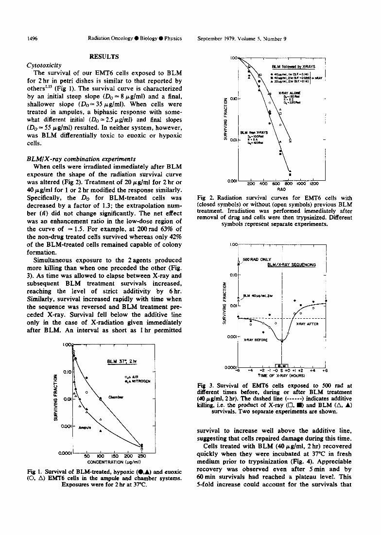
The survival of our EMT6 cells exposed to BLM for 2 hr in petri dishes is similar to that reported by others”” (Fig 1). The survival curve is characterized by an initial steep slope (Do = 8 pglml) and a final, shallower slope (Do = 35 Fg/ml). When cells were treated in ampules, a biphasic response with some- what different initial (Do = 2.5 &ml) and final slopes (Do = 55 Fe/ml) resulted. In neither system, however, was BLM differentially toxic to euoxic or hypoxic cells.
BLMIX-ray combination experiments When cells were irradiated immediately after BLM
exposure the shape of the radiation survival curve was altered (Fig 2). Treatment of 20 pg/ml for 2 hr or 40 pg/ml for 1 or 2 hr modified the response similarly. Specifically, the Do for BLM-treated cells was decreased by a factor of 1.3; the extrapolation num- ber (fi) did not change significantly. The net effect was an enhancement ratio in the low-dose region of the curve of = 1.5. For example, at 200 rad 63% of the non-drug treated cells survived whereas only 42% of the BLM-treated cells remained capable of colony formation.
Simultaneous exposure to the 2 agents produced more killing than when one preceded the other (Fig. 3). As time was allowed to elapse between X-ray and subsequent BLM treatment survivals increased, reaching the level of strict additivity by 6 hr. Similarly, survival increased rapidly with time when the sequence was reversed and BLM treatment pre- ceded X-ray. Survival fell below the additive line only in the case of X-radiation given immediately after BLM. An interval as short as 1 hr permitted
omcl’ I 1
50 loo I!50 200 250 CONCENTRATION (ug/ml)
Fii 1. Survival of BLM-treated, hypoxic (.,A) and euoxic (0, A) EMT6 cells in the ampule and chamber systems.
Exposures were for 2 hr at 37°C.
I . 0.001 I I I I I I 1
200 400 600 800 lo00 I200
RAO
Fig 2. Radiation survival curves for EMT6 cells with (closed symbols) or without (open symbols) previous BLM treatment. Irradiation was performed immediately after removal of drug and cells were then trypsinixed. Different
symbols represent separate experiments.
X.RAY AFTER
I I I
am!6 -4 i
-2 -I -0 S +0 +I +2 +4 +6
TIME OF X-RAY (HOURS)
Fig 3. Survival of EMT6 cells exposed to 500 rad at difierent times before, during or after BLM treatment (40 &nl, 2 hr). The dashed line (------) indicates additive killing, i.e. the product of X-ray (Cl, D and BLM (A, A)
survivals. Two separate experiments are shown.
survival to increase well above the additive line, suggesting that cells repaired damage during this time.
Cells treated with BLM (40 pg/ml, 2 hr) recovered quickly when they were incubated at 37°C in fresh medium prior to trypsinization (Fig. 4). Appreciable recovery was observed even after 5 min and by 60 min survivals had reached a plateau level. This S-fold increase could account for the survivals that

Effects of bleomycin and irradiation on euoxic and hypoxic cells 0 D. C. SHRIEVE and J. W. HARRIS 1497
I .oo I I I 1 I 1
L g 0.10-p 0
5 5 oI 0 a
RECOVERY FROM BLM MOWn-d,2hr)
0.01’ I I I I I I 2 3 4 5+-k-
HO!_% AFTER BLM
Fig 4. Recovery of EMT6 cells from BLM as a function of time (at 37°C) between drug treatment and trypsinization. @ represents cells that were irradiated (500 tad) at different times after BLM and held for a total of 90 min at 37°C between BLM and trypsinization. Data have been corrected for killing by X-ray alone (500 rad) which gave a surviving
fraction of 0.12.
exceed the additive line in the previous sequencing experiment (Fig. 3). X-radiation (500 rads) given dur- ing the recovery process did not affect recovery (Fig 4).
Cells irradiated immediately after a 2 hr exposure to 40 @g/ml of BLM had decreased survival com- pared to non-drug treated cells. The survival curves were characterized by a Do of = 140 rad for non- drug treated cells and a Do = 110 rad for BLM- treated cells. When 90 min elapsed between X-irradia- tion and trypsinization, however, the radiation sur- vival curves for BLM-treated and non-drug treated cells were superimposable (Fig. 5). This indicates that under these circumstances cells not only recovered from BLM damage (Fig. 4) but also recovered from the radiosensitizing effect of the drug.
DISCUSSION We found no differential toxicity of BLM to our
EMT6 cells under hypoxic and euoxic conditions, regardless of whether hypoxia was induced by the chamber method or by the ampule technique. Differential toxicity (i. e. hypoxic cells less sensitive than euoxic cells) has been reported for V79 cells exposed to the drug for 4 hr at 37°C in the latter system.‘” It is possible that the effect depends on
1.
2.
0.001 I I I I I
200 400 600 BOO 000 RAD
Fig 5. Survival of EMT6 cells exposed to BLM (40 &II.& 2 hr) and then to X-radiation. (0), X-ray alone; (0) BLM followed by X-ray and immediate trypsinization; 0 BLM
followed by X-ray with trypsinization 90 min later.
exposure time or is cell line dependent. The difference in the level of survival between the ampule and chamber systems (Fig. 1) may be related to the fact that cells were treated in 15% FCS in the cham- bers but no serum was present in the ampules.
When cells were exposed to BLM immediately prior to X-irradiation the Do was decreased by a factor of 1.3 (Figs. 1 and 5). In the low-dose region of the curve (e.g. 2OOrad) killing was enhanced by a factor of 1.5. The latter is of particular interest since radiotherapy generally involves doses of 200 rad or less per fraction. These modifications of the radia- tion response were seen only when the combined treatment was immediately followed by trypsiniza- tion. When 90 min elapsed between treatment and trypsinization, the shape of the curve returned to that for non-drug treated cells (Fig. 5). Therefore recovery after combination treatment involved 2 processes: repair of some of the BLM damage, and recovery from the radiosensitizing effect of BLM. The mechanism of these recovery processes is not clear.
Further work is necessary to identify the nature of the damage induced by combined BLM/X-ray treat- ment and the mechanism by which cells recover. Of particular interest and possible use would be a means for inhibiting the recovery process. These matters will be the subject of continuing investigation.
REFERENCES Barranco, S.C., Humphrey, R.M.: The effects of bleomycin on survival and cell progression in Chinese hamster cells in aim. Cuncer Res. 31: 1218-1223, 1971. Barranco, S.C., Novak, J.K., Humphrey, R.M.: Res-
ponse of mammalian cells following treatment with bleomycin and 1 ,fbis (Zchloroethyl)-I-nitrosourea during plateau phase. Cuncer Res. 33: 6916% 1973.
3. Barranco, S.C., Novak, J.K., Humphrey, R.M.: Studies

1498 Radiation Oncology 0 Biology ??Physics October 1979, Volume 5, Number 9
on recovery from chemically induced damage in mammalian cells. Cancer Res. 35: 11%1204, 1975.
4. Bienkowsa, Z.M., Dawson, K.B., Peacock, J.H.: Action of actinomycin D, bleomycin and X-rays on HeLa cells. Br. J. Radio1 46: 619-622, 1973.
5. Bistovic, M., MariCiC, Z., Kolaric, K.: Interaction of bleomycin and radiation in combined treatment on mouse L cells. Int. J. Cancer 1%: 540-542, 1976.
6. Bleehen, N.M., Gillies, N.E., Twentyman, P.R.: The effect of bleomycin and radiation in combination on bacteria and mammalian cells in culture. Br. J. Radiof 47: 346-351, 1974.
7. Byfield, J.E., Young, L.C., Tu, L., Kulhanian, F.: Molecular interactions of the combined effects of bleomycin and X-rays on mammalian cell survival. Cancer Res. 31: 1218-1223, 1971.
8. Hall, E.J.. Lehnert, S., Roizin-Towle, L.A.: Split-dose experiments with hypoxic cells. Radiology 112: 425- 430, 1974.
9. Jorgensen, S.J.: Time-dose relationships in combined bleomycin treatment and radiotherapy. Europ. J. Cancer 8: 531-534, 1972.
10. Koch, C.J., Painter, R.B.: The effect of extreme hypoxia on the repair of DNA single-strand breaks in mammalian cells. Radiur. Res. 64: 256-269, 1975
11. Power, J-A., Harris, J.W.: Response of extremely hypoxic cells to hyperthermia: Survival and oxygen
enhancement ratios. Radiology 123: 767-770, 1970. 12. Terasima, T., Takabe, Y., Yasukawa. M.: Combined
effect of X-ray and bleomycin on cultured mammalian cells. Gann 66: 701-703, 1975.
13. Roixin-Towle, L.A., Hall, E.J.: Studies with bleomycin and misonidaxole on aerated and hypoxic cells. Br. J. Cancer 37: 254-260. 1978.
14. Twentyman, P.R., Bleehen, N.M: The sensitivity to bleomycin of spleen colony forming units in the mouse. Br. J. Cancer 28: 66-70, 1973.
15. Twentyman, P.R., Bleehen, N.M.: The sensitivity of cells in exponential and stationary phases of growth to bleomycin and to 1,3-bis(2-chloroethyl)-I-nitrosourea. Er. J. Cancer 28: 500-507, 1973.
16. Twentyman, P.R., Bleehen, N.M.: Changes in the sen- sitivity to bleomycin occurring during the life history of monolayer cultures of a mouse tumour cell line. Br. J. Cancer 32: 68-74, 1975.
17. Twentyman, P.R., Bleehen, N.M.: Studies of “poten- tially lethal damage” in EMT6 mouse tumour cells treated with bleomycin either in vitro or in viva. Br. J. Cancer 32: 491-501. 1975.
18. Wharam, M.D., Phillips, T.L., Kane, L.S., Utley, J.F.: Response of a solid murine tumor to in uiuo combined chemotherapy and irradiation. Radiology 109: 451-455, 1973.