effects of a cytosolic protein on the interaction of rat pancreatic zymogen granules in vitro
TRANSCRIPT

Biochimica et Biophysica Acta 897 (1987) 217-228 217 Elsevier
BBA72947
Ef fec t s of a cy tosol ic p ro te in on the in te rac t ion
of ra t pancrea t i c z y m o g e n granules in vi tro
J a n e Rogers , E .K. M a t t h e w s and D.B. M c K a y *
Department of Pharmacology, University of Cambridge, Cambridge (U.K.)
(Received 11 June 1986)
Key words: Zymogen granule; Granule aggregation; Stabilization factor; Secretion; Quasi-elastic light scattering; (Rat pancreas)
Photon correlation spectroscopy has been used to study the kinetics of aggregation of isolated rat pancreatic zymogen granules in vitro by monitoring time-dependent changes in mean particle size derived from the photon count autocorrelation function, g2(~). Isolated granules were stable in isotonic sucrose (pH 5.4-7.0). At pH 6.0 they maintained a mean diameter of 1225 + 18 nm with a polydispersity index of 0.199 + 0.007. The mean granule diameter showed a limited decrease (approx. 20%) with increasing pH within the range 5.4-7.0, but the polydispersity index was unaltered. At pH > 7.0 granule instability was indicated by a rapid reduction in total photon counts. In solutions of monovalent cations ([M +l > 10 raM) and divalent cations ([M 2+ ] > 0.5 raM) zymogen granules aggregated at a rate dependent upon both ion and granule concentra- tion. These effects were consistent with the bimolecular nature of the interaction mechanism and were clearly distinguishable from the limited size changes associated with osmolarity. At concentrations of Na + or K + salts > 50 mM granule aggregation was accompanied by anion-dependent solubilisation. A soluble protein fraction separated from the pancreatic acinar cell cytosol by gel filtration reduced the mean diameter and polydispersity index of zymogen granules suspended in isotonic sucrose, inhibited cation-induced aggregation and stabilised granules to solubilisation induced by raising pH > 7.0 or exposure to high ionic strength media. The inhibitory effects of this protein were apparent at concentrations ~< 10/tg • ml - t (i.e. at inhibitor: granule protein ratios < 1:20) and could not be mimicked by bovine serum albumin, the Ca2+-binding proteins calmodulin and troponin C (~< 100 pg .mi -1 ) , nor the highly negatively charged polymer polyglutamate (~< 10 / tg .ml - l ) . Inhibitory activity was also absent from fractions of rat liver cytosoi prepared identically to pancreatic acinar cytosol. These observations are consistent with the presence in pancreatic acinar cells of a specific cytosolic granule stabilisation factor (or factors) that normally restricts zymogen granule interaction and may therefore play an important role in the regulation of granule mobility and exocytosis.
* Present address: Department of Pharmacology, College of Pharmacy, Ohio State University, Columbus, Ohio 43210- 1291, U.S.A.
Abbreviations: EGTA, ethyleneglycol bis(fl-aminoethyl ether)- N,N,N',N'-tetraacetate; Mes, 4-morphofineethanesulphonic acid; [M n+ ]i, intracellular free cation concentration; PMSF phenylmethylsulphonyl fluoride.
Correspondence: E.K. Matthews, Department of Pharmacol- ogy, University of Cambridge, Hills Road, Cambridge, CB2 2QD, U.K.
Introduction
The secretion of digestive enzymes by exocyto- sis from pancreatic acinar cells involves controlled fusion between the membranes of the zymogen granules and the apical region of the cell. The process is triggered by secretagogue activation of membrane receptors which initiates a change in cellular membrane conductance [1,2] and genera-
0005-2736/87/$03.50 © 1987 Elsevier Science Publishers B.V. (Biomedical Division)

218
tion of one or more of a variety of putative second messengers such as Ca 2+, inositol phosphates, di- acylglycerol or cyclic GMP [3-7]. The precise role of any of these intracellular mediators in the mobilisation of zymogen granules and the exoc- ytotic extrusion of their contents is, however, still unknown. Attempts to elucidate the various stages of the secretory process by subcellular fractiona- tion have revealed that, when compared to storage granules isolated from other types of secretory cell, isolated zymogen granules are less stable at physiological ionic strength and display an opti- mal stability in non-electrolyte solutions at pH 4.5-6.0 [8,9]. Since from measurements of total ionic content the intracellular milieu of pancreatic acinar cells appears to be similar to that of other cells, i.e. [K+]i approx. 140 mM, [Na÷]i approx. 50 mM, pH 6.8-7.0 [10], it might be anticipated that in vivo the zymogen granule population would be unstable and undergo spontaneous lysis. Elec- tron microscopic studies, however, have indicated no dissolution of granules in intact cells, even when maximally stimulated [11]. The differences in zymogen granule behaviour in vivo and in vitro have been ascribed by De Lisle et al. [12] to damage during the isolation procedure which re- nders the granules susceptible to ionic dissolution of the contents. An alternative possibility is that a cytoplasmic granule stabilisation factor normally restricts granule interaction in vivo and therefore plays an important role in the regulation of gran- ule mobilisation and exocytosis. O'Connor and Matthews [13] have described some evidence for such a factor in cells of the endocrine pancreas. We report here the isolation and partial purifica- tion of a soluble protein factor in cells of the exocrine pancreas that stabilises zymogen granules to ion and pH-induced dissolution and potently inhibits granule aggregation in vitro.
The new technique of photon correlation spec- troscopy based on dynamic laser light scattering has been used to investigate the kinetics of granule interaction, since this method allows time-depen- dent changes in granule dimension and aggrega- tion to be monitored. The effects of osmolarity on granule size per se and the influence of pH and cations on zymogen granule interaction in the absence and presence of the cytosolic factor are described.
Methods
Zymogen granule preparation Pancreases were excised from male Sprague-
Dawley rats of 250-300 g and placed immediately in ice-cold Krebs-Henseleit solution (pH 7.4). After removal of accessory adipose tissue and blood vessels the pancreases were finely chopped in 10 volumes of cold 0.28 M sucrose solution and homogenised using three strokes in a Dounce ho- mogeniser. Zymogen granules were obtained from the homogenised tissue essentially as described by Meldolesi et al. [14]. Nuclei and cellular debris were removed by centrifugation in a swing-out rotor at 300 x g for 10 rain and the supernatant was transferred to a fixed angle rotor and centri- fuged at 700 x g for 10 rain. The resulting white zymogen granule pellet was washed free of the brown overlay of mitochondria, lysosomes etc. with 2 X 1 ml of 0.28 M sucrose, 5 mM Mes(pH 6.0) and resuspended in this medium to give a protein concentration of approx. 2 rag- ml-1. For light scattering measurements granules were routinely diluted into the suspension medium to give a granule protein concentration of approxi- mately 200/~g. ml-1.
Cytosol fractionation To obtain the pancreatic acinar cell cytosol
fraction, PMSF was added at a final concentration of 0.1 mM to the supernatant from the zymogen granule preparation and the solution centrifuged at 100000 x g in an ultracentrifuge for 60 min. The supernatant was dialysed overnight against 150 mM NaCI, 5 mM Mes (pH 6.0), concentrated to 1/40 of the original volume using Minicon B filters (Amicon) and subjected to gel filtration on a 60 x 1.5 cm column of Sephadex G-150 equi- librated with 150 mM NaC1, 5 mM Mes (pH 6.0). Cytosol fractions were eluted from the column in 2-ml aliquots of this buffer.
Biochemical analyses The protein content of zymogen granule and
cytoplasmic fractions was determined by the method of Bradford [15] using bovine serum al- bumin as a standard.
Cytochrome c oxidase was measured by the method of Hodges and Leonard [16] with the

modifications of Pacquet et al. [17]. N-Acetyl- fl-glucosaminidase and NADPH-cytochrome c re- ductase were assayed by the methods of Findlay et al. [18] and Omura and Takesue [19], respectively, and a-amylase was determined according to the method of Rinderknecht et al. [20]. Enzyme activi- ties were calculated as relative specific activity i.e. the ratio of the percent of enzyme marker to the percent of total protein in a given fraction, taking 100% activity as the activity in the cell homo- genate.
Electron microscopy Zymogen granules were fixed overnight at 4 ° C
in 0.1 M sodium cacodylate buffer (pH 6.0) con- taining 2% glutaraldehyde, 2% paraformaldehyde and 2% sucrose and centrifuged at 8700 × g for 2 rain (Beckmann microfuge). Pellets were post-fixed with 2% osmium tetroxide for 2 h at room temper- ature, transferred to 0.05 M sodium maleate buffer (pH 5.2) and stained overnight with 2% uranyl acetate prior to dehydration in a graded ethanol series and embedding in Spurr's epoxy resin. Sec- tions were cut with a diamond knife and stained with uranyl acetate and lead citrate before observation with a Philips EM 300 electron micro- scope.
Photon correlation spectroscopy Particle size determinations. Coherent light from
a 15 mW He-Ne laser (Spectra Physics Model 124A) was focussed at the centre of the sample (0.6 ml) contained in a glass cuvette maintained at a temperature of 37 +_ 0.05 ° C in an optical index matching bath. Photon scattering was detected at 90 ° to the incident collimated laser beam by a photomultiplier tube (EMI D260E) equipped with an amplifier/discriminator. The digitized output of the photomultiplier tube was analysed with the 96 channel Autocorrelator (K7023) of a Malvern System 4300 Photon Correlation Spectrometer (Malvern Instruments Ltd., Malvern, U.K.) and on-line data fitting routines performed by an MPD 7023S microprocessor and printer [21].
The normalised autocorrelation function for quasi-elastic light scattering from a suspension of identical, freely diffusing particles is given by
219
where ,r is the sample time in ms, A is an instru- ment optical constant, F = D K 2, and D is the diffusion coefficient. The scattering wave vector K is defined by
4 ~/'/'/ . 0 K = ~ sm -~ (2)
and is constant for a given angle, 0, where n is the refractive index of the scattering medium and h is the wavelength of the He-Ne laser light source (632.8 nm).
The gradient of the semi-log regression (de- termined by a least-squares polynomial fitting routine) from Eqn. 1, see Fig. 1, therefore yields D and from the Stokes-Einstein relationship
kT D = ,,3,,--~ (3)
the particle diameter, d, can be determined, where k is Boltzmann's constant, T is temperature (K),
is medium viscosity. For each correlation func- tion the time-averaged scattered light intensity, i.e. total photon counts, were also recorded for the correlation period. The computed quadratic func- tion of the semi-log regression from Eqn. 1 also yielded an initial polydispersity index, ~t 2 / F 2 (see Refs. 22 and 23), for samples prior to aggregation.
Measurement of particle interaction and aggrega- tion. If there are present initially in the scattering system n o monomers per unit volume and aggre- gation begins at time t = 0 then at t > 0 aggregates of 2, 3, 4 . . . . . j particles will be formed assuming that each particle encounter leads to lasting con- tact. At time t the number of j-fold particles will be given by
n o nj j=l l +4qrDn°t (4)
where D is the particle diffusion constant. Now D, the average diffusion constant mea-
sured by photon correlation spectroscopy, is the average of the diffusion constants of all the par- ticles in the suspension weighted by the intensity of the light scattered.
Thus for samples which are aggregating, i.e. are polydispersed, Eqn. 1 becomes:
g2(~) =1+ Ae-2r~ (1) g2(r)=l+Ae-2r~+higherorderterms (5)
220
The initial gradient of log g2 ( ' r ) is n o w D K 2 and D is the so-called z-averaged diffusion coefficient [22] i.e.
Y'Ci D i Mi EC, M, (6)
where for the i th species, C i is the concentrat ion, Mi is the mass and D~ is the diffusion coefficient. For completely mono-disp__ersed particles of diffu- sion coefficient, D, then D = D, and higher order cumulants are small or negligible, but as aggrega- tion proceeds, i.e. the number of j -mers increase (see Eqn. 4), then D becomes smaller than D. This progressive decrease in D will be reflected by the Stokes-Einstein relationship as a t ime-depen- dent increase in the mean particle diameter, a~ Measurements of mean particle diameter can be therefore be used as a sensitive measure of the interaction and aggregation of zymogen granules.
For particle aggregation studies using laser-light scattering techniques the optimal size range of the initial monomer is defined by
X/4 < d/2 < X
where X is the wavelength of the incident radia- tion and d is the particle diameter [24].
Statistical analysis The s tandard error of the mean (S.E.) was
calculated and the significance of difference be- tween means determined using Student 's t-test.
Results
Granule characteristics and purity Zymogen granules were obtained in high yield,
approx. 2 mg granule p r o t e i n / g r a m pancreas, using the two stage centrifugation procedure described in Methods. Enzyme assays for subcellu- lar organelles indicated that mitochondrial , lyso- somal and microsomal contaminat ion of the
1.0
8
~ 0.5
)
B
%
k -- \
0 1.8 3.a 5.4 r,2
1- (mlec)
| I
CHANNEL NUMBER
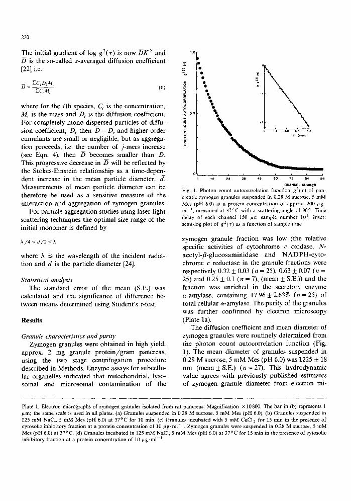
Fig. 1. Photon count autocorrelation function g2('r) of pan- creatic zymogen granules suspended in 0.28 M sucrose, 5 mM Mes (pH 6.0) at a protein concentration of approx, 200 gg- m l, measured at 37°C with a scattering angle of 90 °. Time delay of each channel 150 /~s; sample number 105. Inset: semi-log plot of g2(~.) as a function of sample time.
zymogen granule fraction was low (the relative specific activities of cy tochrome c oxidase, N- ace ty l - f l -g lucosaminidase and N A D P H - c y t o - chrome c reductase in the granule fractions were respectively 0.32 + 0.03 (n = 25), 0.63 __+ 0.07 (n = 25) and 0.25 + 0.1 (n = 7), (mean _+ S.E.)) and the fraction was enriched in the secretory enzyme a-amylase, containing 17.96 + 2.63% (n = 25) of total cellular a-amylase. The puri ty of the granules was further confirmed by electron microscopy (Plate la).
The diffusion coefficient and mean diameter of zymogen granules were routinely determined f rom the pho ton count autocorrelat ion function (Fig. 1). The mean diameter of granules suspended in 0.28 M sucrose, 5 m M Mes (pH 6.0) was 1225 ___ 18 n m ( m e a n + S.E.) (n = 27). This hydrodynamic value agrees with previously published estimates of zymogen granule diameter f rom electron mi-
Plate 1. Electron micrographs of zymogen granules isolated from rat pancreas. Magnification x 10800. The bar in (b) represents 1 /zm; the same scale is used in all plates. (a) Granules suspended in 0.28 M sucrose, 5 mM Mes (pH 6.0). (b) Granules suspended in 125 mM NaC1, 5 mM Mes (pH 6.0) at 37°C for 10 rain. (c) Granules incubated with 5 mM CaC12 for 15 min in the presence of cytosolic inhibitory fraction at a protein concentration of 10 #g.ml -a. Zymogen granules were suspended in 0.28 M sucrose, 5 mM Mes (pH 6.0) at 37°C. (d) Granules incubated in 125 mM NaC1, 5 mM Mes (pH 6.0) at 37°C for 15 min in the presence of cytosolic inhibitory fraction at a protein concentration of 10 t~g. ml-1.

221

222
120
w 110 N
z < 1 0 0 (lc
90
80
'G 9
x v
r z 0
z 0 5 t- O 0.
°OoOO o oee Se8 & &
& ,&
A &
i i i ' ' ' . . . . 5.0 5 5 6 0 6 5 T.0 7.5 0 5 10 15 20
pH TIME (rain
Fig. 2. Effect of pH on zymogen granules. (a) pH dependence of mean granule diameter. Zymogen granules were suspended in 0.28 M sucrose, 5 mM Mes at a protein concentration of 200/,g.m1-1 and granule diameter determined as the mean of 10 successive values calculated from the autocorrelation function as described in Methods. Granule size is expressed as a percentage of the mean diameter of granules measured at pH 6.0. The data are taken from four separate experiments. (b) Time dependence of zymogen granule stability monitored by total photon counts of granule suspensions buffered at pH 6.0 (O), pH 6.9 (e) and pH 7.3 (A).
croscopy (924-1240 nm) [12,25]. The mean poly- dispersity index of zymogen granules suspended in sucrose /Mes was 0.199 +__ 0.007. This value indi- cates that although the size range of the granules was greater than that of a homogeneous synthetic particle suspension (e.g., the mean diameter of polystyrene latex particles was 109 + 0.4 nm with a polydispersity index of 0.073 + 0.008), the range is similar to that shown by preparations of gran- ules from other secretory cells [26].
Ionic dependence of granule stability When compared with many other isolated
secretory granule preparations, zymogen granules are relatively unstable under conditions approxi- mating to normal intracellular ion concentrations and p H [8,9]. In 0.28 M sucrose, 5 mM Mes, at 37°C, however, the granules were stable in the laser beam over a p H range of 5.4 to 7.0. Increas- ing the p H within this range reduced the mean granule diameter (Fig. 2a) but the polydispersity index was unchanged. At pH > 7.0 the granules were unstable. The granule contents became solubilised and this phenomenon, assayed as a-amylase release from granules, correlated with a decrease in the total light scattering (photon counts) detected by the photomultiplier (Fig. 2b). Although the granules were relatively stable in
sucrose solution between pH 5.4 and 7.0, they were routinely prepared and maintained at pH 6.0 before use, at which p H they were stable for > 90 rain at 37 o C.
When zymogen granules were suspended in 125 mM KC1, 5 mM Mes (pH 6.0), an immediate rapid, decrease in diffusion coefficient was observed. This was monitored continuously over the first 10 min by autocorrelation spectroscopy and used to determine the initial rate of increase in mean diameter of the granules. The rate of increase was dependent upon the concentration of KC1 (Fig. 3) and showed a similar concentration dependence in NaC1, indicating that the response was not selective with respect to either cation. Measurement of the increase in mean diameter was limited to the first 10 min after raising the ionic strength by a decrease in total light scatter- ing (photon counts) when [M +] > 50 mM. The photon count reduction correlated with loss of a-amylase from the granules and the presence of empty vesicles in electron micrographs (Plate lb), indicating that the granule contents are solubilised in high ionic strength media. For granules sus- pended in 125 m M solutions of Na ÷ or K ÷ salts containing 5 mM Mes (pH 6.0), both the rate of solubilisation and the aggregation rate were dependent upon the anion present. Thus for the

223
200
t¢:
,~E~ 160
rT- t,U I,- uJ
~ 1 2 0
W . J
Z
z w IE <1 40
o
o o 40 80 12o
[KCI] mM
Fig. 3. Relationship between the rate of increase in mean diameter of zymogen granules and KC1 concentration. Zymo- gen granules were suspended at a protein concentration of approx. 200 g g . m l 1 in 5 m M Mes (pH 6.0) with various concentrations of KC1. The rate of increase in mean granule diameter was determined from the values of granule diameter calculated for the first 10 minutes following exposure to ions. The results shown are from one experiment representative of five others.
following anion species the decreasing order of potency with respect to solubilisation was acetate > nitrate > chloride > bromide > isethionate > glutamate, which was strikingly similar to the in- creasing order of potency with respect to aggrega- tion, viz. nitrate < acetate < chloride < isethionate < bromide < glutamate. The decrease in cation- induced aggregation rate in granule samples un- dergoing rapid solubilisation may be due to a decrease in photon counts reducing the accuracy of size determination. It is more probable, how- ever, that solubilisation induced by permeant an- ions alters the light scattering properties of the granules and thus obscures the cation-induced ag- gregation under these conditions.
Effects o f Ca 2 + Since a rise in intracellular [Ca 2+] is an early
consequence of acinar cell stimulation by several secretagogues, the effects of Ca 2÷ on isolated zymogen granules were investigated. Addition of C a 2+ at concentrations > 0.5 mM to granules suspended in 0.28 M sucrose 5 mM Mes (pH 6.0), evoked a sustained increase in mean granule di- ameter which could not be reversed by the ad- dition of EGTA. The mean rate of size increase
2000
i w
w 18oo
D 1600 Z
Z
~ 1400
1200
1000
(a)
1 0 0
0 00000000 00000~00000
L I l a I a I i i i
0 5 10 15 20
TIME (mln)
• 0 0
0 0 0 0
5
3
1
25
11 2 0 0 0 11
f (b) 9 1800F 9 D
0 c 5
7 1600 0 0 0 0 0 0 0 0 0 0 • 7
o • • 1 4 0 0 5
o
• • • O o • 12oo • • o
0 • • 0 0 • • • 0 0 0 0
1000
i i
0 5 10 15 20 TIME ( ra in ' )
Fig. 4. Time dependence of the effect of Ca 2+ on zymogen granule behaviour. The increase in mean granule diameter (e) and the change in total photon counts (O) induced by the addition of 5 m M CaCI 2 (at arrow) to zymogen granules (200 ~g protein, m l - l ) suspended in 0.28 M sucrose, 5 m M Mes were monitored continuously at (a) pH 6.0 and (b) pH 6.9. The results shown are taken from one experiment typical of at least five others.

224
induced by 5 mM Ca 2+ was 54 + 2 n m / m i n over the first 15 rain (Fig. 4a). In contrast to the behaviour of granules exposed to high concentra- tions of monovalent cations, the Ca2÷-induced increase in diameter at pH 6.0 was not accompa- nied by a rapid decrease in total photon counts ( < 0.5% min) and a large proportion of the gran- ules appeared intact on electron microscopy. With increasing pH, however, both the rate of increase in diameter induced by Ca 2÷ and the stability of the granules were reduced until, at pH 7.0, 5 mM Ca 2÷ induced immediate breakdown of the gran- ules, as evidenced by a rapid decrease in photon counts (Fig. 4b). Although Ca 2÷ is thought to be the major ion involved in the mechanism of secre- tion, the response of zymogen granules to divalent metal cations was not CaZ+-specific. In decreasing order of potency in 0.28 M sucrose, 5 mM Mes (pH 6.0) Zn 2+, Mn 2+, Ca 2+, Sr 2+, Ba 2+ and Mg 2+ each increased mean granule diameter.
Osmotic effects To determine whether the cation-induced in-
crease in granule diameter represents aggregation or is due, at least in part, to osmotic swelling, the effect of osmolarity on granule diameter was mea- sured using sucrose to adjust solution tonicity. As illustrated in Fig. 5, there was only a small change
1300
~ 1 2 0 0
1100
1000
9 0 0
i i i ! i m
0 0.2 0,4 0.6 0.8 1.0 OSMOLARITY
Fig. 5. Effect of osmolarity on zymogen granule diameter. The osmolarity of zymogen granule suspensions (approx. 200 /tg protein.rn1-1) was controlled by changing the sucrose con- centration in solutions containing 5 m M Mes (pH 6.0). The results are expressed as mean _+ S.E. for four different experi- meats.
140
t E s. ~2o
I w 100 f- Lu
< 80
Z < 6 O
Z
< 4 0
< 2O
0
0
rl
13
0
0 0
0 []
r'l
0 E]
L , ~ , i i i i
0.1 0.2 0,3
GRANULE PROTEIN CONCENTRATION (rng.mi "1)
Fig. 6. Dependence of zymogen granule aggregation rate on granule concentration. Aggregation was induced by suspension of granules in 125 m M KC1 @3) or by addition of 5 mM CaCI 2 to granules suspended in 0.28 M sucrose (©) and the aggrega- tion rate determined as the increase in mean granule diameter measured over the first 10 minutes following exposure to ions. All solutions contained 5 m M Mes buffered at pH 6.0. Granule concentration is expressed as concentration of granule protein in m g . m l 1.
(< 12%) in granule diameter in solutions ranging from 0 to 1 M sucrose, indicating that intact granules have only a very limited distensibility. Furthermore, if the ion-induced size increase were due simply to osmotic swelling it would be expected to be independent of granule concentra- tion, but this is not so for either the K ÷ or the Ca2+-induced increase in mean granule diameter (Fig. 6). We therefore attribute the initial increase in mean granule diameter evoked by cations to aggregation. In the absence of aggregation the diffusion coefficient and mean granule diameter remained constant over a wide range of particle concentrations (0.04-0.4 protein, ml-X), in agree- ment with quasi-elastic light scattering theory for non-constrained, non-interacting particles.
Granule stabilisation factor(s) As noted in the Introduction, there is evidence
that in some secretory cells granule mobilisation and exocytosis is controlled, at least in part, by soluble protein factors. To investigate whether such proteins exist in rat pancreatic acinar cells the 100 000 × g supernatant from homogenised rat

225
2.5
uJ 0 Z
2.( E O (/)
1.5
"I.C
0.5
2O O
30 40 50 60 70 80
FRACTION NUMBER
1.25
1.00
).75
3.50
0.25
Fig. 7. A typical elution profile of pancreatic acinar cell cytosol fractionated on Sephadex G-150 (see Methods). 2-ml fractions were eluted in 150 mM NaCI, 5 mM Mes (pH 6.0). Absorbance at 280 nm (e); absorbance at 260 nm (©); protein concentra- tion (mg-ml-]) (zx). The bar indicates fractions that inhibited cation-induced aggregation of zymogen granules when added to granule suspensions at concentrations ~< 10/~g. ml- i.
pancreas was fract ionated on Sephadex G-150 as described in Methods. A typical elution profile of cytosolic componen ts f rom the column is shown in Fig. 7. Fract ions 4 8 - 5 4 consistently exhibited marked stabilising effects on granule suspensions and inhibited cat ion-induced aggregation at con- centrat ions of approx. 10 # g . m l - t . The molecular weight range of proteins eluting f rom the column in these fractions was estimated, by compar ison with fract ionation of molecular weight markers, to be 20-27 kDa. All other fractions, or bovine serum albumin, when added at the same final protein
?, £ m z (3 o z Z
>
0 Z
3_,
concentra t ion (5-200 /~g-ml - t ) were without ef- fect on the ion-induced changes in granule laser light scattering. Identically prepared fractions from rat liver cytosol also showed no inhibition of cat ion-induced zymogen granule aggregation. The inhibitory fractions had four effects on zymogen granules: (1) the mean granule diameter in 0.28 M sucrose, 5 m M Mes (pH 6.0) was reduced by approx. 20%, f rom 1225 + 18 nm to 998 + 15 ( p <0 .001) ( n = 2 7 ) ; (2) the mean polydispersity
index in 0.28 M sucrose was reduced f rom 0.199 + 0.007 to 0.154 + 0.004 ( P < 0.001) (n = 27); (3) the cat ion-induced increase in mean granule diam- eter i.e,, aggregation, was inhibited; and (4) the ion-induced decrease in total light scattering, i.e., pho ton counts, was markedly reduced. The effects of the inhibitory cytoplasmic fractions on Ca 2÷- and KCl- induced aggregation are shown in Figs. 8 and 9, respectively, and in Plate l c and ld. The inhibition of the ionic effects on zymogen granules at p H 6.0 was also seen in the normal cellular pH range (pH 6.8-7.0). Preliminary at tempts to purify further the inhibitory factor by DEAE-cel lulose ion-exchange chromatography indicate that it is negatively charged at p H 6.0.
One way in which the inhibitory protein(s) might modulate the act ion of Ca 2÷ on zymogen granules is to act as a Ca 2÷ binding protein. However, using a method based on that of Cuatrecasas [27], there was no detectable 45Ca2÷
2000' I o o o o
o 15oo o o oo
<[ o o
°ooO °oO %~ '~°I ..s.;.. ..
~oo t
o
0 5 10 15 20 25 0 5 10 15 20 25 TIME (rain) TIME (min)
Fig. 8. Time dependence of the ~esponse of mean granule diameter (a) and photon counts (b) of zymogen granules to the addition of 5 mM CaC12 (at arrow) in the absence (open symbols) and presence (closed symbols) of cytosolic inhibitory fraction. Zymogen granules were suspended in 0.28 M sucrose, 5 rnM Mes (pH 6.0). The final protein concentration of the inhibitory fraction was 10/xg. ml- 1.

226
2000 o a
o 2
o o o
o 1500 o
° o O
o o • o e
I ~ e e e e ~ e e ~
1000 % • • • •
5OO
TIME ( ra in )
b O
z
() b o c z
u
TIME ( m l n )
Fig. 9. Time dependence of the response of mean granule diameter (a) and photon counts (b) of zymogen granules suspended in 125 mM KCI, 5 mM Mes (pH 6.0) at t = 0, in the absence (open symbols) or presence (filled symbols) of cyto- solic inhibitory fraction. The final protein concentration of the inhibitory fraction was 10 t~g.ml 1.
binding to the protein in fractions 48-54. Further- more, addition of purified calmodulin or troponin C (~< 5 t~M--100 #g . m1-1) did not reduce the mean diameter of zymogen granules suspended in sucrose, nor inhibit the effects of Ca 2+ or KC1 in increasing granule size (i.e., aggregation) or de- creasing photon counts (i.e., solubilisation). Another possibility is that the negatively charged inhibitory protein may bind to the granule mem- brane and increase the electrostatic repulsion be- tween adjacent granules. In each of three experi- ments, the addition of the polyanion, polygluta- mate (~< 10 fig. ml-1), had no effect on the prop- erties or ion-induced responses of isolated zymo- gen granules. Any electrostatic effect of the inhibi- tory protein must therefore be dependent on specific adsorption to the granule surface.
Discussion
In this paper we describe the first isolation of a cytosolic factor that stabilises rat pancreatic zymogen granules. This factor reduced the hydro- dynamic diameter of granules suspended in sucrose and inhibited cation-induced aggregation. The in- hibitory factor also reduced the solubilisation of granule content when the pH was raised above 7.0 or the granules suspended in high ionic strength media. The aggregating effect of cations in the absence of inhibitory protein is most probably due
to a decrease in the net negative surface charge of the zymogen granule membrane [28]. As divalent cations will be much more effective (> 50-fold) than monovalent cations in compressing the ionic double layer and reducing the interfacial potential energy barrier between individual zymogen gran- ules, they will promote aggregation at much lower concentrations. This may result in fusion and loss of granule contents, but cannot be the only mech- anism of solubilisation, since at pH > 7.0 loss of granule content occurred rapidly with no indica- tion of prior aggregation. Furthermore, in 5 mM Mes (pH 6.0) in the presence of 125 mM sodium glutamate, aggregation proceeded rapidly without extensive solubilisation, while in the presence of 125 mM sodium acetate rapid solubilisation was accompanied by only a low rate of aggregation. These observations suggest that solubilisation re- sults from permeation of ions across the granule membrane, a process determined by its selective permeability to anions.
The action of the cytoplasmic fraction in in- hibiting the aggregation process cannot be attri- buted to simple electrostatic effects or cationic charge neutralisation lowering bulk phase electro- lyte concentration, because the protein was eluted from the column in 150 mM NaCI and added to zymogen granules in this solution. Furthermore, the absence of detectable Ca z+ binding to the inhibitory factor and the inability of troponin C or calmodulin to mimic the inhibitory effects clearly indicates that granule fusion is not regu- lated by Ca 2+ binding proteins alone. Although changes in viscosity may exert some effect it is likely that the inhibitory factor(s) undergoes ad- sorption to the zymogen granule surface and both spatially opposes the electrostatic action of cations and stabilises membrane structure; a polyanionic protein would be particularly effective in this re- gard. Such a protein could also reduce solubilisa- tion by decreasing the exchange of free anions with, and permeation across, the granule mem- brane. The inability of bovine serum albumin or the highly negatively charged polymer, poly- glutamate, to mimic the effects of the cytosolic factor points to an important specificity in the surface adsorption and aggregation-inhibition re- action. This specificity is also suggested by the absence from rat liver cytosol of an effective in-

hibitory factor. Thus, in addition to modulation of the ion-induced short-range interactions which would otherwise lead to wholesale aggregation within the acinar cell, the cytosolic factor(s) may have an important functional role to play when adsorbed to the granule surface or in close proximity to it.
The in vivo stability of zymogen granules in the resting acinar cell compared with their instability in an in vitro ionic environment approximating to that of the intracellular compartment has hitherto been rationalised by three proposals: (1) the gran- ule isolation procedures may damage the granules, making them unstable in cytosol-like media [12]; (2) granule proteins may equilibrate across the granule membrane and thus be lost down the concentration gradient in vitro [29]; and (3) the activity of ions in the cytosol of acinar cells must be considerably lower than their concentrations [30]. The first proposal, based on the observation that zymogen granules prepared by density gradi- ent centrifugation in Percoll are stable in isotonic NaC1 or KC1 at physiological pH, has been criti- cized by Rothman and Liebow [31]. They pointed out that, when prepared by other methods, more than 90% of granules would have to be damaged to explain their behaviour in ionic media although, in electron microscopy sections, they appear very similar to granules prepared in Percoll. We have found that extensive washing partially restored the response of granules prepared in Percoll to cation-induced aggregation without increasing their instability in isosmotic sucrose; this treat- ment also reduced a component of the total light scattering contributed by small particles (data not shown). We therefore suggest that the refractori- ness of Percoll-prepared granules to isotonic NaC1 or KC1 can be attributed to the prevention of granule interaction by Percoll binding to the gran- ule surface. Zymogen granules prepared by the two-step procedure described in Methods were very stable in sucrose solution in the pH range 5.4-7.0, at 37°C, as determined by the absence of any change in total photon counts for at least 30 rain. Furthermore, this stability persisted upon dilution, a finding inconsistent with equilibration of granule proteins across the granule membrane, as proposed by Liebow and Rothman.
The third rationalisation, i.e. that the activity of
227
ions in the cytosol is considerably lower than their concentrations, has been shown to be untenable. Intracellular free ion concentrations of 10.5 mM and 124 mM for Na ÷ and K ÷, respectively, have been recorded with ion-selective microelectrodes [32,33] and the intracelhilar pH has been esti- mated as 6.8-7.0 from 5,5-dimethyl-2,4-oxazoli- dinedione (DMO) partitioning experiments [10]. The [Ca2+]i in resting acinar cells has also been shown to be similar to that in other cell types, i.e. approx. 150 nM, when measured by quin2 fluores- cence [4,34]. Cellular compartmentalisation might permit maintenance of a specialised ionic micro- environment in the vicinity of the zymogen gran- ules, which are largely confined to the apical re- gion of the cell. There is, however, no convincing evidence for any cellular structure which would effectively isolate the mature granules of the api- cal region from cytosolic continuity with more distal regions [35]. The presence of a cytosolic protein factor, reported here to stabilise isolated zymogen granules against the aggregating effects of solutions of physiological ionic strength, would readily explain the maintenance of granule integr- ity observed in vivo and has several important implications for control of normal secretion. Fur- ther experiments are required to resolve the mech- anism whereby the inhibitory effects of the protein may be counteracted during exocytosis: this may be regulated, directly or indirectly, by the phosphorylation of cytosolic or granule proteins that occurs when acinar cells are stimulated with a variety of ligands [36,37]. This process may be similar to the regulation of the inhibitory protein synapsin by CaZ+-calmodulin dependent phos- phorylation in neurosecretory cells [38].
Finally, the results of this study illustrate the advantages of photon correlation spectroscopy for investigating the kinetics of granule interaction. By use of this technique we have been able to distinguish between the limited changes in zymo- gen granule size resulting from osmotic swelling and the extensive changes due to granule aggrega- tion upon exposure to cations. Furthermore, by continuously monitoring the total light scattering, the stability of the granules over the experimental period can be assessed under precisely controlled conditions.
228
Acknowledgements
We thank the W e l l c o m e Trus t a n d the Roya l Socie ty for f i nanc i a l s u p p o r t a n d a re gra tefu l to Mr. R. H u g h e s for technica l ass is tance.
References
1 Dean, P.M. and Matthews, E.K. (1972) J. Physiol. (London) 225, 1-13
2 Nishiyama, A. and Petersen, O.H. (1975) J. Physiol. 244, 431-465
3 Matthews, E.K., Petersen, O.H. and Williams, J.A. (1973) J. Physiol. (London) 234, 689-701
4 Merritt, J.E. and Rubin, R.P. (1985) Biochem. J. 230, 151-159
5 Streb, H. and Schulz, I. (1983) Am. 1. Physiol. 245, G347-G357
6 Putney, J.W., Burgess, G.M., Halenda, S.P., McKinney, J.S. and Rubin, R.P. (1983) Biochem. J, 212, 483-488
7 Cristophe, J.P., Frandsen, E.F., Conlon, T,P., Krishna, G. and Gardner, J.D. (1976) J. Biol. Chem. 251, 4640-4645
8 Hokin, L.E. (1955) Biochim. Biophys. Acta. 18, 379-385 9 Rothman, S.S. (1971) Biochim. Biophys. Acta 241,567-577
10 Preissler, M. and Williams, J.A. (1981) J. Physiol. (London) 321,437-448
11 Ichikawa, A. (1965) J. Cell. Biol. 24, 369-385 12 De Lisle, R.C., Schultz, I., Tyrakowski, T., Haase, W. and
Hopfer, V. (1984) Am. J. Physiol. 246 (Gastrointest. Liver Physiol. 9), G411-G418
13 O'Connor, M.D.L. and Matthews, E.K. (1980) Horm. Metab. Res. 12, Suppl. 10, 149-153
14 Mddolesi, J., Jamieson, J.D. and Palade, G.E. (1971) J. Cell. Biol. 49, 109-129
15 Bradford, M. (1976) Anal. Biechem. 72, 248-254 16 Hodges, T.K. and Leonard, R.T. (1974) Methods in En-
zymology, Vol. 32, (Colowick, S.P. and Kaplan, N.O., (eds.), Academic Press, New York, pp. 392-406
17 Pacquet, M.R., St-Jean, P., Roberge, M. and Beaudoin, A.R. (1982) Eur. J. Cell Biol. 28, 20-26
18 Findlay, J. , Levvy, G.A. and Marsh, C.A. (1958) Biochem. J. 69, 467-476
19 Omura, T. and Takesue, S. (1970) J. Biochemistry 67, 249-257
20 Rinderknecht, H., Wilding, P. and Haverback, B.J. (•967) Experientia 23, 805
21 Matthews, E.K. and O'Connor, M.D.L. (1978) J. Physiol. 278, 1-2P
22 Koppel, D.E. (1972) J. Chem. Phys. 57, 4814-4820 23 Brown, J.C., Pusey, P.N. and Dietz, R. (1975) J. Chem.
Phys. 62, 1136-1144 24 Von Schultess, G.K., Giglio, M., Cormell, D.S. and Be-
nedek, G.B. (1980) Mol. Immunol. 17, 81-92 25 Liebow, C. and Rothman, S.S. (1973) Am. J. Physiol. 225,
258-262 26 Matthews, E.K., O'Connor, M.D.L., McKay, D.B., Fergu-
son, D.R. and Schuz, A.D. (1982) in Biomedical Applica- tions of Laser Light Scattering, (Sattelle, D.B., Lee, W.L and Ware, B.R., eds.), Elsevier Biomedical Press, Amster- dam
27 Cuatrecasas, P. (1972) Proc. Natl. Acad. Sci. USA 69, 318-322
28 Dean, P.M. (1974) Diabetologia 10, 427-430 29 Liebow, C. and Rothman, S.S. (1976) Bioc~m. Biophys.
Acta 455,241-253 30 Burwen, S.J. and Rothman, S.S. (1972) Am. J. Physiol. 222,
1177-1181 31 Rothman, S.S. and Liebow, C. (1985) Am. J, Physiol. 248
(Gastrointest. Liver. Physiol 11), G385-G392 32 Poulsen, J.H. and Oakley, B. (1979) Proc. R, Soc. Lond.
Ser. B. 204, 90-104 33 O'Doherty, J. and Stark, R.J. (1982) Am. J. Physiol. 242,
G513-G521 34 Ochs, D.L., Korenbrot, LI. and Williams, J.A. (1983) Bio-
chem. Biophys. Res. Commun. 117, 122-128 35 Case, R.M. (1978) Biol. Rev. 53, 211-354 36 Burnham, D.B. and Williams, J.A. (1982) J. Biol. Chem.
257, 10523-10528 37 Wrenn, R.W. (1984) Biochim. Biophys. Acta 775, 1-6 38 Llinas, R., McGuinness, T.I., Leonard, C.S., Sugimori, M.
and Greengard, P. (1985) Prec. Natl. Acad. Sci. USA 82, 3035-3039