effective in vivo and ex vivo gene transfer to intestinal mucosa by vsv-g-pseudotyped lentiviral...
TRANSCRIPT
Matsumoto et al. BMC Gastroenterology 2010, 10:44http://www.biomedcentral.com/1471-230X/10/44
Open AccessR E S E A R C H A R T I C L E
Research articleEffective in vivo and ex vivo gene transfer to intestinal mucosa by VSV-G-pseudotyped lentiviral vectorsHiroshi Matsumoto1, Takahiro Kimura1, Kazunori Haga1, Noriyuki Kasahara1,2, Peter Anton1 and Ian McGowan*3
AbstractBackground: Gene transfer to the gastrointestinal (GI) mucosa is a therapeutic strategy which could prove particularly advantageous for treatment of various hereditary and acquired intestinal disorders, including inflammatory bowel disease (IBD), GI infections, and cancer.
Methods: We evaluated vesicular stomatitis virus glycoprotein envelope (VSV-G)-pseudotyped lentiviral vectors (LV) for efficacy of gene transfer to both murine rectosigmoid colon in vivo and human colon explants ex vivo. LV encoding beta-galactosidase (LV-β-Gal) or firefly-luciferase (LV-fLuc) reporter genes were administered by intrarectal instillation in mice, or applied topically for ex vivo transduction of human colorectal explant tissues from normal individuals. Macroscopic and histological evaluations were performed to assess any tissue damage or inflammation. Transduction efficiency and systemic biodistribution were evaluated by real-time quantitative PCR. LV-fLuc expression was evaluated by ex vivo bioluminescence imaging. LV-β-Gal expression and identity of transduced cell types were examined by histochemical and immunofluorescence staining.
Results: Imaging studies showed positive fLuc signals in murine distal colon; β-Gal-positive cells were found in both murine and human intestinal tissue. In the murine model, β-Gal-positive epithelial and lamina propria cells were found to express cytokeratin, CD45, and CD4. LV-transduced β-Gal-positive cells were also seen in human colorectal explants, consisting mainly of CD45, CD4, and CD11c-positive cells confined to the LP.
Conclusions: We have demonstrated the feasibility of LV-mediated gene transfer into colonic mucosa. We also identified differential patterns of mucosal gene transfer dependent on whether murine or human tissue was used. Within the limitations of the study, the LV did not appear to induce mucosal damage and were not distributed beyond the distal colon.
BackgroundGene transfer to the gastrointestinal (GI) mucosa is atherapeutic strategy which could prove particularlyadvantageous for treatment of various hereditary andacquired intestinal disorders, including inflammatorybowel disease (IBD), GI infections, and cancer [1-5].
Non-viral vectors for delivery of exogenous DNA arelimited by low efficiency of transduction in vivo, and donot provide long-term expression [6]. First-generationretroviral vectors can achieve long-term expressionthrough their ability to integrate permanently in the
genome of target cells, but gene transfer to the GI tract bythese vectors was also found to be inefficient [7-10]. Con-versely, adenoviral vectors can infect a wide range of cells,including intestinal epithelial cells, and show high levelsof transgene expression [11,12], but these are non-inte-grating vectors and so expression is transient [13]. Fur-thermore, adenoviral vectors induce robust cellular andhumoral immune responses in vivo, resulting in elimina-tion of transduced cells and neutralization of the vectorupon repeat administration [14]. In contrast, adeno-asso-ciated virus (AAV) vectors lack all viral genes and havelimited capacity to induce cell mediated immuneresponses. In addition, AAV may have the potential to* Correspondence: [email protected]
3 Magee-Womens Research Institute, Division of Gastroenterology, Hepatology, and Nutrition, University of Pittsburgh School of Medicine, Pittsburgh, PA, USAFull list of author information is available at the end of the article
BioMed Central© 2010 Matsumoto et al; licensee BioMed Central Ltd. This is an Open Access article distributed under the terms of the Creative Com-mons Attribution License (http://creativecommons.org/licenses/by/2.0), which permits unrestricted use, distribution, and reproduc-tion in any medium, provided the original work is properly cited.
Matsumoto et al. BMC Gastroenterology 2010, 10:44http://www.biomedcentral.com/1471-230X/10/44
Page 2 of 10
transduce intestinal crypt progenitor cells resulting inextended transgene expression [15].
LV, such as those derived from human immunodefi-ciency virus (HIV), are distinct from earlier generationretroviral vectors in their ability to infect quiescent cellsthrough active import of the viral preintegration complexacross the intact nuclear membrane, even in post-mitoticcells [16,17]. LV pseudotyped with the vesicular stomati-tis virus envelope glycoprotein (VSV-G) exhibit anexpanded host range that allows entry into most celltypes in a wide variety of species ranging from zebrafishto man [18]. Highly efficient gene delivery by VSV-G-pseudotyped LV has been reported in various types ofterminally differentiated primary cells in vitro and in vivo,including neurons, hepatocytes, cardiomyocytes, vascu-lar endothelium, alveolar pneumocytes, and keratino-cytes [19-29]. With regard to intestinal cells, these vectorshave been shown to be capable of transducing human andcanine colonic epithelial cell lines via the apical mem-brane in polarized monolayer cultures in vitro [30]. How-ever, to date there have been no studies evaluating VSV-G-pseudotyped LV for gene delivery to primary intestinalcells, especially those subjacent to the mucosal epithelia,particularly in the context of the architecturally complexnative tissue.
Therefore, in these studies we tested the ability of VSV-G-pseudotyped LV to deliver reporter genes to colonicmucosa via the apical surface in vivo, after intraluminalinstillation per rectum in a murine model, and ex vivo in ahuman intestinal explant system [31].
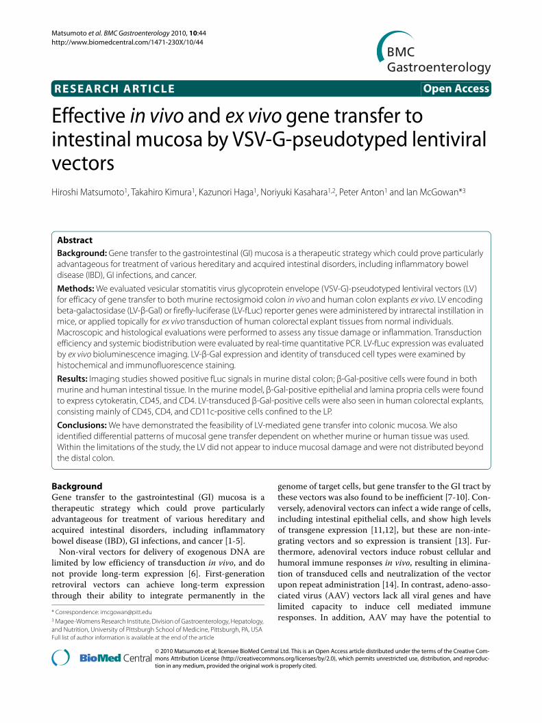
MethodsVector constructionThe pRRLsin-hCMV-β-Gal vector was constructed byinsertion of the β-Gal reporter gene from plasmid pSV-β-Gal (Promega, Madison, WI, USA), and the pRRLsin-hCMV-fLuc vector was constructed by insertion of thefLuc gene [32], respectively, into the multiple cloning site(MCS) of pRRLsin-hCMV-MCS-pre, a third-generation,self-inactivating LV construct kindly provided by Dr.Luigi Naldini (University of Milan, Italy) [33] (Figure 1A).
Cell culture, virus production, and in vitro gene transferHuman embryonic kidney cell line 293T, and humancolon cancer cell lines Caco-2, LoVo, and WiDr, (ATCC,Manassas, VA, USA), were cultured in Dulbecco's modi-fied Eagle's medium (DMEM), Ham's F12K, or RPMI-1640 medium, respectively, with 10% fetal bovine serumand 1% penicillin-streptomycin. LV virus was producedin 293T cells using a third-generation packaging systemas previously described [34], using either pRRLsin-hCMV-β-Gal or pRRLsin-hCMV-fLuc, to produce LV-β-Gal or LV-fLuc, respectively. Virus titers were determinedby HIV-1 p24 ELISA (Perkin Elmer, Waltham, MA, USA)
and expressed as p24 equivalent units (ng/ml). Vectortransductions were performed on 1 × 10⁵ target cells with8 μg/mL polybrene (Sigma, St. Louis, MO, USA) at 37°C.Polybrene is a small, positively charged molecule thatbinds to cell surfaces and neutralizes surface charge andhas been shown to enhance cell transduction by retrovi-ruses [35].
After replacement of the medium and further incuba-tion for 24 hr, cells were washed twice in phosphate-buff-ered saline (PBS), fixed in 2% formaldehyde/0.2%glutaraldehyde (Sigma, St. Louis, MO) for 10 min at roomtemperature, and stained with 20 mg/ml X-Gal solution(5-bromo-4-chloro-3-indolyl beta-D-galactoside; Sigma,St. Louis, MO, USA) at 37°C for 2 hr.
In vivo studiesAll in vivo studies were performed according to institu-tional guidelines under protocols approved by the UCLAAnimal Research Committee. Briefly, 6 to 8-week-oldfemale BALB/c mice (Charles River Laboratories Inc.,Wilmington, MA, USA) were divided into a non-treatedcontrol (NC) group, and two groups that receivedintrarectal instillation of either LV or vehicle solution(DMEM). Prior to instillation, anesthetized mice weregiven an intrarectal enema of 50% ethanol (vol/vol in dis-tilled H2O). Pretreatment with ethanol enemas has beenshown to increase intestinal transduction with other vec-tors [36]. Two hours after the enema, 100 μL of vehicle orviral vector solution containing a total of approximately1000 ng p24 equivalent units was instilled intrarectally.The mice were inverted for 30 sec after administration ofintrarectal products to prevent leakage.
Macroscopic assessment and ex vivo bioluminescence imagingHealth status and body weight was monitored through-out the study. On days 2, 7, and 21 after vector or vehicleinstillation, cohorts of mice were sacrificed, and the
Figure 1 Lentiviral vector (LV) constructs and ex vivo explant cul-ture system. (A) Schematic representation of self-inactivating vectors containing a central polypurine tract (cPPT)/central termination se-quence, immediate early cytomegalovirus (CMV) promoter. Vectors were constructed for expression of beta-galactosidase (β-Gal) and fire-fly luciferase (fLuc).

Matsumoto et al. BMC Gastroenterology 2010, 10:44http://www.biomedcentral.com/1471-230X/10/44
Page 3 of 10
entire colon was removed. Colon length from cecum toanus, weight, and a macroscopic colonic damage scorewere recorded The macroscopic colonic damage scorewas based on the degree of tissue adhesion, mucosalulceration, and intestinal wall thickness (Table 1) [37].
Ex vivo bioluminescence imaging was performed 2 daysafter rectal instillation of LV-fLuc using the Xenogen IVISsystem (Caliper Life Sciences, Alameda, CA, USA). Bio-luminescent signal intensity was expressed as photonsper second per cm2 per steridian (p/s/cm2/sr), and colorimages were processed with Living Image and IGOR-PRO analysis software (Wave Metrics, Portland, OR,USA).
Histological evaluation and X-Gal histochemical stainingFor routine histology, colon and other tissues (spleen,liver, lung, kidney) were fixed in 4% paraformaldehydeovernight, placed in 30% sucrose/PBS for 2 hr, andembedded in OCT compound. Serial 5-μm cryosectionswere stained with hematoxylin/eosin (H&E) for evalua-tion of histopathology score. Mucosal inflammation wasscored using a semi quantitative technique (Table 2) [38].
For X-Gal histochemistry, tissues were fixed in 2% glu-taraldehyde for 2 hr, embedded in OCT compound, and10-μm cryosections were stained in 1 mg/ml X-Gal solu-tion at 37°C for 24sr, rehydrated, and counterstained with0.1% Nuclear Fast Red (Sigma, St. Louis, MO, USA). The
number of positive staining cells was counted in fiveindependent fields in random areas on two non-consecu-tive slides at 200× magnification.
Real-time quantitative PCR (qPCR) analysisQuantification of vector copy number was performed ateach time point by TaqMan qPCR assay to detect theHIV-1 packaging signal sequence, using 300 ng genomicDNA (equivalent to 5 × 10⁴ genomes) isolated frommurine colon and other tissues, including stomach, smallintestine, liver, kidney, spleen, lung, heart, brain, andbone marrow [39]. A reference curve was prepared byamplifying serial dilutions of LV-encoding plasmid in abackground of genomic DNA isolated from untrans-duced murine colon. Genomic DNA from a PC3 cell linepreviously confirmed by flow cytometry to be 100%transduced by LV-GFP vector was used as a positive assaycontrol.
Human colorectal tissue explant culture and ex vivo gene transferAll human endoscopic biopsies were collected fromhealthy HIV-negative volunteers at UCLA Medical Cen-ter, Los Angeles, USA. The protocol for the use of humanendoscopic biopsies was approved by InstitutionalReview Board of the David Geffen School of Medicine atUCLA (#02-05-001-13).
Explant cultures were established as previouslydescribed [31]. Briefly, explants were placed on presoakedGelfoam® (Pharmacia and Upjohn Company, Kalamazoo,MI, USA) rafts with the epithelium uppermost. Tissueswere transduced by pipetting LV solution and polybrene(8 μg/mL) onto the top of each Gelfoam®-supportedexplant. After 2 hr incubation, tissues were washed threetimes, placed on fresh Gelfoam® rafts, and incubated at
Table 2: Histological Scoring System
Infiltration of inflammatory cells
Score Observation
0 Rare inflammatory cells in the lamina propria
1 Increased numbers of inflammatory cells in the lamina propria
2 Confluence of inflammatory cells extending into the submucosa
3 Transmucosal infiltrates
Tissue Damage
Score Observation
0 No mucosal damage
1 A discrete lymphoepithelial lesion
2 Surface mucosal erosion
3 Extensive mucosal damage and ulceration
Table 1: Macroscopic Colonic Damage Score System
Tissue Adhesion
Score Observation
0 No adhesion
1 Little effort required to separate the colon from the surrounding tissue
2 Moderate effort required to separate the colon from the surrounding tissue
3 Severe adhesions
Degree of ulceration
Score Observation
0 Normal appearance of the colon
1 Focal hyperemia with no ulcer
2 Presence of an ulcer and inflammation
3 Two (2) or more ulcers and regions of inflammation
Wall thickness
Score Observation
0 Normal thickness
1 Mild thickening
2 Moderate thickening
3 Severe bowel thickening
Matsumoto et al. BMC Gastroenterology 2010, 10:44http://www.biomedcentral.com/1471-230X/10/44
Page 4 of 10
37°C for a total of 24 hr. Explants were then either fixed inglutaraldehyde, embedded in OCT, and 10-μm cryosec-tions prepared for X-Gal as above, or were fixed in form-aldehyde, embedded in OCT, and 7-μm sectionsprepared for immunofluorescence staining.
Immunofluorescence staining and quantitative morphometryThe identity of transgene-expressing cells in harvestedmurine colon tissues or human colorectal explant tissueswas examined by immunofluorescence double-stainingwith antibodies against β-Gal and cell-specific pheno-typic markers. Briefly, serial 7-μm cryosections werestained for E. coli β-Gal (Promega, Madison, WI, USA).Additional staining included antibodies directed againsthuman and murine cytokeratin AE1/AE3 and CD45(Dako North America Inc., Carpinteria, CA, USA); andCD4, CD8, and CD11c (BD Pharmingen, Franklin Lakes,NJ, USA). β-Gal staining was then visualized with AlexaFluor® 594. Cytokeratin and CD45 were visualized withAlexa Fluor® 488 (Invitrogen Corporation, Carlsbad, CA,USA). The CD4, CD8, and CD11c antibodies werealready conjugated with Alexa Fluor 488. The Vectormouse-on-mouse (M.O.M™) Immunodetection Kit (Vec-tor Laboratories, Burlingame, CA, USA) was used toavoid the high background staining caused by antibodybinding to endogenous murine immunoglobulin whensecondary antibodies were used to amplify primarymurine antibodies. Slides were mounted and nuclei coun-terstained using VECTASHIELD® and DAPI (4',6-diamid-ino-2-phenylindole; Vector Laboratories, Burlingame,CA, USA). In order to quantify LV-mediated β-Gal trans-duction efficiency, the total number of cells showing co-localization of β-Gal-positive and CD45 or CD4 positivesignals in 3 randomly selected fields per section at 200 ×magnification were identified, and expressed as percent-ages of the total number of β-Gal-positive cells or thetotal number of CD45 +/CD4+ cells.
Statistical analysisAll values were expressed as means ± SD. Comparisonsbetween groups were made using the Student t-test andthe Mann-Whitney U test, and p values <0.05 were con-sidered statistically significant.
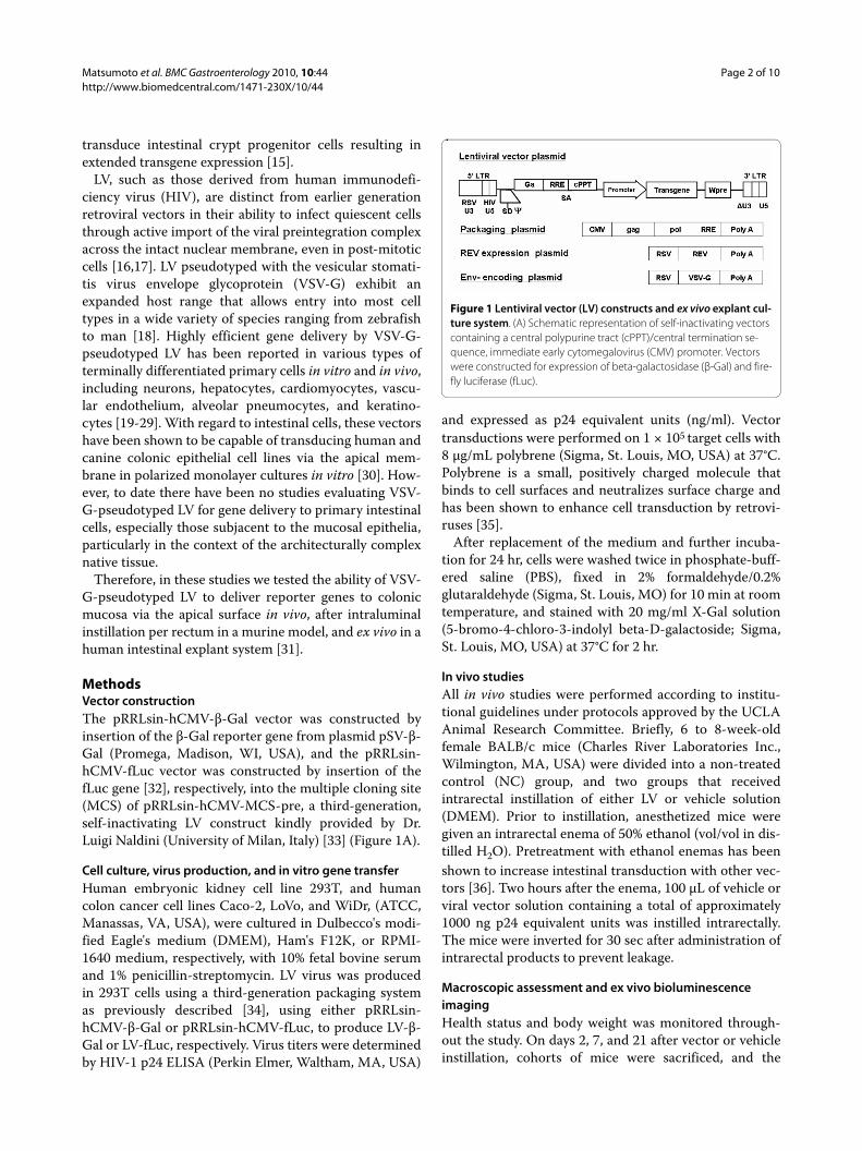
ResultsLV-mediated gene transfer to human colorectal cancer cell lines in vitroLV-mediated transduction efficiency was first assessed invitro using human colorectal adenocarcinoma cell linesCaco-2, WiDr, and LoVo. In each cell line, a dose-depen-dent increase in the number of cells showing positive sig-nals upon X-Gal staining was observed with increasingLV concentration (Figure 2A, B). The highest dose testedwas 10 ng p24 equivalent units of LV, which corresponds
to a biological infectious titer of 106 transducing units(TU) on a standardized cell line such as 293T [40]. At thisdose, transduction rates per 1 × 10⁵ target cells were40.6% for WiDr, 13.7% for Caco-2, and 13.3% for LoVo,indicating that although these colon adenocarcinoma celllines are less permissive for LV compared to 293T embry-onic kidney cells, significant gene transfer can beachieved with higher multiplicities of infection. Seppen etal. have previously reported higher rates of transductionin Caco-2 cells but used a transwell system with GFPexpressing first and third generation lentiviral vectors[41].
Administration of LV for transduction of murine colorectal mucosa in vivoThe safety of gene transfer procedures using LV express-ing either fLuc or β-Gal was assessed by both macro-scopic and histopathologic criteria in three experimentalgroups; mice receiving the LV, mice receiving a mockinstallation (the viral plasmid solution in DMEM), and acontrol group (N = 8 per condition) Intrarectal adminis-tration of 1000 ng p24 equivalent units of VSV-G-pseudotyped LV, corresponding to a 293T cell-basedstandardized biological titer of 108 TU, did not affectbody weight or induce any gross abnormalities upon rou-tine observation. The ratio of colon weight-to-length was0.33 ± 0.04 in the non-treated control (NC) group, 0.34 ±0.5 in the mock instillation control group, and 0.27 ± 0.4in the LV instillation group. Thus, there were no signifi-
Figure 2 In vitro transduction of vesicular stomatitis virus G pro-tein (VSV-G)-pseudotyped lentivirus (LV) encoding β-Gal in co-lonic cell lines. (A) Percentage of cells transduced by the VSV-G-pseudotyped LV. One hundred cells were counted in three randomly selected, non-adjacent fields in triplicate. (B) Colon adenocarcinoma cell lines showing evidence of β-Gal transduction following exposure to 10 ng p24 of vector. All values were expressed as means ± SD. *p < 0.05 compared with the results of 0.1 ng p24 VSV-G LV transduction.
Matsumoto et al. BMC Gastroenterology 2010, 10:44http://www.biomedcentral.com/1471-230X/10/44
Page 5 of 10
cant differences among these three groups in macro-scopic damage assessment or histopathologicalevaluation scores (Additional File 1). More specifically,the use of ethanol enemas did not appear to induce signif-icant mucosal damage. No pathological findings could beobserved in kidney, spleen, lung, and liver harvested fromcontrol and LV-treated mice at each time point (data notshown).
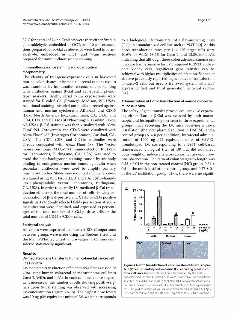
Ex vivo bioluminescence murine imaging following rectal instillation of LV-fLucStrong positive signals were observed in the distal colonadjacent to the rectum in 8/8 (100%) of mice exposed to
VSV-G-pseudotyped LV encoding the fLuc reporter gene(Figure 3A). Quantitative measurement of biolumines-cent photon emission from LV-transduced distal colonshowed a significantly higher level of transduction thanthat of mock-treated colon (57560 ± 28960 p/s/cm2/sr forthe LV instillation group vs. 6260 ± 813.3 p/s/cm2/sr forthe mock instillation group; p < 0.001) (Figure 3B). Addi-tional studies are needed to determine whether the physi-cal distribution of the ethanol enemas and/or the LVinfluenced the location of the bioluminescence signal.
Figure 3 Bioluminescence imaging analysis. (A) Ex vivo bioluminescence imaging analysis after rectal administration of vesicular stomatitis virus-G protein (VSV-G)-pseudotyped lentivirus (LV) expressing firefly-luciferase (fLuc). This pseudocolor image, which is superimposed on a grayscale refer-ence image, uses color (blue, least intense; red, most intense) to illustrate signal strength. (B) Photon emission (p/s/cm2/sr) in the region of interest (ROI) of colon transduced by VSVG-pseudotyped LV shows significantly higher levels of transduction compared to the mock control. (C) Biodistribu-tion of VSV-G LV after rectal administration demonstrated by real-time quantitative polymerase chain reaction (qPCR) analysis of viral copy number in 300 ng of genomic DNA of in vivo (equivalent to approximately 50,000 cells). The detection limit was 50 copies per 300 ng genomic DNA.

Matsumoto et al. BMC Gastroenterology 2010, 10:44http://www.biomedcentral.com/1471-230X/10/44
Page 6 of 10
Real-time qPCR analysis of LV transduction in murine colon tissueReal-time qPCR was employed to quantify the vectorcopy number of LV integrated into genomic DNA frommurine colon tissues after in vivo administration. Usingspiked samples to obtain a reference curve, this methodwas determined to be sensitive enough to detect 50 cop-ies of LV per 5 × 10⁴ cellular genomes. Genomic DNAfrom both positive control cells (data not shown) andtransduced rectosigmoid colon showed amplification ofproviral LV sequences. The average copy number per 300ng genomic DNA from LV-transduced colon was 231.3 ±183.7 (Figure 3C). As expected, no detectable qPCR sig-nals were observed in genomic DNA from colon tissuesof non-treated or mock-treated controls. Importantly,even in LV-treated animals at 2 days post-vector instilla-tion, no detectable qPCR signals were found in any extra-intestinal tissues examined, including stomach, smallintestine, liver, kidney, spleen, lung, heart, brain, andbone marrow (Figure 3C). As the LV was not treated withDNAse prior to injection, we cannot exclude the possibil-ity that the data in Fig 3C in part reflect plasmid contami-nation.
X-Gal histochemistry and double immunofluorescence staining of LV-βGal transduction in murine colon tissuesHistochemistry was performed using optimized X-Galconcentrations and pH conditions to minimize back-ground staining due to endogenous mammalian β-galac-tosidase in the GI tract. As expected, under theseconditions, no staining was observed in mouse colon tis-sues from non-treated and mock-treated control groups,nor in the non-intestinal tissues (kidney, spleen, lung, andliver) from any animals including the LV-treated group.
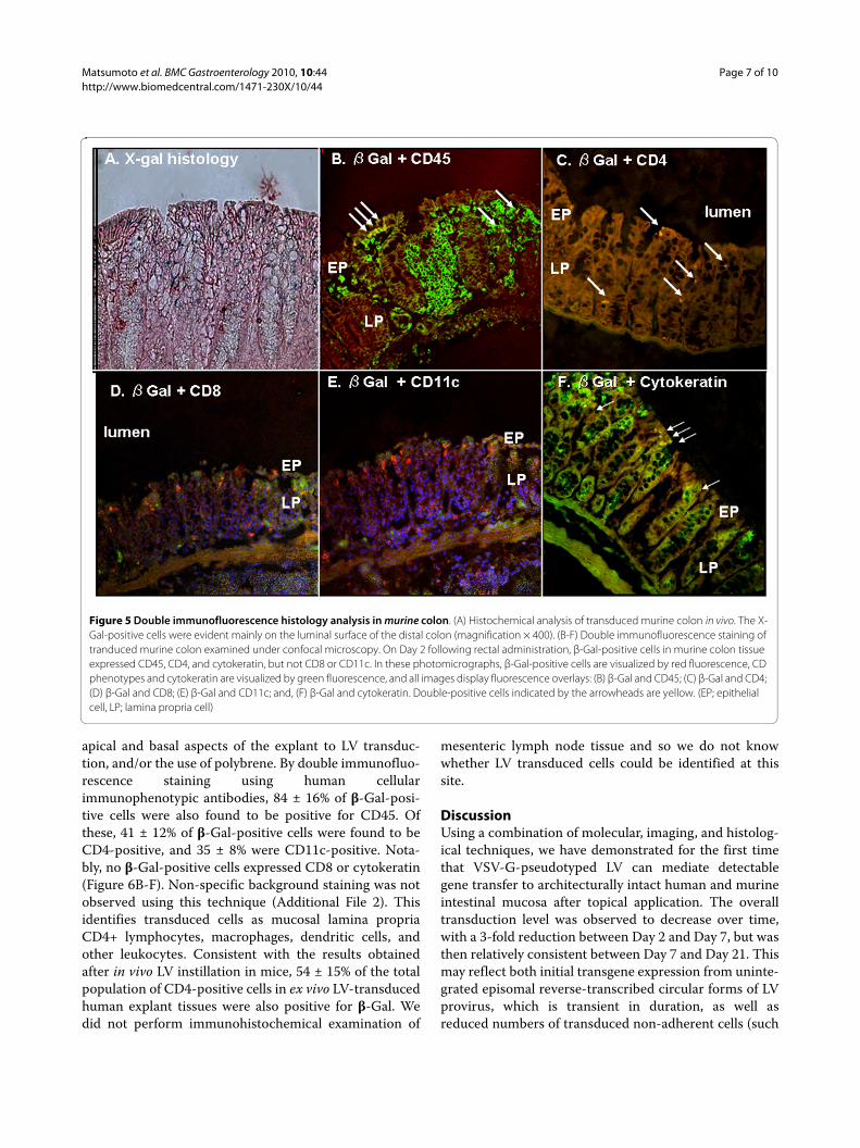
In contrast, positive-staining cells were observed incolon tissues from animals treated with LV expressing theE. coli β-Gal reporter gene. The initial number of posi-tive-staining cells in the LV-treated group was 62.3 ± 18.5per microscopic field at 200× magnification on Day 2post-vector instillation (Figure 4). However, the numberof positive-staining cells was then observed to decrease,to 19.1 ± 5.3 per 200 × field on Day 7, and 15.7 ± 5.1 per200 × field on Day 21. These positive-staining cells werefound predominantly toward the luminal surface of thecolon, but were identified in both mucosal epitheliumand, importantly, the lamina propria (LP) (Figure 5A).
To further characterize the identity of the transducedcells, immunofluorescence double-staining was per-formed using an E. coli β-Gal-specific antibody in combi-nation with various cellular immunophenotypicantibodies. Quantification of double-positive staining incolon tissues harvested on Day 2 post-LV instillation
demonstrated that 27 ± 5% of the β-Gal-positive cellswere also positive for cytokeratin. Consistent with theirhistological origin, β-Gal/cytokeratin double-positivecells were observed solely in the epithelial layers of colontissues from transduced animals. Cells exhibiting double-positive staining for both β-Gal and the common leuko-cyte antigen CD45 were found in both mucosal epithe-lium and lamina propria (LP) regions. These β-Gal/CD45double-positive cells represented 70 ± 11% of the totalpopulation of β-Gal-positive cells.
A subset of LV-transduced white blood cells was fur-ther identified to consist of T lymphocytes, dendriticcells, or macrophages, as 27 ± 2% of the total populationof β-Gal-positive cells were also positive for CD4. In fact,of the total population of CD4-positive cells, 48 ± 13%were β-Gal-positive, indicating a significant transductionof half of this cell population. These β-Gal/CD4 double-positive cells were observed only within the LP of trans-duced murine colon. Notably, there were no cells showingco-expression of both β-Gal and CD8 or CD11c (Figure5B-F).
LV-β Gal-mediated ex vivo gene transfer to human colorectal explant tissuesHuman colon tissue specimens were incubated with 1000ng p24 units of LV-β-Gal, and examined within 24 hr ofexplant culture. The number of X-Gal-positive cells aftertransduction was 12.4 ± 1.5 per low power (×200) field. Incontrast to the murine studies, the transduced cells inhuman samples were primarily found within the LP (Fig-ure 6A). These data might reflect the availability of both
Figure 4 X-Gal histology in murine colon transduced by vesicular stomatitis virus G protein (VSV-G)-pseudotyped lentivirus (LV) encoding β-Gal in vivo. The time course of β-Gal expression in the co-lon of BALB/c mice. Each group had 6 mice. The colon was removed at the indicated time points. The number of X-Gal-positive cells in murine colon transduced by LV-β-Gal in vivo. All values were expressed as means ± SD. *p < 0.05 compared with the results of normal control (NC).
Matsumoto et al. BMC Gastroenterology 2010, 10:44http://www.biomedcentral.com/1471-230X/10/44
Page 7 of 10
apical and basal aspects of the explant to LV transduc-tion, and/or the use of polybrene. By double immunofluo-rescence staining using human cellularimmunophenotypic antibodies, 84 ± 16% of β-Gal-posi-tive cells were also found to be positive for CD45. Ofthese, 41 ± 12% of β-Gal-positive cells were found to beCD4-positive, and 35 ± 8% were CD11c-positive. Nota-bly, no β-Gal-positive cells expressed CD8 or cytokeratin(Figure 6B-F). Non-specific background staining was notobserved using this technique (Additional File 2). Thisidentifies transduced cells as mucosal lamina propriaCD4+ lymphocytes, macrophages, dendritic cells, andother leukocytes. Consistent with the results obtainedafter in vivo LV instillation in mice, 54 ± 15% of the totalpopulation of CD4-positive cells in ex vivo LV-transducedhuman explant tissues were also positive for β-Gal. Wedid not perform immunohistochemical examination of
mesenteric lymph node tissue and so we do not knowwhether LV transduced cells could be identified at thissite.
DiscussionUsing a combination of molecular, imaging, and histolog-ical techniques, we have demonstrated for the first timethat VSV-G-pseudotyped LV can mediate detectablegene transfer to architecturally intact human and murineintestinal mucosa after topical application. The overalltransduction level was observed to decrease over time,with a 3-fold reduction between Day 2 and Day 7, but wasthen relatively consistent between Day 7 and Day 21. Thismay reflect both initial transgene expression from uninte-grated episomal reverse-transcribed circular forms of LVprovirus, which is transient in duration, as well asreduced numbers of transduced non-adherent cells (such
Figure 5 Double immunofluorescence histology analysis in murine colon. (A) Histochemical analysis of transduced murine colon in vivo. The X-Gal-positive cells were evident mainly on the luminal surface of the distal colon (magnification × 400). (B-F) Double immunofluorescence staining of tranduced murine colon examined under confocal microscopy. On Day 2 following rectal administration, β-Gal-positive cells in murine colon tissue expressed CD45, CD4, and cytokeratin, but not CD8 or CD11c. In these photomicrographs, β-Gal-positive cells are visualized by red fluorescence, CD phenotypes and cytokeratin are visualized by green fluorescence, and all images display fluorescence overlays: (B) β-Gal and CD45; (C) β-Gal and CD4; (D) β-Gal and CD8; (E) β-Gal and CD11c; and, (F) β-Gal and cytokeratin. Double-positive cells indicated by the arrowheads are yellow. (EP; epithelial cell, LP; lamina propria cell)

Matsumoto et al. BMC Gastroenterology 2010, 10:44http://www.biomedcentral.com/1471-230X/10/44
Page 8 of 10
as lymphocytes) as they migrate or circulate out of colontissue in vivo, or reflect cells lost through apoptosis orkaryolysis in the ex vivo explant model.
An additional finding is the demonstration that inhuman intestinal tissue, the transduced cells are predom-inantly in the lamina propria, an observation differingfrom previous beliefs that it is predominantly epitheliallineage cells that are transduced. Co-localization of phe-notypic cell surface markers and transgene expressionshowed notable differences in the types of cells trans-duced between experiments involving LV-mediatedtransduction of murine vs. human colon tissue. Signifi-cant transduction of cytokeratin-positive cells wasobserved after in vivo transduction of murine colon, butnot after ex vivo transduction of human explants colontissue. Conversely, after ex vivo transduction of humanexplant colon tissue, but not after in vivo transduction ofmurine colon, we observed significant transduction ofcells positive for CD11c, a cellular marker of mac-
rophages and dendritic cells, but which is also weaklyexpressed on B cells, NK cells, and T cell subsets.
A number of parameters may have contributed to thesedifferences. For example, to avoid degradation of the tis-sue architecture, LV-transduced human colon tissues hadto be analyzed after no more than 24 hours of explant cul-ture, and the time course for accumulating a detectablelevel transgene product may require a longer time inter-val for epithelial cells. Of course, LV transduction afterrectal instillation in vivo also necessitates penetrationfrom the intestinal lumen, and the mucosal epitheliumrepresents the only tissue surface directly in contact withthe virus solution. In contrast, virus applied ex vivo to thesurface of endoscopically acquired human tissue explantsamples has access to sub-epithelial cell layers that wouldnot normally be available to virus administered intralu-minally to intact intestine in vivo. Another differencebetween the in vivo and ex vivo experiments was the useof ethanol in the former, and polybrene in the latter, to
Figure 6 X-Gal histology and double immunofluorescence histology analysis in human colonic explants transduced by VSV-G-pseudo-typed lentivirus (LV) encoding β-Gal. (A) Histological appearance of transduced human explants after X-Gal staining. X-Gal-positive cells, here stained blue, were found mainly in the LP. (B-F) Double immunofluorescence confocal microscopy of human explants transduced with VSV-G. β-Gal-positive cells expressed CD4, CD45, and CD11c, but not CD8, or cytokeratin. In these photomicrographs, β-Gal is visualized by red fluorescence, each cellular phenotype marker is visualized by green fluorescence, and nuclei are visualized by blue DAPI staining, respectively, and all images display flu-orescence overlays: (B) β-Gal and CD45; (C) β-Gal and CD4; (D) β-Gal and CD8; (E) β-Gal and CD11c; and, (F) β-Gal and cytokeratin. Double-positive cells, indicated by the arrowheads, are yellow (EP; epithelial cell, LP; lamina propria cell).
Matsumoto et al. BMC Gastroenterology 2010, 10:44http://www.biomedcentral.com/1471-230X/10/44
Page 9 of 10
facilitate lentiviral transduction. We did not explore theextent to which these different experimental parametersmight influence the results of our experiments.
We did observe a difference between the present studyand previous studies using explant tissues, in terms of thespecific localization of transduced cells within the LP. Inthe present study, LV-transduced cells in human explantsamples were found primarily in the subepithelial regionof the LP whereas in other colorectal explant studiesusing adenoviral vectors [42] transduction was com-monly observed in the basolateral region. However, it isdifficult to compare our data with other published studiesas the cell tropism may differ between vectors. In addi-tion, differences in the histological distribution of vector-specific cellular receptors, such as the coxsackievirus/adenovirus receptor (CAR) required for efficient bindingof adenovirus to target cells may influence experimentalresults.
There were also similarities in cell transduction resultsfrom the murine rectal in vivo transduction and humanexplant ex vivo transduction models. In both cases, VSV-G-pseudotyped LV exhibited the ability to transduceCD4-positive cell population. Notably, LV pseudotypedwith the VSV-G envelope, which only requires binding tophospholipid constituents of most mammalian cells [43],achieved much more efficient infection of T cells than ispossible with identical vectors pseudotyped with ampho-tropic retrovirus envelope [44]. However, CD4 is not onlyexpressed by helper T lymphocytes and T regulatorycells, but also by macrophages and dendritic cells at lowerlevels. All of these cell types generally reside in the LP,which is where the predominant staining was observed inhumans. It is not clear why the LV was unable to trans-duce CD8 positive cells and this finding warrants addi-tional in vitro studies with purified T cell populations.
As the vectors used are replication-defective and canonly mediate a single cycle of infection, this finding sug-gests that LV may be efficiently transported intact acrossthe mucosal epithelium as has been suggested as a naturalroute of HIV infection. It has also been reported that den-dritic cells may form projections into the intestinal lumento sample incoming antigens, which may permit HIVinfection as well as LV transduction of this cell popula-tion.
ConclusionsIn summary, these studies have demonstrated the feasi-bility of using VSV-G-pseudotyped LV to safely achieveappreciable levels of localized gene transfer into architec-turally intact primary murine and human intestinal tis-sues.
Additional material
Competing interestsThe authors declare that they have no competing interests.
Authors' contributionsHM conducted the majority of the experiments described in this paper. TK car-ried out the molecular genetic studies, made the LV, and performed the statis-tical analysis of the experimental data. KH helped to analyze the in vivo animaldata. NK provided technical assistance for the LV experiments, PA collected theendoscopic biopsies, and IM supervised the research, and edited the final man-uscript. All authors read and approved the final manuscript.
AcknowledgementsThe authors would like to thank the staff of the Center for HIV Prevention Research (CPR), the UCLA Vector Core & Shared Resource, and the Blinder Research Foundation for Crohn's Disease. HM was supported by a fellowship from the Blinder Research Foundation for Crohn's Disease, Los Angeles, CA. Additional support was provided by a Pilot Award from the UCLA AIDS Institute (IM, NK), funding from the CURE Digestive Diseases Research Center (P30 DK41301) and Jonsson Comprehensive Cancer Center (P30 CA82103) to the Vector Core & Shared Resource (NK), and the CFAR Mucosal Immunology Core (AI28697).
Author Details1Department of Medicine, Division of Digestive Diseases, University of California Los Angeles (UCLA) David Geffen School of Medicine, Los Angeles, CA, USA, 2Department of Molecular & Medical Pharmacology, University of California Los Angeles (UCLA) David Geffen School of Medicine, Los Angeles, CA, USA and 3Magee-Womens Research Institute, Division of Gastroenterology, Hepatology, and Nutrition, University of Pittsburgh School of Medicine, Pittsburgh, PA, USA
References1. Prieto J, Herraiz M, Sangro B, Qian C, Mazzolini G, Melero I, et al.: The
promise of gene therapy in gastrointestinal and liver diseases. Gut 2003, 52(Suppl 2):ii49-ii54.
2. Raper SE, Wilson JM: Making space for intestinal gene therapy. Gastroenterology 1997, 112:1753-1756.
3. Forbes SJ, Hodgson HJ: Review article: gene therapy in gastroenterology and hepatology. Aliment Pharmacol Ther 1997, 11:823-836.
4. van MC, Te Velde AA, venter SJ van De, Rodriguez Pena MS: Gene therapy in the treatment of intestinal inflammation. Int J Colorectal Dis 2004, 19:79-86.
5. Wirtz S, Neurath MF: Gene transfer approaches for the treatment of inflammatory bowel disease. Gene Ther 2003, 10:854-860.
6. Schmid RM, Weidenbach H, Draenert GF, Lerch MM, Liptay S, Schorr J, et al.: Liposome mediated in vivo gene transfer into different tissues of the gastrointestinal tract. Z Gastroenterol 1994, 32:665-670.
Additional file 1 The effect of vesicular stomatitis virus G protein (VSV-G)-pseudotyped lentivirus (LV) rectal gene transduction on healthy murine cells. BALB/c mice were divided into three groups; a normal healthy control (NC) group and two groups that received either placebo or VSV-G LV fol-lowing a preliminary ethanol enema (EtOH). Mice received 1000 ng p24 VSV-G LV by rectal administration under anesthesia. (A) VSV-G LV did not affect weight loss in healthy mice. Results are shown as a percentage of original weight for each group. (B) There was no significant difference in the ratio of colon length to weight between groups. Further, there was no sig-nificant difference in (C) macroscopic damage score or (D) histomorpho-logic score among all groups.Additional file 2 The negative control pictures of immunofluorence stain-ing using AF488 secondary antibody. Non-specific green staining is not observed in either (A) AE1/AE3 or (B) CD45 examination of murine intestinal tissue or (C) AE1/AE3 (D) CD45 examination of ex vivo human explant tissue
Received: 30 June 2009 Accepted: 11 May 2010 Published: 11 May 2010This article is available from: http://www.biomedcentral.com/1471-230X/10/44© 2010 Matsumoto et al; licensee BioMed Central Ltd. This is an Open Access article distributed under the terms of the Creative Commons Attribution License (http://creativecommons.org/licenses/by/2.0), which permits unrestricted use, distribution, and reproduction in any medium, provided the original work is properly cited.BMC Gastroenterology 2010, 10:44

Matsumoto et al. BMC Gastroenterology 2010, 10:44http://www.biomedcentral.com/1471-230X/10/44
Page 10 of 10
7. Lau C, Soriano HE, Ledley FD, Finegold MJ, Wolfe JH, Birkenmeier EH, et al.: Retroviral gene transfer into the intestinal epithelium. Hum Gene Ther 1995, 6:1145-1151.
8. Noel RA, Shukla P, Henning SJ: Optimization of gene transfer into intestinal epithelial cells using a retroviral vector. J Pediatr Gastroenterol Nutr 1994, 19:43-49.
9. Lozier JN, Yankaskas JR, Ramsey WJ, Chen L, Berschneider H, Morgan RA: Gut epithelial cells as targets for gene therapy of hemophilia. Hum Gene Ther 1997, 8:1481-1490.
10. Laine F, Blouin V, Ferry N: Evaluation of recombinant retrovirus and adenovirus for gene transfer to normal and pathologic intestinal tissue. Gastroenterol Clin Biol 1999, 23:221-228.
11. Kesisoglou F, Schmiedlin-Ren P, Fleisher D, Roessler B, Zimmermann EM: Restituting intestinal epithelial cells exhibit increased transducibility by adenoviral vectors. J Gene Med 2006, 8:1379-1392.
12. Brown GR, Thiele DL, Silva M, Beutler B: Adenoviral vectors given intravenously to immunocompromised mice yield stable transduction of the colonic epithelium. Gastroenterology 1997, 112:1586-1594.
13. Russell WC: Update on adenovirus and its vectors. J Gen Virol 2000, 81:2573-2604.
14. Bessis N, GarciaCozar FJ, Boissier MC: Immune responses to gene therapy vectors: influence on vector function and effector mechanisms. Gene Ther 2004, 11(Suppl 1):S10-S17.
15. Polyak S, Mah C, Porvasnik S, Herlihy JD, Campbell-Thompson M, Byrne BJ, et al.: Gene delivery to intestinal epithelial cells in vitro and in vivo with recombinant adeno-associated virus types 1, 2 and 5. Dig Dis Sci 2008, 53:1261-1270.
16. Bukrinsky MI, Haffar OK: HIV-1 nuclear import: in search of a leader. Front Biosci 1999, 4:D772-D781.
17. Zennou V, Petit C, Guetard D, Nerhbass U, Montagnier L, Charneau P: HIV-1 genome nuclear import is mediated by a central DNA flap. Cell 2000, 101:173-185.
18. Schubert M, Joshi B, Blondel D, Harmison GG: Insertion of the human immunodeficiency virus CD4 receptor into the envelope of vesicular stomatitis virus particles. J Virol 1992, 66:1579-1589.
19. Blomer U, Naldini L, Verma IM, Trono D, Gage FH: Applications of gene therapy to the CNS. Hum Mol Genet 1996, 5(Spec No):1397-1404.
20. Sakoda T, Kasahara N, Hamamori Y, Kedes L: A high-titer lentiviral production system mediates efficient transduction of differentiated cells including beating cardiac myocytes. J Mol Cell Cardiol 1999, 31:2037-2047.
21. Shichinohe T, Bochner BH, Mizutani K, Nishida M, Hegerich-Gilliam S, Naldini L, et al.: Development of lentiviral vectors for antiangiogenic gene delivery. Cancer Gene Ther 2001, 8:879-889.
22. Borok Z, Harboe-Schmidt JE, Brody SL, You Y, Zhou B, Li X, et al.: Vesicular stomatitis virus G-pseudotyped lentivirus vectors mediate efficient apical transduction of polarized quiescent primary alveolar epithelial cells. J Virol 2001, 75:11747-11754.
23. Li W, Nadelman C, Gratch NS, Li W, Chen M, Kasahara N, et al.: An important role for protein kinase C-delta in human keratinocyte migration on dermal collagen. Exp Cell Res 2002, 273:219-228.
24. Chen M, Li W, Fan J, Kasahara N, Woodley D: An efficient gene transduction system for studying gene function in primary human dermal fibroblasts and epidermal keratinocytes. Clin Exp Dermatol 2003, 28:193-199.
25. Naldini L, Blomer U, Gage FH, Trono D, Verma IM: Efficient transfer, integration, and sustained long-term expression of the transgene in adult rat brains injected with a lentiviral vector. Proc Natl Acad Sci USA 1996, 93:11382-11388.
26. Kafri T, Blomer U, Peterson DA, Gage FH, Verma IM: Sustained expression of genes delivered directly into liver and muscle by lentiviral vectors. Nat Genet 1997, 17:314-317.
27. Miyoshi H, Smith KA, Mosier DE, Verma IM, Torbett BE: Transduction of human CD34+ cells that mediate long-term engraftment of NOD/SCID mice by HIV vectors. Science 1999, 283:682-686.
28. Johnson LG, Olsen JC, Naldini L, Boucher RC: Pseudotyped human lentiviral vector-mediated gene transfer to airway epithelia in vivo. Gene Ther 2000, 7:568-574.
29. Kremer KL, Dunning KR, Parsons DW, Anson DS: Gene delivery to airway epithelial cells in vivo: a direct comparison of apical and basolateral transduction strategies using pseudotyped lentivirus vectors. J Gene Med 2007, 9:362-368.
30. Seppen J, Barry SC, Klinkspoor JH, Katen LJ, Lee SP, Garcia JV, et al.: Apical gene transfer into quiescent human and canine polarized intestinal epithelial cells by lentivirus vectors. J Virol 2000, 74:7642-7645.
31. Fletcher PS, Elliott J, Grivel JC, Margolis L, Anton P, McGowan I, et al.: Ex vivo culture of human colorectal tissue for the evaluation of candidate microbicides. AIDS 2006, 20:1237-1245.
32. Koya RC, Kimura T, Ribas A, Rozengurt N, Lawson GW, Faure-Kumar E, et al.: Lentiviral vector-mediated autonomous differentiation of mouse bone marrow cells into immunologically potent dendritic cell vaccines. Mol Ther 2007, 15:971-980.
33. Zufferey R, Dull T, Mandel RJ, Bukovsky A, Quiroz D, Naldini L, et al.: Self-inactivating lentivirus vector for safe and efficient in vivo gene delivery. J Virol 1998, 72:9873-9880.
34. Dull T, Zufferey R, Kelly M, Mandel RJ, Nguyen M, Trono D, et al.: A third-generation lentivirus vector with a conditional packaging system. J Virol 1998, 72:8463-8471.
35. Landazuri N, Le Doux JM: Complexation of retroviruses with charged polymers enhances gene transfer by increasing the rate that viruses are delivered to cells. J Gene Med 2004, 6:1304-1319.
36. Vallance BA, Gunawan MI, Hewlett B, Bercik P, Van KC, Galeazzi F, et al.: TGF-beta1 gene transfer to the mouse colon leads to intestinal fibrosis. Am J Physiol Gastrointest Liver Physiol 2005, 289:G116-G128.
37. Wallace JL, Keenan CM, Gale D, Shoupe TS: Exacerbation of experimental colitis by nonsteroidal anti-inflammatory drugs is not related to elevated leukotriene B4 synthesis. Gastroenterology 1992, 102:18-27.
38. Cooper HS, Murthy SN, Shah RS, Sedergran DJ: Clinicopathologic study of dextran sulfate sodium experimental murine colitis. Lab Invest 1993, 69:238-249.
39. Sastry L, Johnson T, Hobson MJ, Smucker B, Cornetta K: Titering lentiviral vectors: comparison of DNA, RNA and marker expression methods. Gene Ther 2002, 9:1155-1162.
40. Stripecke R, Koya RC, Ta HQ, Kasahara N, Levine AM: The use of lentiviral vectors in gene therapy of leukemia: combinatorial gene delivery of immunomodulators into leukemia cells by state-of-the-art vectors. Blood Cells Mol Dis 2003, 31:28-37.
41. Seppen J, Barry SC, Klinkspoor JH, Katen LJ, Lee SP, Garcia JV, et al.: Apical gene transfer into quiescent human and canine polarized intestinal epithelial cells by lentivirus vectors. J Virol 2000, 74:7642-7645.
42. Schmiedlin-Ren P, Kesisoglou F, Mapili JA, Sabek SE, Barnett JL, Chey WD, et al.: Increased transduction of human intestinal epithelial cells by adenoviral vectors in inflammatory bowel disease. Inflamm Bowel Dis 2005, 11:464-472.
43. Yee JK, Friedmann T, Burns JC: Generation of high-titer pseudotyped retroviral vectors with very broad host range. Methods Cell Biol 1994, 43(Pt A):99-112.
44. Sharma S, Cantwell M, Kipps TJ, Friedmann T: Efficient infection of a human T-cell line and of human primary peripheral blood leukocytes with a pseudotyped retrovirus vector. Proc Natl Acad Sci USA 1996, 93:11842-11847.
Pre-publication historyThe pre-publication history for this paper can be accessed here:http://www.biomedcentral.com/1471-230X/10/44/prepub
doi: 10.1186/1471-230X-10-44Cite this article as: Matsumoto et al., Effective in vivo and ex vivo gene trans-fer to intestinal mucosa by VSV-G-pseudotyped lentiviral vectors BMC Gastro-enterology 2010, 10:44