targeted lentiviral vectors: current applications and future
TRANSCRIPT
Chapter 14
Targeted Lentiviral Vectors: Current Applications andFuture Potential
Cleo Goyvaerts, Therese Liechtenstein,Christopher Bricogne, David Escors andKarine Breckpot
Additional information is available at the end of the chapter
http://dx.doi.org/10.5772/52770
1. Introduction
About two decades ago recombinant human immunodeficiency virus type 1 (HIV-1) wasproposed as a blueprint for the development of lentiviral vectors (LVs) (Naldini, Blomer etal. 1996). Lentiviral vectors exhibit several characteristics that make them favorable tools forgene therapy, including sustained gene delivery through vector integration, transduction ofboth dividing and non-dividing cells, applicability to different target cell types, absence ofexpression of viral proteins after transduction, delivery of complex genetic elements, lowgenotoxicity and the relative ease of vector manipulation and production (Cattoglio, Facchi‐ni et al. 2007; Bauer, Dao et al. 2008). This is reflected in the numerous applications such as:transgene (tg) overexpression (Lopez-Ornelas, Mejia-Castillo et al. 2011), persistent gene si‐lencing (Wang, Hu et al. 2012), immunization (Breckpot, Emeagi et al. 2008), generation oftransgenic animals (Baup, Fraga et al. 2010), in vivo imaging (Roet, Eggers et al. 2012), induc‐tion of pluripotent cells, stem cell modification (Sanchez-Danes, Consiglio et al. 2012), line‐age tracking and site-directed gene editing (Lombardo, Genovese et al. 2007) as well asmany applications targeting cancer cells (Petrigliano, Virk et al. 2009).
Recombinant LVs can be derived from primate as well as non-primate lentiviruses such asHIV-1 and simian immunodeficiency virus (SIV) next to the equine infectious anemia virus,caprine arthritis-encephalitis virus, maedi-visna virus, feline immunodeficiency virus (FIV)and bovine immunodeficiency virus respectively (Escors and Breckpot 2010). They are allmembers of the Retroviridae family with ‘retro’ referring to their capacity to retro-transcribetheir diploid single stranded (ss) RNA genome into a double stranded (ds) DNA copy that is
© 2013 Goyvaerts et al.; licensee InTech. This is an open access article distributed under the terms of theCreative Commons Attribution License (http://creativecommons.org/licenses/by/3.0), which permitsunrestricted use, distribution, and reproduction in any medium, provided the original work is properly cited.

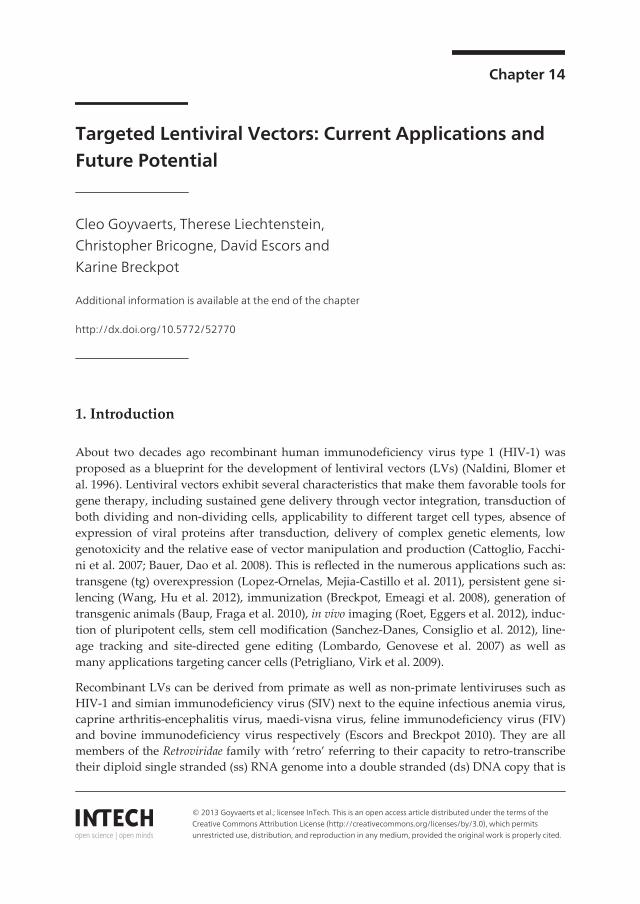
integrated in the genome of the infected host cell (Figure 1A). Since LVs are most often de‐rived from HIV-1, the generation of recombinant LVs has been accompanied by several safetyconcerns such as the generation of replication-competent lentiviruses (RCLs). Another poten‐tial biosafety concern is the induction of insertional mutagenesis, a major genotoxic problemthat emerged in gene therapy clinical trials using their γ-retroviral counterparts (Manilla, Re‐bello et al. 2005). Generally, LVs are produced by transiently transfecting HEK 293 or 293Tcells with plasmids encoding structural and functional sequences, imperative for proper LVparticle generation. Over the last decades, vector development has largely focused on the de‐sign of these plasmids. Firstly, only critical viral structural and functional sequences are pro‐vided and secondly, these sequences are divided over a certain number of individualplasmids either in cis (encoded by the LV) or trans (packaged as a protein within the LV parti‐cle), with a minimal overlap between viral sequences. This led to a LV production procedurewhere at least three different plasmids are used: (1) a packaging plasmid which provides allviral structural and enzymatic sequences (encoded by gag and pol) in trans to generate a func‐tional particle, (2) a transfer plasmid providing the expression cassette in cis, cloned into thenon-coding remains of the original lentiviral genome (Figure 1B, adapted from (Delenda2004)) including a packaging signal and the two long terminal repeats (LTRs) of which thepromoter activity has been deleted from the 3’ LTR and (3) an envelope plasmid encoding anenvelope glycoprotein (gp) consisting of a transmembranary domain (TM) and a receptor-binding domain (SU) that determines the LVs’ tropism (Figure 1A).
Figure 1. Schematic representation of an HIV-1 particle (A) and its genome (B). The diploid ssRNA genome ofHIV-1 is stabilized by structural nucleocapsid proteins and together with the enzymatic proteins reverse transcriptase,protease and integrase packaged in a nucleocapsid structure, which in turn is enclosed by capsid proteins. This nucleo‐capsid is surrounded by a matrix protein layer and a producer cell derived phospholipid bilayer in which the envelopeproteins consisting of an SU and TM part, are embedded (A). All proviral genes (gag, pol, pro, vif, vpr, vpu, ref, tat, enven nef) are flanked by two identical LTRs that consist of three regions: U3, R and U5. Within the U3 region, all proviraltranscriptional control elements are situated such as the promoter and several enhancer sequences. Ψ represents thepackaging signal. At the 3’ end of the pol gene the central polypurine tract (red) and central termination sequence(green) are located. Both ensure the formation of a triple stranded DNA flap, crucial for nuclear entry of the pre-inte‐gration complex in non-dividing cells (B).
Gene Therapy - Tools and Potential Applications344

Besides this division over different plasmids, other important construct optimizationsteps have been implemented. While in the first generation LV packaging plasmids theentire gag and pol genes were encoded together with all accessory regulatory and viru‐lence genes, the second generation was multiply attenuated by removal of the four viru‐lence genes, but not the regulatory genes tat and rev (Zufferey, Nagy et al. 1997). In thethird generation, the rev gene is expressed from a separate plasmid and the tat gene isremoved by insertion of a strong constitutive promoter replacing the U3 region in the 5’LTR of the transfer plasmid (Dull, Zufferey et al. 1998). A major improvement was ach‐ieved with the development of SIN or self-inactivating LVs where a deletion in the U3region of the 3’ LTR of the transfer plasmid abolished the production of full-length vec‐tor RNA in transduced cells. This not only minimizes the risk for RCLs, but also re‐duces the chance that the viral LTR enhancers interfere with the expression cassette,which minimizes aberrant expression of adjacent cellular coding regions. Subsequentlythese and many other optimization steps paved the way towards a more effective andsafer version of the lentiviral gene delivery vehicle (Romano, Claudio et al. 2003).
In addition to packaging and transfer plasmid optimization, also the envelope plasmidwas modified by replacing the natural HIV-1 envelope gp with an alternative gp gene,most often the gp of vesicular stomatitis virus (VSV.G). This concept is called pseudo‐typing and VSV.G endowed the LV particle with an increased stability and broad cellu‐lar tropism (to most if not all mammalian cells). However, it became clear that fornumerous in vivo applications, a broad tropism may not be desirable. First, the tg that isencoded could be toxic to many cell types, e.g. pro-apoptotic or suicide genes, so a strin‐gent control over the induction of tg expression in time and/or place is a necessity (Uch,Gerolami et al. 2003; Seo, Kim et al. 2009). A second point of concern is the risk for in‐sertional mutagenesis; the more cells get infected, the higher this risk becomes. Althoughit has been demonstrated that LVs intrinsically exhibit low genotoxicity, clonal expansionand dominance of transduced hematopoietic progenitors have been reported in a clinicaltrial in which hematopoietic stem cells were genetically modified with a LV that ex‐pressed the β-globin gene for treatment of β-thalassemia (Fehse and Roeder 2008; Cavaz‐zana-Calvo, Payen et al. 2010). Thirdly, while a broad tropism LV is favorable in anti-tumor immunotherapy to efficiently transduce antigen-presenting cells (APCs) which caninduce an antigen specific immune response (Palmowski, Lopes et al. 2004), this is notdesirable when a genetic disorder has to be restored as in this case the tg may not be‐come an immunological target (Annoni, Battaglia et al. 2007). Finally, during productionof pantropic viruses encoding oncogenes, narrow tropism vectors would be more valua‐ble due to biosafety level handling requirements and safety issues (Barrilleaux andKnoepfler 2011). Therefore, in view of safety as well as applicability aspects, four maintargeting strategies can be brought forward: targeted gene expression or transcriptionaltargeting, targeted gene translation or microRNA based (de)targeting, targeted infectionor transductional targeting, and targeted integration of the proviral DNA.
Targeted Lentiviral Vectors: Current Applications and Future Potentialhttp://dx.doi.org/10.5772/52770
345

2. Transcriptional targeting
Most often a strong constitutive promoter with or without enhancer sequences is used todrive the LV encoded tg. These include the cytomegalovirus (CMV), spleen focus formingvirus (SFFV), human polypeptide chain elongation factor-1alpha (EF-1alpha), phosphogly‐cerate kinase (PGK) and ubiquitin C promoters (Kim, Kim et al. 2007; Gilham, Lie et al. 2010;Li, Husic et al. 2010). Although these promoters generally induce strong and ubiquitous ex‐pression of the tg, they present some disadvantages. A first drawback is that they are moreprone to promoter inactivation than cell-specific promoters. In addition, they are more po‐tent in terms of activating the host-cell defense machinery and increasing the long-distanceeffects of insertional mutagenesis caused by their enhancer sequences (Liu, Wang et al. 2004;Stein, Ott et al. 2010; Singhal, Deng et al. 2011). These downsides resulted in the develop‐ment of various strategies to allow cell-specific tg expression by incorporating cell type spe‐cific regulatory elements and/or promoter(s) in the expression cassette of the LV. Because ofthe availability of a large number of endogenous cellular promoters, targeted expression canbe achieved to potentially any cell type or tissue. In addition, its advantage over unselectiveexpression has been demonstrated in numerous studies (Di Nunzio, Maruggi et al. 2008;Kerns, Ryu et al. 2010; Cao, Sodhi et al. 2011). This is exemplified by a study where LV en‐coding iduronidase under the control of the hepatocyte specific albumin gene promoter wasinjected intravenously to treat mucopolysaccharidosis type I. While the same LV with aCMV promoter resulted in the induction of an immune response that diminished the tg ex‐pression over time, the albumin gene promoter enabled stable and prolonged tg expressionwith a partial correction of the pathology (Di Domenico, Di Napoli et al. 2006). In additionto hepatocyte specific targeting, an ever-growing list of cell-type specific promoters has beenused for the specific expression in tissues such as the erythroid lineage, endothelial cells,myocardial cells, retinal cells, B cells, epidermal, hematopoietic stem cells, etcetera (Hanawa,Persons et al. 2002; De Palma, Venneri et al. 2003; Semple-Rowland, Eccles et al. 2007; DiNunzio, Maruggi et al. 2008; Leuci, Gammaitoni et al. 2009; Kerns, Ryu et al. 2010; Semple-Rowland, Coggin et al. 2010; Cao, Sodhi et al. 2011; Lee, Fan et al. 2011; Friedrich, Nopora etal. 2012).
Besides the advantage of increased and prolonged expression levels when expressed in thetarget cell of choice, targeted expression can also be a necessity when the tg causes undesira‐ble damage in non-target cells. For the treatment of Mpl-deficient aplastic anemia, for exam‐ple, targeted transfer to hematopoietic stem cells is inevitable since ectopic Mpl expressioncauses lethal adverse reactions (Heckl, Wicke et al. 2011). The same holds true for toxin, pro-apoptotic or suicide gene encoding LVs used in anti-tumor therapy (Zheng, Chen et al. 2003;Hsieh, Chen et al. 2011). LVs are excellent candidates to modulate the tumor and its environ‐ment since they transduce both dividing cells such as most cancer cells but also non- or veryslowly dividing cells such as cancer stem cells. Furthermore LVs are able to integrate in thegenome of transduced cells, potentially generating clonal populations of genetically modi‐fied cancer cells, which may then spread throughout the tumor mass (Steffens, Tebbets et al.2004). Vector targeting can be attempted by local vector delivery, although this raises practi‐cal concerns for non-solid and metastatic tumor cells. Consequently, systemic delivery of a
Gene Therapy - Tools and Potential Applications346

targeted LVs and subsequent exclusive tg expression in cancer cells is the ultimate goal.Metastatic prostate cancer, for example, has been transcriptionally targeted in various ways(1) using a prostate-specific antigen (PSA) promoter to drive the expression of diphtheriatoxin A, (2) using the prostate-stem cell antigen (PSCA) promoter to drive the expression ofthe Herpes Simplex Virus thymidine kinase (HSV-TK) suicide gene, or (3) combining theprostate-specific promoter ARR2PB and a short DNA sequence in the 5’-untranslated regionthat is recognized by the translation initiation factor eIF4E, often overexpressed in malignantcells, to drive the expression of the HSV-TK suicide gene (Yu, Chen et al. 2001; Zheng, Chenet al. 2003; Yu, Scott et al. 2006; Kimura, Koya et al. 2007; Petrigliano, Virk et al. 2009). Addi‐tionally, the tumor vasculature has been transcriptionally targeted using the endothelial spe‐cific Tie2 promoter to drive the conditionally toxic nitroreductase and subsequentlydiminish tumor growth (De Palma, Venneri et al. 2003). Another cancer cell type specific tar‐geting strategy to limit tg expression to hepatocarcinoma was applied by Uch et al. Theyconstructed a LV expressing HSV-TK under the control of the rat alpha-fetoprotein promot‐er elements (Uch, Gerolami et al. 2003). Besides cancer cell type specific strategies, also moregeneralized cancer targeting strategies have been developed. For example, as the human te‐lomerase reverse transcriptase (hTERT) is expressed in most malignant tumors, its promoterhas been used to drive the expression of the cytosine deaminase gene together with a greenfluorescent protein (GFP) reporter gene. It was demonstrated that hTERT-positive tumorscould be visualized after intratumoral injection of the LV in tumor-bearing nude mice and,more importantly, that significant tumor growth suppression was observed after delivery ofthe pro-drug 5-fluorocytosine (Yu, Li et al. 2011). Besides avoidance of toxic tg expression ina non-tumor cell, tumor specific gene therapy is also interesting for targeted imaging. Forexample, the use of the chimeric promoter EIIAPA containing the alpha-fetoprotein promot‐er and hepatitis B virus enhancer II was used to control the downstream expression of luci‐ferase genes to subsequently assay the selective transcriptional activity by bioluminescenceimaging (Hsieh, Chen et al. 2011).
As LVs efficiently infect non-dividing cells, they provide suitable platforms for tg deliveryto multiple mammalian neuronal cell types. It has been shown that stereotactic injection ofLVs in the brain parenchyma leads to transduction of the striatum, hippocampus and thala‐mus (Watson, Kobinger et al. 2002). Moreover, transcriptional targeting has proven to be areliable technique to unravel the complexity of the nervous system by neuron and brain spe‐cific assessment of the effects of therapeutic proteins and RNA interference, or to investigateneuronal gene expression (Hioki, Kameda et al. 2007; Gascon, Paez-Gomez et al. 2008; Kuro‐da, Kutner et al. 2008; Peviani, Kurosaki et al. 2012). Regulatory sequences of rat neuron spe‐cific enolase, human glial fibrillary acidic protein and myelin basic protein have alreadybeen exploited to obtain LV-mediated selective gene targeting of neurons, astrocytes and oli‐godendrocytes, respectively (Jakobsson, Ericson et al. 2003; McIver, Lee et al. 2005). This hasled to applications like subregional tg expression in the hippocampus using the hybridhEF1alfa/HTLV promoter or neuron specific synapsin I promoter or targeting the central se‐rotonergic neurons using a two-step transcriptional amplification strategy co-expressing thetryptophan hydroxylase-2 gene promoter with the chimeric enhancer GAL4/p65 (Kuroda,Kutner et al. 2008; Benzekhroufa, Liu et al. 2009). Next to the central nervous system, Bend‐
Targeted Lentiviral Vectors: Current Applications and Future Potentialhttp://dx.doi.org/10.5772/52770
347

otti et al. recently focused on selective tg expression in mouse spinal cord motor neurons us‐ing motor neuron specific regulatory sequences derived from the promoter of the homeoboxgene Hb9 (Benzekhroufa, Liu et al. 2009; Peviani, Kurosaki et al. 2012). However, neuronspecific gene expression is not always very efficient and therefore several groups have at‐tempted to improve the endogenous promoters using extra enhancers or artificial transcrip‐tional activators such as the bidirectional promoter. For the latter, Liu et al. based theirbidirectional promoter on the transcriptional activity of the human synapsin-1 promoter andthe compact glial fibrillary acidic protein (GfaABC1D) promoter. In the opposite orientation,a minimal core promoter of 65 basepairs (bp) derived from the CMV promoter was joinedupstream of both promoters, which were flanked with two gene expression cassettes. The 5’cassette transcribed the artificial transcriptional activator while the downstream cassettedrove the expression of the gene of interest (Liu, Paton et al. 2008).
To fulfill the high expectations of gene therapy, both efficient delivery and sustained ex‐pression of the therapeutic gene are essential requirements. However, one of the majorbarriers to stable gene transfer by LVs is the development of innate and adaptive im‐mune responses to the delivery vector and the transferred therapeutic tg. It became clearthat in vivo administered broad tropism LVs efficiently transduce APCs and that theseplay a major role in the induction of tg specific immune responses (Annoni, Battaglia etal. 2007; Vandendriessche, Thorrez et al. 2007). Consequently transcriptional targetingcan be applied to avoid tg expression in APCs. Brown et al. demonstrated stable GFPproduction by modified cells in vivo when tg expression was prevented in APCs (Brown,Venneri et al. 2006). Another study combined the hepatocyte specific enhanced transthyr‐etin promoter with an APC-detargeting microRNA strategy, and showed the inductionof GFP-specific regulatory T cells and the promotion of immunological tolerance (Anno‐ni, Brown et al. 2009 ). Moreover, Matrai et al. demonstrated that hepatocyte-targeted ex‐pression by an integrase-defective LV (IDLV) induced tolerance to coagulation factor IXwith prevention of the induction of neutralizing antibodies in mice (Matrai, Cantore etal. 2011). In contrast to gene therapy, immunotherapy pursuits the induction of a tg-spe‐cific immune response where APC-specific transduction is imperative. Therefore, LVsthat drive tg expression via an APC-specific promoter have been developed. For instanceCui et al. used the HLA-DR promoter to target human MHC class II+ cells like dendriticcells (DCs, CD83+) and macrophages (CD14+). They demonstrated the induction of an al‐logeneic T cell response in vitro (Cui, Golob et al. 2002). The dectin-2 promoter was usedto target the expression of the human melanoma antigen NY-ESO-1 to murine APCs. Af‐ter intravenous injection of the targeted LVs, selective tg expression in dectin-2+ splenicmyeloid and plasmacytoid DCs as well as in F4/80+ macrophages was reported. Further‐more CD11c+ draining lymph node residing DCs were targeted after subcutaneous injec‐tion which resulted in strong NY-ESO-1 specific CD8+ and CD4+ T cell responses (Lopes,Dewannieux et al. 2008). On the other hand, DC-induced tg specific tolerance has alsobeen achieved after the use of a DC-specific promoter. When LVs carrying a CD11c pro‐moter were used to make DC-specific transgenic mice by injecting the purified virus intothe perivitelline space of single-cell embryos, the tg became an autologous antigen to
Gene Therapy - Tools and Potential Applications348

which immunological tolerance was induced. Furthermore, this tg was only expressed inCD11c+ cells derived from the spleen, lymph nodes as well as the thymus (Zhang, Zouet al. 2009). Dresch et al. made use of the DC-STAMP promoter to engineer bone mar‐row-targeted LVs. Therefore, ex vivo transduced hematopoietic stem cells (HSC) were in‐jected in lethally irradiated mice to make HSC chimeric animals (Dresch, Edelmann et al.2008). When GFP expression was analyzed in the leukocyte population isolated from thespleen, the main DC subpopulations such as CD11b−CD8+ DCs, CD11b+CD8− DCs, andplasmacytoid DCs were GFP positive next to a small percentage of CD11c−CD11b+ mono‐cytes. Furthermore, tg expression could only be detected in CD11c+ cells in the thymus.While the previous two tolerance inducing studies could be explained by the fact thatundifferentiated DC precursors were transduced, Kimura et al. intravenously injectedLVs encoding Trp2 driven by the MHCII promoter and also observed persistent tg ex‐pression selectively in the CD11c, CD11b and CD19+ MHCII+ cells of the spleen withoutCD8+ T cell responses against Trp2 in contrast to a CMV carrying construct (Kimura,Koya et al. 2007; Dresch, Edelmann et al. 2008). The induction of tolerance in this studymight be explained by the activation status of the transduced APCs. Induction of tg spe‐cific effector T cells requires fully activated APCs. Since, DC activation by LVs wasshown to be dose-dependent, the LV titers used in these studies could explain the tol‐erogenic instead of stimulatory outcome (Breckpot, Emeagi et al. 2007; Breckpot, Escorset al. 2010).
Finally, also controllable or inducible tg expression can be a prerequisite. Reasons to usetg regulation are: to maintain appropriate levels of a gene product within the therapeu‐tic range, to modulate, stop or resume tg expression in response to disease evolution, orin response to an endogenous molecule as e.g. the secretion of insulin induced by hyper‐glycemia. For human gene therapy, several ligand dependent transcription regulatorysystems have been developed. For clinical applications, such systems need to be safe,specific, highly inducible, reversible and only show dose dependent activation with lowbasal activity while their ligands need to be bioavailable and low in immunogenicity(Toniatti, Bujard et al. 2004). One of the first and most widely used ligands is Tetracylin(Tet) or its more potent analog Doxycycline (Dox) (Efrat, Fusco-DeMane et al. 1995; Reis‐er, Lai et al. 2000). In contrast to the bacterial lac repressor/operator or the Cre-loxP re‐combinase system, it is applicable in vivo and reversible (Deuschle, Hipskind et al. 1990;Lakso, Sauer et al. 1992). The original bacterial Tet system is based on a Tet repressorprotein (TetR) that inhibits the expression of the bacterial Tet resistance genes by bind‐ing to cognate operator sequences (TetO) in their regulatory regions. Upon the additionof Tet, the repressor is inactivated by allosteric change, allowing gene transcription (Gos‐sen and Bujard 1992). The artificial Tet-off system is based on the generation of a hybridtransactivator (tTA) by fusion of the TetR to the transcription activation domain of theHSV VP16 protein. This fusion product will bind and activate transcription at promotersthat include TetO while the presence of Dox impairs this binding, resulting in the shutoff of gene expression (Furth, St Onge et al. 1994) (Figure 2A, adapted from (Ramezaniand Hawley 2002). In contrast, the reverse Tet transactivator (rtTA), generated by ran‐
Targeted Lentiviral Vectors: Current Applications and Future Potentialhttp://dx.doi.org/10.5772/52770
349

dom mutagenesis of tTA, requires Dox to bind to cognate operator sequences and acti‐vate transcription resulting in the inducible Tet-on system (Figure 2B).
Figure 2. Representation of the artificial Tet-off (A) and Tet-on system (B). While the Dox binding transactivator(tTA) binds to the tetracycline-responsive promoter element (TRE) and stimulates tg transcription in the absence ofDox(A), the mutant reverse Tet-controlled transactivator (rtTA) binds to the TRE in the presence of Dox and stimulatestranscription(B).
However, the in vivo applicability of the Tet system remained limited due to leakiness andinsufficient induction levels. Therefore the Tet-on system has been optimized e.g. by isolat‐ing novel rtTA variants and incorporating a Dox-dependent trans-silencer called tTS whichconsists of the KRAB (Kruppel-Associated Box) trans-repressing domain of the human Kid-1protein fused to the wild type TetR. This tTS has been used by the group of Szulc et al. todevelop a LV-based conditional gene expression system for drug-controllable expression ofinhibitory short hairpin RNAs (shRNAs), and reported on a robust and versatile system thatgoverned the tight control over the tg expression both in vitro as well as in vivo among oth‐ers to generate transgenic mice (Szulc, Wiznerowicz et al. 2006). Moreover, Dox is orally bio‐available, has a half-life of 14-22 hrs and has an excellent tissue penetration. Thereforenumerous groups have used both the Tet-on and Tet-off system within LV-based gene re‐porter and therapeutic applications (Blomer, Naldini et al. 1997; Bahi, Boyer et al. 2004;Blesch, Conner et al. 2005; Liu, Wang et al. 2008; Hioki, Kuramoto et al. 2009; Adriani, Boyeret al. 2010). This is exemplified by a study of Seo et al. who developed an oncolytic LV-
Gene Therapy - Tools and Potential Applications350

mediated Tet-on inducible system based on co-transduction of two LVs to drive the expres‐sion of a pro-apoptotic gene by the promoter of matrix-metalloproteinase-2 (MMP-2), whichis highly expressed in several cancer cell lines. The first LV expressed a rtTA under the con‐trol of the MMP-2 promoter, whereas the second LV expressed the pro-apoptotic gene Bax,under the control of the tetracycline-responsive element (Seo, Kim et al. 2009). While mostDox inducible systems are based on the co-transduction of two LVs, all-in-one vectors havealso been described recently (Ogueta, Yao et al. 2001; Barde, Zanta-Boussif et al. 2006; Her‐old, van den Brandt et al. 2008; Wiederschain, Wee et al. 2009; Benabdellah, Cobo et al.2011). Furthermore, an extra Dox-regulated system based on the original TetR protein wasdeveloped in 1998. It serves as an alternative to the tTA- and rtTA-based systems becausethe latter were accompanied by secondary effects due to expression of the transactivator do‐mains. Benabdellah et al. made use of the Dox-responsive cassette driving the expression ofeGFP and the SFFV promoter expressing high amounts of the TetR protein in an all-in onevector system. This LV efficiently produced Dox-regulated cell lines, including primary hu‐man fibroblasts and human mesenchymal stem cells. However, a major concern using Doxremains the possibility to develop resistance to the antibioticum Tet, and although it seems anon-immunogenic system in several mouse strains, studies with intramuscularly deliveredTet-on activators in non-human primates did elicit a cellular and humoral response (Latta-Mahieu, Rolland et al. 2002).
Besides the Tet on/off systems, a plethora of inducible systems has been examined both invitro and in vivo. An interesting strategy is based on the use of small molecules with distinctbinding surfaces for two different polypeptides to modulate the activity of dimerizer-regu‐lated systems. The prototype molecule is rapamycin, which mediates the heterodimer for‐mation between two molecules (FK506-binding protein and FKBP rapamycin binding) thatare coupled to a DNA binding domain (DBD) and transcription activation domain (AD) re‐spectively (Pollock, Issner et al. 2000). The rapamycin inducible system has low basal activi‐ty because of the physical separation of the DBD and AD molecules, the ligand has a shorthalf-life of about 4.5 hrs although the induced gene expression lasts for days due to thestrong stability of the DBD-AD assembled complex (Toniatti, Bujard et al. 2004). Tian et al.used a variant of this system to engineer LVs that produce a fusion protein between the fur‐in-cleavable proinsulin and the self-dimerization mutant of FK506-binding protein to yieldbioactive insulin in keratinocytes. Epidermal keratinocytes in culture, in stratified bioengi‐neered epidermis as well as implanted in diabetic athymic mice released insulin within max‐imally 1 hr after addition of rapamycin. Secretion slowed or stopped within 2-3 hrs afterremoval of the inducing agent. Even in diabetic animals with severe hyperglycemia, de‐creased serum glucose levels to normal levels were reported (Tian, Lei et al. 2008). The ma‐jor disadvantage of this technique is the immunosuppressing activity of rapamycin and theonly partial oral availability, which renders this system impractical for clinical applications.
Another strategy is based on the fact that heterologous proteins can be made hormone re‐sponsive by fusing them with the hormone-binding domain of steroid receptors. The best-characterized system is regulated by mifepristone or RU486, a synthetic progesteroneantagonist. Prototypically the RU486-binding chimera known as GeneSwitch® consists of the
Targeted Lentiviral Vectors: Current Applications and Future Potentialhttp://dx.doi.org/10.5772/52770
351

GAL4 DBD from Saccharomyces cerevisiae fused to the ligand-binding domain of a mutantprogesterone receptor and the activation domain of the p65 subunit of human NF-κB(Abruzzese, Godin et al. 2000; Sirin and Park 2003). Upon ligand binding the GeneSwitch®
protein binds to GAL4 upstream activating sequences in the promoter driving the expres‐sion of the tg of interest. An advantage of the GeneSwitch® system is that the majority of itscomponents are modified human proteins with no impact on cell viability. Furthermore, us‐age of a mifepristone-inducible (auto-inducible) promoter to regulate expression of the chi‐meric transactivator dramatically reduced basal expression of the tg in the absence of theinducer, thereby improving the dynamic range of in vivo tg regulation (Shinoda, Hieda et al.2009). In addition, although mifepristone has anti-progesterone and -glucocorticoid activi‐ties, the concentration needed for ligand-inducible transactivation of the target gene is muchlower than the concentration producing an anti-progesterone effect in humans. However, itis thought that the lower dosage may still affect the ovarian cycle and exert a contraceptiveactivity. Therefore the search for other inducers that are unable to interact with endogenousprogesterone would be more appropriate for clinical use (Sarkar 2002). As an alternative ste‐roid-receptor based inducible system, the glucocorticosteroid responsive element (GRE5)was cloned into a LV (LV-GRE-IL10) encoding interleukin-10 (IL-10). Expression of IL-10 byLV-GRE-IL-10 appeared rapidly, was sustained and inducible in both ovine and human cor‐neas in the presence of dexamethasone (Parker, Brereton et al. 2009). Another alternative canbe the steroid hormone ecdysone, which plays a fundamental role during insect molting andmetamorphosis. Ecdysteroids are considered safe because they are found in large amountsas phytoecdysteroids in vegetables, present in the human diet without detrimental effects.Mouse hematopoietic progenitors transduced with LVs containing an ecdysone-regulatedGFP expression cassette efficiently turned GFP expression on and off in transplanted ani‐mals with low basal activity (Xu, Mizuguchi et al. 2003; Galimi, Saez et al. 2005). Possibly,several other systems will be developed to control tg expression after LV transduction. Po‐tential systems could be based on the cell-cell communication quorum sensing process(Neddermann, Gargioli et al. 2003) or the naturally evolved mechanisms of antibiotic resist‐ance to pristinamycin, a composite streptogramin antibiotic or erythromicin, a member ofthe macrolide antibiotics (Fussenegger, Morris et al. 2000; Roberts 2002).
3. microRNA detargeting
Recently, the concept of microRNA (miRNA) mediated post-transcriptional tg regulationwas introduced in LV-based targeting. miRNAs are 21-22 nucleotide long non-coding frag‐ments which are partially or extensively complementary to an endogenous mRNA molecule(Lai 2002). In mammals, over 400 different miRNAs have been identified so far, most ofwhich are well conserved among species ranging from plants, worms, insects to humans(Brown and Naldini 2009). Some of these miRNAs are expressed ubiquitously whereas oth‐ers are only expressed at certain developmental stages or in a certain cell type. Upon bind‐ing of a miRNA molecule to its complementary target sequence, repression of translation ordirect destruction of the mRNA is induced. The detailed mechanisms involved in this post-
Gene Therapy - Tools and Potential Applications352

transcriptional regulation process, do not lie within the scope of this book chapter but arereviewed elsewhere (Nelson, Kiriakidou et al. 2003; Bartel 2004). A brief description togetherwith a schematic representation is depicted in Figure 3 (adapted from http://www.micro‐rna.ic.cz/mirna4.html).
Figure 3. miRNA-based post-transcriptional gene silencing. Briefly, endogenous miRNA genes are transcribed byRNA polymerase II to pri-miRNA precursor molecules in the nucleus. These are processed to pre-miRNA by a special‐ized enzymatic pathway called Pasha/Drosha and will release the pre-miRNA in short hairpin RNA (shRNA). Then,these pre-miRNAs are exported to the cytoplasm where Dicer degrades most of the shRNA, leaving a miRNA duplexwhich is loaded onto the AGO complex (Argonaut), forming the preRISC (RNA Interference Silencing Complex). Subse‐quently the miRNA strand is degraded, leaving its complementary miRNA intact within the RISC complex. Then, thiscomplex scans mRNAs and when complementation is found, the mRNA is degraded or the poly-A tail is removed,leading to mRNA destabilization or stalled mRNA translation.
In order to limit undesired vector tg expression, LVs encoding target sequences of endoge‐nous miRNAs have been developed. By incorporating at the 3 ’UTR region of the expressioncassette one or more copies of a sequence that is perfectly complementary to a miRNA (miR‐NA tagging), the transgenic mRNA will be degraded or repressed in cells where the comple‐mentary miRNA is expressed. This new way of controlling tg expression at the level of the
Targeted Lentiviral Vectors: Current Applications and Future Potentialhttp://dx.doi.org/10.5772/52770
353

mRNA product came as a complementary strategy to transcriptional targeting since the lat‐ter is associated with some disadvantages such as: (1) difficulty to identify and faithfully re‐constitute a gene’s promoter; (2) for integrating LVs, promoters and enhancers can betrapped, leading to aberrant expression (De Palma, Montini et al. 2005), (3) transcription canbe promiscuous and (4) only few genes have truly cell-specific transcriptional patterns whileseveral promoters are active in many different cell types or states. Moreover, as miRNAsregulate expression at the post-transcriptional level, copy number and vector integration sitehave no appreciable effect on their regulation, which ensures consistent control throughoutthe transduced cell population.
Successful outcomes of LV-based gene therapy have long been precluded by the develop‐ment of tg-specific immunity as a consequence of the direct expression of the tg product byprofessional APCs. Therefore Brown et al. challenged mice with LVs encoding a target se‐quence for miRNA-142-3p, a microRNA specifically expressed in the hematopoietic lineag‐es. Upon injection, they demonstrated a 100-fold suppression of reporter gene expression inintravascular and extravascular hematopoietic lineages, including APCs (Brown, Venneri etal. 2006). One year later, its usefulness was evidenced by the miRNA-142-3p regulated LVmediated stable correction of hemophilia B in mice (Brown, Cantore et al. 2007). Its expres‐sion leads to reduced tg expression in APCs and subsequently lower anti-tg immune re‐sponses. Moreover it was demonstrated that in vivo delivery of this post-transcriptionallyregulated LV induced tg-specific Foxp3+ regulatory CD4+ T cells, which promoted immuno‐logic tolerance (Annoni, Brown et al. 2009 ). Curiously, they also reported the necessity of ahepatocyte specific promoter for this immunological tolerance. So, these studies showed theimpressive potential of miRNA-based detargeting to overcome a major hurdle for clinicalgene therapy, however also other factors than tg expression in APCs seem to influence theimmunological outcome of a gene transfer procedure. Examples are the type of vector used,the tissue targeted and the presence of inflammation (Brown and Lillicrap 2002; Cao, Furlan-Freguia et al. 2007).
Another reason to pursue stringent tg regulation, is to express the tg in a specific develop‐mental state. Brown et al. showed that multiple endogenous miRNAs can be used to achievetg expression patterns that rapidly adjust and sharply discriminate among the myeloid andlymphoid lineage in therapeutically relevant HSCs and their progeny with miRNA-223, oramong immature and mature APCs using miRNA-155 (Brown, Gentner et al. 2007). Anotherexample is provided by Gentner et al. who used the miRNA-126 target sequence to detargettg expression from stem cells and progenitors from the hematopoietic cell lineage in order toavoid expression of the highly toxic GALC in these stages, while inducing GALC expressionin mature cells from the hematopoietic lineage to correct globoid cell leukodystrophy (Gent‐ner, Visigalli et al. 2010). Furthermore the group of Sachdeva et al. used miRNA-292 regulat‐ed LVs to visualize and segregate differentiating neural progenitors in pluripotent culturesand demonstrated that miRNA-regulated vectors allow a potentially broad use on stem cellapplications (Sachdeva, Jonsson et al. 2010). Finally, Sadelain et al. used LVs that encode an‐tigen specific receptors together with target sites for miRNA-181a to suppress the expressionof the receptor in late thymocytes. This avoided clonal deletion of antigen specific T cells in
Gene Therapy - Tools and Potential Applications354

the thymus and subsequent challenge with antigen expressing tumors did not result in tu‐mor growth (Papapetrou, Kovalovsky et al. 2009).
Furthermore this technology is useful as a mechanism to increase vector safety and efficacyby limiting the expression of a toxic or pro-apoptotic tg to certain target cells. For exampleLachmann et al. used the miRNA-150 target sequence to suppress GFP expression in lym‐phocytes and thereby prevented tg-induced lymphotoxicity (Lachmann, Jagielska et al.2011). On the other hand unrestrained growth of transduced cells could also be avoided us‐ing miRNA-based detargeting when growth-promoting gens are replaced (Hawley, Fong etal. 1998). Moreover, miRNA-based regulation could be desirable when targeted gene expres‐sion is needed to assess the contribution of a particular cell type to physiological processesor for the development of new therapeutic strategies. This is exemplified by the work of Col‐in et al. who segregated tg expression between neurons and astrocytes following injectioninto the brain by exploiting the activity of miRNA-124 (Colin, Faideau et al. 2009). AnothermiRNA-based targeting strategy developed a few years ago was the concept of miRNAsponges, decoys, erasers, antagomirs or knockdowns (Ebert, Neilson et al. 2007; Scherr, Ven‐turini et al. 2007; Gentner, Schira et al. 2009). Therefore vectors expressing miRNA targetsites can effectively saturate an endogenous miRNA and prevent it from regulating its natu‐ral targets. This technology enables a new way of investigating miRNA biology and has al‐ready been used to study the role of miRNAs in cancer, cardiac function and hematopoiesis(Scherr, Venturini et al. 2007; Bonci, Coppola et al. 2008; Kumar, Erkeland et al. 2008; Sayed,Rane et al. 2008; Gentner, Schira et al. 2009; Valastyan, Reinhardt et al. 2009).
A possible concern of miRNA-based detargeting is whether sufficient target knockdown canbe achieved for specific applications without escape mutants arising (Kelly, Hadac et al.2008). In addition, it is highly likely that overexpression of the synthetic target sites will sat‐urate their corresponding endogenous miRNAs and deregulate expression of natural targetswith deleterious consequences. However, the latter has not been reported so far (Brown,Gentner et al. 2007). Moreover, miRNA-based regulation is a very robust system since at lowcopy vector number miRNA regulation of tg expression remains effective. Apparently,when a threshold miRNA concentration is present, the tg will be suppressed. This robust‐ness can probably be explained by the perfect complementarity of the target sequence andthe endogenous miRNA sequence. Indeed, when imperfectly complementary sites wereused, this did result in a detectable decrease in target suppression, although only at veryhigh vector copy numbers. So, although it should be recognized that the knowledge regard‐ing miRNA biology and function is still limited, this strategy holds great potential to care‐fully move towards clinical translation (Brown and Naldini 2009)
4. Transductional targeting
Although the strategies described above demonstrate cell-specific gene expression, they of‐ten require broad tropism LVs which does not reduce the risk for RCL formation or inser‐tional mutagenesis. Therefore transductional targeting of LVs seems a more interesting
Targeted Lentiviral Vectors: Current Applications and Future Potentialhttp://dx.doi.org/10.5772/52770
355
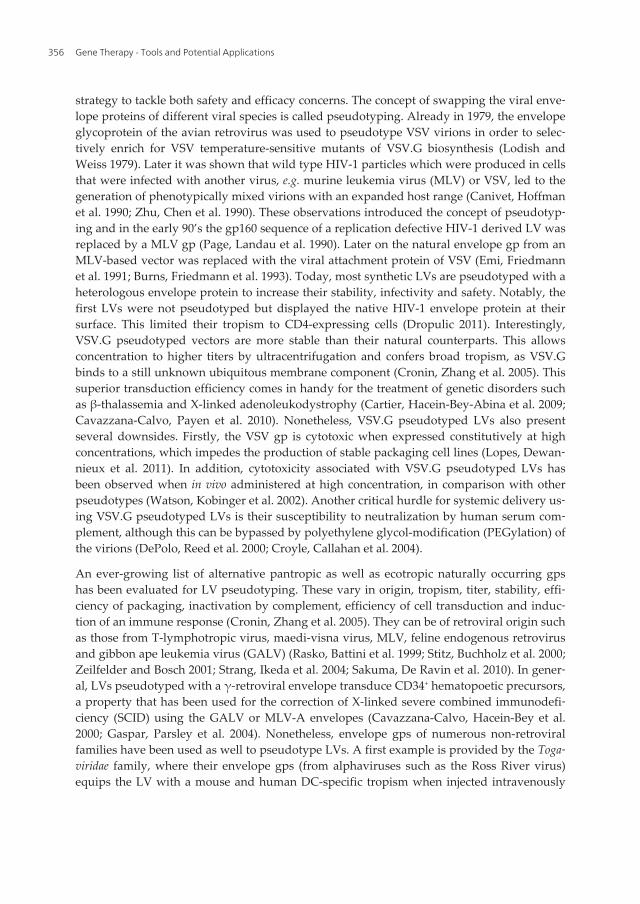
strategy to tackle both safety and efficacy concerns. The concept of swapping the viral enve‐lope proteins of different viral species is called pseudotyping. Already in 1979, the envelopeglycoprotein of the avian retrovirus was used to pseudotype VSV virions in order to selec‐tively enrich for VSV temperature-sensitive mutants of VSV.G biosynthesis (Lodish andWeiss 1979). Later it was shown that wild type HIV-1 particles which were produced in cellsthat were infected with another virus, e.g. murine leukemia virus (MLV) or VSV, led to thegeneration of phenotypically mixed virions with an expanded host range (Canivet, Hoffmanet al. 1990; Zhu, Chen et al. 1990). These observations introduced the concept of pseudotyp‐ing and in the early 90’s the gp160 sequence of a replication defective HIV-1 derived LV wasreplaced by a MLV gp (Page, Landau et al. 1990). Later on the natural envelope gp from anMLV-based vector was replaced with the viral attachment protein of VSV (Emi, Friedmannet al. 1991; Burns, Friedmann et al. 1993). Today, most synthetic LVs are pseudotyped with aheterologous envelope protein to increase their stability, infectivity and safety. Notably, thefirst LVs were not pseudotyped but displayed the native HIV-1 envelope protein at theirsurface. This limited their tropism to CD4-expressing cells (Dropulic 2011). Interestingly,VSV.G pseudotyped vectors are more stable than their natural counterparts. This allowsconcentration to higher titers by ultracentrifugation and confers broad tropism, as VSV.Gbinds to a still unknown ubiquitous membrane component (Cronin, Zhang et al. 2005). Thissuperior transduction efficiency comes in handy for the treatment of genetic disorders suchas β-thalassemia and X-linked adenoleukodystrophy (Cartier, Hacein-Bey-Abina et al. 2009;Cavazzana-Calvo, Payen et al. 2010). Nonetheless, VSV.G pseudotyped LVs also presentseveral downsides. Firstly, the VSV gp is cytotoxic when expressed constitutively at highconcentrations, which impedes the production of stable packaging cell lines (Lopes, Dewan‐nieux et al. 2011). In addition, cytotoxicity associated with VSV.G pseudotyped LVs hasbeen observed when in vivo administered at high concentration, in comparison with otherpseudotypes (Watson, Kobinger et al. 2002). Another critical hurdle for systemic delivery us‐ing VSV.G pseudotyped LVs is their susceptibility to neutralization by human serum com‐plement, although this can be bypassed by polyethylene glycol-modification (PEGylation) ofthe virions (DePolo, Reed et al. 2000; Croyle, Callahan et al. 2004).
An ever-growing list of alternative pantropic as well as ecotropic naturally occurring gpshas been evaluated for LV pseudotyping. These vary in origin, tropism, titer, stability, effi‐ciency of packaging, inactivation by complement, efficiency of cell transduction and induc‐tion of an immune response (Cronin, Zhang et al. 2005). They can be of retroviral origin suchas those from T-lymphotropic virus, maedi-visna virus, MLV, feline endogenous retrovirusand gibbon ape leukemia virus (GALV) (Rasko, Battini et al. 1999; Stitz, Buchholz et al. 2000;Zeilfelder and Bosch 2001; Strang, Ikeda et al. 2004; Sakuma, De Ravin et al. 2010). In gener‐al, LVs pseudotyped with a γ-retroviral envelope transduce CD34+ hematopoetic precursors,a property that has been used for the correction of X-linked severe combined immunodefi‐ciency (SCID) using the GALV or MLV-A envelopes (Cavazzana-Calvo, Hacein-Bey et al.2000; Gaspar, Parsley et al. 2004). Nonetheless, envelope gps of numerous non-retroviralfamilies have been used as well to pseudotype LVs. A first example is provided by the Toga‐viridae family, where their envelope gps (from alphaviruses such as the Ross River virus)equips the LV with a mouse and human DC-specific tropism when injected intravenously
Gene Therapy - Tools and Potential Applications356

(Strang, Takeuchi et al. 2005), and with an astrocyte and oligodendrocyte specific tropismwhen injected into the mouse brain (Kang, Stein et al. 2002). Another example is providedby the family of the Baculoviridae where the gp64 gp ensures high particle stability in addi‐tion to a hepatocyte specific tropism (Matsui, Hegadorn et al. 2011). LVs pseudotyped withthe lymphocytic choriomeningitis virus (LCMV) envelope from the Arenaviridae preferential‐ly transduce cells from the central nervous system such as neural stem cells and progenitorcells, and also to glioma cells and insulin secreting β-cells (Kobinger, Deng et al. 2004; Milet‐ic, Fischer et al. 2004; Stein, Martins et al. 2005). As there is an increasing interest in the de‐velopment of gene therapeutic strategies for malignant gliomas, the most frequent primarybrain tumors with very poor prognosis, several groups report on the use of LCMV gp pseu‐dotyped LVs to target almost exclusively astrocytes, the main source of malignant gliomacells (Beyer, Westphal et al. 2002; Miletic, Fischer et al. 2004; Steffens, Tebbets et al. 2004).The H and F envelope proteins from the Paramyxoviridae family, such as those derived frommeasles viruses, provide LVs with the capacity to bind to SLAM and CD46, which confersefficient virus entry, nuclear transport and integration in non-activated B and T lympho‐cytes. This property is particularly important, since primary unstimulated B and T cells aregenerally difficult to transduce if not pre-treated to induce progression through the cell cy‐cle (e.g. through stimulation with anti-CD3/anti-CD28 antibodies or cytokines) (Frecha, Levyet al. 2010; Frecha, Levy et al. 2011). To transduce airway epithelial cells efficiently, envelopeproteins from several viruses that infect respiratory tissues or cells have been evaluated. Forefficient transduction of unconditioned airway epithelial cells from the apical side, enve‐lopes derived from the ebola virus (Filoviridae), members of the Paramyxoviridae such as therespiratory syncytial (RSV) and sendai viruses, and members of the Orthomyxoviridae such asthe influenza and fowl plaque viruses have been evaluated (Kobinger, Weiner et al. 2001;Nefkens, Garcia et al. 2007; Mitomo, Griesenbach et al. 2010). Surprisingly, it has been re‐ported that the S protein of the severe acute respiratory syndrome-associated coronavirus(Coronaviridae) mediates entry into hepatoma cell lines (Hofmann, Hattermann et al. 2004).Finally, although the vesicular stomatitis, mokola and rabies virus are all derived from theRhabdoviridae family, only LVs pseudotyped with the rabies-G envelope enable retrogradetransport to motoneurons of the spinal cord upon intramuscular injection or to the thalamusupon striatal injection. In contrast, VSV.G displaying LVs transduce cells only locally whilemokola-pseudotyped LVs preferentially target non-neuronal glial cells (Mazarakis, Azzouzet al. 2001; Azzouz, Ralph et al. 2004; Wong, Azzouz et al. 2004; Colin, Faideau et al. 2009;Calame, Cachafeiro et al. 2011).
Although the use of an existing viral envelope gp seems the most straightforward way topseudotype LVs, a natural variant with the desired delivery properties is not available forevery therapeutic application. Moreover, natural gps can come with limitations such as sen‐sitivity to neutralization by the host immune response, lack of specificity and/or insufficienttransduction efficiency. Also their production and purification can be inefficient (Schaffer,Koerber et al. 2008). Therefore, the development of LVs with customized, user-defined genedelivery properties by molecular engineering of the envelope gps is an alternative strategyto retarget the LV to specific cell-surface receptors. This molecular engineering has become acollective term for many different strategies, which will be described below.
Targeted Lentiviral Vectors: Current Applications and Future Potentialhttp://dx.doi.org/10.5772/52770
357

A first strategy to alter the tropism of a virally derived gp is by rational point and domainmutagenesis. This is exemplified by the DC-specific targeting strategy from Yang et al. Cer‐tain subsets of DCs carry the DC-SIGN protein (also known as CD209) on their surface,which is a C-type lectin-like receptor that potentiates rapid binding and endocytosis of ma‐terials. The sindbis virus envelope gp consists of two integral membrane gps that form a het‐erodimer and function as one unit. The fusogenic monomer is E1 and needs binding via E2to mediate low pH-dependent fusion. The latter binds to the DC-SIGN receptor, next to thecanonical viral receptor heparin sulfate, expressed by many cell types. Since both proteinbinding sites are physically separated, selective mutation at the E2 monomer is possible, ab‐rogating the heparin sulfate binding part while leaving the DC-SIGN binding part intact. Bypseudotyping a LV with this mutated sindbis virus derived envelope gp, targeted infectionof DCs in vivo after direct subcutaneous administration was achieved. Moreover, this elicit‐ed a strong antigen-specific immune response (Yang, Yang et al. 2008; Hu, Dai et al. 2010).Another example is the substitution of the V3-loop region of the SIV envelope gp with thecorresponding region of a T cell tropic HIV-1 to create a T-cell targeted MLV vector, pseudo‐typed with this engineered SIV gp (Steidl, Stitz et al. 2002). A final example is provided byDylla et al. who diminished the alfa-dystroglycan affinity of the LCMV WE45 strain enve‐lope gp by a point mutation. When a FIV derived LV was pseudotyped with this point mu‐tated LCMV gp, their intravenous injection in adult mice yielded low transductionefficiencies in hepatocytes in contrast to abundant liver and cardiomyocyte transductionwith the wild type LCMV gp pseudotyped FIVs (Dylla, Xie et al. 2011).
Apart from genetic alterations, chemical modifications can also alter LV tropism. PEGyla‐tion of VSV.G pseudotyped LVs is one such example where the LVs’ tropism is not altered.Nevertheless, chemical modifications can lead to targeted gene delivery vehicles, for exam‐ple by tagging the MLV vector with galactose to selectively transduce human hepatoma celllines expressing asialo-gp receptors specific for oligosaccharides with terminal galactose res‐idues (Neda, Wu et al. 1991). Furthermore, Morizono et al. reported the production of LVspseudotyped with sindbis virus gps in the presence of deoxymannojirimycin. This modifica‐tion altered the structures of N-glycans from complex to high mannose structures as it inhib‐its mannosidase. This led to DC-SIGN specific binding although the gps were geneticallymodified to prevent interaction with DC-SIGN (Morizono, Ku et al. 2010). Furthermore itwas demonstrated that binding of sindbis virus gp to DCs is directly related to the amountof high-mannose structures on the gp (Tai, Froelich et al. 2011). Unfortunately, the effective‐ness of the chemically modified particles strongly depends on the reaction conditions of theapplied modifications.
Other chimeric envelope gps can be generated by covalently fusing a short peptide, a ligandor an antibody to an envelope gp. The advantages of short peptides are that they don’t se‐verely disrupt the original envelope gps’ function and that via high-throughput library ap‐proaches, targeted peptides with strong binding affinity and unlimited specificity within thecontext of a particular gp can be generated (Schaffer, Koerber et al. 2008). However, they canhinder multimerisation of capsid monomers, create fusion products with lower thermosta‐bility and hinder proper intracellular trafficking of the gp during viral production. The latter
Gene Therapy - Tools and Potential Applications358
is exemplified by the blockage in trafficking in the producer cells to the plasma membraneof VSV.G when linked to a collagen-binding motif (Guibinga, Hall et al. 2004). Differentkinds of ligands such as cytokines and growth factors have been linked to the amino-termi‐nal region or receptor-binding domain of the envelope gp, most often derived of MLV. Thisis amongst others exemplified by fusion of the MLV gp to hepatocyte growth factor to targetthe LV to hepatocytes (Nguyen, Pages et al. 1998), or to the insulin-like growth factor (IGF-I)(Chadwick, Morling et al. 1999). Interestingly, these ligands can elevate the transduction ef‐ficiency by altering the targets’ physiological state. When the fusion product of the MLV gpand IL-2 is used to pseudotype LVs, a 34-fold higher infection efficiency was observed ofquiescent IL-2 receptor expressing cells compared to LVs pseudotyped with the wild typeMLV gp. This was explained by IL-2 induced activation of the cell cycle from the otherwisebarely transducible quiescent cells (Maurice, Mazur et al. 1999). However, a very low to un‐observable transduction profile is often reported which can be attributed to sequestration ofthe LV particles at the target cell surface, directing the viral particle to a degradation path‐way after endocytosis and/or inability of the fusion product to trigger a conformationalchange essential for viral fusion and subsequent infection (Lavillette, Russell et al. 2001; Ka‐tane, Takao et al. 2002). In addition to peptides and ligands, also antibodies and their deriva‐tives can be used. In general, single chain variable fragments or scFvs offer higher specificitythan short peptides but as they are larger in size, the chance that they disrupt the process ofconformational changes of the gp to mediate membrane fusion increases. Therefore scFvsare most often linked to a spacer peptide that permits proper conformation of both the scFvdomain and the envelope gp as exemplified by the fusion of the MLV gp to a scFv againstMHC class I (Karavanas, Marin et al. 2002). For LV targeting to APCs, several attempts havebeen made to couple an anti-MHC class II scFv to an ecotropic gp such as MLV or VSV.G(Dreja and Piechaczyk 2006; Gennari, Lopes et al. 2009). Recently, the use of DARPins or de‐signed ankyrin repeat proteins has been reported. These can be fused to the H protein ofmeasles virus for example and then be co-displayed with the fusogenic F protein on the sur‐face of the LV. The advantage is that DARPins can be selected to become high-affinity bind‐ers to any kind of target molecule thus this seems a promising alternative to scFvs forretargeting LVs (Munch, Muhlebach et al. 2011). So, in general, the use of chimeric envelopeproteins for LV targeting has proven to offer tremendous opportunities but at the same timeto be a challenge as the function of chimeric gps is often severely compromised which leadsto a very inefficient transduction profile (Fielding, Maurice et al. 1998; Dreja and Piechaczyk2006; Waehler, Russell et al. 2007; Buchholz, Duerner et al. 2008).
Several solutions have been created to circumvent the problems associated with the forma‐tion of conformational dysfunctional fusion products. One solution is the inclusion of a pro‐tease cleavable peptide between the gp and the ligand. This is certainly an interestingstrategy for the targeting of tumor cells, as they secrete MMP, which degrade the extracellu‐lar matrix to metastasize. By linking a proline-rich hinge and an MMP cleavage site to thefusion product of a scFv recognizing carcinoembryonic antigen (CEA) and the MLV gp, se‐lective targeting of CEA-positive cells after in vivo injection of producer cells at the tumorsite was observed (Chowdhury, Chester et al. 2004). Taking this hinge region idea one stepfurther, the concept of ‘molecular bridges’ was introduced where a bispecific linker mole‐
Targeted Lentiviral Vectors: Current Applications and Future Potentialhttp://dx.doi.org/10.5772/52770
359

cule recognizes both the viral gp as well as the molecular determinant on the target cell. Thisconcept is based on a bridging system that was introduced more than 20 years ago andwhere three different linker molecules were involved: two biotinylated antibodies thatbound the MLV gp and MHC class I or II proteins on the target cells respectively, and abridging streptavidin molecule linking both antibodies. This led to the generation of a MLVthat was capable of transducing MHC class I and II expressing cells (Roux, Jeanteur et al.1989). Subsequently, two-protein molecular bridges have been exploited based on the avi‐din-biotin system. A recent example is provided by O’Leary et al. who used a detoxified re‐combinant form of the full-length botulinum neurotoxin, fused to core streptavidin that forits part was coupled to a biotinylated LV. This envelope gp construct endowed the LV parti‐cle with considerable neuron selectivity in vitro as well as in vivo after injection into the tra‐chea (O'Leary, Ovsepian et al. 2011). Nowadays, alternative linkers such as ligand-receptor,chemical conjugations and monoclonal antibodies have been exploited to retarget LVs aswell (Roux, Jeanteur et al. 1989; Boerger, Snitkovsky et al. 1999). For the latter, the E2 proteinof the sindbis gp has been modified to contain the Fc-binding domain (ZZ domain) of pro‐tein A, making it possible to bind to a monoclonal antibody specific for a target molecule viaits Fab antigen recognition end (Morizono, Xie et al. 2005). However, doubts are raisedabout the affinity of the adaptor-virus complex, as this may not be sufficient to prevent dis‐sociation within the patient’s blood. Moreover, complexity ascends as both the virion as theadaptor must be produced, purified and fully characterized for clinical approval. Anotheralternative possibility is to co-display a chimeric envelope gp together with a wild type gpsuch as VSV.G to enhance the transduction efficiency (Maurice, Verhoeyen et al. 2002; Ver‐hoeyen, Dardalhon et al. 2003; Verhoeyen, Wiznerowicz et al. 2005). However, this had alsolimited success due to partial loss of targeting specificity. Therefore, a final alternative is theusage of a mutated fusogenic but binding-defective envelope gp to mediate fusion uponbinding by the chimeric gp. The group of Lin et al. co-expressed the MLV gp fused to solu‐ble Fms-like tyrosine kinase 3 (Flt3)-ligand together with a binding-defective influenza he‐magglutinin protein from the fowl plague virus rostock 34 (HAmu). When LVs werepseudotyped with both of these gps, Flt3-targeted transduction was enhanced when com‐pared to LVs without HAmu and could be competed away by the addition of soluble Flt3-ligand (Lin, Kasahara et al. 2001). Another more straightforward strategy is the use of theE1/E2 heterodimer gp of sindbis virus as the fusion and binding functions are already sepa‐rated over two different monomers. By mutating the binding E2 monomer, its binding prop‐erty can be completely abolished. Therefore, this binding defective E2 protein forms an idealscaffold for cell-specific antibody conjugation to confer specific tropism to an endless list ofcell types such as P-gp-expressing melanoma progenitor cells and endothelial cells (Morizo‐no, Xie et al. 2005; Pariente, Mao et al. 2008). A drawback is that they only induce fusionupon low pH. Therefore alternatives were explored such as the H and F protein of the mea‐sles virus, which induce fusion without the need for endocytosis (Earp, Delos et al. 2005;Funke, Schneider et al. 2009). This is exemplified by a study were a binding defective formof the H protein was fused to a CD20 specific scFv to pseudotype LVs. When these LVs wereused to kill cells in culture, they selectively killed the CD20+ human lymphocytes in co-cul‐ture with CD20- cells. This demonstrated the ability of these LVs to exclusively transfer a po‐
Gene Therapy - Tools and Potential Applications360

tentially hazardous therapeutic protein into targeted cell populations with virtual absence ofbackground transduction in non-target cells (Funke, Maisner et al. 2008). Meanwhile, abroad variety of surface antigens has been successfully targeted using this strategy (Blechaczand Russell 2008)
A fourth strategy to target LVs is based on two concepts: (1) the separation of binding andfusion functions over two distinct envelope molecules and (2) the ability of LVs to incorpo‐rate host cell proteins into their envelope as they bud from the plasmamembrane of theirproducer cells (Chandrashekran, Gordon et al. 2004; Kueng, Leb et al. 2007). Chandrashek‐ran et al. reported on efficient and specific targeting to human cells expressing stem cell fac‐tor (SCF) receptor (c-kit) by an ecotropic gp pseudotyped LV which also displayed surfaceSCF. Another example is the overexpression of the HIV-1 derived primary receptor CD4 andfusogenic co-receptor CXCR4 or CCR5 on the membrane of producer cells. From these cells,LVs were generated that infect HIV-1 envelope gp expressing cells next to cells infected withHIV-1, enabling the development of novel antiviral therapy approaches (Somia, Miyoshi etal. 2000). Since the transduction efficiency was relatively low, LV co-enveloped with theHIV-1 cellular receptor CD4 and the E2 protein from sindbis virus were created. Theseturned out to have a higher infectivity level than in the former strategy (Lee, Dang et al.2011). In another study the binding defective but fusogenic E1/E2 heterodimer was used tobe co-displayed with a separate membrane bound anti-CD20 antibody in order to transduceexclusively CD20+ B cells (Lei, Joo et al. 2009). Today, numerous examples are found that ap‐ply this principle to target the following: immunoglobulin-expressing B cells, CD3+ T cellsand CD117+ HSCs (Ziegler, Yang et al. 2008; Froelich, Ziegler et al. 2009; Yang, Joo et al.2009). However, clinical applications with LVs displaying scFvs are hampered by lack of sta‐bility, size and immunogenicity leading to the development of neutralizing antibodies. Tosolve these problems, we developed the Nanobody (Nb) display technology (Goyvaerts, DeGroeve et al. 2012). In this strategy, a fusogenic but binding-defective form of VSV.G(VSV.GS) (Zhang, Kutner et al. 2010) is co-displayed with a surface bound form of a cell-spe‐cific Nb to confer target binding (Figure 4). Some twenty years ago, Hamers-Casterman et al.discovered that part of the humoral response of Camelids is based on a unique repertoire ofantibodies, which only consisted of two heavy chains (Hamers-Casterman, Atarhouch et al.1993). The antigen binding part of these antibodies is composed of only one single variableregion, termed VHH or Nb. These Nbs have unique characteristics and offer many advan‐tages over scFvs to target LVs to specific cell types. These include (1) they are highly soluble,(2) they can refold after denaturation whilst retaining their binding capacity, (3) cloning andselection of antigen-specific Nbs obviate the need for construction and screening of large li‐braries, (4) as Nbs can be fused to other proteins, it is possible to present them on the cellmembrane of a producer cell line, thus generating LVs that incorporate a cell-specific Nb intheir envelope during budding. Using the Nb display technology, we demonstrated produc‐tion of stable Nb pseudotyped LV stocks at high titers with a DC subtype specific transduc‐tion profile both in vitro as well as in vivo (Goyvaerts, De Groeve et al. 2012). As ligandspecific Nbs can be generated to potentially every cell surface molecule, this technology willbe applicable to target LVs to every cell type for which cell specific surface molecules arecharacterized (Gainkam, Huang et al. 2008; Vaneycken, Devoogdt et al. 2011).
Targeted Lentiviral Vectors: Current Applications and Future Potentialhttp://dx.doi.org/10.5772/52770
361

The downside of the use of the above-described strategies is that they rely on the fusogeniccapacity of a gp that is derived from viruses infectious to humans such as VSV, measles vi‐rus, sindbis virus and MLV. Their exposure to the complement or immune system, leadingto anti-gp antibodies, might limit their clinical applicability. To surmount these obstacles,Frecha et al. pseudotyped LVs with a mutant fusogenic gp derived from an endogenous fe‐line virus, named RD114. The mutant RD114 gp is an attractive candidate for in vivo use as itis resistant to degradation by the human complement. By co-displaying the early-acting-cy‐tokine SCF together with mutant RD114 gp, human CD34+ HSCs could be targeted in vivo(Frecha, Fusil et al. 2011; Frecha, Costa et al. 2012). SCF was responsible for a slight and tran‐sient stimulation of the HSCs while preserving the ‘stemness’ of the targeted HSCs. In thatway, the need for CD3/CD28 or cytokine pretreatment was obviated. Springfeld et al. recent‐ly pseudotyped LVs with the H and F gps of the Tupaia paramyxovirus (TPMV), an animalvirus without close human pathogenic relatives. Moreover, as this virus does not infect hu‐man cells, detargeting the H protein from its natural receptors is unnecessary. When LVswere pseudotyped with the TPMV envelope protein linked to an anti-CD20 single chain an‐tibody, selective transduction of CD20+ cells, including quiescent primary human B cells,was reported (Enkirch, Kneissl et al. 2012).
Figure 4. Principle of the Nb display technology. The Nb display technology is based on the fact that LVs need tobind and fuse with the membrane of a target cell for proper infection. While VSV.G accounts for both of these func‐tions, we propose to separate these functions over two different molecules: (1) binding via a membrane bound Nbagainst the target cell of choice and (2) fusion via VSV.GS, which is a binding defective truncated version of VSV.G.
Recently an innovative alternative strategy has been described by Mannell et al. for site spe‐cific vascular gene delivery. In this case, the LVs were first coupled to magnetic nanoparti‐
Gene Therapy - Tools and Potential Applications362

cles, which were in turn coupled to lipid microbubbles. LVs coupled to magneticnanoparticles to target them to specific cell types in vitro using an external magnetic fieldhas been described before. However, when these LV-nanoparticle constructs are consideredfor in vivo use, a sufficient magnetic moment is needed as the particles are subject to flowvelocity within the blood vessels. As the magnetic moment is proportional to particle size,Mannell et al. coated the LV-nanoparticle constructs with magnetic microbubbles for en‐largement. Upon intravenous delivery, the LV magnetic microbubbles were first trapped atthe site of interest. Next ultrasound mediated destruction of the microbubbles resulted infast release of the LVs at the site of interest with high transduction efficiency without thecost of higher cytotoxicity (Mannell, Pircher et al. 2012).
In conclusion, there seem to be some general prerequisites for successful transductional tar‐geting of LVs: (1) use envelope gps with defined receptor binding sites, (2) abolish the natu‐ral recognition sites of the attachment gp, (3) separate fusion and attachment functions overtwo different molecules, (4) avoid the construction of large fusion constructs since their fu‐sogenic capacities can be severely compromised and (5) avoid the use of immunogenic gps(Buchholz, Muhlebach et al. 2009).
5. Genomic targeting
Nowadays, LVs have become valuable tools for the treatment of several monogenic disor‐ders such as hemophilia B, β-thalassemia and X-linked adrenoleukodystrophy (Cartier, Ha‐cein-Bey-Abina et al. 2012; Payen, Colomb et al. 2012). However, the use of viral vectors thatintegrate their cargo into the genome of the host cell can trigger oncogenesis by insertionalmutagenesis. This is exemplified by the incident where two out of 11 patients treated with aγ-retroviral vector to correct X-linked SCID, developed leukemia. This was caused by the γ-retroviral construct’s tendency to insert into active genes, in this case the LMO-2 oncogene(Marshall 2002). Later on, using the same vector type to treat chronic granulomatous dis‐ease, genomic instability and myelodysplasia was observed (Stein, Ott et al. 2010). These in‐cidents prompted substantial research into design, safety testing and optimization ofintegrating vectors. Thus far several measures have been taken to pose a reduced risk on in‐sertional mutagenesis such as the development of SIN LVs containing a moderate cellularpromoter (Modlich and Baum 2009; Montini, Cesana et al. 2009). Furthermore LVs are in‐trinsically less genotoxic than their retroviral counterparts (Montini, Cesana et al. 2006).Nevertheless, LVs have a higher transduction efficiency, which could counterbalance the re‐duced risk of mutagenic vector integration into the patient’s genome. In addition, accumu‐lating studies report the concept of LV-induced clonal dominance related to growth and/orsurvival advantage e.g. induced by vector integration and subsequent formation of aberrant‐ly spliced mRNA forms (Fehse and Roeder 2008; Cavazzana-Calvo, Payen et al. 2010; Cesa‐na, Sgualdino et al. 2012; Moiani, Paleari et al. 2012). In an extensive analysis to explore theeffect of promoter-enhancer selection on efficacy and safety of LVs, no clear underlyingmechanism could be provided for the observed. They concluded that other ill-defined riskfactors must be involved for oncogenesis, including replicative stress (Ginn, Liao et al. 2010).
Targeted Lentiviral Vectors: Current Applications and Future Potentialhttp://dx.doi.org/10.5772/52770
363

Finally, next to transcriptional activation of neighboring genes, also transcriptional shut offof the tg has been reported. This was due to chromatin remodeling at the site of insertionand cessation of the therapeutic effect (Stein, Ott et al. 2010).
Therefore additional strategies have been considered to reduce the side effects related torandom insertion. The most straightforward strategy is to prevent integration of the proviralcargo by the use of IDLVs. These IDLVs are produced with a mutated integrase, which re‐sults in prevention of proviral integration and generation of increased levels of circular vec‐tor episomes within the infected cells. They appear to be safer with only a 0,1 to 2,3% chancethat the episomal transcript gets integrated without a marked loss in effectiveness in termsof immune stimulatory potential of the IDLV-based vaccines (Vargas, Gusella et al. 2004;Philippe, Sarkis et al. 2006; Karwacz, Mukherjee et al. 2009; Wanisch and Yanez-Munoz2009). However, as the lentiviral episomes lack replication signals, they are gradually lost bydilution in dividing cells and only stable in quiescent cells, which is undesirable for perma‐nent correction of any genetic disorder. Furthermore also lower tg expression levels havebeen reported compared to integrative vectors (Bayer, Kantor et al. 2008). Therefore severalalternative strategies have been brought forward to target the integrative process to a specif‐ic ‘safe’ genomic site.
In a first attempt, several groups tried to fuse a heterologous DNA binding domain directlyto the integrase. Bushman et al. were the first to evaluate the activity of a hybrid, composedof the HIV-1 integrase and the lambda repressor. They reported on integration primarilynear the lambda operator sites on the same face of the β-DNA helix (Bushman 1994). Later amodel system was used were the integrase, derived from the avian sarcoma virus or HIV-1respectively was fused to the Escherichia coli LexA repressor protein DNA binding domain(Katz, Merkel et al. 1996; Holmes-Son and Chow 2000). When this construct was packagedinto the virion in trans either by replacing the original integrase gene or by cloning it adja‐cent to the HIV-1 accessory protein Vpr, they observed that this enhanced the use of integra‐tion sites adjacent to the lexA operators. In another study, the HIV-1 derived integrase wasfused to a synthetic polydactyl zinc finger protein E2C, which binds specifically to a contig‐uous 18 bp E2C recognition site (Tan, Dong et al. 2006). Although in all studies clearly ahigher preference for integration near the target sequence of choice was observed, this alsoimplicated reduced DNA-binding specificity of the fusion protein with associated decreaseof integration frequency of about 80 percent compared to viruses containing wild type inte‐grase. Furthermore this strategy is also limited by the difficulty to incorporate the fusionprotein into infectious virions (Michel, Yu et al. 2010).
Another strategy is targeting the integration away from genes using tethering domainslinked to the host cell-encoded transcriptional co-activator lens epithelium-derived growthfactor/p75 (LEDGF/p75), a cellular integrase binding protein. For example the LEDGF/p75chromatin interaction-binding domain has been replaced with CBX1, which binds histoneH3 di- or trimethylated on K9. Subsequently proviral integration was directed to pericentricheterochromatin and intergenic regions (Llano, Vanegas et al. 2006; Ferris, Wu et al. 2010;Gijsbers, Ronen et al. 2010; Silvers, Smith et al. 2010). As this requires engineering of a hostcell protein, it is not feasible for clinical applications at the present stage (Izmiryan, Basma‐
Gene Therapy - Tools and Potential Applications364

ciogullari et al. 2011). Site-specific proviral integration can also be mediated by the use ofsite-specific recombinases. The best known are derived from the lambda integrase family ofenzymes and include the bacteriophage P1 Cre recombinase, bacteriophage lambda inte‐grase, the yeast Flp recombinase and bacterial XerCD recombinase. They catalyze site specif‐ic recombination by a transient DNA-protein covalent linkage that brings two specific DNArepeats together (Van Duyne 2001). Depending on the orientation of the DNA repeats, theDNA segment will either be excised or inverted when in the same or opposite orientationrespectively (Figure 5A, adapted from http://www.ruf.rice.edu/~rur/issue1_files/norman.html). The Cre-loxP system has been developed for gene studies to conditionallyknock out a target gene in a cell- or tissue specific manner to overcome embryonic lethalitydue to permanent inactivation of the target gene in an early developmental stage (Ray, Fa‐gan et al. 2000). This system is based on two palindromic loxP sites of 34 bp that flank thegene of interest. Although these loxP sites are prevalent in the genomes of bacteriophages,they are absent in the mouse genome where they have to be introduced by targeted muta‐genesis (Kos 2004). Throughout the human genome, however, loxP-like sequences or pseu‐do-loxP sites are present that can be recognized by either wild-type Cre or Cre variants. Thislast feature enables site-specific insertion of a gene in a defined loxP site in the human ge‐nome if a Cre recombinase is provided in cis or trans. Michel et al. evaluated the feasibility ofcombining the Cre-loxP system for gene targeting with the versatile gene delivery system ofLVs for site-specific gene insertion in human cell lines. They transduced a loxP site contain‐ing cell line with a LV containing Cre recombinase in trans as a fusion protein to the HIVaccessory protein Vpr. Moreover the LV contained a cassette containing a loxP site followedby the neomycin resistant gene, inserted in the U3 region of the 3’LTR. Upon reverse tran‐scription, the loxP-neo sequence would appear in both LTRs, thereby providing a substratefor recombination that could be catalyzed by the virion-associated Vpr-Cre. Upon this re‐combination step, a circular product was produced that was on his turn inserted into theloxP site of the cell line, again catalyzed by virion-associated Vpr-Cre. Another example isprovided by the group of Jiang et al. who demonstrated a selective inhibitory effect on thelens epithelial cells and not the retinal pigment epithelial cells (Jiang, Lu et al. 2011). There‐fore they used an enhanced Cre/loxP system with a LV expressing Cre under the control ofthe lens-specific promoter LEP503 in combination with another LV that contained a stiffersequence encoding eGFP with a functional polyadenylation signal between two loxP sites,followed by the HSV-TK gene, both under the control of the human phosphoglycerate kin‐ase promoter. Expression of the downstream HSV-TK was activated by co-expression of Creunder the control of the lens-specific promoter LEP503. Although this technology allowssite-specific tg insertion, there are only a limited amount of pseudo-loxP sites in the humangenome and even none in the mouse genome, which makes this technique unusable for fun‐damental research in laboratory animals. Furthermore, two recombination events are re‐quired which has a major impact on its efficiency.
A recent strategy makes use of site-specific endonucleases to target the tg to neutral ‘safeharbor’ genome regions or stimulate the process of homologous recombination for gene re‐pair (Fischer, Hacein-Bey-Abina et al. 2011). Endonucleases induce site-specific ds breaksthat can be repaired by homology-directed repair, a form of homologous recombination that
Targeted Lentiviral Vectors: Current Applications and Future Potentialhttp://dx.doi.org/10.5772/52770
365

uses a copy of the genetic information from the broken DNA molecule. When the latter isprovided by the same or another LV, this copy will be used to repair the ds break (Urnov,Miller et al. 2005; O'Driscoll and Jeggo 2006). The advantage of gene repair/correction is thatboth function and expression of the affected gene are restored while the risk associated withrandom vector integration is avoided. Besides the advantage of the reduced risk for inser‐tional mutagenesis, this strategy is also used to target genes in order to knock them down orreplace them with another gene by homologous recombination. The disadvantage is that thenuclease coding sequences are expressed for several days, which is not optimal for transla‐tion to the clinic due to the background off-target generation of dsDNA breaks.
One possibility is the use of the zinc finger nuclease strategy. For this, the Cys2His2 class ofzinc finger DNA binding domains is engineered to recognize a DNA sequence of interest,fused to the nuclease domain of the FokI type II restriction endonuclease to yield a highlyspecific zinc finger (Figure 5 B, adapted from http://biol1020-2011-2.blogspot.be/2011/09/zinc-finger-nucleases-zfn-emerging.html) (Kim, Cha et al. 1996; Pabo, Peisach et al. 2001).When two different zinc fingers are designed to bind the same sequence of interest in theopposite orientation, this will allow dimerization of the FokI domains which leads to a zincfinger induced dsDNA break (Bitinaite, Wah et al. 1998). Various strategies have been devel‐oped to engineer the Cys2His2 zinc fingers in order to bind a specific sequence either bymodular assembly or by selection strategies using phage display or a cellular selection sys‐tem. Naldini et al. evaluated the use of zinc finger nucleases in combination with an IDLVfor gene editing. Therefore they co-transduced several cell lines with three different IDLVs,one encoding the donor sequence and two encoding the two zinc fingers (Lombardo, Geno‐vese et al. 2007). A few years later they also used this strategy to assess zinc finger specificitygenome-wide by comprehensively mapping the locations of the IDLV integration sites incells co-transduced with GFP and zinc finger encoding LVs (Gabriel, Lombardo et al. 2011).They observed a very high efficiency and specificity, yet a measurable rate of vector integra‐tion at unidentified sites occurred with this approach, which is the sum of zinc finger medi‐ated and background levels of IDLV integration. Moreover co-transduction with threedifferent LVs may be a rate-limiting step in this system. Therefore the use of a single con‐struct to express the zinc fingers and deliver the donor tg must be evaluated, especially forless permissive cells such as hematopoietic progenitors.
Another way to target the proviral genome is by the provision of a vector-associated mega‐nuclease encoded by a separate vector or supplied as a protein within the viral particle.(Izmiryan, Basmaciogullari et al. 2011). For the latter, Ismiryan et al. fused the prototypicmeganuclease I-SceI from yeast to Vpr. This avoided the potentially toxic sustained expres‐sion of the introduced endonuclease. IDLVs encoding the donor sequence and containingthe meganuclease-SceI fusion construct were tested in reporter cells in which targetingevents were scored by the repair of a puromycin resistance gene. They reported a two-foldhigher frequency of the expected recombination event when the nuclease was delivered as aprotein rather than encoded by a separate vector and therefore improved both the safetyand efficacy of this LV-based gene targeting system. In conclusion, although the field of ge‐
Gene Therapy - Tools and Potential Applications366
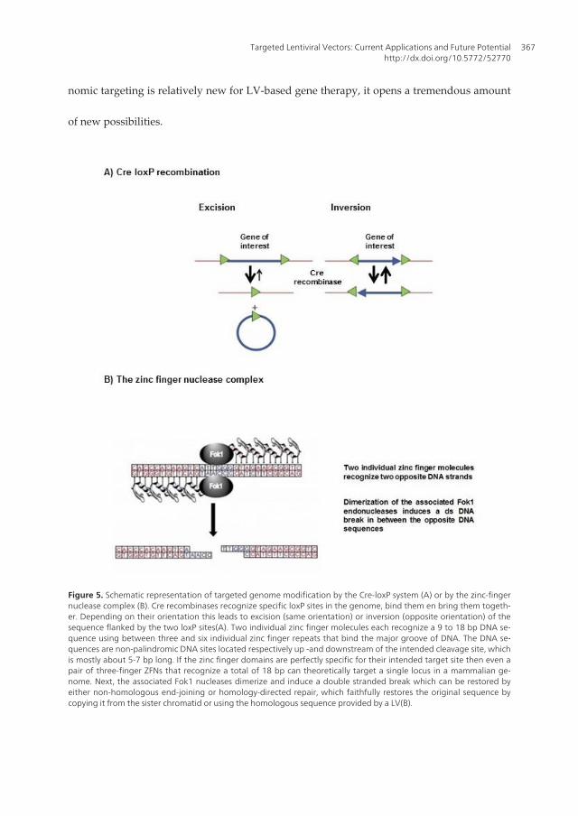
nomic targeting is relatively new for LV-based gene therapy, it opens a tremendous amount
of new possibilities.
Figure 5. Schematic representation of targeted genome modification by the Cre-loxP system (A) or by the zinc-fingernuclease complex (B). Cre recombinases recognize specific loxP sites in the genome, bind them en bring them togeth‐er. Depending on their orientation this leads to excision (same orientation) or inversion (opposite orientation) of thesequence flanked by the two loxP sites(A). Two individual zinc finger molecules each recognize a 9 to 18 bp DNA se‐quence using between three and six individual zinc finger repeats that bind the major groove of DNA. The DNA se‐quences are non-palindromic DNA sites located respectively up -and downstream of the intended cleavage site, whichis mostly about 5-7 bp long. If the zinc finger domains are perfectly specific for their intended target site then even apair of three-finger ZFNs that recognize a total of 18 bp can theoretically target a single locus in a mammalian ge‐nome. Next, the associated Fok1 nucleases dimerize and induce a double stranded break which can be restored byeither non-homologous end-joining or homology-directed repair, which faithfully restores the original sequence bycopying it from the sister chromatid or using the homologous sequence provided by a LV(B).
Targeted Lentiviral Vectors: Current Applications and Future Potentialhttp://dx.doi.org/10.5772/52770
367

6. Concluding remarks
LVs have proven to be efficient vehicles to deliver one or more tgs to any cell type of choice,which has led to a promising list of therapeutic applications. As the demand for experimen‐tation in gene delivery to specific cell types increases, technologies that precisely target LV-based gene expression will become more important for research and clinical applications.Four main groups of strategies with their own possibilities as well as difficulties have beendeveloped so far. Self-evidently, further optimization and fine-tuning of these strategies is anecessity to fulfill the expectations for targeted LV delivery in vivo. In addition to extra opti‐mization steps, combinations of two or more of these strategies can also lead to an overallmore selective, efficient and most importantly, safe LV system. Several attempts to combinethe different strategies have been reported (Brown, Cantore et al. 2007; Pariente, Morizonoet al. 2007; Escors and Breckpot 2011). Pariente et al. for example, reported on a LV that wastransductionally targeted to prostate cancer bone metastases by a modified sindbis virus en‐velope that interacts with PSCA and transcriptionally targeted with a prostate cell specificpromoter. This dual-targeted LV enhanced specificity to prostate cancer bone metastases af‐ter systemic delivery with respect to individual transcriptional or transductional targeting.As the developed targeting strategies already resulted in a major step forward for LV-basedgene therapy, their potential will most likely be more exploited in the future, paving theway towards an all-embracing LV-based tg vehicle for the gene therapeutic field.
Author details
Cleo Goyvaerts1, Therese Liechtenstein2, Christopher Bricogne2, David Escors2 andKarine Breckpot1
1 Laboratory of Molecular and Cellular Therapy, Department of Immunology-Physiology,Vrije Universiteit Brussel, Jette, Belgium
2 Division of Infection and Immunity, Rayne Institute, University College London, London,UK
References
[1] Abruzzese, R. V., D. Godin, et al. (2000). "Ligand-dependent regulation of vascularendothelial growth factor and erythropoietin expression by a plasmid-based autoin‐ducible GeneSwitch system." Mol Ther 2(3): 276-87.
[2] Adriani, W., F. Boyer, et al. (2010). "Social withdrawal and gambling-like profile afterlentiviral manipulation of DAT expression in the rat accumbens." Int J Neuropsycho‐pharmacol 13(10): 1329-42.
Gene Therapy - Tools and Potential Applications368

[3] Annoni, A., M. Battaglia, et al. (2007). "The immune response to lentiviral-deliveredtransgene is modulated in vivo by transgene-expressing antigen-presenting cells butnot by CD4+CD25+ regulatory T cells." Blood 110(6): 1788-96.
[4] Annoni, A., B. D. Brown, et al. (2009). "In vivo delivery of a microRNA-regulatedtransgene induces antigen-specific regulatory T cells and promotes immunologic tol‐erance." Blood 114(25): 5152-61.
[5] Azzouz, M., G. S. Ralph, et al. (2004). "VEGF delivery with retrogradely transportedlentivector prolongs survival in a mouse ALS model." Nature 429(6990): 413-7.
[6] Bahi, A., F. Boyer, et al. (2004). "CD81-induced behavioural changes during chroniccocaine administration: in vivo gene delivery with regulatable lentivirus." Eur J Neu‐rosci 19(6): 1621-33.
[7] Barde, I., M. A. Zanta-Boussif, et al. (2006). "Efficient control of gene expression in thehematopoietic system using a single Tet-on inducible lentiviral vector." Mol Ther13(2): 382-90.
[8] Barrilleaux, B. and P. Knoepfler (2011). "Transduction of human cells with polymer-complexed ecotropic lentivirus for enhanced biosafety." J Vis Exp 2011(53).
[9] Bartel, D. P. (2004). "MicroRNAs: genomics, biogenesis, mechanism, and function."Cell 116(2): 281-97.
[10] Bauer, G., M. A. Dao, et al. (2008). "In vivo biosafety model to assess the risk of ad‐verse events from retroviral and lentiviral vectors." Mol Ther 16(7): 1308-15.
[11] Baup, D., L. Fraga, et al. (2010). "Variegation and silencing in a lentiviral-based mur‐ine transgenic model." Transgenic Res 19(3): 399-414.
[12] Bayer, M., B. Kantor, et al. (2008). "A large U3 deletion causes increased in vivo ex‐pression from a nonintegrating lentiviral vector." Mol Ther 16(12): 1968-76.
[13] Benabdellah, K., M. Cobo, et al. (2011). "Development of an all-in-one lentiviral vec‐tor system based on the original TetR for the easy generation of Tet-ON cell lines."PLoS One 6(8): e23734.
[14] Benzekhroufa, K., B. H. Liu, et al. (2009). "Targeting central serotonergic neuronswith lentiviral vectors based on a transcriptional amplification strategy." Gene Ther16(5): 681-8.
[15] Beyer, W. R., M. Westphal, et al. (2002). "Oncoretrovirus and lentivirus vectors pseu‐dotyped with lymphocytic choriomeningitis virus glycoprotein: generation, concen‐tration, and broad host range." J Virol 76(3): 1488-95.
[16] Bitinaite, J., D. A. Wah, et al. (1998). "FokI dimerization is required for DNA cleav‐age." Proc Natl Acad Sci U S A 95(18): 10570-5.
[17] Blechacz, B. and S. J. Russell (2008). "Measles virus as an oncolytic vector platform."Curr Gene Ther 8(3): 162-75.
Targeted Lentiviral Vectors: Current Applications and Future Potentialhttp://dx.doi.org/10.5772/52770
369

[18] Blesch, A., J. Conner, et al. (2005). "Regulated lentiviral NGF gene transfer controlsrescue of medial septal cholinergic neurons." Mol Ther 11(6): 916-25.
[19] Blomer, U., L. Naldini, et al. (1997). "Highly efficient and sustained gene transfer inadult neurons with a lentivirus vector." J Virol 71(9): 6641-9.
[20] Boerger, A. L., S. Snitkovsky, et al. (1999). "Retroviral vectors preloaded with a viralreceptor-ligand bridge protein are targeted to specific cell types." Proc Natl Acad SciU S A 96(17): 9867-72.
[21] Bonci, D., V. Coppola, et al. (2008). "The miR-15a-miR-16-1 cluster controls prostatecancer by targeting multiple oncogenic activities." Nat Med 14(11): 1271-7.
[22] Breckpot, K., P. Emeagi, et al. (2007). "Activation of immature monocyte-deriveddendritic cells after transduction with high doses of lentiviral vectors." Hum GeneTher 18(6): 536-46.
[23] Breckpot, K., P. U. Emeagi, et al. (2008). "Lentiviral vectors for anti-tumor immuno‐therapy." Curr Gene Ther 8(6): 438-48.
[24] Breckpot, K., D. Escors, et al. (2010). "HIV-1 lentiviral vector immunogenicity ismediated by Toll-like receptor 3 (TLR3) and TLR7." J Virol 84(11): 5627-36.
[25] Brown, B. D., A. Cantore, et al. (2007). "A microRNA-regulated lentiviral vector me‐diates stable correction of hemophilia B mice." Blood 110(13): 4144-52.
[26] Brown, B. D., B. Gentner, et al. (2007). "Endogenous microRNA can be broadly ex‐ploited to regulate transgene expression according to tissue, lineage and differentia‐tion state." Nat Biotechnol 25(12): 1457-67.
[27] Brown, B. D. and D. Lillicrap (2002). "Dangerous liaisons: the role of "danger" signalsin the immune response to gene therapy." Blood 100(4): 1133-40.
[28] Brown, B. D. and L. Naldini (2009). "Exploiting and antagonizing microRNA regula‐tion for therapeutic and experimental applications." Nat Rev Genet 10(8): 578-85.
[29] Brown, B. D., M. A. Venneri, et al. (2006). "Endogenous microRNA regulation sup‐presses transgene expression in hematopoietic lineages and enables stable genetransfer." Nat Med 12(5): 585-91.
[30] Buchholz, C. J., L. J. Duerner, et al. (2008). "Retroviral display and high throughputscreening." Comb Chem High Throughput Screen 11(2): 99-110.
[31] Buchholz, C. J., M. D. Muhlebach, et al. (2009). "Lentiviral vectors with measles virusglycoproteins - dream team for gene transfer?" Trends Biotechnol 27(5): 259-65.
[32] Burns, J. C., T. Friedmann, et al. (1993). "Vesicular stomatitis virus G glycoproteinpseudotyped retroviral vectors: concentration to very high titer and efficient genetransfer into mammalian and nonmammalian cells." Proc Natl Acad Sci U S A 90(17):8033-7.
Gene Therapy - Tools and Potential Applications370

[33] Bushman, F. D. (1994). "Tethering human immunodeficiency virus 1 integrase to aDNA site directs integration to nearby sequences." Proc Natl Acad Sci U S A 91(20):9233-7.
[34] Calame, M., M. Cachafeiro, et al. (2011). "Retinal degeneration progression changeslentiviral vector cell targeting in the retina." PLoS One 6(8): e23782.
[35] Canivet, M., A. D. Hoffman, et al. (1990). "Replication of HIV-1 in a wide variety ofanimal cells following phenotypic mixing with murine retroviruses." Virology 178(2):543-51.
[36] Cao, J., K. Sodhi, et al. (2011). "Lentiviral-human heme oxygenase targeting endothe‐lium improved vascular function in angiotensin II animal model of hypertension."Hum Gene Ther 22(3): 271-82.
[37] Cao, O., C. Furlan-Freguia, et al. (2007). "Emerging role of regulatory T cells in genetransfer." Curr Gene Ther 7(5): 381-90.
[38] Cartier, N., S. Hacein-Bey-Abina, et al. (2012). "Lentiviral hematopoietic cell genetherapy for X-linked adrenoleukodystrophy." Methods Enzymol 507: 187-98.
[39] Cartier, N., S. Hacein-Bey-Abina, et al. (2009). "Hematopoietic stem cell gene therapywith a lentiviral vector in X-linked adrenoleukodystrophy." Science 326(5954):818-23.
[40] Cattoglio, C., G. Facchini, et al. (2007). "Hot spots of retroviral integration in humanCD34+ hematopoietic cells." Blood 110(6): 1770-8.
[41] Cavazzana-Calvo, M., S. Hacein-Bey, et al. (2000). "Gene therapy of human severecombined immunodeficiency (SCID)-X1 disease." Science 288(5466): 669-72.
[42] Cavazzana-Calvo, M., E. Payen, et al. (2010). "Transfusion independence andHMGA2 activation after gene therapy of human beta-thalassaemia." Nature467(7313): 318-22.
[43] Cesana, D., J. Sgualdino, et al. (2012). "Whole transcriptome characterization of aber‐rant splicing events induced by lentiviral vector integrations." J Clin Invest 122(5):1667-76.
[44] Chadwick, M. P., F. J. Morling, et al. (1999). "Modification of retroviral tropism bydisplay of IGF-I." J Mol Biol 285(2): 485-94.
[45] Chandrashekran, A., M. Y. Gordon, et al. (2004). "Targeted retroviral transduction ofc-kit+ hematopoietic cells using novel ligand display technology." Blood 104(9):2697-703.
[46] Chowdhury, S., K. A. Chester, et al. (2004). "Efficient retroviral vector targeting ofcarcinoembryonic antigen-positive tumors." Mol Ther 9(1): 85-92.
[47] Colin, A., M. Faideau, et al. (2009). "Engineered lentiviral vector targeting astrocytesin vivo." Glia 57(6): 667-79.
Targeted Lentiviral Vectors: Current Applications and Future Potentialhttp://dx.doi.org/10.5772/52770
371

[48] Cronin, J., X. Y. Zhang, et al. (2005). "Altering the tropism of lentiviral vectorsthrough pseudotyping." Curr Gene Ther 5(4): 387-98.
[49] Croyle, M. A., S. M. Callahan, et al. (2004). "PEGylation of a vesicular stomatitis virusG pseudotyped lentivirus vector prevents inactivation in serum." J Virol 78(2):912-21.
[50] Cui, Y., J. Golob, et al. (2002). "Targeting transgene expression to antigen-presentingcells derived from lentivirus-transduced engrafting human hematopoietic stem/progenitor cells." Blood 99(2): 399-408.
[51] De Palma, M., E. Montini, et al. (2005). "Promoter trapping reveals significant differ‐ences in integration site selection between MLV and HIV vectors in primary hemato‐poietic cells." Blood 105(6): 2307-15.
[52] De Palma, M., M. A. Venneri, et al. (2003). "In vivo targeting of tumor endothelialcells by systemic delivery of lentiviral vectors." Hum Gene Ther 14(12): 1193-206.
[53] Delenda, C. (2004). "Lentiviral vectors: optimization of packaging, transduction andgene expression." J Gene Med 6 Suppl 1: S125-38.
[54] DePolo, N. J., J. D. Reed, et al. (2000). "VSV-G pseudotyped lentiviral vector particlesproduced in human cells are inactivated by human serum." Mol Ther 2(3): 218-22.
[55] Deuschle, U., R. A. Hipskind, et al. (1990). "RNA polymerase II transcription blockedby Escherichia coli lac repressor." Science 248(4954): 480-3.
[56] Di Domenico, C., D. Di Napoli, et al. (2006). "Limited transgene immune responseand long-term expression of human alpha-L-iduronidase in young adult mice withmucopolysaccharidosis type I by liver-directed gene therapy." Hum Gene Ther17(11): 1112-21.
[57] Di Nunzio, F., G. Maruggi, et al. (2008). "Correction of laminin-5 deficiency in humanepidermal stem cells by transcriptionally targeted lentiviral vectors." Mol Ther16(12): 1977-85.
[58] Dreja, H. and M. Piechaczyk (2006). "The effects of N-terminal insertion into VSV-Gof an scFv peptide." Virol J 3: 69.
[59] Dresch, C., S. L. Edelmann, et al. (2008). "Lentiviral-mediated transcriptional target‐ing of dendritic cells for induction of T cell tolerance in vivo." J Immunol 181(7):4495-506.
[60] Dropulic, B. (2011). "Lentiviral vectors: their molecular design, safety, and use in lab‐oratory and preclinical research." Hum Gene Ther 22(6): 649-57.
[61] Dull, T., R. Zufferey, et al. (1998). "A third-generation lentivirus vector with a condi‐tional packaging system." J Virol 72(11): 8463-71.
Gene Therapy - Tools and Potential Applications372

[62] Dylla, D. E., L. Xie, et al. (2011). "Altering alpha-dystroglycan receptor affinity ofLCMV pseudotyped lentivirus yields unique cell and tissue tropism." Genet VaccinesTher 9: 8.
[63] Earp, L. J., S. E. Delos, et al. (2005). "The many mechanisms of viral membrane fusionproteins." Curr Top Microbiol Immunol 285: 25-66.
[64] Ebert, M. S., J. R. Neilson, et al. (2007). "MicroRNA sponges: competitive inhibitors ofsmall RNAs in mammalian cells." Nat Methods 4(9): 721-6.
[65] Efrat, S., D. Fusco-DeMane, et al. (1995). "Conditional transformation of a pancreaticbeta-cell line derived from transgenic mice expressing a tetracycline-regulated onco‐gene." Proc Natl Acad Sci U S A 92(8): 3576-80.
[66] Emi, N., T. Friedmann, et al. (1991). "Pseudotype formation of murine leukemia viruswith the G protein of vesicular stomatitis virus." J Virol 65(3): 1202-7.
[67] Enkirch, T., S. Kneissl, et al. (2012). "Targeted lentiviral vectors pseudotyped with theTupaia paramyxovirus glycoproteins." Gene Ther.
[68] Escors, D. and K. Breckpot (2010). "Lentiviral vectors in gene therapy: their currentstatus and future potential." Arch Immunol Ther Exp (Warsz) 58(2): 107-19.
[69] Escors, D. and K. Breckpot (2011). "Lentiviral vectors in gene therapy: their currentstatus and future potential." Arch Immunol Ther Exp (Warsz) 58(2): 107-19.
[70] Fehse, B. and I. Roeder (2008). "Insertional mutagenesis and clonal dominance: bio‐logical and statistical considerations." Gene Ther 15(2): 143-53.
[71] Ferris, A. L., X. Wu, et al. (2010). "Lens epithelium-derived growth factor fusion pro‐teins redirect HIV-1 DNA integration." Proc Natl Acad Sci U S A 107(7): 3135-40.
[72] Fielding, A. K., M. Maurice, et al. (1998). "Inverse targeting of retroviral vectors: se‐lective gene transfer in a mixed population of hematopoietic and nonhematopoieticcells." Blood 91(5): 1802-9.
[73] Fischer, A., S. Hacein-Bey-Abina, et al. (2011). "Gene therapy for primary adaptiveimmune deficiencies." J Allergy Clin Immunol 127(6): 1356-9.
[74] Frecha, C., C. Costa, et al. (2012). "A novel lentiviral vector targets gene transfer intohuman hematopoietic stem cells in marrow from patients with bone marrow failuresyndrome and in vivo in humanized mice." Blood 119(5): 1139-50.
[75] Frecha, C., F. Fusil, et al. (2011). "In vivo gene delivery into hCD34+ cells in a human‐ized mouse model." Methods Mol Biol 737: 367-90.
[76] Frecha, C., C. Levy, et al. (2010). "Advances in the field of lentivector-based transduc‐tion of T and B lymphocytes for gene therapy." Mol Ther 18(10): 1748-57.
[77] Frecha, C., C. Levy, et al. (2011). "Measles virus glycoprotein-pseudotyped lentiviralvector-mediated gene transfer into quiescent lymphocytes requires binding to bothSLAM and CD46 entry receptors." J Virol 85(12): 5975-85.
Targeted Lentiviral Vectors: Current Applications and Future Potentialhttp://dx.doi.org/10.5772/52770
373

[78] Friedrich, R. I., K. Nopora, et al. (2012). "Transcriptional targeting of B cells with viralvectors." European Journal of Cell Biology 91(1): 86-96.
[79] Froelich, S., L. Ziegler, et al. (2009). "Targeted gene delivery to CD117-expressingcells in vivo with lentiviral vectors co-displaying stem cell factor and a fusogenicmolecule." Biotechnol Bioeng 104(1): 206-15.
[80] Funke, S., A. Maisner, et al. (2008). "Targeted cell entry of lentiviral vectors." MolTher 16(8): 1427-36.
[81] Funke, S., I. C. Schneider, et al. (2009). "Pseudotyping lentiviral vectors with thewild-type measles virus glycoproteins improves titer and selectivity." Gene Ther16(5): 700-5.
[82] Furth, P. A., L. St Onge, et al. (1994). "Temporal control of gene expression in trans‐genic mice by a tetracycline-responsive promoter." Proc Natl Acad Sci U S A 91(20):9302-6.
[83] Fussenegger, M., R. P. Morris, et al. (2000). "Streptogramin-based gene regulationsystems for mammalian cells." Nat Biotechnol 18(11): 1203-8.
[84] Gabriel, R., A. Lombardo, et al. (2011). "An unbiased genome-wide analysis of zinc-finger nuclease specificity." Nat Biotechnol 29(9): 816-23.
[85] Gainkam, L. O., L. Huang, et al. (2008). "Comparison of the biodistribution and tu‐mor targeting of two 99mTc-labeled anti-EGFR nanobodies in mice, using pinholeSPECT/micro-CT." J Nucl Med 49(5): 788-95.
[86] Galimi, F., E. Saez, et al. (2005). "Development of ecdysone-regulated lentiviral vec‐tors." Mol Ther 11(1): 142-8.
[87] Gascon, S., J. A. Paez-Gomez, et al. (2008). "Dual-promoter lentiviral vectors for con‐stitutive and regulated gene expression in neurons." J Neurosci Methods 168(1):104-12.
[88] Gaspar, H. B., K. L. Parsley, et al. (2004). "Gene therapy of X-linked severe combinedimmunodeficiency by use of a pseudotyped gammaretroviral vector." Lancet364(9452): 2181-7.
[89] Gennari, F., L. Lopes, et al. (2009). "Single-chain antibodies that target lentiviral vec‐tors to MHC class II on antigen-presenting cells." Hum Gene Ther 20(6): 554-62.
[90] Gentner, B., G. Schira, et al. (2009). "Stable knockdown of microRNA in vivo by lenti‐viral vectors." Nat Methods 6(1): 63-6.
[91] Gentner, B., I. Visigalli, et al. (2010). "Identification of hematopoietic stem cell-specificmiRNAs enables gene therapy of globoid cell leukodystrophy." Sci Transl Med 2(58):58ra84.
[92] Gijsbers, R., K. Ronen, et al. (2010). "LEDGF hybrids efficiently retarget lentiviral in‐tegration into heterochromatin." Mol Ther 18(3): 552-60.
Gene Therapy - Tools and Potential Applications374
[93] Gilham, D. E., A. L. M. Lie, et al. (2010). "Cytokine stimulation and the choice of pro‐moter are critical factors for the efficient transduction of mouse T cells with HIV-1vectors." J Gene Med 12(2): 129-36.
[94] Ginn, S. L., S. H. Liao, et al. (2010). "Lymphomagenesis in SCID-X1 mice followinglentivirus-mediated phenotype correction independent of insertional mutagenesisand gammac overexpression." Mol Ther 18(5): 965-76.
[95] Gossen, M. and H. Bujard (1992). "Tight control of gene expression in mammaliancells by tetracycline-responsive promoters." Proc Natl Acad Sci U S A 89(12): 5547-51.
[96] Goyvaerts, C., K. De Groeve, et al. (2012). "Development of the Nanobody displaytechnology to target lentiviral vectors to antigen-presenting cells." Gene Ther.
[97] Guibinga, G. H., F. L. Hall, et al. (2004). "Ligand-modified vesicular stomatitis virusglycoprotein displays a temperature-sensitive intracellular trafficking and virus as‐sembly phenotype." Mol Ther 9(1): 76-84.
[98] Hamers-Casterman, C., T. Atarhouch, et al. (1993). "Naturally occurring antibodiesdevoid of light chains." Nature 363(6428): 446-8.
[99] Hanawa, H., D. A. Persons, et al. (2002). "High-level erythroid lineage-directed geneexpression using globin gene regulatory elements after lentiviral vector-mediatedgene transfer into primitive human and murine hematopoietic cells." Hum GeneTher 13(17): 2007-16.
[100] Hawley, T. S., A. Z. Fong, et al. (1998). "Leukemic predisposition of mice transplant‐ed with gene-modified hematopoietic precursors expressing flt3 ligand." Blood 92(6):2003-11.
[101] Heckl, D., D. C. Wicke, et al. (2011). "Lentiviral gene transfer regenerates hemato‐poietic stem cells in a mouse model for Mpl-deficient aplastic anemia." Blood 117(14):3737-47.
[102] Herold, M. J., J. van den Brandt, et al. (2008). "Inducible and reversible gene silencingby stable integration of an shRNA-encoding lentivirus in transgenic rats." Proc NatlAcad Sci U S A 105(47): 18507-12.
[103] Hioki, H., H. Kameda, et al. (2007). "Efficient gene transduction of neurons by lentivi‐rus with enhanced neuron-specific promoters." Gene Ther 14(11): 872-82.
[104] Hioki, H., E. Kuramoto, et al. (2009). "High-level transgene expression in neurons bylentivirus with Tet-Off system." Neurosci Res 63(2): 149-54.
[105] Hofmann, H., K. Hattermann, et al. (2004). "S protein of severe acute respiratory syn‐drome-associated coronavirus mediates entry into hepatoma cell lines and is targetedby neutralizing antibodies in infected patients." J Virol 78(12): 6134-42.
[106] Holmes-Son, M. L. and S. A. Chow (2000). "Integrase-lexA fusion proteins incorpo‐rated into human immunodeficiency virus type 1 that contains a catalytically inactiveintegrase gene are functional to mediate integration." J Virol 74(24): 11548-56.
Targeted Lentiviral Vectors: Current Applications and Future Potentialhttp://dx.doi.org/10.5772/52770
375

[107] Hsieh, Y. J., F. D. Chen, et al. (2011). "The EIIAPA Chimeric Promoter for Tumor Spe‐cific Gene Therapy of Hepatoma." Mol Imaging Biol.
[108] Hu, B., B. Dai, et al. (2010). "Vaccines delivered by integration-deficient lentiviral vec‐tors targeting dendritic cells induces strong antigen-specific immunity." Vaccine28(41): 6675-83.
[109] Izmiryan, A., S. Basmaciogullari, et al. (2011). "Efficient gene targeting mediated by alentiviral vector-associated meganuclease." Nucleic Acids Res 39(17): 7610-9.
[110] Jakobsson, J., C. Ericson, et al. (2003). "Targeted transgene expression in rat brain us‐ing lentiviral vectors." J Neurosci Res 73(6): 876-85.
[111] Jiang, Y. X., Y. Lu, et al. (2011). "Using HSV-TK/GCV suicide gene therapy to inhibitlens epithelial cell proliferation for treatment of posterior capsular opacification."Mol Vis 17: 291-9.
[112] Kang, Y., C. S. Stein, et al. (2002). "In vivo gene transfer using a nonprimate lentiviralvector pseudotyped with Ross River Virus glycoproteins." J Virol 76(18): 9378-88.
[113] Karavanas, G., M. Marin, et al. (2002). "The insertion of an anti-MHC I ScFv into theN-terminus of an ecotropic MLV glycoprotein does not alter its fusiogenic potentialon murine cells." Virus Res 83(1-2): 57-69.
[114] Karwacz, K., S. Mukherjee, et al. (2009). "Nonintegrating lentivector vaccines stimu‐late prolonged T-cell and antibody responses and are effective in tumor therapy." JVirol 83(7): 3094-103.
[115] Katane, M., E. Takao, et al. (2002). "Factors affecting the direct targeting of murineleukemia virus vectors containing peptide ligands in the envelope protein." EMBORep 3(9): 899-904.
[116] Katz, R. A., G. Merkel, et al. (1996). "Targeting of retroviral integrase by fusion to aheterologous DNA binding domain: in vitro activities and incorporation of a fusionprotein into viral particles." Virology 217(1): 178-90.
[117] Kelly, E. J., E. M. Hadac, et al. (2008). "Engineering microRNA responsiveness to de‐crease virus pathogenicity." Nat Med 14(11): 1278-83.
[118] Kerns, H. M., B. Y. Ryu, et al. (2010). "B cell-specific lentiviral gene therapy leads tosustained B-cell functional recovery in a murine model of X-linked agammaglobuli‐nemia." Blood 115(11): 2146-55.
[119] Kim, S., G. J. Kim, et al. (2007). "Efficiency of the elongation factor-1alpha promoterin mammalian embryonic stem cells using lentiviral gene delivery systems." StemCells Dev 16(4): 537-45.
[120] Kim, Y. G., J. Cha, et al. (1996). "Hybrid restriction enzymes: zinc finger fusions toFok I cleavage domain." Proc Natl Acad Sci U S A 93(3): 1156-60.
Gene Therapy - Tools and Potential Applications376

[121] Kimura, T., R. C. Koya, et al. (2007). "Lentiviral vectors with CMV or MHCII promot‐ers administered in vivo: immune reactivity versus persistence of expression." MolTher 15(7): 1390-9.
[122] Kobinger, G. P., S. Deng, et al. (2004). "Transduction of human islets with pseudotyp‐ed lentiviral vectors." Hum Gene Ther 15(2): 211-9.
[123] Kobinger, G. P., D. J. Weiner, et al. (2001). "Filovirus-pseudotyped lentiviral vectorcan efficiently and stably transduce airway epithelia in vivo." Nat Biotechnol 19(3):225-30.
[124] Kos, C. H. (2004). "Cre/loxP system for generating tissue-specific knockout mousemodels." Nutr Rev 62(6 Pt 1): 243-6.
[125] Kueng, H. J., V. M. Leb, et al. (2007). "General strategy for decoration of envelopedviruses with functionally active lipid-modified cytokines." J Virol 81(16): 8666-76.
[126] Kumar, M. S., S. J. Erkeland, et al. (2008). "Suppression of non-small cell lung tumordevelopment by the let-7 microRNA family." Proc Natl Acad Sci U S A 105(10):3903-8.
[127] Kuroda, H., R. H. Kutner, et al. (2008). "A comparative analysis of constitutive andcell-specific promoters in the adult mouse hippocampus using lentivirus vector-mediated gene transfer." J Gene Med 10(11): 1163-75.
[128] Lachmann, N., J. Jagielska, et al. (2011). "MicroRNA-150-regulated vectors allow lym‐phocyte-sparing transgene expression in hematopoietic gene therapy." Gene Ther.
[129] Lai, E. C. (2002). "Micro RNAs are complementary to 3' UTR sequence motifs thatmediate negative post-transcriptional regulation." Nat Genet 30(4): 363-4.
[130] Lakso, M., B. Sauer, et al. (1992). "Targeted oncogene activation by site-specific re‐combination in transgenic mice." Proc Natl Acad Sci U S A 89(14): 6232-6.
[131] Latta-Mahieu, M., M. Rolland, et al. (2002). "Gene transfer of a chimeric trans-activa‐tor is immunogenic and results in short-lived transgene expression." Hum Gene Ther13(13): 1611-20.
[132] Lavillette, D., S. J. Russell, et al. (2001). "Retargeting gene delivery using surface-engi‐neered retroviral vector particles." Curr Opin Biotechnol 12(5): 461-6.
[133] Lee, C. J., X. Fan, et al. (2011). "Promoter-specific lentivectors for long-term, cardiac-directed therapy of Fabry disease." J Cardiol 57(1): 115-22.
[134] Lee, C. L., J. Dang, et al. (2011). "Engineered lentiviral vectors pseudotyped with aCD4 receptor and a fusogenic protein can target cells expressing HIV-1 envelope pro‐teins." Virus Res 160(1-2): 340-50.
[135] Lei, Y., K. I. Joo, et al. (2009). "Engineering fusogenic molecules to achieve targetedtransduction of enveloped lentiviral vectors." J Biol Eng 3: 8.
Targeted Lentiviral Vectors: Current Applications and Future Potentialhttp://dx.doi.org/10.5772/52770
377

[136] Leuci, V., L. Gammaitoni, et al. (2009). "Efficient transcriptional targeting of humanhematopoietic stem cells and blood cell lineages by lentiviral vectors containing theregulatory element of the Wiskott-Aldrich syndrome gene." Stem Cells 27(11):2815-23.
[137] Li, M., N. Husic, et al. (2010). "Optimal promoter usage for lentiviral vector-mediatedtransduction of cultured central nervous system cells." J Neurosci Methods 189(1):56-64.
[138] Lin, A. H., N. Kasahara, et al. (2001). "Receptor-specific targeting mediated by the co‐expression of a targeted murine leukemia virus envelope protein and a binding-de‐fective influenza hemagglutinin protein." Hum Gene Ther 12(4): 323-32.
[139] Liu, B., J. F. Paton, et al. (2008). "Viral vectors based on bidirectional cell-specificmammalian promoters and transcriptional amplification strategy for use in vitro andin vivo." BMC Biotechnol 8: 49.
[140] Liu, B., S. Wang, et al. (2008). "Enhancement of cell-specific transgene expressionfrom a Tet-Off regulatory system using a transcriptional amplification strategy in therat brain." J Gene Med 10(5): 583-92.
[141] Liu, B. H., X. Wang, et al. (2004). "CMV enhancer/human PDGF-beta promoter forneuron-specific transgene expression." Gene Ther 11(1): 52-60.
[142] Llano, M., M. Vanegas, et al. (2006). "Identification and characterization of the chro‐matin-binding domains of the HIV-1 integrase interactor LEDGF/p75." J Mol Biol360(4): 760-73.
[143] Lodish, H. F. and R. A. Weiss (1979). "Selective isolation of mutants of vesicular sto‐matitis virus defective in production of the viral glycoprotein." J Virol 30(1): 177-89.
[144] Lombardo, A., P. Genovese, et al. (2007). "Gene editing in human stem cells usingzinc finger nucleases and integrase-defective lentiviral vector delivery." Nat Biotech‐nol 25(11): 1298-306.
[145] Lopes, L., M. Dewannieux, et al. (2008). "Immunization with a lentivector that targetstumor antigen expression to dendritic cells induces potent CD8+ and CD4+ T-cell re‐sponses." J Virol 82(1): 86-95.
[146] Lopes, L., M. Dewannieux, et al. (2011). "A lentiviral vector pseudotype suitable forvaccine development." J Gene Med 13(3): 181-7.
[147] Lopez-Ornelas, A., T. Mejia-Castillo, et al. (2011). "Lentiviral transfer of an inducibletransgene expressing a soluble form of Gas1 causes glioma cell arrest, apoptosis andinhibits tumor growth." Cancer Gene Ther 18(2): 87-99.
[148] Manilla, P., T. Rebello, et al. (2005). "Regulatory considerations for novel gene thera‐py products: a review of the process leading to the first clinical lentiviral vector."Hum Gene Ther 16(1): 17-25.
Gene Therapy - Tools and Potential Applications378

[149] Mannell, H., J. Pircher, et al. (2012). "Targeted endothelial gene delivery by ultrasonicdestruction of magnetic microbubbles carrying lentiviral vectors." Pharm Res 29(5):1282-94.
[150] Marshall, E. (2002). "Gene therapy. What to do when clear success comes with an un‐clear risk?" Science 298(5593): 510-1.
[151] Matrai, J., A. Cantore, et al. (2011). "Hepatocyte-targeted expression by integrase-de‐fective lentiviral vectors induces antigen-specific tolerance in mice with low genotox‐ic risk." Hepatology 53(5): 1696-707.
[152] Matsui, H., C. Hegadorn, et al. (2011). "A microRNA-regulated and GP64-pseudotyp‐ed lentiviral vector mediates stable expression of FVIII in a murine model of Hemo‐philia A." Mol Ther 19(4): 723-30.
[153] Maurice, M., S. Mazur, et al. (1999). "Efficient gene delivery to quiescent interleukin-2(IL-2)-dependent cells by murine leukemia virus-derived vectors harboring IL-2 chi‐meric envelope glycoproteins." Blood 94(2): 401-10.
[154] Maurice, M., E. Verhoeyen, et al. (2002). "Efficient gene transfer into human primaryblood lymphocytes by surface-engineered lentiviral vectors that display a T cell-acti‐vating polypeptide." Blood 99(7): 2342-50.
[155] Mazarakis, N. D., M. Azzouz, et al. (2001). "Rabies virus glycoprotein pseudotypingof lentiviral vectors enables retrograde axonal transport and access to the nervoussystem after peripheral delivery." Hum Mol Genet 10(19): 2109-21.
[156] McIver, S. R., C. S. Lee, et al. (2005). "Lentiviral transduction of murine oligodendro‐cytes in vivo." J Neurosci Res 82(3): 397-403.
[157] Michel, G., Y. Yu, et al. (2010). "Site-specific gene insertion mediated by a Cre-loxP-carrying lentiviral vector." Mol Ther 18(10): 1814-21.
[158] Miletic, H., Y. H. Fischer, et al. (2004). "Selective transduction of malignant glioma bylentiviral vectors pseudotyped with lymphocytic choriomeningitis virus glycopro‐teins." Hum Gene Ther 15(11): 1091-100.
[159] Mitomo, K., U. Griesenbach, et al. (2010). "Toward gene therapy for cystic fibrosis us‐ing a lentivirus pseudotyped with Sendai virus envelopes." Mol Ther 18(6): 1173-82.
[160] Modlich, U. and C. Baum (2009). "Preventing and exploiting the oncogenic potentialof integrating gene vectors." J Clin Invest 119(4): 755-8.
[161] Moiani, A., Y. Paleari, et al. (2012). "Lentiviral vector integration in the human ge‐nome induces alternative splicing and generates aberrant transcripts." J Clin Invest122(5): 1653-66.
[162] Montini, E., D. Cesana, et al. (2009). "The genotoxic potential of retroviral vectors isstrongly modulated by vector design and integration site selection in a mouse modelof HSC gene therapy." J Clin Invest 119(4): 964-75.
Targeted Lentiviral Vectors: Current Applications and Future Potentialhttp://dx.doi.org/10.5772/52770
379

[163] Montini, E., D. Cesana, et al. (2006). "Hematopoietic stem cell gene transfer in a tu‐mor-prone mouse model uncovers low genotoxicity of lentiviral vector integration."Nat Biotechnol 24(6): 687-96.
[164] Morizono, K., A. Ku, et al. (2010). "Redirecting lentiviral vectors pseudotyped withSindbis virus-derived envelope proteins to DC-SIGN by modification of N-linkedglycans of envelope proteins." J Virol 84(14): 6923-34.
[165] Morizono, K., Y. Xie, et al. (2005). "Lentiviral vector retargeting to P-glycoprotein onmetastatic melanoma through intravenous injection." Nat Med 11(3): 346-52.
[166] Munch, R. C., M. D. Muhlebach, et al. (2011). "DARPins: an efficient targeting do‐main for lentiviral vectors." Mol Ther 19(4): 686-93.
[167] Naldini, L., U. Blomer, et al. (1996). "In vivo gene delivery and stable transduction ofnondividing cells by a lentiviral vector." Science 272(5259): 263-7.
[168] Neda, H., C. H. Wu, et al. (1991). "Chemical modification of an ecotropic murine leu‐kemia virus results in redirection of its target cell specificity." J Biol Chem 266(22):14143-6.
[169] Neddermann, P., C. Gargioli, et al. (2003). "A novel, inducible, eukaryotic gene ex‐pression system based on the quorum-sensing transcription factor TraR." EMBO Rep4(2): 159-65.
[170] Nefkens, I., J. M. Garcia, et al. (2007). "Hemagglutinin pseudotyped lentiviral parti‐cles: characterization of a new method for avian H5N1 influenza sero-diagnosis." JClin Virol 39(1): 27-33.
[171] Nelson, P., M. Kiriakidou, et al. (2003). "The microRNA world: small is mighty."Trends Biochem Sci 28(10): 534-40.
[172] Nguyen, T. H., J. C. Pages, et al. (1998). "Amphotropic retroviral vectors displayinghepatocyte growth factor-envelope fusion proteins improve transduction efficiencyof primary hepatocytes." Hum Gene Ther 9(17): 2469-79.
[173] O'Driscoll, M. and P. A. Jeggo (2006). "The role of double-strand break repair - in‐sights from human genetics." Nat Rev Genet 7(1): 45-54.
[174] O'Leary, V. B., S. V. Ovsepian, et al. (2011). "Innocuous full-length botulinum neuro‐toxin targets and promotes the expression of lentiviral vectors in central and auto‐nomic neurons." Gene Ther 18(7): 656-65.
[175] Ogueta, S. B., F. Yao, et al. (2001). "Design and in vitro characterization of a singleregulatory module for efficient control of gene expression in both plasmid DNA anda self-inactivating lentiviral vector." Mol Med 7(8): 569-79.
[176] Pabo, C. O., E. Peisach, et al. (2001). "Design and selection of novel Cys2His2 zinc fin‐ger proteins." Annu Rev Biochem 70: 313-40.
Gene Therapy - Tools and Potential Applications380

[177] Page, K. A., N. R. Landau, et al. (1990). "Construction and use of a human immuno‐deficiency virus vector for analysis of virus infectivity." J Virol 64(11): 5270-6.
[178] Palmowski, M. J., L. Lopes, et al. (2004). "Intravenous injection of a lentiviral vectorencoding NY-ESO-1 induces an effective CTL response." J Immunol 172(3): 1582-7.
[179] Papapetrou, E. P., D. Kovalovsky, et al. (2009). "Harnessing endogenous miR-181a tosegregate transgenic antigen receptor expression in developing versus post-thymic Tcells in murine hematopoietic chimeras." J Clin Invest 119(1): 157-68.
[180] Pariente, N., S. H. Mao, et al. (2008). "Efficient targeted transduction of primary hu‐man endothelial cells with dual-targeted lentiviral vectors." J Gene Med 10(3): 242-8.
[181] Pariente, N., K. Morizono, et al. (2007). "A novel dual-targeted lentiviral vector leadsto specific transduction of prostate cancer bone metastases in vivo after systemic ad‐ministration." Mol Ther 15(11): 1973-81.
[182] Parker, D. G., H. M. Brereton, et al. (2009). "A steroid-inducible promoter for the cor‐nea." Br J Ophthalmol 93(9): 1255-9.
[183] Payen, E., C. Colomb, et al. (2012). "Lentivirus vectors in beta-thalassemia." MethodsEnzymol 507: 109-24.
[184] Petrigliano, F. A., M. S. Virk, et al. (2009). "Targeting of prostate cancer cells by a cy‐totoxic lentiviral vector containing a prostate stem cell antigen (PSCA) promoter."Prostate 69(13): 1422-34.
[185] Peviani, M., M. Kurosaki, et al. (2012). "Lentiviral vectors carrying enhancer elementsof Hb9 promoter drive selective transgene expression in mouse spinal cord motorneurons." J Neurosci Methods 205(1): 139-47.
[186] Philippe, S., C. Sarkis, et al. (2006). "Lentiviral vectors with a defective integrase al‐low efficient and sustained transgene expression in vitro and in vivo." Proc NatlAcad Sci U S A 103(47): 17684-9.
[187] Pollock, R., R. Issner, et al. (2000). "Delivery of a stringent dimerizer-regulated geneexpression system in a single retroviral vector." Proc Natl Acad Sci U S A 97(24):13221-6.
[188] Ramezani, A. and R. G. Hawley (2002). "Overview of the HIV-1 Lentiviral VectorSystem." Curr Protoc Mol Biol Chapter 16: Unit 16 21.
[189] Rasko, J. E., J. L. Battini, et al. (1999). "The RD114/simian type D retrovirus receptor isa neutral amino acid transporter." Proc Natl Acad Sci U S A 96(5): 2129-34.
[190] Ray, M. K., S. P. Fagan, et al. (2000). "The Cre-loxP system: a versatile tool for target‐ing genes in a cell- and stage-specific manner." Cell Transplant 9(6): 805-15.
[191] Reiser, J., Z. Lai, et al. (2000). "Development of multigene and regulated lentivirusvectors." J Virol 74(22): 10589-99.
Targeted Lentiviral Vectors: Current Applications and Future Potentialhttp://dx.doi.org/10.5772/52770
381

[192] Roberts, M. C. (2002). "Resistance to tetracycline, macrolide-lincosamide-streptogra‐min, trimethoprim, and sulfonamide drug classes." Mol Biotechnol 20(3): 261-83.
[193] Roet, K. C., R. Eggers, et al. (2012). "Non-invasive bioluminescence imaging of olfac‐tory ensheathing glia and Schwann cells following transplantation into the lesionedrat spinal cord." Cell Transplant.
[194] Romano, G., P. P. Claudio, et al. (2003). "Human immunodeficiency virus type 1(HIV-1) derived vectors: safety considerations and controversy over therapeutic ap‐plications." Eur J Dermatol 13(5): 424-9.
[195] Roux, P., P. Jeanteur, et al. (1989). "A versatile and potentially general approach tothe targeting of specific cell types by retroviruses: application to the infection of hu‐man cells by means of major histocompatibility complex class I and class II antigensby mouse ecotropic murine leukemia virus-derived viruses." Proc Natl Acad Sci U SA 86(23): 9079-83.
[196] Sachdeva, R., M. E. Jonsson, et al. (2010). "Tracking differentiating neural progenitorsin pluripotent cultures using microRNA-regulated lentiviral vectors." Proc Natl AcadSci U S A 107(25): 11602-7.
[197] Sakuma, T., S. S. De Ravin, et al. (2010). "Characterization of retroviral and lentiviralvectors pseudotyped with xenotropic murine leukemia virus-related virus envelopeglycoprotein." Hum Gene Ther 21(12): 1665-73.
[198] Sanchez-Danes, A., A. Consiglio, et al. (2012). "Efficient generation of A9 midbraindopaminergic neurons by lentiviral delivery of LMX1A in human embryonic stemcells and induced pluripotent stem cells." Hum Gene Ther 23(1): 56-69.
[199] Sarkar, N. N. (2002). "Mifepristone: bioavailability, pharmacokinetics and use-effec‐tiveness." Eur J Obstet Gynecol Reprod Biol 101(2): 113-20.
[200] Sayed, D., S. Rane, et al. (2008). "MicroRNA-21 targets Sprouty2 and promotes cellu‐lar outgrowths." Mol Biol Cell 19(8): 3272-82.
[201] Schaffer, D. V., J. T. Koerber, et al. (2008). "Molecular engineering of viral gene deliv‐ery vehicles." Annu Rev Biomed Eng 10: 169-94.
[202] Scherr, M., L. Venturini, et al. (2007). "Lentivirus-mediated antagomir expression forspecific inhibition of miRNA function." Nucleic Acids Res 35(22): e149.
[203] Semple-Rowland, S. L., W. E. Coggin, et al. (2010). "Expression characteristics of du‐al-promoter lentiviral vectors targeting retinal photoreceptors and Muller cells." MolVis 16: 916-34.
[204] Semple-Rowland, S. L., K. S. Eccles, et al. (2007). "Targeted expression of two pro‐teins in neural retina using self-inactivating, insulated lentiviral vectors carrying twointernal independent promoters." Mol Vis 13: 2001-11.
[205] Seo, E., S. Kim, et al. (2009). "Induction of cancer cell-specific death via MMP2 pro‐moterdependent Bax expression." BMB Rep 42(4): 217-22.
Gene Therapy - Tools and Potential Applications382

[206] Shinoda, Y., K. Hieda, et al. (2009). "Efficient transduction of cytotoxic and anti-HIV-1 genes by a gene-regulatable lentiviral vector." Virus Genes 39(2): 165-75.
[207] Silvers, R. M., J. A. Smith, et al. (2010). "Modification of integration site preferences ofan HIV-1-based vector by expression of a novel synthetic protein." Hum Gene Ther21(3): 337-49.
[208] Singhal, R., X. Deng, et al. (2011). "Long-distance effects of insertional mutagenesis."PLoS One 6(1): e15832.
[209] Sirin, O. and F. Park (2003). "Regulating gene expression using self-inactivating lenti‐viral vectors containing the mifepristone-inducible system." Gene 323: 67-77.
[210] Somia, N. V., H. Miyoshi, et al. (2000). "Retroviral vector targeting to human immu‐nodeficiency virus type 1-infected cells by receptor pseudotyping." J Virol 74(9):4420-4.
[211] Steffens, S., J. Tebbets, et al. (2004). "Transduction of human glial and neuronal tumorcells with different lentivirus vector pseudotypes." J Neurooncol 70(3): 281-8.
[212] Steidl, S., J. Stitz, et al. (2002). "Coreceptor Switch of [MLV(SIVagm)] pseudotype vec‐tors by V3-loop exchange." Virology 300(2): 205-16.
[213] Stein, C. S., I. Martins, et al. (2005). "The lymphocytic choriomeningitis virus enve‐lope glycoprotein targets lentiviral gene transfer vector to neural progenitors in themurine brain." Mol Ther 11(3): 382-9.
[214] Stein, S., M. G. Ott, et al. (2010). "Genomic instability and myelodysplasia with mon‐osomy 7 consequent to EVI1 activation after gene therapy for chronic granulomatousdisease." Nat Med 16(2): 198-204.
[215] Stitz, J., C. J. Buchholz, et al. (2000). "Lentiviral vectors pseudotyped with envelopeglycoproteins derived from gibbon ape leukemia virus and murine leukemia virus10A1." Virology 273(1): 16-20.
[216] Strang, B. L., Y. Ikeda, et al. (2004). "Characterization of HIV-1 vectors with gammar‐etrovirus envelope glycoproteins produced from stable packaging cells." Gene Ther11(7): 591-8.
[217] Strang, B. L., Y. Takeuchi, et al. (2005). "Human immunodeficiency virus type 1 vec‐tors with alphavirus envelope glycoproteins produced from stable packaging cells." JVirol 79(3): 1765-71.
[218] Szulc, J., M. Wiznerowicz, et al. (2006). "A versatile tool for conditional gene expres‐sion and knockdown." Nat Methods 3(2): 109-16.
[219] Tai, A., S. Froelich, et al. (2011). "Production of lentiviral vectors with enhanced effi‐ciency to target dendritic cells by attenuating mannosidase activity of mammaliancells." J Biol Eng 5(1): 1.
[220] Tan, W., Z. Dong, et al. (2006). "Human immunodeficiency virus type 1 incorporatedwith fusion proteins consisting of integrase and the designed polydactyl zinc finger
Targeted Lentiviral Vectors: Current Applications and Future Potentialhttp://dx.doi.org/10.5772/52770
383

protein E2C can bias integration of viral DNA into a predetermined chromosomal re‐gion in human cells." J Virol 80(4): 1939-48.
[221] Tian, J., P. Lei, et al. (2008). "Regulated insulin delivery from human epidermal cellsreverses hyperglycemia." Mol Ther 16(6): 1146-53.
[222] Toniatti, C., H. Bujard, et al. (2004). "Gene therapy progress and prospects: transcrip‐tion regulatory systems." Gene Ther 11(8): 649-57.
[223] Uch, R., R. Gerolami, et al. (2003). "Hepatoma cell-specific ganciclovir-mediated tox‐icity of a lentivirally transduced HSV-TkEGFP fusion protein gene placed under thecontrol of rat alpha-fetoprotein gene regulatory sequences." Cancer Gene Ther 10(9):689-95.
[224] Urnov, F. D., J. C. Miller, et al. (2005). "Highly efficient endogenous human gene cor‐rection using designed zinc-finger nucleases." Nature 435(7042): 646-51.
[225] Valastyan, S., F. Reinhardt, et al. (2009). "A pleiotropically acting microRNA, miR-31,inhibits breast cancer metastasis." Cell 137(6): 1032-46.
[226] Van Duyne, G. D. (2001). "A structural view of cre-loxp site-specific recombination."Annu Rev Biophys Biomol Struct 30: 87-104.
[227] Vandendriessche, T., L. Thorrez, et al. (2007). "Efficacy and safety of adeno-associat‐ed viral vectors based on serotype 8 and 9 vs. lentiviral vectors for hemophilia B genetherapy." J Thromb Haemost 5(1): 16-24.
[228] Vaneycken, I., N. Devoogdt, et al. (2011). "Preclinical screening of anti-HER2 nano‐bodies for molecular imaging of breast cancer." FASEB J 25(7): 2433-46.
[229] Vargas, J., Jr., G. L. Gusella, et al. (2004). "Novel integrase-defective lentiviral episo‐mal vectors for gene transfer." Hum Gene Ther 15(4): 361-72.
[230] Verhoeyen, E., V. Dardalhon, et al. (2003). "IL-7 surface-engineered lentiviral vectorspromote survival and efficient gene transfer in resting primary T lymphocytes."Blood 101(6): 2167-74.
[231] Verhoeyen, E., M. Wiznerowicz, et al. (2005). "Novel lentiviral vectors displaying"early-acting cytokines" selectively promote survival and transduction of NOD/SCIDrepopulating human hematopoietic stem cells." Blood 106(10): 3386-95.
[232] Waehler, R., S. J. Russell, et al. (2007). "Engineering targeted viral vectors for genetherapy." Nat Rev Genet 8(8): 573-87.
[233] Wang, Y., H. H. Hu, et al. (2012). "Lentiviral transgenic microRNA-based shRNAsuppressed mouse cytochromosome P450 3A (CYP3A) expression in a dose-depend‐ent and inheritable manner." PLoS One 7(1): e30560.
[234] Wanisch, K. and R. J. Yanez-Munoz (2009). "Integration-deficient lentiviral vectors: aslow coming of age." Mol Ther 17(8): 1316-32.
Gene Therapy - Tools and Potential Applications384

[235] Watson, D. J., G. P. Kobinger, et al. (2002). "Targeted transduction patterns in themouse brain by lentivirus vectors pseudotyped with VSV, Ebola, Mokola, LCMV, orMuLV envelope proteins." Mol Ther 5(5 Pt 1): 528-37.
[236] Wiederschain, D., S. Wee, et al. (2009). "Single-vector inducible lentiviral RNAi sys‐tem for oncology target validation." Cell Cycle 8(3): 498-504.
[237] Wong, L. F., M. Azzouz, et al. (2004). "Transduction patterns of pseudotyped lentivi‐ral vectors in the nervous system." Mol Ther 9(1): 101-11.
[238] Xu, Z. L., H. Mizuguchi, et al. (2003). "Regulated gene expression from adenovirusvectors: a systematic comparison of various inducible systems." Gene 309(2): 145-51.
[239] Yang, H., K. I. Joo, et al. (2009). "Cell type-specific targeting with surface-engineeredlentiviral vectors co-displaying OKT3 antibody and fusogenic molecule." Pharm Res26(6): 1432-45.
[240] Yang, L., H. Yang, et al. (2008). "Engineered lentivector targeting of dendritic cells forin vivo immunization." Nat Biotechnol 26(3): 326-34.
[241] Yu, D., D. Chen, et al. (2001). "Prostate-specific targeting using PSA promoter-basedlentiviral vectors." Cancer Gene Ther 8(9): 628-35.
[242] Yu, D., C. Scott, et al. (2006). "Targeting and killing of prostate cancer cells using len‐tiviral constructs containing a sequence recognized by translation factor eIF4E and aprostate-specific promoter." Cancer Gene Ther 13(1): 32-43.
[243] Yu, S. T., C. Li, et al. (2011). "Noninvasive and real-time monitoring of the therapeu‐tic response of tumors in vivo with an optimized hTERT promoter." Cancer 118(7):1884-93.
[244] Zeilfelder, U. and V. Bosch (2001). "Properties of wild-type, C-terminally truncated,and chimeric maedi-visna virus glycoprotein and putative pseudotyping of retroviralvector particles." J Virol 75(1): 548-55.
[245] Zhang, J., L. Zou, et al. (2009). "Rapid generation of dendritic cell specific transgenicmice by lentiviral vectors." Transgenic Res 18(6): 921-31.
[246] Zhang, X. Y., R. H. Kutner, et al. (2010). "Cell-specific targeting of lentiviral vectorsmediated by fusion proteins derived from Sindbis virus, vesicular stomatitis virus, oravian sarcoma/leukosis virus." Retrovirology 7: 3.
[247] Zheng, J. Y., D. Chen, et al. (2003). "Regression of prostate cancer xenografts by a len‐tiviral vector specifically expressing diphtheria toxin A." Cancer Gene Ther 10(10):764-70.
[248] Zhu, Z. H., S. S. Chen, et al. (1990). "Phenotypic mixing between human immunodefi‐ciency virus and vesicular stomatitis virus or herpes simplex virus." J Acquir Im‐mune Defic Syndr 3(3): 215-9.
Targeted Lentiviral Vectors: Current Applications and Future Potentialhttp://dx.doi.org/10.5772/52770
385

[249] Ziegler, L., L. Yang, et al. (2008). "Targeting lentiviral vectors to antigen-specific im‐munoglobulins." Hum Gene Ther 19(9): 861-72.
[250] Zufferey, R., D. Nagy, et al. (1997). "Multiply attenuated lentiviral vector achieves ef‐ficient gene delivery in vivo." Nat Biotechnol 15(9): 871-5.
Gene Therapy - Tools and Potential Applications386