effect of vitamin e on the prevention of peritoneal ... 8(4) 90-94, dec 25, 2018.pdf · cover the...
TRANSCRIPT
90 To cite this paper: Borges LPB, Mattos-Junior E-de, Silva MAM, Pereira dos Santos MAA, Garcia DO, Ayer IM, Pereira da Câmara Barros FF and Teixeira PPM (2018).
Effect of Vitamin E on the Prevention of Peritoneal Adhesions in Sheep. World Vet. J. 8(4): 90-94. www.wvj.science-line.com
2018, Scienceline Publication
World’s Veterinary Journal
World Vet J, 8(4): 90-94, December 25, 2018 ISSN 2322-4568
Effect of Vitamin E on the Prevention of Peritoneal
Adhesions in Sheep
Luisa Pucci Bueno Borges¹, Ewaldo de Mattos-Junior
², Marco Augusto Machado Silva
³, Maria Augusta Adami
Pereira dos Santos², Débora Oliveira Garcia
², Ilan Munhoz Ayer
², Felipe Farias Pereira da Câmara Barros⁴ and
Pedro Paulo Maia Teixeira¹
1 Pará Federal University, Veterinary Institute (BR 316, km 62, PO Box 68743-970, Castanhal, Pará), Brazil 2 Veterinary Science Post Graduation, Franca University, Franca (Armando Salles Oliveira street, 201- Parque Universitário, PO Box 14404-600, Franca, São Paulo), Brazil
3 Goiás Federal University, UFG, Chácaras de Recreio Samambaia (Esperança Street, UFG, PO Box 74690-900, Jataí, Goiás), Brazil 4 Rio de Janeiro Rural Federal University, Veterinary Surgery and Medicine Department (BR 465 road, km 07, Zona Rural, PO Box 23890-000, Seropédica, Rio de
Janeiro), Brazil
* Corresponding author’s Email: [email protected]
ABSTRACT
The objective of this study was to assess vitamin E solution on the prophylaxis of intraperitoneal adhesions in ovine
uterine serosal damage model with bipolar diathermy. Therefore, 19 ewes underwent laparotomy for induction of
adhesions, using a uterine serosal bipolar electrocauterization model. Cauterizations were performed on the right
uterine horn serosa and right ovary. Ewes were randomly divided into three groups: control group (GCT, n=5), with
no treatment following electrocoagulation, another group using local rinse of 20 mL of normal saline (GNS, n=8),
and the last group using local rinse of 20 mL of vitamin E injection solution (GVE, n=6). On day 21 postoperative,
animals underwent laparoscopy for scoring and comparison of intraperitoneal adhesion according to frequency and
number. The number of adhesions was compared among groups using the Kruskal-Wallis test and Dunn’s post-hoc
test. As results, the bipolar uterine serosal coagulation model triggered uterine adhesions in 74% (14/19) of the
animals. Frequency of postoperative intraperitoneal adhesions was similar (P= 0.819) among groups (80% ewes of
GCT, 62.5% of GNS and 83% of GVE). There was no significant difference between treatment groups, however,
number of adhesions was lower in GVE and GNS groups than in control group (P= 0.032), showing that the addition
of these kind of substances are better than not using any type of barrier to prevent the formation of intraperitoneal
adhesions.
Key words: Adhesions, Bipolar diathermy, Laparoscopy, Sheep, Uterus
OR
GIN
AL
AR
TIC
LE
pii: S
23
22
456
81
80
00
10
-8
Receiv
ed: 2
0 O
ct 20
18
Accep
ted: 2
2 N
ov 2
018
INTRODUCTION
Ovine specie has been widely used as an animal model on surgical research and skills training. Moreover, surgical
approaches are extensively performed in ovine reproduction science and biotechnology (Teixeira et al., 2013). However,
post-operative adhesion formation is one of the main concerns in surgical approach for Artificial Insemination (AI) and
Ovum Pick-Up (OPU) in small ruminants (Ward and Panitch, 2011; Menchaca et al., 2016 and Zarkawi and Soukouti,
2018).
Post-operative intraperitoneal adhesions affect both human and veterinary patients, causing chronic abdominal and
pelvic pain, bowel obstructions and infertility (Reijnen et al., 2003; Moris et al., 2017). As a result, patients are under
risk of repeated surgical approaches for adhesiolysis (Koninckx et al., 2016). There is a growing interest for researches
on prophylaxis of adhesions using barrier methods and low molecular weight intraperitoneal solutions (Decherney and
Dizerega, 1997; Kamel, 2010; Yildiz et al., 2011).
Pathogenesis of adhesion formation is complex and multifactorial. Peritoneal surgical damage disrupts mesothelial
cells and exposes connective tissue layers, triggering inflammation, coagulation, fibrin deposition and fibrinolysis. In a
favorable intraperitoneal environment, fibrinolysis overcome fibrin deposition and mesothelial layer heals without
permanent adhesion formation (Reijnen et al., 2003). However, intraperitoneal surgical trauma and inflammation may
comprise fibrinolysis, resulting in coagulation/fibrinolysis imbalance. In that environment, fibroblasts migrate onto the
fibrin matrix and organized collagen fibbers, small blood vessels and connective tissue forms. Finally, mesothelial cells
cover the connective tissue (Koninckx et al., 2017). At that point, permanent adhesions form and surgery for adhesiolysis
is the treatment of choice in cases of adhesion-related complications (Reijnen et al., 2003). Surgical approaches to the

91 To cite this paper: Borges LPB, Mattos-Junior E-de, Silva MAM, Pereira dos Santos MAA, Garcia DO, Ayer IM, Pereira da Câmara Barros FF and Teixeira PPM (2018).
Effect of Vitamin E on the Prevention of Peritoneal Adhesions in Sheep. World Vet. J. 8(4): 90-94. www.wvj.science-line.com
uterus are both traumatic and invasive, requiring meticulous and aseptic technique to avoid postoperative adhesiogenesis
(Ishwar and Memon, 1996; Wilde et al., 2017). As usual, abdominal cavity should be constantly rinsed with proper
solution. The use of an intraperitoneal prophylactic method should also be considered (Decherney and Dizerega, 1997;
Moris et al., 2017).
Carboxymethylcellulose, lidocaine chloride, hyaluronic acid, sodium heparin, methylene blue solution and normal
saline were assessed as anti-adhesion therapies (Elkelani et al., 2002; Durmus et al., 2011; Mariano et al., 2015). Other
management include selection of minimally invasive approaches such as laparoscopy, use of powder free gloves,
meticulous hemostasis, use of NSAIDs and antibiotics, cautious visceral handling and less organ exposition (Reijnen et
al., 2003 and Kamel, 2010). Vitamin E, a natural cell antioxidant, avoids membrane lipid peroxidation. Vitamin E
showed anti-inflammatory, anticoagulant, enzymatic, genic expression and anti-fibroblast properties (Jiang, 2014).
Intraperitoneal vitamin E was efficient in preventing intraperitoneal adhesion formation in rat and mouse models
(Kokcam and Nairoglu, 2002; Yetkin et al., 2009; Yildiz et al., 2011; Durmus et al., 2011). Moreover, intraperitoneal
vitamin E rinse decreased adhesion formation in a mouse uterine horn trauma model (Yildiz et al., 2011).
To the authors’ knowledge, there is a gap in the literature concerning the use of vitamin E on the prophylaxis of
adhesiogenesis in small ruminants. Thus, the purpose of this study was to assess the efficacy of intraperitoneal vitamin E
solution on the prevention of adhesion formation in an ovine uterine and ovarian electrosurgical trauma model.
MATERIALS AND METHODS
Ethical approval
This study was carried out under approval by the committee for ethics in animal use of the university of Franca
(protocol no. 2713070316).
Experimental animals
Nineteen healthy adult female Santa Ines ewes, aging two to five years old, weighting thirty to fifty kg and
presenting mean corporal score three (scale ranging from one to five), were selected for this study. Inclusion criteria was
absence of abnormalities based on clinical, hematological, coproparasitological, gynaecological and abdominal
echography assessments. Animals underwent a thirty-day adaptation period, in the veterinary hospital of the university of
Franca, São Paulo, Brazil, in 16m2 collective stalls, receiving daily diet of Tifton hay, balanced commercial ration for
ovine specie, mineral supplementation and water ad libitum.
Experimental design
Ewes were randomly divided into one out of three groups as follows: group GCT (n=5), the control group, without
intraperitoneal rinse; group GNS (n=8), receiving intraperitoneal rinse of 20 ml of normal saline (Cloreto de sódio 0,9%,
Baxter™, Jurubatuba, São Paulo, Brazil), and group GVE (n=6), undergoing intraperitoneal rinse of 20 ml of vitamin E
(Acetato de Vitamina E, 2g, 20 mL, LaboVET™, Bahia, Brazil).
Surgical procedure
Animals were fasted for 48 hours. Prior surgery, ewes were weighted and undergone premedication with
acepromazine (0.1 mg/kg, IV - Acepran®, Vetnil, Louveira, São Paulo, Brazil) and methadone (0.5 mg/kg, IM -
Mytadon® Cristalia. São Paulo, São Paulo, Brazil). General anesthesia was induced using propofol (6 mg/kg, IV -
Propofol® Cristalia. São Paulo, São Paulo, Brazil), 10 minutes following premedication, and maintained with isoflurane
(Isofluorano® Cristalia. São Paulo, São Paulo, Brazil) vaporized in 100% oxygen delivered by tracheal tube. Ewes were
positioned in dorsal recumbence, followed by aseptic prepare of the ventral abdomen and then positioned in 45º
Trendelenburg as described by Teixeira et al. (2013). A 10-cm pre-pubic midline celiotomy was performed, followed by
exposition of the uterus and ovaries. A uterine and ovarian serosa damage model was used to trigger adhesiogenesis.
Thus, three spots of bipolar electrocoagulation (3-second period each coagulation spot) were performed on the right
uterine horn. One spot was carried out on the right ovary using the same technique. A 42 cm laparoscopic bipolar forceps
(Lina Tripol Powerblade™, LiNA Medical, Glostrup, Denmark) was used for electrocoagulation. After cauterization,
animals were submitted to one of experimental treatments. Group GVE received 20 ml of vitamin E rinsed over the
electrocoagulation spots, while group GNS received 20 ml of normal saline in the same fashion and group GCT (control)
received no rinse. The reproductive tract was repositioned within the abdominal cavity. Linea alba was closed using
cross mattress sutures, followed by subcutaneous space closure and skin synthesis with interrupted horizontal mattress
sutures, using nylon 0 thread (Nylon 0 Bioline®, São Paulo, São Paulo, Brazil). A repellent ointment (Unguento Plus®,
Pearson, São Paulo, São Paulo, Brazil) was applied on the skin around the surgical site. Skin sutures were removed
following 10 days.
92 To cite this paper: Borges LPB, Mattos-Junior E-de, Silva MAM, Pereira dos Santos MAA, Garcia DO, Ayer IM, Pereira da Câmara Barros FF and Teixeira PPM (2018).
Effect of Vitamin E on the Prevention of Peritoneal Adhesions in Sheep. World Vet. J. 8(4): 90-94. www.wvj.science-line.com
Exploratory laparoscopy
Ewes undergone diagnostic laparoscopy to assess uterine and ovarian adhesion formation 21 days after surgery.
Anesthetic protocol and positioning for laparoscopy the same as the one employed for adhesion induction approach. A
12 mm trocar cannula was inserted through a small midline celiotomy slightly cranial to the previous surgery’s scar.
Carbon dioxide pneumoperitoneum was set to eight mmHg intra-abdominal pressure and 5 l/min flow rate. A 10 mm
straight angle telescope was used. A 5 mm working port was established under laparoscopic assistance for insertion of an
atraumatic Babcock laparoscopic forceps. The right uterine horn and ovary were handled and primarily assessed for
presence of adhesion. Adhesions were classified using an adapted scoring system (Farzad et al., 2012 and Mariano et al.,
2015). Scores could range from zero to five according to the number or adhesions, with five meaning ≥ 5 adhesion sites.
Adhesions were also scored regarding organs involved as follows: (0) no adhesions; (1) on the uterine horn and ovarian
bursa; (2) involving the uterus, ovary and uterine tube; (3) among the uterus, ovary and other organs or tissues (bowel,
bladder, peritoneum). After laparoscopic adhesion scoring by macroscopic evaluation and manipulation with the forceps
to analyze the integrity of viscera, pneumoperitoneum was drained and trocars were withdrawn from the abdominal wall.
Skin incisions were closed with simple interrupted nylon 0 USP sutures. Animals were kept in collective stalls following
complete anesthesia recovery.
Statistical analysis
All analysis was performed using the Rs (R foundation for statistical computing®, Vienna, Austria). Incidence of
adhesion were compared among groups using the Fischer’s exact test. Scores of the number of intraperitoneal adhesions
were compared among groups using the Kruskal-Wallis test and Dunn’s post-hoc test. Significance level was set to 5%
(P<0.05) for all tests.
RESULTS
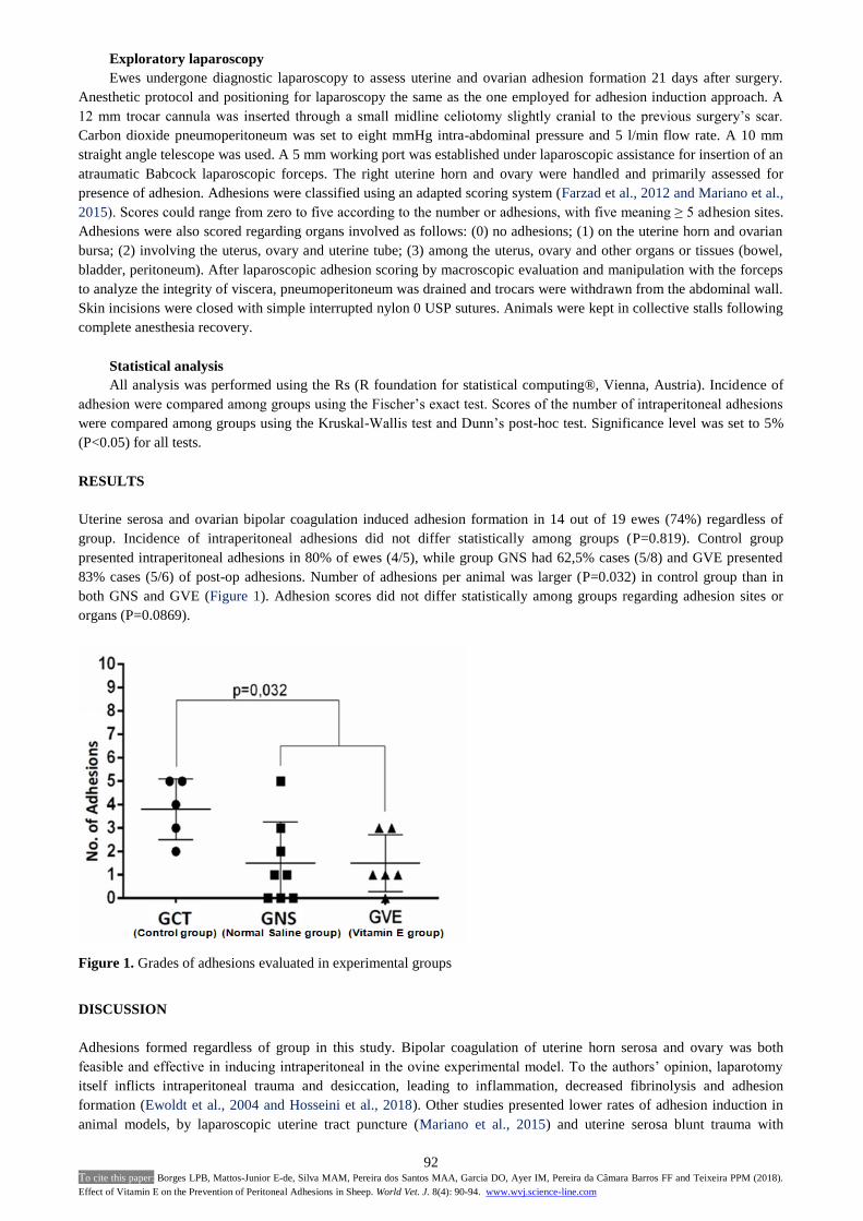
Uterine serosa and ovarian bipolar coagulation induced adhesion formation in 14 out of 19 ewes (74%) regardless of
group. Incidence of intraperitoneal adhesions did not differ statistically among groups (P=0.819). Control group
presented intraperitoneal adhesions in 80% of ewes (4/5), while group GNS had 62,5% cases (5/8) and GVE presented
83% cases (5/6) of post-op adhesions. Number of adhesions per animal was larger (P=0.032) in control group than in
both GNS and GVE (Figure 1). Adhesion scores did not differ statistically among groups regarding adhesion sites or
organs (P=0.0869).
Figure 1. Grades of adhesions evaluated in experimental groups
DISCUSSION
Adhesions formed regardless of group in this study. Bipolar coagulation of uterine horn serosa and ovary was both
feasible and effective in inducing intraperitoneal in the ovine experimental model. To the authors’ opinion, laparotomy
itself inflicts intraperitoneal trauma and desiccation, leading to inflammation, decreased fibrinolysis and adhesion
formation (Ewoldt et al., 2004 and Hosseini et al., 2018). Other studies presented lower rates of adhesion induction in
animal models, by laparoscopic uterine tract puncture (Mariano et al., 2015) and uterine serosa blunt trauma with

93 To cite this paper: Borges LPB, Mattos-Junior E-de, Silva MAM, Pereira dos Santos MAA, Garcia DO, Ayer IM, Pereira da Câmara Barros FF and Teixeira PPM (2018).
Effect of Vitamin E on the Prevention of Peritoneal Adhesions in Sheep. World Vet. J. 8(4): 90-94. www.wvj.science-line.com
traumatic grasping forceps (Ewoldt et al., 2004). Besides decreased adhesion formation, those experimental models did
not provide controlled focal damage to the reproductive tract.
Although adhesions formed in group GVE, intraperitoneal use of vitamin E resulted in fewer adhesion sites in
comparison to the control group, as seen in other studies in rats undergone surgical serosa trauma (Corrales et al., 2008
and Yetkin et al., 2009). Another trial reported a decrease of 80% in adhesion formation with intraperitoneal use of
vitamin E following laparotomy in rats (De La Portilla et al., 2004). Intraperitoneal vitamin E also decreased
substantially adhesion formation following scalpel blade uterine horn scarification in rats (Durmus et al., 2011). Those
authors attributed few adhesion formations due to the reactive oxygen specimen (ROS) scavenger and antioxidant
properties of vitamin E. Normal saline did not avoid adhesion formation, as well as vitamin E, as reported in other trial
(Kappas et al., 1981). That study also reported lower adhesion scores in normal saline group than in control group in a rat
laparotomy model, which is in theory less traumatic adhesion induction model than the method applied in this study.
Unlike Hosseini et al. (2018), who reported the same amount of adhesions after laparotomy in rats using normal saline
and the control group, which no substance was applied. Vitamin E is well known as a natural anti-inflammatory agent,
specifically by neutralizing ROS. However, vitamin E did not avoid adhesion formation in this reproductive tract
electrocoagulation injury trial.
CONCLUSION
Vitamin E does not affect the incidence of intraperitoneal adhesions following uterine and ovary bipolar
electrocoagulation injury in ewes. However, vitamin E reduces the number of adhesion sites and severity, as well as
normal saline does when compared to not using any adhesion prevention. We hypothesize that a dose-dependent study
should indicate if higher doses of vitamin E given intraperitoneal affect adhesiogenesis, as other studies pointed towards
such property. Thus, further research is warranted to fill this gap in the literature. The experimental model of
adhesiogenesis was safe and showed to be a controlled method of intraperitoneal adhesion induction since bipolar
diathermy provided precise localized spot thermal injury to the ovine reproductive tract.
DECLARATIONS
Acknowledgments
The authors are thankful to the University of Franca and Fapesp (Fundação de Amparo a Pesquisa) for the support
and allowing this research work to be performed. We also would like to appreciate all participants who contributed
during the performance of the research, especially Dr. Ricardo Uscategui, who helped in the statistical analisys. Also, all
the gratitude for Dr Lucas Pereira, who had his particular and unforgettable way of helping the involved ones in the
team.
Competing interests
The authors declare that they have no competing interests.
Author’s contribution
All authors have revised the work critically for important intellectual content, given their final approval of the
version to be published and agreed to be accountable for all aspects of the work in ensuring that questions related to the
accuracy or integrity of any part of the work are appropriately investigated and resolved. Furthermore, the authors have
contributed as follows: substantial contributions to the conception, design and planning of the work, the acquisition,
analysis and interpretation of data, and drafting of the work; contributions to the design of the work, and the acquisition
and analysis of data. Therefore, all authors contributed equally to this work.
REFERENCES
Corrales F, Corrales M and Schirmer CC (2008). Preventing Intraperitoneal Adhesions with Vitamin E and Sodium
Hyaluronate/Carboxymethylcellulose. A Comparative Study in Rats. Acta Cirurgica Brasileira, 23: 36-41.
Doi:http://dx.doi.org/10.1590/S0102-86502008000100007.
Decherney AH and Dizerega DS (1997). Clinical problem of intraperitoneal postsurgical adhesion formation following general
surgery and the use of adhesion prevention barriers. Surgical Clinics of North America, 77: 671-688. Doi:10.1016/s0039-
6109(05)70574-0.
De La Portilla F, Ynfante I, Bejarano D, Conde J, Fernández A, Ortega J M and Carranza G (2004). Prevention of Peritoneal
Adhesions by Intraperitoneal Administration of Vitamin E: An Experimental Study in Rats. Diseases of the Colon & Rectum,
47: 2157–2161. Doi: 10.1007 / s10350-004-0741-6.

94 To cite this paper: Borges LPB, Mattos-Junior E-de, Silva MAM, Pereira dos Santos MAA, Garcia DO, Ayer IM, Pereira da Câmara Barros FF and Teixeira PPM (2018).
Effect of Vitamin E on the Prevention of Peritoneal Adhesions in Sheep. World Vet. J. 8(4): 90-94. www.wvj.science-line.com
Durmus AS, Yildiz H, Yaman I and Simsek H (2011). Efficacy of vitamin E and selenium for the prevention of intra-abdominal
adhesions in rats: uterine horn models. Clinics, 66: 1247-1251. Doi:10.1590/S1807-59322011000700021.
Elkelani OA, Molinas CR, Mynbaev O and Koninckx RP (2002). Prevention of Adhesions with Crystalloids during Laparoscopic
Surgery in Mice. The Journal of the American Association of Gynecologic Laparoscopists, 9: 447-452. Doi:10.1016/s1074-
3804(05)60517-8.
Ewoldt JM, Anderson DHJ and Weisbrode SE (2004). Evaluation of A Sheep Laparoscopic Uterine Trauma Model and Repeat
Laparoscopy for Evaluation of Adhesion Formation and Prevention with Sodium Carboxymethylcellulose. Veterinary Surgery,
33: 668-672. Doi:10.1111/j.1532-950X.2004.04090.x.
Farzad P, Seyed H, Sadraie B, Hadi K, Elias S, Gholamreza K and Mohammad H (2012). Macroscopic and Pathological Assessment
of Methylene Blue and Normal Saline on Postoperative Adhesion Formation in A Rat Cecum Model. International Journal of
Surgery, 10: 537-541. Doi:http://dx.doi.org/10.1016/j.ijsu.2012.08.009.
Hosseini A, Akhavan S, Menshaei M and Feizi A (2018). Effects of Streptokinase and Normal Saline on The Incidence of Intra-
adbominal Adhesion 1 Week and 1 Month after Laparotomy in Rats. Advanced Biomedical Research, 7: 1-11. Doi:
10.4103/abr.abr_225_16: 10.4103/abr.abr_225_16.
Ishwar AK and Memon MA (1996). Embryo transfer in sheep and goat: a review. Small Ruminants Research, 19: 35–43.
Doi:https://doi.org/10.1016/0921-4488(95)00735-0.
Jiang Q (2014). Natural forms of vitamin E: metabolism, antioxidant, and anti-inflammatory activities and their role in disease
prevention and therapy. Free Radical Biology and Medicine, 72: 76–90. Doi:10.1016/j.freeradbiomed.2014.03.03.
Kamel RM (2010). Prevention of postoperative peritoneal adhesions. European Journal of Obstetrics and Gynecology and
Reproductive Biology, 150: 111–118. Doi:10.1016/j.ejogrb.2010.02.003.
Kappas M, Fatouros M, Papadimitriou K, Katsouyannopoulost V and Cassioumis D (1981). Effect of Intraperitoneal Saline Irrigation
at Different Temperatures on Adhesion Formation. British Journal of Surgery, 854-856. Doi:http://dx.doi.org/10.1590/S0100-
69912000000200004.
Kokcam I and Nairoglu M (2002). Effects of vitamin E supplementation on blood antioxidant levels in patients with Behcet’s disease.
Clinical Biochemistry, 35: 633-639. Doi:10.1016/s0009-9120(02)00400-9.
Koninckx PR, Gomel V, Ussia A and Adamyan L (2016). Role of the peritoneal cavity in the prevention of postoperative adhesions,
pain, and fatigue. Fertility and Sterility, 106: 998–1010. Doi:10.1016/j.fertnstert.2016.08.012.
Mariano RSG, Uscategui RAR, Nociti RP, Santos VJC, Padilha-Nakaghi LC, Barros FFPC, Silva MAM, Malta CAS, Bonato DV,
Vicente WRR and Teixeira PPM (2015). Intraperitoneal Lidocaine Hydrochloride for prevention of intraperitoneal adhesions
following laparoscopic genitourinary tract surgery in ewes. Veterinarni Medicina, 60: 403-406. Doi: 10.17221/8414-VETMED.
Menchaca A, Barrera N, Dos Santos PCN, Cuadro F and Crispo M (2016). Advances and limitations of in vitro embryo production in
sheep and goats. Animal Reproduction, 13: 273-275. Doi: 10.21451/1984-3143-AR871.
Moris D, Chakedis J, Rahnemai-Azar AA, Wilson A, Hennessy MM, Athanasiou A, Beal EW, Argyrou C, Felekouras E and Pawlik
TM (2017). Postoperative Abdominal Adhesions: Clinical Significance and Advances in Prevention and Management. Journal
of Gastrointestinal Surgery, 21: 1713-1722. Doi: 10.1007/s11605-017-3488-9.
Reijnen MMPJ, Bleichrodt RP and Van Goor H (2003). Pathophysiology of Intra-abdominal adhesion and abscess formation, and the
effect of hyaluronan. British Journal of Surgery, 90: 533–541. Doi:10.1002/bjs.4141.
Teixeira PPM, Padilha LC, Mariano RS, Coutinho LN, Barros FFPC, Silva MAM and Silva ASL (2013). Aspiração folicular por
laparoscopia. In: Biotécnicas Reprodutivas em Ovinos e Caprinos, 1st Edition. Editora MedVet. São Paulo. Brazil. pp. 147-153.
Ward BC and Panitch A (2011). Abdominal adhesions: current and novel therapies. Journal of Surgical Research, 165: 9-11.
Doi:10.1016/j.jss.2009.09.015.
Wilde RL, Alvarez J , Brölmann H, Campo R, Cheong Y, Sardo ADS, Koninckx P, Lundorff P, Pawelczyk L, Roman H, Torres-de-la-
Roche LA and Wallwiener M (2017). Prevention of Adhesions in Gynecological Surgery: The 2016 Experts Recommendations
on Adhesion Prophylaxis. Gynecology and Obstetrics, 7: 1-4. Doi: 10.4172/2161-0932.1000428.
Yetkin G, Uludag M, Citgez B, Karakoc S, Polat N and Kabukcuoglu F (2009). Prevention of peritoneal adhesions by intraperitoneal
administration of vitamin E and human amniotic membrane. International Journal of Surgery, 7: 561-565.
Doi:10.1016/j.ijsu.2009.09.007.
Yildiz H, Durmus AS, Simsek H and Yaman I (2011). The comparison of methylene blue and vitamin E in prevention of abdominal
postoperative adhesion formation in rat uterine horn models. Biochemical and histopathologic evaluation. Acta Cirurgica
Brasileira, 26: 51-57. Doi: 10.1590/S0102-86502011000100010.
Zarkawi M and Soukouti A (2018). Response of Damascus does to FGA and eCG treatments assessed by laparoscopy, progesterone
and cortisol concentrations. Archiva Zootechnica, 21: 61-73.