effect of static magnetic field on amylase and protease
TRANSCRIPT

Republic of Iraq
Ministry of Higher Education and Scientific Research
Al- Nahrain University/ College of Science
Department of Biotechnology
Effect of Static Magnetic Field on
Amylase and Protease Produced by
some Fungal isolates using Solid
State Fermentation
A thesis
Submitted to the Council of College of Science / Al-Nahrain
University as a partial fulfillment of the requirements for the
Degree of Master of Science in Biotechnology
By
Marowa Sabah Hashim
B.Sc. Biotechnology/ College of Science/ Al-Nahrain University, 2013
Supervised by
Ramadan June
1437 2016
Dr. Abdul Wahid Sh. Jabir
Assistant professor

Supervisors Certification
I, certify that this thesis entitled "Effect of Static Magnetic Field on
Amylase and Protease Produced by some Fungal isolates using Solid
State Fermentation" was prepared by "Marowa Sabah Hashim" under
our supervision at the College of Science / Al-Nahrain University as a
partial fulfillment of the requirements for the Degree of Master collage of
Science in Biotechnology.
Signature:
Name: Dr. Abdul Wahid Sh. Jabir
Scientific Degree: Asst. Professor
Address: College of Applied
Biotechnology / Al-Nahrain University
Date: / / 2016
In view of available recommendation, I forward this thesis for debate
by examining committee
Signature:
Name: Dr. Hameed M. Jasim
Scientific Degree: Professor
Address: Head of Biotechnology Department
Date: / / 2016

Committee Certification
We, the examining committee certify that we have read this thesis
entitled "Effect of Static Magnetic Field on Amylase and Protease
Produced by some Fungal isolates using Solid State Fermentation"
and examined the student "Marowa Sabah Hashim" in its contents and
that in our opinion, it is accepted for the degree of Master of Science in
Biotechnology.
Signature:
Name: Dr. Ghazi M. Aziz
Scientific Degree: Professor
Address: College of Science/ Baghdad University
Date: / / 2016
(Chairman)
Signature: Signature:
Name: Dr. Ali S. Ahmed Name: Dr. Firas A. Hassan
Scientific Degree: Asst. Professor Scientific Degree: Asst. Professor
Address: College of Applied Address: College of Science/
Biotechnology/ Al-Nahrain University Al-Nahrain University
Date: / / 2016 Date: / / 2016
(Member) (Member)
Signature:
Name: Dr. Abdul Wahid Sh. Jabir
Scientific Degree: Asst. Professor
Address: College of Applied
Biotechnology / Al-Nahrain University
Date: / / 2016
(Member and Supervisor)
I, hereby certify upon the decision of the examining committee.
Signature:
Name: Dr. Hadi M. A. Abood
Scientific Degree: Professor
Address: College of Science / Al-Nahrain University
Title: Dean of College of Science
Date: / / 2016

حيم حمن الره الره بسم الله
ر لي أمري )62( قال رب اشرح لي صدري )62( ويس
واحلل عقدة من لساني )62( يفقهىا قىلي )62(
صدق الل العظيم
(62-62طه/ (

Acknowledgments
First of all Praise to Allah the lord of the universe who gave me the faith and strength to accomplish this work. My special thanks with respect and appreciation to my supervisor Asst. Prof. Dr. Abdulwahid S. Jabir for his guidance and support throughout my study.
Special Thanks are due to Prof. Dr. Abdul Al-Ghani I. Yahya for his help and guidance during this study.
Grateful thanks are conveyed to Dr. Yaseen Ismaeel for his invaluable assistance, to overcome difficulties in all hard times and encouragement throughout the study. Grateful thanks are conveyed to the staff of Biotechnology Department ⁄ College of Science ⁄ Al-Nahrain University for their assistance and encouragement I would like to extend my thanks to Dr. Khalid Waleed for his efforts in solving the difficulties that faced me during this work, supporting and continuous encouragement. Deep thanks are presented to my family for their patience and support. I would like to thank Dr. Emad Abbas for providing information and assistance. Finally, my sincere and deeply thanks to all my friends for their great friendship and support.
Marowa

Dedications
To the candle that burned to enlighten my way in
life
My Mother
To the people who were always encouraging me
in my life
My Brothers
To the soul that he was always beside me
Majeed Ali
To the woman who were always encouraging me
in my life
Riyam

I
Summary
In this study, the effect of static magnetic field on the production of
amylase and protease enzymes using solid state fermentations from five
different fungal species Alternaria sp., Aspergillus niger, Fusarium sp.,
Mucor sp., and Penicillium sp. were investigated. The substrate used for
fungi growth was bread only. The above species were exposed to the
Northern pole, Southern pole and both poles together (South + North) and
their effects were compared with the control treatment (The control of all
experiments was the solid medium without the effect of the magnetic
field). The results were statistically analyzed by GenStat programm and
the Least Significant Differences (LSD) was determined. The results
showed that the Northern pole significantly decreased the specific activity
of amylase enzyme of Alternaria sp., Fusarium sp., and Penicillium sp.
which were 2.50, 2.12, and 3.27 U/mg respectively. The Southern pole
significantly increased the amylase specific activity of Fusarium sp. (3.84
U/mg) and Mucor sp. (3.36 U/mg), while it significantly decreased
amylase specific activity of Alternaria sp. (2.77 U/mg) and Penicillium
sp. (3.88 U/mg) compared with control. The effect of both poles was as
follow: they were significantly decreased the amylase specific activity of
Alternaria sp. (2.62 U/mg) and Penicillium sp. (2.26 U/mg), on the other
hand, they significantly increased the amylase specific activity of
Fusarium sp. (3.84 U/mg) and Mucor sp. (3.83 U/mg). Regarding the
effect of the magnetic field on the protease specific activity, Northern
pole significantly decreased the protease specific activity of Alternaria
sp., Aspergillus niger, and Penicillium sp., which were 16.86, 14.69,
12.03 U/mg respectively. The Southern pole significantly increased the
protease specific activity of Fusarium sp. (23.04 U/mg) and Mucor sp.
(12.15 U/mg) except Alternaria sp. in which its protease specific activity
II
was decreased significantly (19.30 U/mg). Both poles significantly
increased protease specific activity of Fusarium sp. (21.03 U/mg) and
Mucor sp. (9.75 U/mg), whereas they significantly decreased protease
specific activity of Alternaria sp. (13.65 U/mg) and Penicillium sp., (8.19
U/mg). This study clearly showed that there are significant effect of the
electrostatic magnetic field in increasing and decreasing the enzymes
activities of the fungal species which could be exploited industrially in
increasing the production of important enzymes in industry such as
proteases and amylase tested in this study.

III
LIST OF CONTENTS
Page
No. Subject
I Summary
III List of contents
VI List of tables
VII List of figures
VIII List of Abbreviations
Chapter one
Introduction and Literature Review
1 1 Introduction and Literature review
1 1.1 Introduction
4 1.2 Literature review
4 1.2.1 Magnetism and Electromagnetism
4 1.2.1.1 Magnate and Magnetic Field Energy
6 on Living Organisms The Effects of Magnetic Field1.2.2
8 1.2.2.1 The influence of Magnetic Field on DNA and Cell
Division
11 1.2.3 Fungi
14 1.2.3.1 Amylase Enzyme
15 1.2.3.2 Protease Enzyme
16 1.2.4 Solid-State Fermentation
17 1.2.4.1 Moisture and Water Activity in SSF
19 1.2.4.2 Temperature and Heat Transfer
Chapter Two
Materials and Methods
21 2 Materials and Methods
21 2.1 Instruments and Chemicals

IV
21 2.1.1 Instruments
21 2.1.2 Chemicals
22 2.2 Culture Media
22 2.2.1 Ready to use media
23 2.3 Buffers and Reagents
23 2.3.1 Potassium phosphate dibasic (K2HPO4, 1 M)
23 2.3.2 Potassium phosphate monobasic (KH2PO4, 1 M)
23 2.3.3 Potassium phosphate buffer (0.1 M, pH 7.0)
23 2.3.4 Soluble Starch Solution (1%)
23 2.3.5 Sodium Potassium Tartrate Solution
24 2.3.6 3,5-Dinitrosalicylic Acid Solution (96 mM)
24 2.3.7 Colour Reagent Solution
24 2.3.8 Maltose Standard Solution (0.2%)
24 2.3.9 Potassium Phosphate Buffer (50 mM, pH 7.5)
24 2.3.10 Casein solution (0.65%)
24 2.3.11 Trichloroacetic acid solution (1 N)
25 2.3.12 Sodium Carbonate solution (500 mM)
25 2.3.13 L-tyrosine Standard Stock Solution (1.1 mM)
25 2.3.14 Bovine Serum Albumin Stock Solution (2mg/ml)
25 2.4.1 Coomassie Brilliant Blue G-250
26 2.2 Methods
26 2.2.1 Fungal species
26 2.2.1.1 Slide Culture Preparations
27 2.2.2 Static magnetic field
27 2.2.3 Spores’ suspension preparation
28 2.2.4 Spores Counting
28 2.2.5 Effect of magnetic field poles on enzyme activity using SSF

V
29 2.2.6 Crude enzyme extraction
29 2.2.7 Determination of protein concentration by Bradford
method
30 2.2.8 Protein specific activity estimation
30 2.2.8.1 Preparation of tyrosine standard curve
32 2.2.8.2 Assay of Protease activity
33 2.2.8.3 Protease specific activity (U/mg)
34 2.2.9 Amylase specific activity estimation
34 2.2.9.1 Preparation of maltose standard curve
35 2.2.9.2 Assay of amylase activity
36 2.2.9.3 Amylase specific activity (U/mg)
36 2.10 Statistical Analysis
Chapter Three
Results and Discussion
38 3 Results and Discussion
38 3.1 Fungal species identification
41 3.2 Effect of Magnetic Field on Amylase Specific Activity
41 3.2.1 Effect of Magnetic Field on the Amylase Specific
Activity of Alternaria sp.
42 3.2.2 Effect of Magnetic Field on the Amylase Specific
Activity of Aspergillus niger
43 3.2.3 Effect of Magnetic Field on the Amylase Specific
Activity of Fusarium sp.
44 3.2.4 Effect of Magnetic Field on the Amylase Specific
Activity of Mucor sp.
45 3.2.5 Effect of Magnetic Field on the Amylase Specific
Activity of Penicillium sp.
47 3.3 Effect of Magnetic Field on the Protease Specific Activity
47 3.3.1 Effect of Magnetic Field on the Protease Specific
Activity of Alternaria sp.
48 3.3.2 Effect of Magnetic Field on the Protease Specific
Activity of Aspergillus niger
VI
49 3.3.3 Effect of Magnetic Field on the Protease Specific
Activity of Fusarium sp.
50 3.3.4 Effect of Magnetic Field on the Protease Specific
Activity of Mucor sp.
51 3.3.5 Effect of Magnetic Field on the Protease Specific
Activity of Penicillium sp.
Conclusions and Recommendations
55 Conclusions and Recommendations
55 Conclusions
56 Recommendations
RReeffeerreenncceess
5577 RReeffeerreenncceess
LIST OF TABLES
Page
No. Table
21 Table (2-1): Instruments used in this study
21 Table (2-2): Chemicals used in this study
30 Table (2-3): Preparation of Bovine serum albumin standard
curve.
31 Tables (2-4): Preparation of tyrosine standard curve.
34 Table (2-5): Preparation of maltose standard curve
LIST OF FIGURES
Page
No. Figure
5 Figure (1-1): The magnetic field is represented by the

VII
magnetic field lines.
30 Figure (2-1): Bovine serum albumin standard curve.
32 Figure (2-2): Tyrosine standard curve.
35 Figure (2-3): Maltose standard curve.
38 Figure (3-1): Alternaria sp. Morphology on (PDA) agar at 28
°C for 4 days
39 Figure (3-2): Aspergillus niger morphology on (PDA) agar at
28 °C for 4 days.
39 Figure (3-3): Fusarium sp. Morphology on (PDA) agar at
28 °C for 4 days.
40 Figure (3-4): Mucor sp. Morphology on (PDA) agar at 28 °C
for 4 days.
40 Figure (3-5): Penicillium sp. Morphology on (PDA) agar at
28 °C for 4 days.
41 Figure (3-6): Effect of magnetic field on amylase specific
activity of Alternaria sp.
42 Figure (3-7): Effect of magnetic field on amylase specific
activity of Aspergillus niger.
43 Figure (3-8): Effect of magnetic field on amylase specific
activity of Fusarium sp.
44 Figure (3-9): Effect of magnetic field on amylase specific
activity of Mucor sp.
45 Figure (3-10): Effect of magnetic field on amylase specific
activity of Penicillium sp.
47 Figure (3-11): Effect of magnetic field on protease specific
activity of Alternaria sp.
48 Figure (3-12): Effect of magnetic field on protease specific
activity of Aspergillus niger.
49 Figure (3-13): Effect of magnetic field on protease specific
activity of Fusarium sp.
50 Figure (3-14): Effect of magnetic field on protease specific
activity of Mucor sp.
51 Figure (3-15): Effect of magnetic field on protease specific
activity of Penicillium sp.

VIII
LIST OF ABBERVIATION
Abbreviation Meaning
T Tesla
SMFs Static magnetic fields
EMF Electromagnetic field /fields
SSF Solid state fermentation
Hsp Heat shock protein
DNA Deoxyribonucleic acid
DNS Dinitro-sylcilic acid
G Gauss
LSD Least significant differences
MF Magnetic field
mg/ml Milligram per milliliter
Na/K ATPase Sodium – potassium Adenosine
tri- phosphatase
OMF Oscillating Magnetic Field
ELF Extremely low frequency
UV Ultra-Violette
Bp Base pair
PDA Potato dextrose agar
BSA Bovine serum albumin
Hz Hertz
Aw Water activity
DC Direct current
AC Alternate current
µm Micrometer

IX
BC Before Christ
A.D Anno Domini
RNA Ribonucleic acid
mRNA Messenger RNA
TCA Trichloroacetic acid
µmole micromole
Fe Mn Ferromanganese

Chapter One
Introduction
and
Literature Review

Chapter One Introduction and Literature Review
1
1. Introduction and Literature Review
1.1 Introduction
Mycology is a term derived from a Greek word myke means
„mushroom‟, and logos means „study‟. So, mycology is the study of
mushrooms. However, mycology is commonly used to refer to the study
of organisms called fungi. Fungi are found in two structural forms. One
of them yeast cells which are unicellular. The other is hyphae which
made up of thread like structures. A group of hyphae are known as
mycelium. Outer cell wall of Fungi are typically made up of a porous
made up of chitin. Inner to the cell wall is a cell membrane that is
wrapped up in places to increase its surface area for exchange of
materials. Heterotrophs can either be saprobes, symbionts or parasites.
Reproduction occur both sexual and asexual means. Fungi are important
in the food industry. In many counties, mushrooms which considered a
delicacy at the dining table. Species such as Penicillium sp. are used to
add flavour to cheese. The yeasts are important in the fermentation
processes of wine and beer manufacturing. Fungi are important in the
breakdown of organic matter and organic wastes, it useful in cleaning up
of wastes in an ecosystem and contribute to the recycling of nutrients.
Some of fungi cause various diseases for plants, but only a few species
cause disease in animals and humans, these diseases are permanent
(Alexopoulos et al., 1996).
The history of magnetic materials development cannot be denied and
it is fabulous. Magnetic objects played an important role in the discovery
of the new world and in the development of modern technology. The
magnetic properties of materials are important to understand them which
led to a deep understanding the main structure of materials. One of the

Chapter One Introduction and Literature Review
2
basic properties of materials is magnetization; these materials appear in
different forms, but the studied form known as ferromagnetism. Magnetic
field only refers to the elements that exhibit ferromagnetism properties. A
magnet is an object that has a magnetic field. "Magnítis líthos", is a
Greek word mean magnet which means "magnesian stone". Magnetite
has been discovered in Magnesia which is an area in Greece where
Magnetite deposits (Peter et al., 2002).
For long time period the effect of magnetic field on biological
systems has been an area of interest. Unlike time-varying
(electromagnetic) field, which are not related with induced electric
currents except during activation and deactivation or when there is
motion within the magnetic field. It is effect on the molecular structure of
spasmodic membranes, an influence sufficient to alter the role of
embedded ion-specific channels.
The effect of the magnetic field energy causes significant changes in
the characteristics metabolism of organisms; these changes occur in the
exchange of ions through the cell membrane and in the movement of
cells. Magnetic field (MF) and electromagnetic field (EMF) that are
generated from both external MF and EMF and internal sources natural
metabolisms of organisms effect on biological systems. Depending on
several studies magnetic and electromagnetic fields have various
responses for biosystems such as neuromuscular activity, repair and
tissue growth, glandular secretion, and cell membrane function.
Electromagnetic field has an effect mostly on charged units and related
metabolisms and usually has influence on biochemical reactions that
include more than one unpaired electron. The impact of an external
magnetic field on enzymatic reaction rates can be determined in the same
way classical enzyme kinetic parameters are determined. Enzymes

Chapter One Introduction and Literature Review
3
with chromogenic substrates or products can be followed
spectrophotometrically (Koch et al., 2003).
The purpose of the study was to:
Investigate the effect of static magnetic field on amylase and protease
production using solid state fermentation.

Chapter One Introduction and Literature Review
4
1.2 Literature Review
1.2.1 Magnetism and Electromagnetism
The force between magnets that pull or push other objects is a
physical phenomenon known as magnetism. The materials which exhibit
magnetism, some of them are stronger than others. The only form of
magnetism strong enough to be felt by people is a ferromagnetism which
is permanent magnets, made from iron, which experience the strongest
effects (Jiles, 1991).
The force between electrically charged particles defined at the
subatomic level as an electromagnetism. It is considered one of the
essential interactions of matter. Electromagnetic waves results from
oscillating electrical charges. Attraction and repulsion of electrically
charged particles resulted from electromagnetism which is related to the
electromagnetic force. Which is in nature has been considered one of the
essential forces that also include nuclear and gravitational forces.
Electrically charged particles, such as electrons, create a magnetic field
when they are put into motion and they create electromagnetic radiation
when made to oscillate. Depending on the frequency of the oscillation,
this can include radio waves, visible light, or x-rays. On a larger scale the
creation of a magnetic field from the movement of electrical charges
known as electromagnetism. Electrodynamics is the use of electric effect
that uses varying magnetic field an electromagnet to induce an electric
current (Grant and Phillips, 2008).
1.2.1.1 Magnate and Magnetic Field Energy
Magnetism is the result of electrically charged molecules. Particles in
the field can be affected by a magnetic field force which is called Lorentz
force. This force depends on three factors namely: particles speed,

Chapter One Introduction and Literature Review
5
magnetic field strength, and the charge magnitude. Though connected,
electrical and magnetic fields are different and do different things.
Magnetic fields and their lines were first tested by Michael Faraday then
by James Clerk Maxwell (Kronmüller, 2007).
Magnetic field is the force surrounding the magnet in which magnetic
materials affected by this force. Around a magnet the shape of the
magnetic field can be shown as lines. From these lines arrows will point
out toward the south and point away from the north to show the direction
of the magnetic field. These magnetic force lines do not cross each other.
As shown in the Figure (1-1).
Figure (1-1): The magnetic field is represented by the magnetic field
lines.
Magnetic poles are the points where the lines of magnetic fields begin
and end. Fields lines move away from each other then it will come close
to each other on the poles. These poles are places in which our earth field
lines come close to each other. These poles named north and south
because of their location on our planet. Every magnetic material has poles
and field lines. If a magnet is divided into two pieces, this will make two
magnets with new north and south poles. At the fracture point, new north
and south poles will be formed (Cullity and Graham, 2008).

Chapter One Introduction and Literature Review
6
1.2.2 The Effects of Magnetic Field on Living Organisms
The influences of magnets and magnetic fields have been mentioned
many years ago. Galen, a Greek physician about 200 BC, in book, De
Simplicium Medicamentorum Facultatibus, noted about the use of
magnets. Also, it had been found that the using magnetism to relieve
various disorders in 1000 A.D. In the early 1500s, the great medical
doctor, Paracelsus studied several cases on the use of magnet and
magnetism. Paracelsus introduced many hints for the nature and use of
magnetism, who taught a whole philosophy of universal magnetism
(Presman, 1970; Binhi and Savin, 2003).
Several studies have been carried out to examine of the impact of
magnetic field on the enzymes activity (Blank et al., 1995; Blank and
Soo, 1998) and DNA (Ivancsits et al., 2002; Nikolai et al., 2004).
Enzymes are important in the biological processes and cell
communication, any change in the enzyme activity may effect on these
biological processes.
Some studies mentions that the influence of the magnetic field on
cellulolytic microorganisms regarding metabolism. The influence of the
magnetic field on the microorganisms was recognized since (1937), when
Kimball found that the wine yeasts cells were not affected after exposure
for several period of time to the magnetic field (Kimball, 1937).
Staphylococcus aureus growth rate was increased when exposure to a
magnetic field for (3-6) hr, while there was no effect after 7 hr of
exposure. The growth rate was unchanged when Serratia marcescens was
exposed to the magnetic field for 6 hr, but it increased after exposition to
a long time (Gerencser et al., 1962). The morphology of Pseudomonas
aeruginosa was changed by the influences of different levels of static
magnetic field. The growth of Escherichia coli, was enhanced by static
Chapter One Introduction and Literature Review
7
magnetic field and this enhancement of the growth is proportional to the
increase of the magnetic field frequency (Moore, 1979).
The effects of magnetic field on the growth rate of plants have been
object of many scientists. The first studies were reported by Savostin,
(1930), who found that the increase 100% in seedlings elongation rate by
the effect of magnetic field. The strong magneto tropic affection on root
development was reported by (Audus, 1960; Pittman, 1977).
Magnetic field has distinctive influence on biological systems. There
are many reports about the exposure to magnetic field which effect on
different plant functions such as growth (Racuciu and Creanga, 2006),
development (Yano et al., 2003; Rakosy-Tican et al., 2005), protein
biosynthesis and enzyme activity (Alikamanoglu and Sen, 2011). The
mechanism of interaction of fields with the living cells is still unclear
(Atak and Emiroglu, 2003). Magnetic field causes an oxidative stress,
that may increases the activity and lifetime of the free radicals which are
very reactive byproducts of normal metabolism and immune defense
(Scaiano et al., 1994). It has been known that the reactive oxygen species
which are produced during stress can harm many cellular components
like nucleic acids, carbohydrates, proteins and lipids (Halliwell, 1982).
Therefore, MF could be altering the activity of antioxidant enzyme
(Sahebjamei et al., 2007; Alikamanoglu and Sen, 2011).
Many studies have shown that the influence of static magnetic fields
on biological systems and time-varying magnetic or electromagnetic
fields on transport of membrane cations and other functions. These
studies show that the influences of exposures to different magnetic fields
are not identical. While some reports display an inhibitory effect by the
fields, others show activation, while others showed no significant effects
on the transport of cation. Many studies indicated to the effect of
magnetic field on Ca2+
influx across the cell membrane or intracellular

Chapter One Introduction and Literature Review
8
Ca2+
movements. Significantly exposed to the time-varying magnetic
field suppressed the increase in [Ca2+
] and partially inhibited the K+
influx through Ca2+
-dependent K+ channels, proposing the inhibition of
Ca2+
influx and/or Ca2+
release by the exposure. However, the inhibition
of K+ influx is due to a direct magnetic field exposure on K
+ channels
rather than suppression of the increase in [Ca2+
] (Ikehara et al., 1997).
Liburdy found that the magnetic field affects Ca2+
uptake across the cell
membrane, but the release of Ca2+
from its stores is not affected. This is
because eddy current induced by the magnetic field or electric field could
not enter the outer cell membrane. The cell membrane is bilayers that act
as an electric insulator (Liburdy, 1992). Magnetic fields influence the
charge of the cell membrane, which open up the membrane channels.
These channels are similar to windows and doors of a house. By opening
cell channels, nutrients are capable to enter the cell, and waste is more
easily removed from the cell. This would assist to equilibrate and restore
optimum cell function.
Also, other studies indicate that the static magnetic fields affect the
diffusion of particles in solutions by induction of Maxwell stress or
Lorentz force. Lorentz force would influence the diffusion of charged
particles like different ions that passes in and out the plasma proteins
(Kinouchi, 1988).
1.2.2.1The influence of Magnetic Field on DNA and Cell Division
The interactions between biological activities and electromagnetic
fields have focus on different time period exposure to Alternating Circuit
fields. Effects on in vitro biochemical reactions have been reported .The
studies revealed that the DC (static) magnetic fields can interact with
living activities at different levels. (Adamkiewicz and Pilon, 1983; Kim,

Chapter One Introduction and Literature Review
9
1976; Markov et al., 1992; Richardson et al., 1992; Harkins and Grissom,
1994).
Many researchers have found that the magnetic field affects the
growth of bacteria, which includes an increase in mass and cell division.
Exposure of Escherichia coli to an AC field (0−2 mT, 16 and 50 Hz),
shows shortened in generation time (Aarholt et al., 1981). Powerful
magnetic fields (5.2 − 6.1 T) caused delayed cell death in stationary
cultures of Bacillus subtilis (Nakamura et al., 1997). Whether or not an
AC magnetic field exerts an inhibitory or else a stimulatory mode of
action based on a complex manner on the frequency and the field
strength. Moore found high or even no growth rates for Staphylococci, B.
subtilis, Halobacterium, Salmonella typhimurium and Candida albicans,
after exposure to AC frequencies ranging from (0 - 0.3 Hz) and magnetic
flux densities of (5−90 mT) (Moore ,1979).
It has been found that the fungus Physarum polycephalum responds
to weak AC fields (0.2 mT, 60, 75 Hz) with a delay mitotic cycle (Marron
et al., 1978; Marron et al., 1975), displayed by an enlarged mitotic cycle
span at (0.2 mT and 75 Hz) (Goodman et al., 1976; Goodman et al.,
1979). Also, AC fields (0.1 mT, 60 Hz) caused decrease in ATP levels in
Physarum polycephalum, but no reduction in respiration (Marron et al.,
1986). Reduced respiration found with (0.2 mT 60 and 75 Hz) (Marron et
al., 1975).
Some other studies revealed that weak magnetic fields (0−110 μT)
affect DNA-protein conformations in E. coli. (Binhi et al., 2001). In
Salmonella, AC fields (14.6 mT, 60 Hz) do not cause DNA breaks
(Williams et al., 2006). Also, AC magnetic fields (120 μT, 50 Hz) cause a
reduction in the survival of Saccharomyces cerevisiae after UV

Chapter One Introduction and Literature Review
10
irradiation, while sustaining no effect on cell cycle kinetics (Markkanen
et al., 2001).
The magnetic fields (AC) can enhance specific sets of genes. In E.
coli, σ32 mRNA (transcription factor) was increased when exposed to
(1.1 mT and 60 Hz) (Cairo et al., 1998). Pulsed square fields (1.5 mT)
enhanced the increase in α subunit of RNA polymerase. The translation
machinery itself must be magneto sensitive and was not, for example,
relying on the existence of a biomembrane. An important observation in
this subject was the fact that AC fields can induce translation, even in an
in vitro system (Goodman et al., 1993). The weak, alternating, magnetic
field (8 and 80 μT, 60 Hz) was able to increase the transcription of human
or mouse c-myc genes (Lin et al., 1994). The same response elements
were also detected the heat shock gene hsp70 in the promoter region
(Goodman and Blank, 2002). This influence depends on the presence of
response of specific electromagnetic elements localized between (−353
and −1257 bp) relative to the promoter (Lin et al., 2001).
After exposing E. coli to AC fields, there was no alteration in the
profile of stress proteins occurred (7.8 − 14 mT, 5 − 100 Hz) (Nakasono
and Saiki, 2000). In Shewanella oneidensis strong magnetic fields (14.1
T) caused the transcriptional up-regulation of 21 genes and the down-
regulation of 4 genes, while at the similar time caused no substantial
alterations in growth (Gao et al., 2005). Furthermore, in S. cerevisiae no
changes in protein profile (2-D gel analysis) or differential gene
expression (microarray analysis) were found when exposed to AC
magnetic fields (10 − 300 mT, 50 Hz) (Nakasono et al., 2003).

Chapter One Introduction and Literature Review
11
1.2.3 Fungi
Fungi are an essential kingdom of organisms, comprising about
77,000 named species. Mycologists and scientists believe there may be
more than 1.2 million fungi species existed on earth. Although fungi have
traditionally been classified under the plant kingdom, they do not have
chlorophyll (non-photosynthetic) and resemble plants only in their
general appearance and lack of mobility (Alexopoulos et al., 1996).
Fungi are eukaryotic organisms classified into yeasts (unicellular
microorganisms) or molds (multicellular filamentous fungi). Fungal cell
wall differs from that of plant by containing chitin instead of cellulose.
The energy reserve of fungi is not starch like plants but it is glycogen like
animals. Fungi are aerobic and non-motile. Most are heterotrophic and
consume organic matter. Fungi reproduce sexually by mating of two
opposite types and fusing of their hyphae. Asexual reproduction by
spores or fragmentation (breakage) or binary fission or by budding,
hyphae which can produce new mycelium (Terborgh, 1992).
A key characteristic of fungi is the creation of a mycelium
(filamentous thallus). A mycelium is composed of branching microscopic
tubular cells called hyphae (singular, hypha). These hyphae are typically
composed of long chains of cells divided by cross-partitions termed septa
(singular, septum). The fungi have unique features which indicate that
these microorganisms are not related to other group of organisms. The
septa rarely form a complete barrier except when they isolate the
reproductive cells. Cytoplasm moves freely throughout the hyphae
passing right through major pores in the septa (Harris, 2008).
The mycelium (plural, mycelia) is the vegetative part of a fungus, is
a collection of hyphae that grow through and across substrates or food
sources, enzymes secreting to break down difficult substrates into simple
compounds which can be diffused through the cell wall, resulting in a

Chapter One Introduction and Literature Review
12
unique relationship between the fungi and their environment. Whole
components of a fungus are metabolically active continuously interacting
with their environment such as soil, plants, or any other materials which
enhance the growth of mycelium (Kirk et al., 2008).
Fungi are different from most other organisms in mitosis. The
nuclear envelope does not break down and re-form. Mitosis occurs inside
the nucleus. Centrioles are lacking in all fungi instead, fungi adjust the
construction of microtubules throughout mitosis with relatively small
shapeless configurations called spindle plaques. These unique
characteristics of fungi suggesting that these microorganisms originated
from some unidentified group of unicellular eukaryotes. A spindle
apparatus forms there, pulling chromosomes to the poles on each side of
the nucleus (Moens and Rapport, 1971).
Fungi are achlorophylls meaning that they do not contain
chlorophyll which make them incapable organisms to make their own
food like plants do. Fungi based on other organisms for their carbon
source. Hence, fungi are heterotrophic organisms. These are fungi that
derive their nutrition from other organism protoplasm named the host.
Fungi have a distinct mode of nutrition which involves the liberation of
enzymes to the surrounding environment to break down the substrates
needed for nutrition. Then this substrate is processed outside the cell and
absorption of the products (monomer forms) occur through the permeable
cell wall and the selectively porous membrane for ultimate digestion by
the cells (Raven and Johnson, 1999).
Fungi play important ecological and commercial roles. The
organisms that have the capability to decompose lignin (one of the major
constituents of wood) are fungi. By breaking down such substances fungi
release essential chemical elements like carbon, nitrogen, and phosphorus
from the bodies of dead organisms and make these elements accessible to

Chapter One Introduction and Literature Review
13
other organisms. Some fungi attack living plants and animals through in
breakdown of organic matter some as a source of organic particles while
other fungi invade dead ones (Boerjan et al., 2003).
Two types of mutualistic relations between fungi and autotrophic
organisms are important ecologically. Lichens are two organisms
associated symbiotically which involved a green algae or less frequently
cyanobacteria with a fungus. The fungus gets carbohydrates formed by
photosynthesis from the algae or cyanobacteria and the fungus provide
them with protection from dryness and ultraviolet light. They are
prominent nearly everywhere in different environments around the world
particularly in abnormal severe habitats like plain rock. Mycorrhizae, a
specialized mutualistic symbiotic association between the fungi and the
roots of plants, are typical of about 90% of whole plants kingdom. In
each of them, the photosynthetic organisms captured Co2 from the
atmosphere and thus make organic material available to the fungi. The
metabolic activities of the fungi consecutively improve the overall ability
of the symbiotic association to exist in a particular habitat. The fungal
partner accelerates the absorption of important nutrients like phosphorus
into the plants (Rodriguez et al., 2009).
Classification of fungi depends on reproductive structures. There
are three phyla of fungi. In zygomycetes the hyphae fusion leads to the
formation of a zygote, (Phylum zygomycota). (Phylum Ascomycota), in
ascomycetes, hyphal fusion leads to constant dikaryons that grow into
dense webs of hyphae that form zygotes within a characteristic saclike
structure the ascus, mostly ascomycetes that play many important
commercial and medical roles. In basidiomycetes, dikaryons also form
but zygotes are produced within reproductive structures named basidia,
(Phylum basidiomycota). The imperfect fungi have not been observed to

Chapter One Introduction and Literature Review
14
reproduce sexually. Therefore, it cannot be classified into one of the three
phyla (Hibbett, 2007).
1.2.3.1 Amylase Enzyme
A group of enzymes known as amylase are secreted by several
microorganisms like fungi and bacteria to the outside of the cells to
performed extra-cellular digestion (Ellaiah et al., 2003). Source of Fungi
is restricted to terrestrial isolates; mostly to Aspergillus spp. It has been
found that many fungi will be good sources of amylolytic (amylase)
enzymes. The highest amylase activity was possessed by Aspergillus spp.
due to the ubiquitous nature and non-fastidious nutritional requirements
of Aspergillus niger, studies on fungal amylase particularly in the
developing countries have been done mainly on this organism (Abe et al.,
1988).
In 1894, amylase was the first enzyme produced industrially from a
fungal source, which has been used for the treatment of digestive disorder
(Crueger and Crueger, 1984). Amylase is an enzyme that breaks down
starch into sugar. In starch processing industries, chemical hydrolysis of
starch have been successfully replaced by microbial amylases, they also
have potential application in many industrial processes like food, baking,
textile, paper, and detergent industries. Besides their use of starch
saccharification application of amylase has been extended into many
other disciplines like medical, clinical, and analytical chemistry (Pandey
et al., 2001).
The solid state fermentation (SSF) processes have been progressively
applied for the production of amylases which have been produced by
submerged fermentation. By comparison between solid state fermentation
and submerged fermentation (SSF), the latter is more simple, has superior
productivity, require lower capital, reduce energy requirement, simple

Chapter One Introduction and Literature Review
15
fermentation media and lack of severe control of fermentation conditions,
uses less water and produces lower waste water, has easier regulation of
bacterial contamination and a low cost is needed for downstream
processing (Facciotti et al., 1989).
1.2.3.2 Protease Enzyme
Proteases “previously consider as enzymes of breakdown” are one of
the first and major families of enzymes known and are participate in each
part of organism‟s function. They contain a very large and complex group
of hydrolytic enzymes that breaks down the peptide bonds of proteins to
produce amino acids and other smaller peptides (Mitchell et al., 2007). It
was established in all living systems: animals, plants, and
microorganisms including viruses. Proteases have a wide range of
applications was mostly used in foodstuff and cleaner productions
(Yandri et al., 2008). Also, these enzymes vary in their properties like
active site, substrate specificity and catalytic mechanism; possess
different mechanical stress responses for chemical environment,
temperature and pH for stability and activity. Fungal proteases are
dynamic over an extensive pH range (pH 4-11) and display broad
substrate specificity (Rao et al., 1998).
Proteases work optimally in acidic environments excluding alkaline
proteases which has its optimal activity shown in alkaline (basic) pH
(Mitchell et al., 2007). They are one of the enzymes classes that have
great importance in industry and constitute about 60% of the total world-
wide enzyme sales (Barrette and Rawlings, 2003). They are application in
several of biotechnological processes such as in pharmaceuticals, food
processing, leather industry, and detergent formulations, (Nascimento and
Martins, 2004; Beg and Gupta, 2003).

Chapter One Introduction and Literature Review
16
Proteases are one of the main groups of enzymes which secreted by
fungi. The submerged as well as solid state fermentation are useful
techniques that used for protease production, when cultured in solid-state
fermentation usually show better results as compared to bacteria (Pandey
et al., 1999). In addition, the conditions in solid state fermentation (SSF)
system especially the low moisture content in the system lead to some
potential benefits. (i) low moisture content lower the risk of bacterial
contamination during the fermentation (ii) SSF conditions favor growth
of filamentous fungi since wood, leaves, roots and other organic matter
are their natural habitats (iii) The environmental conditions in solid-state
fermentation can enhance the microbe to produce enzymes whose
features could be dissimilar to those enzymes produced by the identical
organism in the submerged fermentation environments.
Molds of the genera Aspergillus, Penicillium and Rhizopus are
especially important for producing proteases. Several species of these
genera are generally considered safe (Devi et al., 2008). Among fungi,
the ability of many species of Aspergillus to produce proteases is well
known. Aspergillus oryzae produces neutral, acid, and alkaline proteases
(Godfrey and West, 1996). Aspergillus clavatus ES1 has been recently
discovered as a producer of an extracellular decolorizing constant
alkaline protease (Hajji et al., 2008). Protease is an important group of
enzymes which occupy an essential place with respect to their application
in both biological and industrial fields (Pastor et al., 2001; Ward, 1985).
1.2.4 Solid-State Fermentation
The fermentation technique in which microorganisms can grow on
solid substances without the presence of free liquid is called solid-state
fermentation (SSF) (Cannel and Moo-Youn, 1980).The idea of using
solid substrates is probably the oldest method used by man to make

Chapter One Introduction and Literature Review
17
microorganisms work for him. In solid-state when fungi are used, unlike
other microorganisms, fungi naturally grow in environment on solid
substrates like a pieces of wood, seeds, stems, roots and dried parts of
animals like bones, skin, and fecal matter that is low in moisture
(Hesseltine, 1977).
In SSF, the moisture important for microbial growth appears in an
absorbed state or in complex with solid matrix. However, SSF differs
from solid substrate fermentation. In solid substrate fermentation the
substrate itself acts as a carbon source and occurs in absence or near
absence of free water. However, the process occurs in absence or near
absence of free water by employing an inactive substrate or a natural
substrate as solid support is called solid-state fermentation (Kumar et al.,
2003). The concept of SSF is to bring cultivated fungi or bacteria in tight
contact with the insoluble substrate and to achieve the maximum nutrient
concentration from the substrate needed for fermentation. This method so
far is to run only on a small scale, but has a benefit over submerged
fermentation. Solid-state fermentation processes can also be classified
according to whether the seed culture for fermentation is pure or mixed.
In pure culture SSF, individual strains are used for substrate consumption
and with varied culture, different microorganisms are utilized for the
bioconversion of agro-industrial remains at once (Oojikaas et al., 2000).
1.2.4.1 Moisture and Water Activity in SSF
The idea of water availability in substrate becomes important. Water
activity (Aw) activity is defined as the ratio of the vapor pressure of water
in substances to the vapor pressure of pure water at the same temperature
(Oriol et al., 1988). During solid-state, the low level of moisture inside
the substrate reduces the growth and metabolism of microorganisms
when compared to submerged fermentation. Water activity is a very

Chapter One Introduction and Literature Review
18
important parameter for testing water potential, characterizing the
energetic state of water (Scott, 1953).
Water activity of substrates has a very important influence on
microbial activity and it is determines type of organisms which can grow
in solid state fermentation. Aw of the medium has been attributed as a
basic parameter for mass transfer of water throughout the microscopic
cells. The control of parameter of Aw could be used to modify microbial
metabolic production and its excretion (Pandey et al., 1999).
The study of Grevais and Molin, (2003) showed that the effect of
water in solid culture medium on fungal physiology, like radial growth
rate and cellular mechanisms. They found that the radial extension rate of
mycelia associated to the water activity value. The optimum water
activity was found to be (0.99) for Trichoderma viride and below (0.90),
no fungal development occurred. For Penicillum roquefortii the optimal
water activity was (0.97).
Water activity has also an effect on the fungal spore production.
Maximum sporulation value of Penicillum roquefortii was obtained at
water activity of (0.96). However, at lower water activity maximum
sporulation occurs. The impact of low water activity value on fungal
fermentation is not well understood. There is loss of essential nutrients
for fungal growth (Charlang and Horowitz, 1971; Charlang and
Horowitz, 1974).
Production of secondary metabolites also based on water activity. It was
found that there is a direct relationship between the quantities of enzyme
produced versus Aw. In a solid culture medium of Trichoderma viride,
biosynthesis of polygalactouronase, beta-galactosidase and D-xylanase
enzyms was influenced by water activity of the substrate. It was found
that maximum Aw of polygalactouronase and D-xylanase production is

Chapter One Introduction and Literature Review
19
(0.99). However, beta-galactosidase production was optimum between
Aw of (0.96) and (0.98) (Grajek and Grevais, 1987).
Moreover, higher Aw also promoted substrate conversion to fungal
biomass and reduced water activity causes lower mass transfer and little
water availability for microorganisms (Oriol et al., 1988). Grajek and
Grevais, (1987) observed that the reduction in biomass production and
protein content of culture medium due to the decrease of water activity by
(0.01) (equivalent to 1 % of relative air humidity). Narahara, (1977)
noticed that optimum Aw for Aspergillus spp. was between (0.97) and
(0.99). Fungus was unable to grow below Aw of (0.97). These data prove
that fungal growth and their secondary metabolite production during SSF
are strongly influenced by Aw of substrate (Grajek and Grevais, 1987;
Narahara, 1977; Grevais and Molin, 2003).
1.2.4.2 Temperature and Heat Transfer
In SSF the fungal growth and secondary metabolite production are
greatly affected by temperature and heat transfer processes in the
substrate bed. A large amount of heat generated through the SSF which
is proportional to the activities of metabolisms of the microorganism.
Nevertheless, fungi can grow on a broad range of temperatures (20°C to
55°C). Nevertheless, ideal temperature for fungal growth might be
different from that required for product formation (Yadav, 1988).
The substrates used for SSF have low thermal conductivities that
decrease heat removal and increase its accumulation. Therefore, the
important issues in SSF are heat removal, and hence most studies are
focused on maximizing heat removal. The heat transfer out or in of an
SSF technique is related closely with aeration of the fermentation system
and microbial metabolic activity. The fungal germination, metabolites

Chapter One Introduction and Literature Review
20
production and sporulation are effected by high temperature
(Raghavarao et al., 2003).
Coupled control of temperature and moisture is also an important
issue for consideration. In SSF heat transfer and temperature control
would be difficult because of the low moisture and poor thermal
conductivity of the substrate. Solving heat transfer problems; is possible
only in small-scale solid-state fermentation by minimizing the substrate
bed height or by good mixing of substrate with sparged oxygen.
Moreover mixing also ensures an effective heat and mass transfer not
only aids in the homogeneity of the bed. In large-scale SSF continuous
mixing along with addition of water is beneficial for control of
temperature and moisture (Raghavarao et al., 2003).

Chapter Two
Materials
and
Methods
Chapter Two Materials and Method
21
2. Materials and Methods
2.1 Instruments and Chemicals
2.1.1 Instruments
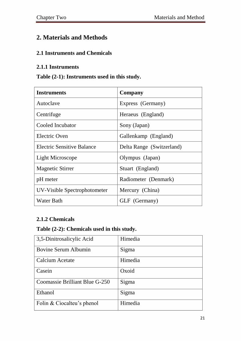
Table (2-1): Instruments used in this study.
Instruments Company
Autoclave Express (Germany)
Centrifuge Heraeus (England)
Cooled Incubator Sony (Japan)
Electric Oven Gallenkamp (England)
Electric Sensitive Balance Delta Range (Switzerland)
Light Microscope Olympus (Japan)
Magnetic Stirrer Stuart (England)
pH meter Radiometer (Denmark)
UV-Visible Spectrophotometer Mercury (China)
Water Bath GLF (Germany)
2.1.2 Chemicals
Table (2-2): Chemicals used in this study.
3,5-Dinitrosalicylic Acid Himedia
Bovine Serum Albumin Sigma
Calcium Acetate Himedia
Casein Oxoid
Coomassie Brilliant Blue G-250 Sigma
Ethanol Sigma
Folin & Ciocalteu’s phenol Himedia

Chapter Two Materials and Method
22
Hydrochloric Acid Sigma
K2HPO4 BDH
KH2PO4 BDH
Lacto phenol cotton blue Himedia
L-Tyrosine Himedia
Maltose Fluka
Methanol Sigma
Phosphoric Acid Sigma
Sodium Acetate, Trihydrate Fluka
Sodium Carbonate Himedia
Sodium Chloride BDH
Sodium Hydroxide BDH
Sodium Phosphate Himedia
Sodium Potassium Tartrate Sigma
Starch Oxoid
Trichloroacetic acid Sigma
2.2 Culture Media
2.2.1 Ready to use media
Medium used in this study Potato Dextrose Agar – PDA was
prepared according to the instruction on the containers of their
manufacturing companies.
Chapter Two Materials and Method
23
2.3 Buffers and Reagents
2.3.1 Potassium phosphate dibasic (K2HPO4, 1 M)
It was prepared by dissolving 174.18 g of K2HPO4 in 800 ml distilled
water and completed to 1 L with DW. This buffer was stored at 4 °C.
2.3.2 Potassium phosphate monobasic (KH2PO4, 1 M)
It was prepared by dissolving 136.09 g of KH2PO4 in 800 ml DW and
completed to 1 L with DW. This buffer was stored at 4 °C.
2.3.3 Potassium phosphate buffer (0.1 M, pH 7.0) (Green and
Sambrook, 2012)
It was prepared by mixing 61.5 ml of K2HPO4 and 38.5 ml of
KH2PO4.This buffer was stored at 4 °C.
2.3.4 Soluble Starch Solution (1%)
It was prepared by dissolving 1 g of soluble starch in 100 ml of DW.
Solubilization of starch was facilitated by heating the solution in a glass
beaker directly on a heating-stirrer plate using constant stirring. The
solution was brought to boil for 15 minutes. The solution was permit to
cool to room temperature with stirring and the volume was completed to
100 ml using DW.
2.3.5 Sodium Potassium Tartrate Solution
It was prepared by weighing 12 g of Sodium Potassium Tartrate,
Tetrahydrate and added in 8 ml of 2 M NaOH which was previously
heated to (50 - 70) °C. The solution was dissolved by heating in a water
bath with constant stirring.

Chapter Two Materials and Method
24
2.3.6 3,5-Dinitrosalicylic Acid Solution (96 mM)
It was prepared by adding 0.438 g of 3,5-Dinitrosalicylic Acid in 20
ml of DW which was previously heated to (50 - 70) °C. The solution was
dissolved by heating in a water bath with constant stirring.
2.3.7 Colour Reagent Solution
It was prepared by slowly adding with stirring Sodium Potassium
Tartrate solution followed by 3,5-Dinitrosalicylic Acid solution to 12 ml
of DW which was previously heated to (50 - 70) °C, and mixed. The
solution should be stored in an amber container at room temperature.
2.3.8 Maltose Standard Solution (0.2%)
It was prepared by weighing 0.2 g of Maltose and dissolved in 100
ml of DW.
2.3.9 Potassium Phosphate Buffer (50 mM, pH 7.5)
It was prepared by dissolving 1.14 g of potassium phosphate dibasic,
trihydrate in 100 ml of DW. pH was adjusted to 7.5 with 1M HCl.
2.3.10 Casein solution (0.65%)
It was prepared by mixing 0.65 g in 100 ml of the 50 mM potassium
phosphate buffer. The temperature of solution is gradually increased with
gentle stirring to 80-85 °C for about 10 minutes.
2.3.11 Trichloroacetic acid solution (1 N)
It was prepared by dissolving 16.33 g of Trichloroacetic acid in 80 ml
of DW. The volume was completed to 100 ml with DW. A 100 ml of
110mM of this solution was prepared by mixing 11 ml of 1 N solution
with 89 ml DW.

Chapter Two Materials and Method
25
2.3.12 Sodium Carbonate solution (500 mM)
It was prepared by dissolving 5.3 g of anhydrous sodium carbonate in
90 ml. The volume was completed to 100 ml with DW.
2.3.13 L-tyrosine Standard Stock Solution (1.1 mM)
It was prepared by mixing 0.02 g of L-tyrosine in 100 DW and heated
gently until the tyrosine dissolves. The L-tyrosine solution was allowed to
cool to room temperature.
2.3.14 Bovine Serum Albumin Stock Solution (2mg/ml)
Bovine Serum Albumin (BSA) stock solution was prepared by
dissolving 0.02 g of BSA in 10 ml of 0.1 M phosphate buffer.
2.4.1 Coomassie Brilliant Blue G-250 (Bradford, 1976)
Prepared by dissolving 100 mg of Commassie in 50 ml of 95%
ethanol, then 100 ml of 85% phosphoric acid was added. The volume was
then completed to 1000 ml with DW. The reagent was filtered through
Whatman No.1 filter paper just before use.
Chapter Two Materials and Method
26
2.2 Methods
2.2.1 Fungal species
The fungi species, Penicillium sp., Alternaria sp., Fusarium sp.,
Mucor sp., and Aspergillus niger were obtained from the Department of
Biology/ College of Science/ University of Baghdad. The fungi species
were identified after growing on Potato Dextrose Agar (PDA) medium by
observing the growth characteristics (color, texture appearance and
diameter of colonies) and microscopic (microstructure) (Baijal and
Mehrotra, 1980; Bisset, 1991). All the cultures were maintained on PDA
slants. Then, stored in refrigerator and sub-cultured regularly at an
interval of three months.
2.2.1.1 Slide Culture Preparations ( Riddle, 1950)
A simple modification of this method using a single agar plate is
described below:
A sterile blade was used to cut out an agar block, small
enough to fit under a cover slip.
The block was flip onto the surface of the agar plate.
Four sides of the agar block inoculated with spores or
mycelial fragments of the fungus to be grown.
A flamed coverslip was placed centrally upon the agar block.
The plate was incubated at 28 °C until growth and
sporulation have occurred.
The cover slip removed from the agar block.
A drop of 95% alcohol was applied as a wetting agent.
The cover slip gently lowering on to a small drop of
Lactophenol cotton blue on a clean glass slide.

Chapter Two Materials and Method
27
The slide is then left overnight to dry, later sealed with
fingernail polish.
When sealing with nail polish was used a coat of clear polish
followed by one coat of red coloured polish.
2.2.2 Static magnetic field
A special magnetic bar of thickness (2.9 cm) with single field strength
of 100 Gauss which was measured by a Gaussmeter. The magnetic bars
(North Pole, South Pole, or both poles) were put on the side of the
cultured flasks using adhesive tape.
2.2.3 Spores’ suspension preparation
Spores’ suspension was prepared according to (Faraj, 1990) with slight
modifications as follows:
Plates containing PDA medium inoculated with fungal
isolates were incubated at 28 °C for 3-4 days.
Spores were harvested by adding 5 ml of sterile DW on the
plate.
The spores’ suspension was transferred by a micropipette to
a flask containing sterilized bread and incubated at 28 °C for 5
days.
One hundred ml of DW was added to the flask and mixed
vigorously by hand.
The suspension of spores was filtered through sterile cotton
wool.
The suspension was centrifuged at (3000 rpm for 5 min).
Then, the supernatant was discarded and the spores then washed
twice by DW and further centrifugation.

Chapter Two Materials and Method
28
Then, 1 ml of DW was added to the deposit and mixed
vigorously.
2.2.4 Spores Counting
Spores were counted according to (Sambrook and Russell, 2001) as
follows:
The hemocytometer was set up by placing it flat on the
surface with the glass coverslip on top of the grids.
The spores’ suspension was vortexed for 5 s.
By using sterile tips, ten microliter of the suspension was
transferred to the divot on the hemocytometer.
The total number of spores was counted in each of the five
small squares.
The following equation was used to estimate the number of
spores /ml in original suspension:
Spores /ml = (Average no. of spores / 5) × (25) × (104) × (Dilution
Factor).
2.2.5 Effect of magnetic field poles on enzyme activity using solid
state fermentation
To test the effect of the magnetic field on the fungi cultures under
solid state fermentation the following steps were carried out:
Bread loafs were cut into pieces (approximately 1 cm3) using
bread loaf knife.
Twelve flasks 500 ml were loaded with 30 g of bread pieces
and autoclaved at 121 °C for 15 min.

Chapter Two Materials and Method
29
Each flask was inoculated with 108 spores/ml of fungus.
The moisture content was constant.
The twelve flasks were divided into four groups as follow:
Three flasks used as control, three flasks were put under the effect
of northern pole, three flasks were put under the effect of southern
pole and three flasks were put under the effect of both poles.
The flasks were incubated at 28 °C for 7 days with shaking
every day.
2.2.6 Crude enzyme extraction
After 7 days of fungal fermentation, extraction of crude enzyme was
done as follow:
To each culture flask, 100 ml of phosphate buffer (pH 7) was
added and shake vigorously for 15 min to suspend the bread pieces.
The whole content of the flask was filtered through sterile
cotton wool and centrifuged at (3000 rpm for 5 min).
The supernatant was assembled and filtered by filter paper
Whatman No.1 under vacuum.
The filtrate was stored at 4 °C.
2.2.7 Determination of protein concentration by Bradford method
(1976)
The concentration of protein was determined by using the Bradford
method, as follows:
The Bradford reagent was gently mixed and brought to room
temperature.

Chapter Two Materials and Method
30
A protein standard was prepared ranging from (0.2 to 1
mg/ml). Dilutions are given in Table (2-3) below. Each
concentration was done in triplicate.
The unknown samples were prepared in a similar way to
make the final volume (0.1 ml).
To all the tubes, (3.0 ml) of Bradford reagent were added
with continuous mixing, then incubated at room temperature for 5
min.
The absorbance was measured at 595nm using
spectrophotometer.
Table (2-3): Preparation of Bovine serum albumin standard curve.
BSA standard (µl)
Dilution buffer (µl)
Final concentration (mg/ml)
Absorbance (595 nm)
10 90 0.2 0.238 20 80 0.4 0.410 30 70 0.6 0.568
40 60 0.8 0.750 50 50 1 0.921

Chapter Two Materials and Method
31
Figure (2-1): Bovine serum albumin standard curve.
2.2.8 Protease specific activity estimation
2.2.8.1 Preparation of tyrosine standard curve
Tyrosine standard curve was prepared as follow:
From the tyrosine stock solution, five concentrations,
ranging from (0.055 to 0.553 µMole) were prepared. Each
concentration was done in triplicate. A test tube containing a blank
solution was also prepared.
The volume was brought up to (2 ml) in each test tube,
including the test tube by DW containing the blank solution.
In each tube, (5 ml) of sodium carbonate were added to
regulate any pH drop due to the addition of the Folin’s reagent.
One ml of Folin’s reagent was added immediately. The tubes
were mixed by swirling and incubated at 37 °C for 30 min, blue
colour is development.
The absorbance of our samples was measured by a
spectrophotometer at 660 nm.
Y = 0.9432X
R² = 0.9862
0
0.2
0.4
0.6
0.8
1
1.2
0 0.2 0.4 0.6 0.8 1 1.2
Ab
sorb
an
ce (
59
5 n
m)
BSA Concentration (mg/ml)

Chapter Two Materials and Method
32
A graph with the concentration of tyrosine on X axis against
absorbance at 660 nm on Y axis was plotted.
Tables (2-4): preparation of tyrosine standard curve.
Figure (2-2): Tyrosine standard curve for protease activity
determination.
2.2.8.2 Assay of Protease activity
The activity of protease was assayed using casein as a substrate
according (Anson, 1938):
Thirteen sterile test tubes were prepared. One tube was used
as a blank and the others were used to assay the activity of
protease.
y = 0.3736x
R² = 0.9952
0
0.05
0.1
0.15
0.2
0.25
0.3
0 0.1 0.2 0.3 0.4 0.5 0.6 0.7 0.8Ab
sorb
an
ce (
660 n
m)
Tyrosine Concentration (μmol/ml)
Tyrosine standard
(ml) DW (ml)
Tyrosine
Final concentration
(μmol/ml)
Absorbance
(660 nm)
0.05 1.95 0.0275 0.050
0.1 1.9 0.055 0.124
0.2 1.8 0.110 0.277
0.4 1.6 0.220 0.603
0.5 1.5 0.275 0.736

Chapter Two Materials and Method
33
In each tube, (5 ml) of 0.65% casein solution was added and
these tubes were placed in a water bath at 37°C for about 5 min.
In each tube, (1 ml) of the enzyme extract was added, except
the blank in which 1 ml of DW was added, mixed by swirling and
incubated at 37 °C for exactly 10 min.
Five milliliters of TCA reagent were added in each tube to
stop the reaction and incubated at 37 °C for 30 min.
In each tube, (5 ml) of sodium carbonate were added to
regulate any pH drop created by the addition of the Folin’s reagent.
One ml of Folin’s reagent was added immediately. The tubes
were mixed by swirling and incubated at 37 °C for 30 min, Folin’s
reagent react with protein to produce a blue color.
The absorbance was measured by a spectrophotometer at
660 nm.
2.2.8.3 Protease specific activity (U/mg)
Absorbance values obtained from protease activity were put
in the standard curve equation (Figure 2-1) to get the amount of
tyrosine in micromoles.
From the values generated above, total activity of protease
samples can be calculated in terms of Units, which is the amount of
tyrosine released from casein in micromoles per minute, according
to the following equation:
Protease activity (Units/ml) =
Where:

Chapter Two Materials and Method
34
11 = Assay total volume (ml)
1 = Volume of enzyme used (ml)
10 = Assay time (min) as the Unit definition
Finally the enzyme specific activity was estimated by
dividing the enzyme activity on the protein activity obtained from
bovine serum albumin standard curve (Figure 2-1) according to the
following equation:
Protease specific activity (U/mg) =
2.2.9 Amylase specific activity estimation
2.2.9.1 Preparation of maltose standard curve
Maltose standard curve was prepared as follow:
From the maltose stock solution, seven concentrations,
ranging from (0.2 to 1 mg/ml) were prepared. Each concentration
was prepared in triplicate. A test tube containing a blank solution
was also prepared.
Using DW, the volume was brought up to (2 ml) in each test
tube, including the test tube containing the blank solution.
To each tube, (1 ml) of DNS reagent was added.
The contents of the tubes were heated in a boiling water bath
for 5 min.
The test tubes were cooled to room temperature, after taking
them out of the water bath.

Chapter Two Materials and Method
35
To each test tube, (9 ml) of DW were added and mixed well.
From each test tube, the mixture was transferred into
cuvettes and the absorbance was measured at 540 nm.
A graph with the concentration of maltose on X axis against
absorbance at 540nm on Y axis was plotted.
Tables (2-5): Preparation of maltose standard curve.
Maltose standard (ml)
DW (ml) Maltose Final concentration
(mg/ml)
Absorbance (540 nm)
0.2 1.8 0.2 0.065 0.4 1.6 0.4 0.106 0.6 1.4 0.6 0.145 0.8 1.2 0.8 0.190 1 1 1 0.221
Figure (2-3): Maltose standard curve for amylase activity
determination.
2.2.9.2 Assay of amylase activity
Amylase activity was assayed as described by (Bernfeld, 1955):
Y = 0.2347X
R² = 0.9574
0
0.05
0.1
0.15
0.2
0.25
0.3
0 0.2 0.4 0.6 0.8 1 1.2
Ab
sorb
an
ce (
540 n
m)
Maltose Concentration (mg/ml)

Chapter Two Materials and Method
36
Thirteen test tubes were prepared. One tube was used as a
blank and the others were used to assay amylase activity.
To each tube, (1 ml) of starch solution was added.
To each tube, (1 ml) of enzyme extract was added, except
the blank to which (1 ml) of DW was added, the tubes were mixed
by swirling and incubated at 37 °C for exactly 3 min.
To each tube, (1 ml) of colour reagent (DNS) was added and
boiled in water bath for 15 min.
The tubes were cooled on ice for 3 min.
To each tube, (9 ml) of DW were added with mixing.
The absorbance for all the test tubes was measured at 540
nm using spectrophotometer.
2.2.9.3 Amylase specific activity (U/mg)
One unit of amylase enzyme can be defined as the amount needed for
liberation of (1.0 mg) of maltose from starch in 3 min at pH 6.9 at 20°C.
The absorbance of all sample tests obtained above were put
in the slope of the standard curve (Figure 2-3) to give the liberated
maltose from starch in mg.
Total activity of amylase in unit/ml can be obtained using
the following equation:
Amylase activity (U/ml) =
Where:

Chapter Two Materials and Method
37
df = Dilution Factor
1 = Enzyme volume (ml)
Ultimately, the enzyme specific activity was estimated by
dividing the enzyme activity on the protein activity obtained from
bovine serum albumin standard curve (Figure 2-1) according to the
following equation:
Amylase specific activity (U/mg) =
2.10 Statistical Analysis
All the experiments were performed in triplicates. The statistical analysis
was done using GenStat program. Least significant difference (LSD) was
determined for each fungus to both enzymes (Amylase and Protease). The
mean of specific activity for each treatment was calculated and subtracted
from the control of this treatment manually. If the result of this
subtraction is more than LSD then the difference is significant.
Alternatively the difference will be not significant if the subtraction
results are less than LSD.

Chapter Three
Results
and
Discussion
Chapter Three Results and Discussion
38
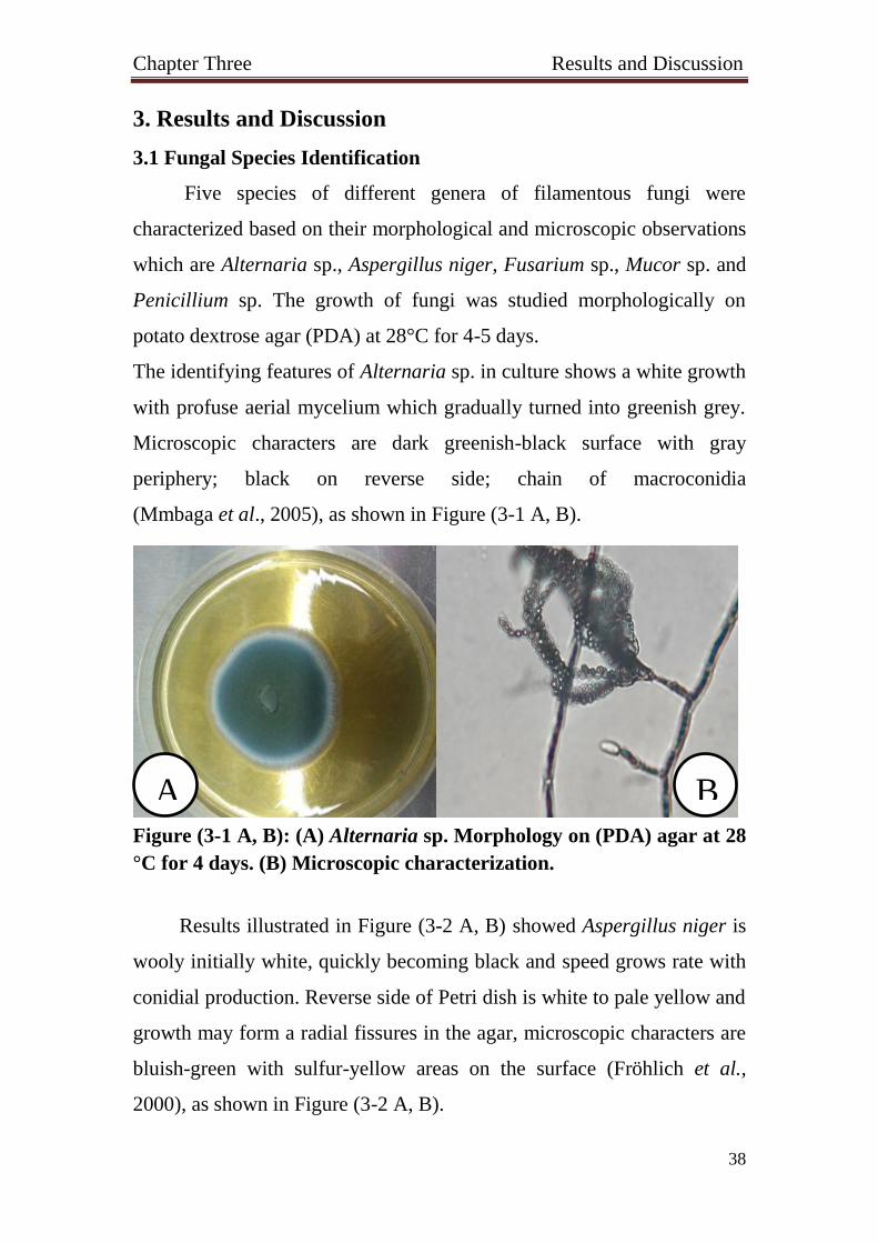
3. Results and Discussion
3.1 Fungal Species Identification
Five species of different genera of filamentous fungi were
characterized based on their morphological and microscopic observations
which are Alternaria sp., Aspergillus niger, Fusarium sp., Mucor sp. and
Penicillium sp. The growth of fungi was studied morphologically on
potato dextrose agar (PDA) at 28°C for 4-5 days.
The identifying features of Alternaria sp. in culture shows a white growth
with profuse aerial mycelium which gradually turned into greenish grey.
Microscopic characters are dark greenish-black surface with gray
periphery; black on reverse side; chain of macroconidia
(Mmbaga et al., 2005), as shown in Figure (3-1 A, B).
Figure (3-1 A, B): (A) Alternaria sp. Morphology on (PDA) agar at 28
°C for 4 days. (B) Microscopic characterization.
Results illustrated in Figure (3-2 A, B) showed Aspergillus niger is
wooly initially white, quickly becoming black and speed grows rate with
conidial production. Reverse side of Petri dish is white to pale yellow and
growth may form a radial fissures in the agar, microscopic characters are
bluish-green with sulfur-yellow areas on the surface (Fröhlich et al.,
2000), as shown in Figure (3-2 A, B).
B A

Chapter Three Results and Discussion
39
Figure (3-2 A, B): (A) Aspergillus niger morphology on (PDA) agar at
28 °C for 4 days. (B) Microscopic characterization.
Fusarium sp. colonies are fast growing, pale or brightly colored and
may have a cottony aerial mycelium. The color of the thallus varies from
white to yellow, brown, pink, red or lilac shades, different microscopic
characteristics can be observed such as yellow, purple, orange and red
colonies; sickle shaped macroconidia, as shown in Figure (3-3 A, B).
Mucor sp. colonies are fast-growing, white to beige or grey and colonies
on culture medium may grow to several centimeters in height,
microscopic characters are sporangia with slimy texture spores with dark
pigment (Domsch et al., 1980), as shown in Figure (3-4 A, B).
Figure (3-3 A, B): (A) Fusarium sp. Morphology on (PDA) agar at
28 °C for 4 days. (B) Microscopic characterization.
B A
B A

Chapter Three Results and Discussion
40
Figure (3-4 A, B): (A) Mucor sp. Morphology on (PDA) agar at 28 °C
for 4 days. (B) Microscopic characterization.
Suryanarayanan et al., (2000), reported that Penicillium sp. colonies
are fast growing in shades of green or white, mostly composed of a dense
felt of conidiophores, microscopic characters are bluish-green brush
arrangement of phialospores, as shown in Figure (3-5 A, B).
Figure (3-5 A, B): (A) Penicillium sp. Morphology on (PDA) agar at
28 °C for 4 days. (B) Microscopic characterization.
The slide cultures technique permits fungi to be studied virtually in
situ with as less disturbance as possible. It is a fast method to prepare
fungal colonies for examination, identification and preserves the
morphological features (Riddle, 1950).
A B
A B

Chapter Three Results and Discussion
41
3.2 Effect of Magnetic Field on Amylase Specific Activity
3.2.1 Effect of Magnetic Field on the Amylase Specific Activity of
Alternaria sp.
The effect of magnetic field on specific activity of amylase of
Alternaria sp. was investigated. It was measured after 7 days of
fermentation on solid medium (bread cubes) at 28 °C. The Control of all
experiments was the solid medium without exposure to the effect of the
magnetic field. The crude extract of the Control flasks was assayed and
the specific activity was (3.66 U/mg). Least Significant Differences
(LSD) was (0.375). As shown in the Figure (3-6), the Northern, Southern,
and both Poles significantly decreased the amylase specific activity (2.50
U/mg), (2.77 U/mg), and (2.62 U/mg) respectively when compared to the
Control.
*LSD is 0.375 and the mean difference is significant at the 0.05 level.
Figure (3-6): Effect of magnetic field on amylase specific activity of
Alternaria sp.
3.66
2.50* 2.77*
2.62*
0
1
2
3
4
5
Amylase Specific Activity (U/mg)
Control North South South + North
Chapter Three Results and Discussion
42
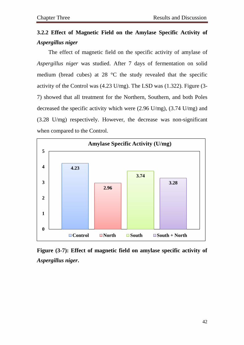
3.2.2 Effect of Magnetic Field on the Amylase Specific Activity of
Aspergillus niger
The effect of magnetic field on the specific activity of amylase of
Aspergillus niger was studied. After 7 days of fermentation on solid
medium (bread cubes) at 28 °C the study revealed that the specific
activity of the Control was (4.23 U/mg). The LSD was (1.322). Figure (3-
7) showed that all treatment for the Northern, Southern, and both Poles
decreased the specific activity which were (2.96 U/mg), (3.74 U/mg) and
(3.28 U/mg) respectively. However, the decrease was non-significant
when compared to the Control.
Figure (3-7): Effect of magnetic field on amylase specific activity of
Aspergillus niger.
4.23
2.96
3.74
3.28
0
1
2
3
4
5
Amylase Specific Activity (U/mg)
Control North South South + North
Chapter Three Results and Discussion
43
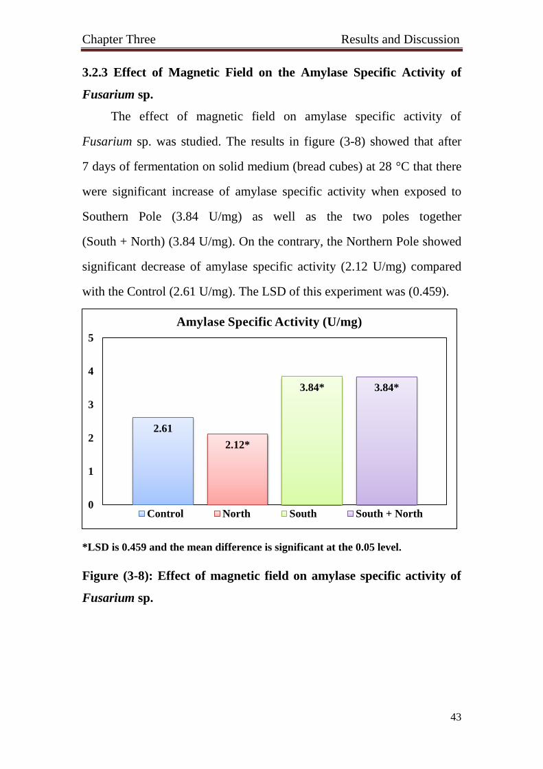
3.2.3 Effect of Magnetic Field on the Amylase Specific Activity of
Fusarium sp.
The effect of magnetic field on amylase specific activity of
Fusarium sp. was studied. The results in figure (3-8) showed that after
7 days of fermentation on solid medium (bread cubes) at 28 °C that there
were significant increase of amylase specific activity when exposed to
Southern Pole (3.84 U/mg) as well as the two poles together
(South + North) (3.84 U/mg). On the contrary, the Northern Pole showed
significant decrease of amylase specific activity (2.12 U/mg) compared
with the Control (2.61 U/mg). The LSD of this experiment was (0.459).
*LSD is 0.459 and the mean difference is significant at the 0.05 level.
Figure (3-8): Effect of magnetic field on amylase specific activity of
Fusarium sp.
2.61
2.12*
3.84* 3.84*
0
1
2
3
4
5
Amylase Specific Activity (U/mg)
Control North South South + North
Chapter Three Results and Discussion
44
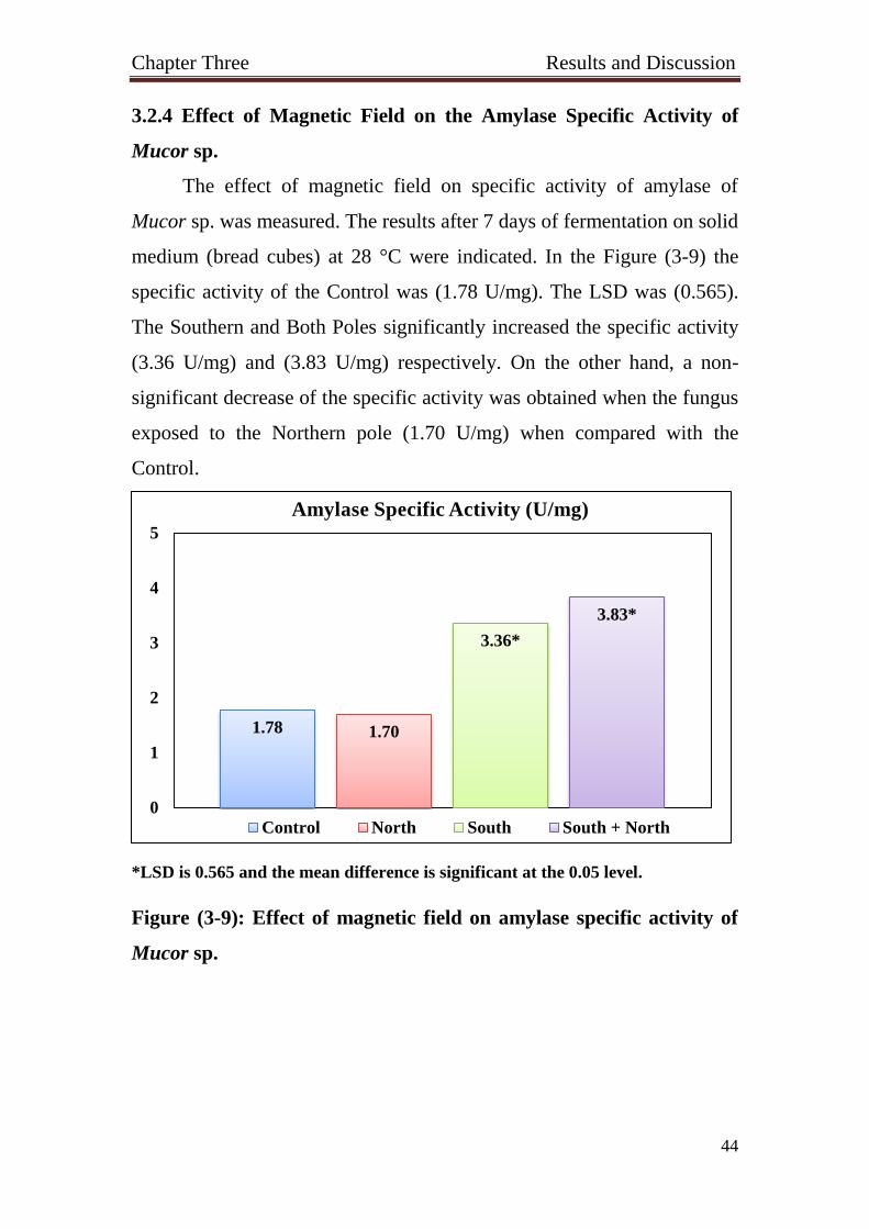
3.2.4 Effect of Magnetic Field on the Amylase Specific Activity of
Mucor sp.
The effect of magnetic field on specific activity of amylase of
Mucor sp. was measured. The results after 7 days of fermentation on solid
medium (bread cubes) at 28 °C were indicated. In the Figure (3-9) the
specific activity of the Control was (1.78 U/mg). The LSD was (0.565).
The Southern and Both Poles significantly increased the specific activity
(3.36 U/mg) and (3.83 U/mg) respectively. On the other hand, a non-
significant decrease of the specific activity was obtained when the fungus
exposed to the Northern pole (1.70 U/mg) when compared with the
Control.
*LSD is 0.565 and the mean difference is significant at the 0.05 level.
Figure (3-9): Effect of magnetic field on amylase specific activity of
Mucor sp.
1.78 1.70
3.36*
3.83*
0
1
2
3
4
5
Amylase Specific Activity (U/mg)
Control North South South + North

Chapter Three Results and Discussion
45
3.2.5 Effect of Magnetic Field on the Amylase Specific Activity of
Penicillium sp.
The effect of magnetic field on specific activity of amylase of
Penicillium sp. was tested. After 7 days of fermentation on solid medium
(bread cubes) at 28 °C, the specific activity was measured. The LSD of
this experiment was (0.508). The Control specific activity was (4.60
U/mg). As seen in the figure (3-10), all treatments were significantly
decreased the specific activity. The Northern, the Southern, and the two
poles together (South + North) specific activities were (3.27 U/mg), (3.88
U/mg) and (2.62 U/mg) respectively as compared with the Control.
*LSD is 0.508 and the mean difference is significant at the 0.05 level.
Figure (3-10): Effect of magnetic field on amylase specific activity of
Penicillium sp.
The statistical analysis of the results in Figures (3-(6, 7, 8, 9, 10))
showed that the Southern and Both Poles treatment significantly
increased the amylase specific activity of Fusarium sp. and Mucor sp.
and conversely significantly decreased amylase specific activity of
4.60
3.27*
3.88*
2.62*
0
1
2
3
4
5
Amylase Specific Activity (U/mg)
Control North South South + North

Chapter Three Results and Discussion
46
Alternaria sp. and Penicillium sp. compared with the Control. On the
other hand, the Northern Pole treatment significantly decreased the
amylase specific activity of Alternaria sp., Fusarium sp., and Penicillium
sp.
It is well known that there are many factors affect the enzyme
activity. These include temperature, pH, substrate concentration and the
presence of activator or inhibitor.
The effect of the magnetic field of the Southern pole seems to
potentiate the action of (Ca2+
) ion leading to increase in amylase specific
activity in Fusarium sp. and Mucor sp. It seems that other fungi
(Alternaria sp., and Penicillium sp.) respond to the action of the southern
pole of the magnetic field differently which could be explained by the
difference of the amino acid composing these proteins’ active sites which
led to this decrease of their activity by the effect of the magnetic field.
The action of both poles of the magnetic fields which led to increase the
specific activity of amylase of Mucor sp. and Fusarium sp. could be due
to the Southern pole only as it competes with Northern pole and
suppressed its effect.
The Moving Charge Interaction (MCI) model p suggest that
electrons motion affect enzyme activity. Theoretical models have
depicted existence of low frequency (ELF) magnetic field interactions
with biosystems at ion cyclotron resonance which is, at frequencies
corresponding to charge to mass ratios of ions such as Ca2+
, Mg2+
and K+
(Bruce et al., 1992). Experimental investigations detect that low
frequency electromagnetic fields affects living systems through the
calcium signaling pathways and systolic calcium oscillator (Galvanovskis
and Sandblom, 1998). The theories on the effects of MFs on activities of
enzyme refer to changes of motion of ions at the active site (Edmonds,
1993).

Chapter Three Results and Discussion
47
3.3 Effect of Magnetic Field on the Protease Specific Activity
3.3.1 Effect of Magnetic Field on the Protease Specific Activity of
Alternaria sp.
The effect of magnetic field on the specific activity of protease of
Alternaria sp. was tested. After 7 days of fermentation on solid medium
(bread cubes) at 28 °C. The LSD of this experiment was (2.644). All
treatments significantly decreased the protease specific activity
(Figure 3-11). The Northern and Southern Poles as well as the two poles
together (South + North) specific activities were (16.86 U/mg), (19.30
U/mg), and (13.65 U/mg) respectively when compared to the Control
(24.05 U/mg).
*LSD is 2.644 and the mean difference is significant at the 0.05 level.
Figure (3-11): Effect of magnetic field on protease specific activity of
Alternaria sp.
24.05
16.86*
19.30*
13.65*
0
5
10
15
20
25
30Protease Specific Activity (U/mg)
Control North South South + North

Chapter Three Results and Discussion
48
3.3.2 Effect of Magnetic Field on the Protease Specific Activity of
Aspergillus niger
The effect of magnetic field on the specific activity of protease of
Aspergillus niger was studied after 7 days of fermentation on solid
medium (bread cubes) at 28 °C. The LSD of this experiment was (2.932).
The study showed that the specific activity of the Control was (20.15
U/mg) (without MF), the Northern Pole significantly decreased specific
activity (14.69 U/mg). The two poles together (South + North) non-
significantly decreased the specific activity (19.16 U/mg). On the other
hand, the Southern Pole increased specific activity (21.35 U/mg),
however, the increase was non-significant (Figure 3-12).
*LSD is 2.932 and the mean difference is significant at the 0.05 level.
Figure (3-12): Effect of magnetic field on protease specific activity of
Aspergillus niger.
20.15
14.69*
21.35 19.16
0
5
10
15
20
25
30
Protease Specific Activity (U/mg)
Control North South South + North

Chapter Three Results and Discussion
49
3.3.3 Effect of Magnetic Field on the Protease Specific Activity of
Fusarium sp.
The effect of magnetic field on specific activity of protease of
Fusarium sp. was studied. The results after 7 days of fermentation on
solid medium (bread cubes) at 28 °C indicated that the Control specific
activity was (10.70 U/mg). The LSD of this experiment was (3.478).The
Southern Pole significantly increased specific activity (23.04 U/mg) as
well as the two poles together (South + North) (21.03 U/mg). The
Northern Pole non- significantly decreased specific activity (10.21 U/mg)
(Figure 3-13).
*LSD is 3.478 and the mean difference is significant at the 0.05 level.
Figure (3-13): Effect of magnetic field on protease specific activity of
Fusarium sp.
10.70 10.21
23.04* 21.03*
0
5
10
15
20
25
30
Protease Specific Activity (U/mg)
Control North South South + North

Chapter Three Results and Discussion
50
3.3.4 Effect of Magnetic Field on the Protease Specific Activity of
Mucor sp.
The effect of magnetic field on specific activity of protease of
Mucor sp. was measured. After 7 days of fermentation on solid medium
(bread cubes) at 28 °C, the results displayed that the specific activity of
the Control was (6.06 U/mg). The LSD of this trial was (2.424). As seen
in the Figure (3-14), the Southern Pole significantly increased specific
activity (12.15 U/mg) as well as the two poles together (South + North)
(9.75 U/mg), while the Northern Pole non-significantly decreased specific
activity (5.16 U/mg).
*LSD is 2.424 and the mean difference is significant at the 0.05 level.
Figure (3-14): Effect of magnetic field on protease specific activity of
Mucor sp.
6.06 5.16
12.15*
9.75*
0
5
10
15
20
25
30
Protease Specific Activity (U/mg)
Control North South South + North

Chapter Three Results and Discussion
51
3.3.5 Effect of Magnetic Field on the Protease Specific Activity of
Penicillium sp.
The effect of magnetic field on protease specific activity of
Penicillium sp. has been investigated. The Figure (3-15) represent after
7 days of fermentation on solid medium (bread cubes) at 28 °C that the
Control specific activity was (14.84 U/mg). The LSD of this test was
(2.684). The Northern Pole significantly decreased the specific activity
(12.03 U/mg) as well as the two poles together (South + North)
(8.19 U/mg). On contrary, the Southern Pole non-significantly increased
the specific activity (16.04 U/mg).
*LSD is 2.684 and the mean difference is significant at the 0.05 level.
Figure (3-15): Effect of magnetic field on protease specific activity of
Penicillium sp.
14.84
12.03*
16.04
8.19*
0
5
10
15
20
25
30Protease Specific Activity (U/mg)
Control North South South + North

Chapter Three Results and Discussion
52
The statistical analysis of the results in Figures (3-(11, 12, 13, 14,
15)) showed that the Southern Pole treatment significantly increased the
protease specific activity of Fusarium sp. and Mucor sp., while it
significantly decrease protease specific activity of Alternaria sp. Both
Poles treatment significantly increased protease specific activity of
Fusarium sp., Mucor sp. whereas decreased protease specific activity of
Alternaria sp. and Penicillium sp. The Northern Pole significantly
decreased the protease specific activity of Alternaria sp., Aspergillus
niger, and Penicillium sp.
Regarding the Southern pole magnetic field effect on protease
specific activity, it is logical to expect the increase effect of the enzyme
activity due to the probable potentiation of the positivity of cations.
However, it is not easy to explain the peculiar decrease of protease
activity in the case of the fungus Alternaria sp.
The most reasonable explanation of the decrease of enzyme specific
activity (protease and amylase) after exposing the fungi to the Northern
the magnetic field is attributable to the probable interface of the negative
charge of magnetic field with the release of the stimulating effect of
(Ca2+
).
Blank and Soo (2001), demonstrated the EMF interaction
mechanism with Na+- K
+ ATPase and suggested to be due to acceleration
of the electron regardless of direction the electrons move regularly and

Chapter Three Results and Discussion
53
the threshold force producing the effect on the enzyme . Therefore, it is
very likely that the influence of the field on the acid phosphatase could
include electron density at the anti-ferromagnetically coupled binuclear
Fe(III)-Mn(II) center and the active site.
A second theory concluded that the effect of SMF and OMF on
calcium ions bound in calcium-binding proteins, like calmodulin. The
calcium ions always vibrate about an equilibrium position in calmodulin
binding site. A static magnetic field to calmodulin causes the level of
vibration to rotate, or forward in the direction of the magnetic field at a
frequency that equal to the cyclotron frequency of the bound calcium.
Adding a "wobbling" MF at the cyclotron frequency disturbs the accuracy
to such an extent that it loosens the bond between the calmodulin and the
calcium ion (Pothakamury et al., 1993).
The ELF EMF influence enzyme activity irrespective of
temperature, pH and substrate concentration. Optimal pH and temperature
are very important for the activity of enzymes. Changing in pH and
temperature affect the shape of an enzyme and change the charge or
shape properties of the substrate so that the substrate cannot bind to the
active site or undergo catalysis. The studies search for whether ELF EMF
cause specific changes in the optimal pH and optimal temperature.
However, when the samples were exposed to EMF at various pH and
temperature the OD value was changed, which indicates there was a

Chapter Three Results and Discussion
54
change in the activity of enzyme. But there was no significant effect of
ELF EMF on optimal pH and temperature (Prashanth et al., 2008).

Conclusions
and
Recommendations

Conclusions and Recommendations
55
Conclusions
Magnetic field poles (100 gauss) showed different effects on
protease and amylase specific activity produced from Alternaria
sp., Aspergillus niger, Fusarium sp., Mucor sp., and Penicillium sp.
The Northern pole had a negative effect on the amylase
specific activity of Alternaria sp., Fusarium sp., and Penicillium
sp. as well as on the protease specific activity of Alternaria sp.,
Aspergillus niger, and Penicillium sp.
The Southern pole had a positive effect on both amylase and
protease specific activity of Fusarium sp., and Mucor sp. While it
had a negative effect on amylase specific activity of Alternaria sp.
and Penicillium sp. and protease specific activity of Alternaria sp.
as compared with the Control under the same conditions.
The two poles together (South + North) showed negative
effects on both amylase and protease specific activity of Alternaria
sp. and Penicillium sp. While they showed positive effects on
amylase and protease specific activity of Fusarium sp. and Mucor
sp. as compared with the Control under the same conditions.
Conclusions and Recommendations
56
Recommendations
Demonstrate the effect of magnetic field poles on the gene
expression of amylase and protease enzymes of Alternaria sp.,
Aspergillus niger, Fusarium sp., Mucor sp., and Penicillium sp.
Study the optimum conditions for maximum production of
amylase and protease enzymes from the five fungal genera.
Purification of amylase and protease enzymes of fungal
source and show the effect of MF poles on them.
Study the effect of MF poles on other enzyme like cellulase
that produced from these fungi.
Study the effect of MF poles on the amylase and protease
produced from other fungi like Rhizopus.

References

References
57
References
Aarholt, E., Flinn, E.A. and Smith, C.W. (1981). Effects of low-frequency
magnetic fields on bacterial growth rate. Phys. Med. Biol., 26: 613-621.
Abe, J., Bergmann, F.W., Obata, K. and Hizukuri, S. (1988). Production
of the starch digesting amylases of Aspergillus sp. Appl. Microbiol. Biot.,
27: 447-450.
Adamkiewicz, V.W. and Pilon, D.R. (1983). Magnetic stimulation of
polysaccharide accumulation in cultures of Streptococcus mutans, Can. J.
Microbiol., 29: 464-467.
Alexopoulos, C.J., Mims, C.M. and Blackwell, M. (1996). Introductory
Mycology (4th
Ed.). John Wiley and Sons Inc., New York.
Alikamanoglu, S. and Sen, A. (2011). Stimulation of growth and some
biochemical parameters by magnetic field in wheat (Tritium aestivum L)
tissue cultures. Afr. J. Biotechnol., 10(53): 10957-10963.
Anson, M.L. (1938). J. Gen. Physiol; 22: 79-89.
Atak, C. and Emiroglu, O. (2003). Stimulation of regeneration by
magnetic field in soybean (Glycine max L. Merrill) tissue cultures'.
J. Cell. Mol. Biol., 2: 113-119.
Audus, L.J. (1960). Magnetotropism: A new plant growth response,
"Nature; 185: 132-134.
Baijal, U. and Mehrotra, B.S. (1980). The genus Cunninghamella sp. a
reassessment. Sydowia; 33: 1-13.
Barrette, A.J. and Rawlings, N.O. (2003). Evolutionary families of
peptidases. Biochem. J., 290: 205-218.
Beg, K.B. and Gupta, R. (2003). Purification and characterization of an
oxidation-stable, thiol-dependent serine alkaline protease from Bacillus
mojavensis. Enzyme and Microb. Technol., 32: 294-304.
Bernfeld, P. (1955). Meth. Enzymol., 1: 149-158.

References
58
Binhi, V.N., Alipov, Y.D and Belyaev, I.Y. (2001). “Effect of static
magnetic field on E. coli cells and individual rotations of ion-protein
complexes”, Bioelectromagnetics, 22: 79-86.
Binhi, V.N. and Savin, A.V. (2003). Effect of weak magnetic fields on
biological system: Physical aspects. Physics_Uspekhi; 46(3): 259-291.
Bissett, J. (1991). A revision of the genus Trichoderma sp. III section
Pachybasium, Can. J. Bot., 69: 2373-2420.
Blank, M. and Soo, L. (1998). Enhancement of cytochrome oxidase
activity in 60 Hz magnetic fields. J. Bioelectroch. Bioenerg., 45: 253-259.
Blank, M., Soo, L. and papstein, V. (1995). Effects of low frequency
magnetic field on Na, KATPase activity. J. Bioelectroch. Bioenerg., 38:
267-273.
Blank, M. and Soo, L. (2001). Electromagnetic acceleration of electron
transfer reactions. J. Cell. Biochem., 81: 278-283.
Boerjan, W., Ralph, J. and Baucher, M. (2003). Lignin biosynthesis.
Annu. Rev. Plant Biol., 54: 519-546.
Bradford, M.M. (1976). A rapid and sensitive method for the
quantification of microorganism quantities of protein utilizing the
principle of protein _ dye binding. Anal. Biochem., 72: 248-254.
Bruce, M., Abraham, R., Liboff, A. and Stephen, D.S. (1992).
Electromagnetic gating in ion channels. J. Theor. Biol., 158: 15-31.
Cairo, P., Greenebaum, B. and Goodman, E. (1998). Magnetic field
exposure enhances mRNA expression of σ32 in E. coli. J. Cell Biochem.,
68: 1-7.
Cannel, E. and Moo-Young, M. (1980). Solid-state fermentation system.
Process Biochem., 15 (5): 2-7.
Charlang, G.W. and Horowitz, N.H. (1971). Germination and growth of
neurospora at low water activities. Proc. Natl. Acad. Sci. U. S. A. 68(2):
260-262.

References
59
Charlang, G.W. and Horowitz, N.H. (1974). Membrane permeability and
the loss of germination factor from Neurospora crassa at low water
activities. J. Bacteriol., 117(1): 261-264.
Cullity, B.D. and Graham, C.D. (2008). Introduction to Magnetic
Materials (2th Ed.). Wiley-IEEE Press; 103.
Crueger, W. and Crueger, A. (1984). Enzymes. In Bio-Technology. A
Text book of Industrial Microbiology; 161-186.
Devi, M.K., Banu, A.R., Gnanaprabhal, G.R., Pradeep, B.V. and
Palaniswamy, M. (2008). Purification, characterization of alkaline
protease enzyme from native isolate Aspergillus niger and its
compatibility with commercial detergents. Indian J. Sci. Technol., 1: 7.
Domsch, K.H., Gams, W. and Anderson, T.H. (1980). Compendium of
soil fungi. Vol 1. Academic Press, London, UK.
Edmonds, D.T. (1993). Larmor precession as a mechanism for the
detection of static and alternating magnetic fields. J. Bioelectrochem.
Bioenerg., 30: 3-12.
Ellaiah, P., Adinarayana, K., Bhavani, Y., Padmaja, P. and Srinivasula, B.
(2003). Optimization on process parameters for amylase production under
SSF by a newly isolated Aspergillus sp. Process Biochem., 38: 615-620.
Facciotti, M.C.R., Kilikian, B.V., Schmidell, W. and Fachini, E.R.
(1989). Glucoamylase synthesis in batch process by Aspergillus awamori.
Rev. Microbiol., 20: 108-114.
Faraj, M.K. (1990). Regulation of mycotoxins formation in Zea mays.
Ph.D. thesis. Department of Bioscience and Biotechnology, University of
Strathclyde, Glascow, U.K; 117.
Fröhlich, J., Hyde, K.D. and Petrini, O. (2000). Endophytic fungi
associated with palms. Mycol. Res., 104: 1202-1212.
Galvanovskis, J. and Sandblom, J. (1998). Periodic forcing of
intracellular calcium oscillators theoretical studies of the effects of low

References
60
frequency fields on the magnitude of oscillations. J. Bioelectrochem.
Bioenerg., 46: 161-174.
Gao, W., Liu, Y., Zhou, J. and Pan. H. (2005). Effects of a strong static
magnetic field on bacterium Shewanella oneidensis: An assessment by
using whole genome microarray. Bioelectromagnetics; 26: 558-563.
Gerencser, V.E., Barnothy, M.F. and Bamothy, J.M. (1962). Inhibition of
bacterial growth by magnetic fields, Nature, 196: 539-541.
Godfrey, T. and West, S. (1996). Industrial Enzymology, (2nd
Edn.), NY:
Macmillan Publishers, Inc. ISBN 0-33359464-9,3.
Goodman, E.A., Greenebaum, B. and Marron, M.T. (1976). Effects of
extremely low frequency electromagnetic fields on Physarum
polycephalum. Rad. Res., 6: 531-540.
Goodman, E.A., Greenebaum, B. and Marron, M.T. (1979). Bioeffects of
extremely low frequency electromagnetic fields: variation with intensity,
waveform, and individual or combined electric and magnetic fields. Rad.
Res., 78: 485-501.
Goodman, E.A., Greenebaum, B. and Marron, M.T. (1993). Altered
protein synthesis in a cell-free system exposed to a sinusoidal magnetic
field. Biochim. Biophys. Acta., 1202: p. 107-112.
Goodman, R. and Blank, M. (2002). Insights into Electromagnetic
Interaction Mechanisms. J. Cellular Physiol., 192: 16-22.
Grajek, W. and Grevais, P. (1987). Influence of water activity on the
enzyme biosynthesis and enzyme activities produced by Trichoderma
viride, in solid-state fermentation; 9 (11): 658-662.
Grant, I.S. and Phillips, W.R. (2008). Manchester Physics.
Electromagnetism (2nd
Ed.). John Wiley & Sons.
Green, M. R. and Sambrook, J. (2012). Molecular cloning: A laboratory
manual. Cold Spriner Harbor Laboratory Press. Cold Spring Harbor. New
York. USA.

References
61
Grevais, P. and Molin, P. (2003). The role of water in solid-state
fermentation. Biochem. Eng. J. 13: 85-101.
Hajji, M., Rebai, A., Gharsallah, N. and Nasri, M. (2008). Optimization
of alkaline protease production by Aspergillus clavatus ES1 in Mirabilis
jalapa tuber powder using statistical experimental design. J. Appl.
Microbiol. Biotechnol., 79: 915-923.
Halliwell, B. (1982). 'The toxic effects of oxygen on plant tissues'. In
Superoxide Dismutase [ L.W. Oberley (ed.)]. Boca. Raton, FL: CRC.
Press; 1: 89-123.
Harkins, T.T. and Grissom, C.B. (1994). Magnetic field effects on B12
ethanolamine ammonia lyase: evidence for a radical mechanism, Science;
263: 958-959.
Harris, S.D. (2008). Branching of fungal hyphae: regulation mechanisms
and comparison with other branching systems. Mycologia; 100(6): 823-
832.
Hesseltine, C.W. (1977). Solid state fermentation part_1. Process
Biochem; 24-27.
Hibbett, D.S. (2007). A phylogenetic overview of the agaricomycotina.
Mycologia; 98: 917-925.
Ikehara, T., Park, K.H., Kinouchi, Y., Yamaguchi, H., Yoshizaki, K. and
Miyamoto, H. (1997). Effects of strong magnetic fields on membrane ion
transport of cultured cells. Tech. Rep. IEICE. MBE97-40: 7-12.
Ivancsits, S., Diem, E., Pilger, A., Rudiger, H.W. and Jahn, O. (2002).
Induction of DNA strand breaks by intermittent exposure to extremely-
low-frequency electromagnetic fields in human diploid fibroblasts. J.
Mutation Res., 519: 1-13.
Jiles, D.C. (1991). Introduction to Magnetism and Magnetic Materials.
Chapman and Hall, New York.

References
62
Kim, Y.S. (1976). Some possible effects of static magnetic fields on
cancer, Tower International Technomedical Institute of Life Sciences; 5:
11-28.
Kimball, G.C. (1937). The growth of yeast on magnetic fields, J.
Bacteriol., 35: 109-122.
Kinouchi, Y., Tanimoto, S., Ushita, T., Sato, K., Yamaguchi, H. and
Miyamoto, H. (1988). Effects of static magnetic fields on diffusion in
solutions. Bioelectromagnetics, 9: 159-166.
Kirk, P.M., Cannon, P.F., Minter, D.W. and Stalpers, J.A. (2008).
Dictionary of the Fungi. (10th
Ed.). CAB International, Wallingford, UK.
Koch, B., Sommarin, M., Persson, B., Salford, L.G. and Eberhardt, J.L.
(2003). Interaction between week low frequency magnetic fields ans cell
membranes. Bioelectromagnetics, 24: 395-402.
Kronmüller, H. (2007). Handbook of Magnetism and Advanced Magnetic
Materials, Vol. 5, Set. John Wiley and Sons.
Kumar, D., Jain, V.K., Shanker, G. and Srivastava, A. (2003). Citric acid
production by solid state fermentation using sugarcane bagasse. Process
Biochem., 38: 1731-1738.
Liburdy, R.P. (1992). Calcium signaling in lymphocytes and ELF
magnetic fields. E: vidence for an electric field metric and site of
interaction involving the calcium ion channel. FEBS Lett, 301: 53-59.
Lin, H., Blank, M., Rossol-Haseroth, K. and Goodman, R. (2001).
Regulating genes with electromagnetic response elements. J. Cell.
Biochem., 81: 143-148.
Lin, H., Goodman, R. and Shirley-Henderson, A. (1994). Specific region
of the c-myc promoter is responsive to electric and magnetic fields”, J.
Cell. Biochem., 54: 281-288.
Markkanen, A., Juutilainen, J., Lang, S., Pelkonen, J., Rytomaa, T. and
Naarala, J. (2001). Effects of 50 Hz magnetic field on cell cycle kinetics

References
63
and colony forming ability of budding yeast exposed to ultraviolet
radiation. Bioelectromagnetics., 2: 345-350.
Markov, M.S., Wang, S. and Pilla, A.A. (1992): Weak pulsing sinusoidal
and DC magnetic fields affect myosin phosphorylation in a cell free
preparation, in First World Congress on Electricity and Magnetism in
Biology and Medicine.
Marron, M.T., Goodman, E.M. and Greenebaum, B. (1975). Mitotic
delay in the slime mould Physarum polycephalum induced by low
intensity 60 and 75 Hz electromagnetic fields. Nature, 254: 6-67.
Marron, M.T., Goodman, E.M. and Greenebaum, B. (1978). Effects of
weak electromagnetic fields on Physarum polycephalum: mitotic delay in
heterokaryons and decreased respiration”, Experientia; 34: 589-591.
Marron, M.T., Goodman, E.M., Greenebaum, B. and Tipnis, P. (1986).
Effects of sinusoidal 60-Hz electric and magnetic fields on ATP and
oxygen levels in the slime mold, Physarum polycephalum.
Bioelectromagnetics; 7: 307-314.
Mitchell, R.S., Kumar, V., Abbas, A.K. and Fausto, N. (2007). Robbins
Basic Pathology. (8th
Edn.). Philadelphia; Sanders; 122.
Mmbaga, M.T., Sauvé, R.J., Nnodu, E. and Zhou, S. (2005). Multiple
disease resistance to powdery mildew, bacterial blight, and Alternaria
blight in lilac (Syringa spp.). J. Arboriculture, 31: 1-9.
Moens, P.B. and Rapport, E. (1971). Spindles, spindle plaques, and
meiosis in the yeast Saccharomyces cerevisiae (Hansen). J. Cell. Biol.,
50(2): 344-361.
Moore, R.L. (1979). Biological effects of magnetic fields: studies with
microorganisms, Can. J. Microbiol., 25 (10): 1145-1151.
Nakamura, K., Okuno, K., Ano, T. and Shoda, M. (1997). Effect of high
magnetic field on growth of Bacillus subtilis measured in a newly

References
64
developed superconducting magnetic biosystem. Bioelectrochem.
Bioenerg., 43: 123-128.
Nakasono, S. and Saiki, H. (2000). Effect of ELF magnetic fields on
protein synthesis in Escherichia coli K12. Radiat. Res., 154: 208-216.
Nakasono, S., Laramee, C., Saiki, H. and McLeod, K.J. (2003). Effect of
power-frequency magnetic fields on genome-scale gene expression in
Saccharomyces cerevisiae. Radiat. Res., 160: 25-37.
Narahara, H.J. (1997). Solid-State Fermentation: An Overview.
Fermentation Technology, 55: 254-256.
Nascimento, W.C.A. and Martins, M.L.L. (2004). Production and
properties of an extracellular protease from thermophilic Bacillus spp.
Brazilian. J. Microbiol., 35: p. 1-2.
Nikolai, K.C., Gapeyev, A.B., Sirota, N. P., Gudkov, O.Y., Kornienko, N.
V., Tankanag, A.V., Konovalov, I.V., Buzoverya, M.E., Suvorov, V.G.,
and Logunov, V.A. (2004). DNA damage in frog erythrocytes after in
vitro exposure to a high peak power pulsed electromagnetic field. J.
Mutation Res., 558: 27-34.
Oojikaas, L.P., Weber, F.J., Buitelaar, R.M., Tramper, J. and Rinzema, A.
(2000). Defined media and inert supports: their potential as solid-state
fermentation production systems. Trends in Biotechnology; 18: 356-360.
Oriol, E., Raimbault, M., Roussos, S. and Gonzales, G.V. (1988). Water
and water activity in the solid state fermentation of cassava starch by
Aspergillus niger. Appl. Microbiol. Biotechnol., 27: 498-503.
Pandey, A., Selvakumar, P., Soccol, C.R and Nigam, P. (1999). Solid
state fermentation for the production of industrial enzymes, J Microbiol.,
77: 149-162.
Pandey, A., Soccol, C.R. Rodriguez, L.J. and Nigam, P. (2001).
Biotechnological potential of agro-industrial residues II: cassava bagasse.
Bioresource Technol., 74: 81-87.

References
65
Pastor, M.D., Lorda, G.S. and Balatti, A. (2001). Protease obtention using
Bacillus subtilis 3411 and amaranth seed meal medium at different
aeration rates. Brazilian J. Microbiol., 32: 1-8.
Peter, H. Wald and Greg M. Stave, (2002). Physical and Biological
Agents of the Workplace, (2nd
Ed.). New York: John Wiley and Sons.
Pittman, U.J. (1977). Effects of magnetic seed treatment on yields of
barley, wheat and oats on Suthern Alberta,"Can. J. Plant. Sci., 57: 37-45.
Pothakamury, U.R., Barbosa-Cánovas, G.V., and Swanson, B.G. (1993).
Magnetic-field inactivation of microorganisms and generation of
biological changes. Food Technol., 47(12): 85-93.
Prashanth, K.S., Chouhan, T.R.S., Nadlger, S. (2008). Effect of 50 Hz
electromagnetic fields on alpha amylase activity. Romanian J. Biophys.,
18(3): 255-263.
Presman, A.S. (1970). Electromagnetic Fields and Life, New York,
Plenum.
Racuciu, M. and Creanga, D.E. (2006). 'Plant growth under static
magnetic field influence'. Rom. J. Phys., 53(1-2): 353-359.
Raghavarao, K.S.M., Ranganathan, T.V. and Karanth, N.G. (2003). Some
engineering aspects of Solid-State Fermentation. Biochem. Eng. J., 13:
127-135.
Rakosy-Tican, L., Aurori, C.M. and Morariu, V.V. (2005). 'Influence of
near null magnetic field on in vitro growth of potato and wild Solanum
species'. Bioelectromagnetics., 26(7): 548-57.
Rao, M.B., Tanksale, A.M., Ghatge, M.S and Deshpande, V.V. (1998).
Molecular and biotechnological aspects of microbial proteases, Microbiol
Mol. Biol. Rev., 62: 597-635.
Raven, H. and Johnson, G.B. (1999). Biology. Fifth Edition. McGraw-
Hill, Boston. P. 1284.

References
66
Richardson, B.A., Yaga, K., Reiter, R.J. and Morton, D.J. (1992). Pulsed
static magnetic field effects on in-vitro pineal indoleamine metabolism,
Biochimica Biophysica Acta., 1137: 59-64.
Riddle, R.W. (1950). Permanent stained mycological preparation
obtained by slide culture. Mycologia., 42: 265-70.
Rodriguez, R.J., White, J.F., Arnold, A.E. and Redman, R.S. (2009).
Fungal endophytes: diversity and functional roles. New Phytologist., 182:
314-330.
Sahebjamei, H., Abdolmaleki, P. and Ghanati, F. (2007). 'Effects of
magnetic field on the antioxidant enzyme activities of suspension
cultured Tobacco cells'. Bioelectromagnetics; 28: 42-47.
Sambrook, J. and Russell, D.W. (2001). Molecular cloning: a laboratory
manual. (3rd
Ed.). New York: Cold Spring Harbor Laboratory Press.
Savostin, P.W. (1930). Magnetic growth relations in plants,"Planta., 12:
327.
Scaiano, J.C., Cozens, F.L. and McLean, J. (1994). 'Model of the
rationalization of magnetic field effects in vivo. Application of radical-
pair mechanism to biological systems'. Photochem Photobiol., 59(1): 585-
589.
Scott, W.J. (1953). Water relations of Staphylococcus aureus at 30
degrees. Aust. J. Biol. Sci. Nov., 6(4): 549-564.
Suryanarayanan, T.S., Senthilarasu, G. and Muruganandam, V. (2000).
Endophytic fungi from Cuscutarejlexa and its host plants. Fungal
Diversity; 4: 117-123.
Terborgh, J. (1992). Diversity in the Tropical Rainforest, Scientific
American Books, New York, p. 31-51.
Yadav, J. (1988). Solid-State Fermentation of wheat straw with
alcaliphilic Coprinus. Biotechnol. Bioeng., 31(5): 414-417.

References
67
Yandri, A.S., Suhartati, T., Herasari, D. and Hadi, S. (2008). The
chemical modification of protease enzyme isolated from local bacteria
isolate Bacillus subtilis 1TBCCB148 with cynauric chloride – poly –
ethyleneglycol. Eur. J. Sci. Res., 23(1): 177-186.
Yano, A., Ohashi, Y. and Hirasaki, T. (2004). 'Effects of a 60 Hz
magnetic field on photosynthetic CO2 uptake and early growth of Radish
seedlings'. Bioelectromagnetics; 25 (8): 572-581.
Ward, O.P. (1985). Proteolytic enzymes. In: Blanch, H.W., Drew, S.,
Wang, D.I., eds. Comprehensive Biotechnology. Oxford U.K. Pergamon
Press, 3: p. 789-818.
Williams, P.A., Ingebretsen, R.J. and Dawson, R.J. (2006). 14.6 mT ELF
magnetic field exposures yields no DNA breaks in model system
Salmonella, but provides evidence of heat stress protection.
Bioelectromagnetics., 27: 445-450.

ةـلاصـــــــــــــــــالخ
ػهى إخاج اشى الأيهش انبزحش انغاطظ انثابج، أخخبز حاثز اندال ف ذ انذراطت
Alternaria sp., Aspergillusانخ ػائذة لأخاص يخخهفت، خظت أاع فطزتن
niger, Fusarium sp., Mucor sp. and Penicillium sp. حخزاث انحانت باططت
إنى اندال حؼزط ذ انفطزاثحى ن انفطزاث. أطخخذو انخبش فقط كطط. انصهبت
نهقطب اندب انشان الاث يؼا، ثى قر حاثز اندال انغاطظ ػهى انغاطظ
انخائح احصائا . حى ححهم نهدال( نفض انفؼانت الاشت يغ يؼايهت انظطزة )انخ نى حخؼزض
اظزث انخائح بأ . (LSD( حذد الاخخلاف الاقم انؼي )GenStatباططت بزايح )
بخاثز انقطب انشان نهدال انغاطظ نهفطزاث يؼا يهش حقم لاشى الا انفؼانت انػت
Alternaria sp., Fusarium sp. and Penicillium sp. 0..0، 2..0انخ كاج ،
نفطز بخاثز انقطب اندب لاشى الايهش انفؼانت انػت حشداد .ػهى انخان غىحذة/يه 7.03
Fusarium sp 3.84 /يهغى نفطز حذةMucor sp. 3.36 ا قههج يؼا يهغى ، ب/حذة
Penicilliumحذة/يهغى نفطز Alternaria sp. 2.77لاشى الايهش نفطز انفؼانت انػت
sp. 3.88 :خفظانقطب كلا احذة/يهغى يقارت يغ انظطزة. كا حأثز كلا انقطب كالأح
حذة/يهغى Alternaria sp. 2.62لاشى الايهش نفطز يؼا انفؼانت انػت
لاشى م، سادا يؼا انفؼانت انػت، ف انقابحذة/يهغى Penicillium sp. 2.26نفطز
. فا حذة/يهغى Mucor sp. 3.83فطز حذة/يهغى Fusarium sp. 3.84 الايهش نفطز
قهم ، انقطب انشانانفؼانت انػت لاشى انبزحشنغاطظ ػهى خص حأثز اندال ا
Aspergillusفطز .Alternaria spلاشى انبزحش نفطز يؼا ي انفؼانت انػت
niger فطزPenicillium sp. حذة/يهغى ػهى 12.03 ,14.69 ,16.86 انخ كاج
.Fusarium spلاشى انبزحش نفطز ي انفؼانت انػت انخان. انقطب اندب ساد يؼا
.Alternaria spياػذا فطز حذة/يهغى Mucor sp (12.15) .حذة/يهغى نفطز (23.04)
حذة/يهغى. كلا انقطب سادا (19.30)لاشى انبزحش يؼا ف انفؼانت انػت قهجانذي
فطز حذة/يهغى Fusarium sp. (21.03)لاشى انبزحش نفطز انفؼانت انػتيؼا
Mucor sp. ((9.75 لاشى انبزحش نفطز انفؼانت انػت حذة/يهغى، با قهلا يؼا ي
Alternaria sp. (13.65) فطز حذة/يهغى Penicillium sp. (8.19) ذ حذة/يهغى.
انذراطت اظحج با اك حأثز يؼي نهدال انغاطظ انثابج ف سادة حقهم انفؼانت

الاشت لااع انفطزاث انخ ي انك اطخغلانا صاػا ف سادة اخاج الاشاث انت
هش انخ اخخبزث ف ذ انذراطت.صاػا يثم انبزحش الاي

جوهىريت العراق
وزارة التعلين العالي والبحث العلوي
جاهعت النهرين / كليت العلىم
الأحيائيت التقانت قسن
أوزيمي على الثابث جاثير المجال المغىاطيسي
العزلات بعضمه المىحجةزالاميليز و البروجي
جخمرات الحالة الصلبة باسحعمال الفطرية
رسالت
جاهعت النهرين –كليت العلىم هجلس هقذهت الى
التقانت الاحيائيت هن هتطلباث نيل درجت هاجستير في علىموهي جسء
هن قبل
هروة صباح هاشن
1023- الىهريهجامعة -كلية العلىم -احيائية اوةبكالىريىس جق
أشراف
د عبذ الىاحذ شوخي جابر.م.أ
حزيران رمضان
2437 1026