effect of salicylate on the rectal absorption of lidocaine, levodopa, and cefmetazole in rats
TRANSCRIPT

Effect of Salicylate on the Rectal Absorption of Lidocaine, Levodopa, and Cefmetazole in Rats
a 2 40
0 0
-I LL 0
30,
9 $ 2 0 . W V IT W 0 1 0 .
n.
TOSHIAKI NISHIHATA, J. HOWARD RYTTING'", and TAKERU HIGUCHI Received October 27,1980, from the Pharmaceutical Chemistry Department, T h e University of Kansas, Lawrence, K S 66045. for publication November 3,1981.
Accepted
Abstract Salicylic acid and sodium salicylate have been found to en- hance the absorption of lidocaine, levodopa, and cefmetazole after rectal administration to rats. These drugs represent a base (lidocaine), an acid (cefmetazole), and a substance (levodopa) which exists as a zwitterion in solution. The rectal absorption of each type of drug, as well as theo- phylline, a neutral compound, was enhanced by salicylate, particularly at pH values where the substances exist primarily in their ionic form. A requirement for the observed enhancement is that salicylate be present in the rectal membrane. The loss of drug from the perfusing solution was greater from solutions having an ionic strength of 0.75 than from those with p = 0.15.
Keyphrases Absorption, rectal-effect of salicylate on rectal ab- sorption of lidocaine, levodopa, and cefmetazole, rats Salicylate--effect on rectal absorption of lidocaine, levodopa, and cefmetazole, rats 0 Lidocaine-effect of salicylate on rectal absorption, rats Levodopa- effect of salicylate on rectal absorption, rats Cefmetazole-effect of salicylate on rectal absorption, rats
It has been reported (1,2) that salicylate markedly en- hanced the rectal absorption of theophylline. It was sug- gested that the effect of salicylate depends on its presence in the rectal membrane. It was also observed that the changes in absorption required the presence of salicylate in the formulation and that the mechanism of enhance- ment is different from that of surfactants.
The present report describes the extension of the use of salicylate to enhance the absorption of lidocaine, levodopa, and cefmetazole after rectal administration to rats. The drugs selected represent four different chemical classes: theophylline is a neutral substance, lidocaine is a basic material, cefmetazole is acidic, and levodopa exists as a
50 z
zwitterion in solution. Furthermore, each of these drugs exhibits some difficulties in its administration and ab- sorption. For example, it has been reported (3) that levo- dopa given rectally resulted in no rise in blood levodopa concentrations and no clinical benefit after rectal admin- istration to parkinsonian patients. The purpose of this study was to examine to what extent the effects of salicy- late are general and the extent rectal absorption en- hancement depends on the specific drugs used. In addition, studies involving intravenous administration of salicylate were included to provide additional information about the mechanism of enhancement.
EXPERIMENTAL
Materials-Sodium salicylate', lidocaine2, sodium cefmetazole3, and levodopa3 were used as obtained from the manufacturers.
Animals-Sprague-Dawley male rats (200-300 g) were used in this study and were fasted for 16 hr prior to the experiment. During the ex- periment, the rats were kept on a 38' surface and were anesthetized with pentobarbital (60 mglkg).
The in situ perfusion of the rat rectum and the in uivo absorption studies were carried out as described previously (1,2) with the following modifications: The pH of the perfusate was maintained constant by either the addition of 0.1 N NaOH or 0.1 N HCI as needed. Some experiments were also carried out at an ionic strength of 0.15 and 0.75 using sodium chloride to obtain the desired ionic strength. The in uivo absorption studies involved administering 0.3 ml of the drug solution into a 2-cm section of the rectum which was isolated by ligation with thread.
Intravenous Infusion Studies-To maintain constant plasma sali- cylate concentrations, an intravenous infusion method was used for some experiments. After the rats were anesthetized with pentobarbital, a so- dium salicylate solution (300 mg/ml), adjusted to pH 7.4 with 0.0114 M phosphate buffer, was infused into the leg vein of the rat through a polyethylene canula at a flow rate of 0.05 ml/min during the first 10 min and then at a flow rate of 0.02 ml/min for another 60 min. Beginning 10 min after starting the infusion of the salicylate solution, blood samples
- , I 1 I I I
4.0 5.0 6.0 7.0 8.0 9.0 PH
Figure 1-Effect o f pH, ionic strength, and salicylate on the disap- pearance of lidocaine hydrochloride from a perfusate in the rat rectum after 1 hr. The initial lidocaine hydrochloride concentration was 500 pglml, and the sodium salicylate concentration was 0.5 (0 and 0) or 0% (0 and A). The ionic strength was 0.75 (-) or 0.15 (- - -).
w o n 4.0 5.0 6.0 7.0 8.0 9.0 10.0
I 1 I
PH Figure 2-The percent loss of salicylate from a perfusate as a function of pH having a n initial concentration of 0.5% and ionic strength of 0.75 (0) and 0.15 (0).
Aldrich Chemical Co., 99+%.
Sankyo. *Sigma Chemical Co.
0022-35491 821 0800-0869$0 1.001 0 @ 1982, American Pharmaceutical Association
Journal of Pharmaceutical Sciences I 069 Vol. 71. No. 8, August 1982
a
MINUTES
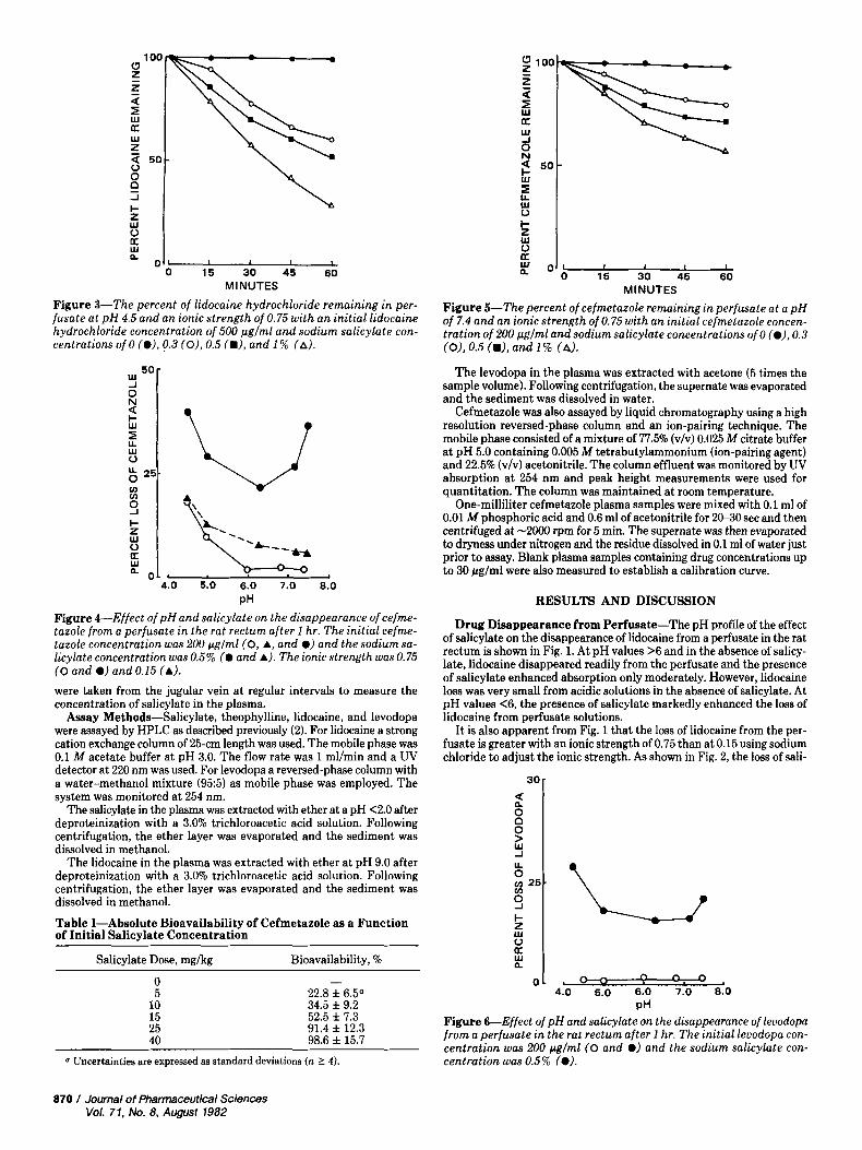
Figure 3-The percent of lidocaine hydrochloride remaining in per- fusate at pH 4.5 and an ionic strength of 0.75 with an initial lidocaine hydrochloride concentration of 500 pglml and sodium salicylate con- centrations of 0 (*), 0.3 (O), 0.5 (m), and 1 % (A).
3 5 0 -
lL W 0
t
5 W u a
0 4.0 5.0 6.0 7.0 0.0
k! 1 , PH
Figure &--Effect of pH and salicylate on the disappearance ofcefme- tazole from a perfusate in the rat rectum after 1 hr. The initial cefme- tazole concentration was 200 p g l m l ( 0 , A, and 0) and the sodium sa- licylate concentration was 0.5% (0 and A). The ionic strength was 0.75 (0 and 0) and 0.15 (A). were taken from the jugular vein at regular intervals to measure the concentration of salicylate in the plasma.
Assay Methods-Salicylate, theophylline, lidocaine, and levodopa were assayed by HPLC as described previously (2). For lidocaine a strong cation exchange column of 25-cm length was used. The mobile phase was 0.1 M acetate buffer at pH 3.0. The flow rate was 1 mllmin and a UV detector at 220 nm was used. For levodopa a reversed-phase column with a water-methanol mixture (95:5) as mobile phase was employed. The system was monitored at 254 nm.
The salicylate in the plasma was extracted with ether at a pH <2.0 after deproteinization with a 3.0% trichloroacetic acid solution. Following centrifugation, the ether layer was evaporated and the sediment was dissolved in methanol.
The lidocaine in the plasma was extracted with ether a t pH 9.0 after deproteinization with a 3.0% trichloroacetic acid solution. Following centrifugation, the ether layer was evaporated and the sediment was dissolved in methanol.
Table I-Absolute Bioavailability of Cefmetazole as a Function of Initial Salicylate Concentration
Salicylate Dose, mglkg Bioavailability, '?6
0 5
10 15 25 40
- 22.8 f 6.5a 34.5 f 9.2 52.5 f 7.3 91.4 f 12.3 98.6 f 15.7
~ ~
Uncertainties are expressed as standard deviations (n 1 4).
RESULTS AND DISCUSSION
Drug Disappearance from Perfusate-The pH profile of the effect of salicylate on the disappearance of lidocaine from a perfusate in the rat rectum is shown in Fig. 1. At pH values >6 and in the absence of salicy- late, lidocaine disappeared readily from the perfusate and the presence of salicylate enhanced absorption only moderately. However, lidocaine loss was very small from acidic solutions in the absence of salicylate. At pH values <6, the presence of salicylate markedly enhanced the loss of lidocaine from perfusate solutions.
It is also apparent from Fig. 1 that the loss of lidocaine from the per- fusate is greater with an ionic strength of 0.75 than at 0.15 using sodium chloride to adjust the ionic strength. As shown in Fig. 2, the loss of sali-
a
U 0 g 25.
s k
0
a
f ::
01 ,- 4.0 5.0 6.0 7.0 8.0
PH Figure &Effect of pH and salicylate on the disappearance of levodopa from a perfusate in the rat rectum after 1 hr. The initial levodopa con- centration was 200 pglml (0 and *) and the sodium salicylate con- centration was 0.5% (0).
870 / Journal of Pharmaceutical Sciences Vol. 71, No. 8, August 7982
20 - E z a
. K U 4 a z z
D
1 c
9 0
J
0
T
0 30 60 90 120 MINUTES
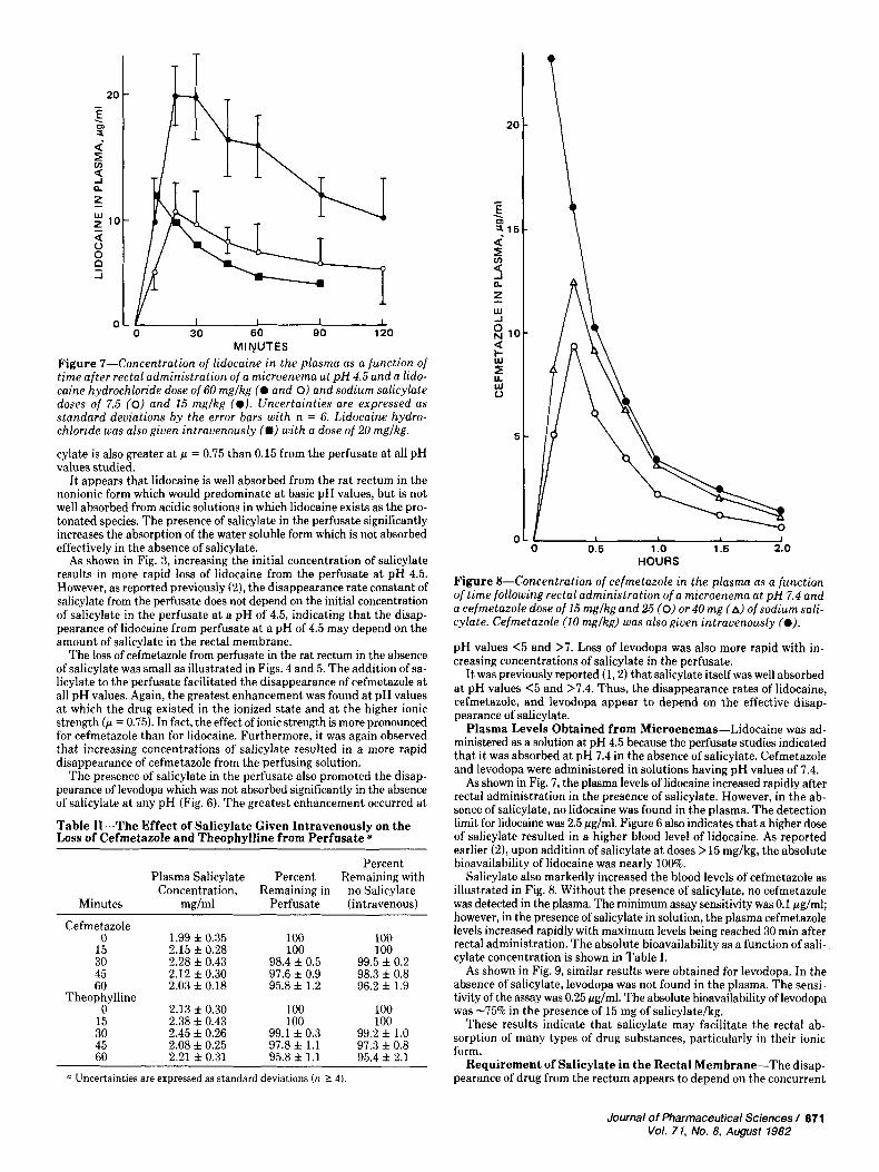
Figure 7-Concentration of lidocaine in the plasma as a function of time after rectal administration of a microenema a t pH 4.5 and a lido- caine hydrochloride dose of 60 mglkg (@ and 0) and sodium salicylate doses of 7.5 (0) and 15 mglkg (@). Uncertainties are expressed as standard deviations by the error bars with n = 6. Lidocaine hydro- chloride was also given intravenously (.) with a dose of 20 mglkg.
cylate is also greater a t p = 0.75 than 0.15 from the perfusate a t all pH values studied.
I t appears that lidocaine is well absorbed from the rat rectum in the nonionic form which would predominate a t basic pH values, but is not well absorbed from acidic solutions in which lidocaine exists as the pro- tonated species. The presence of salicylate in the perfusate significantly increases the absorption of the water soluble form which is not absorbed effectively in the absence of salicylate.
As shown in Fig. 3, increasing the initial concentration of salicylate results in more rapid loss of lidocaine from the perfusate at pH 4.5. However, as reported previously (2), the disappearance rate constant of salicylate from the perfusate does not depend on the initial concentration of salicylate in the perfusate a t a pH of 4.5, indicating that the disap- pearance of lidocaine from perfusate a t a pH of 4.5 may depend on the amount of salicylate in the rectal membrane.
The loss of cefmetazole from perfusate in the rat rectum in the absence of salicylate was small as illustrated in Figs. 4 and 5. The addition of sa- licylate to the perfusate facilitated the disappearance of cefmetazole a t all pH values. Again, the greatest enhancement was found a t pH values at which the drug existed in the ionized state and a t the higher ionic strength ( p = 0.75). In fact, the effect of ionic strength is more pronounced for cefmetazole than for lidocaine. Furthermore, it was again observed that increasing concentrations of salicylate resulted in a more rapid disappearance of cefmetazole from the perfusing solution.
The presence of salicylate in the perfusate also promoted the disap- pearance of levodopa which was not absorbed significantly in the absence of salicylate a t any pH (Fig. 6). The greatest enhancement occurred a t
Table 11-The Effect of Salicylate Given Intravenously on the Loss of Cefmetazole and Theophylline from Perfusate a
Percent Plasma Salicylate Percent Remaining with
Concentration, Remaining in no Salicylate Minutes mglml Perfusate (intravenous)
Cefmetazole 0 1.99 f 0.35 100
15 2.15 f 0.28 100 100 100
30 2.28 f 0.43 98.4 f 0.5 99.5 f 0.2 45 2.12 f 0.30 97.6 f 0.9 98.3 f 0.8 60 2.03 f 0.18 95.8 f 1.2 96.2 f 1.9
Theophylline 0 2.13 f 0.30 100 100
15 2.38 f 0.43 100 100 30 2.45 f 0.26 99.1 f 0.3 99.2 f 1.0 45 2.08 f 0.25 97.8 f 1.1 97.3 f 0.8 60 2.21 f 0.31 95.8 f 1.1 95.4 f 2.1
0 Uncertainties are expressed as standard deviations ( n 1 4).
20 -
- E -5
!15-
2 5
8 10- a t; 3
v)
n z Lu -J
Lu 0
5 -
~ ~
0 0.5 1 .o 1.5 2.0 HOURS
Figure 8-Concentration of cefmetazole in the plasma as a function of time following rectal administration of a microenema a t pH 7.4 and a cefmetazole dose of 15 mglkg and 25 (0) or 40 mg (A) of sodium sali- cylate. Cefmetazole (10 mglkg) was also given intravenously (a).
pH values <5 and >7. Loss of levodopa was also more rapid with in- creasing concentrations of salicylate in the perfusate.
I t was previously reported (1,2) that salicylate itself was well absorbed a t pH values <5 and >7.4. Thus, the disappearance rates of lidocaine, cefmetazole, and levodopa appear t o depend on the effective disap- pearance of salicylate.
Plasma Levels Obtained from Microenemas-Lidocaine was ad- ministered as a solution a t pH 4.5 because the perfusate studies indicated that it was absorbed a t pH 7.4 in the absence of salicylate. Cefmetazole and levodopa were administered in solutions having pH values of 7.4.
As shown in Fig. 7, the plasma levels of lidocaine increased rapidly after rectal administration in the presence of salicylate. However, in the ab- sence of salicylate, no lidocaine was found in the plasma. The detection limit for lidocaine was 2.5 pglml. Figure 6 also indicates that a higher dose of salicylate resulted in a higher blood level of lidocaine. As reported earlier (2), upon addition of salicylate at doses >15 mg/kg, the absolute bioavailability of lidocaine was nearly 100%.
Salicylate also markedly increased the blood levels of cefmetazole as illustrated in Fig. 8. Without the presence of salicylate, no cefmetazole was detected in the plasma. The minimum assay sensitivity was 0.1 pglml; however, in the presence of salicylate in solution, the plasma cefmetazole levels increased rapidly with maximum levels being reached 30 min after rectal administration. The absolute bioavailability as a function of sali- cylate concentration is shown in Table I.
As shown in Fig. 9, similar results were obtained for levodopa. In the absence of salicylate, levodopa was not found in the plasma. The sensi- tivity of the assay was 0.25 ~ g l m l . The absolute bioavailability of levodopa was -75% in the presence of 15 mg of salicylate/kg.
These results indicate that salicylate may facilitate the rectal ab- sorption of many types of drug substances, particularly in their ionic form.
Requirement of Salicylate in the Rectal Membrane-The disap- pearance of drug from the rectum appears to depend on the concurrent
Journal of Pharmaceutical Sciences 1 871 Vol. 71, No. 8, August 1982
6 ) a
F w a
0 0 0
0 30 60 90 120 MINUTES
1 I I 0 30 60 90 120
MINUTES
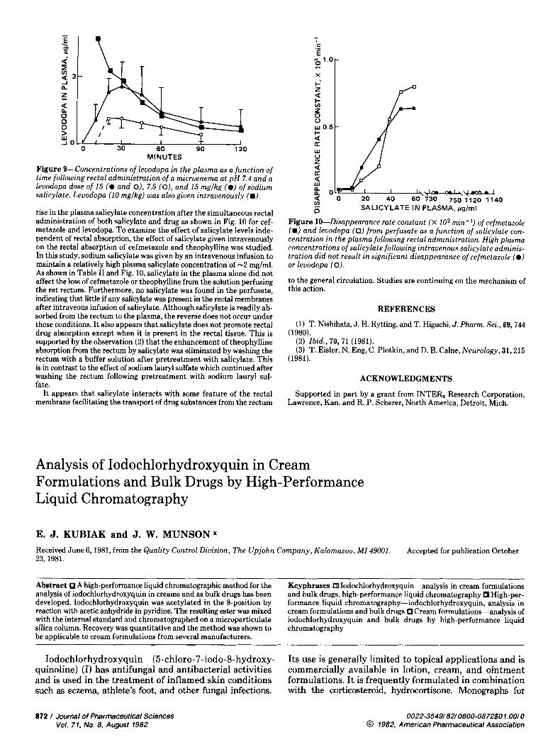
Figure 9-Concentrations of levodopa in the plasma as a function of t ime following rectal administration of a microenema at pH 7.4 and a levodopa dose o f 15 (@ and O), 7.5 (O), and 15 mglkg (@) of sodium salicylate. Levodopa (10 mglkg) was also given intravenously (m),
rise in the plasma salicylate concentration after the simultaneous rectal administration of both salicylate and drug as shown in Fig. 10 for cef- metazole and levodopa. To examine the effect of salicylate levels inde- pendent of rectal absorption, the effect of salicylate given intravenously on the rectal absorption of cefmetazole and theophylline was studied. In this study, sodium salicylate was given by an intravenous infusion to maintain a relatively high plasma salicylate concentration of -2 mglml. As shown in Table I1 and Fig. 10, salicylate in the plasma alone did not affect the loss of cefmetazole or theophylline from the solution perfusing the rat rectum. Furthermore, no salicylate was found in the perfusate, indicating that little if any salicylate was present in the rectal membranes after intraveous infusion of salicylate. Although salicylate is readily ab- sorbed from the rectum to the plasma, the reverse does not occur under these conditions. It also appears that salicylate does not promote rectal drug absorption except when it is present in the rectal tissue. This is supported by the observation (2) that the enhancement of theophylline absorption from the rectum by salicylate was eliminated by washing the rectum with a buffer solution after pretreatment with salicylate. This is in contrast to the effect of sodium lauryl sulfate which continued after washing the rectum following pretreatment with sodium lauryl sul- fate.
It appears that salicylate interacts with some feature of the rectal membrane facilitating the transport of drug substances from the rectum
Figure 10-Disappearance rate constant (X 102 min-I) of cefmetazole (m) and levodopa (0) f rom perfusate as a function of salicylate con- centration in the plasma following rectal administration. High plasma concentrations of salicylate following intravenous salicylate adminis- tration did not result in significant disappearance of cefmetazole (@) or levodopa (0).
to the general circulation. Studies are continuing on the mechanism of this action.
REFERENCES
(1) T. Nishihata, J. H. Rytting, and T. Higuchi, J. Pharm. Sci., 69,744
(2) Ibid., 70,71 (1981). (3) T. Eider, N. Eng, C. Plotkin, and D. B. Calne, Neurology, 31,215
(1980).
(1981).
ACKNOWLEDGMENTS
Supported in part by a grant from INTER, Research Corporation, Lawrence, Kan. and R. P. Scherer, North America, Detroit, Mich.
Analysis of Iodochlorhydroxyquin in Cream Formulations and Bulk Drugs by High-Performance Liquid Chromatography
E. J. KUBIAK and J. W. MUNSONX Received June 6,1981, from the Quality Control Division, The Upjohn Company, Kalamazoo, M I 49001. 23, 1981.
Accepted for publication October
Abstract A high-performance liquid chromatographic method for the analysis of iodochlorhydroxyquin in creams and as bulk drugs has been developed. Iodochlorhydroxyquin was acetylated in the &position by reaction with acetic anhydride in pyridine. The resulting ester was mixed with the internal standard and chromatographed on a microparticulate silica column, Recovery was quantitative and the method was shown to be applicable to cream formulations from several manufacturers.
Keyphrases Iodochlorhydroxyquin-analysis in cream formulations and bulk drugs, high-performance liquid chromatography High-per- formance liquid chromatography-iodochlorhydroxyquin, analysis in cream formulations and bulk drugs Cream formulations-analysis of iodochlorhydroxyquin and bulk drugs by high-performance liquid chromatography
Iodochlorhydroxyquin (5-chloro-7-iodo-8-hydroxy- quinoline) (I) has antifungal and antibacterial activities and is used in the treatment of inflamed skin conditions such as eczema, athlete's foot, and other fungal infections.
Its use is generally limited to topical applications and is commercially available in lotion, cream, and ointment formulations. It is frequently formulated in combination with the corticosteroid, hydrocortisone. Monographs for
872 1 Journal of Pharmaceutical Sciences Vol. 71. No. 8. August 1982
0022-3549/82/0800-0872$0 1.00/0 @ 1982, American Pharmaceutical Association