effect of mechanical properties of hydrogel nanoparticles ...peter/publications/soft.pdf · effect...
TRANSCRIPT

PAPER www.rsc.org/softmatter | Soft Matter
Effect of mechanical properties of hydrogel nanoparticles on macrophage celluptake†
Xavier Banquy,a Fernando Suarez,b Anteneh Argaw,c Jean-Michel Rabanel,a Peter Grutter,b
Jean-Francois Bouchard,c Patrice Hildgenab and Suzanne Giasson*adef
Received 2nd December 2008, Accepted 30th June 2009
First published as an Advance Article on the web 3rd August 2009
DOI: 10.1039/b821583a
Uptake and intracellular trafficking of hydrogel nanoparticles (NPs) of N,N-diethyl acrylamide and
2-hydroxyethyl methacrylate crosslinked with N,N0-methylene-bis-acrylamide were studied with
a RAW 264.7 murine macrophage cell line. Results show that the uptake rate, the mechanism of
internalization and the concentration of internalized NPs are correlated to the NP Young modulus.
Soft NPs are found to be internalized preferentially via macropinocytosis while the uptake of
stiff NPs is mediated by a clathrin-dependent mechanism. NPs with an intermediate Young modulus
exhibit multiple uptake mechanisms. The accumulation rate of the NPs into lysosomal
compartments of the cell is also dependent on the NP elasticity. Our results suggest that control over the
mechanical properties of hydrogel NPs can be used to tailor the cellular uptake mechanism and
kinetics of drug delivery.
Introduction
Different drug-carrier systems, such as liposomes, polymeric
micelles, polymer conjugates and polymeric nanoparticles have
been investigated for intracellular delivery of therapeutics.
Nanoparticles (NPs) based on crosslinked water-soluble poly-
mers forming hydrogels have shown to be very good candidates
for drug carriers1 because of their low cytotoxicity2 and their
tunable properties such as swelling and mechanical properties
which can be controlled by the crosslinking density. Moreover,
hydrogel NPs are relatively stable against biodegradation,3,4
show excellent transfection capabilities,5 promote uptake by
a large number of cell lines,2 and can also be used as intracellular
nanosensors.6–8
The efficiency of therapeutic carriers for intracellular delivery
largely depends on their interactions with the cell membrane and the
uptake mechanism. Most NPs are found to be internalized into cells
by means of endocytic mechanisms.9 The interactions of carriers
with cell membranes can be made specific by using ligands that can
bind to their receptors on cell membranes. This biorecognition
interaction usually increases the intracellular internalization.9
aFaculty of Pharmacy, Universit�e de Montr�eal, C.P. 6128, succursaleCentre-Ville, Montr�eal, Qu�ebec, H3C 3J7, CanadabDepartment of Physics, McGill University, 3600 University Street,Montr�eal, Qu�ebec, H3A 2T8, CanadacEcole d’Optom�etrie, Universit�e de Montr�eal, C.P. 6128, succursaleCentre-Ville, Montr�eal, Qu�ebec, H3C 3J7, CanadadGroupe de Recherche Universitaire sur le M�edicament de l’Universit�e deMontr�eal, C.P. 6128, succursale Centre-Ville, Montr�eal, Qu�ebec, H3C3J7, CanadaeDepartment of Chemistry, Universit�e de Montr�eal, C.P. 6128, succursaleCentre-Ville, Montr�eal, Qu�ebec, H3C 3J7, Canada. E-mail: [email protected]; Fax: +01 514 340 5290; Tel: +01 514 340 5175fCenter for Self-Assembled Chemical Structures, McGill University, 801Sherbrooke St. West, Montr�eal, Qu�ebec, H3A 2K6, Canada
† Electronic supplementary information (ESI) available: Cytotoxicitydata for the NPs by LDH and MTT assays. See DOI: 10.1039/b821583a
3984 | Soft Matter, 2009, 5, 3984–3991
The physicochemical parameters (i.e. surface charge, NP size
and substrate elasticity) of polymer-based carriers have been
shown to influence the interaction with cells or the intracellular
fate of the carriers. For example, the surface charge of poly-
amidoamine dendrimers was found to be a determinant factor in
their intracellular trafficking.5 Anionic dendrimers were found to
be internalized via caveolae-mediated endocytosis while neutral
and cationic dendrimers were mostly internalized by non-clathrin
and non-caveolae mediated endocytosis mechanisms in A549
cells.5 The surface charge of poly-D,L-lactic acid (PLA) NPs has
been shown to strongly affect the intracellular fate in Madin–
Darby canine kidney (MDCK) cells but to a less extent the entry
mechanism.10 Both cationic and anionic PLA NPs were found to
be mainly internalized by a clathrin-dependent mechanism. Only
anionic NPs were found in the lysosomal compartments while
significant amounts of cationic NPs were found to be exocytosed.
Particle size has also been shown to affect the uptake mechanism
and the intracellular fate.11 Small polystyrene particles (nano-
metre size) were found to exhibit higher intracellular uptake
compared to microspheres. Moreover, the small particles were
found to be internalized by B16F10 murine melanoma cells
almost exclusively via a clathrin-dependent pathway and their
intracellular trafficking was found to be microtubule dependent.
Larger particles were found to be internalized via a caveolae-
related mechanism and systematically exhibited slower kinetics to
reach lysosomal compartments. It was recently reported that
polystyrene NPs of 24 nm diameter can be internalized via a non-
clathrin and non-caveolae dependent mechanism. It was shown
that these NPs accumulate in non-digestive vesicles close to the
perinuclear membrane. These findings may open new routes to the
efficient delivery of sensitive molecules like DNA or proteins.12
The elasticity of the cell substrate was recently found to have
a significant effect on the uptake of polyplexes by murine
MC3T3-E1 preosteoblasts.13 An increase in substrate elasticity
led to an increase in the NP uptake which was correlated with
enhanced cell proliferation and survival. This study suggests that
This journal is ª The Royal Society of Chemistry 2009

the mechanical properties of the cell–surface (or cell–particle)
interface may influence the cellular uptake and intracellular
trafficking of drug carriers. In the present study, we investigated
the effect of the mechanical properties of hydrogel NPs, more
specifically their elasticity, on the cellular uptake and intracel-
lular fate in murine macrophage cells. Macrophage cells repre-
sent a good model system for the evaluation of the cytotoxicity of
nanoparticulate vectors and for the investigation of treatments
against pathologies related to bacterial invasion as most bacteria
and pathogens are sheltered from immune defense system in host
macrophages. The mechanical properties of the NPs were
controlled by varying the concentration of the crosslinking agent
used for the NP synthesis and measured using atomic force
microscopy (AFM). The NP elasticity was quantified by the
Young modulus. The uptake mechanisms of the different NPs
were identified using different inhibitors of known entry routes.
The intracellular localization of the NPs was assessed by fluo-
rescence microscopy. Colocalization assays with endosomal and
lysosomal markers allowed identification of the intracellular
pathways pursued by the NPs after their internalization.
Experimental
Materials
N,N-Diethyl acrylamide (DEA) was synthesized as previously
described.14 Unless stated, all chemical products were purchased
from Sigma-Aldrich (Oakville, ON, Canada). Solvents used were
analytical grade and all other chemicals were commercially
available reagent grade. 2-Hydroxyethyl methacrylate (HEMA)
was vacuum distilled prior to use. N,N0-Methylene-bis-acryl-
amide (BIS) was recrystallized twice from hexane. Sodium
dodecyl sulfate (SDS) was crystallized from pure ethanol.
Amonium persulfate (APS), rhodamine B, 1-ethyl-3-(3-dime-
thylaminopropyl)carbodiimide hydrochloride (EDC) and
N-hydroxysuccinimide (NHS) were used as received. Water was
obtained from a Milli-Q� Gradient System from Millipore
equipped with a Quantum� cartridge.
Nanoparticle synthesis
The NPs were synthesized as previously reported.14 The mono-
mers (DEA 90 mol% and HEMA 10 mol%, total amount of
5.3 � 10�3 mol), BIS and SDS (0.040 g) were added to 200 mL of
Milli-Q� water under stirring followed by nitrogen purging for
30 min at 70 �C. An aqueous solution of APS (0.120 g in 15 mL)
was subsequently added to the reacting medium. The reaction was
allowed to proceed for 4 h and then cooled at room temperature
before filtering the milky suspension through 2.0 mm Millipore
Isopore� membrane filters. The filtrate was centrifuged for 15 min
at 15 000 rpm. The polymer NPs were collected and purified by
dialysis for 1 week against Milli-Q� water. Four different batches of
NPs were prepared using four different concentrations of BIS. The
NPs used in this study are known to be thermosensitive with a lower
critical temperature (LCT) value ranging from 23 to 28 �C
depending on the monomer concentration.14 All nanoparticles were
labeled with rhodamine B using standard EDC/NHS coupling
reactions. After labeling, NPs were centrifuged for 15 min at
15 000 rpm and the supernatant was replaced by Milli-Q� water.
The polymer NPs were collected and purified by dialysis in Milli-Q�
water for 1 week.
This journal is ª The Royal Society of Chemistry 2009
Nanoparticle characterization
Particle size and zeta-potential measurements, and surface
topography. NP suspensions were prepared at a final concen-
tration of 0.1 mg mL�1 in filtered phosphate buffer saline (PBS)
and the particle size distribution was measured using dynamic
light scattering with a Nano-ZS Zetasizer (Malvern Instrument
Ltd., Malvern, Worcestershire, UK) at 37 �C in back-scattering
mode. The zeta potential of the NP suspensions was measured in
triplicate at 37 �C using the Zetasizer. Surface imaging was
carried out in tapping mode using a multimode AFM equipped
with a NanoScope IIIa controller (Digital Instruments, Santa
Barbara, CA). The NPs were deposited by spreading a 20 mL
suspension droplet on a freshly cleaved mica surface and stored
in 30% relative humidity (RH) atmosphere 2 h prior to AFM
analysis. All images were obtained in air and at room
temperature using commercial etched silicon cantilevers with
a tip of radius ranging from 5 to 10 nm and a spring constant of
20–100 N m�1. Image analysis was performed using the
NanoScope III software (version 512r2).
Immobilization of the nanoparticles on mica surfaces. To
determine their mechanical properties, the NPs were grafted onto
modified mica surfaces as described elsewhere.15 Briefly, large
sheets (1.5 � 4 cm) of freshly cleaved mica surfaces were first
plasma treated to activate OH groups on their surface as
described in detail elsewhere.16,17 Plasma activation was per-
formed for 5 min at 40 W using argon and water vapors at
a partial pressure of 80 mTorr and 300 mTorr respectively. After
the plasma treatment, the mica surfaces were left in the plasma
chamber under vacuum (0.5 mTorr) for 5 min. Then, the surfaces
were transferred to an evaporation chamber and stored under
vacuum (1.6 mmHg). The evaporation chamber was connected
via a valve to a small glass reservoir containing 100 mL of
APTES. After purging the evaporation chamber for 15 min, the
valve was opened allowing APTES vapors to react with the
activated mica surfaces. Evaporation was allowed to proceed for
4 h. The valve was closed and remnant APTES vapors were
pumped out for 2 h. The grafting reaction of APTES was
completed by annealing the surfaces for 30 min at 90 �C under
atmospheric pressure. Then, the surfaces bearing grafted APTES
molecules were immersed overnight in a 1% (w/w) water solution
of glutaraldehyde where the coupling reaction between APTES
amine function and glutaraldehyde carbonyl functional groups
took place in presence of the catalytic agent NaBH3CN. The
resulting glutaraldehyde-functionalized mica surfaces were
thoroughly rinsed with Milli-Q� water prior to NP deposition.
Deposition was performed using the horizontal convective
evaporation method.18 A small drop (25 mL) of a NP suspension
(0.1% w/w in DMEM) was placed between a glass applicator
and the functionalized mica surface prior to substrate trans-
lation. As the surface was translated at a fixed velocity, the NP
suspension drop could spread on the substrate forming a uniform
monolayer of nanoparticles. The final concentration of grafted
NPs on the surfaces was 2.6 � 0.4 NPs/mm2 as determined by
AFM imaging.
Mechanical properties of the nanoparticles. The mechanical
properties of the NPs grafted on mica surfaces were measured in
Soft Matter, 2009, 5, 3984–3991 | 3985
PBS at 37 �C using a Bioscope AFM equipped with a G-type
piezotube scanner, Nanoscope IIIa controller, and version 4.43r8
of the Nanoscope software (Veeco Metrology, Santa Barbara, CA).
Silicon nitride microlevers with spring constant k ¼ 0.01 N m�1
were used. Mica substrates bearing grafted NPs were immersed
in PBS at 37 �C 1 h prior to measurement. Localization of the
NPs was realized by scanning the surface in contact mode over
a 3� 3 mm2 area. The scanning area was progressively decreased to
visualize only one NP in the scanned area. Force profiles were
measured by approaching the tip to the NP on the surface at a fixed
velocity and recording the cantilever deflection. Young moduli
were extracted from experimental curves by fitting the force
profiles with the Hertz model for a conical indenter:19,20
F¼ 2E tanq
pð1�n2Þ d2 (1)
where F is the indentation force, E and n are the Young modulus
and the Poisson ratio of the NPs respectively, d is the indentation
and q is the half-opening angle of the indenter. One monolayer of
NPs was prepared for each of the different NP batches. Five to
ten force profiles were recorded on one single NP and six
different NPs were analyzed for all surfaces. The Young moduli
reported in this study represent the average value obtained from
all recorded force profiles.
Nanoparticle uptake by living cells
Cell culture. Macrophage cells, RAW 264.7 were a kind gift
from Professor J. C. Leroux (Universit�e de Montr�eal). Cells were
grown in complete medium consisting of Dulbecco’s modified
Eagle culture medium (DMEM) containing 10% (v/v) heat-
inactivated fetal bovine serum, 100 U mL�1 penicillin-G, and
100 mg mL�1 streptomycin (Invitrogen, Burlington, ON, Can-
ada) in a 5% CO2 atmosphere at 37 �C. The cells were used
between passage 4 and 15.
Uptake study. RAW 264.7 cells were plated in 96-well flat
bottom plates (2 � 105 cells/well) and allowed to adhere over-
night in complete medium. Then, the medium was removed and
replaced with DMEM. Various volumes of a fluorescently-
labeled NP suspension (0.1% w/w in DMEM) were added to the
cells to obtain different NP concentrations. The cellular uptake
of the NPs was allowed to occur for 2 h at 37 �C. The cells were
washed with PBS and lysed with 0.2% (w/v) Triton-X 100 in
0.2 N NaOH solution. The lysate fluorescence was measured on
a microplate reader at excitation and emission wavelengths of
550 and 580 nm respectively. The concentration of internalized
NPs was determined from an internal calibration curve. This
internal calibration curve, i.e. fluorescence intensity versus NP
concentration, was established by measuring the fluorescence of
a known NP concentration under the same conditions of cell
incubation but without washing the cells with PBS. The uptake
kinetics of the NPs were also investigated via fluorescence anal-
ysis. For a fixed NP concentration of 70 mg mL�1, the cells were
treated with the NPs at different incubation times and lysed as
previously described.
To determine the uptake mechanism of the NPs, cells were
incubated with endocytic and metabolic inhibitors prior to NP
introduction. The cells were incubated in complete medium in the
3986 | Soft Matter, 2009, 5, 3984–3991
presence of one inhibitor, whose concentration is given in Fig. 3,
for 1 h and then incubated with the NP solution (70 mg mL�1,
serum free medium) for 2 h. Cells were washed three times with
PBS, lysed and the lysate fluorescence was measured.
All uptake experiments were repeated three times in 96-well
plates from Corning (Costar, Corning, NY, USA).
The toxicity of the inhibitors at the concentrations used in the
study was assessed by standard MTT assay. Results showed that
no significant effect of cell viability could be observed of a period
of time of 24 h (data not shown).
Intracellular trafficking. To establish the intracellular path-
ways pursued by the NPs, RAW 264.7 cells were plated on round-
shape cover slips (Fisher Scientific, CA) and incubated overnight
at 37 �C in the presence of fluorescein isothiocyanate-labeled
dextran (DX-FITC, Mw 77 000 g mol�1, 1 mg mL�1 in complete
medium) as lysosome marker. Then, the cells were washed with
DMEM and 8 mL of NP suspension were added to reach a final
concentration of 70 mg NPs/mL per well. After 20 min of incu-
bation, the cells were washed with DMEM and incubated in
complete medium for different incubation times allowing intra-
cellular trafficking. Following incubation, the cells were fixed
with a 4 % (v/v) formaldehyde solution for 10 min and mounted
on glass slides for subsequent fluorescence microscopy analysis.
Staining of endocytic vesicles was done as follows. After
fixation, the cells were incubated in 250 mL of blocking solution
(2 % w/v bovine serum albumin in PBS) for 2 h. Then the cells
were incubated overnight at 4 �C in the presence of goat anti-
mouse EEA1 (clone 281.7 from Santa Cruz Biotechnology, CA,
USA) antibody at a concentration of 10 mg mL�1. After washing
with PBS, anti-goat ALEXA FLUOR� 488 (Invitrogen, Bur-
lington, CA) secondary antibody was added for 2 h.
All samples were analyzed on a fluorescence microscope
(Olympus IX71 microscope, Olympus, Markham, ON) using
filters optimized for the different fluorescent probes. Images were
obtained using an Evolution VF camera (MediaCybernetics,
Bethesda, MD) with a 100� objective lens and a constant
optimal exposure time. All images were analyzed with Image-
Pro� 5.0 (MediaCybernetics, Bethesda, MD) image analysis
software.
All trafficking experiments were repeated three times in 24-well
plates from Corning (Costar, Corning, NY, USA).
Statistical analysis of in vitro experiments. All in vitro experi-
ments were repeated three times and values are presented as the
mean � standard deviation. Statistical significance was deter-
mined by a Student’s t-test.
Results and discussion
Physical properties of the nanoparticles
All NPs were synthesized by emulsion polymerization at
a constant ratio of 9 : 1 of DEA:HEMA monomers. Four
different batches were prepared using four different crosslinker
concentrations ranging from 1.7 to 15 mol% (Table 1). A typical
AFM image of the particles deposited via horizontal convective
assembly on a bare mica substrate at 25 �C is illustrated in
Fig. 1(a). All NPs exhibit a round shape and a smooth surface
This journal is ª The Royal Society of Chemistry 2009
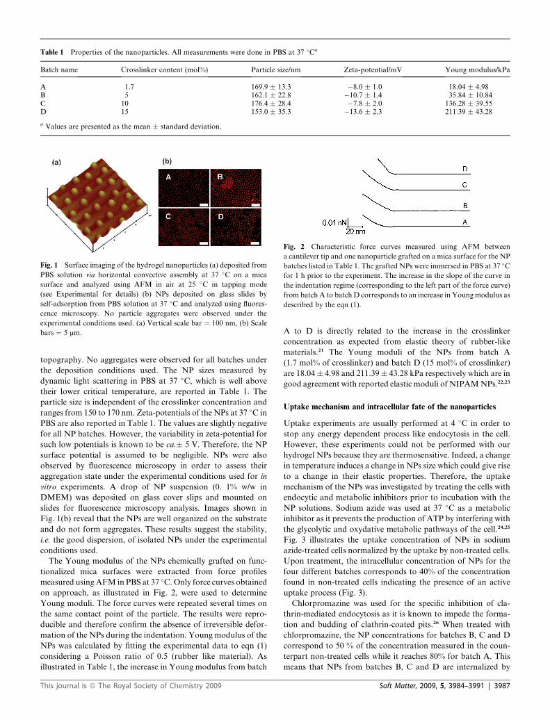
Table 1 Properties of the nanoparticles. All measurements were done in PBS at 37 �Ca
Batch name Crosslinker content (mol%) Particle size/nm Zeta-potential/mV Young modulus/kPa
A 1.7 169.9 � 13.3 �8.0 � 1.0 18.04 � 4.98B 5 162.1 � 22.8 �10.7 � 1.4 35.84 � 10.84C 10 176.4 � 28.4 �7.8 � 2.0 136.28 � 39.55D 15 153.0 � 35.3 �13.6 � 2.3 211.39 � 43.28
a Values are presented as the mean � standard deviation.
Fig. 2 Characteristic force curves measured using AFM between
a cantilever tip and one nanoparticle grafted on a mica surface for the NP
batches listed in Table 1. The grafted NPs were immersed in PBS at 37 �C
for 1 h prior to the experiment. The increase in the slope of the curve in
the indentation regime (corresponding to the left part of the force curve)
from batch A to batch D corresponds to an increase in Young modulus as
described by the eqn (1).
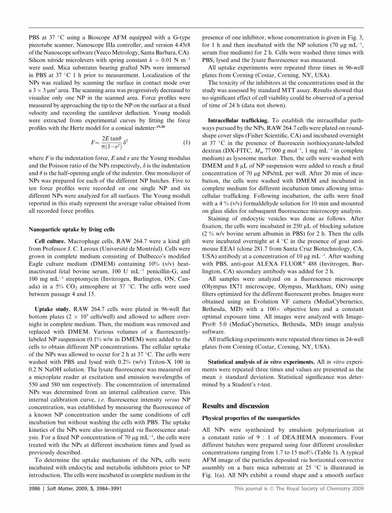
Fig. 1 Surface imaging of the hydrogel nanoparticles (a) deposited from
PBS solution via horizontal convective assembly at 37 �C on a mica
surface and analyzed using AFM in air at 25 �C in tapping mode
(see Experimental for details) (b) NPs deposited on glass slides by
self-adsorption from PBS solution at 37 �C and analyzed using fluores-
cence microscopy. No particle aggregates were observed under the
experimental conditions used. (a) Vertical scale bar ¼ 100 nm, (b) Scale
bars ¼ 5 mm.
topography. No aggregates were observed for all batches under
the deposition conditions used. The NP sizes measured by
dynamic light scattering in PBS at 37 �C, which is well above
their lower critical temperature, are reported in Table 1. The
particle size is independent of the crosslinker concentration and
ranges from 150 to 170 nm. Zeta-potentials of the NPs at 37 �C in
PBS are also reported in Table 1. The values are slightly negative
for all NP batches. However, the variability in zeta-potential for
such low potentials is known to be ca.� 5 V. Therefore, the NP
surface potential is assumed to be negligible. NPs were also
observed by fluorescence microscopy in order to assess their
aggregation state under the experimental conditions used for in
vitro experiments. A drop of NP suspension (0. 1% w/w in
DMEM) was deposited on glass cover slips and mounted on
slides for fluorescence microscopy analysis. Images shown in
Fig. 1(b) reveal that the NPs are well organized on the substrate
and do not form aggregates. These results suggest the stability,
i.e. the good dispersion, of isolated NPs under the experimental
conditions used.
The Young modulus of the NPs chemically grafted on func-
tionalized mica surfaces were extracted from force profiles
measured using AFM in PBS at 37 �C. Only force curves obtained
on approach, as illustrated in Fig. 2, were used to determine
Young moduli. The force curves were repeated several times on
the same contact point of the particle. The results were repro-
ducible and therefore confirm the absence of irreversible defor-
mation of the NPs during the indentation. Young modulus of the
NPs was calculated by fitting the experimental data to eqn (1)
considering a Poisson ratio of 0.5 (rubber like material). As
illustrated in Table 1, the increase in Young modulus from batch
This journal is ª The Royal Society of Chemistry 2009
A to D is directly related to the increase in the crosslinker
concentration as expected from elastic theory of rubber-like
materials.21 The Young moduli of the NPs from batch A
(1.7 mol% of crosslinker) and batch D (15 mol% of crosslinker)
are 18.04� 4.98 and 211.39� 43.28 kPa respectively which are in
good agreement with reported elastic moduli of NIPAM NPs.22,23
Uptake mechanism and intracellular fate of the nanoparticles
Uptake experiments are usually performed at 4 �C in order to
stop any energy dependent process like endocytosis in the cell.
However, these experiments could not be performed with our
hydrogel NPs because they are thermosensitive. Indeed, a change
in temperature induces a change in NPs size which could give rise
to a change in their elastic properties. Therefore, the uptake
mechanism of the NPs was investigated by treating the cells with
endocytic and metabolic inhibitors prior to incubation with the
NP solutions. Sodium azide was used at 37 �C as a metabolic
inhibitor as it prevents the production of ATP by interfering with
the glycolytic and oxydative metabolic pathways of the cell.24,25
Fig. 3 illustrates the uptake concentration of NPs in sodium
azide-treated cells normalized by the uptake by non-treated cells.
Upon treatment, the intracellular concentration of NPs for the
four different batches corresponds to 40% of the concentration
found in non-treated cells indicating the presence of an active
uptake process (Fig. 3).
Chlorpromazine was used for the specific inhibition of cla-
thrin-mediated endocytosis as it is known to impede the forma-
tion and budding of clathrin-coated pits.26 When treated with
chlorpromazine, the NP concentrations for batches B, C and D
correspond to 50 % of the concentration measured in the coun-
terpart non-treated cells while it reaches 80% for batch A. This
means that NPs from batches B, C and D are internalized by
Soft Matter, 2009, 5, 3984–3991 | 3987
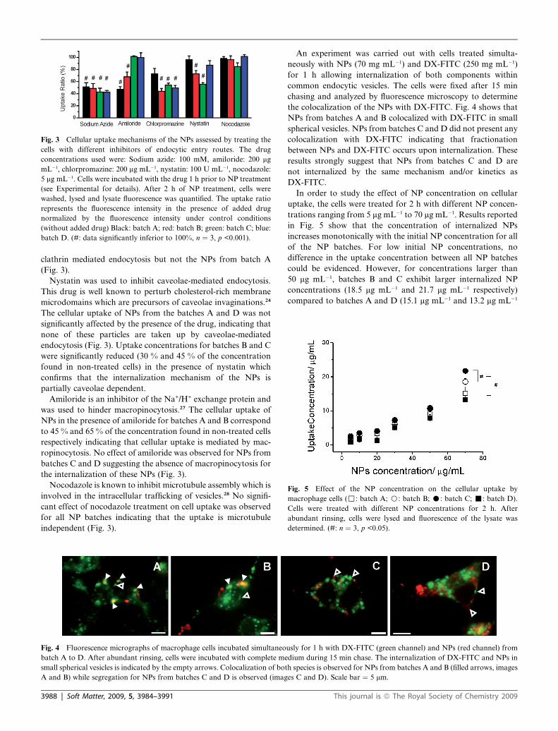
Fig. 3 Cellular uptake mechanisms of the NPs assessed by treating the
cells with different inhibitors of endocytic entry routes. The drug
concentrations used were: Sodium azide: 100 mM, amiloride: 200 mg
mL�1, chlorpromazine: 200 mg mL�1, nystatin: 100 U mL�1, nocodazole:
5 mg mL�1. Cells were incubated with the drug 1 h prior to NP treatment
(see Experimental for details). After 2 h of NP treatment, cells were
washed, lysed and lysate fluorescence was quantified. The uptake ratio
represents the fluorescence intensity in the presence of added drug
normalized by the fluorescence intensity under control conditions
(without added drug) Black: batch A; red: batch B; green: batch C; blue:
batch D. (#: data significantly inferior to 100%, n ¼ 3, p <0.001).
Fig. 5 Effect of the NP concentration on the cellular uptake by
macrophage cells (,: batch A; B: batch B; C: batch C; -: batch D).
Cells were treated with different NP concentrations for 2 h. After
abundant rinsing, cells were lysed and fluorescence of the lysate was
determined. (#: n ¼ 3, p <0.05).
clathrin mediated endocytosis but not the NPs from batch A
(Fig. 3).
Nystatin was used to inhibit caveolae-mediated endocytosis.
This drug is well known to perturb cholesterol-rich membrane
microdomains which are precursors of caveolae invaginations.24
The cellular uptake of NPs from the batches A and D was not
significantly affected by the presence of the drug, indicating that
none of these particles are taken up by caveolae-mediated
endocytosis (Fig. 3). Uptake concentrations for batches B and C
were significantly reduced (30 % and 45 % of the concentration
found in non-treated cells) in the presence of nystatin which
confirms that the internalization mechanism of the NPs is
partially caveolae dependent.
Amiloride is an inhibitor of the Na+/H+ exchange protein and
was used to hinder macropinocytosis.27 The cellular uptake of
NPs in the presence of amiloride for batches A and B correspond
to 45 % and 65 % of the concentration found in non-treated cells
respectively indicating that cellular uptake is mediated by mac-
ropinocytosis. No effect of amiloride was observed for NPs from
batches C and D suggesting the absence of macropinocytosis for
the internalization of these NPs (Fig. 3).
Nocodazole is known to inhibit microtubule assembly which is
involved in the intracellular trafficking of vesicles.28 No signifi-
cant effect of nocodazole treatment on cell uptake was observed
for all NP batches indicating that the uptake is microtubule
independent (Fig. 3).
Fig. 4 Fluorescence micrographs of macrophage cells incubated simultaneo
batch A to D. After abundant rinsing, cells were incubated with complete me
small spherical vesicles is indicated by the empty arrows. Colocalization of bot
A and B) while segregation for NPs from batches C and D is observed (imag
3988 | Soft Matter, 2009, 5, 3984–3991
An experiment was carried out with cells treated simulta-
neously with NPs (70 mg mL�1) and DX-FITC (250 mg mL�1)
for 1 h allowing internalization of both components within
common endocytic vesicles. The cells were fixed after 15 min
chasing and analyzed by fluorescence microscopy to determine
the colocalization of the NPs with DX-FITC. Fig. 4 shows that
NPs from batches A and B colocalized with DX-FITC in small
spherical vesicles. NPs from batches C and D did not present any
colocalization with DX-FITC indicating that fractionation
between NPs and DX-FITC occurs upon internalization. These
results strongly suggest that NPs from batches C and D are
not internalized by the same mechanism and/or kinetics as
DX-FITC.
In order to study the effect of NP concentration on cellular
uptake, the cells were treated for 2 h with different NP concen-
trations ranging from 5 mg mL�1 to 70 mg mL�1. Results reported
in Fig. 5 show that the concentration of internalized NPs
increases monotonically with the initial NP concentration for all
of the NP batches. For low initial NP concentrations, no
difference in the uptake concentration between all NP batches
could be evidenced. However, for concentrations larger than
50 mg mL�1, batches B and C exhibit larger internalized NP
concentrations (18.5 mg mL�1 and 21.7 mg mL�1 respectively)
compared to batches A and D (15.1 mg mL�1 and 13.2 mg mL�1
usly for 1 h with DX-FITC (green channel) and NPs (red channel) from
dium during 15 min chase. The internalization of DX-FITC and NPs in
h species is observed for NPs from batches A and B (filled arrows, images
es C and D). Scale bar ¼ 5 mm.
This journal is ª The Royal Society of Chemistry 2009
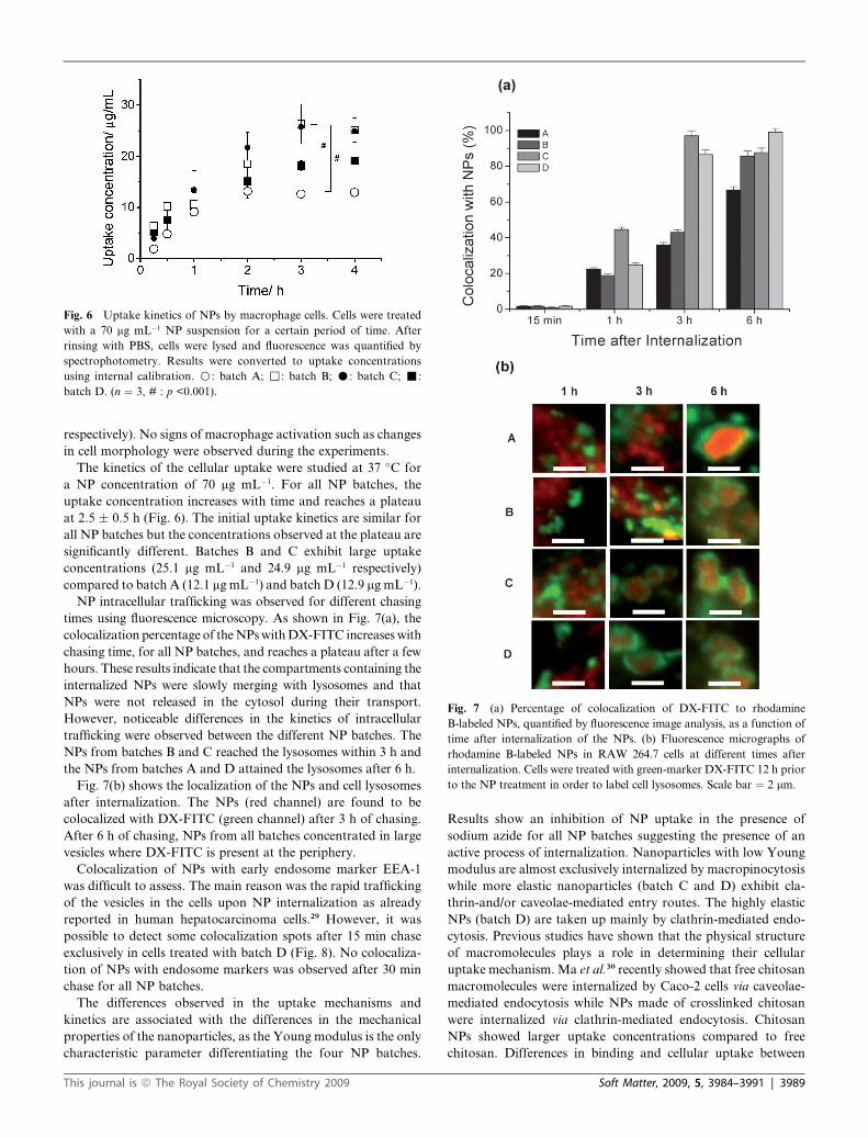
Fig. 6 Uptake kinetics of NPs by macrophage cells. Cells were treated
with a 70 mg mL�1 NP suspension for a certain period of time. After
rinsing with PBS, cells were lysed and fluorescence was quantified by
spectrophotometry. Results were converted to uptake concentrations
using internal calibration. B: batch A; ,: batch B; C: batch C; -:
batch D. (n ¼ 3, # : p <0.001).
Fig. 7 (a) Percentage of colocalization of DX-FITC to rhodamine
B-labeled NPs, quantified by fluorescence image analysis, as a function of
time after internalization of the NPs. (b) Fluorescence micrographs of
rhodamine B-labeled NPs in RAW 264.7 cells at different times after
internalization. Cells were treated with green-marker DX-FITC 12 h prior
to the NP treatment in order to label cell lysosomes. Scale bar ¼ 2 mm.
respectively). No signs of macrophage activation such as changes
in cell morphology were observed during the experiments.
The kinetics of the cellular uptake were studied at 37 �C for
a NP concentration of 70 mg mL�1. For all NP batches, the
uptake concentration increases with time and reaches a plateau
at 2.5 � 0.5 h (Fig. 6). The initial uptake kinetics are similar for
all NP batches but the concentrations observed at the plateau are
significantly different. Batches B and C exhibit large uptake
concentrations (25.1 mg mL�1 and 24.9 mg mL�1 respectively)
compared to batch A (12.1 mg mL�1) and batch D (12.9 mg mL�1).
NP intracellular trafficking was observed for different chasing
times using fluorescence microscopy. As shown in Fig. 7(a), the
colocalization percentage of the NPs with DX-FITC increases with
chasing time, for all NP batches, and reaches a plateau after a few
hours. These results indicate that the compartments containing the
internalized NPs were slowly merging with lysosomes and that
NPs were not released in the cytosol during their transport.
However, noticeable differences in the kinetics of intracellular
trafficking were observed between the different NP batches. The
NPs from batches B and C reached the lysosomes within 3 h and
the NPs from batches A and D attained the lysosomes after 6 h.
Fig. 7(b) shows the localization of the NPs and cell lysosomes
after internalization. The NPs (red channel) are found to be
colocalized with DX-FITC (green channel) after 3 h of chasing.
After 6 h of chasing, NPs from all batches concentrated in large
vesicles where DX-FITC is present at the periphery.
Colocalization of NPs with early endosome marker EEA-1
was difficult to assess. The main reason was the rapid trafficking
of the vesicles in the cells upon NP internalization as already
reported in human hepatocarcinoma cells.29 However, it was
possible to detect some colocalization spots after 15 min chase
exclusively in cells treated with batch D (Fig. 8). No colocaliza-
tion of NPs with endosome markers was observed after 30 min
chase for all NP batches.
The differences observed in the uptake mechanisms and
kinetics are associated with the differences in the mechanical
properties of the nanoparticles, as the Young modulus is the only
characteristic parameter differentiating the four NP batches.
This journal is ª The Royal Society of Chemistry 2009
Results show an inhibition of NP uptake in the presence of
sodium azide for all NP batches suggesting the presence of an
active process of internalization. Nanoparticles with low Young
modulus are almost exclusively internalized by macropinocytosis
while more elastic nanoparticles (batch C and D) exhibit cla-
thrin-and/or caveolae-mediated entry routes. The highly elastic
NPs (batch D) are taken up mainly by clathrin-mediated endo-
cytosis. Previous studies have shown that the physical structure
of macromolecules plays a role in determining their cellular
uptake mechanism. Ma et al.30 recently showed that free chitosan
macromolecules were internalized by Caco-2 cells via caveolae-
mediated endocytosis while NPs made of crosslinked chitosan
were internalized via clathrin-mediated endocytosis. Chitosan
NPs showed larger uptake concentrations compared to free
chitosan. Differences in binding and cellular uptake between
Soft Matter, 2009, 5, 3984–3991 | 3989
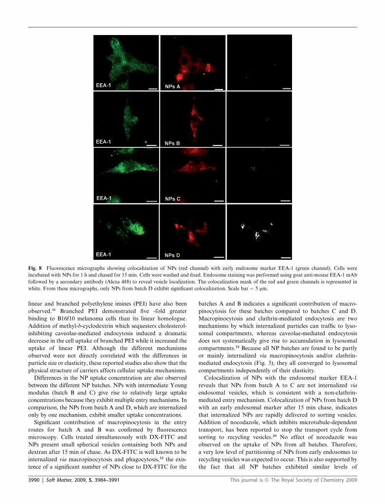
Fig. 8 Fluorescence micrographs showing colocalization of NPs (red channel) with early endosome marker EEA-1 (green channel). Cells were
incubated with NPs for 1 h and chased for 15 min. Cells were washed and fixed. Endosome staining was performed using goat anti-mouse EEA-1 mAb
followed by a secondary antibody (Alexa 488) to reveal vesicle localization. The colocalization mask of the red and green channels is represented in
white. From these micrographs, only NPs from batch D exhibit significant colocalization. Scale bar ¼ 5 mm.
linear and branched polyethylene imines (PEI) have also been
observed.31 Branched PEI demonstrated five -fold greater
binding to B16f10 melanoma cells than its linear homologue.
Addition of methyl-b-cyclodextrin which sequesters cholesterol-
inhibiting caveolae-mediated endocytosis induced a dramatic
decrease in the cell uptake of branched PEI while it increased the
uptake of linear PEI. Although the different mechanisms
observed were not directly correlated with the differences in
particle size or elasticity, these reported studies also show that the
physical structure of carriers affects cellular uptake mechanisms.
Differences in the NP uptake concentration are also observed
between the different NP batches. NPs with intermediate Young
modulus (batch B and C) give rise to relatively large uptake
concentrations because they exhibit multiple entry mechanisms. In
comparison, the NPs from batch A and D, which are internalized
only by one mechanism, exhibit smaller uptake concentrations.
Significant contribution of macropinocytosis in the entry
routes for batch A and B was confirmed by fluorescence
microscopy. Cells treated simultaneously with DX-FITC and
NPs present small spherical vesicles containing both NPs and
dextran after 15 min of chase. As DX-FITC is well known to be
internalized via macropinocytosis and phagocytosis,32 the exis-
tence of a significant number of NPs close to DX-FITC for the
3990 | Soft Matter, 2009, 5, 3984–3991
batches A and B indicates a significant contribution of macro-
pinocytosis for these batches compared to batches C and D.
Macropinocytosis and clathrin-mediated endocytosis are two
mechanisms by which internalized particles can traffic to lyso-
somal compartments, whereas caveolae-mediated endocytosis
does not systematically give rise to accumulation in lysosomal
compartments.33 Because all NP batches are found to be partly
or mainly internalized via macropinocytosis and/or clathrin-
mediated endocytosis (Fig. 3), they all converged to lysosomal
compartments independently of their elasticity.
Colocalization of NPs with the endosomal marker EEA-1
reveals that NPs from batch A to C are not internalized via
endosomal vesicles, which is consistent with a non-clathrin-
mediated entry mechanism. Colocalization of NPs from batch D
with an early endosomal marker after 15 min chase, indicates
that internalized NPs are rapidly delivered to sorting vesicles.
Addition of nocodazole, which inhibits microtubule-dependent
transport, has been reported to stop the transport cycle from
sorting to recycling vesicles.29 No effect of nocodazole was
observed on the uptake of NPs from all batches. Therefore,
a very low level of partitioning of NPs from early endosomes to
recycling vesicles was expected to occur. This is also supported by
the fact that all NP batches exhibited similar levels of
This journal is ª The Royal Society of Chemistry 2009
colocalization with lysosomal marker DX-FITC after 6 h chase.
It is noteworthy that microtubule-independent transport mech-
anism to lysosomes discards the possibility of uptake by
phagocytosis.34,35
The particle size can strongly affect the uptake mechanism.
Recently, it has been reported that particles of diameter lower
than 200 nm were preferentially internalized via clathrin-coated
pits, whereas particles of 500 nm diameter penetrated into the
cells via the caveolae.11 The multiplicity of entry mechanism
observed in our study for batches B and C can not be attributed
to particle size or aggregation of particles because the particle
size is not significatly different between the different NP batches
and no signs of aggregation induced by the high salinity of the
medium were evident during fluorescence microscopy. NP
aggregation in vitro was observed only after 2 to 3 h of chase. Our
results clearly indicate that the cellular uptake mechanism and
kinetics of the NPs are also regulated by the particle elasticity.
The mechanism by which NP elasticity modulates the entry
route remains unknown. However, the dynamics of the
membrane–NP interface deformation is most probably playing
a role in controlling the cellular uptake. As a NP approaches the
cell membrane, the thermal fluctuations of the membrane–NP
interface could impede the formation of stable contacts and
subsequent receptor recruitment. As soft NPs are expected to
deform more easily under thermal fluctuation forces than the
more elastic NPs, their binding with the cell membrane could be
less favorable. Therefore, soft NP uptake would be expected to
be mediated by a receptor-independent mechanism such as
macropinocytosis, while more elastic NPs could trigger receptor-
dependent internalization. This hypothesis is well supported
by our experimental data but also by studies using hard nano-
particles. Indeed, the internalization of hard polymeric or
non-polymeric nanoparticles has been reported to be clathrin-
dependent in the vast majority of cases.36
Conclusions
We investigated the relationship between the mechanical prop-
erties of hydrogel nanoparticles (NPs) assessed by the Young
modulus, and their cellular uptake mechanisms and kinetics. All
NPs were internalized by an energy-dependent mechanism but
the endocytic mechanism was shown to be dependant on the NP
elasticity. Soft NPs are preferentially internalized by macro-
pinocytosis while hard sphere uptake involves clathrin-mediated
routes. NPs exhibiting intermediate elasticity are found to be
internalized via multiple mechanisms which gives rise to a larger
uptake rate.
The present study confirmed that the elasticity of hydrogel
NPs influences their cellular entry routes. The control of
mechanical properties of hydrogel NPs represents a promising
route to the design of new drug carriers with specific subcellular
delivery.
Acknowledgements
This research was funded by the Groupe de Recherche Uni-
versitaire de l’Universit�e de Montr�eal and the Natural Sciences
and Engineering Research Council of Canada. A. A. is supported
by the E. A. Baker Foundation and The Institute of
This journal is ª The Royal Society of Chemistry 2009
Neuroscience, Mental Health and Addiction (INMHA) of the
Canadian Institutes of Health Research (CIHR). F. S. is sup-
ported by CONACYT. J-F. B. is supported by a Health
Research Foundation R&D Canadian Institutes of Health
Research Scholarship. The authors thank Professor R. E.
Prud’homme for providing access to his AFM facilities.
References
1 J. O. You and D. T. Auguste, Biomaterials, 2008, 29, 1950.2 K. T. Nguyen, K. P. Shukla, M. Moctezuma, A. R. C. Braden,
J. Z. Zhibing and H. L. Tang, J. Biomed. Mater. Res., Part A, 2008,88A, 1020.
3 Y. Li, G. Sun, J. Xu and K. L. Wooley, in Nanotechnology inTherapeutics: Current technology and applications, ed. J. Z. Hilt,Horizon Bioscience, Wymondham, UK, 2007, pp. 381–407.
4 J. L. Turner, Z. Y. Chen and K. L. Wooley, J. Controlled Release,2005, 109, 189.
5 O. P. Perumal, R. Inapagolla, S. Kannan and R. M. Kannan,Biomaterials, 2008, 29, 3469.
6 H. A. Clark, M. Hoyer, M. A. Philbert and R. Kopelman, Anal.Chem., 1999, 71, 4831.
7 H. A. Clark, R. Kopelman, R. Tjalkens and M. A. Philbert, Anal.Chem., 1999, 71, 4837.
8 H. Xu, J. W. Aylott, R. Kopelman, T. J. Miller and M. A. Philbert,Anal. Chem., 2001, 73, 4124.
9 I. A. Khalil, K. Kogure, H. Akita and H. Harashima, Pharmacol.Rev., 2006, 58, 32.
10 O. Harush-Frenkel, E. Rozentur, S. Benita and Y. Altschuler,Biomacromolecules, 2008, 9, 435.
11 J. Rejman, V. Oberle, I. S. Zuhorn and D. Hoekstra, Biochem. J.,2004, 377, 159.
12 S. K. Lai, K. Hida, S. T. Man, C. Chen, C. Machamer, T. A. Schroerand J. Hanes, Biomaterials, 2007, 28, 2876.
13 H. J. Kong, J. D. Liu, K. Riddle, T. Matsumoto, K. Leach andD. J. Mooney, Nat. Mater., 2005, 4, 460.
14 M. Colonne, Y. Chen, K. Wu, S. Freiberg, S. Giasson and X. X. Zhu,Bioconjugate Chem., 2007, 18, 999.
15 X. Banquy, X. X. Zhu and S. Giasson, J. Phys. Chem. B, 2008, 112,12208.
16 B. Liberelle, X. Banquy and S. Giasson, Langmuir, 2008, 24, 3280.17 B. Liberelle and S. Giasson, Langmuir, 2007, 23, 9263.18 B. G. Prevo and O. D. Velev, Langmuir, 2004, 20, 2099.19 K. L. Johnson, Contact mechanics, Cambridge University Press,
Cambridge, 1st edn, 1987.20 M. Radmacher, M. Fritz and P. K. Hansma, Biophys. J., 1995, 69,
264.21 P. J. Flory, Polymer, 1979, 20, 1317.22 O. Tagit, N. Tomczak and G. J. Vancso, Small, 2008, 4, 119.23 J. Wiedemair, M. J. Serpe, J. Kim, J. F. Masson, L. A. Lyon,
B. Mizaikoff and C. Kranz, Langmuir, 2007, 23, 130.24 C. Lamaze and S. L. Schmid, Curr. Opin. Cell Biol., 1995, 7, 573.25 J. Saraste, G. E. Palade and M. G. Farquhar, Proc. Natl. Acad. Sci.
U. S. A., 1986, 83, 6425.26 L. H. Wang, K. G. Rothberg and R. G. W. Anderson, J. Cell Biol.,
1993, 123, 1107.27 L. J. Hewlett, A. R. Prescott and C. Watts, J. Cell Biol., 1994, 124,
689.28 G. W. Zieve, D. Turnbull, J. M. Mullins and J. R. McIntosh, Exp.
Cell Res., 1980, 126, 397.29 C. Goncalves, E. Mennesson, R. Fuchs, J. P. Gorvel, P. Midoux and
C. Pichon, Mol. Ther., 2004, 10, 373.30 Z. S. Ma and L. Y. Lim, Pharm. Res., 2003, 20, 1812.31 F. P. Seib, A. T. Jones and R. Duncan, J. Controlled Release, 2007,
117, 291.32 U. Hacker, R. Albrecht and M. Maniak, J. Cell Sci., 1997, 110, 105.33 A. Ferrari, V. Pellegrini, C. Arcangeli, A. Fittipaldi, M. Giacca and
F. Beltram, Mol. Ther., 2003, 8, 284.34 S. D. Conner and S. L. Schmid, Nature, 2003, 422, 37.35 L. A. H. Allen and A. Aderem, Curr. Opin. Immunol., 1996, 8, 36.36 O. Harush-Frenkel, Y. Altschuler and S. Benita, Crit. Rev. Ther. Drug
Carrier Syst., 2008, 25, 485.
Soft Matter, 2009, 5, 3984–3991 | 3991