effect of mandibular advancement therapy on inflammatory
TRANSCRIPT

HAL Id: hal-02323256https://hal.archives-ouvertes.fr/hal-02323256
Submitted on 17 Dec 2020
HAL is a multi-disciplinary open accessarchive for the deposit and dissemination of sci-entific research documents, whether they are pub-lished or not. The documents may come fromteaching and research institutions in France orabroad, or from public or private research centers.
L’archive ouverte pluridisciplinaire HAL, estdestinée au dépôt et à la diffusion de documentsscientifiques de niveau recherche, publiés ou non,émanant des établissements d’enseignement et derecherche français ou étrangers, des laboratoirespublics ou privés.
Effect of mandibular advancement therapy oninflammatory and metabolic biomarkers in patients with
severe obstructive sleep apnoea: a randomisedcontrolled trial
Sylvain Recoquillon, Jean-Louis Pépin, Bruno Vielle, RamarosonAndriantsitohaina, Vanessa Bironneau, Frédérique Chouet-Girard, Bernard
Fleury, François Goupil, Sandrine Launois, M Carmen Martinez, et al.
To cite this version:Sylvain Recoquillon, Jean-Louis Pépin, Bruno Vielle, Ramaroson Andriantsitohaina, Vanessa Biron-neau, et al.. Effect of mandibular advancement therapy on inflammatory and metabolic biomarkers inpatients with severe obstructive sleep apnoea: a randomised controlled trial. Thorax, BMJ PublishingGroup, 2019, 74 (5), pp.496-499. �10.1136/thoraxjnl-2018-212609�. �hal-02323256�

Confidential: For Review Only
1
Effect of mandibular advancement therapy on inflammatory and metabolic biomarkers
in patients with severe obstructive sleep apnoea: A randomized controlled trial.
Sylvain Recoquillon1, Jean-Louis Pépin2,3, Bruno Vielle4, Ramaroson Andriantsitohaina1,
Vanessa Bironneau5; Frédérique Chouet-Girard6, Bernard Fleury7, François Goupil8, Sandrine
Launois7, M. Carmen Martinez1, Nicole Meslier1,9, Xuan-Lan Nguyen7, Audrey Paris8,
Pascaline Priou1,9, Renaud Tamisier2,3, Wojciech Trzepizur1,9, Frédéric Gagnadoux1,9
1INSERM UMR 1063, Angers, France; 2Université Grenoble Alpes, HP2, INSERM UMR
1042, Grenoble, France; 3CHU de Grenoble, Laboratoire EFCR, Clinique Universitaire de
Physiologie, Grenoble, France; 4Centre de Recherche Clinique, CHU d’Angers, Angers,
France ; 5Université de Poitiers, CHU, Service de Pneumologie, Poitiers ; 6Service de
Chirurgie Maxillo-faciale et Stomatologie, Centre Hospitalier, Le Mans, France; 7Université
Paris VI, Hôpital Saint-Antoine, Unité de Sommeil, Paris, France; 8Service de Pneumologie,
Centre Hospitalier, Le Mans, France; 9Département de Pneumologie, CHU d’Angers, Angers,
France.
Corresponding author:
Frédéric Gagnadoux, Département de Pneumologie, CHU, 4 rue Larrey, 49100, Angers,
France; Phone: 33 241353695; Fax: 33 241354974
e-mail: [email protected]
Word count: 1110
Page 1 of 24
https://mc.manuscriptcentral.com/thorax
Thorax
123456789101112131415161718192021222324252627282930313233343536373839404142434445464748495051525354555657585960

Confidential: For Review Only
2
Abstract
Systemic inflammation and metabolic disorders are among the mechanisms linking
obstructive sleep apnoea (OSA) and cardiovascular disease (CVD). In 109 patients with
severe OSA and no overt CVD, biomarkers of inflammation (C-reactive protein, interleukin-
6, tumour necrosis factor-α and its receptors, adiponectin, leptin and P-selectin), glucose and
lipid metabolism, and N-terminal pro-brain natriuretic peptide, were measured before and
after 2 months of treatment with a mandibular advancement device (MAD) (n=55) or a sham
device (n=54). MAD reduced the apnoea-hypopnoea index (p < 0.001) but had no effect on
circulating biomarkers compared to the sham device, despite high treatment adherence (6.6
h/night).
Trial registration number: Results, NCT01426607
Key words: obstructive sleep apnoea syndrome; mandibular advancement device;
inflammation; cytokines.
Page 2 of 24
https://mc.manuscriptcentral.com/thorax
Thorax
123456789101112131415161718192021222324252627282930313233343536373839404142434445464748495051525354555657585960
Confidential: For Review Only
3
Introduction
Systemic inflammation and metabolic disorders are among the intermediary mechanisms
linking obstructive sleep apnoea (OSA) with cardiovascular diseases (CVD).[1] Even still
debated owing to confounding factors, many studies have established an association between
inflammatory cytokines and indices of OSA severity.[2] Exposure of rodents to intermittent
hypoxia, a hallmark of OSA, stimulates inflammatory pathways and leads to cardiovascular or
metabolic disorders.[3]
Mandibular advancement devices (MAD) have emerged as the main therapeutic alternative to
continuous positive airway pressure (CPAP) for OSA. Despite the superior efficacy of CPAP
in reducing OSA severity, most trials comparing MAD and CPAP have reported similar
health outcomes.[4,5] In a recent multicentre randomized controlled trial, our group evaluated
the impact of 2 months of effective MAD therapy versus a sham device on endothelial
function in patients with severe OSA.[6] The aim of the present ancillary study was to
investigate the effects of MAD therapy on circulating levels of inflammatory and metabolic
biomarkers.
Methods
Study Design and Interventions
As described previously,[6] patients with severe OSA (Apnoea-Hypopnoea Index [AHI] > 30)
and no overt CVD, were randomly assigned to receive 2 months of treatment with either a
custom-made effective MAD or a sham device with objective measurement of treatment
adherence by an embedded microsensor (see online supplement Figure E1). Overnight in-lab
polysomnography and blood sample collections were performed at baseline and after the 2-
month treatment period. Additional details regarding patient recruitment, randomization
procedure, interventions, and the laboratory techniques that were used for measuring
circulating biomarkers of inflammation, metabolism, and N-terminal pro-brain natriuretic
Page 3 of 24
https://mc.manuscriptcentral.com/thorax
Thorax
123456789101112131415161718192021222324252627282930313233343536373839404142434445464748495051525354555657585960
Confidential: For Review Only
4
peptide (NT-ProBNP) are described in the online supplement. (ClinicalTrials.gov number:
NCT01426607, Results).
Statistics
A sample size calculation was performed for the primary endpoints of the main study.[6] Only
patients with available biomarkers before and after the 2-month intervention were included in
the present per-protocol analysis. Treatment effects (effective MAD versus sham device) were
modelled using linear regression (STATA version 13.1; STATA Corp., College Station, TX),
adjusting for baseline values and potential covariates. (See online supplement for further
details).
Results
Study flow and baseline characteristics
Among 150 randomized patients, 109 had available biomarkers before and after 2 months of
effective MAD (n=55) or sham device (n=54), and were included in the analysis (Figure 1).
There were no differences between baseline characteristics of all randomized patients,
patients included and not included in the present analysis (see online supplemental Table E1).
As shown in table 1, only gender was significantly different between the effective and the
sham device groups (p=0.04).
Page 4 of 24
https://mc.manuscriptcentral.com/thorax
Thorax
123456789101112131415161718192021222324252627282930313233343536373839404142434445464748495051525354555657585960

Confidential: For Review Only
5
Table 1: Baseline characteristics of the study population
All patients Effective MAD Sham device p value N 109 55 54 - Age, years 53.6 (10.1) 54.2 (10.1) 53.0 (10.2) 0.56 BMI, kg/m2 27.1 (3.3) 27.0 (3.1) 27.2 (3.4) 0.78 Women, % 12.1 18.5 5.7 0.04 Hypertension, % 20.9 21.1 20.7 0.96 Diabetes, % 5.7 3.8 7.5 0.68 Dyslipidaemia, % 11.2 13.0 9.4 0.56 Current smoker, % 21.1 13.7 28.3 0.07 ESS 9.1 (4.2) 9.0 (4.1) 9.2 (4.3) 0.81 AHI, n 41.0 [34.0-52.0] 40.0 [34.0-51.0] 44.5 [35.0-56.0] 0.27 ODI, n 31.7 (17.1) 30.2 (18.5) 33.2 (15.6) 0.39 SBP, mmHg 126.4 (15.1) 127.1 (14.3) 125.8 (16.0) 0.66 DBP, mmHg 77.6 (11.4) 76.6 (12.5) 78.6 (10.2) 0.38 Data are expressed as mean (standard deviation), median [interquartile range] or percentages Abbreviations: MAD, mandibular advancement device; BMI, body mass index; ESS, Epworth sleepiness score; AHI, apnoea-hypopnoea index; ODI, 3% oxygen desaturation index, SBP, office systolic blood pressure; DBP, office diastolic blood pressure.
Outcomes
In 84 patients with available objective adherence data, mean objective use rate was 6.6 (1.4)
h/night in the effective MAD group (n=42) versus 6.0 (2.0) h/night in the sham device group
(n=42) (p = 0.10). Only minor changes in body weight were observed during intervention
with no significant intergroup differences (p=0.9). Effective MAD was superior to sham
device in reducing the AHI (p<0.001), the oxygen desaturation index (p<0.001), and the
micro-arousal index (p=0.009) (see online supplemental Table E2). A complete response
(AHI reduced by ≥50% to less than 5/h) was obtained in 10% patients, a partial response
(AHI reduced by ≥50% to but persistent ≥ 5/h) in 50% of patients and 40% of patients were
poor responders with less than 50% reduction in AHI.
As shown in Table 3, we observed a decrease in tumour necrosis factor-α (TNF-α) in the
sham device group (p=0.01), an increase in leptin levels in the 2 groups (p=0.01), and an
increase in triglyceride levels in the effective MAD group (p=0.009). However, after
Page 5 of 24
https://mc.manuscriptcentral.com/thorax
Thorax
123456789101112131415161718192021222324252627282930313233343536373839404142434445464748495051525354555657585960

Confidential: For Review Only
6
adjustment for baseline value, age, gender, body mass index and baseline AHI, no significant
intergroup differences were observed for the outcome of inflammatory, metabolic biomarkers
and NT-ProBNP.
As effective and sham device groups were not well balanced for gender, we performed a post-
hoc analysis restricted to male patients. No significant intergroup differences in biomarker
outcomes were observed in the male population, except for a significant decrease in TNF-α
receptor 1 (TNF-R1) levels with effective MAD compared to the sham device group (-118.5
pg/mL [95%CI, -230.9; -6.2]; p=0.04). No significant intergroup differences in biomarkers
outcome were observed when the analysis was restricted to complete and partial responders to
effective MAD.
Page 6 of 24
https://mc.manuscriptcentral.com/thorax
Thorax
123456789101112131415161718192021222324252627282930313233343536373839404142434445464748495051525354555657585960

Confidential: For Review Only
7
Table 3: Impact of effective mandibular advancement device (MAD) versus sham device on
inflammatory, metabolic biomarkers and N-terminal pro-brain natriuretic peptide
Data are expressed as mean (standard deviation) or mean (95% confidence interval [CI]) Abbreviations: CRP, C-reactive protein; IL-6, interleukin-6; TNF-α, tumour necrosis factor-α; TNF-R1, tumour necrosis factor receptor 1; TNF-R2, tumour necrosis factor receptor 2; HDL-c, high-density lipoprotein cholesterol; LDL-c, low-density lipoprotein cholesterol; HOMA-IR, homeostasis model assessment-insulin resistance index; NT-ProBNP, N- terminal pro-brain natriuretic peptide. * Adjusted for baseline value, age, gender and body mass index and baseline apnoea-hypopnoea index. ** p<0.01 versus baseline. † p<0.05 versus effective MAD group at baseline. § p<0.05 versus baseline.
Effective MAD Sham device Adjusted intergroup
differences *
Baseline Follow-up Baseline Follow-up Mean
(95%CI) p value
CRP, mg/L 3.8 (6.9) 3.6 (7.3) 1.7 (1.4) † 3.6 (8.1) -0.9
(-3.5;1.7) 0.49
IL-6, pg/mL 1.1 (0.5) 1.3 (1.2) 1.1 (0.8) 1.3 (2.0) 0.3
(0.0;0.7) 0.07
TNF-α, pg/mL 6.7 (2.5) 6.4 (2.4) 8.1 (3.6) † 6.7 (3.3) § 0.2
(-0.9;1.3) 0.71
P-Selectin, pg/mL
72.8 (31.2) 71.8 (26.6) 76.0 (34.4) 79.2 (45.4) -6.7
(-21.1;7.7) 0.36
TNF-R1, pg/mL
1859.3 (485.6)
1831.5 (487.9)
1824.1 (507.5)
1909.8 (550.1)
-80.7 (-184.2;22.9)
0.12
TNF-R2, pg/mL
4119.6 (1501.1)
4139.3 (1793.6)
3985.2 (1406.3)
3941.2 (1265.6)
108.2 (-368.9;585.3)
0.65
Leptin, pg/mL 14.0 (12.8) 16.3 (15.2) § 11.4 (8.3) 13.4 (9.2) § 0.1
(-2.3;2.6) 0.91
Adiponectin, µg/mL
18.3 (8.9) 18.2 (9.3) 14.9 (6.8) † 14.3 (6.7) 0.5
(-1.0;2.0) 0.47
Triglycerides, mmol/L
1.4 (0.6) 1.6 (0.9) ** 1.5 (0.7) 1.5 (0.6) 0.3 (0.0;0.6) 0.05
Cholesterol, mmol/L
5.3 (0.8) 5.4 (1.0) 5.3 (0.8) 5.2 (0.8) 0.2 (-0.1;0.4) 0.23
HDL-c, mmol/L 1.3 (0.3) 1.3 (0.3) 1.3 (0.3) 1.3 (0.3) 0.0 (0.0;0.1) 0.30
LDL-c, mmol/L
3.4 (0.7) 3.4 (0.9) 3.3 (0.8) 3.3 (0.8) 0.0 (-0.2;0.3) 0.73
Glucose, mmol/L
5.2 (1.3) 5.4 (1.0) 5.4 (1.0) 5.6 (1.4) 0.0 (-0.2;0.3) 0.81
Insulin, mU/L 10.4 (7.4) 11.6 (8.3) 11.7 (9.3) 11.7 (8.3) 0.7 (-2.1;3.5) 0.60 HOMA-IR 2.6 (3.2) 3.0 (3.3) 3.0 (3.0) 3.2 (3.0) 0.4 (-0.4;1.1) 0.31 NT-ProBNP, pg/mL
296.8 (401.6)
252.5 (301.0)
189.8 (173.5) 184.3
(177.8) 12.0
(-40.9;64.9) 0.65
Page 7 of 24
https://mc.manuscriptcentral.com/thorax
Thorax
123456789101112131415161718192021222324252627282930313233343536373839404142434445464748495051525354555657585960

Confidential: For Review Only
8
Discussion
Only few studies flawed by small sample sizes and the absence of placebo group have
evaluated the impact of MAD therapy on circulating biomarkers, and reported discrepant
findings.[7–9] In this randomized controlled trial, 2 months of effective MAD therapy had no
effect on inflammatory and metabolic biomarkers in patients with severe OSA and no overt
CVD, despite high objective device adherence and a significant reduction in OSA severity.
As expected, the mean reduction in AHI obtained in the effective MAD group (about 53%)
was lower than that is usually obtained with CPAP.[4] However, the hypothesis of an
insufficient reduction of AHI with MAD is unlikely, as no significant changes in biomarkers
were observed when the analysis was restricted to complete and partial responders. It also
cannot be formally excluded that the 2-month intervention was too short. Previous studies
have reported improvements of inflammatory and metabolic profiles after 3 months to 1 year
of MAD therapy, but these studies were uncontrolled with no sham device group.[7–9]
Conversely, recent randomized trials showed no impact of 6 months to 1 year of CPAP
therapy on biomarkers despite a complete suppression of apneic events.[10,11] We observed a
higher proportion of women in the effective MAD group. It has been reported that female
patients with OSA exhibit a less severe inflammatory profile than men.[2] Interestingly, we
found that effective MAD therapy was associated with a significant improvement of TNF-R1
levels only in male patients. Between-group differences were also observed for baseline levels
of adiponectin, C-reactive protein and TNF-α, but adjustments for baseline values were
performed to mitigate this potential bias.
We acknowledge that our ancillary study may have been underpowered to detect small
changes in biomarkers, as the sample size was based on endothelial function, the primary
outcome of the main study.[6] Furthermore, our study population presented several
characteristics that may have contributed to a lower cardiovascular response and/or a floor
Page 8 of 24
https://mc.manuscriptcentral.com/thorax
Thorax
123456789101112131415161718192021222324252627282930313233343536373839404142434445464748495051525354555657585960

Confidential: For Review Only
9
effect of the intervention, including a relatively small proportion of metabolic disorders, no
overt CVD, and moderate daytime sleepiness at baseline. Further studies are required to
determine whether MAD therapy for OSA can improve circulating biomarkers in patients
who exhibit more severe inflammatory and metabolic dysfunction at baseline. However,
recent randomized trials and a meta-analysis have shown remarkably small effects of CPAP
on inflammatory and metabolic biomarkers, even in patients at high cardio-metabolic
risk.[10–12]
Conclusion
Two months of MAD therapy in patients with severe OSA reduced OSA severity, but had no
effect on inflammatory and metabolic biomarkers despite high treatment adherence.
Page 9 of 24
https://mc.manuscriptcentral.com/thorax
Thorax
123456789101112131415161718192021222324252627282930313233343536373839404142434445464748495051525354555657585960

Confidential: For Review Only
10
Acknowledgements
The authors thank Jean-Marie Chrétien, Sonia Dias Domingo, Caroline Erignoux, Anaïg le
Cam, Mathieu Lelay, and Marie Peeters for their help with trial administration, monitoring
and coordination, as well as data management.
Contributors
SR, J-LP, BV, RA, FC-G, BF, FGou, SL, CM, NM, VB, X-LN, AP, PP, RT, WT, and FGag
were substantially involved in the design of the study and critical revision of the paper for
important intellectual content.
SR, FGag, J-LP, BV, WT, were substantially involved in drafting the article.
All authors were substantially involved in data acquisition, data analysis, and/or interpretation
of data. All authors critically revised the article for important intellectual content.
Competing interests
The authors declare no competing interests.
Funding
This study was supported by a grant from the French Ministry of Health (PHRC-I 2010-06)
JLP and RT are supported supported by the French National Research Agency in the
framework of the "Investissements d’avenir” program (ANR-15-IDEX-02).
Patient consent
Obtained
Ethics approval
Comité de Protection des Personnes (CPP), Ouest II, Angers, France; No. 2010/14
Page 10 of 24
https://mc.manuscriptcentral.com/thorax
Thorax
123456789101112131415161718192021222324252627282930313233343536373839404142434445464748495051525354555657585960

Confidential: For Review Only
11
References
1 Drager LF, McEvoy RD, Barbe F, et al. Sleep Apnea and Cardiovascular Disease:
Lessons From Recent Trials and Need for Team Science. Circulation 2017;136:1840–50.
2 Gaines J, Vgontzas AN, Fernandez-Mendoza J, et al. Gender differences in the
association of sleep apnea and inflammation. Brain Behav Immun 2015;47:211–7.
3 Unnikrishnan D, Jun J, Polotsky V. Inflammation in sleep apnea: an update. Rev
Endocr Metab Disord 2015;16:25–34.
4 Sharples LD, Clutterbuck-James AL, Glover MJ, et al. Meta-analysis of randomised
controlled trials of oral mandibular advancement devices and continuous positive airway
pressure for obstructive sleep apnoea-hypopnoea. Sleep Med Rev 2016;27:108–24.
5 Bratton DJ, Gaisl T, Wons AM, et al. CPAP vs Mandibular Advancement Devices and
Blood Pressure in Patients With Obstructive Sleep Apnea: A Systematic Review and Meta-
analysis. JAMA 2015;314:2280–93.
6 Gagnadoux F, Pépin J-L, Vielle B, et al. Impact of Mandibular Advancement Therapy
on Endothelial Function in Severe Obstructive Sleep Apnea. Am J Respir Crit Care Med
2017;195:1244–52.
7 Galic T, Bozic J, Ivkovic N, et al. Effects of mandibular advancement device
treatment on arterial stiffness and glucose metabolism in patients with mild to moderate
obstructive sleep apnea: a prospective 1 year study. Sleep Breath 2016;20:69–77.
8 Niżankowska-Jędrzejczyk A, Almeida FR, Lowe AA, et al. Modulation of
inflammatory and hemostatic markers in obstructive sleep apnea patients treated with
mandibular advancement splints: a parallel, controlled trial. J Clin Sleep Med 2014;10:255–
62.
9 Yalamanchali S, Salapatas AM, Hwang MS, et al. Impact of mandibular advancement
devices on C-reactive protein levels in patients with obstructive sleep apnea. The
Page 11 of 24
https://mc.manuscriptcentral.com/thorax
Thorax
123456789101112131415161718192021222324252627282930313233343536373839404142434445464748495051525354555657585960

Confidential: For Review Only
12
Laryngoscope 2015;125:1733–6.
10 Chirinos JA, Gurubhagavatula I, Teff K, et al. CPAP, weight loss, or both for
obstructive sleep apnea. N Engl J Med 2014;370:2265–75.
11 Thunström E, Glantz H, Yucel-Lindberg T, et al. CPAP Does Not Reduce
Inflammatory Biomarkers in Patients With Coronary Artery Disease and Nonsleepy
Obstructive Sleep Apnea: A Randomized Controlled Trial. Sleep 2017;40.
12 Ning Y, Zhang T-S, Wen W-W, et al. Effects of continuous positive airway pressure
on cardiovascular biomarkers in patients with obstructive sleep apnea: a meta-analysis of
randomized controlled trials. Sleep Breath Published Online First: 22 April 2018.
doi:10.1007/s11325-018-1662-2
Page 12 of 24
https://mc.manuscriptcentral.com/thorax
Thorax
123456789101112131415161718192021222324252627282930313233343536373839404142434445464748495051525354555657585960

Confidential: For Review Only
13
Legends
Figure 1: Flow diagram showing trial allocation
Abbreviation: MAD, mandibular advancement device
Page 13 of 24
https://mc.manuscriptcentral.com/thorax
Thorax
123456789101112131415161718192021222324252627282930313233343536373839404142434445464748495051525354555657585960

Confidential: For Review Only
Figure 1: Flow diagram showing trial allocation Abbreviation: MAD, mandibular advancement device
273x210mm (144 x 144 DPI)
Page 14 of 24
https://mc.manuscriptcentral.com/thorax
Thorax
123456789101112131415161718192021222324252627282930313233343536373839404142434445464748495051525354555657585960

Confidential: For Review Only
1
Online only supplementary material
Effect of mandibular advancement therapy on inflammatory and metabolic biomarkers
in patients with severe obstructive sleep apnoea: A randomized controlled trial.
Authors: Sylvain Recoquillon1, Jean-Louis Pépin2,3, Bruno Vielle4, Ramaroson
Andriantsitohaina1, Vanessa Bironneau5; Frédérique Chouet-Girard6, Bernard Fleury7,
François Goupil8, Sandrine Launois7, M. Carmen Martinez1, Nicole Meslier1,9, Xuan-Lan
Nguyen7, Audrey Paris8, Pascaline Priou1,9, Renaud Tamisier2,3, Wojciech Trzepizur1,9,
Frédéric Gagnadoux1,9
1INSERM UMR 1063, Angers, France; 2Université Grenoble Alpes, HP2, INSERM UMR
1042, Grenoble, France; 3CHU de Grenoble, Laboratoire EFCR, Clinique Universitaire de
Physiologie, Grenoble, France; 4Centre de Recherche Clinique, CHU d’Angers, Angers,
France ; 5Université de Poitiers, CHU, Service de Pneumologie, Poitiers ; 6Service de
Chirurgie Maxillo-faciale et Stomatologie, Centre Hospitalier, Le Mans, France; 7Université
Paris VI, Hôpital Saint-Antoine, Unité de Sommeil, Paris, France; 8Service de Pneumologie,
Centre Hospitalier, Le Mans, France; 9Département de Pneumologie, CHU d’Angers, Angers,
France;
Page 15 of 24
https://mc.manuscriptcentral.com/thorax
Thorax
123456789101112131415161718192021222324252627282930313233343536373839404142434445464748495051525354555657585960

Confidential: For Review Only
2
Methods (expanded version)
Study Design and Patients
This randomized, single-blind, parallel-group trial conducted in five French sleep centres was
approved by our local ethics committee (Comité de Protection des Personnes, Ouest II,
Angers; No. 2010/14), and registered with ClinicalTrial.gov (NCT01426607). Patients with
severe obstructive sleep apnoea (OSA; Apnoea-Hypopnoea Index [AHI] > 30), aged 18-70
years, for whom mandibular advancement device (MAD) therapy was considered as second-
line therapy because of continuous positive airway pressure (CPAP) intolerance, were
assessed for eligibility. Exclusion criteria were body mass index (BMI) greater than or equal
to 32 kg/m2; history of cardiovascular disease (CVD) including coronary heart disease, heart
failure, arrhythmias, and stroke; coexisting sleep disorders other than OSA; central sleep
apnoea defined by a central apnoea index greater than or equal to 5; severe daytime sleepiness
defined by an Epworth Sleepiness Scale greater than or equal to 16; and inadequate dental
structure or temporomandibular joint disease contraindicating MAD treatment as assessed by
a dentist. All patients provided their written informed consent to participate in the study.
Randomization
Patients were randomly assigned to receive 2 months of treatment with either effective MAD
or a sham device according to a 1:1 allocation using a computer-generated randomization list
stratified by site with permuted blocks of random sizes. The effective MAD was custom-
made, consisting of an adjustable two-piece acrylic oral appliance (AMO; Orthosom,
Beaucouzé, France) with attachments of various sizes allowing adjustment of mandibular
advancement (Figure E1). The sham device consisted of the upper appliance only and did not
advance the mandible. As previously described,[1] treatment adherence with the effective
MAD and the sham device was objectively measured by a validated embedded microsensor
thermometer (TheraMon®, IFT Handels- und Entwicklungsgesellschaft GmbH,
Page 16 of 24
https://mc.manuscriptcentral.com/thorax
Thorax
123456789101112131415161718192021222324252627282930313233343536373839404142434445464748495051525354555657585960

Confidential: For Review Only
3
Handelsagentur Gschladt, Hargelsberg, Austria). With ethics committee approval, patient
blinding was achieved by concealing the less effective nature of the sham device.[1,2]
Interventions
At baseline, patients underwent clinical assessment, overnight in-lab polysomnography (PSG)
followed by collection of blood samples. All patients underwent a 6-week MAD
acclimatization period, during which the mandible was incrementally advanced by 1-mm
steps every 1 or 2 weeks until symptom relief or until adverse effects prevented further
advancement. Patients were then submitted to a one-week washout period, after which they
were allocated to receive 2 months of treatment with either effective MAD or the sham
device. Clinical assessment, PSG with effective MAD or sham device, and blood sample
collection were repeated after the 2-month treatment period. Objective treatment adherence
was also calculated after the 2-month treatment period.
Measurements of inflammatory and metabolic biomarkers
After overnight fasting, blood samples were collected in EDTA tubes (Vacutainers, Becton
Dickinson, Le Pont de Claix, France) from a peripheral vein using a 21-gauge needle to
minimize platelet activation, and were processed for assays within 2 hours. Samples were
centrifuged for 20 minutes at 250 g. Platelet-rich plasma was harvested and centrifuged 20
minutes at 1500 g to obtain platelet-free plasma (PFP). PFP was aliquoted and stored at 80°C
for subsequent use.
The assessment of inflammatory biomarkers was performed on PFP with an enzyme-linked
immunosorbent assay (ELISA) using MESO QuickPlex SQ 120 assay (MSD, Rockville, Md.,
USA). Each assay was performed in duplicate in order to verify the intra-assay variability
using samples randomly prepared. Then, if the coefficient of variability was less than 10%,
the mean of each duplicate was calculated. Measures with a coefficient of variability > 10%
were considered as unavailable biological data (n=5). A single assay was used to measure
Page 17 of 24
https://mc.manuscriptcentral.com/thorax
Thorax
123456789101112131415161718192021222324252627282930313233343536373839404142434445464748495051525354555657585960

Confidential: For Review Only
4
plasma levels of adiponectin, C-reactive protein (CRP) and leptin. Multiplex assays were used
for the assessment of interleukin-6 (IL-6), tumor necrosis factor-α (TNF-α), N- terminal pro-
brain natriuretic peptide (NT-ProBNP), tumor necrosis factor receptor 1 and 2 (TNF-R1 and
TNF-R2) and P-selectin. Both assays depended on the electro chemiluminescent compound
SULFO-TAG™ linked to a detection antibody. All the solutions were supplied by MSD and
experiments were performed according to manufacturer’s protocol.
For single assays, PFP was first 1000-fold diluted for adiponectin and CRP in an assay diluent
whereas detection of leptin did not require any dilution. Then, the samples were incubated in
96-wells plate coated by a capture antibody directed against the protein of interest. After 2
hours of incubation at room temperature with vigorous shaking, the plate was washed three
times with PBS supplemented with 0.05% of Tween-20. Then, SULFO-TAG-labelled
detection antibodies were added for another 2 hours at room temperature with vigorous
shaking. At the end, three other washes were performed before addition of a read solution.
Then, voltage stimulation of plate electrodes induced chemilumescence read by the
instrument (MESO QuickPlex SQ 120, USA). The level of the protein of interest is
proportional to the emitted light present in the PFP and is calculated thanks to a calibration
curve. The limits of detection (LOD) of CRP, adiponectin and leptin were 1.33 pg/mL, 0.005
ng/mL and 43 pg/mL respectively. None of the measured values were outside of these limits.
For the multiplex assay, samples were 2-fold diluted and then incubated for 2 hours with
vigorous shaking in a 96-wells plate. Each well is seeded in 6 distinct spot coated with capture
antibody specific of the protein of interest. After three washes with PBS supplemented with
0.05% of Tween-20, SULFO-TAG-labelled detection antibodies were added for another 2
hours at room temperature with vigorous shaking. At the end, three other washes were
performed before addition of a read solution. Then, voltage stimulation of plate electrodes
induced chemilumescence read by the instrument (MESO QuickPlex SQ 120, USA). The
Page 18 of 24
https://mc.manuscriptcentral.com/thorax
Thorax
123456789101112131415161718192021222324252627282930313233343536373839404142434445464748495051525354555657585960
Confidential: For Review Only
5
level of the protein of interest is proportional to the emitted light present in the PFP and is
calculated thanks to a calibration curve. The calculated LOD of IL-6, TNF-α, TNF-RI, TNF-
RII, P-selectin and NT-proBNP were 0.06 pg/mL, 0.04 pg/mL, 0.569 pg/mL, 0.102 pg/mL,
29.9 pg/mL and 0.311 pg/mL respectively. None of the measured values were outside of these
limits.
Plasma glucose, insulin, triglycerides, total serum cholesterol, and high-density lipoprotein
serum cholesterol (HDL-c) were directly measured in accredited laboratories using standard
techniques. Low-density lipoprotein serum cholesterol (LDL-c) was calculated. The
homeostasis model assessment resistance index (HOMA-IR.) was calculated from fasting
glucose and insulin concentrations, as follows: insulin (mIU/l) * glucose (mmol/l)/22.5.
Statistics
A sample size calculation was performed for the primary endpoints of the main study.[1] In
the present ancillary study, we performed a per-protocol analysis including patients with
available biomarkers before after 2 months of effective MAD or sham device. Continuous
variables were described as mean (SD) or mean (95% confidence interval [CI]) for variables
with a normal distribution and as median (interquartile range) for variables with a non-normal
distribution. Normality of distribution was assessed using the Kolmogorov–Smirnov test.
Normal variables were analysed using an unpaired Student’s t test for intergroup difference
and a paired t test for intragroup difference. Linear regression analysis was used to adjust for
baseline values and potential covariates. Non-normal variables were analysed using the
Mann-Whitney test for intergroup difference and the Wilcoxon signed rank test for intragroup
difference. The Chi-square test and Fisher’s exact test were used for categorical variables, as
appropriate. All reported p values are two-sided. A p value less than or equal to 0.05 was
considered to indicate statistical significance. All analyses were performed using STATA
version 13.1 (STATA Corp., College Station, TX).
Page 19 of 24
https://mc.manuscriptcentral.com/thorax
Thorax
123456789101112131415161718192021222324252627282930313233343536373839404142434445464748495051525354555657585960
Confidential: For Review Only
6
Results
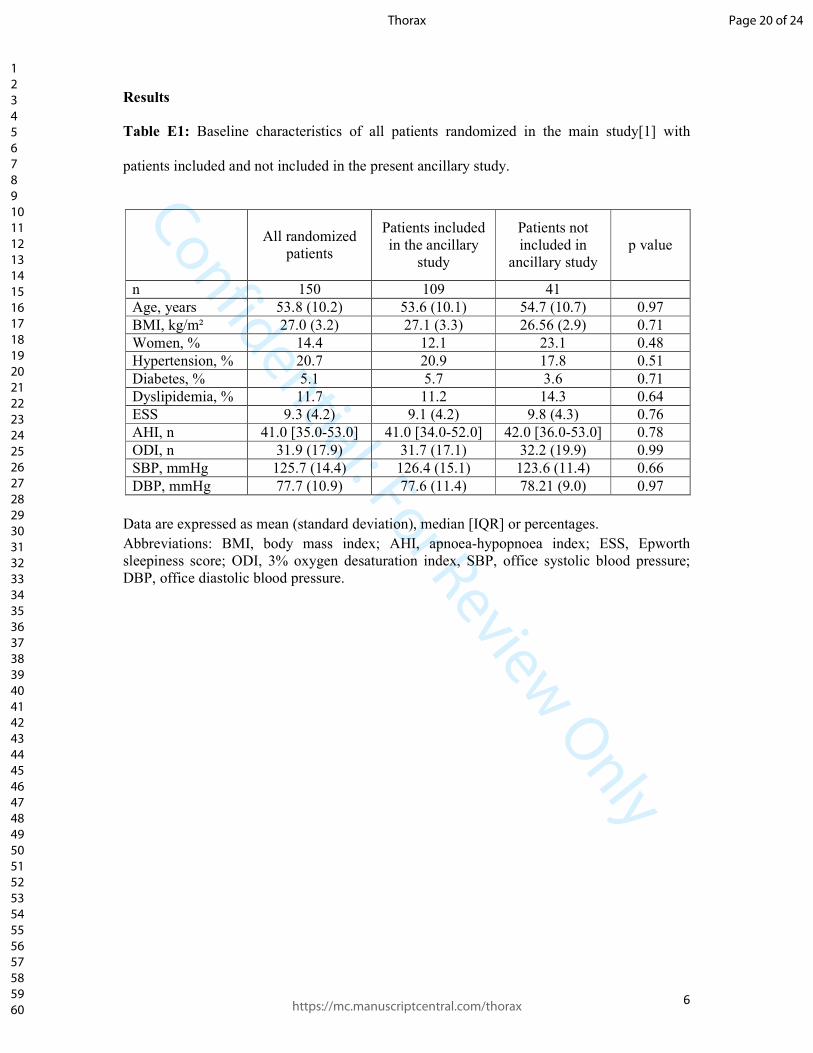
Table E1: Baseline characteristics of all patients randomized in the main study[1] with
patients included and not included in the present ancillary study.
Data are expressed as mean (standard deviation), median [IQR] or percentages. Abbreviations: BMI, body mass index; AHI, apnoea-hypopnoea index; ESS, Epworth sleepiness score; ODI, 3% oxygen desaturation index, SBP, office systolic blood pressure; DBP, office diastolic blood pressure.
All randomized
patients
Patients included in the ancillary
study
Patients not included in
ancillary study p value
n 150 109 41 Age, years 53.8 (10.2) 53.6 (10.1) 54.7 (10.7) 0.97 BMI, kg/m² 27.0 (3.2) 27.1 (3.3) 26.56 (2.9) 0.71 Women, % 14.4 12.1 23.1 0.48 Hypertension, % 20.7 20.9 17.8 0.51 Diabetes, % 5.1 5.7 3.6 0.71 Dyslipidemia, % 11.7 11.2 14.3 0.64 ESS 9.3 (4.2) 9.1 (4.2) 9.8 (4.3) 0.76 AHI, n 41.0 [35.0-53.0] 41.0 [34.0-52.0] 42.0 [36.0-53.0] 0.78 ODI, n 31.9 (17.9) 31.7 (17.1) 32.2 (19.9) 0.99 SBP, mmHg 125.7 (14.4) 126.4 (15.1) 123.6 (11.4) 0.66 DBP, mmHg 77.7 (10.9) 77.6 (11.4) 78.21 (9.0) 0.97
Page 20 of 24
https://mc.manuscriptcentral.com/thorax
Thorax
123456789101112131415161718192021222324252627282930313233343536373839404142434445464748495051525354555657585960

Confidential: For Review Only
7
Table E2: Impact of effective mandibular advancement device (MAD) versus sham device on
daytime polysomnographic indices
Data are expressed as mean (standard deviation), median [interquartile range] or mean (95% confidence interval [CI]) Abbreviations: AHI, apnoea-hypopnoea index; AI, apnoea index; ODI, 3% oxygen desaturation index; TST, total sleep time; REM, rapid eye movement sleep; MAI, micro-arousal index. * Adjusted for baseline value, age, gender and body mass index † p<0.001 versus baseline; ‡ p<0.01 versus baseline; § p<0.05 versus baseline ll p<0.001; ** p<0.01
Effective MAD Sham device Adjusted intergroup
differences * Baseline Follow-up Baseline Follow-up Mean (95%CI)
AHI, n 40.0
[34.0-51.0] 17.5 †
[11.5-25.0] 44.5
[35.0;56.0] 38.5 †
[19.0;51.0] -17.2 (-23.6;-10.8) ll
ODI, n 30.2 (18.5) 15.2 (10.8) † 33.2 (15.6) 28.0 (17.4) -11.9 (-17.6;-6.1) ll TST, min 402.7 (74.4) 397.9 (61.9) 376.2 (67.8) 372.3 (71.2) 14.9 (-11.2;41.0) N1sleep, min
40.9 (32.8) 32.4 (24.5) 36.6 (30.1) 29.6 (21.8) 0.2 (-7.4;7.8)
N2 sleep, min
200.3 (67.3) 194.9 (46.6) 202.0 (69.9) 196.5 (58.7) -2.3 (-22.6;18.0)
N3 sleep, min
69.5 (45.7) 78.1 (38.7) 61.4 (43.0) 65.8 (40.7) 11.4 (-2.1;24.9)
REM, min
87.0 (30.7) 91.8 (28.6) 68.2 (36.0) 77.3 (34.7) § 6.0 (-6.3;18.2)
MAI, n 32.7 (16.9) 23.1 (11.9) ‡ 36.3 (12.8) 31.9 (13.9) § -7.2 (-12.5;-1.8) **
Page 21 of 24
https://mc.manuscriptcentral.com/thorax
Thorax
123456789101112131415161718192021222324252627282930313233343536373839404142434445464748495051525354555657585960

Confidential: For Review Only
8
E References
1 Gagnadoux F, Pépin J-L, Vielle B, et al. Impact of Mandibular Advancement Therapy on
Endothelial Function in Severe Obstructive Sleep Apnea. Am J Respir Crit Care Med
2017;195:1244–52.
2 Gotsopoulos H, Kelly JJ, Cistulli PA. Oral appliance therapy reduces blood pressure in
obstructive sleep apnea: a randomized, controlled trial. Sleep 2004;27:934–41.
Page 22 of 24
https://mc.manuscriptcentral.com/thorax
Thorax
123456789101112131415161718192021222324252627282930313233343536373839404142434445464748495051525354555657585960

Confidential: For Review Only
9
Figure Legends
Figure E1: The effective mandibular advancement device used in the study. Full-coverage
acrylic appliances designed to fit onto the upper and lower dental arches are connected by
acrylic plates of various sizes allowing adjustment of mandibular advancement. The
microsensor thermometer was sealed into the upper arch of the device. The sham device
consisted of the upper appliance alone
Page 23 of 24
https://mc.manuscriptcentral.com/thorax
Thorax
123456789101112131415161718192021222324252627282930313233343536373839404142434445464748495051525354555657585960

Confidential: For Review Only
Figure E1: The effective mandibular advancement device used in the study. Full-coverage acrylic appliances designed to fit onto the upper and lower dental arches are connected by acrylic plates of various sizes
allowing adjustment of mandibular advancement. The microsensor thermometer was sealed into the upper arch of the device. The sham device consisted of the upper appliance alone
342x240mm (144 x 144 DPI)
Page 24 of 24
https://mc.manuscriptcentral.com/thorax
Thorax
123456789101112131415161718192021222324252627282930313233343536373839404142434445464748495051525354555657585960