complications related to mandibular advancement by ... · complications related to mandibular...
TRANSCRIPT
Complications related to mandibular advancement
by bilateral sagittal split osteotomy:
a retrospective study of 132 patients
Ane Landsverk Vagle
Faculty of Dentistry
University of Oslo, Norway
May 2007

Complications related to mandibular advancement
by bilateral sagittal split osteotomy:
a retrospective study of 132 patients
Ane Vagle
Thesis presented as partial fulfillment of the requirements for
the degree Master of Science in Dentistry
Supervised by
Professor Lisen Espeland, Department of Orthodontics, Dental Faculty and
Professor Per Skjelbred, Department of Maxillofacial Surgery, Ullevaal University Hospital
University of Oslo, Norway
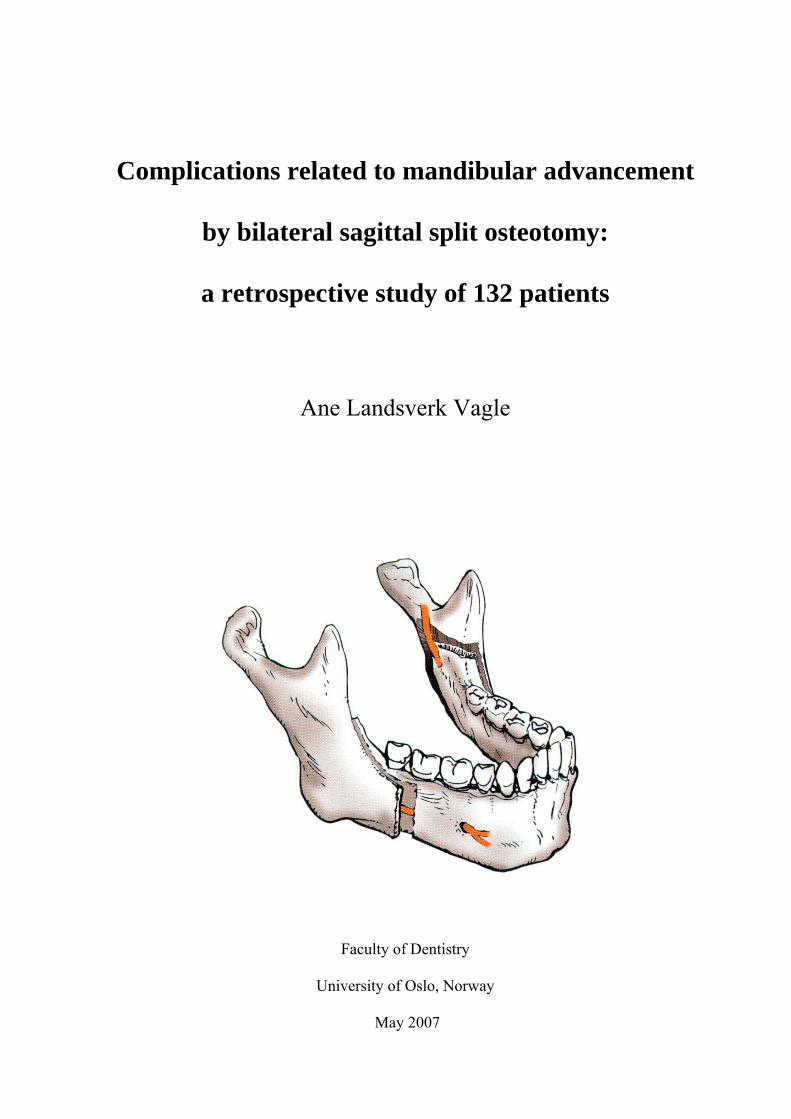
Illustration on front page: from Westermark 1999
2
CONTENTS
page Summary 4
Introduction 5
Aim 6
Materials and Methods 7
Results 11
Discussion 18
Conclusions 25
References 27
Tables 35
Appendix 41

Summary Purpose: The aim of the present study was to evaluate intraoperative and postoperative
complications associated with bilateral sagittal split osteotomy (BSSO), and to analyse
whether complications were associated with age at operation and patient’s satisfaction with
the result. An additional objective was to analyse whether nerve damage during surgery was
related to persistent neurosensory disturbance in the facial skin.
Sample: 132 patients with skeletal Class II malocclusion operated between 1990 and 2002
with BSSO for advancement of the mandible participated in the study. Surgery was
performed at Department of Maxillofacial Surgery Ullevaal University Hospital, and all
subjects were followed for 3 years at Department of Orthodontics, University of Oslo.
Methods: Intra- and postoperative complications were assessed from the medical records.
Neurosensory function and patient’s satisfaction with outcome were assessed from clinical
examination and patient’s response in questionnaires 3 years after surgery. Associations
between variables were analyzed by Chi square test, Fisher exact test or t- test.
Results and Conclusions: The inferior alveolar nerve was inadvertently injured in 36 sides
(14%), suboptimal splits occurred in 15 sides (6% of the sides, 11% of the patients), 18
patients (14%) experienced postoperative infection, and osteosynthesis was removed in 10
patients (8%). Three years after operation, 76% of the patients reported not having normal
sensation and 15% reported the alterations in sensibility to be distressing during daily life.
Age had no significant effect on the prevalence of complications with exception of distress
related to sensory disturbance. Dissatisfaction tended to increase among patients
experiencing complications, but the only statistically significant relationship was observed
among subjects reporting distress because of their altered sensation. Registration of visible
nerve injury during surgery was significantly reflected in the degree of neurosensory
disturbances.
4

Introduction
Bilateral sagittal split osteotomy (BSSO) is a well documented standardized and relatively
safe operation to correct jaw deformities such as mandibular retrognathism. The surgical
procedure consists of bilateral osteotomies of the mandible, in which the angulus area is
exposed by intraoral incisions and split in a near sagittal plane on both sides. The distal
segments are then slid relative to the proximal segments and fixed with screws or plates. The
inferior alveolar nerve enters the mandibular foramen at the medial side of the ramus,
courses through the mandibular body and innervates teeth and associated structures. A
terminal branch, the mental nerve, exits at the mental foramen and innervates the skin of lip
and chin.
During the procedure the inferior alveolar nerve is invariably damaged as evidenced by
initial, complete anaesthesia in the distribution of the mental nerve followed by prolonged or
permanent sensory deficits in the area of its distribution. The relative roles of direct
mechanical trauma and indirect trauma (vascular compromise, oedema, etc.) is not known.
Although the bilateral sagittal split operations is a common and safe procedure several
complications have been reported including nerve injury,1-13 excessive bleeding,14
suboptimal splits,2,11 infections,1,2,7,15-18 bone necrosis,19 temporomandibular joint (TMJ)
problems,2,6,20-22 dysphagia,23 and psychological problems.24 Nerve injury with resultant
neurosensory disturbance is considered to be the main complication of this procedure. As
stated above, the exact mechanisms underlying the nerve injury is complex and
unknown,1,5,8,10,12,13,25,26 but several possibilities have been discussed including surgical
technique5,12,26 and age. 5,12,27,28
5

In the past decades patient centered health care has been emphasized as an important factor
in evaluating treatment outcome.29,30 Several long-term studies have shown that the majority
of patients are satisfied after orthognathic surgery.29,31-34 It has been observed that patients
reporting dissatisfaction after orthognathic treatment have in common occurrence of
unanticipated postsurgical events.35,36 The importance of proper information about the
treatment, including postoperative complications, for patient satisfaction has also been
stressed by several authors.33, 35, 36
Aim
The aim of the present study was to evaluate intra- and postoperative complications
associated with surgical advancement of the mandible by bilateral sagittal split osteotomy
(BSSO), and to analyse whether occurrence of complications was associated with age at
operation and patient’s satisfaction with outcome.
Another objective was to analyse whether recorded nerve injury during surgery was related
to persistent sensory disturbances in the lip and chin area.
6

Materials and Methods
Subjects
The material consisted of patients with skeletal Class II malocclusion and mandibular
retrognathism. They all underwent mandibular advancement surgery (BSSO) at the
Department of Maxillofacial Surgery, Ullevaal University Hospital, Oslo in the period
between February 1990 and September 2002. All received pre- and postoperative
orthodontic treatment carried out by local practicing orthodontic specialists or postgraduate
students under supervision. The dental and skeletal movements were planned by a university
team of surgeons and orthodontists. All patients were followed for 3 years after the operation
at the Department of Orthodontics, University of Oslo.
Information was collected from the medical records at the Department of Maxillofacial
Surgery and records and questionnaires from the Department of Orthodontics. The latter is
part of a protocol where the short- and long-term outcomes of the treatment are
systematically reviewed. Data collected at the final 3-year review is included in the present
study.
Of 135 consecutively operated patients who attended the 3-year review, three were excluded
because the medical records were not available in the archives at the Department of
Maxillofacial Surgery. Of the 132 subjects who constituted the sample 83 (62.8%) were
women and 49 (37.1%) were men. One case with simultaneous genioplasty was included in
the study, but there were no cases of concomitant maxillary osteotomies. Three patients had
a history of trauma to the jaw and face area. None of the patients had a record of
neurosensory disturbance in the inferior alveolar nerve (IAN) prior to surgery.
7
Surgical technique
A team of 6 senior surgeons were involved in the treatment. In addition, several resident
surgeons participated. The patients were operated according to a modified Obwegeser
bilateral sagittal split osteotomy (BSSO). After completion of the split, the distal segment
was repositioned in the planned position. Before fixation, the mandibular and maxillary
dental arches were wired together. The bony segments were fixed using 3 bicortical screws
(Salzburg system, Leibinger/ Howmedica, Germany) at each osteotomy site in the majority
of the patients (92.4 %) In the remaining cases miniplates (Leibinger/Howmedica, Germany)
alone or along with bicortical screws were used. Following fixation, the wiring was released
and the occlusion and the position of the condyles were checked. The patients received
standard regimens with antibiotics and glucocorticoids (see Appendix).
Data collected from the surgical charts
Nine variables were defined from the data which was collected from the medical records.
These variables were classified into one of two main categories: intraoperative complications
(3 variables) and postoperative complications (6 variables).
Intraoperative complications
1. Excessive bleeding (rupture of vessel)
no excessive bleeding
excessive bleeding
2. Suboptimal split
successful split
suboptimal split (right side / left side / both sides)
bad split (right side / left side / both sides)
3. Visible lesion or injury to the inferior alveolar nerve (IAN):
no visible lesion
visible lesion with sustained continuity (right side / left side / both sides)
8

total transsection (right side / left side / both sides)
uncertain (right side / left side / both sides)
Postoperative complications
1. Surgical site infection (recorded for right and left side separately)
no infection
infectio
2. Removal of osteosyntesis
no removal
removal (right side / left side / both sides)
3. Pseudarthrosis
no pseudarthrosis
pseudarthrosis (right side / left side / both sides)
4. Reoperation within 1 month
no reoperation
reoperation
5. Pain related sensory changes
no pain related sensory changes
pain related sensory changes
6. Visit at the out-patient clinic after being dismissed from hospital
no visit
one or more visits
Data collected from orthodontic charts and questionnaires 3 years after surgery
Mapping area of sensory disturbances.
During the clinical examination 3 years after surgery, information about nerve injury was
collected by mapping out the affected area. The skin was lightly touched by a cotton wisp
which was moved across the area until the patient stated the sensation to be normal. The size
of the affected area was recorded in cm2.
9

Temporomandibular joint dysfunction
Patients reporting symptoms related to the temporomandibular joint were examined for signs
of dysfunction. The dysfunction was classified as no problem, slight/moderate, or severe.
Treatment outcome
The questionnaires which were distributed during the clinical examination addressed
attitudes to the treatment and treatment outcome. The questions (Q1 to Q5) applied in the
present study are presented below (response alternatives in parenthesis):
Q1: How would you describe the sensation in the face/lips at present?
normal / not normal
Q2: Does the impaired sensation cause you distress during daily life?
no or minor / yes / not relevant (normal sensation)
Q3: Are you satisfied with the result of treatment?
yes / no
Q4: If you are dissatisfied, what is the reason?
(Free text)
Q5: With your current experience, would you have decided to have this treatment?
yes / no
Statistical analyses
Differences between subgroups were analysed by Chi-square test or Fisher exact test for
categorical and ordinal variables, and by t-test for continuous variables.
10
Results
Age of patients
The patients’ ages at time of surgery ranged from 15.7 to 60.5 years (mean 30.9 years ± 10.4
years). Distribution of subjects in various age groups is presented in Table 1. In order to
investigate possible effects of age on occurrence of complications this variable was
dichotomized: < 30 years (n = 70, 53%), ≥ 30 years (n = 62, 47%). There was no statistically
significant difference in age between genders.
Period of surgery
Possible trends related to time of operation were analyzed by defining three 4-year periods;
1990-94 (n = 43), 1994-98 (n = 43), and 1998-2002 (n = 46).
Intraoperative complications
Excessive bleeding
Excessive bleeding was recorded in 4 out of 132 patients. One case was caused by lesion of
the facial artery, while the second case was caused by a similar lesion of another artery in the
operating field. The third and forth cases were described as profuse bleeding with no
visualization of the bleeding vessel.
Suboptimal split
A total of 15 sides (5.7%) and 14 patients (10.6%) were subjected to suboptimal osteotomies
There were seven cases (2.6%) recorded as bad split; three sides with fracture of the lingual
fragment, two sides with fracture of the buccal fragment and two sides where the term was
used without clarifying the fragment in question. Eight sides (3.0%) were exposed to smaller
fractures, of which two were fractures of the coronoid process and one was a fracture of the
11
anterior part of the proximal segment (Table 2). No association was observed between
suboptimal splits and gender, age at operation, and the period of surgery.
Nerve injury
The number of patients with visible nerve injury during the operation is presented in Table 3.
The IAN was visibly injured during surgery in 36 (13.6%) sides, of which 3 sides (in 3
patients) represented total transsection of the nerve. In 204 sites (77.2%) there was no visible
damage, which leaves 24 sites (9.1%) where the condition of the IAN did not appear clearly
from the medical records. No significant differences in frequency of nerve injury were
observed between genders, age at operation, and period of operation.
No intraoperative cardiovascular, allergic or other severe complications were recorded in the
charts.
Postoperative complications
Surgical site infection
Table 4 shows that infection occurred in 18 patients (13.6%). Among the 17 patients with
unilateral infection, 10 had infection on right side and 7 on the left side. Eleven patients
developed infection within 2 months and 7 patients developed infection after 2 months. No
differences in rate of infection were observed between genders, age groups, and period of
operation.
Reoperation
Four patients (3.0%) were reoperated within one month. The causes were loosening of
fixation screws in one case, poor occlusion in another, infection in a third and the forth
needed additional surgery after a bad fracture.
12

Failure/removal of osteosynthesis
Ten patients experienced failure of osteosynthesis (unilateral in 7 and bilateral in 3 patients).
The most prevalent cause of failure with following removal was infection (6 patients)
followed by prominent Salzburger screws (2 patients). In one patient removal was due to
loosening of screws with pain, but no infection, and in one subject osteosynthesis was
removed in conjunction with a pseudarthrosis operation.
Other complications
One patient had a hematoma when discharged from the hospital and one patient acquired a
hematoma 4 days postoperatively. Two patients presented a complaint concerning intraoral
scarring at the outpatient clinic, with surgical revision being done in one of the cases. One
patient experienced difficulties swallowing and presented this complaint 8 years
postoperatively.
According to the medical records pain related sensory changes were reported in 7 patients, 3
being noted early postoperatively (the term neuropathic pain was used in only one of the
cases). In addition, 3 patients presented a complaint at the out- patient clinic about
hypoesthesia in the lip and chin area.
Number of visits in out-patient clinic
Thirty-five patients (26.5%) had one or more visits at the out-patient clinic after they had
been discharged from the hospital. There were no significant differences between genders or
age groups, although there was a tendency that visits increased with increasing age.
Furthermore, of those patients being operated during the period 1998-2002, 41.3% visited
the out-patient clinic compared to 18.6% of those being operated both during 1990-94 and
1994-98 (p = 0.019). This was also reflected in an increased prevalence of suboptimal splits,
13

visible nerve injury, postoperative infection and removal of ostheosynthesis, although neither
showed a significant difference between time periods.
Clinical recordings and questionnaire data 3 years after surgery
Temporomandibular joint dysfunction
At the 3-year follow-up problems related to the TMJ were recorded in 25 patients (19.8%)
(Table 5) and occurred significantly more often among women than men (25.6% vs 10.4%, p
= 0.037).
Area of sensory disturbances
Area mapping showed that 19.2% reported normal sensory function while 80.8%
experienced skin areas with impaired sensory function (Table 6). Clinical examination
revealed that for the total sample the size of the area with persistent disturbed sensation
ranged from 0 to 33.0 cm2 (mean 9.8 cm2 ± 8.4 cm2). No significant difference was found in
the mean size of the area between genders, age at operation (dichotomized < 30 years, ≥ 30
years), or time period for operation.
Subjectively reported disturbance in sensory function
Normal sensibility was reported by 24.2% of the subjects, while the remaining 75.8% had
some degree of disturbances (Table7).
Subjective distress related to sensory impairment
Subjective distress related to the nerve injury was reported by 15% (table 8). A higher rate of
subjects older than 30 years reported this disability compared to the younger group below 30
years (17.7% vs 5.7% p= 0.030)
14

Patient satisfaction with the treatment outcome
Sixteen subjects (12.1%) indicated in the questionnaires that they were not satisfied with the
treatment result (Table 9). A tendency to higher rate of women reporting dissatisfaction with
the result compared to men was observed. The difference did, however, not reach a
statistically significant level. No significant association was found between satisfaction and
age at time of operation.
From the patients’ answers the reason for dissatisfaction could be categorized in one of 4
groups; displeasure about dental appearance and/or occlusion (n = 4), TMJ related problems
(n = 4), impaired sensation (n = 3) and relapse (n =2).
Re-election of treatment
Based on their experiences 81.1% of the subjects reported that they would have gone
through the same surgical procedure one more time, 13.6% would not, while 5.3% were not
sure (Table 10).
Relationship between complications and patient’s satisfaction with result
None of the recorded intra- or postoperative complications showed statistically significant
association with patients’ report about satisfaction/dissatisfaction 3 years after surgery.
However, there was a tendency towards dissatisfaction among patients with experience of
suboptimal splits, postoperative infection, removal of osteosynthesis and/or had visited the
out-patient clinic after being discharged from the hospital.
There were relatively more dissatisfied patients among those who reported persistent
impaired sensation, but neither the subjectively reported sensory function nor the clinically
assessed size of the affected area showed a significant relationship to satisfaction/
dissatisfaction.
15

A significantly increased frequency of dissatisfied individuals was observed among those
who reported distress caused by altered sensation (p = 0.020) (Table 11).
Satisfaction and temporomandibular joint dysfunction
A significant relationship was found between dissatisfaction with treatment outcome and
temporomandibular joint dysfunction. Of the 16 dissatisfied patients (Table 9) 10 reported
temporomandibular joint problems (p = 0.000). The remaining 15 patients who reported
tempormandibular joint problems were satisfied with the treatment
Relationship between nerve injury during surgery and persistent sensory dysfunction 3
years after operation
A significant relationship existed between patient’s report about sensory function in the
questionnaire and the size of the area with impaired sensation as assessed clinically at the 3-
year review. The mean area among those who reported impaired sensation was 12.6 cm2 (±
7.6 cm2) compared to 1.4 cm2 (± 3.6 cm2) among those who expressed that they had normal
sensation (p = 0.000).
Registration of visible nerve injury during surgery was significantly reflected in the
clinically assessed (p = 0.029) and subjectively reported sensation (p = 0.01) as well as the
increased distress felt because of sensory impairment (Chi-square = 7.222; df = 2, p = 0.027)
(Tables 12 and 13). Visible nerve injury was however not reflected in any increased
dissatisfaction.
16

Relationship between other complications and sensory impairment 3 years after
surgery
None of the patients who experienced postoperative infection (n = 18) reported normal
sensation at the final 3 year check-up, a tendency that proved significant (p = 0.01). There
was, however, no relationship between infection and size of area with sensory impairment.
No other complication was significantly related to persistent sensory dysfunction. However
visits to the out- patient clinic showed a significant connection to distress caused by sensory
impairment (p = 0.001).
17

Discussion
Neurosensory disturbance
The most common complaint after bilateral sagittal split osteotomy was neurosensory
disturbances. The peripheral neural basis of normal cutaneous sensation is relatively well
understood. The afferent fibers that innervate the skin provide the central nervous system
with neural representations of the external world that vary in intensive (total numbers of
active fibers and action potentials), temporal (timing of action potentials), spatial (patterns of
activity across fibers) and modal (distribution of activity between fibre classes) structure.
Any factors that affect the intensive or temporal response properties of individual afferent
fibres, the spatial structure of the afferent population discharge, the balance of activity
between fibre classes or the central mechanisms that operate upon the peripheral neural
representations will affect a subject’s sensory experience and perceptual capacity.37
In the present study 75.8% of the subjects reported disturbances in cutaneous sensation after
3 years. According to a literature study by Schreuder et al. in 2007, long standing (one year
postoperatively) neurosensory disturbance has been reported whit an incidence ranging from
0 to 75%.38 The wide range of incidences reported is probably due to lack of standardised
and reliable methods for evaluating and defining neurosensory disturbance.8 It may also
reflect that the nerve injuries in patients undergoing sagittal split osteotomy are not
homogenous. It is more likely that some nerve fascicles sustained neurapraxia (temporary
blockage of axon potential conduction without axonal degeneration), some sustained damage
like that which occurs in crush (axonal degeneration and regeneration with connective tissue
guidance to the original site of innervation), some like that which occurs in transection and
some that sustained a mixture of these lesions.
18

The sagittal split osteotomy has been used as a clinical model for evaluation of nerve
regeneration in humans.37 A battery of psychophysical tests in which the neural mechanisms
underlying performance are understood, have been used to study the basis of recovery
following nerve injury.
It is understandable that modern neurophysiological methods and current understanding of
neural coding mechanisms not have reached most orthodontists and oral and maxillofacial
surgeons working in the field of nerve injury after orthognatic surgery. This is illustrated by
the frequent use of two-point discrimination tests and area mapping which appears to be less
sensitive than subjective magnitude estimation. It has been shown that Pacinian receptors are
activated by mechanical stimuli at great distances, and therefore poorly controlled
mechanical stimuli in the middle of a denervated region might activate mechanoreceptors in
distant, normally innervated tissue regions, confusing both patient and observer to draw the
wrong conclusion about the somatosensory state of the tested area.37 This is illustrated by the
different results found in evaluating nerve injuries after orthognatic surgery. Some authors
have found higher incidence of sensory disturbance with subjective evaluation3 compared to
objective assessments, while others found the opposite.39 Objective measurements have in
some studies been found to correlate well with subjective sensation.12 In the present study
mapping of the area was performed. 80.2% was found to have an area with reduced
sensitivity. Subjectively reported disturbances in sensory function were reported by 75.8%.
The correlation between these two methods were significant (p = 0.000).
The most important parameter in evaluating the extent and degree of neurosensory
disturbances in the present study appears to be the subjective evaluation. Subjective distress
related to the nerve injury was reported by 15%. This is in line with Blomquist et al who
found that constant discomfort was reported by 14 % of the patients.11
19

Among those reporting distress due to sensory impairment, there was a greater frequency of
patients aged ≥ 30 years at the time of operation. However, we did not find a significant
correlation between age and prevalence of reported sensory dysfunction as found in many
previous reports.5, 12, 27, 28 This relationship has been explained by the influence of age on
recovery from nerve injury and the better ability for young individuals to adapt to the new
pattern of sensory impulses.40
Nerve damage
Westermark et al. reported in 1998 an incidence of 33% sides with visible nerve injury and
Ylikontiola et al. in 2000 reported an incidence of 40.5,28 In comparison, the incidence of
visible nerve damage recorded in this material is quite low (13.6% of the sides), although
one must keep in mind that in 12 (9 % of the sides) the charts were not clear on the condition
of the IAN (Table 3).
Nerve encounter with resultant neurosensory disturbance can occur both during and after the
operation. The IAN can be damaged by medial protecting retractors, when sawing of the
bone, splitting with chisels, on advancing the distal fragment or as a result of direct injury or
compression by rigid fixation.10, 12, 25 The nerve can also be injured in an indirect manner by
immediate postoperative haematoma or oedema.3 All these potential ways of nerve damage
could explain why patients have neurosensory dysfunction despite the fact that the IAN
seems unharmed intraoperatively.
According to Bell, Proffit and White the incidence of nerve injury with SSRO cannot be
minimized greatly by good surgical technique, but the severity of the damage can be
minimized1. Many authors have found that surgical skill significantly influences the
incidence of postoperative neurosensory disturbance.5, 12, 26
20

The vide range of surgeons (6 senior surgeons and a number of surgeons in training), 1-2
participating in one operation, without indicating which side was operated by whom, made it
difficult to analyse the operator variable. Instead, the year of the operation was used as a less
specific measurement to correlate to nerve encounter and other complications. No significant
relation of occurrence of visible nerve injury and the date of operation was found.
It could be argued that the categories for degree of nerve injury used are too wide. The cause
of this lies in the ambiguous terms used in the medical records when describing the condition
of the nerve. Another shortcoming of this study is that the clinical assessment of sensation
does not discriminate between sides. Westermark et al. (1998) found a significant correlation
between increasing degree of intraoperative nerve encounter and increasing severity of nerve
dysfunction5, an observation supported by other authors.3,12 This is in line with the findings
in this study, which showed that visible nerve injury resulted in an increased area of sensory
disturbance and self reported sensory impairment.
Excessive bleeding
Troublesome bleeding was encountered in four patients. This was controlled by local
measures and no one needed blood transfusions. Previous studies also indicate that severe
intraoperative bleeding is very rare.2, 6, 11
Suboptimal splits
Bad splits usually involve the lingual part of the distal segment. Martis found an incidence of
1.93% of such fractures2 and Panula, Finne and Oikarinen reported an occurrence of 2% of
bad splits6. Bothur and Blomqvist reported difficult splits in 8.8 % of the sides and bad splits
in 1.3 %.11 In this material some kind of fracture occurred in 6% of the sides, 3 % being
recorded as bad splits (including the proximal segment as well as the distal).
21

Surgical site infection
All the patients received prophylactic antibiotics for two days. Wound infections related to
the immediate postoperative course were few. Infection within two months postoperatively
occurred in 11 subjects (8%) and after two months in 7 subjects (5%, totally 13%) This is in
accordance with other studies suggesting infection rates in the range of 10-15 %.7, 17
Failure of osteosynthesis
Removal of osteosynthesis was performed in 8 % of the patients, a prevalence which is in
agreement with previous reports.41 The most prevalent cause of removal was infection, a
finding also supported by previous studies.41-43 Routinely asymptomatic titanium screws or
miniplates are not removed, in spite of some controversy on the subject.44
Visits to out-patient clinic
There was an increase in visits to the out- patient clinic observed in patients operated during
the period 1998-2002 compared to those being operated both during 1990-94 and 1994-98 (p
= 0.019). This could reflect an increased awareness of the patients with regards to their
health as a result media focus on medical issues and adverse outcomes of treatment. Another
possibility is operator dependent, with a shift in surgeons, as there was an increased
prevalence of intra- and postoperative complications in 1998-2002. The difference in
prevalence of complications between the groups was not significant. As mentioned the use of
these year groups this is a rather inaccurate measurement.
Temporomandibular joint function
At the 3- year follow-up problems related to the TMJ were recorded in 25 patients (20%) and
occurred significantly more often in women. This is in line with frequency of signs and
symptoms of TMD in the general population, and cannot be viewed as a complication of the
operation.45 Several authors have concluded that orthognathic surgery has a positive impact
22

on TMJ dysfunction. However, both improvement and appearance of new signs and
symptoms have been reported after orthognathic surgery.
20-22
21, 22 Unfortunately, in the present
study no systematic recordings of the pretreatment TMJ situation were available.
Patient’s satisfaction with the outcome
In modern medicine success of the treatment is not only dependent on clinical measures, but
also on the quality of care apprehended by the patient. The majority of patients is satisfied
with the result after orthognathic surgery 29, 31-34 and would re-elect operation based on their
present experience.33, 46 The frequency of satisfied patients in the present study (87%) as well
as the rate of patients reporting willingness to make the same decision (86%) is in
accordance with these previous studies. However, the frequency of satisfied subjects in this
sample of patients treated by surgical mandibular advancement is lower than the overall rate
of 93% satisfied among all patient (n = 741) receiving orthognathic surgery during the actual
period (1990-2002) and being followed for 3 years.47
Of 16 dissatisfied patients, nine indicated that they would re-elect surgery based on their
current experience, which indicates that the treatment had some positive impact on their lives
although they were not overall satisfied with the result. Flanary et al 1985 found that surgical
goal fulfilment didn’t guarantee that the patient would re-elect the treatment.35
An interesting finding in this study was also that 13 of the satisfied patients would not re-
elect surgery. This suggests differences in perception of the terms satisfaction and treatment
result which can be comprehended in several dimensions such as the functional, aesthetic,
psychological and social. Fulfilment of expectations is a contributing factor for patient
satisfaction/dissatisfaction, and it has been found that patients with realistic expectations are
more satisfied in long term36. The importance of proper presurgical preparation and advice
23

about complications has been highlighted by many authors.33, 35 Careful patient selection is
also an imperative.32, 36
Flanary et al 1985 found that one of the most important factors leading to dissatisfaction
with surgery was the patient's experience of postoperative "surprises”.33, 35 Although not
statistically significant, we found a tendency towards dissatisfaction when patients
experienced postoperative complications. The results showed that the frequency of
dissatisfied patients was significantly greater among those who reported distress because of
altered sensation (p = 0.020) (Table 12). Maurer et al 2002 also found that dissatisfaction
was related to postoperative sensory function.46
The observation in the present study that women showed an increased tendency to
dissatisfaction (not statistically significant) might be explained by an increased prevalence of
TMJ problems compared to men (p = 0.037), as postoperative TMJ problems were
significantly related to dissatisfaction (p = 0.000).
24
Conclusions
Severe complications were rather rare in this study of 132 patients undergoing mandibular
advancement with bilateral sagittal split osteotomy. With a low frequency of complications it
is difficult to reveal significant associations, and larger samples might be needed to identify
relationships between complications and variables such as patient satisfaction and sensory
function.
• The most frequent intra- and postoperative complications observed:
The inferior alveolar nerve was visibly injured in 14% of the sides
Suboptimal splits was encountered in 6% of the sides (11% of the patients)
Postoperative infection occurred in 14% of the patients
Osteosynthesis was removed in 8% of the patients, mainly because of infection
• After being discharged from the hospital 27% of the patients visited the out-patient
clinic.
• Three years after surgery 75% of the patients reported not having normal sensation and
15% of these indicated distress caused by the alterations in sensation.
• Registration of visible nerve injury during surgery was significantly reflected in the
clinically assessed and subjectively reported sensation as well as the increased distress
felt because of sensory impairment.
• Age had no significant effect on the prevalence of complications. However, distress
caused by sensory disturbance was reported by a higher rate of subjects ≥ 30 years
compared to those < 30 years (p = 0.030).
• There was a tendency to increased rate of dissatisfaction with treatment outcome among
patients who experienced complications, although not statistically significant. The only
25

significant relationship observed was increased dissatisfaction among subjects reporting
distress because of altered sensation.
26

References 1. Bell, Proffit, white, Sugical correction of dentofacial deformities, W. B. Saunders
Company,1980
2. Martis CS Complications after Mandibular Sagittal Split Osteotomy J Oral Maxillofac
Surg 1984 Feb;42(2):101-7
3. Leira JI, Gilhuus- Moe OT Sensory impairment following sagittal split osteotomy for
correction of mandibular retrognathism Int J Adult Orthodon Orthognath Surg
1991;6(3):161-7
4. Leira J. I. Funksjonelle forstyrrelser av nervus alveolaris inferior, Spesialistoppgave i
oral kirurgi og medisin Institutt for oral kirurgi og oral medisin, Det odontologiske
fakultet, UiB 1991
5. Westermark A, Bystedt H, von Konow L Inferior alveolar nerve function after sagittal
split osteotomy of the mandible: correlation with degree of intraoperative nerve
encounter and other variables in 496 operations Br J Oral Maxillofac Surg 1998
Dec;36(6):425-8
6. Panula K, Finne K, Oikarinen K Incidence of complications and problems related to
orthognathic surgery: A review of 655 patients J Oral Maxillofac Surg 2001
Oct;59(10):1128-36
27

7. Girod A, Odin G, Yachouh J Complications of orthognathic surgery. Apropos of a series
of 84 patients Rev Stomatol Chir Maxillofac 2001 Feb;102(1):21-5
8. Teerijoki-Oksa T, Jaaskelainen SK, Forssell K, Forssell H, Vahatalo K, Tammisalo T,
Virtanen A. Risk factors of nerve injury during mandibular split osteotomy Int J Oral
Maxillofac Surg 2002 Feb;31(1):33-9
9. Gianni AB, D’Orto O, Biglioli F, Bozzetti A, Brusati R Neurosensory alterations of the
inferior alveolar and mental nerve after genioplatsy alone or associated with sagittal
osteotomy of the mandibular ramus Journal of Cranio-Maxillofacial Surgery 2002 Oct;
30(5):295-303
10. Yamamoto R, Nakamura A, Ohno K, Michi K Relationship of the mandibular canal to
the lateral cortex of the mandibular ramus as a factor in the development of neurosensory
disturbance after bilateral sagittal split osteotomy J Oral Maxillofac Surg 2002
May;60(5)490-5
11. Bothur S, Blomqvist JE. Patient perception of neurosensory deficit after sagittal split
osteotomy in the mandible Plast Reconstr Surg 2003 Jan;111(1):373-7
12. Panula K, Finne K, Oikarinen K Neurosensory deficits after bilateral sagittal split ramus
osteotomy of the mandible- influence of soft tissue handling medial to the ascending
ramus Int J Oral Maxillofac Surg 2004 Sep;33(6):543-8
28

13. Nesari S, Kahnberg KE, Rasmusson L Neurosensory function of the inferior alveolar
nerve after bilateral sagittal ramus osteomy: a retrospective study of 68 patients Int J
Oral Maxillofac Surg 2005 Jul 34(5)495-8
14. Lanigan DT, Hey J, West RA, Hemorrhage following mandibular osteotomies: a report
of 21 cases, J Oral Maxillofac Surg 1991 Jul;49(7):713-24
15. Gallagher DM, Epker BN Infection following intraoral surgicalcorrection of dentofacial
deformities: A review of 140 consecutive cases J Oral Surg 1980 Feb;38(2):117-20
16. Chow LK, Singh B, Chiu WK, Samman N Prevalence of postoperative complications
after orthognathic surgery: a 15-year review, J Oral Maxillofac Surg 2007
May;65(5):984-92
17. Zijderveld SA, Smeele LE, Kostense PJ, Tuinzing DB Preoperative antibiotic
prophylaxis in orthognatic surgery: a randomized, double-blind, and placebo- controlled
clinical study J Oral Maxillofac Surg 1999 Dec;57(12):1403-6
18. Martis C, Karabouta I Infection after orthognathic surgery, with and without preventive
antibiotics. Int J Oral Surg 1984 Dec;13(6):490-4
19. Lanigan DT, West RA: Aseptic necrosis of the mandible: Report of two cases. J Oral
Maxillofac Surg 1990 Mar;48(3):296-300
29

20. Egermark I, Blomqvist JE, Cromvik U, Isaksson S Temporomandibular dysfunction in
patients treated with orthodontics in combination with orthognathic surgery Eur J Orthod
2000 Oct;22(5):537-44
21. Pahkala R, Heino J Effects of sagittal split ramus osteotomy on temporomandibular
disorders in seventy-two patients Acta Odontol Scand 2004; 62:238-244
22. Aoyama S, Kino K, Kobayashi J, Yoshimasu H, Amagasa T Clinical evaluation of the
temporomandibular joint following orthognathic surgery- multiple logistic regression
analysis J Med Dent Sci 2005 Jun;52(2):109-14
23. MC Gaukroger Dysphagia following bimaxillary osteotomy Br J Oral Maxillofac Surg
1993 Jun;31(3):189-90
24. Stewart TD, Sexton J: Depression: A possible complication of orthognathic surgery J
Oral Maxillofac Surg 1987 Oct;45(10):847-51
25. Nakagawa K, Ueki K, Takatsuka S, Yamamoto E Trigeminal Nerve Hypesthesia after
sagittal split Osteotomy in setback cases: Correlation of postoperative computed
tomography and long- term trigeminal Somatosensory evoked potensials Int J Oral
Maxillofac Surg 2003 Aug;61(8):898-903
30

26. Kobayashi A, Yoshimasu H, Kobayashi J, Amagasa T neurosensory alterations in the
lower lip and chin area after orthognathic surgery: bilateral sagittal split osteotomy
versus inverted L ramus osteotomy J Oral Maxillofac Surg 2006 May;64(5):778- 84
27. Blomqvist JE, Alberius P, Isaksson S, Sensibility following sagittal split osteotomy in
the mandible: a prospective clinical study Plast Reconstr Surg 1998 Aug;102(2):325-33
28. Ylikontiola L, Kinnunen J, Oikarinen K Factors affecting neurosensory disturbance after
mandibular sagittal split osteotomy J Oral Maxillofac Surg 2000 Nov;58(11):1234-9
29. Philips C Patient- centred outcomes in surgical and orthodontic treatment, Semin Orthod
1999 Dec;5 (4):223-30
30. Travess HC; Newton JT, Sandy JR, Williams AC The development of a patient- centred
measure of the process and outcome of combined orthodontic and orthognatic treatment,
J Orthod 2004 Sep;31(3):220-34; discussion 201-2
31. Flanary CM, Alexander JM, 1983, Patient responses to the orthognatic surgical
experience: factors leading to dissatisfaction, J Oral Maxillofac Surg 1983 Dec: 41(12):
770-4
32. Finlay PM, Atkinson JM, Moos KF, Orthognathic surgery: patient expectations;
psychological profile and satisfaction with outcome Br J Oral Macillofac Surg 1995;
Feb: 33(1): 9-14
31
33. Cunningham Sj, Crean SJ, Hunt NP, Harris M Preparation, perceptions, and problems: a
long- term follow-up study of orthognathic surgery Int J Adult Orthod Orthognath Surg
1996;11(1):41-7
34. Zhou YH, Hagg U, Rabie AB Patient satisfaction following orthognathic surgical
correction of skeletal Class III malocclusionInt J Adult Orthodon Orthognath Surg
2001;16(2):99-107
35. Flanary CM, Barnwell GM Jr, Alexander JM Patient perceptions of orthognathic surgery
Am J Orthod 1985 Aug 88(2):137-45
36. Chen B, Zhang ZK, Wang X Factors influencing postoperative satisfaction of
orthognathic surgery patients Int J Adult Orthodon Orthognath Surg 2002
Fall;17(3):217-22
37. Van Boven RW, Johnson KO A psychophysical study of the mechanisms of sensory
recovery following nerve injury in humans Brain1994 Feb;117 ( Pt 1):149-67
38. Schreuder WH, Jansma J, Biermann MWJ, Vissik A, Distraction osteogenesis versus
bilateral sagittal split osteotomy for advancement of the regrognathic mandible: a review
of the literature Int J Oral Maxillofac Surg 2007 Feb; 36(2); 103-110
39. Fridrich KL, Holton TJ, Pansegrau KJ, Buckley MJ. Neurosensory recovery following
the mandibular bilateral sagittal split osteotomy. J Oral Maxillofac Surg. 1995
Nov;53(11):1300-6
32

40. Almquist EE, Smith OA, Fry L, Nerve conduction velocity, microscopic, and electron
microscopy studies comparing repaired adult and baby monkey median nerves, J Hand
Surg 1983 Jul;8(4):406-10
41. Bouwman JP, Husak A, Putnam GD, Becking AG, Tuinzing DB. Screw fixation
following bilateral sagittal ramus osteotomy for mandibular advancement- complications
in 700 consecutive cases Br J Oral Maxillofac Surg 1995 Aug;33(4):231-4
42. Alpha C, O'Ryan F, Silva A, Poor D The incidence of postoperative wound healing
problems following sagittal ramus osteotomies stabilized with miniplates and
monocortical screws J Oral Maxillofac Surg 2006 Apr;64(4):659-68
43. Becelli R, Fini G, Renzi G, Giovannetti F, Roefaro E Complications of bicortical screw
fixation observed in 482 mandibular sagittal osteotomies J Craniofac Surg 2004
Jan;15(1):64-8
44. Haug RH; Retention of asymptomatic bone plates used for orthognathic surgery and
facial fractures J Oral Maxillofac Surg 1996 May;54(5):611-7
45. De Kanter RJ, Truin GJ, Burgersdijk RC, Van 't Hof MA, Battistuzzi PG, Kalsbeek H,
Kayser AF Prevalence in the Dutch adult population and a meta-analysis of signs and
symptoms of temporomandibular disorder J Dent Res. 1993 Nov; 72(11): 1509-18
33

46. Maurer P, Otto C, Bock JJ, Eckert AW, Scheubert J, Patient satisfaction with the
outcome of surgical orthodontic intervention and effort of aesthetic and functional
criteria Mund Kiefer Gesichtschir 2002 Jan;6(1):15-8
47. Espeland L, Stenvik A Satisfaction and dissatisfaction among 741 patients 3 years after
orthognathic surgery Eur J Orthod 2006;28(6):e202
34

Table 1. Distribution of the 132 subjects (83 women, 49 men) according to age group.
n %
< 20 years 24 18.2
20-29 years 46 34.8
30-39 years 37 28.0
40-49 years 15 11.4
≥ 50 years 10 7.6
Table 2. Frequency of successful, suboptimal and bad splits among 132 subjects.
n %
Successful split 118 89.4
Suboptimal split, unilateral 6 4.5
Suboptimal split, bilateral 1 0.8
Bad split, unilateral 7 5.3
Bad split, bilateral 0 0
Table 3. Frequency of nerve injury during operation among 264 sides in 132 subjects.
n %
No visible injury 204 77,3
uncertain 24 9,1
Visible injury 36 13,6
35
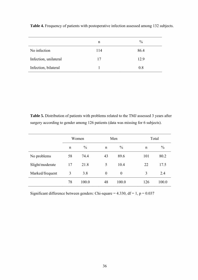
Table 4. Frequency of patients with postoperative infection assessed among 132 subjects.
n %
No infection 114 86.4
Infection, unilateral 17 12.9
Infection, bilateral 1 0.8
Table 5. Distribution of patients with problems related to the TMJ assessed 3 years after
surgery according to gender among 126 patients (data was missing for 6 subjects).
Women Men Total
n % n % n %
No problems 58 74.4 43 89.6 101 80.2
Slight/moderate 17 21.8 5 10.4 22 17.5
Marked/frequent 3 3.8 0 0 3 2.4
78 100.0 48 100.0 126 100.0
Significant difference between genders: Chi-square = 4.330, df = 1, p = 0.037
36

Table 6. Distribution of patients according to size of area with sensory disturbance recorded
3 years after surgery. n = 130(data missing for 2 patients)
Area (cm2) n %
0 (normal sensation) 25 19.2
< 4 18 13.8
4 – 16 59 45.4
≥ 16 28 21.5
Total 130 100.0
Table 7. Distribution of answers to question about sensory function (Q1) reported among
132 patients 3 years after surgery.
Normal sensation
Not normal Total
n % n % n %
Women 24 28.9 59 71.1 83 100.0
Men 8 16.3 41 83.3 49 100.0
Total 32 24.2 100 75.8 132 100.0
37

Table 8. Distribution of answers to question about distress related to sensory dysfunction
(Q2) among the 100 patients reporting impaired sensation 3 years after surgery (patients
reporting normal sensation(n=32) are excluded).
No / minor Yes Total
n % n % n %
Women 48 81.4 11 18.6 59 100.0
Men 37 90.2 4 9.8 41 100.0
Total 85 85.0 15 15.0 100 100.0
Table 9. Distribution of answers to question about satisfaction with treatment result (Q3)
reported among 132 patients 3 years after surgery.
Satisfied Dissatisfied Total
n % n % n %
Women 70 84.3 13 15.7 83 100.0
Men 46 93.9 3 6.1 49 100.0
Total 116 87.9 16 12.1 132 100.0
38

Table 10. Distribution of answers to question about whether the individuals would have re-
elected surgery based on their present experience (Q5) reported among 132 patients 3 years
after surgery.
Yes, re-elect surgery
No, not re-elect surgery
Not sure
n % n % n %
Women 67 80.7 11 13.3 5 6.0
Men 40 81.6 7 14.1 2 4.1
Total 107 81.1 18 13.6 7 5.3
Table 11. Association between satisfaction/dissatisfaction with result (Q3) and
reported distress caused by impaired sensory function (Q2) reported
among 132 patients 3 years after surgery.
Satisfied Dissatisfied Total
n % n % n %
No / minor distress / not relevant
106 90.6 11 9.4 117 100.0
Distress 10 66.7 5 33.3 15 100.0
Total 116 87.9 16 12.1 132 100.0
Fisher exact test: p = 0.020
39

Table 12. Size of area (mean value and SD) with clinically assessed sensory impairment 3
years after surgery among subjects with and without visible nerve
injury as recorded intraoperatively (n = 132).
n Mean (cm2)
SD (cm2)
No visible inury 81 8.6 8.4
Visible injury or uncertain 49 11.9 8.0
t-test: p = 0.029 Table 13.
Frequency of patients reporting normal sensation and not normal sensation 3 years after
surgery (Q1) among subjects with and without visible nerve injury as recorded
intraoperatively (n = 132).
Normal sensation
Not normal sensation
Total
n % n % n %
No visible injury 26 31.7 56 68.3 82 100.0
Visible injury / uncertain 6 12.0 44 88.0 50 100.0
Total 32 24.2 100 75.8 132 100.0
Chi-square = 6.568, df = 1, p = 0.010
40

Appendix
Regimens with antibiotics and glucocorticoids. If patients were allergic to penicillin
erythromycin was given as the alternative
___________________________________________________________________________ Drug: Day of surgery First p.o. day Second p.o. day Penicillin 5 mill. I.E. x 3 i.v. 5 mill I.E. x 3 i.v. Erythromycin 250mg x 4 i.v. 250mg x 4 i.v. Methylprednisolone * 125 mg i.v. at start of 40mg x 4 i.v. 40mg i.m.** surgery. 40 mg x 3 i.v.
• Solu- Medrol (Pfizer) ** Depo- Medrol (Pfizer)
41