early online release - renovatio biomédica · an early online release. early online release...
TRANSCRIPT

EARLY ONLINE RELEASENote: This article was posted on the Archives Web site as an Early Online Release. Early Online Release articles have been peer reviewed, copyedited, and reviewed by the authors. Additional changes or corrections may appear in these articles when they appear in a future print issue of the Archives. Early Online Release articles are citable by using the Digital Object Identifier (DOI), a unique number given to every article. The DOI will typically appear at the end of the abstract.
The DOI for this manuscript is doi: 10.5858/arpa.2017-0388-CP
The final published version of this manuscript will replacethe Early Online Release version at the above DOI once it is available.
© College of American Pathologists2018

CAP Laboratory Improvement Programs
Updated Molecular Testing Guideline for the Selection ofLung Cancer Patients for Treatment With Targeted
Tyrosine Kinase InhibitorsGuideline From the College of American Pathologists, the International Association
for the Study of Lung Cancer, and the Association for Molecular Pathology
Neal I. Lindeman, MD; Philip T. Cagle, MD; Dara L. Aisner, MD, PhD; Maria E. Arcila, MD; Mary Beth Beasley, MD;Eric Bernicker, MD; Carol Colasacco, MLIS, SCT(ASCP); Sanja Dacic, MD, PhD; Fred R. Hirsch, MD, PhD; Keith Kerr, MB, ChB;David J. Kwiatkowski, MD, PhD; Marc Ladanyi, MD; Jan A. Nowak, MD, PhD; Lynette Sholl, MD; Robyn Temple-Smolkin, PhD;
Benjamin Solomon, MBBS, PhD; Lesley H. Souter, PhD; Erik Thunnissen, MD, PhD; Ming S. Tsao, MD;Christina B. Ventura, MPH, MT(ASCP); Murry W. Wynes, PhD; Yasushi Yatabe, MD, PhD
� Context.—In 2013, an evidence-based guideline waspublished by the College of American Pathologists, theInternational Association for the Study of Lung Cancer, andthe Association for Molecular Pathology to set standardsfor the molecular analysis of lung cancers to guidetreatment decisions with targeted inhibitors. New evi-dence has prompted an evaluation of additional laboratorytechnologies, targetable genes, patient populations, andtumor types for testing.
Objective.—To systematically review and update the2013 guideline to affirm its validity; to assess the evidenceof new genetic discoveries, technologies, and therapies;and to issue an evidence-based update.
Design.—The College of American Pathologists, theInternational Association for the Study of Lung Cancer,and the Association for Molecular Pathology convened anexpert panel to develop an evidence-based guideline tohelp define the key questions and literature search terms,review abstracts and full articles, and draft recommenda-tions.
Results.—Eighteen new recommendations were drafted.The panel also updated 3 recommendations from the 2013guideline.
Conclusions.—The 2013 guideline was largely reaf-firmed with updated recommendations to allow testing ofcytology samples, require improved assay sensitivity, andrecommend against the use of immunohistochemistry forEGFR testing. Key new recommendations include ROS1testing for all adenocarcinoma patients; the inclusion ofadditional genes (ERBB2, MET, BRAF, KRAS, and RET) forlaboratories that perform next-generation sequencingpanels; immunohistochemistry as an alternative to fluo-rescence in situ hybridization for ALK and/or ROS1testing; use of 5% sensitivity assays for EGFR T790Mmutations in patients with secondary resistance to EGFRinhibitors; and the use of cell-free DNA to ‘‘rule in’’targetable mutations when tissue is limited or hard toobtain.
(Arch Pathol Lab Med. doi: 10.5858/arpa.2017-0388-CP)
Accepted for publication November 29, 2017.Supplemental digital content is available for this article. See text
for hyperlink.From the Departments of Pathology (Drs Lindeman and Sholl) and
Medicine (Dr Kwiatkowski), Brigham and Women’s Hospital, Boston,Massachusetts; the Department of Pathology and Genomic Medicine,Houston Methodist Hospital, Houston, Texas (Dr Cagle); the Departmentof Pathology, University of Colorado School of Medicine, Denver (DrAisner); the Diagnostic and Molecular Pathology Laboratory (Dr Arcila)and the Molecular Diagnostics Service (Dr Ladanyi), Memorial SloanKettering Cancer Center, New York, New York; the Department ofPathology & Medicine, Pulmonary, Critical Care and Sleep Medicine,New York, New York (Dr Beasley); the Cancer Research Program, HoustonMethodist Research Institute, Houston, Texas (Dr Bernicker); the Pathologyand Laboratory Quality Center, College of American Pathologists, North-field, Illinois (Mss Colasacco and Ventura); the Department of Pathology,University of Pittsburgh, Pittsburgh, Pennsylvania (Dr Dacic); theDepartment of Medicine and Pathology, University of Colorado, Denver(Dr Hirsch); the Department of Pathology, University of Aberdeen,Aberdeen, Scotland (Dr Kerr); the Department of Molecular Pathology,Roswell Park Cancer Institute, Buffalo, New York (Dr Nowak); the Clinicaland Scientific Affairs Division, Association for Molecular Pathology,Bethesda, Maryland (Dr Temple-Smolkin); the Molecular Therapeutics andBiomarkers Laboratory, Peter Maccallum Cancer Center, Melbourne,Australia (Dr Solomon); the Department of Pathology, VU UniversityMedical Center, Amsterdam, the Netherlands (Dr Thunnissen); theDepartment of Laboratory Medicine and Pathobiology, Princess MargaretCancer Center, Toronto, Ontario, Canada (Dr Tsao); Scientific Affairs,International Association for the Study of Lung Cancer, Aurora, Colorado(Dr Wynes); and the Department of Pathology and Molecular Diagnostics,Aichi Cancer Center, Nagoya, Japan (Dr Yatabe). Dr Souter is in privatepractice in Wellanport, Ontario, Canada.
This guideline was developed through collaboration among the Collegeof American Pathologists, the International Association for the Study ofLung Cancer, the Association for Molecular Pathology, and the AmericanSociety for Investigative Pathology and has been jointly published byinvitation and consent in the Archives of Pathology & LaboratoryMedicine, Journal of Thoracic Oncology, and The Journal of MolecularDiagnostics. Copyright 2018 College of American Pathologists, Interna-tional Association for the Study of Lung Cancer, Association for MolecularPathology, and American Society for Investigative Pathology.
Authors’ disclosures of potential conflicts of interest and authorcontributions are found in the Appendix at the end of this article.
Reprints: Neal I. Lindeman, MD, Brigham and Women’s Hospital,Department of Pathology, 75 Francis St, Shapiro 5, Room 020,Boston, MA 02115 (email: [email protected]).
Arch Pathol Lab Med Lung Cancer Molecular Testing Guideline Update—Lindeman et al 1
Patients with advanced lung cancer have a poor progno-sis, with a median survival of 1 year. However, for many
patients whose tumors harbor certain specific molecularalterations (eg, activating alterations in the EGFR, ALK, andROS1 genes), particularly in lung adenocarcinoma, targetedtyrosine kinase inhibitor (TKI) therapy provides significantimprovement in survival and quality. Accordingly, patientswith the types of advanced lung cancer in which thesetargetable molecular alterations typically occur shouldreceive the molecular testing required to identify them,and thereby receive appropriate targeted treatments. Im-portantly, this testing should extend beyond those molec-ular alterations for which targeted therapies are approved byregulatory agencies such as the US Food and DrugAdministration (FDA) to include molecular alterations forwhich there is compelling evidence of effective investiga-tional targeted therapies (and, more recently, immunother-apies) from published clinical trials.
In 2010, 3 professional societies—the College of AmericanPathologists (CAP), the International Association for theStudy of Lung Cancer (IASLC), and the Association forMolecular Pathology (AMP)—recruited specialists in thebiology, diagnosis, and treatment of lung cancer to form ajoint working group to systematically assess the evidencesupporting the clinical utility of molecular analysis of lungcancer samples. In 2013, this working group published anevidence-based guideline1–3 for standard-of-care clinicalpractice concerning which lung cancer patients and samplesshould be tested, which genes should be tested, and howthese tests should be designed, validated, and executed. Thisguideline was subsequently endorsed by the AmericanSociety of Clinical Oncology,4 and has been cited inguidelines developed by numerous professional societiesaround the world.5–26 However, the field has continued toadvance rapidly, with the emergence of new geneticdiscoveries, new therapies, and new technologies, such thatthese same 3 organizations convened a second workinggroup to systematically assess new evidence and to issue anevidence-based revision of the lung cancer molecularpathology practice guideline.
The revision focuses on new recommendations in 5specific content areas: (1) Which new genes should routinelybe tested for alterations in lung cancers? (2) What methodsare appropriate for lung cancer testing, with particularemphases on the use of immunohistochemistry (IHC) andnext-generation sequencing (NGS)? (3) Is there a need totest patients with squamous cell, small cell, or othernonadenocarcinoma lung cancers? (4) What testing shouldbe performed for patients with a targetable alteration whohave progressed following initial response to appropriatelytargeted therapy? (5) What is the role of testing circulatingcell-free DNA (cfDNA) in lung cancer patient management?In addition, new evidence supporting the original 2013guideline was reviewed and used to either modify thestrength of those recommendations or change thementirely. Finally, a sixth question, regarding diagnosticsupport for the role of immunomodulatory therapies (eg,programmed death ligand-1 or PD-L1), emerged during therevision process. Although this topic was not subject to thesystematic review of evidence, the expert panel decided toissue an opinion statement addressing this question, awarethat separate efforts are currently underway to developevidence-based recommendations regarding the use ofbiomarkers to select patients for immunomodulatorytherapies.
One particular challenge for this evidence-based guideline revision was the rapid pace of discovery in this field. During the time between literature review and guideline drafting, major new discoveries were published and treatment advanced for BRAF-mutant lung cancers and for the use of immunotherapies. We expect that many additional advances will emerge in the fields of targeted therapy, cfDNA diagnostics, and immunotherapies in the near term. Although we make strong recommendations for the molecular biomarkers for which there was good evidence at the time we conducted our analysis, we also fully recognize the importance of emerging biomarkers to enable lung cancer patients to be eligible for clinical trials of investigational therapies. Accordingly, we have stratified the biomarkers in this guideline into 3 categories, rather than 2. The first are ‘‘must-test’’ biomarkers, which are standard of care for all patients with advanced lung cancer with an adenocarcinoma component who are being considered for an approved targeted therapy. Second are ‘‘should-test’’ biomarkers, which are used to direct patients to clinical trials and which should be included in any large sequencing panel that is performed for lung cancer patients, but which are not required for laboratories that perform only single-gene assays. All remaining candidate biomarkers are investiga-tional and are not appropriate for clinical use at this time.
PANEL COMPOSITION
The CAP, IASLC, and AMP convened an expert panel consisting of practicing pathologists and oncologists with expertise and experience in lung carcinoma. The CAP, IASLC, and AMP approved the appointment of the project cochairs and expert panel members. In addition, a meth-odologist experienced in systematic review and guideline development consulted with the panel throughout the project.
CONFLICT OF INTEREST POLICY
Prior to acceptance on the expert panel, potential members completed a joint conflict of interest disclosure process, whose policy and form require disclosure of material financial interest in, or potential for benefit of significant value from, the guideline’s development or its recommendations. The potential members completed the conflict of interest disclosure form, listing any relationship that could be interpreted as constituting an actual, potential, or apparent conflict. Potential conflicts were managed by the cochairs. All expert and advisory panel members were required to disclose conflicts prior to beginning and continuously throughout the project’s timeline. Disclosed conflicts of the expert panel members are listed in the Appendix. The CAP, IASLC, and AMP provided funding for the administration of the project; no industry funds were used in the development of the guideline. All panel members volunteered their time and were not compensated for their involvement. Please see the supplemental digital content (SDC) for full details on the conflict of interest policy.
OBJECTIVE
The expert panel was charged with the review and update of the CAP-IASLC-AMP molecular testing guideline for selection of lung cancer patients for EGFR and ALK tyrosine kinase inhibitors. The panel reviewed any new studies that would change or refute the statements from the 2013
2 Arch Pathol Lab Med Lung Cancer Molecular Testing Guideline Update—Lindeman et al

guideline. In addition, the panel also addressed additionalkey questions:
1. Which new genes should be tested for lung cancerpatients?
2. What methods should be used to perform moleculartesting?
3. Is molecular testing appropriate for lung cancers that donot have an adenocarcinoma component?
4. What testing is indicated for patients with targetablemutations who have relapsed on targeted therapy?
5. What is the role of testing for circulating cell-free DNAfor lung cancer patients?
Key questions 1 through 3 relate to patients diagnosed withnonsquamous non–small cell lung cancer (NSCLC) of allstages. The key questions are included in full detail in theSDC.
METHODS
A detailed account of the methods used to create this guidelinecan be found in the SDC, including additional scope questions.
Systematic Literature Review and Analysis
A systematic literature review was completed with 2 compre-hensive searches. The first search was designed to assess the 2013guideline statements and was based on the original search strategy.It included medical subject headings and keywords to address theconcepts lung cancer, tumor biomarkers, and laboratory testing andwas run in Ovid MEDLINE (Ovid Technologies, Inc, New YorkCity, New York) on May 17, 2015, to locate studies published inEnglish with publication dates from January 1, 2012 through May17, 2015. Publication filters were applied to identify guidelines,systematic reviews, meta-analyses (MAs), and randomized clinicaltrials. The search was rerun on June 27, 2016, to identify relevantnew literature published since May 17, 2015.
The second search was based on new key questions that focusedon additional biomarkers not included in the 2013 guideline, withspecific search strategies designed for each key question. Allsearches were performed in Ovid MEDLINE and PubMed (USNational Library of Medicine, Bethesda, Maryland) (June 28, 2015)and were limited to English-language studies. Supplementalsearches were run in Scopus (Amsterdam, Netherlands) (June 25,2015) to identify relevant publications not indexed in MEDLINE. Asearch for relevant clinical trials was completed using theclinicaltrials.gov Web site, and focused searches on guidelinerepository sites (eg, guideline.gov, g-i-n.net) and organizations’Web sites were undertaken to identify relevant publications.Further detail about the systematic literature search, including theOvid search strings, can be found in the SDC.
Eligible Study Designs
Studies were not limited to randomized controlled trials but alsoincluded other study types, including cohort designs, case series,evaluation studies, and comparative studies. Letters, commentaries,editorials, narrative reviews, case reports, studies in mouse models,in vitro studies, consensus documents, abstracts, and non-Englisharticles were excluded a priori.
Inclusion Criteria
Published studies were selected for inclusion in the systematicreview of evidence if they were peer-reviewed full-text articles thatmet the following criteria:
1. The study population consisted of patients with nonsquamous,non–small cell lung adenocarcinoma, small cell lung carcinoma,or squamous cell lung cancer of any stage.
2. The study evaluated, prospectively or retrospectively, sensitivity,specificity, negative predictive value, or positive predictive valueof EGFR, ALK, KRAS, ROS1, RET, MET, BRAF, or ERBB2 (HER2)tests for detection of gene-specific mutation, rearrangement,translocation, amplification, overexpression, or response to atargeted gene-specific therapy.
3. The study examined potential testing algorithms for NSCLCmolecular testing.
4. The study examined the correlation of EGFR, ALK, KRAS, ROS1,RET, MET, BRAF, or ERBB2 (HER2) status in primary ormetastatic tumors from the same patients.
5. The study included primary outcomes such as accuracy,sensitivity, specificity, positive predictive value, and negativepredictive value of tests and concordance across platforms todetermine EGFR, ALK, KRAS, ROS1, RET, MET, BRAF, or ERBB2(HER2) status or treatment response, alone or in combination.
Quality Assessment
An assessment of the quality of the evidence was performed forall retained studies following application of the inclusion andexclusion criteria. Using this method, studies deemed low qualitywould not be excluded from the systematic review, but would beretained and their methodologic strengths and weaknessesdiscussed where relevant. Each guideline statement includes arating of the strength of the evidence as described in Table 1 (alsoin SDC Table 1). The process used to assess the quality of theevidence base is fully detailed in the SDC.
Assessing the Strength of Recommendations
In order to articulate recommendation statements that wereclearly written and easy to implement, the expert panel usedGLIDES (Guidelines Into Decision Support) methodology andaccompanying BridgeWiz software (Yale University, New Haven,Connecticut).27 This methodology prioritizes the use of activelanguage; however, in some situations, the person responsible forensuring guidance is implemented is dependent on the organiza-tion of the clinic and/or laboratory. To ensure clarity of guidance inthese situations, the expert panel used passive-voice language toemphasize the recommended action. Development of recommen-dations required that the panel review the identified evidence andmake a series of key judgments (using procedures described in theSDC). This guideline uses a 3-tier system to rate the strength ofrecommendations, as well as a ‘‘no recommendation’’ categorywhen there is insufficient evidence to support a recommendation.Table 2 (also in SDC Table 2) summarizes the strength of evidenceand net benefits and harms, as well as obligatory language that wasused for each of the recommendation types.
Guideline Revision
This guideline will be reviewed every 4 years or earlier in theevent of publication of substantive and high-quality evidence thatcould potentially alter the original guideline statements. Ifnecessary, the entire panel will reconvene to discuss potentialchanges and, if indicated, recommend revision of the guideline toCAP, IASLC, and AMP.
Disclaimer
Practice guidelines and consensus statements reflect the bestavailable evidence and expert consensus supported in practice.They are intended to assist physicians and patients in clinicaldecision making and to identify questions and settings for furtherresearch. With the rapid flow of scientific information, newevidence may emerge between the time a practice guideline orconsensus statement is developed and when it is published or read.Guidelines and statements are not continually updated and maynot reflect the most recent evidence. Guidelines and statementsaddress only the topics specifically identified therein and are notapplicable to other interventions, diseases, or stages of diseases.Furthermore, guidelines and consensus statements cannot accountfor individual variation among patients and cannot be considered
Arch Pathol Lab Med Lung Cancer Molecular Testing Guideline Update—Lindeman et al 3

inclusive of all proper methods of care or exclusive of othertreatments. It is the responsibility of the treating physician or otherhealth care provider, relying on independent experience andknowledge, to determine the best course of treatment for thepatient. Accordingly, adherence to any practice guideline orconsensus statement is voluntary, with the ultimate determinationregarding its application to be made by the physician in light ofeach patient’s individual circumstances and preferences. The CAP,IASLC, and AMP make no warranty, express or implied, regardingguidelines and statements and specifically exclude any warrantiesof merchantability and fitness for a particular use or purpose. TheCAP, IASLC, and AMP assume no responsibility for any injury ordamage to persons or property arising out of or related to any useof this statement or for any errors or omissions.
RESULTS
For the reaffirmation of the 2013 guideline recommenda-tions, a total of 610 studies met the search termrequirements. Following a review of the 610 abstracts, thefull texts of 77 studies that met the inclusion criteria andwere likely to refute or change the 2013 recommendationswere reviewed. A total of 21 articles were included for dataextraction. Excluded articles were available as discussion orbackground references.
For the new key questions, 1654 articles met the searchterm requirements. Based on review of these abstracts, 488articles met the inclusion criteria and continued to full-textreview. Articles that addressed any of the new key questionswere moved to a second-level full-text–review phase. A totalof 118 articles were included for data extraction. Excluded
articles were available as discussion or background refer-ences.
The panel convened 5 times (3 times by teleconferenceand 2 face-to-face meetings) to develop the scope, draftrecommendations, review and respond to solicited feed-back, and assess the quality of evidence that supports thefinal recommendations. A nominal group technique wasused by the panel for consensus decision making toencourage unique input with balanced participation amonggroup members. An open comment period was held fromJune 28 to August 2, 2016, during which the 2013 guidelinestatements and new draft recommendations and statementswere posted for public comment. The public commentperiod was posted on the AMP Web site at www.amp.org.All 2013 recommendations received strong agreement(95%–99%) from the open comment period participants.There were 20 new draft statements with strong agreement,ranging from 86% to 97%, from the open comment periodparticipants (refer to Outcomes in the SDC for full details).The expert panel members were assigned to review thepublic comments in small groups. The panel modified thedraft statements and recommended the deletion of 1 expertconsensus opinion and a no recommendation statementbased on the feedback during the considered judgmentprocess. The final recommendations were approved by theexpert panel with a vote. The panel considered benefits andharms, required resources, feasibility, and acceptabilitythroughout the entire process, although neither cost nor
Table 1. Grades for Strength of Evidencea
Designation Description Quality of Evidence
Convincing High confidence that available evidence reflects true effect. Furtherresearch is very unlikely to change the confidence in the estimateof effect.
High/intermediate quality of evidence.
Adequate Moderate confidence that available evidence reflects true effect.Further research is likely to have an important impact on theconfidence in estimate of effect and may change the estimate.
Intermediate/low quality of evidence.
Inadequate Little confidence that available evidence reflects true effect. Furtherresearch is very likely to have an important impact on theconfidence in the estimate of effect and is likely to change theestimate.
Low/insufficient evidence and expert panel uses formalconsensus process to reach recommendation.
Insufficient Evidence is insufficient to discern net effect. Any estimate of effect isvery uncertain.
Insufficient evidence and expert panel uses formalconsensus process to reach recommendation.
a Adapted from J Clin Epidemiol. 2011;64(4):401–406, Balshem H, Helfand M, Schunemann HJ, et al. GRADE guidelines: 3. Rating the quality ofevidence, copyright 2011, with permission from Elsevier.262
Table 2. Strength of Recommendationsa
Designation Recommendation Rationale
Strong recommendation Recommend for or against a particular moleculartesting practice in lung cancer (can include mustor should).
Supported by convincing (high) or adequate (intermediate)quality of evidence and clear benefit that outweighs anyharms.
Recommendation Recommend for or against a particular moleculartesting practice in lung cancer (can includeshould or may).
Some limitations in quality of evidence (adequate[intermediate] or inadequate [low]), balance of benefitsand harms, values, or costs, but panel concludes thatthere is sufficient evidence to inform arecommendation.
Expert consensus opinion Recommend for or against a particular moleculartesting practice in lung cancer (can includeshould or may).
Serious limitations in quality of evidence (inadequate[low, very low] or insufficient), balance of benefits andharms, values, or costs, but panel consensus is that astatement is necessary.
No recommendation No recommendation for or against a particularmolecular testing practice in lung cancer.
Insufficient evidence, confidence, or agreement to providea recommendation.
a Data derived from Andrews et al.263
4 Arch Pathol Lab Med Lung Cancer Molecular Testing Guideline Update—Lindeman et al

cost-effectiveness analyses were performed. A description ofthe benefits and harms of implementing the guidelinestatements is included in the SDC (SDC Table 3).
Each organization instituted a review process to approvethe guideline. For the CAP, an independent review panelrepresenting the Council on Scientific Affairs was assembledto review and approve the guideline. The independentreview panel was masked to the expert panel and vettedthrough the conflict of interest process. The IASLC approvalprocess required review and approval by the IASLC Board ofDirectors. The AMP approval process required contentreview by an independent subject matter expert panel, ledby the Publications & Communications chair, with repre-sentation from the Clinical Practice Committee and SolidTumors Subdivision leadership, and organizational approvalby the AMP Executive Committee.
GUIDELINE STATEMENTS
Reaffirmation of 2013 Recommendations
The 2013 guideline recommended universal testing oflung cancer patients with advanced-stage cancers with anadenocarcinoma component, using molecular diagnosis foractivating ‘‘hot-spot’’ mutations in EGFR exons 18 to 21 withat least 1% prevalence (ie, codons 709 and 719, exon 19deletion 768, and exon 20 insertions 790, 858, and 861), andusing fluorescence in situ hybridization (FISH) for rear-rangements involving ALK. Any methodology or testingalgorithm with suitable analytic sensitivity (ability to detectmutations in formalin-fixed samples with 50% or moremalignant cells) and turnaround time (10 days betweensample receipt and reporting of all results), with appropriatevalidation and deployment under the Clinical LaboratoryImprovement Act of 1988, was acceptable.
The 2013 guideline recommended against applyingclinical parameters (eg, tobacco exposure, age, sex, ethnicity)to select patients for testing, testing pure squamouscarcinomas, using KRAS negativity as a determinant ofanti-EGFR therapy, using IHC for EGFR or ALK testing, andusing FISH for EGFR testing.
The 2013 guideline left several decisions open to eachinstitution to set policy, such as whether or not to test early-stage patients, whether or not to use clinical predictors toselect patients with minimally sampled squamous carcino-ma biopsies such that a mixed adenosquamous carcinomacould not be excluded, and whether or not to use asimultaneous or sequential testing approach. Of these, the
question concerning testing early-stage disease remainsopen, and awaits data from more clinical trials before anevidence-based recommendation can be made. Althoughthe American Society of Clinical Oncology Clinical PracticeGuidelines Committee highlighted consideration of molec-ular testing for early-stage lung cancer patients,4 ouropinion remains that each institution should set its ownpolicy regarding testing patients with early-stage disease,balancing the benefit of having results on record fromtesting a high-quality resection sample for alterations thatare likely to become necessary at a time of futureprogression when a high-quality sample could be hard toobtain against the cost of testing patients for whom a subsetwill be surgically cured and never need the test result.Accordingly, the testing recommended below applies topatients with advanced-stage (stages IIIB and IV) lungcancer.
Following review of literature published since 2013, theoriginal recommendations are largely reaffirmed. Severalstatements have gained strength with the publication ofadditional supporting evidence (SDC Tables 4a, 4b, and 5).Some warranted a complete reevaluation in this revision,and will appear subsequently (Table 3); these include theuse of IHC for ALK, the use of multigene NGS panels, andthe question of testing nonadenocarcinoma samples.
Of the remaining 2013 recommendations, the followingchanges are made:
1. Any Cytology Sample With Adequate Cellularityand Preservation May Be Tested.—The original recom-mendation preferred cell blocks over smears. A recentsystematic review28 identified by the literature search hasindicated that numerous studies have been publishedshowing excellent performance of smear preparations, suchthat this preference is no longer appropriate. It is incumbentupon any laboratory that tests cytopathology specimens toperform appropriate validation studies of these as separatesample types, distinct from tissue and blood samples.
2. Analytic Methods Must Be Able to Detect Mutationin a Sample With 20% or More Malignant CellContent.—Although the original studies demonstratingresponse of EGFR-mutated lung cancers to treatment withEGFR inhibitors used unmodified Sanger sequencing with asensitivity limit of 50% tumor cellularity, this is insufficientin practice because many lung cancer samples are small andcomprise a majority of benign stromal cells, and most of thelarger phase III clinical trials that confirmed the clinicalutility of EGFR mutation testing used polymerase chain
Table 3. Summary of the Updated Statements With Strength of Recommendationsa
2013 Statement 2017 Statement
Expert consensus opinion: Cytologic samples are also suitable forEGFR and ALK testing, with cell blocks being preferred oversmear preparations.
Recommendation: Pathologists may use either cell blocks or othercytologic preparations as suitable specimens for lung cancerbiomarker molecular testing.
Expert consensus opinion: Laboratories should use EGFR testmethods that are able to detect mutations in specimens with atleast 50% cancer cell content, although laboratories are stronglyencouraged to use (or have available at an external referencelaboratory) more sensitive tests that are able to detect mutations inspecimens with as little as 10% cancer cells.
Expert consensus opinion: Laboratories should use, or have availableat an external reference laboratory, clinical lung cancer biomarkermolecular testing assays that are able to detect molecularalterations in specimens with as little as 20% cancer cells.
Recommendation: Immunohistochemistry for total EGFR is notrecommended for selection of EGFR TKI therapy.
Strong recommendation: Laboratories should not use total EGFRexpression by IHC testing to select patients for EGFR-targeted TKItherapy.
Abbreviations: IHC, immunohistochemistry; TKI, tyrosine kinase inhibitor.a Supplemental Table 4b includes a list of the 2013 reaffirmed statements.
Arch Pathol Lab Med Lung Cancer Molecular Testing Guideline Update—Lindeman et al 5

reaction (PCR)–based methods that were more sensitive
than unmodified Sanger sequencing. Given the widespread
availability of technology capable of reliably detecting
lower-frequency mutational events in small samples, it is
no longer appropriate to offer a low-sensitivity test that
cannot test tumors with 20% to 50% tumor content and
requires patients to undergo more procedures, and poten-
tially more invasive procedures, solely to procure a tissuesample with high tumor content.
3. It Is Not Appropriate to Use IHC for EGFRMutation Testing.—There is no role whatsoever for IHCagainst total EGFR protein as a determinant of treatmentwith an EGFR kinase inhibitor. The targetable mutationslead to activation of the cytoplasmic kinase of thistransmembrane protein, but that has no bearing on the
Table 4. Summary of 2017 Guideline Statements
Guideline Statements Strength of Recommendation
Key Question 1: Which new genes should be tested for lung cancer patients?
1. ROS1 testing must be performed on all lung adenocarcinoma patients, irrespective of clinicalcharacteristics.
Strong recommendation
2. ROS1 IHC may be used as a screening test in lung adenocarcinoma patients; however, positive ROS1IHC results should be confirmed by a molecular or cytogenetic method.
Expert consensus opinion
3. BRAF molecular testing is currently not indicated as a routine stand-alone assay outside the context of aclinical trial. It is appropriate to include BRAF as part of larger testing panels performed either initiallyor when routine EGFR, ALK, and ROS1 testing are negative.
Expert consensus opinion
4. RET molecular testing is not recommended as a routine stand-alone assay outside the context of aclinical trial. It is appropriate to include RET as part of larger testing panels performed either initially orwhen routine EGFR, ALK, and ROS1 testing are negative.
Expert consensus opinion
5. ERBB2 (HER2) molecular testing is not indicated as a routine stand-alone assay outside the context of aclinical trial. It is appropriate to include ERBB2 (HER2) mutation analysis as part of a larger testingpanel performed either initially or when routine EGFR, ALK, and ROS1 testing are negative.
Expert consensus opinion
6. KRAS molecular testing is not indicated as a routine stand-alone assay as a sole determinant of targetedtherapy. It is appropriate to include KRAS as part of larger testing panels performed either initially orwhen routine EGFR, ALK, and ROS1 testing are negative.
Expert consensus opinion
7. MET molecular testing is not indicated as a routine stand-alone assay outside the context of a clinicaltrial. It is appropriate to include MET as part of larger testing panels performed either initially or whenroutine EGFR, ALK, and ROS1 testing are negative.
Expert consensus opinion
Key Question 2: What methods should be used to perform molecular testing?
8. IHC is an equivalent alternative to FISH for ALK testing. Recommendation
9. Multiplexed genetic sequencing panels are preferred over multiple single-gene tests to identify othertreatment options beyond EGFR, ALK, and ROS1.
Expert consensus opinion
10. Laboratories should ensure test results that are unexpected, discordant, equivocal, or otherwise of lowconfidence are confirmed or resolved using an alternative method or sample.
Expert consensus opinion
Key Question 3: Is molecular testing appropriate for lung cancers that do not have an adenocarcinomacomponent?
11. Physicians may use molecular biomarker testing in tumors with histologies other than adenocarcinomawhen clinical features indicate a higher probability of an oncogenic driver.
Expert consensus opinion
Key Question 4: What testing is indicated for patients with targetable mutations who have relapsedon targeted therapy?
12. In lung adenocarcinoma patients who harbor sensitizing EGFR mutations and have progressed aftertreatment with an EGFR-targeted tyrosine kinase inhibitor, physicians must use EGFR T790M mutationaltesting when selecting patients for third-generation EGFR-targeted therapy.
Strong recommendation
13. Laboratories testing for EGFR T790M mutation in patients with secondary clinical resistance to EGFR-targeted kinase inhibitors should deploy assays capable of detecting EGFR T790M mutations in as littleas 5% of viable cells.
Recommendation
14. There is currently insufficient evidence to support a recommendation for or against routine testing forALK mutational status for lung adenocarcinoma patients with sensitizing ALK mutations who haveprogressed after treatment with an ALK-targeted tyrosine kinase inhibitor.
No recommendation
Key Question 5: What is the role of testing for circulating cell-free DNA for lung cancer patients?
15. There is currently insufficient evidence to support the use of circulating cell-free plasma DNAmolecular methods for the diagnosis of primary lung adenocarcinoma.
No recommendation
16. In some clinical settings in which tissue is limited and/or insufficient for molecular testing, physiciansmay use a cell-free plasma DNA assay to identify EGFR mutations.
Recommendation
17. Physicians may use cell-free plasma DNA methods to identify EGFR T790M mutations in lungadenocarcinoma patients with progression or secondary clinical resistance to EGFR-targeted tyrosinekinase inhibitors; testing of the tumor sample is recommended if the plasma result is negative.
Expert consensus opinion
18. There is currently insufficient evidence to support the use of circulating tumor cell molecular analysisfor the diagnosis of primary lung adenocarcinoma, the identification of EGFR or other mutations, or theidentification of EGFR T790M mutations at the time of EGFR TKI resistance.
No recommendation
Abbreviations: FISH, fluorescence in situ hybridization; IHC, immunohistochemistry; TKI, tyrosine kinase inhibitor.
6 Arch Pathol Lab Med Lung Cancer Molecular Testing Guideline Update—Lindeman et al

extent of expression at the cell surface, which is what isdetected by the total EGFR immunostain. Although EGFRexpression by IHC was performed for some of the veryearly studies of EGFR kinase inhibitors in the start of thiscentury, clinical responses were seen in patients withmutations but absent/weak IHC expression, and poorresponses were seen in patients with strong IHC expres-sion but no mutations.
Following the discovery of EGFR mutations, antibodieswere developed for IHC directed at the most commonmutated forms of the protein, most notably the L858Rsubstitution and the 746 to 750 ELREA deletion. Theoriginal guideline allowed for the use of the mutant-specificEGFR antibodies by IHC in a setting with extremely limitedmaterial. Although published evidence for these antibodiesshows good accuracy for the L858R activating mutation andfor some of the exon 19 deletions, these antibodies havepoor sensitivity for other exon 19 deletions, insensitivity toless common mutations (eg, codon 719 mutations), andfalse-positive results with exon 20 insertions.29 Overall, theperformance is suboptimal for reliable detection of EGFRmutations. Given that advances in molecular diagnostictechnology now enable analysis of very limited samples aswell as circulating tumor DNA (see below), at this timethere is no role for routine use of mutant-specific IHC inselecting anti-EGFR treatment for lung cancer patients.
New Recommendations
Question 1: Which New Genes Should Be Tested forLung Cancer Patients?—In the 2013 guideline, genes fellinto 1 of 2 categories: testing is necessary (EGFR, ALK), ortesting is investigational. One gene, KRAS, was consideredconditionally necessary in the context of sequential testingalgorithms because of its ease of analysis and mutualexclusivity with EGFR and ALK. By 2017, however, webelieve that there are now 3 categories into which genesshould be placed. One set of genes must be offered by alllaboratories that test lung cancers, as an absolute minimum:EGFR, ALK, and ROS1. A second group of genes should beincluded in any expanded panel that is offered for lungcancer patients: BRAF, MET, RET, ERBB2 (HER2), andKRAS, if adequate material is available. KRAS testing mayalso be offered as a single-gene test to exclude patients from
expanded panel testing. All other genes are consideredinvestigational at the time of publication.
In this context, institutions providing care for lung cancerpatients have a choice: (1) offer a comprehensive cancerpanel that includes all of the genes in the first 2 categories(EGFR, ALK, ROS1, BRAF, MET, ERBB2 [HER2], KRAS, RET)for all appropriate patients, or (2) offer targeted testing forthe genes in the must-test category (EGFR, ALK, ROS1) forall appropriate patients and offer as a second test anexpanded panel containing the second-category genes(BRAF, MET, ERBB2 [HER2], and RET) for patients whoare suitable candidates for clinical trials, possibly afterperforming a single-gene KRAS test to exclude patients withKRAS-mutant cancers from expanded panel testing. Table 4includes a list of the recommendation statements with thestrength of recommendations.
1. Strong Recommendation.—ROS1 testing must be per-formed on all lung advanced-stage adenocarcinoma pa-tients, irrespective of clinical characteristics.
The strength of evidence was convincing to support theuse of ROS1 molecular (ie, reverse transcription PCR [RT-PCR] or sequencing) or cytogenetic (ie, FISH or other in situhybridization) testing to identify ROS1 rearrangements. Thestrength of evidence supporting the use of any clinicalcharacteristic to identify patients who should receive ROS1testing was adequate. This recommendation is evidence-based and supported by 9 studies,30–38 6 of which informedon the association between ROS1 rearrangement andpatient or tumor characteristics30,31,34–37 and consisted of 1prospective cohort study (PCS),35 1 prospective-retrospec-tive cohort study (PRCS),31 and 4 retrospective cohortstudies (RCSs).30,34,36,37 The 3 remaining studies assessedclinical outcomes of patients treated with the ROS1-targetedtherapy crizotinib32,33,38 and included 1 nonrandomizedclinical trial33 and 3 RCSs.32,38 All included studies wereassessed for quality and none were found to havemethodologic flaws that would raise concerns about thestudies’ findings (SDC Table 6). Refer to SDC Table 7 for asummary of findings from studies supporting the use ofROS1 molecular or cytogenetic testing to enable selection ofpatients for ROS1-targeted therapy.
Although relatively rare, accounting for less than 2% ofnon–small cell lung carcinomas30,31,34 and 2% to 3% of lungadenocarcinomas,30,34,35 structural rearrangements involvingthe ROS1 gene generate an oncogenic fusion that can betreated successfully with targeted inhibitors. A single phase Iclinical trial of 50 NSCLC patients demonstrated that thepresence of a ROS1 rearrangement by FISH or RT-PCRpredicts response to targeted inhibition using crizotinib,with a response rate of 72% and median progression-freesurvival of 19.2 months.33 Based on this trial, the FDAapproved the expanded use of crizotinib in patients withROS1-rearranged NSCLC in 2016. A European multi-institutional retrospective study of 32 patients with ROS1-rearranged NSCLC treated with crizotinib demonstrated an80% response rate and 9.1-month progression-free surviv-al.32 Overall survival for patients with ROS1-rearrangedtumors irrespective of use of targeted therapy appearslonger than that for patients with other molecular alter-ations undergoing targeted therapy.38,39
As with ALK, ROS1 activation is driven by structuralvariants, with multiple different partners fusing to the C-terminal portion of ROS1 containing the cytoplasmictyrosine kinase and driving downstream signaling throughMAPK, JAK/STAT, and PI3K pathways. Common fusion
Table 5. Emerging Markers for Molecular Testing inLung Cancer
Mitogen-activated protein kinase kinase 1 (MEK1/MAP2K1)
Fibroblast growth factor receptor 1–4 (FGFR 1–4)
Neurotrophic tyrosine kinase, receptor, type 1–3 (NTRK1-3)
Neuregulin 1 (NRG1)
Ras-like without CAAX 1 (RIT1)
Neurofibromin 1 (NF1)
Phosphatidylinositol-4,5-bisphosphate 3-kinase catalytic subunitalpha (PIK3CA)
AKT serine/threonine kinase 1 (AKT1)
NRAS proto-oncogene, GTPase (NRAS)
Mechanistic target of rapamycin (MTOR)
Tuberous sclerosis 1 (TSC1)
Tuberous sclerosis 2 (TSC2)
KIT proto-oncogene receptor tyrosine kinase (KIT)
Platelet-derived growth factor receptor alpha (PDGFRA)
Discoidin domain receptor tyrosine kinase 2 (DDR2)
Arch Pathol Lab Med Lung Cancer Molecular Testing Guideline Update—Lindeman et al 7

partners include SLC34A2, CD74, and TPM3, among others.The role of wild-type ROS1 is still being elucidated, but itshares similar structure with ALK, albeit with significantdifferences, notably absence of a dimerization domain, anextracellular domain with some resemblance to cell adhe-sion molecules, and no clear ligand.
As with EGFR mutations and ALK rearrangements, light tonever smoking history has been associated with an increasedincidence of ROS1 rearrangements in patients with lungadenocarcinoma.30,37 However, this association has not beenconsistently observed across studies.34 Other clinical charac-teristics, such as younger age, female sex, and non-Asianethnicity, have been associated with ROS1 rearrangement inisolated studies only.30,31,35 Therefore, clinical characteristicsshould not be used to either select or exclude patients fromtesting for ROS1 rearrangements. ROS1 rearrangementsoccur in a mutually exclusive fashion with other oncogenicdriver alterations (such as EGFR and KRAS mutation and ALKrearrangement). In recognition of the rarity of ROS1rearrangement, it may be reasonable to perform sequentialtesting of EGFR and ALK followed by ROS1 testing. Indeed,the frequency of ROS1 rearrangements is enriched to 5% to10% in otherwise driver (ie, EGFR, ALK, KRAS, BRAF)-negative lung adenocarcinomas.31,37
Notably, in the United States in 2016, crizotinib therapy inROS1-rearranged tumors does not require the use of anFDA-approved companion diagnostic. Published methodsthat have established clinical utility of testing ROS1 in orderto choose ROS1-targeted therapy have relied primarily uponFISH and RT-PCR. Outside the United States, a diagnostictest using RT-PCR was used for an international phase IIclinical trial,40 involving mainly East Asian countries, forselection of tumors with ROS1 rearrangement. This assayhas been approved as an in vitro diagnostic in Europe andChina, and may be recognized as a companion diagnostictest in some countries. Although targeted RT-PCR assaysmay be challenging because of variation in ROS1 breakpoints (typically introns 31–35) and partner genes, capture-based sequencing strategies for RNA or DNA may be used,provided they are properly validated on known positivesamples. Within the United States, FISH methods have beenpublished more frequently. Fluorescence in situ hybridiza-tion testing should be performed with a break-apart probedesign given the multiple fusion partners, and should showrearrangement, defined as signals split by at least 1 probediameter, in 15% or more of tumor cells.41
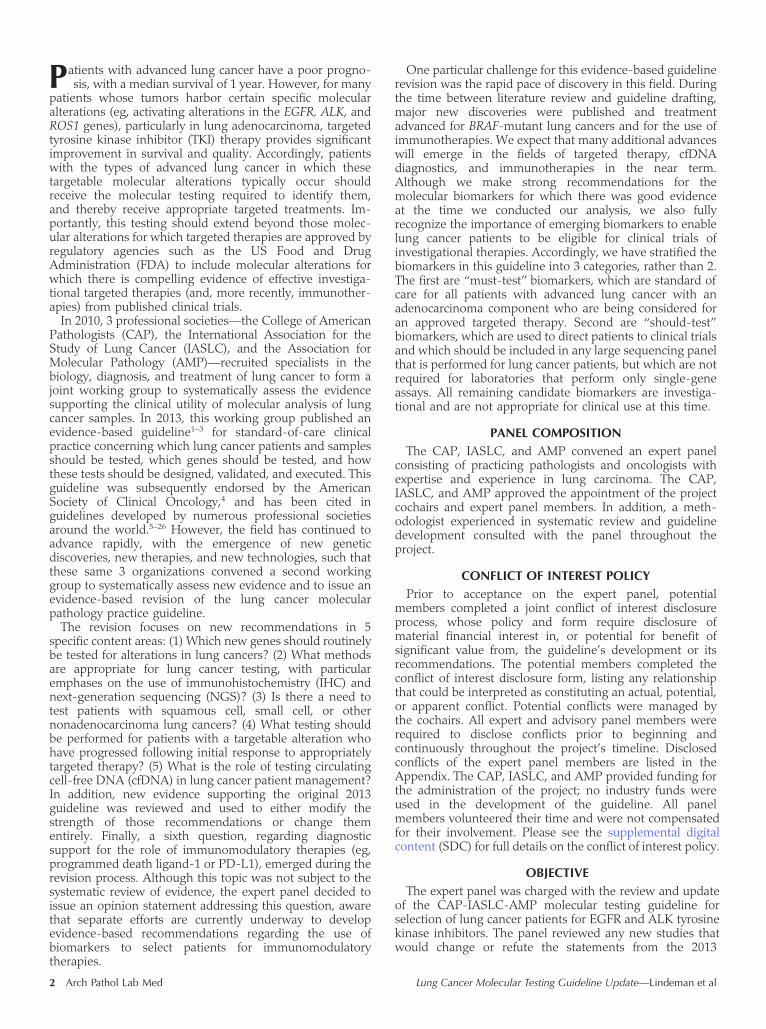
2. Expert Consensus Opinion.—ROS1 IHC may be used as ascreening test in advanced-stage lung adenocarcinomapatients; however, positive ROS1 IHC results should beconfirmed by a molecular or cytogenetic method.
The strength of evidence is inadequate supporting the useof IHC as a screening assay for ROS1 molecular testing. Thisstatement is evidence-based and supported by 6 studies,42–47
consisting of 2 PCSs,43,44 1 PRCS,42 and 3 RCSs.45–47 Fivestudies compared ROS1 IHC with a FISH referencetest42–45,47 and 1 study compared ROS1 IHC with an RT-PCR reference test.46 Using reported true-positive, false-positive, true-negative, and false-negative data from studiescomparing IHC with FISH, an MA was conducted todetermine a pooled estimate of sensitivity and specificity forROS1 IHC (Figure 1). All included studies were assessed forquality and none were found to have methodologic flawsthat would raise concerns about the studies’ findings (SDCTable 8). Refer to SDC Table 9 for a summary of findings
from studies supporting the use of IHC as a screening assayfor ROS1 molecular testing.
In light of the relative rarity of ROS1 rearrangementevents in NSCLC, screening by IHC may be preferable toFISH or molecular techniques in some settings. Interpreta-tion of ROS1 IHC is challenging, however, as expression canbe seen in a patchy pattern, typically at weak intensity, in upto a third of tumors that do not have an underlyingrearrangement.44,45,48 Although some studies suggest thatROS1 expression in the absence of a rearrangement mayhave prognostic significance,48 focal or patchy expression intumor cells is rarely associated with a ROS1 rearrangementand therefore is unlikely to predict response to ROS1-targeted therapy. Moreover, the pattern of staining can varyamong fusion types, including granular to globular stainingin CD74-ROS1 fusions, weak membranous staining in EZR-ROS1 fusions, and vesicular localization staining in GOPC-ROS1 fusions.45
A single commercially available antibody clone (D4D6, CellSignaling Technology, Danvers, Massachusetts) has beenused in studies published to date. Most retrospective studiesof ROS1 IHC using the D4D6 antibody demonstrate asensitivity of 100% relative to FISH or RT-PCR.35,42–47 Tumorslacking ROS1 expression can be safely interpreted as lackinga ROS1 fusion. However, the specificity of ROS1 IHC is morevariable, ranging from 92% to 100% using different methodsand interpretive cutoffs.35,42–47 Meta-analysis of 5 studiesidentified by the literature search determined a pooledsensitivity of 96% (95% CI, 71%–99%) and specificity of94% (95% CI, 89%–96%) for IHC compared with FISH whenthe D4D6 antibody with a staining intensity of at least 2þ (asdefined within the study) was used (Figure 1). Several cutoffshave been proposed using intensity alone or H score(intensity 3 percentage of tumor cells staining). In moststudies, FISH- or molecularly confirmed ROS1-rearrangedtumors have at least moderate-intensity ROS1 proteinexpression, but published evidence is insufficient to recom-mend one specific cutoff or scoring system,42,45 and eachlaboratory must validate its own interpretive cutoff fromknown positive and negative samples.
Because of imperfect specificity and challenges related tointerpretation of nonspecific expression, we recommendthat all ROS1 IHC positive results undergo confirmation byFISH or a molecular method (ie, RT-PCR, NGS) prior toconsidering a patient for ROS1-targeted therapy. Given thehigh sensitivity of IHC, however, tumors that clearly lackROS1 staining can be interpreted as negative for ROS1fusion.
Additional Genes
Of the genes newly included in this guideline, only ROS1testing must be offered to all appropriate lung cancerpatients. Testing for the following genes should be includedwith any expanded multigene panel testing performed forlung cancer patients, whether or not the panel is offered forall lung cancer patients, or if the panel is reserved as asecond-line test for EGFR/ALK/ROS1 wild-type patientsseeking clinical trials.
3. Expert Consensus Opinion.—BRAF molecular testing iscurrently not indicated as a routine stand-alone assay outsidethe context of a clinical trial. It is appropriate to include BRAFas part of larger testing panels performed either initially orwhen routine EGFR, ALK, and ROS1 testing is negative.
The strength of evidence was inadequate to support theuse of BRAF molecular testing. This statement was
8 Arch Pathol Lab Med Lung Cancer Molecular Testing Guideline Update—Lindeman et al

evidence-based and supported by 9 studies: 4 PCSs49–52 and3 RCSs,53–55 all of which informed on the associationbetween BRAF mutation and patient or tumor characteris-tics,49–55 and 2 additional nonrandomized clinical trials thatassessed the activity of a BRAF inhibitor in p.V600E mutantpatients.56,57 All included studies were assessed for qualityand none were found to have methodologic flaws thatwould raise concerns about the studies’ findings (SDC Table10). Refer to SDC Table 11 for a summary of findings fromstudies supporting the use of BRAF molecular testing.
Activating mutations in BRAF, especially p.V600E, lead tooncogenic signaling through MAPK, and are rare recurrentalterations in lung adenocarcinoma, seen in 0.5% to 4.9% of
tumors.49–52,54 In lung cancer, data from a 2016 phase IIsingle-arm clinical trial56 showed that (1) single-agentdabrafenib given in second line to stage IV BRAF p.V600Emutant NSCLC had a partial response rate of 33% anddisease control rate of 58% and (2) combination dabrafenib-trametinib therapy given in second line to stage IV BRAFp.V600E mutant lung adenocarcinoma had a partialresponse rate of 63% and disease control rate of 75%.
Based on these data, the FDA conferred a breakthroughtherapy designation for the combination treatment in BRAFp.V600E mutation–positive NSCLC, and FDA approval wasgranted in 2017. Hence, this was the most controversial ofall recommendations among the working panel. Although
Figure 1. Forest plot of sensitivity and specificity for immunohistochemistry (IHC)-based determination of ROS1 rearrangement positivity comparedwith fluorescence in situ hybridization. Pooled estimate of sensitivity and specificity based on bivariate analysis of included studies. All includedstudies used an IHC staining intensity of at least 2þwith a D4D6 antibody to define ROS1 rearrangement positivity. Abbreviations: FN, false-negative;FP, false-positive; TN, true-negative; TP, true-positive.
Figure 2. Forest plot of sensitivity and specificity for immunohistochemistry-based determination of ALK translocation positivity compared withfluorescence in situ hybridization. Pooled estimate of sensitivity and specificity based on bivariate analysis of included studies. Included studiesassessed 5A4, D5F3, or either 5A4 or D5F3 antibodies with positivity cutoffs based on either presence of any staining or staining intensity.Abbreviations: FN, false-negative; FP, false-positive; neg, negative; pos, positive; TN, true-negative; TP, true-positive.
Figure 3. Forest plot of sensitivity and specificity for various assays determining EGFR mutation positivity with cell-free DNA compared with tumortissue. Pooled estimate of sensitivity and specificity based on bivariate analysis of included studies. Four included studies compared tumor tissuesamples with plasma samples using the same detection system,234,235,242,243 and a fifth study232 obtained plasma samples from patients with knownEGFR and KRAS tumor mutation status. Abbreviations: ARMs, amplification refractory mutation system; ddPCR, droplet digital PCR; FN, false-negative; FP, false-positive; PCR, polymerase chain reaction; PNA/LNA, peptide nucleic acid–locked nucleic acid; TN, true-negative; TP, true-positive.
Arch Pathol Lab Med Lung Cancer Molecular Testing Guideline Update—Lindeman et al 9

there was a strong opinion in the working group that BRAFmutation analysis should be performed at the time of initialmolecular testing in lung adenocarcinoma, the publishedevidence available at the time of publication lackedcontrolled prospective trials, and therefore lacked thestrength to warrant an international recommendation forsingle-gene testing for BRAF for all lung adenocarcinomapatients. We anticipate the publication of stronger evidencesupporting the utility of BRAF inhibition in BRAF-mutantlung cancer, and our opinion is that BRAF testing will beproven necessary. We expect that the next revision of thisguideline will include a recommendation to include single-gene testing for BRAF alongside EGFR, ALK, and ROS1, butwe are unable to make that recommendation in the springof 2017. Although stand-alone single-gene testing for BRAFis not currently recommended, if a panel strategy is used,either initially or for patients who are known wild type forEGFR, ALK, and ROS1, then BRAF should be included.
As with EGFR and KRAS mutations, selected hot-spotmutations in BRAF exert an oncogenic effect. The V-rafmurine sarcoma homolog b (BRAF) gene encodes for anonreceptor serine-threonine kinase in the MAPK kinasesignaling pathway, between RAS and MEK. The mostcommon BRAF mutation in NSCLC is the c.1799T.A(p.V600E) point mutation that is the predominant mutationin many other cancers, including melanoma, papillarythyroid cancer, colorectal cancer, hairy cell leukemia, andganglioglioma. However, in contrast to other cancers withBRAF mutations, lung cancers frequently have non-p.V600EBRAF mutations, including other mutations at codon 600(eg, p.V600K) and nearby codons in exon 15, andsubstitutions at codons 466 and 469 in exon 11.
Like many other targetable oncogenes in lung cancer,BRAF mutations are more frequent in adenocarcinomasthan in squamous cell carcinomas. BRAF p.V600E mutationis more frequent in females52,54 and never smokers54 in somestudies, but several studies failed to show these associa-tions.49,50,53,58 One distinction between BRAF mutations andother targetable oncogenes is that non-p.V600E BRAFmutations (particularly the exon 11 mutations) may coexistwith mutations in KRAS,49,52,53,59 whereas the p.V600Emutations are mutually exclusive of KRAS, EGFR, or ALKalterations.
Single-gene assays for BRAF are in wide use for othercancer types, particularly for melanoma patients beingconsidered for targeted therapy, but most of these methodscannot detect the exon 11 mutations that are seen in lungcancer. Although the evidence supporting utility of BRAFtesting was restricted to the p.V600E mutations, our opinionis that testing for BRAF, done as part of a large panel or forclinical trial enrollment, should use a method that evaluatesat a minimum exons 11 and 15.
A similar challenge arises concerning the use of mutation-specific IHC using antibodies against the p.V600E mutantprotein (VE1), which have been widely used in melanomadiagnosis. Reported data on small numbers of lung cancercases58,60 demonstrate the VE1 clone can stain between 90%and 100% of p.V600E-mutant adenocarcinomas. In 1 ofthese studies, all non-p.V600E cases were negative on IHCtesting,61 whereas in another, a single non-p.V600–mutatedcase out of 21, with a unique 599 insertion T mutation,showed positive staining. There is currently insufficientevidence to support a recommendation either for or againstBRAF p.V600E IHC (VE1) testing in NSCLC.
4. Expert Consensus Opinion.—RET molecular testing is notrecommended as a routine stand-alone assay outside thecontext of a clinical trial. It is appropriate to include RET aspart of larger testing panels performed either initially orwhen routine EGFR, ALK, and ROS1 testing is negative.
The strength of evidence to support the use of RETmolecular testing was inadequate. This statement isevidence-based and supported by 3 studies,37,62,63 consistingof 1 PCS62 and 2 RCSs.37,63 All included studies wereassessed for quality and none were found to havemethodologic flaws that would raise concerns about thestudies’ findings (SDC Table 12). Refer to SDC Table 13 for asummary of findings from studies supporting the use of RETmolecular testing.
Structural variants causing RET fusions are rare, beingfound in 0.6% to 0.9% of NSCLCs and in 1.2% to 2% ofadenocarcinomas.62,64–67 The potential to treat RET-positivelung cancers with inhibitors of the RET kinase is beingexplored in phase II clinical trials,68,69 although small seriesand case reports have shown promise.70,71 Given the rarityof RET rearrangements and limited evidence of therapeuticbenefit, testing for RET alterations is not recommended as astand-alone test for all lung adenocarcinoma patients.However, any large multigene panel test developed for lungcancer patients, either for initial workup or for patients whoare wild type for EGFR, ALK, and ROS1, should include RET.
As with ALK and ROS1 rearrangements, RET is activatedby rearrangements that fuse the tyrosine kinase domain ofRET with coiled-coil dimerization domains of one of avariety of recurring partner genes, including KIF5B (the mostcommon, at 90%),64,72,73 CCDC6,65,74 NCOA4,62 andTRIM33.72 RET rearrangement is mutually exclusive withaberrations in EGFR, KRAS, ALK, HER2, and BRAF in lungcancer.62,64,65
RET fusion occurs more frequently in never smokers thanever smokers.37,62,64,66,72 Patients with RET fusion harboringtumors are usually younger than patients with an EGFRmutation and have an equal sex distribution.67 RET fusionproteins have been detected in adenocarcinoma37,62,64 and inadenosquamous carcinoma.62 Histologic subtypes in ade-nocarcinomas include those with mucinous/signet ring cellsand those with a cribriform37,62,65 or solid growth pattern.37,62
However, no clinical or histologic features (other thanexcluding from testing pure squamous histology cases)should be used to select a patient for RET testing.
Multiple methods have been applied for RET analysis,including break-apart FISH analysis,75 IHC,37 RT-PCR,75 andNGS.37 RET FISH is particularly challenging, however,because of the narrow spacing between the split probesignals seen in the common fusion types, and a pattern ofsplit RET signals separated by as little as 1 signal diameterdistance is interpreted as positive.37 Similar to ALKrearrangement testing by FISH, the threshold for RET FISHpositivity for rearrangement is 15% of cells with split signalsor single 30 probe signals. In another study, a 4-colored RETFISH assay was used62; samples were positive for RETrearrangement or KIF5B-RET fusion if more than 20% oftumor cells exhibited split red-green signals or touchinggolden-green signals, respectively.
One recent retrospective study used RET IHC (anti-RETantibody ab134100, Abcam, Cambridge, United Kingdom)showing diffusely granular cytoplasm staining and occa-sionally membranous or perinuclear staining, with moderateto strong intensity. A sensitivity of 100% and specificity of
10 Arch Pathol Lab Med Lung Cancer Molecular Testing Guideline Update—Lindeman et al

88% were reported,37 although corroborating evidence isnot strong enough to warrant a recommendation.
Although multiplex RT-PCR may be successful forcommon fusions involving KIF5B-RET and CCDC6-RET,75
as with ALK and ROS1, targeted RT-PCR alone is usuallyinsufficient to detect new partners or isoforms. However,although the diversity of treatable rearrangements in ALKand ROS1 has matured sufficiently through years of testingand clinical trials, such that targeted RT-PCR assays forthese genes can be designed with adequate clinicalsensitivity, the diversity of treatable RET rearrangements isearlier in evolution. A capture-based sequencing approach,involving DNA or RNA, may be more sensitive and morereadily integrated into a large multigene panel.76
5. Expert Consensus Opinion.—ERBB2 (HER2) moleculartesting is not indicated as a routine stand-alone assayoutside the context of a clinical trial. It is appropriate toinclude ERBB2 (HER2) mutation analysis as part of a largertesting panel performed either initially or when routineEGFR, ALK, and ROS1 testing is negative.
The strength of evidence was inadequate to support theuse of ERBB2 (HER2) molecular testing. This recommenda-tion was evidence-based and supported by 10 studies, 9 thatreported on the association between ERBB2 (HER2) andpatient or tumor characteristics49,77–84 and 1 that assessedthe use of ERBB2-targeted therapy (dacomitinib)85 inpatients with ERBB2 (HER2) mutations and amplifications.All included studies were assessed for quality and none werefound to have methodologic flaws that would raise concernsabout the studies’ findings (SDC Table 14). Refer to SDCTable 15 for a summary of findings from studies supportingthe use of ERBB2 (HER2) molecular testing.
Alterations in the human epidermal growth factorreceptor 2 gene (HER2, ERBB2) have emerged as oncogenicdrivers and as potential therapeutic targets in lungcancer.81,83,84,86 Sequence alterations and gene amplificationoccur in this setting and constitute approximately 2% to 3%and 2% to 5% of reported recurrent alterations, respectively.Therapeutic targeting of HER2 (the protein product of theERBB2 gene) remains an area of active investigation at thistime. Earlier clinical trials selecting patients based on proteinexpression by IHC or ERBB2 amplification by FISH did notdemonstrate a clear benefit.87,88 An additional phase II trialusing ERBB2 mutation and ERBB2 amplification for patientselection demonstrated durable responses to dacomitinib,but only in patients with specific HER2 mutations.85
In-frame insertions in exon 20 and substitutions at S310are the most common mutations seen, and are typicallymutually exclusive with other recurrent alterations, includ-ing mutations in EGFR, KRAS, and BRAF, as well asrearrangements involving ALK and ROS1. Insertions inexon 20 are variable, with most being a 12–base pairduplication of codons 775–778 encoding amino acidsYVMA,81 and are more commonly observed in youngerpatients and patients with no smoking history. De novoERBB2 amplification may occur with or without ERBB2mutation,82,84,86 with highly variable reported rates of co-occurrence from 0% to 87%.81,84,86 Although differences inmethods and criteria defining amplification levels may beresponsible for these observed discrepancies and requirestandardization, the higher prevalence of ERBB2 amplifica-tion independent of ERBB2 mutation suggests that mutationand amplification could represent distinct markers andtherapeutic targets in lung cancer.89 ERBB2 amplification hasalso been reported rarely as a secondary event in patients
with sensitizing EGFR mutations and as a potentialmechanism of resistance following treatment with EGFRinhibitors.90
In this context and with current evidence, routine stand-alone testing for ERBB2 mutations is not indicated outside aclinical trial. Nevertheless, when broader testing is per-formed through a multiplex assay or NGS, it is appropriateto include ERBB2 as part of the testing, as it may identifypatients to be directed to clinical trials—in this context,testing for sequence alterations in ERBB2, particularlyinsertions/duplications in exon 20, which have beenassociated with response to treatment with targetedinhibitors of ERBB2 in case reports and small series.85,91
6. Expert Consensus Opinion.—KRAS molecular testing isnot indicated as a routine stand-alone assay as a soledeterminant of targeted therapy. It is appropriate to includeKRAS molecular testing as part of larger testing panelsperformed either initially or when routine EGFR, ALK, andROS1 testing is negative.
The strength of evidence was adequate to support the useof KRAS molecular testing when selecting patients fortargeted therapy. The strength of evidence supporting theuse of any clinical characteristic to identify patients whoshould receive KRAS testing was inadequate. This statementis evidence-based and supported by 7 studies,49,51,52,92–95
comprising 2 MAs,93,94 4 PCSs,49,51,52,92 and 1 RCS.95 Fivestudies attempted to identify associations between patientor tumor characteristics and KRAS mutational sta-tus.49,52,92,93,95 Two MAs93,94 reported on overall survivaland EGFR-TKI response rates when KRAS mutation–positive patients were treated with standard care. Allincluded studies were assessed for quality and none werefound to have methodologic flaws that would raise concernsabout the studies’ findings (SDC Table 16). Refer to SDCTable 17 for a summary of findings from studies supportingthe use of KRAS molecular testing.
KRAS mutations are reported in 20% to 30% of lungadenocarcinomas. KRAS mutations are encountered morefrequently in people with tobacco exposure, but have beenreported in approximately 5% of lung cancer patients whohave never used tobacco. Most studies indicate an increasedincidence in males and those of white or African ancestry, incomparison with females and those of Asian ancestry. KRASmutations occur most frequently in codon 12 and 13, muchless commonly in codon 61, and rarely in codon 146, andcan readily be detected by quick targeted hot-spot assays (ie,real-time PCR, droplet digital PCR, or pyrosequencing)interrogating these codons, as well as incorporated intolarger panel tests. They are typically mutually exclusive withother driver mutations such as EGFR mutations and ALKrearrangements.49,51,92,93,95–101
Therapies directed against mutated KRAS have not beenproven clinically effective. For example, although promisingresults (37% objective response rate) were obtained in aphase II study of selumetinib, an inhibitor of MEK1(downstream of KRAS), plus docetaxel102 in KRAS-mutantadvanced lung cancer, this combination failed to demon-strate an outcome benefit in the Selumetinib Evaluation asCombination Therapy-1 (SELECT-1) phase III trial,103 and aphase II study of selumetinib þ erlotinib in KRAS-mutantlung cancers failed to show response to selumetinibindependent of erlotinib.104 Hence, intense research inves-tigation into therapeutic strategies against this commonmutation continues, and it is appropriate to include KRAS in
Arch Pathol Lab Med Lung Cancer Molecular Testing Guideline Update—Lindeman et al 11

a larger testing panel used for directing patients toinvestigational therapies.
Another application of KRAS mutation testing is in asequential testing algorithm, with a positive result greatlydiminishing the likelihood of another, targetable oncogenicalteration. If the KRAS test is performed prior to EGFR, ALK,or ROS1 testing, however, the laboratory must ensure thatsufficient tumor is available for EGFR, ALK, and ROS1testing within the recommended time frame, particularly inthe event of a negative KRAS result. Similarly, the presenceof a KRAS mutation renders unlikely the other oncogenesrecommended for larger panels, such as RET, ERBB2(HER2), and BRAF. In this context, a rapid, targeted assayfor KRAS may have value in helping to determine whetheror not an EGFR/ALK/ROS1 wild-type patient would benefitfrom expanded panel testing, in that panel testing would beless likely to benefit KRAS-mutant cancer patients. Thismodel may, however, change as technology evolves, asnewer ultrasensitive methods have shown co-occurrence ofdriver oncogenes, including KRAS, in subpopulations withintumors that previously had not been detected by lesssensitive methods.105,106
7. Expert Consensus Opinion.—MET molecular testing isnot indicated as a routine stand-alone assay outside thecontext of a clinical trial. It is appropriate to include MET aspart of larger testing panels performed either initially orwhen routine EGFR, ALK, and ROS1 testing is negative.
The strength of evidence is inadequate supporting the useof MET molecular testing. This statement was evidence-based and supported by 7 studies,107–113 comprising 1 MA,107
1 phase II randomized controlled trial,109 1 PCS,110 and 4RCSs.108,111–113 All included studies were assessed for qualityand none were found to have methodologic flaws thatwould raise concerns about the studies’ finding (SDC Table18). Refer to SDC Table 19 for a summary of findings fromstudies supporting the use of MET molecular testing.
Initially reported as a mechanism of secondary resistanceto EGFR TKI therapy in EGFR-mutant lung cancer,114,115
both the understanding of the mechanism of activation ofMET and the utility of MET testing in lung cancer have gonethrough several phases. MET copy gain was initiallyrecognized in association with secondary resistance toEGFR inhibitors,114 prompting the development of targetedtherapies that showed disappointing results.116 More re-cently, interest in targeting MET has been rekindled by thediscovery of activating mutations that may respond totargeted inhibition.
The MET gene encodes for the receptor for hepatocytegrowth factor (HGFR), and its activation has pleotropicfunctions in promoting cell survival, proliferation, motility,invasion, and epithelial-mesenchymal transition.117–120
HGFR can become activated and drive oncogenesis throughseveral different mechanisms, including (1) amplificationresulting in high expression of the receptor,121,122 (2) tyrosinekinase domain mutations resulting in constitutive activationof the receptor,123 and (3) splicing mutations resulting inskipping of exon 14 and loss of Y1003, the Casitas B-lineagelymphoma proto-oncogene (CBL) binding site required forthe ubiquitin-mediated degradation of the protein.124
Although most of the exon-skipping mutations involvecanonical splice sites, some are located further into theintronic sequence, and thus can be difficult to interpret ormay be missed by assays examining only exons and theimmediately adjacent 50 and 30 splice acceptor and donorsites.
Activating MET alterations are inhibited by crizotinib, atreatment for ALK- and ROS1-rearranged lung cancers.Despite the reported association of the MET gene amplifi-cation and high protein expression as a poor prognosticmarker107,125 and recent reports that patients with METamplification or MET exon 14 mutation are sensitive tocrizotinib in some cases, there is as yet no approved targetedtherapy to treat patients whose tumor harbor these METgenomic aberrations.126–130 In this context, a routine stand-alone testing for these MET genomic aberrations or HGFRprotein level is not indicated outside a clinical trial.Nevertheless, when multiplex testing for putative oncogenicdriver mutations is applied to lung cancer patients, eitherinitially when testing for EGFR/ALK/ROS1 or after they arefound to be negative, these MET gene aberrations should beincluded in the test panels.
To date, more than 100 somatic splice site alterationsresulting in MET exon 14 skipping have been described.Mutations exhibit a highly diverse sequence composition,encompassing small insertions, deletions, complex indels,and single-nucleotide variants, which are primarily locatedin splice donor and acceptor sites. Point mutations deeper inthe introns, up to 25 base pairs into the intronic noncodingregions, adjacent to the splice acceptor sites, have also beenreported at a lower frequency, although many assays do notinterrogate this region. In general, the overall incidence andthe effect of these less common mutations in exon splicinghave not been defined.
Given the wide variability and complexity of mutationsaffecting MET exon 14, comprehensive diagnostic testingcould prove challenging depending on the method used.Targeted NGS-based assays interrogating MET as part of awider gene panel are preferred for screening purposes. ForDNA-based testing, assay design should be such as to allowaccurate and full sequencing of exon 14 and its flankingintrons. Novel mutations, particularly those alterationsaffecting regions adjacent to splice sites but deeper intothe introns, may require confirmation of exon 14 skippingusing an RNA-based assay. Alternatively, up-front RNA-based testing interrogating MET as part of a wider genepanel designed for comprehensive assessment of structuralvariants or gene expression may also be used.
Fluorescence in situ hybridization has been traditionallyused for detection of gene amplification in clinical practice.Currently, there is no guideline for cutoff of MET positivityin lung cancer specimens. MET amplification has beenclassified by using MET:CEP7 ratio as low (�1.8 to �2.2),intermediate (.2.2 to ,5), and high (�5).131 Otherexamples of MET FISH-positive criteria include 5 or moreMET signals per cell132 and a MET:CEP7 ratio of 2 or higher(PathVysion, Abbott Park, Illinois). Low and intermediatelevels of MET amplification can occur synchronously withother oncogenic mutations and gene rearrangements(KRAS, EGFR, BRAF, ERBB2 [HER2], ALK, ROS1, RET) inup to 63% of lung carcinomas.133 However, this overlap hasnot been observed in high–MET-amplification tumors(MET:CEP7 ratio �5), suggesting that MET amplificationis probably a true oncogenic driver.133 This group of tumors,with high-level amplification, showed a response tocrizotinib, whereas no response was seen in tumors withlow- or intermediate-level amplification. Importantly, about20% of lung adenocarcinomas with MET exon 14–skippingmutations have concurrent high-level MET amplification,confounding the interpretation of each.125,126,129 Regarding
12 Arch Pathol Lab Med Lung Cancer Molecular Testing Guideline Update—Lindeman et al

the significance of amplification alone, case reports haveshown response to crizotinib.134,135
The same challenges defining a clinically valid cutoff ofMET amplification positivity exist in the setting of acquiredresistance to EGFR TKI. A recent phase II study showed40% response rate in patients with acquired EGFR TKIresistance and a MET copy number of 5 or higher whentreated with a combination of gefitinib and capmatinib; noresponse was observed in a group with a MET copy numberless than 5.136
Immunohistochemistry for MET protein expression per-formed on formalin-fixed, paraffin-embedded tissue sam-ples has been the most frequently used method in lungcancer specimens. A number of commercially availablemonoclonal and polyclonal antibodies are directed againstvarious epitopes of MET, with different sensitivities andspecificities for both total and phosphorylated MET.Immunohistochemistry procedures and scoring methodsfor MET assessment have not been standardized. As a result,MET protein overexpression in unselected NSCLC cases hasbeen reported to range from 20% to 70%.137,138 An MA hasfound that MET expression by IHC in NSCLC is a negativeprognostic factor in patients with surgically resectedNSCLC.107 A frequently used commercially available anti-body, particularly in clinical trials, is the CONFIRM anti-total MET (SP44) rabbit monoclonal primary antibody(Ventana Medical Systems, Tucson, Arizona) directedagainst a membranous and cytoplasmic epitope of MET.109
At the time of publication, it remains unclear whether totalMET or phosphoMET protein overexpression represents areliable indicator of MET activation. Both MET IHC andFISH are not predictive of efficacy of onartuzumabcombined with erlotinib in advanced NSCLC patients.116
Other Genes.—The spectrum of recurring alterations inlung cancer continues to evolve, and several promisingalterations have been reported that were not included in thisrecommendation. This includes fusions involving genes inthe NTRK and FGFR families, both of which haveexperimental targeted inhibitors with supporting in vitrodata and case reports. No guideline can be completely up todate, and practitioners of lung cancer care are advised tokeep abreast of these and other developments. Table 5includes a list of emerging biomarkers for molecular testingin lung cancer.
Question 2: What Methods Should Be Used toPerform Molecular Testing?
8. Recommendation.—Immunohistochemistry is an equiv-alent alternative to FISH for ALK testing.
The strength of evidence supporting the use of IHC forALK testing was adequate. This recommendation is evi-dence-based and was supported by 20 studies,61,111,139–156
comprising 6 PCSs,61,139,141–143,154 3 PRCSs,140,146,155 and 11RCSs.* Of the 20 studies, 19 used FISH as the referencestandard when assessing the diagnostic potential ofIHC.111,145,147–153,156 The remaining study used IHC as thereference standard and FISH as the index test.144 Usingreported true-positive, false-positive, true-negative, andfalse-negative data from 14 studies using FISH as thereference standard, an MA was conducted to determine apooled estimate of sensitivity and specificity for ALK IHC(Figure 2). All included studies were assessed for quality andnone were found to have methodologic flaws that would
raise concerns about the studies’ findings (SDC Table 20).Refer to SDC Table 21 for a summary of findings from studiessupporting the use of the IHC assay for ALK testing.
At the time of the original guideline, the only assay thathad evidence of clinical utility from prospective studies toselect patients for crizotinib therapy was an ALK FISHbreak-apart assay, which is interpreted as positive if at least15% of tumor cells show signals separated by at least 2probe diameters or a single 30 signal (deleted 50). The ALKFISH assay can be technically challenging, particularly withtumors showing positive signals near the cutoff of 15%.FISH assays in general are limited by high cost, need forspecialized personnel for interpretation, and limited avail-ability of equipment, space, and testing personnel. Howev-er, ALK IHC can also show variation in staining amongantibodies, protocols, and interpretation.
For all of these reasons, different assay approaches havebeen proposed for identification of ALK rearrangements inlung carcinoma. Many studies have focused on IHC as awidely available and cost-effective screening assay. In 2013,IHC had been shown comparable with FISH in somestudies, but significant variations among antibodies andmethods, and limited market availability of some of themore accurate antibodies, precluded making an evidence-based recommendation at that time.
By 2016, however, numerous publications61,111,139–156 hadestablished the technical performance of several ALK IHCassays and their correlation with ALK FISH results. Animportant early observation was that the amount of ALKfusion protein expression in the ALK rearranged non–smallcell carcinomas tends to be lower than is found in anaplasticlarge cell lymphoma, from which the gene gets its name andfor which the first IHC antibodies were developed.157 TheALK1 antibody (mouse monoclonal anti-human CD246,clone ALK1) typically used to diagnose anaplastic large celllymphoma failed to identify a significant number of the ALKrearranged NSCLCs using standard techniques.140,158–161 Toovercome this problem, several technical steps have beenapplied, such as tyramide amplification and enhancedpolymer-based detection systems. Despite these advances,although the ALK1 antibody has good specificity (91%–99%), sensitivity is still poor, ranging from 67% to 100%,and therefore the ALK1 antibody is not recommended forALK rearrangement screening in lung carcinoma.
Subsequently, 2 commercially available clones, mousemonoclonal 5A4 (Novocastra, Leica Biosystems, BuffaloGrove, Illinois)– and rabbit monoclonal D5F3 (Ventana)–based assays, showed clinically acceptable sensitivities andspecificities, ranging from 95% to 100%, when comparedwith ALK FISH results.61,111,139,141–156 Studies also showedthat IHC-positive ALK protein expression correlates withtumor response to ALK inhibitors even in ALK FISH–negative cases.162 In the United States, an assay using theD5F3 antibody (Ventana) is now approved by the FDA forselection of lung cancer patients to receive treatment withcrizotinib.
Based on published evidence with 5A4 and D5F3monoclonal antibodies, properly validated IHC assays arean equivalent alternative to ALK FISH. A meta-analysis thatpooled 14 studies using FISH as a reference standarddetermined a pooled sensitivity of 97% (95% CI, 93%–98%)and a pooled specificity of 99% (95% CI, 97%–100%) forALK IHC for both the 5A4 or D5F3 assays (Figure 2). Thelaboratory may choose which antibody to use based onanalytic precision, clinical sensitivity, and clinical specificity* References 111, 144, 145, 147–153, 156.
Arch Pathol Lab Med Lung Cancer Molecular Testing Guideline Update—Lindeman et al 13

in accordance with published standards. ALK IHC is anacceptable alternative to FISH and treatment decisions canbe made when IHC results are clearly positive, asmanifested by strong granular cytoplasmic staining with/without membrane accentuation, or negative; however,weak staining can be challenging to interpret, and thespecificity of weak staining relative to FISH should bedetermined in each laboratory during validation. Occasionalcases may be difficult to interpret because of heterogeneousfixation/preservation and/or nonspecific staining artifacts,such as light cytoplasmic staining in alveolar macrophages,neural cells, extracellular mucin, necrosis, and glandularepithelium. In these settings, these cases should also betested by a validated method (eg, ALK FISH, RT-PCR,NGS).141,146
Discordant results between ALK FISH and IHC assayshave been described in rare cases. ALK IHC–negative caseswere reported in association with ALK FISH–positive assaysthat showed a lower percentage of tumor cells withrearrangement (15%–20%). Technical errors cannot bereliably excluded in a case with a lower percentage of nucleipositive for rearrangement. Recent studies suggest that a 50
deletion FISH pattern may more commonly represent afalse-positive result with discrepant IHC results than caseswith a split FISH signal.163–165 Importantly, however, clinicaloutcomes of patients with discrepant FISH and IHC resultshave not shown a consistent pattern of superiority of onemethod over the other.156,166
Although at the time of writing RT-PCR and NGS are notapproved by FDA in the United States as first-line methodsfor determining ALK status in selection of patients for ALKinhibitor therapy, these approaches have shown comparableperformance with IHC163–165 when designed to detect themajority of fusions, and are standard practice in many othercountries.163–165,167 These methods are highly specific formost fusions,97,168,169 and patients with positive resultsshould be treated with an ALK inhibitor, although patientswith negative results may benefit from a more sensitivemethod to exclude the possibility of a variant fusion.Similarly, amplicon-based NGS assays of DNA maylikewise fail to detect all fusion variants, and therefore acapture-based DNA or RNA approach is preferred for NGStesting for ALK fusions. Current data are still too limited todevelop a specific recommendation either for or against theuse of NGS for ALK fusions as a sole determinant of ALK-TKI therapy.
Next-Generation Sequencing.9. Expert Consensus Opinion.—Multiplexed genetic se-
quencing panels are preferred over multiple single-genetests to identify other treatment options beyond EGFR, ALK,and ROS1.
The strength of evidence is inadequate supporting the useof multiplexed genetic sequencing panels compared withsingle-gene tests. The statement is evidence-based andsupported by 5 studies,169–173 comprising 1 PCS,172 2PRCSs,171,173 and 2 RCSs.169,170 All included studies wereassessed for quality and none were found to havemethodologic flaws that would raise concerns about thestudies’ findings (SDC Table 22). Refer to SDC Table 23 for asummary of findings from studies supporting the use ofmultiplexed genetic sequencing panels.
The rapid recent emergence of so-called NGS, ormassively parallel sequencing, has changed the practice ofmolecular diagnostics considerably, in lung cancer and inother contexts. This technology involves spatial separation
of individual DNA molecules from a sample, PCR ampli-fication (either with or without a preceding hybridizationcapture step) of predetermined regions of the genome, andparallel sequencing by synthesis of each of the amplifiedsingle DNA molecules on a massive scale, followed bycomputational data processing to recombine and identifythe sequences and provide a digital display of each sample’sgenomic features. This technology enables sensitive andspecific assessment of multiple genomic regions at once, upto and including the entire genome. The resources necessaryfor clinical NGS implementation are substantial and theassays are complex in design, performance, and interpreta-tion. Consequently, this technology is not universallydeployed. Nonetheless, multiple academic institutions andprivate companies have used this technology at scale,providing a relatively DNA- and cost-efficient method forassessment of cancer gene mutations as compared withperforming multiple single-gene analyses. In the firstguideline, this was a new and unproven technology, but itis now well established. In early 2017, professional practiceguidelines were published regarding the validation174 andinterpretation/reporting17 of NGS assays for cancer.
NGS enables the simultaneous assessment of all 3 of the‘‘must-test’’ genes in lung cancer—EGFR, ALK, ROS1—aswell as each of the genes suggested for inclusion in largerpanels—BRAF, RET, ERBB2 (HER2), KRAS, MET—andhundreds to thousands of other genes that may havepotential roles in cancer development. In addition to smallmutations, NGS assays are able to detect fusions/rearrange-ments and copy number changes in the targeted genes, ifdesigned with these alterations in mind.
Numerous studies169–173 have demonstrated the excellentsensitivity of NGS methods relative to single-gene targetedassays, particularly for single-nucleotide–substitution muta-tions. Next-generation sequencing methods typically re-quire less input DNA and can accommodate smallersamples with lower concentrations of malignant cells, and,although typically slower than 1 single-gene assay, canoften be performed more rapidly than sequential multiplesingle-gene assays.175,176 A reduced need for repeat biopsy isan additional benefit of panel testing.
Two basic designs of NGS assays are used in molecularoncology: amplicon based and hybrid capture based. Both ofthese designs generate a ‘‘library’’ of amplified DNA that issequenced as single molecules, using one of severalsequencing platforms. Amplicon sequencing uses multiplePCR reactions to generate the library, which is generallyeasier, faster, and able to detect mutations at lower allelefrequencies (ie, subclonal populations). However, it is moresuitable for simpler assays of fewer genes, and typically usedfor analysis of oncogene hot spots or small, selected regionsof selected genes of interest. In its basic form, it cannotreliably detect fusions or copy number variations. Thegreater analytic sensitivity of this method makes it suitablefor very small or heterogeneous samples. Capture-basedmethods, by contrast, use hybridization to generate thelibrary and are more complex and involve more steps,resulting in a longer turnaround time, but are better forlarger set of genes or genomic regions to be analyzed;however, these methods typically are less sensitive in highlyheterogeneous or small samples. In general, capture-basedmethods may be preferable for initial testing of lung cancersamples in order to detect rearrangements, such as in ALKand ROS1, as well as a broader range of potential geneticmarkers. For monitoring during secondary clinical resistance
14 Arch Pathol Lab Med Lung Cancer Molecular Testing Guideline Update—Lindeman et al

(ie, EGFR), where a more narrow range of possiblemutations is needed and smaller or more heterogeneoussamples may be desired, amplicon sequencing may bepreferred; however, either method may be designed anddeployed, successfully, for either application.
10. Expert Consensus Opinion.—Laboratories should en-sure test results that are unexpected, discordant, equivocal,or otherwise of low confidence are confirmed or resolvedusing an alternative method or sample.
The strength of evidence supporting the routine use oforthogonal methods to confirm results for any of themolecular markers is insufficient. However, our opinion isthat good laboratory practice for somatic alterations is toperform confirmatory testing for results that are unusual,suboptimal, or inconsistent with other laboratory findings orclinical information.
All assays should be appropriately validated before beingoffered for clinical use. This typically includes an assessmentof sensitivity, specificity, and reproducibility, in addition toother performance characteristics, as required by mostlaboratory-certifying authorities. The performance charac-teristics of sequencing-based assays can be readily deter-mined for the more common alterations. The reliability ofinfrequently encountered mutations or specific categories ofchromosomal alterations may be more challenging todocument, and laboratories using such technologies shouldhave procedures in place to verify any results that areunexpected, discordant with other results, equivocal, or ofcompromised confidence in order to provide an optimalresult for patient care and to better understand any intrinsicassay limitations. Corroboration of such questionable resultsmight be sought by evaluating a separate specimen from thesame lesion, confirmatory testing in another laboratory, orevaluation using an orthogonal methodology.
Question 3: Is Molecular Testing Appropriate forLung Cancers That Do Not Have an AdenocarcinomaComponent?
11. Expert Consensus Opinion.—Physicians may use mo-lecular biomarker testing in tumors with histologies otherthan adenocarcinoma when clinical features indicate ahigher probability of an oncogenic driver.
The strength of evidence supporting the use of molecularbiomarker testing in lung cancers that lack an adenocarci-noma component is insufficient. This statement is based onexpert consensus opinion.
Upon systematic review, no new evidence established theroutine molecular testing of any genes for typical squamouscell carcinoma, small cell carcinoma, or other neuroendo-crine lung tumors. Although small studies have reportedrare EGFR mutations in squamous cell carcinoma biopsies,these may have represented partial sampling of adenosqu-amous cancers and have not been borne out in fully resectedsamples with confirmed squamous histology.177 Evidencefrom controlled and well-powered studies, supporting theclinical utility of molecular testing of lung cancers forselection of targeted therapies, remains confined to non-squamous non–small cell lung carcinomas, predominantlyadenocarcinomas or mixed cancers with an adenocarcinomacomponent. However, strict reliance upon adenocarcinomahistology may occasionally exclude some patients who donot have a definitive diagnosis of adenocarcinoma (eg, non–small cell lung carcinoma, not otherwise specified), andmight benefit from targeted therapy, particularly for smallbiopsies that partially sample a larger tumor. Althoughactionable mutations have been reported in biopsies with
nonadenocarcinoma non–small cell types, the frequency ofsuch findings is low enough that it is not recommended totest all small biopsy samples with nonadenocarcinomahistology. In this context, molecular testing is appropriate toperform in lung cancers with non–small cell histologiesother than adenocarcinoma when clinical features areatypical and/or consistent with a higher likelihood of atargetable mutation.
Chief among these clinical factors that may indicate ahigher probability of a targetable oncogenic driver in thesetting of a nonadenocarcinoma histology are young ageand absence of tobacco exposure. In nonadenocarcinomanon–small cell histologies, the finding of EGFR, ALK, orROS1 alterations has been most commonly reported insituations in which patients had a minimal (1–10 packs peryear) or no history of tobacco exposure.30,159–161,178–214 Thus,across the spectrum of lung carcinomas, light or absenttobacco exposure should be sufficient rationale to prompttesting, regardless of sampling methodology or complete-ness of exclusion of adenocarcinoma component.
Similarly, some studies have suggested associationsbetween the presence of ALK or ROS1 alterations andyounger patient age.30,35,181,215,216 Although other studieshave indicated that these findings may reflect testing bias,217
the documentation of an association between youngerpatient age and an actionable biomarker is anotherconsideration in selecting patients for testing. The boundarybetween young and not young is not well defined, however,and a clear evidence-based cutoff for this guideline cannotbe established. Systematic review from the original (2013)guideline1 demonstrated that adenocarcinoma patients withEGFR mutations had a significantly lower mean age thanpatients without mutations (56 versus 63, P¼ .03), althoughthe difference in mean ages for patients with and withoutALK fusions was not significant (60 versus 66), nor is thedifference in mean ages for patients with and without ROS1fusions from this review (65 versus 62), and the difference inmeans does not completely capture the distribution of agesand, accordingly, the sensitivity and specificity of any givenage cutoff. In the absence of published evidence, ouropinion is that a reasonable strategy would be to testpatients younger than 50 years with nonadenocarcinomahistology.
Of note, reflex testing algorithms initiated and/or man-aged by pathologists need to accommodate intricacies ofclinical management, which can be challenging as sufficientclinical information is often not available for pathologists toincorporate into their evaluation. Establishing a program forreflex molecular testing of lung cancer samples should be aninstitutional decision, and should include an open dialoguebetween pathologists and oncology teams, in order to put inplace an optimal strategy. Once those practices areestablished by the team, however, reflex testing initiatedby the pathologist is reasonable.
Lastly, in the context of increasing use of panel-based/NGS-based testing, it may become unnecessary to identifyspecific analytes of interest in specific clinical situations,instead identifying clinical situations in which panel-basedtesting may be beneficial.
Question 4: What Testing Is Indicated for PatientsWith Targetable Mutations Who Have Relapsed onTargeted Therapy?
12. Strong Recommendation.—In lung adenocarcinomapatients who harbor sensitizing EGFR mutations and haveprogressed after treatment with an EGFR-targeted TKI,
Arch Pathol Lab Med Lung Cancer Molecular Testing Guideline Update—Lindeman et al 15

EGFR T790M mutational testing should be used to guideselection of treatment with third-generation EGFR inhibi-tors.
The strength of evidence was adequate to support the useof EGFR T790M mutation testing when selecting patients forthird-generation EGFR-targeted therapy. This recommen-dation is evidence-based and supported by 5 studies,218–222
including 1 MA,222 2 single-arm phase I nonrandomizedclinical trials,220,221 1 PCS,219 and 1 RCS.218 All studies wereassessed for quality and none were found to havemethodologic flaws that would raise concerns about thestudies’ findings (SDC Table 24). Refer to SDC Table 25 for asummary of findings from the studies supporting the use ofEGFR T790M mutation testing when selecting patients forthird-generation EGFR-targeted therapy.
13. Recommendation.—Laboratories testing for EGFRT790M mutation in patients with secondary clinical resis-tance to EGFR-targeted kinase inhibitors should deployassays capable of detecting EGFR T790M mutations in aslittle as 5% of EGFR alleles.
The strength of evidence supporting this recommendationis insufficient. This recommendation is based on theanalytical sensitivity of the allele-specific real-time PCRassay used in clinical trials that established the utility ofthird-generation EGFR inhibitors directed against theT790M mutant protein.
The major mechanism of secondary clinical resistance tothe first-generation EGFR TKIs erlotinib and gefitinib is thedevelopment of T790M mutation on the same EGFR allelethat harbors the original sensitizing mutation, blockinginhibition of the mutant protein by these TKIs. Thedetection of the EGFR T790M mutation in this setting hasbecome clinically necessary because of the development ofthird-generation EGFR TKIs, such as osimertinib, which areactive in the presence of this mutation.223,224 However,although rare responses have been reported to third-generation inhibitors in EGFR T790M–negative disease,225
such cases may harbor other resistance mechanisms such asMET or ERBB2 amplification90,114,226 that may be moreeffectively targeted by other agents. Therefore, determiningappropriate therapy in the setting of secondary clinicalresistance to an EGFR inhibitor requires knowledge of thepresence or absence of the T790M mutation.
Importantly, genetic mechanisms of secondary clinicalresistance arise in a subclonal fashion because they conferresistance to the subpopulation of cells in which they arepresent, and this subclone expands gradually under theselective pressure of the EGFR TKI. Experimental studieshave shown that the presence of an EGFR T790M mutationin small proportion of a bulk tumor cell population (as littleas 5%), often undetectable by Sanger sequencing, can leadto increased growth in spite of treatment with EGFRTKI.225,227 Detection is made even more challenging whenbiopsies contain a high proportion of nontumor cells. Theseconsiderations lead us to recommend that laboratoriesshould have a high sensitivity assay available for thedetection of the EGFR T790M mutation in posttreatmentbiopsies from patients who demonstrate progression orrelapse after an initial response to EGFR TKI.
The clinical trials221,228–230 that established the clinicalutility of T790M testing in predicting response to osimerti-nib used a commercial allele-specific real-time PCR assaywith a reported lower limit of detection of 5% mutant allelefraction.231 Several studies232,233 have shown comparable, ifnot superior, analytical sensitivity with droplet digital PCR,
and NGS methods234,235 can also provide this level ofsensitivity, given the appropriate assay design. It isimportant, however, to recognize that unmodified Sangersequencing, which was an acceptable method in the original2013 guideline,1 does not provide adequate sensitivity forthis application. Regardless of the method chosen, carefulvalidation must be performed to establish appropriatesensitivity.
Finally, it should be noted that trials are currentlyunderway to assess the value of these third-generationEGFR inhibitors as first-line treatment of EGFR-mutant lungadenocarcinoma.220,221,236 Data are emerging237 that a secondacquired resistance mutation, C797S, can arise in tumorsthat have progressed after osimertinib treatment for T790Mdisease, but these cases are so far rare and this mutation ispoorly studied and not currently treatable, so testing forC797S is not recommended for routine management at thistime.
14. No Recommendation.—There is currently insufficientevidence to support a recommendation for or againstroutine testing for ALK mutational status for lung adeno-carcinoma patients with sensitizing ALK mutations whohave progressed after treatment with an ALK-targeted TKI.
The evidence was insufficient to inform a recommenda-tion on the association between pretreatment or rebiopsydiscovery of ALK secondary resistance and clinical out-comes.
Several groups have reported a diverse set of secondarymutations in ALK that confer acquired resistance tocrizotinib (eg, L1152R, C1156Y, F1174L, L1196M, L1198P,D1203N, and G1269A).238 For second-line ALK inhibitors,other acquired mutations have been reported (G1202R,G1202del, V1180L, S1206Y, E1201K) as well.
However, although some studies238 have suggested thatdifferent secondary ALK mutations may show sensitivity orresistance to different ALK inhibitors, these data are stilllimited and insufficient to guide selection of treatment in thesetting of acquired resistance. Moreover, second-generationALK inhibitors also show activity in NSCLC without ALKresistance mutations, suggesting that a significant propor-tion of ALK-rearranged lung carcinomas become resistantto crizotinib because of inadequate suppression of ALK.Accordingly, current practice is to administer one of severalsecond-generation ALK inhibitors (ceritinib, brigatinib,lorlatinib, and alectinib) that have received FDA approvalfor the treatment of crizotinib-refractory, ALK-rearrangedNSCLC, without testing for secondary ALK mutations. Asmore patients experience resistance and receive second-generation inhibitors, we anticipate maturation of data tostrengthen the association between secondary mutation andsensitivity/resistance to different inhibitors.
For now, however, we believe there is insufficient clinicalutility to warrant routine testing for secondary ALKmutations in patients who have relapsed after initialresponse to an ALK inhibitor.
Question 5: What Is the Role of Testing for Circulat-ing cfDNA for Lung Cancer Patients?—Numerous recentstudies99,100,239 have demonstrated that lung cancer cellsshed their DNA into the circulation at levels that aredetectable with several modern technologies, such asdroplet digital PCR, allele-specific PCR, and NGS. Thisevent enables testing of plasma cfDNA obtained fromperipheral blood samples, at least in some instances, as analternative to a biopsy sample, to identify mutations
16 Arch Pathol Lab Med Lung Cancer Molecular Testing Guideline Update—Lindeman et al

occurring in lung cancer both at diagnosis and during thecourse of disease.
A theoretical advantage of these assays is the derivation ofcirculating tumor DNA from multiple disease sites, andtherefore it may represent an integrative measure of all sitesof disease. Although not formally proven, this potentialadvantage of cfDNA is particularly important in the settingof secondary clinical resistance,236 to enable broad samplingof different tumor subclones.232,240,241
Analytical methods for cfDNA have high analyticalspecificity, with very low (,5%–20%) false-positiverates,232,234,235,242,243 such that demonstration of a mutation,in the proper clinical context, can be used to guide treatmentwith a targeted inhibitor. However, sensitivity of cfDNAanalysis is lower (60%–70%),232,234,235,242,243 such that theabsence of mutation finding does not exclude the possibilityof a mutation.
It is also important to understand that, despite thepromise afforded by this technology, much is still unknownabout the dynamics of release of DNA from cancer cells.Factors that increase or decrease the release of DNA fromcells, and its half-life in circulation and mechanisms ofelimination, are poorly understood.244–246 There is, however,an overall correlation between burden of disease (bothvolume and number of metastatic sites) and prevalence ofmutations in cfDNA.
Finally, other methods of analysis can be applied to bloodsamples. Circulating tumor cells can be isolated from theblood, as can exosomes bearing DNA that have beenreleased by cancer cells. Analysis of these latter 2 samples ismore challenging technically, and has not been sufficientlystudied in lung cancer to warrant consideration in thisguideline. Similarly, data are emerging regarding theanalysis of cfDNA in other body fluids, particularly urine,but are similarly insufficient to warrant a recommendationat this time. Most of the data to date, and the subject of thecomments that follow, apply to plasma cfDNA.
15. No Recommendation.—There is currently insufficientevidence to support the use of circulating plasma cfDNAmolecular methods for establishing a primary diagnosis oflung adenocarcinoma.
The evidence was insufficient to inform a recommenda-tion on the use of cfDNA for diagnosis of primary lungadenocarcinoma.
Theoretically, because sensitizing mutations in EGFR arecharacteristic and specific alterations in lung cancers, onemay question whether the combination of a cfDNA resultshowing such a mutation in an appropriate clinical context,with radiographic evidence of a lung lesion, could enable adiagnosis of EGFR-mutant lung cancer without requiring ananatomic pathology diagnosis. However, no studies in themedical literature have rigorously evaluated this approach ina prospective manner.
16. Recommendation.—In some clinical settings in whichtissue is limited and/or insufficient for molecular testing,physicians may use a cfDNA assay to identify EGFRmutations.
The strength of evidence supporting the use of cfDNA todetermine EGFR mutation status in situations where tissueis limited or insufficient is adequate. This recommendationis evidence-based and supported by 6 studies,† comprising 2MAs,243,247 2 PCSs,235,242 and 2 PRCSs.232,234 The identified
studies used various EGFR detection methods, but allverified the results from cfDNA with results from tumortissue. Using reported true-positive, false-positive, true-negative, and false-negative data from 4 studies, a meta-analysis was conducted to determine a pooled estimate ofsensitivity and specificity for cfDNA detection of EGFRmutation (Figure 3). All included studies were assessed forquality and none were found to have methodologic flawsthat would raise concerns about the studies’ findings (SDCTable 26). Refer to SDC Table 27 for a summary of findingsfrom studies supporting the use of cfDNA to determineEGFR mutational status in situations where tissue is limitedor insufficient.
Analysis of cfDNA for EGFR mutations has intermediatesensitivity (66.4%; 95% CI, 62.7%–69.9%) and high speci-ficity (95.6%; 95% CI, 83.3%–99.0%) in lung adenocarcino-ma (Figure 3). In some clinical settings in which tissuebiopsy material is unavailable or insufficient and tissuerebiopsy is not feasible, so that a tissue-based EGFR analysiscannot be performed, then a cfDNA assay for an activatingEGFR mutation may be conducted as an alternativemolecular diagnostic procedure. Because the sensitivity ofthis assay is less than 80% in all reports, it should berecognized that not all lung adenocarcinoma patients withEGFR mutation–positive disease will have the mutationdetected in their cfDNA, so that a negative result by cfDNAanalysis is not reliable evidence that there is not an EGFRmutation in a given patient’s cancer. In this context,physicians should renew efforts to obtain an adequatetissue sample for analysis.
17. Expert Consensus Opinion.—Physicians may use plasmacfDNA methods to identify EGFR T790M mutations in lungadenocarcinoma patients with progression or secondaryclinical resistance to EGFR-targeted TKIs; testing of thetumor sample is recommended if the plasma result isnegative.
The strength of evidence supporting the use of cfDNAmethods to identify EGFR T790M mutation is inadequate.This statement is evidence-based and supported by 4studies,233,236,248,249 comprising 2 PCSs233,236 and 2RCSs.248,249 All included studies were assessed for qualityand none were found to have methodologic flaws thatwould raise concerns about the studies’ findings (SDC Table28). Refer to SDC Table 29 for a summary of findings fromstudies supporting the use of cfDNA methods to identifyEGFR T790M mutation.
Molecular testing for EGFR T790M mutation should beperformed in lung adenocarcinoma patients with sensitizingEGFR mutations whose disease progresses on, or showssecondary clinical resistance to, EGFR TKI. Such testing isparticularly appropriate because third-generation EGFRinhibitors (eg, osimertinib) are proven to have significantbenefit for T790M mutant cancers221 and have beenapproved for this indication by health authorities aroundthe world. Analysis of cfDNA or analysis of a new tissuebiopsy is appropriate for EGFR T790M detection. Cell-freeDNA may be preferred for patients unwilling or unable toundergo a biopsy at the time of progression; moreover, asprogression may represent subclonal processes, cfDNAtesting may represent a more global sampling of diseaseas compared with a tissue biopsy. However, cfDNA analysisfor EGFR T790M has intermediate sensitivity (0.40–0.78) andhigh specificity.233,236,248,249 Hence, a negative result does notrule out the possibility that EGFR T790M mutation is themechanism of resistance to TKI therapy, and a new tissue† References 232, 234, 235, 242, 243, 247.
Arch Pathol Lab Med Lung Cancer Molecular Testing Guideline Update—Lindeman et al 17

biopsy from a site of progressive disease should beconsidered if the cfDNA result is wild type.
Note that acquired T790M mutations are often subclonal,and a cfDNA sample could show the original sensitizingEGFR mutation (eg, L858R) and still have a false-negativeresult for T790M, requiring a tissue or cytology sample forconfirmation; implicit in this is the benefit of including theoriginal sensitizing mutation (eg, exon 19 deletion, L858R)in the assay to confirm that tumor DNA is being shed intothe circulation, although this may not be practical for lesscommon sensitizing mutations. On the other hand, becauseof the high specificity of cfDNA, a positive T790M finding incfDNA is equivalent to a tissue biopsy finding of T790M,236
and can be used to guide therapy with osimertinib.18. No Recommendation.—There is currently insufficient
evidence to support the use of circulating tumor cellmolecular analysis for the diagnosis of primary lungadenocarcinoma, the identification of EGFR or othermutations, or the identification of EGFR T790M mutationsat the time of EGFR TKI resistance.
The evidence was insufficient to inform a recommenda-tion on the use of circulating tumor cells for diagnosis ofprimary lung adenocarcinoma.
What Is the Role of Testing to Select Patients forTreatment With Immunomodulatory Therapies?—Opin-ion.—Tissue should be preserved to enable testing forimmunomodulatory therapies.
Evidence.—Subject of upcoming guideline.Since publication of the last guideline, immunotherapy in
lung cancer has rapidly evolved to become a part of standardof care for many patients with advanced NSCLC. Recentstudies250–254 have shown significant benefits in a subset ofadvanced lung cancer patients when treated with theseagents. Government regulatory agencies have approvedimmunomodulatory therapies as second-line agents foradvanced lung cancer patients,255–257 as well as first-linetherapy for patients with high level of PD-L1 expression andabsence of sensitizing EGFR mutations or ALK rearrange-ments. For some of these agents, selection of patients with acompanion diagnostic is required,258 for others a comple-mentary diagnostic is recommended,259–261 and for some nobiomarker selection is indicated. Putative biomarkers topredict response to these agents, and the methods used toassess them, are varied and not yet standardized.
The principle of the immunomodulatory therapies is theirability to disrupt inhibitory signaling between tumor cellsand immune cells (typically T cells), which occurs whentumor cells express proteins that induce immunologictolerance and prevent the immune system from attackingthe tumor. Normally this mechanism is used to control theimmune response and prevent autoimmune disease. Severalsuch inhibitory signaling processes exist, although thegreatest progress in clinical therapy in lung cancer involvesthe interaction between PD-L1 on tumor cells andprogrammed death receptor-1 (PD-1) on T cells. Thisinteraction effectively silences the T-cell response to atumor. By blocking PD-1 with so-called immune checkpointinhibitors, T cells become enabled to recognize and respondto foreign antigens presented on the cancer cells.
Because most lung cancer cells contain many mutationsbeyond their oncogenic drivers, they typically express a largenumber of mutant proteins, some of which can be displayedon the cell surface by human leukocyte antigen molecules as‘‘foreign’’ neoantigens. The more mutations in a cell, themore neoantigens are probably expressed, and the more
likely the immune system is to destroy the cells, providedthat the tolerance mechanisms, such as PD-L1/PD-1, arenot activated. Expression of PD-L1 by tumor cells (or localmacrophages), expression of PD-1 by tumor-infiltratinglymphocytes, number of mutations and neoantigens, andevidence of an immune infiltrate (‘‘inflamed tumor’’) are allcandidates to predict response to these treatments. Otherinhibitory signaling pathways may also be involved, such asthe interaction between cytotoxic T-lymphocyte–associatedprotein 4 (CTLA-4) and CD80/86, which is targeted by theimmunomodulatory agent ipilimumab, typically given topatients with melanoma.
The results of treatment with these agents have beenimpressive,250–254 with some patients experiencing durableresponses that have lasted years. However, unlike thetargeted therapies, the frequency with which patientsrespond, even in biomarker-selected populations, is closerto 20% to 30% in the second line and 50% in the first line(as opposed to 80% for targeted therapies),252–254 althoughradiographic response may not be the best indicator ofeffectiveness of an immunomodulatory therapy because ofthe impact of active inflammation on the size of a tumor asassessed by standard imaging. In this regard, betterbiomarkers are needed, as the companion diagnosticbiomarkers do not fully address the clinical need todetermine, with sensitivity and specificity, who shouldreceive these drugs. Complicating this further are themultitude of different biomarkers, with different methodol-ogies developed in parallel by competing companies forcompeting drugs, such that no clear message emerges, withevidentiary basis, regarding which biomarkers should betested or how to test for them. Studies are currentlyunderway to compare a variety of candidate markers andto attempt to harmonize different assays targeting eachbiomarker (eg, different IHC methods for PD-L1).
Because of the lack of firm evidence supporting specificmethodology or agents, we cannot make evidence-basedrecommendations regarding testing for these drugs in thisguideline. A subsequent practice guideline is being plannedto focus specifically on evidence-based assessment ofmethods for selecting patients to receive immunomodula-tory therapies.
Despite the exclusion of this question from our systematicliterature review, it is our opinion that samples should bepreserved for assessment of biomarkers that predictresponse to immunomodulatory therapies, in accordancewith the labeling requirements of the drugs under consid-eration. Importantly, often the ideal section of a tumor forthis application is different from the ideal section of tumorrequired for molecular testing. For molecular testing,especially sequencing, an ideal sample of cancer has nearly‘‘pure cancer,’’ with little intervening or adjacent stroma orinflammation. Assessment of biomarkers for immunomod-ulatory therapies, theoretically, should be performed on asection of tumor containing intervening and adjacentstroma, particularly if rich in infiltrating T cells; however,there are no guideline recommendations and this has notbeen prospectively studied. Response to some drugs250 hasbeen shown to associate with the nature of the inflamma-tory cells within a tumor more than with the tumor cellsthemselves. This distinction, with the operational implica-tions of needing, potentially, to identify and recut 2 sectionsfrom each cancer (1 for molecular testing, 1 for the IHCassessment of immune-regulatory molecules) is essential for
18 Arch Pathol Lab Med Lung Cancer Molecular Testing Guideline Update—Lindeman et al

surgical pathologists and histology laboratories to under-stand.
For most applications, the necessary analysis is IHC forPD-L1, although the exact antibody and staining protocolused and the interpretive criteria vary for different treat-ments. Other candidate IHC biomarkers may be required,however, as well as characterization of reactive cellpopulations within a tumor. The potential validity andutility of mutational burden calculations (mutations/basepair of total genomic sequence) as assessed by NGS panelsare being explored as an investigational biomarker, as areneoantigen prediction algorithms derived from whole-exome sequencing data.
CONCLUSIONS
The importance of molecular diagnostics for the care oflung cancer patients continues to develop at a rapid pace.Many of the guideline recommendations for clinical practicethat were published in 2013 were subject for reconsiderationby 2015. Similarly, this guideline will likely need revisionwithin a relatively short period of time. Relying uponpublished prospective studies to serve as a basis for practicerecommendations will always lag behind the latest discov-eries and advancing edge of care as presented in meetings ofprofessional societies. Practitioners today, and those of usmaking recommendations, are faced with the dauntingchallenge of balancing precision oncology, the notion thateach patient has a unique combination of factors that shouldbe incorporated into determining individual treatmentplans, with evidence-based medicine, the notion thatappropriate treatment decisions should be based upon largeinterventional studies of otherwise identical patients.Compounding this is the increasingly small sizes of thepopulations being defined by large-scale genomic analysestoday, which makes designing effective large controlledinterventional studies exceptionally difficult. When a dra-matic clinical response is seen in 0.5% of patients with acondition—even a common condition such as lung cancer—how do we recruit enough patients to study to prove thatthe dramatic response is a general truth that should changepractice, and how do we decide that everyone in the worldshould get the test that can determine if they are in that0.5%?
We have updated the 2013 recommendations to recognizethe changes that have passed this threshold—the impor-tance of ROS1 testing, the value of IHC for ALK, and theimportance of testing for T790M mutations in patients whoprogress on anti-EGFR therapy. In addition, we have laidout the emerging and promising molecular alterations thatare ‘‘1 step lower’’—alterations in BRAF, MET, ERBB2(HER2), and RET—which we anticipate will pass this bar ina short time, and which we believe should be included in theexpanded analyses that are possible because of theemergence of NGS technology. We also see the promiseand results obtained with immunomodulatory therapies andawait the systematic review that will be conducted toidentify and recommend best practices to select patients forthese therapies. We look forward to the continuingevolution in diagnostics and care for lung cancer patientsas technology, scientific understanding, and clinical practiceevolve.
The authors gratefully acknowledge the following: advisory panelmembers Timothy Craig Allen, MD, JD, Julia A. Bridge, MD, LukasBubendorf, MD, Paul Bunn, MD, Federico Cappuzzo, MD, Edmund
S. Cibas, MD, Dhananjay A. Chitale, MD, Marileila Varella Garcia,PhD, A. John Iafrate, MD, PhD, Pasi A. Janne, MD, PhD, Mark G.Kris, MD, Antonio Marchetti, MD, PhD, Tetsuya Mitsudomi, MD,PhD, Tony Mok, MD, Marina Nikiforova, MD, Suresh Ramalin-gam, MD, Natasha Rekhtman, MD, PhD, Charles Powell, MD,Juan-Sebastian Saldivar, MD, Yi-Long Wu, MD, David Yankelevitz,MD, and patient advocates Bonnie J. Addario and Kim Norris, BA,for their review of the key questions, recommendations, and draftmanuscript; and Lisa A. Fatheree, SCT(ASCP), Pia Hirsch, MrudulaPullambhatla, MS, and Mary Steele Williams, MNA,MT(ASCP)SM, CAE, for their support throughout the guidelinedevelopment process.
References
1. Lindeman NI, Cagle PT, Beasley MB, et al. Molecular testing guideline forselection of lung cancer patients for EGFR and ALK tyrosine kinase inhibitors:guideline from the College of American Pathologists, International Association forthe Study of Lung Cancer, and Association for Molecular Pathology. Arch PatholLab Med. 2013;137(6):828–860.
2. Lindeman NI, Cagle PT, Beasley MB, et al. Molecular testing guideline forselection of lung cancer patients for EGFR and ALK tyrosine kinase inhibitors:guideline from the College of American Pathologists, International Association forthe Study of Lung Cancer, and Association for Molecular Pathology. J ThoracOncol. 2013;8(7):823–859.
3. Lindeman NI, Cagle PT, Beasley MB, et al. Molecular testing guideline forselection of lung cancer patients for EGFR and ALK tyrosine kinase inhibitors:guideline from the College of American Pathologists, International Association forthe Study of Lung Cancer, and Association for Molecular Pathology. J Mol Diagn.2013;15(4):415–453.
4. Leighl NB, Rekhtman N, Biermann WA, et al. Molecular testing forselection of patients with lung cancer for epidermal growth factor receptor andanaplastic lymphoma kinase tyrosine kinase inhibitors: American Society ofClinical Oncology endorsement of the College of American Pathologists/International Association for the Study of Lung Cancer/Association for MolecularPathology guideline. J Clin Oncol. 2014;32(32):3673–3679.
5. Ettinger DS, Wood DE, Aisner DL, et al. NCCN Clinical Practice Guidelinesin Oncology (NCCN Guidelines): non-small cell lung cancer. Version 6.2017.National Comprehensive Cancer Network, Inc. https://www.nccn.org. AccessedJune 12, 2017.
6. Camps C, Felip E, Garcia-Campelo R, Trigo JM, Garrido P. SEOM clinicalguidelines for the treatment of non-small cell lung cancer (NSCLC) 2013. ClinTransl Oncol. 2013;15(12):977–984.
7. Cooper W, Fox S, O’Toole S, et al. National Working Group Meeting onALK diagnostics in lung cancer. Asia Pac J Clin Oncol. 2014;10(suppl):211–217.
8. Dietel M, Bubendorf L, Dingemans AM, et al. Diagnostic procedures fornon-small-cell lung cancer (NSCLC): recommendations of the European ExpertGroup. Thorax. 2016;71(2):177–184.
9. Duffy MJ, Sturgeon CM, Soletormos G, et al. Validation of new cancerbiomarkers: a position statement from the European group on tumor markers. ClinChem. 2015;61(6):809–820.
10. Felip E, Concha A, de Castro J, et al. Biomarker testing in advanced non-small-cell lung cancer: a National Consensus of the Spanish Society of Pathologyand the Spanish Society of Medical Oncology. Clin Transl Oncol. 2015;17(2):103–112.
11. Garcia-Campelo R, Bernabe R, Cobo M, et al. SEOM clinical guidelinesfor the treatment of non-small cell lung cancer (NSCLC) 2015. Clin Transl Oncol.2015;17(12):1020–1029.
12. Gridelli C, Balducci L, Ciardiello F, et al. Treatment of elderly patients withnon-small-cell lung cancer: results of an International Expert Panel Meeting of theItalian Association of Thoracic Oncology. Clin Lung Cancer. 2015;16(5):325–333.
13. Joseph L, Cankovic M, Caughron S, et al. The spectrum of clinical utilitiesin molecular pathology testing procedures for inherited conditions and cancer: areport of the Association for Molecular Pathology. J Mol Diagn. 2016;18(5):605–619.
14. Kim H, Shim HS, Kim L, et al. Guideline recommendations for testing ofALK gene rearrangement in lung cancer: a proposal of the Korean Cardiopul-monary Pathology Study Group. Korean J Pathol. 2014;48(1):1–9.
15. Kerr KM, Bubendorf L, Edelman MJ, et al. Second ESMO consensusconference on lung cancer: pathology and molecular biomarkers for non-small-cell lung cancer. Ann Oncol. 2014;25(9):1681–1690.
16. Layfield LJ, Roy-Chowdhuri S, Baloch Z, et al. Utilization of ancillarystudies in the cytologic diagnosis of respiratory lesions: the Papanicolaou Societyof Cytopathology consensus recommendations for respiratory cytology. DiagnCytopathol. 2016;44(12):1000–1009.
17. Li MM, Datto M, Duncavage EJ, et al. Standards and guidelines for theinterpretation and reporting of sequence variants in cancer: a joint consensusrecommendation of the Association for Molecular Pathology, American Society ofClinical Oncology, and College of American Pathologists. J Mol Diagn. 2017;19(1):4–23.
18. Liu SV, Miller VA, Lobbezoo MW, Giaccone G. Genomics-based early-phase clinical trials in oncology: recommendations from the Task Force onMethodology for the Development of Innovative Cancer Therapies. Eur J Cancer.2014;50(16):2747–2751.
Arch Pathol Lab Med Lung Cancer Molecular Testing Guideline Update—Lindeman et al 19

19. Melosky B, Agulnik J, Albadine R, et al. Canadian consensus: inhibition ofALK-positive tumours in advanced non-small-cell lung cancer. Curr Oncol. 2016;23(3):196–200.
20. Monso E, Montuenga LM, Sanchez de Cos J, Villena C; Lung CancerCIBERES-RTICC-SEPAR-Plataforma Biobanco Pulmonar. Biological marker anal-ysis as part of the CIBERES-RTIC Cancer-SEPAR Strategic Project on Lung Cancer.Arch Bronconeumol. 2015;51(9):462–467.
21. Popper HH, Gruber-Mosenbacher U, Hutarew G, et al. Recommendationsof the Austrian Working Group on Pulmonary Pathology and Oncology forpredictive molecular and immunohistochemical testing in non-small cell lungcancer. Memo. 2016;9(4):191–200.
22. Sepulveda AR, Hamilton SR, Allegra CJ, et al. Molecular biomarkers forthe evaluation of colorectal cancer: guideline from the American Society forClinical Pathology, College of American Pathologists, Association for MolecularPathology, and the American Society of Clinical Oncology. J Clin Oncol. 2017;35(13):1453–1486.
23. van der Heijden EH, Casal RF, Trisolini R, et al. Guideline for theacquisition and preparation of conventional and endobronchial ultrasound-guided transbronchial needle aspiration specimens for the diagnosis andmolecular testing of patients with known or suspected lung cancer. Respiration.2014;88(6):500–517.
24. Villar Alvarez F, Muguruza Trueba I, Belda Sanchis J, et al. Executivesummary of the SEPAR recommendations for the diagnosis and treatment of non-small cell lung cancer. Arch Bronconeumol. 2016;52(7):378–388.
25. von Laffert M, Schirmacher P, Warth A, et al. ALK-testing in non-small celllung cancer (NSCLC): immunohistochemistry (IHC) and/or fluorescence in-situhybridisation (FISH)?: statement of the Germany Society for Pathology (DGP) andthe Working Group Thoracic Oncology (AIO) of the German Cancer Society e.V.(Stellungnahme der Deutschen Gesellschaft fur Pathologie und der AG ThorakaleOnkologie der Arbeitsgemeinschaft Onkologie/Deutsche Krebsgesellschaft e.V.).Lung Cancer. 2017;103:1–5.
26. Scottish Intercollegiate Guidelines Network (SIGN). Management of lungcancer. Edinburgh: SIGN; 2014. SIGN publication no. 137. http://www.sign.ac.uk. Accessed June 12, 2017.
27. Yale University School of Medicine. GuideLines Into DEcision Support(GLIDES). http://medicine.yale.edu/cmi/glides/index.aspx. Accessed June 21,2017.
28. Ellison G, Zhu G, Moulis A, Dearden S, Speake G, McCormack R. EGFRmutation testing in lung cancer: a review of available methods and their use foranalysis of tumour tissue and cytology samples. J Clin Pathol. 2013;66(2):79–89.
29. Kitamura A, Hosoda W, Sasaki E, Mitsudomi T, Yatabe Y. Immunohisto-chemical detection of EGFR mutation using mutation-specific antibodies in lungcancer. Clin Cancer Res. 2010;16(13):3349–3355.
30. Bergethon K, Shaw AT, Ou SH, et al. ROS1 rearrangements define aunique molecular class of lung cancers. J Clin Oncol. 2012;30(8):863–870.
31. Go H, Kim DW, Kim D, et al. Clinicopathologic analysis of ROS1-rearranged non-small-cell lung cancer and proposal of a diagnostic algorithm. JThorac Oncol. 2013;8(11):1445–1450.
32. Mazieres J, Zalcman G, Crino L, et al. Crizotinib therapy for advancedlung adenocarcinoma and a ROS1 rearrangement: results from the EUROS1cohort. J Clin Oncol. 2015;33(9):992–999.
33. Shaw AT, Ou SH, Bang YJ, et al. Crizotinib in ROS1-rearranged non-small-cell lung cancer. N Engl J Med. 2014;371(21):1963–1971.
34. Cai W, Li X, Su C, et al. ROS1 fusions in Chinese patients with non-small-cell lung cancer. Ann Oncol. 2013;24(7):1822–1827.
35. Chen YF, Hsieh MS, Wu SG, et al. Clinical and the prognosticcharacteristics of lung adenocarcinoma patients with ROS1 fusion in comparisonwith other driver mutations in East Asian populations. J Thorac Oncol. 2014;9(8):1171–1179.
36. Warth A, Muley T, Dienemann H, et al. ROS1 expression andtranslocations in non-small-cell lung cancer: clinicopathological analysis of1478 cases. Histopathology. 2014;65(2):187–194.
37. Lee SE, Lee B, Hong M, et al. Comprehensive analysis of RET and ROS1rearrangement in lung adenocarcinoma. Mod Pathol. 2015;28(4):468–479.
38. Scheffler M, Schultheis A, Teixido C, et al. ROS1 rearrangements in lungadenocarcinoma: prognostic impact, therapeutic options and genetic variability.Oncotarget. 2015;6(12):10577–10585.
39. Kim HR, Lim SM, Kim HJ, et al. The frequency and impact of ROS1rearrangement on clinical outcomes in never smokers with lung adenocarcino-ma. Ann Oncol. 2013;24(9):2364–2370.
40. Kemner A. Phase II safety and efficacy study of crizotinib in East Asianpatients with ROS1 positive, ALK negative advanced NSCLC (NCT01945021).https://clinicaltrials.gov/ct2/show/results/NCT01945021?term¼NCT01945021&rank¼1. Accessed June 12, 2017.
41. US Food and Drug Administration. XALKORI prescribing information.http://www.accessdata.fda.gov/drugsatfda_docs/label/2016/202570s016lbl.pdf.Accessed June 12, 2017.
42. Cha YJ, Lee JS, Kim HR, et al. Screening of ROS1 rearrangements in lungadenocarcinoma by immunohistochemistry and comparison with ALK rearrange-ments. PLoS One. 2014;9(7):e103333. doi:10.1371/journal.pone.0103333.
43. Mescam-Mancini L, Lantuejoul S, Moro-Sibilot D, et al. On the relevanceof a testing algorithm for the detection of ROS1-rearranged lung adenocarcino-mas. Lung Cancer. 2014;83(2):168–173.
44. Sholl LM, Sun H, Butaney M, et al. ROS1 immunohistochemistry fordetection of ROS1-rearranged lung adenocarcinomas. Am J Surg Pathol. 2013;37(9):1441–1449.
45. Yoshida A, Tsuta K, Wakai S, et al. Immunohistochemical detection ofROS1 is useful for identifying ROS1 rearrangements in lung cancers. Mod Pathol.2014;27(5):711–720.
46. Boyle TA, Masago K, Ellison KE, Yatabe Y, Hirsch FR. ROS1immunohistochemistry among major genotypes of non-small-cell lung cancer.Clin Lung Cancer. 2015;16(2):106–111.
47. Shan L, Lian F, Guo L, et al. Detection of ROS1 gene rearrangement inlung adenocarcinoma: comparison of IHC, FISH and real-time RT-PCR. PLoSOne. 2015;10(3):e0120422. doi:10.1371/journal.pone.0120422.
48. Wiesweg M, Eberhardt WE, Reis H, et al. High prevalence of concomitantoncogene mutations in prospectively identified patients with ROS1-positivemetastatic lung cancer. J Thorac Oncol. 2017;12(1):54–64.
49. Hsu KH, Ho CC, Hsia TC, et al. Identification of five driver gene mutationsin patients with treatment-naive lung adenocarcinoma in Taiwan. PLoS One.2015;10(3):e0120852. doi:10.1371/journal.pone.0120852.
50. Kinno T, Tsuta K, Shiraishi K, et al. Clinicopathological features ofnonsmall cell lung carcinomas with BRAF mutations. Ann Oncol. 2014;25(1):138–142.
51. Li H, Pan Y, Li Y, et al. Frequency of well-identified oncogenic drivermutations in lung adenocarcinoma of smokers varies with histological subtypesand graduated smoking dose. Lung Cancer. 2013;79(1):8–13.
52. Li S, Li L, Zhu Y, et al. Coexistence of EGFR with KRAS, or BRAF, orPIK3CA somatic mutations in lung cancer: a comprehensive mutation profilingfrom 5125 Chinese cohorts. Br J Cancer. 2014;110(11):2812–2820.
53. Cardarella S, Ogino A, Nishino M, et al. Clinical, pathologic, and biologicfeatures associated with BRAF mutations in non-small cell lung cancer. ClinCancer Res. 2013;19(16):4532–4540.
54. Marchetti A, Felicioni L, Malatesta S, et al. Clinical features and outcomeof patients with non-small-cell lung cancer harboring BRAF mutations. J ClinOncol. 2011;29(26):3574–3579.
55. Brustugun OT, Khattak AM, Tromborg AK, et al. BRAF-mutations in non-small cell lung cancer. Lung Cancer. 2014;84(1):36–38.
56. Planchard D, Kim TM, Mazieres J, et al. Dabrafenib in patients withBRAF(V600E)-positive advanced non-small-cell lung cancer: a single-arm,multicentre, open-label, phase 2 trial. Lancet Oncol. 2016;17(5):642–650.
57. Planchard D, Besse B, Groen HJ, et al. Dabrafenib plus trametinib inpatients with previously treated BRAF(V600E)-mutant metastatic non-small celllung cancer: an open-label, multicentre phase 2 trial. Lancet Oncol. 2016;17(7):984–993.
58. Ilie M, Long E, Hofman V, et al. Diagnostic value of immunohistochem-istry for the detection of the BRAFV600E mutation in primary lung adenocarci-noma Caucasian patients. Ann Oncol. 2013;24(3):742–748.
59. Smit E. BRAF mutations in non-small-cell lung cancer. J Thorac Oncol.2014;9(11):1594–1595.
60. Sasaki H, Shimizu S, Tani Y, et al. Usefulness of immunohistochemistry forthe detection of the BRAF V600E mutation in Japanese lung adenocarcinoma.Lung Cancer. 2013;82(1):51–54.
61. Ilie MI, Bence C, Hofman V, et al. Discrepancies between FISH andimmunohistochemistry for assessment of the ALK status are associated with ALK‘‘borderline’’-positive rearrangements or a high copy number: a potential majorissue for anti-ALK therapeutic strategies. Ann Oncol. 2015;26(1):238–244.
62. Wang R, Hu H, Pan Y, et al. RET fusions define a unique molecular andclinicopathologic subtype of non-small-cell lung cancer. J Clin Oncol. 2012;30(35):4352–4359.
63. Tsai TH, Wu SG, Hsieh MS, Yu CJ, Yang JC, Shih JY. Clinical andprognostic implications of RET rearrangements in metastatic lung adenocarcino-ma patients with malignant pleural effusion. Lung Cancer. 2015;88(2):208–214.
64. Kohno T, Ichikawa H, Totoki Y, et al. KIF5B-RET fusions in lungadenocarcinoma. Nat Med. 2012;18(3):375–377.
65. Takeuchi K, Soda M, Togashi Y, et al. RET, ROS1 and ALK fusions in lungcancer. Nat Med. 2012;18(3):378–381.
66. Suehara Y, Arcila M, Wang L, et al. Identification of KIF5B-RET and GOPC-ROS1 fusions in lung adenocarcinomas through a comprehensive mRNA-basedscreen for tyrosine kinase fusions. Clin Cancer Res. 2012;18(24):6599–6608.
67. Lipson D, Capelletti M, Yelensky R, et al. Identification of new ALK andRET gene fusions from colorectal and lung cancer biopsies. Nat Med. 2012;18(3):382–384.
68. Yoh K, Seto T, Satouchi M, et al. Vandetanib in patients with previouslytreated RET-rearranged advanced non-small-cell lung cancer (LURET): an open-label, multicentre phase 2 trial. Lancet Respir Med. 2017;5(1):42–50.
69. Drilon A, Rekhtman N, Arcila M, et al. Cabozantinib in patients withadvanced RET-rearranged non-small-cell lung cancer: an open-label, single-centre, phase 2, single-arm trial. Lancet Oncol. 2016;17(12):1653–1660.
70. Gautschi O, Milia J, Filleron T, et al. Targeting RET in patients with RET-rearranged lung cancers: results from the global, multicenter RET registry. J ClinOncol. 2017;35(13):1403–1410.
71. Gautschi O, Zander T, Keller FA, et al. A patient with lungadenocarcinoma and RET fusion treated with vandetanib. J Thorac Oncol.2013;8(5):e43–e44.
72. Drilon A, Wang L, Hasanovic A, et al. Response to cabozantinib inpatients with RET fusion-positive lung adenocarcinomas. Cancer Discov. 2013;3(6):630–635.
20 Arch Pathol Lab Med Lung Cancer Molecular Testing Guideline Update—Lindeman et al

73. Thunnissen E, van der Oord K, den Bakker M. Prognostic and predictivebiomarkers in lung cancer: a review. Virchows Arch. 2014;464(3):347–358.
74. Li F, Feng Y, Fang R, et al. Identification of RET gene fusion by exon arrayanalyses in ‘‘pan-negative’’ lung cancer from never smokers. Cell Res. 2012;22(5):928–931.
75. Sasaki H, Shimizu S, Tani Y, et al. RET expression and detection of KIF5B/RET gene rearrangements in Japanese lung cancer. Cancer Med. 2012;1(1):68–75.
76. Zheng Z, Liebers M, Zhelyazkova B, et al. Anchored multiplex PCR fortargeted next-generation sequencing. Nat Med. 2014;20(12):1479–1484.
77. Aleric I, Razumovic JJ, Koprivica B. HER-2/neu oncogene and estrogenreceptor expression in non small cell lung cancer patients. Med Pregl. 2012;65(5–6):210–215.
78. Calikusu Z, Yildirim Y, Akcali Z, Sakalli H, Bal N, Ozyilkan O. Prognosticsignificance of the C-erbB-2 expression in Turkish non-small cell lung cancerpatients. Asian Pac J Cancer Prev. 2009;10(3):479–482.
79. Liu L, Shao X, Gao W, et al. The role of human epidermal growth factorreceptor 2 as a prognostic factor in lung cancer: a meta-analysis of publisheddata. J Thorac Oncol. 2010;5(12):1922–1932.
80. Tomizawa K, Suda K, Onozato R, et al. Prognostic and predictiveimplications of HER2/ERBB2/neu gene mutations in lung cancers. Lung Cancer.2011;74(1):139–144.
81. Arcila ME, Chaft JE, Nafa K, et al. Prevalence, clinicopathologicassociations, and molecular spectrum of ERBB2 (HER2) tyrosine kinase mutationsin lung adenocarcinomas. Clin Cancer Res. 2012;18(18):4910–4918.
82. Yoshizawa A, Sumiyoshi S, Sonobe M, et al. HER2 status in lungadenocarcinoma: a comparison of immunohistochemistry, fluorescence in situhybridization (FISH), dual-ISH, and gene mutations. Lung Cancer. 2014;85(3):373–378.
83. Shan L, Qiu T, Ling Y, et al. Prevalence and clinicopathologicalcharacteristics of HER2 and BRAF mutation in Chinese patients with lungadenocarcinoma. PLoS One. 2015;10(6):e0130447. doi:10.1371/journal.pone.0130447.
84. Suzuki M, Shiraishi K, Yoshida A, et al. HER2 gene mutations in non-smallcell lung carcinomas: concurrence with Her2 gene amplification and Her2protein expression and phosphorylation. Lung Cancer. 2015;87(1):14–22.
85. Kris MG, Camidge DR, Giaccone G, et al. Targeting HER2 aberrations asactionable drivers in lung cancers: phase II trial of the pan-HER tyrosine kinaseinhibitor dacomitinib in patients with HER2-mutant or amplified tumors. AnnOncol. 2015;26(7):1421–1427.
86. Li C, Sun Y, Fang R, et al. Lung adenocarcinomas with HER2-activatingmutations are associated with distinct clinical features and HER2/EGFR copynumber gains. J Thorac Oncol. 2012;7(1):85–89.
87. Gatzemeier U, Groth G, Butts C, et al. Randomized phase II trial ofgemcitabine-cisplatin with or without trastuzumab in HER2-positive non-small-cell lung cancer. Ann Oncol. 2004;15(1):19–27.
88. Krug LM, Miller VA, Patel J, et al. Randomized phase II study of weeklydocetaxel plus trastuzumab versus weekly paclitaxel plus trastuzumab in patientswith previously untreated advanced nonsmall cell lung carcinoma. Cancer. 2005;104(10):2149–2155.
89. Li BT, Ross DS, Aisner DL, et al. HER2 amplification and HER2 mutationare distinct molecular targets in lung cancers. J Thorac Oncol. 2016;11(3):414–419.
90. Takezawa K, Pirazzoli V, Arcila ME, et al. HER2 amplification: a potentialmechanism of acquired resistance to EGFR inhibition in EGFR-mutant lungcancers that lack the second-site EGFRT790M mutation. Cancer Discov. 2012;2(10):922–933.
91. Chuang JC, Stehr H, Liang Y, et al. ERBB2-mutated metastatic non-smallcell lung cancer: response and resistance to targeted therapies. J Thorac Oncol.2017;12(5):833–842.
92. Fiala O, Pesek M, Finek J, Benesova L, Belsanova B, Minarik M. Thedominant role of G12C over other KRAS mutation types in the negative predictionof efficacy of epidermal growth factor receptor tyrosine kinase inhibitors in non-small cell lung cancer. Cancer Genet. 2013;206(1–2):26–31.
93. Mao C, Qiu LX, Liao RY, et al. KRAS mutations and resistance to EGFR-TKIs treatment in patients with non-small cell lung cancer: a meta-analysis of 22studies. Lung Cancer. 2010;69(3):272–278.
94. Meng D, Yuan M, Li X, et al. Prognostic value of K-RAS mutations inpatients with non-small cell lung cancer: a systematic review with meta-analysis.Lung Cancer. 2013;81(1):1–10.
95. Yeung SF, Tong JH, Law PP, et al. Profiling of oncogenic driver events inlung adenocarcinoma revealed MET mutation as independent prognostic factor. JThorac Oncol. 2015;10(9):1292–1300.
96. Leidner RS, Fu P, Clifford B, et al. Genetic abnormalities of the EGFRpathway in African American patients with non-small-cell lung cancer. J ClinOncol. 2009;27(33):5620–5626.
97. Reinersman JM, Johnson ML, Riely GJ, et al. Frequency of EGFR and KRASmutations in lung adenocarcinomas in African Americans. J Thorac Oncol. 2011;6(1):28–31.
98. Shigematsu H, Lin L, Takahashi T, et al. Clinical and biological featuresassociated with epidermal growth factor receptor gene mutations in lung cancers.J Natl Cancer Inst. 2005;97(5):339–346.
99. Riely GJ, Kris MG, Rosenbaum D, et al. Frequency and distinctivespectrum of KRAS mutations in never smokers with lung adenocarcinoma. ClinCancer Res. 2008;14(18):5731–5734.
100. Thu KL, Vucic EA, Chari R, et al. Lung adenocarcinoma of never smokersand smokers harbor differential regions of genetic alteration and exhibit differentlevels of genomic instability. PLoS One. 2012;7(3):e33003. doi:10.1371/journal.pone.0033003.
101. Broet P, Dalmasso C, Tan EH, et al. Genomic profiles specific to patientethnicity in lung adenocarcinoma. Clin Cancer Res. 2011;17(11):3542–3550.
102. Janne PA, Shaw AT, Pereira JR, et al. Selumetinib plus docetaxel for KRAS-mutant advanced non-small-cell lung cancer: a randomised, multicentre,placebo-controlled, phase 2 study. Lancet Oncol. 2013;14(1):38–47.
103. Janne PA, van den Heuvel MM, Barlesi F, et al. Selumetinib plus docetaxelcompared with docetaxel alone and progression-free survival in patients withKRAS-mutant advanced non-small cell lung cancer: the SELECT-1 randomizedclinical trial. JAMA. 2017;317(18):1844–1853.
104. Carter CA, Rajan A, Keen C, et al. Selumetinib with and without erlotinibin KRAS mutant and KRAS wild-type advanced nonsmall-cell lung cancer. AnnOncol. 2016;27(4):693–699.
105. Guibert N, Barlesi F, Descourt R, et al. Characteristics and outcomes ofpatients with lung cancer harboring multiple molecular alterations: results fromthe IFCT study Biomarkers France. J Thorac Oncol. 2017;12(6):963–973.
106. Scheffler M, Ihle MA, Hein R, et al. Genetic heterogeneity of KRAS-mutated NSCLC: co-occurrence of potentially targetable aberrations andevolutionary background [ASCO meeting abstract]. J Clin Oncol. 2016;34(suppl):9018.
107. Guo B, Cen H, Tan X, Liu W, Ke Q. Prognostic value of MET gene copynumber and protein expression in patients with surgically resected non-small celllung cancer: a meta-analysis of published literatures. PLoS One. 2014;9(6):e99399. doi:10.1371/journal.pone.0099399.
108. Jin Y, Sun PL, Kim H, et al. MET gene copy number gain is an independentpoor prognostic marker in Korean stage I lung adenocarcinomas. Ann SurgOncol. 2014;21(2):621–628.
109. Spigel DR, Ervin TJ, Ramlau RA, et al. Randomized phase II trial ofOnartuzumab in combination with erlotinib in patients with advanced non-small-cell lung cancer. J Clin Oncol. 2013;31(32):4105–4114.
110. Kowalczuk O, Kozlowski M, Niklinska W, Kisluk J, Niklinska BJ, NiklinskiJ. Increased MET gene copy number but not mRNA level predicts postoperativerecurrence in patients with non-small cell lung cancer. Transl Oncol. 2014;7(5):605–612.
111. Jurmeister P, Lenze D, Berg E, et al. Parallel screening for ALK, MET andROS1 alterations in non-small cell lung cancer with implications for daily routinetesting. Lung Cancer. 2015;87(2):122–129.
112. Noro R, Seike M, Zou F, et al. MET FISH-positive status predicts shortprogression-free survival and overall survival after gefitinib treatment in lungadenocarcinoma with EGFR mutation. BMC Cancer. 2015;15:31. doi:10.1186/s12885-015-1019-1.
113. Weingertner N, Meyer N, Voegeli AC, et al. Correlation between METprotein expression and MET gene copy number in a Caucasian cohort of non-small cell lung cancers according to the new IASLC/ATS/ERS classification.Pathology. 2015;47(4):320–328.
114. Engelman JA, Zejnullahu K, Mitsudomi T, et al. MET amplification leads togefitinib resistance in lung cancer by activating ERBB3 signaling. Science. 2007;316(5827):1039–1043.
115. Piotrowska Z, Sequist LV. Epidermal growth factor receptor-mutant lungcancer: new drugs, new resistance mechanisms, and future treatment options.Cancer J. 2015;21(5):371–377.
116. Spigel DR, Edelman MJ, O’Byrne K, et al. Results from the phase IIIrandomized trial of onartuzumab plus erlotinib versus erlotinib in previouslytreated stage IIIB or IV non-small-cell lung cancer: METLung. J Clin Oncol. 2017;35(4):412–420.
117. Trusolino L, Bertotti A, Comoglio PM. MET signalling: principles andfunctions in development, organ regeneration and cancer. Nat Rev Mol Cell Biol.2010;11(12):834–848.
118. Gherardi E, Birchmeier W, Birchmeier C, Vande Woude G. Targeting METin cancer: rationale and progress. Nat Rev Cancer. 2012;12(2):89–103.
119. Sierra JR, Tsao MS. c-MET as a potential therapeutic target and biomarkerin cancer. Ther Adv Med Oncol. 2011;3(1)(suppl):S21–S35.
120. Feng Y, Thiagarajan PS, Ma PC. MET signaling: novel targeted inhibitionand its clinical development in lung cancer. J Thorac Oncol. 2012;7(2):459–467.
121. Lutterbach B, Zeng Q, Davis LJ, et al. Lung cancer cell lines harboringMET gene amplification are dependent on Met for growth and survival. CancerRes. 2007;67(5):2081–2088.
122. Gainor JF, Niederst MJ, Lennerz JK, et al. Dramatic response tocombination erlotinib and crizotinib in a patient with advanced, EGFR-mutantlung cancer harboring de novo MET amplification. J Thorac Oncol. 2016;11(7):e83–e85.
123. Jeffers M, Schmidt L, Nakaigawa N, et al. Activating mutations for the mettyrosine kinase receptor in human cancer. Proc Natl Acad Sci U S A. 1997;94(21):11445–11450.
124. Ma PC. MET receptor juxtamembrane exon 14 alternative spliced variant:novel cancer genomic predictive biomarker. Cancer Discov. 2015;5(8):802–805.
125. Tong JH, Yeung SF, Chan AW, et al. MET Amplification and exon 14 splicesite mutation define unique molecular subgroups of non-small cell lungcarcinoma with poor prognosis. Clin Cancer Res. 2016;22(12):3048–3056.
126. Paik PK, Drilon A, Fan PD, et al. Response to MET inhibitors in patientswith stage IV lung adenocarcinomas harboring MET mutations causing exon 14skipping. Cancer Discov. 2015;5(8):842–849.
Arch Pathol Lab Med Lung Cancer Molecular Testing Guideline Update—Lindeman et al 21

127. Frampton GM, Ali SM, Rosenzweig M, et al. Activation of MET via diverseexon 14 splicing alterations occurs in multiple tumor types and confers clinicalsensitivity to MET inhibitors. Cancer Discov. 2015;5(8):850–859.
128. Heist RS, Shim HS, Gingipally S, et al. MET exon 14 skipping in non-smallcell lung cancer. Oncologist. 2016;21(4):481–486.
129. Awad MM, Oxnard GR, Jackman DM, et al. MET exon 14 mutations innon-small-cell lung cancer are associated with advanced age and stage-dependent MET genomic amplification and c-Met overexpression. J Clin Oncol.2016;34(7):721–730.
130. Liu X, Jia Y, Stoopler MB, et al. Next-generation sequencing of pulmonarysarcomatoid carcinoma reveals high frequency of actionable MET genemutations. J Clin Oncol. 2016;34(8):794–802.
131. Dziadziuszko R, Wynes MW, Singh S, et al. Correlation between METgene copy number by silver in situ hybridization and protein expression byimmunohistochemistry in non-small cell lung cancer. J Thorac Oncol. 2012;7(2):340–347.
132. Cappuzzo F, Marchetti A, Skokan M, et al. Increased MET gene copynumber negatively affects survival of surgically resected non-small-cell lungcancer patients. J Clin Oncol. 2009;27(10):1667–1674.
133. Noonan SA, Berry L, Lu X, et al. Identifying the appropriate FISH criteriafor defining MET copy number-driven lung adenocarcinoma through oncogeneoverlap analysis. J Thorac Oncol. 2016;11(8):1293–1304.
134. Ou SH, Kwak EL, Siwak-Tapp C, et al. Activity of crizotinib (PF02341066),a dual mesenchymal-epithelial transition (MET) and anaplastic lymphoma kinase(ALK) inhibitor, in a non-small cell lung cancer patient with de novo METamplification. J Thorac Oncol. 2011;6(5):942–946.
135. Caparica R, Yen CT, Coudry R, et al. Responses to crizotinib can occur inhigh-level MET-amplified non-small cell lung cancer independent of MET exon14 alterations. J Thorac Oncol. 2017;12(1):141–144.
136. Wu Y-L, Yang JC-H, Kim D-W, et al. Safety and efficacy of INC280 incombination with gefitinib (gef) in patients with EGFR-mutated (mut), MET-positive NSCLC: a single-arm phase Ib/II study. J Clin Oncol. 2014;32(5s):8017.
137. Tsuta K, Kozu Y, Mimae T, et al. c-MET/phospho-MET protein expressionand MET gene copy number in non-small cell lung carcinomas. J Thorac Oncol.2012;7(2):331–339.
138. Koeppen H, Yu W, Zha J, et al. Biomarker analyses from a placebo-controlled phase II study evaluating erlotinib 6 onartuzumab in advanced non-small cell lung cancer: MET expression levels are predictive of patient benefit.Clin Cancer Res. 2014;20(17):4488–4498.
139. McLeer-Florin A, Moro-Sibilot D, Melis A, et al. Dual IHC and FISH testingfor ALK gene rearrangement in lung adenocarcinomas in a routine practice: aFrench study. J Thorac Oncol. 2012;7(2):348–354.
140. Sholl LM, Weremowicz S, Gray SW, et al. Combined use of ALKimmunohistochemistry and FISH for optimal detection of ALK-rearranged lungadenocarcinomas. J Thorac Oncol. 2013;8(3):322–328.
141. Park HS, Lee JK, Kim DW, et al. Immunohistochemical screening foranaplastic lymphoma kinase (ALK) rearrangement in advanced non-small celllung cancer patients. Lung Cancer. 2012;77(2):288–292.
142. Minca EC, Portier BP, Wang Z, et al. ALK status testing in non-small celllung carcinoma: correlation between ultrasensitive IHC and FISH. J Mol Diagn.2013;15(3):341–346.
143. To KF, Tong JH, Yeung KS, et al. Detection of ALK rearrangement byimmunohistochemistry in lung adenocarcinoma and the identification of a novelEML4-ALK variant. J Thorac Oncol. 2013;8(7):883–891.
144. Blackhall FH, Peters S, Bubendorf L, et al. Prevalence and clinicaloutcomes for patients with ALK-positive resected stage I to III adenocarcinoma:results from the European Thoracic Oncology Platform Lungscape Project. J ClinOncol. 2014;32(25):2780–2787.
145. Conde E, Suarez-Gauthier A, Benito A, et al. Accurate identification ofALK positive lung carcinoma patients: novel FDA-cleared automated fluores-cence in situ hybridization scanning system and ultrasensitive immunohisto-chemistry. PLoS One. 2014;9(9):e107200. doi:10.1371/journal.pone.0107200.
146. Cutz JC, Craddock KJ, Torlakovic E, et al. Canadian anaplastic lymphomakinase study: a model for multicenter standardization and optimization of ALKtesting in lung cancer. J Thorac Oncol. 2014;9(9):1255–1263.
147. Tantraworasin A, Lertprasertsuke N, Kongkarnka S, Euathrongchit J,Wannasopha Y, Saeteng S. Retrospective study of ALK rearrangement andclinicopathological implications in completely resected non-small cell lungcancer patients in northern Thailand: role of screening with D5F3 antibodies.Asian Pac J Cancer Prev. 2014;15(7):3057–3063.
148. Wang J, Cai Y, Dong Y, et al. Clinical characteristics and outcomes ofpatients with primary lung adenocarcinoma harboring ALK rearrangementsdetected by FISH, IHC, and RT-PCR. PLoS One. 2014;9(7):e101551. doi:10.1371/journal.pone.0101551.
149. Yang P, Kulig K, Boland JM, et al. Worse disease-free survival in never-smokers with ALKþ lung adenocarcinoma. J Thorac Oncol. 2012;7(1):90–97.
150. Ying J, Guo L, Qiu T, et al. Diagnostic value of a novel fully automatedimmunochemistry assay for detection of ALK rearrangement in primary lungadenocarcinoma. Ann Oncol. 2013;24(10):2589–2593.
151. Shan L, Lian F, Guo L, Yang X, Ying J, Lin D. Combination of conventionalimmunohistochemistry and qRT-PCR to detect ALK rearrangement. Diagn Pathol.2014;9:3. doi:10.1186/1746-1596-9-3.
152. Zwaenepoel K, Van Dongen A, Lambin S, Weyn C, Pauwels P. Detectionof ALK expression in non-small-cell lung cancer with ALK gene rearrangements—
comparison of multiple immunohistochemical methods. Histopathology. 2014;65(4):539–548.
153. Gruber K, Kohlhaufl M, Friedel G, Ott G, Kalla C. A novel, highly sensitiveALK antibody 1A4 facilitates effective screening for ALK rearrangements in lungadenocarcinomas by standard immunohistochemistry. J Thorac Oncol. 2015;10(4):713–716.
154. Lantuejoul S, Rouquette I, Blons H, et al. French multicentric validation ofALK rearrangement diagnostic in 547 lung adenocarcinomas. Eur Respir J. 2015;46(1):201–218.
155. Savic S, Diebold J, Zimmermann AK, et al. Screening for ALK in non-smallcell lung carcinomas: 5A4 and D5F3 antibodies perform equally well, butcombined use with FISH is recommended. Lung Cancer. 2015;89(2):104–109.
156. Ali G, Proietti A, Pelliccioni S, et al. ALK rearrangement in a large series ofconsecutive non-small cell lung cancers: comparison between a new immuno-histochemical approach and fluorescence in situ hybridization for the screeningof patients eligible for crizotinib treatment. Arch Pathol Lab Med. 2014;138(11):1449–1458.
157. Hutarew G, Hauser-Kronberger C, Strasser F, Llenos IC, Dietze O.Immunohistochemistry as a screening tool for ALK rearrangement in NSCLC:evaluation of five different ALK antibody clones and ALK FISH. Histopathology.2014;65(3):398–407.
158. Mino-Kenudson M, Chirieac LR, Law K, et al. A novel, highly sensitiveantibody allows for the routine detection of ALK-rearranged lung adenocarcino-mas by standard immunohistochemistry. Clin Cancer Res. 2010;16(5):1561–1571.
159. Rodig SJ, Mino-Kenudson M, Dacic S, et al. Unique clinicopathologicfeatures characterize ALK-rearranged lung adenocarcinoma in the westernpopulation. Clin Cancer Res. 2009;15(16):5216–5223.
160. Shaw AT, Yeap BY, Mino-Kenudson M, et al. Clinical features and outcomeof patients with non-small-cell lung cancer who harbor EML4-ALK. J Clin Oncol.2009;27(26):4247–4253.
161. Kim H, Yoo SB, Choe JY, et al. Detection of ALK gene rearrangement innon-small cell lung cancer: a comparison of fluorescence in situ hybridizationand chromogenic in situ hybridization with correlation of ALK proteinexpression. J Thorac Oncol. 2011;6(8):1359–1366.
162. Marchetti A, Di Lorito A, Pace MV, et al. ALK protein analysis by IHCstaining after recent regulatory changes: a comparison of two widely usedapproaches, revision of the literature, and a new testing algorithm. J ThoracOncol. 2016;11(4):487–495.
163. Pekar-Zlotin M, Hirsch FR, Soussan-Gutman L, et al. Fluorescence in situhybridization, immunohistochemistry, and next-generation sequencing fordetection of EML4-ALK rearrangement in lung cancer. Oncologist. 2015;20(3):316–322.
164. Gao X, Sholl LM, Nishino M, Heng JC, Janne PA, Oxnard GR. ClinicalImplications of variant ALK FISH rearrangement patterns. J Thorac Oncol. 2015;10(11):1648–1652.
165. Dacic S, Villaruz LC, Abberbock S, Mahaffey A, Incharoen P, NikiforovaMN. ALK FISH patterns and the detection of ALK fusions by next generationsequencing in lung adenocarcinoma. Oncotarget. 2016;7(50):82943–82952.
166. Cabillic F, Gros A, Dugay F, et al. Parallel FISH and immunohistochemicalstudies of ALK status in 3244 non-small-cell lung cancers reveal majordiscordances. J Thorac Oncol. 2014;9(3):295–306.
167. Li Y, Zhang R, Peng R, et al. Reliability assurance of detection of EML4-ALK rearrangement in non-small cell lung cancer: the results of proficiencytesting in China. J Thorac Oncol. 2016;11(6):924–929.
168. Soda M, Isobe K, Inoue A, et al. A prospective PCR-based screening for theEML4-ALK oncogene in non-small cell lung cancer. Clin Cancer Res. 2012;18(20):5682–5689.
169. Drilon A, Wang L, Arcila ME, et al. Broad, hybrid capture-based next-generation sequencing identifies actionable genomic alterations in lungadenocarcinomas otherwise negative for such alterations by other genomictesting approaches. Clin Cancer Res. 2015;21(16):3631–3639.
170. Su J, Zhang XC, An SJ, et al. Detecting the spectrum of multigenemutations in non-small cell lung cancer by Snapshot assay. Chin J Cancer. 2014;33(7):346–350.
171. Han JY, Kim SH, Lee YS, et al. Comparison of targeted next-generationsequencing with conventional sequencing for predicting the responsiveness toepidermal growth factor receptor-tyrosine kinase inhibitor (EGFR-TKI) therapy innever-smokers with lung adenocarcinoma. Lung Cancer. 2014;85(2):161–167.
172. Tuononen K, Maki-Nevala S, Sarhadi VK, et al. Comparison of targetednext-generation sequencing (NGS) and real-time PCR in the detection of EGFR,KRAS, and BRAF mutations on formalin-fixed, paraffin-embedded tumor materialof non-small cell lung carcinoma—superiority of NGS. Genes ChromosomesCancer. 2013;52(5):503–511.
173. Scarpa A, Sikora K, Fassan M, et al. Molecular typing of lungadenocarcinoma on cytological samples using a multigene next generationsequencing panel. PLoS One. 2013;8(11):e80478. doi:10.1371/journal.pone.0080478.
174. Jennings LJ, Arcila ME, Corless C, et al. Guidelines for validation of next-generation sequencing-based oncology panels: a joint consensus recommenda-tion of the Association for Molecular Pathology and College of AmericanPathologists. J Mol Diagn. 2017;19(3):341–365.
175. Endris V, Penzel R, Warth A, et al. Molecular diagnostic profiling of lungcancer specimens with a semiconductor-based massive parallel sequencing
22 Arch Pathol Lab Med Lung Cancer Molecular Testing Guideline Update—Lindeman et al

approach: feasibility, costs, and performance compared with conventionalsequencing. J Mol Diagn. 2013;15(6):765–775.
176. Head SR, Komori HK, LaMere SA, et al. Library construction for next-generation sequencing: overviews and challenges. Biotechniques. 2014;56(2):61–77.
177. Rekhtman N, Paik PK, Arcila ME, et al. Clarifying the spectrum of driveroncogene mutations in biomarker-verified squamous carcinoma of lung: lack ofEGFR/KRAS and presence of PIK3CA/AKT1 mutations. Clin Cancer Res. 2012;18(4):1167–1176.
178. Ludovini V, Bianconi F, Pistola L, et al. Phosphoinositide-3-kinase catalyticalpha and KRAS mutations are important predictors of resistance to therapy withepidermal growth factor receptor tyrosine kinase inhibitors in patients withadvanced non-small cell lung cancer. J Thorac Oncol. 2011;6(4):707–715.
179. Hirsch FR, Varella-Garcia M, Cappuzzo F, et al. Combination of EGFRgene copy number and protein expression predicts outcome for advanced non-small-cell lung cancer patients treated with gefitinib. Ann Oncol. 2007;18(4):752–760.
180. Massarelli E, Varella-Garcia M, Tang X, et al. KRAS mutation is animportant predictor of resistance to therapy with epidermal growth factorreceptor tyrosine kinase inhibitors in non-small-cell lung cancer. Clin Cancer Res.2007;13(10):2890–2896.
181. Inamura K, Takeuchi K, Togashi Y, et al. EML4-ALK lung cancers arecharacterized by rare other mutations, a TTF-1 cell lineage, an acinar histology,and young onset. Mod Pathol. 2009;22(4):508–515.
182. Shaw AT, Yeap BY, Solomon BJ, et al. Effect of crizotinib on overall survivalin patients with advanced non-small-cell lung cancer harbouring ALK generearrangement: a retrospective analysis. Lancet Oncol. 2011;12(11):1004–1012.
183. Girard N, Sima CS, Jackman DM, et al. Nomogram to predict the presenceof EGFR activating mutation in lung adenocarcinoma. Eur Respir J. 2012;39(2):366–372.
184. Dogan S, Shen R, Ang DC, et al. Molecular epidemiology of EGFR andKRAS mutations in 3,026 lung adenocarcinomas: higher susceptibility of womento smoking-related KRAS-mutant cancers. Clin Cancer Res. 2012;18(22):6169–6177.
185. Wu CC, Hsu HY, Liu HP, et al. Reversed mutation rates of KRAS and EGFRgenes in adenocarcinoma of the lung in Taiwan and their implications. Cancer.2008;113(11):3199–3208.
186. Marks JL, Broderick S, Zhou Q, et al. Prognostic and therapeuticimplications of EGFR and KRAS mutations in resected lung adenocarcinoma. JThorac Oncol. 2008;3(2):111–116.
187. Molina-Vila MA, Bertran-Alamillo J, Reguart N, et al. A sensitive methodfor detecting EGFR mutations in non-small cell lung cancer samples with fewtumor cells. J Thorac Oncol. 2008;3(11):1224–1235.
188. Kawada I, Soejima K, Watanabe H, et al. An alternative method forscreening EGFR mutation using RFLP in non-small cell lung cancer patients. JThorac Oncol. 2008;3(10):1096–1103.
189. Lee YJ, Park IK, Park MS, et al. Activating mutations within the EGFRkinase domain: a molecular predictor of disease-free survival in resectedpulmonary adenocarcinoma. J Cancer Res Clin Oncol. 2009;135(12):1647–1654.
190. Soh J, Toyooka S, Ichihara S, et al. Impact of HER2 and EGFR gene statuson gefitinib-treated patients with nonsmall-cell lung cancer. Int J Cancer. 2007;121(5):1162–1167.
191. Cohen V, Agulnik JS, Ang C, et al. Epidermal growth factor receptormutations detected by denaturing high-performance liquid chromatography innonsmall cell lung cancer: impact on response to therapy with epidermal growthfactor receptor-tyrosine kinase inhibitors. Cancer. 2010;116(18):4309–4317.
192. Harada T, Lopez-Chavez A, Xi L, Raffeld M, Wang Y, Giaccone G.Characterization of epidermal growth factor receptor mutations in non-small-celllung cancer patients of African-American ancestry. Oncogene. 2011;30(15):1744–1752.
193. Hotta K, Kiura K, Toyooka S, et al. Clinical significance of epidermalgrowth factor receptor gene mutations on treatment outcome after first-linecytotoxic chemotherapy in Japanese patients with non-small cell lung cancer. JThorac Oncol. 2007;2(7):632–637.
194. Kondo M, Yokoyama T, Fukui T, et al. Mutations of epidermal growthfactor receptor of non-small cell lung cancer were associated with sensitivity togefitinib in recurrence after surgery. Lung Cancer. 2005;50(3):385–391.
195. Porta R, Sanchez-Torres JM, Paz-Ares L, et al. Brain metastases from lungcancer responding to erlotinib: the importance of EGFR mutation. Eur Respir J.2011;37(3):624–631.
196. Sasaki H, Endo K, Okuda K, et al. Epidermal growth factor receptor geneamplification and gefitinib sensitivity in patients with recurrent lung cancer. JCancer Res Clin Oncol. 2008;134(5):569–577.
197. Satouchi M, Negoro S, Funada Y, et al. Predictive factors associated withprolonged survival in patients with advanced non-small-cell lung cancer (NSCLC)treated with gefitinib. Br J Cancer. 2007;96(8):1191–1196.
198. Taron M, Ichinose Y, Rosell R, et al. Activating mutations in the tyrosinekinase domain of the epidermal growth factor receptor are associated withimproved survival in gefitinib-treated chemorefractory lung adenocarcinomas.Clin Cancer Res. 2005;11(16):5878–5885.
199. Uramoto H, Sugio K, Oyama T, et al. Epidermal growth factor receptormutations are associated with gefitinib sensitivity in non-small cell lung cancer inJapanese. Lung Cancer. 2006;51(1):71–77.
200. Wang Z, Wu YL, Zhang GC, Zhou Q, Xu CR, Guo AL. EGFR/KRASmutations and gefitinib therapy in Chinese NSCLC patients. Onkologie. 2008;31(4):174–178.
201. Xu JM, Han Y, Duan HQ, et al. EGFR mutations and HER2/3 proteinexpression and clinical outcome in Chinese advanced non-small cell lung cancerpatients treated with gefitinib. J Cancer Res Clin Oncol. 2009;135(6):771–782.
202. Wu JY, Shih JY, Chen KY, Yang CH, Yu CJ, Yang PC. Gefitinib therapy inpatients with advanced non-small cell lung cancer with or without testing forepidermal growth factor receptor (EGFR) mutations. Medicine (Baltimore). 2011;90(3):159–167.
203. Asahina H, Yamazaki K, Kinoshita I, et al. A phase II trial of gefitinib asfirst-line therapy for advanced non-small cell lung cancer with epidermal growthfactor receptor mutations. Br J Cancer. 2006;95(8):998–1004.
204. Bae NC, Chae MH, Lee MH, et al. EGFR, ERBB2, and KRAS mutations inKorean non-small cell lung cancer patients. Cancer Genet Cytogenet. 2007;173(2):107–113.
205. Haneda H, Sasaki H, Lindeman N, et al. A correlation between EGFR genemutation status and bronchioloalveolar carcinoma features in Japanese patientswith adenocarcinoma. Jpn J Clin Oncol. 2006;36(2):69–75.
206. Riely GJ, Pao W, Pham D, et al. Clinical course of patients with non-smallcell lung cancer and epidermal growth factor receptor exon 19 and exon 21mutations treated with gefitinib or erlotinib. Clin Cancer Res. 2006;12(3 Pt 1):839–844.
207. Sugio K, Uramoto H, Onitsuka T, et al. Prospective phase II study ofgefitinib in non-small cell lung cancer with epidermal growth factor receptorgene mutations. Lung Cancer. 2009;64(3):314–318.
208. Sunaga N, Tomizawa Y, Yanagitani N, et al. Phase II prospective study ofthe efficacy of gefitinib for the treatment of stage III/IV non-small cell lung cancerwith EGFR mutations, irrespective of previous chemotherapy. Lung Cancer. 2007;56(3):383–389.
209. Tang X, Varella-Garcia M, Xavier AC, et al. Epidermal growth factorreceptor abnormalities in the pathogenesis and progression of lung adenocarci-nomas. Cancer Prev Res (Phila). 2008;1(3):192–200.
210. Cote ML, Haddad R, Edwards DJ, et al. Frequency and type of epidermalgrowth factor receptor mutations in African Americans with non-small cell lungcancer. J Thorac Oncol. 2011;6(3):627–630.
211. Moiseyenko VM, Procenko SA, Levchenko EV, et al. High efficacy of first-line gefitinib in non-Asian patients with EGFR-mutated lung adenocarcinoma.Onkologie. 2010;33(5):231–238.
212. Sahoo R, Harini VV, Babu VC, et al. Screening for EGFR mutations in lungcancer, a report from India. Lung Cancer. 2011;73(3):316–319.
213. Otani H, Toyooka S, Soh J, et al. Detection of EGFR gene mutations usingthe wash fluid of CT-guided biopsy needle in NSCLC patients. J Thorac Oncol.2008;3(5):472–476.
214. Dacic S, Shuai Y, Yousem S, Ohori P, Nikiforova M. Clinicopathologicalpredictors of EGFR/KRAS mutational status in primary lung adenocarcinomas.Mod Pathol. 2010;23(2):159–168.
215. Boland JM, Erdogan S, Vasmatzis G, et al. Anaplastic lymphoma kinaseimmunoreactivity correlates with ALK gene rearrangement and transcriptionalup-regulation in non-small cell lung carcinomas. Hum Pathol. 2009;40(8):1152–1158.
216. Kwak EL, Bang YJ, Camidge DR, et al. Anaplastic lymphoma kinaseinhibition in non-small-cell lung cancer. N Engl J Med. 2010;363(18):1693–1703.
217. Guerin A, Sasane M, Zhang J, et al. ALK rearrangement testing andtreatment patterns for patients with ALK-positive non-small cell lung cancer.Cancer Epidemiol. 2015;39(3):307–312.
218. Hata A, Katakami N, Yoshioka H, et al. Rebiopsy of non-small cell lungcancer patients with acquired resistance to epidermal growth factor receptor-tyrosine kinase inhibitor: Comparison between T790M mutation-positive andmutation-negative populations. Cancer. 2013;119(24):4325–4332.
219. Sun JM, Ahn MJ, Choi YL, Ahn JS, Park K. Clinical implications of T790Mmutation in patients with acquired resistance to EGFR tyrosine kinase inhibitors.Lung Cancer. 2013;82(2):294–298.
220. Janjigian YY, Smit EF, Groen HJ, et al. Dual inhibition of EGFR withafatinib and cetuximab in kinase inhibitor-resistant EGFR-mutant lung cancerwith and without T790M mutations. Cancer Discov. 2014;4(9):1036–1045.
221. Janne PA, Yang JC, Kim DW, et al. AZD9291 in EGFR inhibitor-resistantnon-small-cell lung cancer. N Engl J Med. 2015;372(18):1689–1699.
222. Ding D, Yu Y, Li Z, Niu X, Lu S. The predictive role of pretreatmentepidermal growth factor receptor T790M mutation on the progression-freesurvival of tyrosine-kinase inhibitor-treated non-small cell lung cancer patients: ameta-analysis. Onco Targets Ther. 2014;7:387–393.
223. Cross DA, Ashton SE, Ghiorghiu S, et al. AZD9291, an irreversible EGFRTKI, overcomes T790M-mediated resistance to EGFR inhibitors in lung cancer.Cancer Discov. 2014;4(9):1046–1061.
224. Politi K, Ayeni D, Lynch T. The next wave of EGFR tyrosine kinaseinhibitors enter the clinic. Cancer Cell. 2015;27(6):751–753.
225. Chmielecki J, Foo J, Oxnard GR, et al. Optimization of dosing for EGFR-mutant non-small cell lung cancer with evolutionary cancer modeling. Sci TranslMed. 2011;3(90):90ra59. doi:1126/scitranslmed.3002356.
226. Bean J, Brennan C, Shih JY, et al. MET amplification occurs with or withoutT790M mutations in EGFR mutant lung tumors with acquired resistance togefitinib or erlotinib. Proc Natl Acad Sci U S A. 2007;104(52):20932–20937.
Arch Pathol Lab Med Lung Cancer Molecular Testing Guideline Update—Lindeman et al 23

227. Engelman JA, Mukohara T, Zejnullahu K, et al. Allelic dilution obscuresdetection of a biologically significant resistance mutation in EGFR-amplified lungcancer. J Clin Invest. 2006;116(10):2695–2706.
228. Thress KS, Brant R, Carr TH, et al. EGFR mutation detection in ctDNAfrom NSCLC patient plasma: a cross-platform comparison of leading technologiesto support the clinical development of AZD9291. Lung Cancer. 2015;90(3):509–515.
229. Kim TM, Song A, Kim DW, et al. Mechanisms of acquired resistance toAZD9291: a mutation-selective, irreversible EGFR inhibitor. J Thorac Oncol.2015;10(12):1736–1744.
230. Yu HA, Tian SK, Drilon AE, et al. Acquired resistance of EGFR-mutant lungcancer to a T790M-specific EGFR inhibitor: emergence of a third mutation(C797S) in the EGFR tyrosine kinase domain. JAMA Oncol. 2015;1(7):982–984.
231. cobast EGFR Mutation Test v2 [package insert]. Branchburg, NJ: RocheMolecular Systems Inc; 2015.
232. Oxnard GR, Paweletz CP, Kuang Y, et al. Noninvasive detection ofresponse and resistance in EGFR-mutant lung cancer using quantitative next-generation genotyping of cell-free plasma DNA. Clin Cancer Res. 2014;20(6):1698–1705.
233. Wei Z, Shah N, Deng C, Xiao X, Zhong T, Li X. Circulating DNA addressescancer monitoring in non small cell lung cancer patients for detection andcapturing the dynamic changes of the disease. Springerplus. 2016;5:531. doi:10.1186/s40064-016-2141-5.
234. Kukita Y, Uchida J, Oba S, et al. Quantitative identification of mutantalleles derived from lung cancer in plasma cell-free DNA via anomaly detectionusing deep sequencing data. PLoS One. 2013;8(11):e81468. doi:10.1371/journal.pone.0081468.
235. Mok T, Wu YL, Lee JS, et al. Detection and dynamic changes of EGFRmutations from circulating tumor DNA as a predictor of survival outcomes inNSCLC patients treated with first-line intercalated erlotinib and chemotherapy.Clin Cancer Res. 2015;21(14):3196–3203.
236. Oxnard GR, Thress KS, Alden RS, et al. Association between plasmagenotyping and outcomes of treatment with osimertinib (AZD9291) in advancednon-small-cell lung cancer. J Clin Oncol. 2016;34(28):3375–3382.
237. Wang S, Tsui ST, Liu C, Song Y, Liu D. EGFR C797S mutation mediatesresistance to third-generation inhibitors in T790M-positive non-small cell lungcancer. J Hematol Oncol. 2016;9(1):59. doi:10.1186/s13045-016-0290-1.
238. Huang D, Kim DW, Kotsakis A, et al. Multiplexed deep sequencinganalysis of ALK kinase domain identifies resistance mutations in relapsed patientsfollowing crizotinib treatment. Genomics. 2013;102(3):157–162.
239. Douillard JY, Shepherd FA, Hirsh V, et al. Molecular predictors of outcomewith gefitinib and docetaxel in previously treated non-small-cell lung cancer:data from the randomized phase III INTEREST trial. J Clin Oncol. 2010;28(5):744–752.
240. Schwarzenbach H, Hoon DS, Pantel K. Cell-free nucleic acids asbiomarkers in cancer patients. Nat Rev Cancer. 2011;11(6):426–437.
241. Bettegowda C, Sausen M, Leary RJ, et al. Detection of circulating tumorDNA in early- and late-stage human malignancies. Sci Transl Med. 2014;6(224):224ra224. doi:10.1126/scitranslmed.3007094.
242. Douillard JY, Ostoros G, Cobo M, et al. Gefitinib treatment in EGFRmutated Caucasian NSCLC: circulating-free tumor DNA as a surrogate fordetermination of EGFR status. J Thorac Oncol. 2014;9(9):1345–1353.
243. Li Z, Zhang Y, Bao W, Jiang C. Insufficiency of peripheral blood as asubstitute tissue for detecting EGFR mutations in lung cancer: a meta-analysis.Targeted Oncology. 2014;9(4):381–388.
244. Jiang P, Chan CW, Chan KC, et al. Lengthening and shortening of plasmaDNA in hepatocellular carcinoma patients. Proc Natl Acad Sci U S A. 2015;112(11):E1317–E1325.
245. Mouliere F, Robert B, Arnau Peyrotte E, et al. High fragmentationcharacterizes tumour-derived circulating DNA. PLoS One. 2011;6(9):e23418.doi:10.1371/journal.pone.0023418.
246. Wang BG, Huang HY, Chen YC, et al. Increased plasma DNA integrity incancer patients. Cancer Res. 2003;63(14):3966–3968.
247. Luo J, Shen L, Zheng D. Diagnostic value of circulating free DNA for thedetection of EGFR mutation status in NSCLC: a systematic review and meta-analysis. Sci Rep. 2014;4:6269. doi:10.1038/srep06269.
248. Sakai K, Horiike A, Irwin DL, et al. Detection of epidermal growth factorreceptor T790M mutation in plasma DNA from patients refractory to epidermalgrowth factor receptor tyrosine kinase inhibitor. Cancer Sci. 2013;104(9):1198–1204.
249. Wang Z, Chen R, Wang S, et al. Quantification and dynamic monitoring ofEGFR T790M in plasma cell-free DNA by digital PCR for prognosis of EGFR-TKItreatment in advanced NSCLC. PLoS One. 2014;9(11):e110780. doi:10.1371/journal.pone.0110780.
250. Rittmeyer A, Barlesi F, Waterkamp D, et al. Atezolizumab versus docetaxelin patients with previously treated non-small-cell lung cancer (OAK): a phase 3,open-label, multicentre randomised controlled trial. Lancet. 2017;389(10066):255–265.
251. Herbst RS, Baas P, Kim DW, et al. Pembrolizumab versus docetaxel forpreviously treated, PD-L1-positive, advanced non-small-cell lung cancer(KEYNOTE-010): a randomised controlled trial. Lancet. 2016;387(10027):1540–1550.
252. Reck M, Rodriguez-Abreu D, Robinson AG, et al. Pembrolizumab versuschemotherapy for PD-L1-positive non-small-cell lung cancer. N Engl J Med.2016;375(19):1823–1833.
253. Borghaei H, Paz-Ares L, Horn L, et al. Nivolumab versus docetaxel inadvanced nonsquamous non-small-cell lung cancer. N Engl J Med. 2015;373(17):1627–1639.
254. Brahmer J, Reckamp KL, Baas P, et al. Nivolumab versus docetaxel inadvanced squamous-cell non-small-cell lung cancer. N Engl J Med. 2015;373(2):123–135.
255. FDA approves KEYTRUDA for advanced non-small cell lung cancer [pressrelease]. Silver Spring, MD: US Food and Drug Administration; October 2, 2015.
256. FDA expands approved use of Opdivo in advanced lung cancer [newsrelease]. Silver Spring, MD; US Food and Drug Administration. October 9, 2015.
257. US Food and Drug Administration. Atezolizumab (TECENTRIQ). US Foodand Drug Adminis t rat ion Web si te. ht tps: / /www.fda.gov/drugs/informationondrugs/approveddrugs/ucm525780.htm. Updated October 19,2016. Accessed June 14, 2017.
258. Sholl LM, Aisner DL, Allen TC, et al. Programmed death ligand-1immunohistochemistry—a new challenge for pathologists: a perspective frommembers of the Pulmonary Pathology Society. Arch Pathol Lab Med. 2016;140(4):341–344.
259. KEYTRUDA [package insert]. Whitehouse Station, NJ: Merck & Co, Inc;2014.
260. OPDIVO [package insert]. Princeton, NJ: Bristol-Myers Squibb Company;2015.
261. TECENTRIQ [package insert]. South San Francisco, CA: Genentech, Inc;2016.
262. Balshem H, Helfand M, Schunemann HJ, et al. GRADE guidelines, 3:rating the quality of evidence. J Clin Epidemiol. 2011;64(4):401–406.
263. Andrews J, Guyatt G, Oxman AD, et al. GRADE guidelines, 14: going fromevidence to recommendations: the significance and presentation of recommen-dations. J Clin Epidemiol. 2013;66(7):719–725.
24 Arch Pathol Lab Med Lung Cancer Molecular Testing Guideline Update—Lindeman et al

APPENDIX. Disclosed Interests and Activities January 2015–June 2017a
Name Interest/Activity Type Entity
Dara L. Aisner, MD, PhD Consulting/advisory fees Oxford Oncology
Fred R. Hirsch, MD, PhD Advisory board Pfizer
AstraZeneca
Consulting/advisory fees Novartis
Research grants Lilly/Imclone
Celgene
Research grants, advisory board Bristol-Myers Squibb
Research grants, consulting/advisory fees Amgen
Roche/Genentech
Keith Kerr, MB, ChB Advisory board, consulting/advisory fees Roche/Genentech
Speaker fees/honoraria Eli Lilly
David J. Kwiatkowski, MD, PhD Consulting/advisory fees Astra-Zeneca
Marc Ladanyi, MD Consulting/advisory fees NCCN/Boehringer-Ingelheim Afatinib Targeted TherapyAdvisory Committee
Stock options/bonds Foundation Medicine
Research grants LOXO Pharmaceuticals
Lynette Sholl, MD Research grants, consulting/advisory fees Roche/Genentech
Benjamin Solomon, MBBS, PhD Advisory board Bristol-Myers Squibb
Roche/Genentech
Ming S. Tsao, MD Research grants, consulting/advisory fees Pfizer Canada
AstraZeneca
Merck Canada
Consulting/advisory fees Ventana/Hoffmann La Roche
Erik Thunnissen, MD, PhD Research grants Pfizer
The following disclosures are provided but were determined to represent no financial conflict of interest:
Dara L. Aisner, MD, PhD Speaker fees/honoraria Clovis Oncology
AstraZeneca
Consulting/advisory fees Casdin Capital
Maria E. Arcila, MD Speaker fees/honoraria Raindance Technologies
Mary Beth Beasley, MD Consulting/advisory fees Roche/Genentech
Eric Bernicker, MD Speaker fees/honoraria Myriad Genetics
Consulting/advisory fees Foundation Medicine
Philip T. Cagle, MD Position of influence Editor in chief, Archives of Pathology & LaboratoryMedicine
Research funding Roche
Fred R. Hirsch, MD, PhD Consulting/advisory fees Bristol-Myers Squibb
Merck Sharp Dohme
HTG Molecular
Pfizer
Eli Lilly
Roche
AstraZeneca
Boehringer Ingelheim
Position of influence Chief executive officer, IASLC, 2013–current
Keith Kerr, MB, ChB Speaker fees/honoraria Boehringer Ingelheim
Pfizer
Eli Lilly
Consulting/advisory fees Pfizer
Boehringer Ingelheim
Consulting/advisory fees, speakerfees/honoraria
Roche
AstraZeneca
Novartis
Position of influence Board member, IASLC
Member, Scottish NHS Molecular Pathology EvaluationPanel
David J. Kwiatkowski, MD, PhD Consulting/advisory fees Novartis
Arch Pathol Lab Med Lung Cancer Molecular Testing Guideline Update—Lindeman et al 25

APPENDIX. Continued
Name Interest/Activity Type Entity
Jan A. Nowak, MD, PhD Employment Chief medical officer, OmniSeq, LLC
Position of influence CAP Center Committee, Pathology and Laboratory QualityCenter, 2009–2015
Council on Governmental and Professional Affairs—PHCWorking Group, 2012–2016
PHC
Archives of Pathology & Laboratory Medicine, associateeditor for clinical pathology, 2012–current
CAP Guideline Metrics Expert Panel, member, 2014–current
AMA CPT Editorial Panel member (American HospitalAssociation) 2015–current
AMA CPT MPAG 2015–current
AMP Economic Affairs Committee 2009–current (cochair2013–2014)
Pathology Coding Caucus–AMP representative 2005–2008;2013–2015
Lynette Sholl, MD Consulting/advisory fees Eli Lilly
Benjamin Solomon, MBBS, PhD Royalties University of Colorado for (Veristrat) Biodesix
Consulting/advisory fees Merck Sharp Dohme
Roche
Consulting/advisory fees, speakerfees/honoraria
Bristol-Myers Squibb
AstraZeneca
Speaker fees/honoraria Pfizer
Erik Thunnissen, MD, PhD Speaker fees/honoraria Pfizer
Consulting/advisory fees Merck Sharp Dohme
Clovis Oncology
Bristol-Myers Squibb
Research grants Pfizer
Ming S. Tsao, MD Consulting/advisory fees Boehringer Ingelheim Canada
Bristol-Myers Squibb
Position of Influence Member, Advisory Committee on Research, CanadianCancer Society Research Institute
Cochair, Correlative Science and Tumor BiologyCommittee, Canadian Cancer Trials Group
Yasushi Yatabe, MD, PhD Speaker fees/honoraria AstraZeneca
Pfizer
Chugai-pharm
Yakuruto-pharm
Novartis
Roche
Merck Sharp Dohme
Abbreviations: AMA, American Medical Association; AMP, Association for Molecular Pathology; CAP, College of American Pathologists; CPT,Current Procedural Terminology; IASLC, International Association for the Study of Lung Cancer; MPAG, Molecular Pathology Advisory Group; PHC,Personalized Healthcare Committee.a Carol Colasacco, MLIS, SCT(ASCP), Sanja Dacic, MD, PhD, Neal I. Lindeman, MD, Lesley H. Souter, PhD, Robyn Temple-Smolkin, PhD, Christina
B. Ventura, MPH, MT(ASCP), and Murry W. Wynes, PhD have no reported conflicts of interest to disclose.
26 Arch Pathol Lab Med Lung Cancer Molecular Testing Guideline Update—Lindeman et al