early biochemical events in insulin-stimulated fluid phase ... · c. menzaghi, r. di paola, g. baj,...
TRANSCRIPT

276:94-105, 1999. AJP - EndoPerry J. Blackshear Diana M. Pitterle, Robert T. Sperling, Martin G. Myers, Jr., Morris F. White andphase endocytosis Early biochemical events in insulin-stimulated fluid
You might find this additional information useful...
59 articles, 28 of which you can access free at: This article cites http://ajpendo.physiology.org/cgi/content/full/276/1/E94#BIBL
2 other HighWire hosted articles: This article has been cited by
[PDF] [Full Text] [Abstract]
, March 1, 2003; 284 (3): E514-520. Am J Physiol Endocrinol MetabV. Trischitta C. Menzaghi, R. Di Paola, G. Baj, A. Funaro, A. Arnulfo, T. Ercolino, N. Surico, F. Malavasi and
Insulin modulates PC-1 processing and recruitment in cultured human cells
[PDF] [Full Text] [Abstract], January 1, 2004; 84 (1): 277-359. Physiol Rev
B. CANNON and J. NEDERGAARD Brown Adipose Tissue: Function and Physiological Significance
on the following topics: http://highwire.stanford.edu/lists/artbytopic.dtlcan be found at Medline items on this article's topics
Medicine .. Insulin Physiology .. Endocytosis Oncology .. MAP/ERK Kinase Biochemistry .. Phosphatidylinositol Biochemistry .. Insulin Receptors Biochemistry .. Kinases
including high-resolution figures, can be found at: Updated information and services http://ajpendo.physiology.org/cgi/content/full/276/1/E94
can be found at: AJP - Endocrinology and Metabolismabout Additional material and information http://www.the-aps.org/publications/ajpendo
This information is current as of May 3, 2005 .
http://www.the-aps.org/.20814-3991. Copyright © 2005 by the American Physiological Society. ISSN: 0193-1849, ESSN: 1522-1555. Visit our website at organization. It is published 12 times a year (monthly) by the American Physiological Society, 9650 Rockville Pike, Bethesda MD
publishes results of original studies about endocrine and metabolic systems on any level ofAJP - Endocrinology and Metabolism
on May 3, 2005
ajpendo.physiology.orgD
ownloaded from

Early biochemical events in insulin-stimulatedfluid phase endocytosis
DIANA M. PITTERLE,1 ROBERT T. SPERLING,2 MARTIN G. MYERS, JR.,3MORRIS F. WHITE,3 AND PERRY J. BLACKSHEAR1,4
1Departments of Medicine and Biochemistry, Duke University Medical Center, Durham 27710;4Office of Clinical Research and Laboratory of Signal Transduction, National Institute ofEnvironmental Health Sciences, Research Triangle Park, North Carolina 27709; 2Department ofMedicine, Brigham and Women’s Hospital, Boston 02115; and 3Research Division, Joslin DiabetesCenter, Department of Medicine and Program in Cell and Developmental Biology,Harvard Medical School, Boston, Massachusetts 02215
Pitterle, Diana M., Robert T. Sperling, Martin G.Myers, Jr., Morris F. White, and Perry J. Blackshear.Early biochemical events in insulin-stimulated fluid phaseendocytosis. Am. J. Physiol. 276 (Endocrinol. Metab. 39):E94–E105, 1999.—We examined the initial molecular mecha-nisms by which cells nonselectively internalize extracellularsolutes in response to insulin. Insulin-stimulated fluid phaseendocytosis (FPE) was examined in responsive cells, and theroles of the insulin receptor, insulin receptor substrate-1(IRS-1), phosphatidylinositol 38-kinase (PI 38-kinase), Ras,and mitogen-activated protein kinase kinase (MEK) wereassessed. Active insulin receptors were essential, as demon-strated by the stimulation of FPE by insulin in HIRc-B cells(Rat-1 cells expressing 1.2 3 106 normal insulin receptors/cell) but not in untransfected Rat-1 cells or in Rat-1 cellsexpressing the inactive A/K1018 receptor. IRS-1 expressionaugmented insulin-stimulated FPE, as assessed in 32D cells,a hematopoietic precursor cell line lacking endogenous IRS-1.Insulin-stimulated FPE was inhibited in mouse brown adi-pose tissue (BAT) cells expressing the 17N dominant negativemutant Ras and was augmented in cells expressing wild-typeRas. The MEK inhibitor PD-98059 had little effect on insulin-stimulated FPE in BAT cells. In 32D cells, but not in HIRc-Band BAT cells, insulin-stimulated FPE was inhibited by 10nM wortmannin, an inhibitor of PI 38-kinase. The resultsindicate that the insulin receptor, IRS-1, Ras, and, perhaps incertain cell types, PI 38-kinase are involved in mediatinginsulin-stimulated FPE.
pinocytosis; signaling pathways; wortmannin; PD-98059;horseradish peroxidase
INSULIN ELICITS both metabolic and mitogenic responsesin cells expressing its receptor, and recent studiessuggest that these responses are likely to be mediatedthough separate intracellular signaling pathways (32,45, 56). Metabolic responses include rapid increases inthe cellular uptake and storage of glucose, lipids, andamino acids and the inhibition of cellular processesthat release these molecules in the bloodstream. Thebiochemical mechanisms that mediate many of theseacute metabolic effects of insulin remain poorly under-stood.
In contrast, the growth-promoting or mitogenic ef-fects of insulin appear to involve many of the pathwaysutilized by other growth factors whose receptors haveintrinsic tyrosine kinase activity (47). Common fea-tures include 1) activation of the intrinsic tyrosinekinase activity of the receptor on binding of the growthfactor; 2) autophosphorylation of the receptor on tyro-sine residues; 3) activation of phosphatidylinositol 38-kinase (PI 38-kinase); 4) activation of Ras; and 5)activation of the mitogen-activated protein kinase (MAPkinase) cascade. For most receptor protein tyrosinekinases, the phosphotyrosines generated within theirreceptors become binding sites for the src-homology 2domains of proteins such as PI 38-kinase and theGRB2/son of sevenless (SOS) or GRB2/Shc complexesthat can activate Ras (53).
Although the insulin receptor contains phosphoty-rosines that may be bound in a similar fashion, theprimary signaling pathway is mediated through recep-tor phosphorylation of a large intracellular protein,insulin receptor substrate-1 (IRS-1), on tyrosines; theactivation of PI 38-kinase and Ras occurs when PI38-kinase and the GRB2/SOS complex bind to thephosphotyrosines in IRS-1. In addition, several otherproteins have been shown to bind to the phosphoty-rosines in IRS-1, including the tyrosine phosphataseSyp and the cytoplasmic tyrosine kinase Nck. Many ofthe available data indicate that phosphorylated IRS-1acts as the scaffold upon which an insulin-dependentsignaling complex is built (reviewed in Ref. 36).
Insulin and other growth factors have another fea-ture in common: in cells that express their receptors,they stimulate a rapid increase in ruffling and fluidphase endocytosis (FPE; reviewed in Ref. 42). In thecase of insulin, internalization of fluid phase vesicles isone of at least four different vesicular trafficking eventsthat occur in response to the hormone, the others beingthe internalization of the insulin receptor in clathrin-coated vesicles, the recruitment of the intracellularvesicles carrying the glucose transporter to the plasmamembrane, and the increase in caveolae at the plasmamembrane (4, 6, 46). Each of these vesicular traffickingevents occurs within minutes of receptor occupancy byinsulin.
The term FPE describes the nonspecific internaliza-tion of extracellular fluid in large vesicles derivedfrom the plasma membrane of cells. Addition of sol-
The costs of publication of this article were defrayed in part by thepayment of page charges. The article must therefore be herebymarked ‘‘advertisement’’ in accordance with 18 U.S.C. Section 1734solely to indicate this fact.
E94
on May 3, 2005
ajpendo.physiology.orgD
ownloaded from

uble markers such as horseradish peroxidase (HRP),[14C]sucrose, or fluorescent dextran to the extracellularfluid permits this process to be measured (39, 58). FPEis distinguished from receptor-mediated endocytosis bya variety of criteria: 1) FPE is inhibited by amiloride, areagent that inhibits the Na1/H1 exchanger (55); 2)fluid phase vesicles are not limited by the size con-straints imposed by a clathrin coat as are the vesiclesfor receptor-mediated endocytosis (43); and 3) FPEincreases linearly with marker concentration and incu-bation time while receptor-mediated endocytosis is asaturable process (58). Like other forms of endocytosis,FPE is inhibited at 4°C.
A variety of physiological roles have been proposedfor growth factor-stimulated FPE, including 1) nutrientuptake, 2) removal of insulin and other compoundsfrom the immediate extracellular space, 3) downregula-tion of receptors, 4) plasma membrane turnover, 5)uptake of extracellular components for subsequentpresentation to immunological cells, and 6) rapid read-justment of intracellular ion concentrations (2, 25, 27,57, 59). Although the actual physiological functions ofthis response have not been identified, it is likely thatthey will vary depending on the type of cell in question.In addition, insulin-stimulated FPE may simply be aby-product of other responses such as plasma mem-brane ruffling (5) or cell motility (49). Establishing thesignaling molecules involved in the pathways from thereceptor to these responses (ruffling, FPE, motility) willultimately allow a determination of their identity ordistinctness. To learn more about the mechanism ofinsulin-stimulated FPE, we have studied this processin a variety of insulin-responsive cell lines using vari-ous biochemical tools to assess the involvement of theinsulin receptor tyrosine kinase, IRS-1, PI 38-kinase,Ras, and the MAP kinase cascade. Our results indicatethat the insulin receptor, IRS-1, and Ras are involved inmediating insulin-stimulated FPE, whereas the role ofPI 38-kinase appears to be minimal and/or cell-typedependent.
EXPERIMENTAL PROCEDURES
Cells. Cell lines were maintained at 37°C in a 5% CO2-95%air atmosphere. Media and additives were purchased fromLife Technologies (Grand Island, NY) unless otherwise noted.All media contained 2 mM glutamine (JRH Biosciences,Lenexa, KS), 100 U/ml penicillin (JRH Biosciences), and 100µg/ml streptomycin (JRH Biosciences).
Rat-1, HIRc-B, and A/K1018-B cells (28, 30–32, 51) weregenerous gifts from Drs. D. A. McClain and J. M. Olefsky(University of California, San Diego, CA) and from A. Ullrich(Genentech, South San Francisco, CA). Rat-1 fibroblasts weremaintained in minimal essential medium (with Earle’s salts)supplemented with 10% (vol/vol) fetal calf serum (FCS). Themutant cell lines were grown in F-12/Dulbecco’s modifiedEagle’s medium (1:1) deficient in hypoxanthine and thymi-dine and supplemented with 10% (vol/vol) FCS. NIH 3T3 HIR3.5 cells and C127 HIR 4–60 cells were generously providedby Dr. J. Whittaker (State University of New York, Stony-brook, NY) and were grown in DMEM supplemented with10% (vol/vol) FCS. Chinese hamster ovary (CHO)-T cells werea gift from Dr. R. A. Roth (Stanford University, Palo Alto, CA)and were grown in Ham’s F-12 medium supplemented with
10% (vol/vol) FCS. H35 cells were a gift from Dr. J. W. Koontz(University of Tennessee, Knoxville, TN) and were main-tained in low glucose-DMEM supplemented with 5% (vol/vol)FCS and 5% (vol/vol) calf serum. 32D cells and WEHI cellsthat were used to prepare conditioned medium, a source ofinterleukin-3 necessary for the growth of 32D cells, weredescribed previously (54). Brown adipose tissue (BAT) cellswere a gift from Drs. U. C. Kozak and L. P. Kozak (JacksonLaboratory, Bar Harbor, ME) and were grown in DMEMsupplemented with 10% (vol/vol) FCS, biotin (0.33 µM finalconcentration), calcium pantothenate (0.17 µM final concen-tration), and ascorbic acid (1.0 mM final concentration;Sigma, St. Louis, MO). Because BAT cells are trypsin sensi-tive, collagenase or mechanical methods were employed torelease the cells from flask surfaces.
Addition of agents. Regular insulin (Pork; Novo Nordisk,Princeton, NJ) was stored at 4°C and diluted in DMEM-1%BSA. 5-(N,N-dimethyl)-amiloride and amiloride (SigmaChemical) were dissolved in Me2SO at 60 µM and werediluted 20-fold in media for final concentrations of 3 mMamiloride and 5% (vol/vol) Me2SO. Wortmannin (Sigma) wasdissolved in Me2SO to give a 10 mM stock solution and wasstored at 220°C until use. Final concentrations of wortman-nin in media contained 0.1% (vol/vol) Me2SO. The mitogen-activated protein kinase kinase (MEK) inhibitor PD-98059(1, 10), a generous gift from Dr. Alan Saltiel (Parke-DavisWarner-Lambert, Ann Arbor, MI), was dissolved in Me2SO (20mM) and stored at 220°C until use. Final concentrations ofPD-98059 were 100 µM in 0.5% (vol/vol) Me2SO; controls weretreated in parallel with an equivalent concentration of Me2SO.
Measurement of FPE in adherent cell lines. Two days beforeeach experiment, cells were plated in normal growth mediumat a concentration of 3 3 105 cells/well in 24-well plates(Costar, Cambridge, MA) and incubated overnight at 37°C.Eighteen to 24 h later, the cells were washed in phosphate-buffered saline (PBS) and incubated overnight (16–20 h) at37°C in serum-free DMEM-1% BSA to make them quiescent.Cells were treated with 70 nM insulin in all experimentsexcept where indicated. The fluid phase marker fluoresceinisothiocyanate (FITC)-conjugated dextran (FD; molecularweight 5 70,000; Molecular Probes, Eugene, OR) was dis-solved in starvation medium and added to each well to a finalconcentration of 1 mg/ml. After various times of exposure toFD, the dye solution was aspirated, and each well wasimmediately washed with ice-cold PBS containing 1 mg/mlBSA (US Biochemical, Cleveland, OH), followed by twowashes with ice-cold PBS. After the last PBS wash, 0.200 mlof 0.05% (wt/vol) trypsin (JRH Biosciences) in PBS wasadded. The plates were incubated on ice for ,10 min, untilthe cells were released from the plastic. Next, 0.165 ml of PBScontaining 10% (vol/vol) FCS was added to each well toinactivate the trypsin, and the cells were transferred toFalcon tubes (Falcon 2052; Becton-Dickinson, Lincoln Park,NJ). Formaldehyde (0.135 ml of 3.7%) in PBS containing 10%FCS was added to each tube and mixed gently; the finalformaldehyde concentration was 1%. The cells were stored at4°C until analysis. Fluorescence in individual cells wasmeasured using fluorescence-activated cell sorters (FACS)within 2 days of each experiment. The FACStar Plus andFACScan Flow Cytometers (Becton-Dickinson, MountainView, CA) were equipped with argon lasers operating at 488nm and 50 mW. FITC fluorescence was determined using a530- or 30-nm filter. Fluorescence intensity was recorded onlyfrom cells that were alive during the FD incubation; thesecells exhibited characteristic size and scatter factors, gatingparameters that were determined before analysis. Data wereanalyzed by the Consort 32 utilizing the LysysII analysis and
E95INSULIN-STIMULATED FLUID PHASE ENDOCYTOSIS
on May 3, 2005
ajpendo.physiology.orgD
ownloaded from

software package. In each sample, the fluorescence of ,5,000cells was measured, and these data were used to calculate themean fluorescence per cell. At least three samples wereanalyzed for each treatment condition. Data points representmeans 6 SD of the replicate sample population means.
HRP (Sigma type II) was also used as a marker of FPE (39).Typically, cells were incubated with HRP (final concentrationof 2 mg/ml in DMEM) and then washed as described above,trypsinized, and resuspended in 10% FCS to inhibit thetrypsin. Cells were then pelleted, washed in PBS, and frozenat 220°C until analysis. HRP activity was measured spectro-photometrically as described (48). The initial rate of increasein absorbance at 460 nm, which was linear for 1–2 min, wasused to calculate the concentration of HRP in each sample bycomparison with rates observed with standard HRP solu-tions.
Measurement of FPE in 32D cells. Before each experiment,cells were made quiescent by incubation at 37°C for at least2 h in DMEM-1% BSA. For each sample, 3 3 105 quiescentcells were treated with the indicated agents and 1 mg/ml FD(final concentration). After incubation with FD at 37°C, thecells were chilled on ice and centrifuged for 2 min at 3,000 g at4°C. The dye solution was aspirated, and each cell pellet wasimmediately washed with ice-cold PBS containing 1 mg/mlBSA, followed by two washes with ice-cold PBS. After the lastPBS wash, the cells were fixed in 250 µl of 1.0% formaldehyde(wt/vol) in PBS, transferred to Falcon tubes, and stored at 4°Cuntil FACS analysis as detailed above.
Measurement of endosome recycling. Endosomes in quies-cent HIRc-B cells were loaded with FD by incubating the cellsfor 2 h at 37°C in DMEM containing 1 mg/ml FD. Cells werethen washed one time in ice-cold PBS containing 1 mg/mlBSA and two times in PBS. DMEM-1% BSA was then addedto the cells (0.3 ml/well), followed by a further incubation at37°C for the indicated times. Cells were washed in ice-coldPBS, trypsinized, and fixed. FD fluorescence remaining in thecells was quantitated by FACS analysis as described above.
Northern blot analysis of insulin receptor mRNA in 32D celllines. Total RNA was prepared from log phase 32D cellcultures using the TriReagent-RNA/DNA/Protein IsolationReagent according to the manufacturer’s protocol (MolecularResearch Center, Cincinnati, OH). RNA (20 µg) was fraction-ated on 2.2 M formaldehyde-1.2% agarose gels, transferred toNytran membrane (Schleicher and Schuell, Keene, NH), andhybridized with a 1.1-kb EcoR I fragment of pSG5-HIRc (11)encoding part of the a-subunit of the human insulin receptorcDNA. The probe was labeled using the Random PrimersDNA labeling system according to the manufacturer’s proto-col except that labeling was done for 4 h (Life Technologies).
Western blots of the insulin receptor and IRS-1 in 32D celllines. Equal numbers of cells were concentrated by centrifuga-tion from exponentially growing cultures of 32D, 32D/IR,32D/IRS-1, and 32D/IR/IRS-1 cells (54). The pellets werewashed two times in ice-cold PBS and resuspended in samplebuffer containing 40% glycerol, 1.4 M b-mercaptoethanol, 4%SDS, 0.025 M Tris, and 0.1% bromphenol blue. Samples(,100 µg protein) were subjected to electrophoresis on 9%SDS-polyacrylamide gels and transferred to nylon mem-branes [polyvinylidene difluoride (PVDF), 0.2 µm; Bio-Rad,Hercules, CA]. After blocking the membranes in Tris-bufferedsaline (TBS; 10 mM Tris, pH 7.5, and 100 mM NaCl)containing 5% (wt/vol) nonfat dry milk and 0.05% (vol/vol)Tween 20, the membranes were incubated for 1 h in the sameblocking solution with a rabbit antibody to IRS-1 (catalog no.06–248; Upstate Biotechnolongy, Lake Placid, NY) used at adilution of 1:1,000. Blots were washed four times (at least 10min each wash) in TBS-Tween and then incubated for 1 h
with HRP-conjugated goat anti-rabbit antibody diluted 1:5,000in the blocking solution. Again, four washes in TBS-Tweenwere performed as before, and the bands were visualizedusing the enhanced chemiluminescence reagents (Amer-sham, Arlington Heights, IL). Blots were then stripped,reblocked, and reprobed with a rabbit antibody to the insulinreceptor (catalog no. sc-710; Santa Cruz Biotechnology, SantaCruz, CA) used at a dilution of 1:1,000 in blocking buffer.Subsequent steps were performed as described above.
Isolation of BAT cell lines expressing Ras and dominantnegative 17N Ras. Vectors pZIPNeoSV(X) and pZIP-RasH(17N)for expression of the dominant negative 17N mutant of H-Raswere kindly provided by Dr. Channing Der (University ofNorth Carolina, Chapel Hill, NC; see Refs. 8 and 40). BATcells of line B4–4 were plated at 1 3 106 cells/100 mm dish(Costar) 2 days before transfection. Approximately 20 µg ofplasmid DNA were transfected in cells using the BES-calciumphosphate-DNA precipitation protocol as described (17). Be-cause the wild-type H-Ras expression vector, pHO6T1, usedin these experiments did not contain an intrinsic selectablemarker, the plasmid pSV2Neo was cotransfected with it at aratio of 1:10 (pSV2Neo-pHO6T1; see Ref. 8). Cells wereselected for neomycin resistance initially in medium supple-mented with 250 mg/l Geneticin (Life Technologies). After 2wk, survivors were replated at ,1 3 106 cells/100-mm dish inmedium containing 500 mg/l Geneticin. Resistant colonieswere picked using a pipet tip (BAT cells are trypsin sensitive)and were placed in 24-well plates (Costar), where they wereallowed to grow to confluence before expanding to 100-mmdishes. Colonies that overexpressed Ras or the 17N dominantnegative mutant were identified by Western blot analysis ofwhole cell lysates (100 µg/lane) that had been separated on12% SDS-PAGE gels and transferred to nylon membranes(PVDF, 0.2 µm; Bio-Rad) as described (8). After blockingmembranes in TBS (10 mM Tris pH 7.5, 150 mM NaCl)containing 5% nonfat dry milk and 0.05% Tween 20 for 1 h atroom temperature, the membranes were incubated overnightat 4°C in the same blocking solution containing a rat monoclo-nal antibody to Ras [v-H-Ras (259): sc-35; Santa Cruz Biotech-nology] used at a dilution of 1:300 (vol/vol). After four washesof at least 10 min each in TBS-Tween, membranes wereincubated for 1 h in blocking solution containing an HRP-conjugated rabbit anti-rat antibody at a dilution of 1:3,000.After washing as before, bands were visualized using chemi-luminescence reagents. Clones that overexpressed the Rasproteins were expanded for further analysis of insulin-stimulated FPE.
Western blotting for MEK. Cells plated in 24-well plates asdescribed above were made quiescent by culturing inDMEM-1% BSA overnight; the cells were pretreated with theMEK inhibitor PD-98059 (1, 10) in 0.5% Me2SO or with 0.5%Me2SO alone for 30 min at 37°C and then were treated with70 nM insulin (final concentration) for 5, 10, 15, 20, or 25 minin DMEM-1% BSA or with DMEM-1% BSA as a control. Atthe end of the treatment, the medium was aspirated from thewells, and SDS-PAGE sample buffer was added directly to thecells. Samples were collected in Eppendorf tubes, sonicated toreduce viscosity, and loaded on 12% SDS-PAGE gels. Afterelectrophoresis and transfer to PVDF membranes, blots wereblocked in a solution of 4% BSA-TBS-Tween (10 mM Tris, 100mM NaCl, and 0.05% Tween) and incubated for 1 h at roomtemperature with rabbit anti-active (phospho) MEK antibody(Promega, Madison, WI) used at a dilution of 1:20,000 in thesame blocking buffer. Blots were washed extensively (at least4 times for 15 min each) in TBS-Tween and then with thesecondary antibody (HRP-conjugated goat anti-rabbit) at adilution of 1:5,000 in the BSA blocking buffer for 1 h at room
E96 INSULIN-STIMULATED FLUID PHASE ENDOCYTOSIS
on May 3, 2005
ajpendo.physiology.orgD
ownloaded from

temperature. Blots were washed as before, and bands werevisualized using the Amersham chemiluminescence reagentsas described above.
RESULTS
Initial experiments were done to determine whichinsulin-responsive cell lines exhibited the highest rateof insulin-stimulated FPE. The intracellular accumula-tion of the fluorescent fluid phase marker FD wasmeasured under control and insulin-stimulated condi-tions (70 nM insulin) in an immortalized liver cell line(H35), BAT-derived cell lines [BAT B4–4, B7, andB7(old)], and several cell lines overexpressing the hu-man insulin receptor, including CHO-T cells, Rat-1fibroblasts (HIRc-B), C127 cells (C127 HIR 4–60), andNIH 3T3 fibroblasts (NIH 3T3 HIR 3.5; Fig. 1, A and B).In the presence of insulin, the relative intracellular
fluorescence was increased 0.5- to 8-fold over thatmeasured in the unstimulated cell lines.
HIRc-B cells, which express ,1.2 3 106 insulinreceptors/cell (30), were used for further experiments,since they exhibited the strongest response to insulinwith regard to accumulation of FD (Fig. 1). The use ofFD as a marker of FPE in these cells was validated byshowing that its uptake was nonsaturable with increas-ing FD concentration or time of incubation (Fig. 2, Aand B). In addition, another commonly used marker ofFPE, HRP, was also internalized by HIRc-B cells morerapidly in the presence of insulin (4-fold increase overbasal, data not shown).
Fluorescence microscopy of the insulin-stimulatedHIRc-B cells after incubation with FD revealed thepresence of large fluorescent vesicles (0.6–4.4 µm diam-eter) resembling macropinocytic vesicles (43). Smaller
Fig. 1. Effect of insulin on fluoresceinisothiocyanate-conjugated dextran (FD)accumulation in insulin-sensitive celllines. A: quiescent cells were treatedwith 70 nM (hatched bars) insulin or acontrol solution (DMEM-1% BSA; openbars) for 90 min at 37°C. FD was addedfor the last 30 min. B: adipose tissuecell lines were tested for insulin-stimu-lated fluid phase endocytosis (FPE) es-sentially as described in A, except thatcells were incubated with FD 6 insulinfor 30 min at 37°C. Cells were thenwashed and fixed for fluorescence-activated cell sorter (FACS) analysis.Bars represent mean 6 SD fluores-cence per cell from 5 samples in A and 3samples in B (5,000 cells analyzed/sample). Differences between insulin-stimulated and control samples werehighly significant (P , 0.0005) for eachcell line, using Student’s t-test. CHO,Chinese hamster ovary.
Fig. 2. Characteristics of FD uptake byHIRc-B cells: dependence on FD concen-tration, time, temperature, and sensitiv-ity to amiloride. A: quiescent cells wereincubated with the indicated concentra-tion of FD ([FD]) for 90 min at 37°C.B: quiescent cells were incubated with1 mg/ml FD either at 37°C (s) or 4°C(r) for the indicated lengths of time.C: quiescent cells were pretreated withamiloride (3 mM in 3% Me2SO) or acontrol solution (3% Me2SO) for 10 minbefore addition of FD [control (Con);1 mg/ml, final concentration; open bars]or FD and insulin (Ins; 70 nM, finalconcentration; hatched bars). Cells wereincubated for 30 min at 37°C and thenwere washed and fixed, and intracellu-lar fluorescence was analyzed byFACS. Data points and bars representmeans 6 SD of triplicate samples.DMSO, Me2SO.
E97INSULIN-STIMULATED FLUID PHASE ENDOCYTOSIS
on May 3, 2005
ajpendo.physiology.orgD
ownloaded from

vesicles were also apparent, suggesting that someuptake might be occurring in vesicles derived fromclathrin-coated pits, which are generally 100–150 nmin diameter (data not shown). To determine the propor-tion of FD uptake in HIRc-B cells that was independentof receptor-mediated endocytosis, we took advantage ofthe fact that FPE is more sensitive to amiloride thanreceptor-mediated endocytosis (55). HIRc-B cells pre-treated with 3 mM amiloride exhibited only 12% of theinsulin-stimulated FPE observed in control cells(Fig. 2C), suggesting that most of the insulin-stimu-lated FD internalization in this system was amiloridesensitive and thus due to FPE.
The results shown in Fig. 2B suggested that insulin-stimulated FD uptake was most rapid in the initial 30min of treatment and then continued at a somewhatslower rate. This apparent decrease in uptake rate hasbeen attributed to the return of vesicles containing themarker to the plasma membrane, followed by release ofthe marker in the extracellular fluid. To assess theeffect of insulin on this recycling of endocytic vesicles,we preloaded HIRc-B cells with FD and then, aftercareful washing, incubated them in marker-free me-dium at 37°C or 4°C in the presence or absence ofinsulin. At the indicated times, cells were harvestedand the relative amount of intracellular FD was mea-sured. There was a measureable level of endosomerecycling to the plasma membrane under basal condi-tions (Fig. 3). However, the level of FD remaining in theinsulin-treated cells was similar to or greater than thatseen in the control cells, indicating that insulin did notincrease the recycling of marker-filled endosomes inHIRc-B cells under these conditions.
We also examined the dose dependence of insulin-stimulated FPE in the HIRc-B cells. The stimulatoryeffect of insulin on FPE was concentration dependent,with half-maximal stimulation occurring between 0.07and 0.7 nM insulin (Fig. 4A). To assess the time courseof the insulin effect, HIRc-B cells were treated with70 nM insulin for the indicated times and exposed toFD for the final 15 min (Fig. 4B). After 15 min of insulintreatment, the rate of FD uptake was essentiallymaximal, at 3.7 times control. This rate remainedelevated for at least 4 h, at which time it was 3.8 timescontrol (Fig. 4B).
Insulin-stimulated FPE is dependent on insulin recep-tor tyrosine kinase activity. The HIRc-B cell line is aderivative of Rat-1 cells, a fibroblast line that expressesonly ,1,700 endogenous insulin receptors/cell (30). Todetermine whether the insulin receptor and its intrin-sic protein tyrosine kinase activity were necessary formediating insulin-stimulated FPE, the response of
Fig. 3. Effect of insulin on recycling of FD-loaded endosomes to theplasma membrane. Quiescent, FD-loaded HIRc-B cells were incu-bated for various lengths of time at 37°C or 4°C in DMEM-1% BSA(squares) or in DMEM-1% BSA and 70 nM insulin (circles). At theindicated times, cells were washed and fixed, and the fluorescenceremaining in the cells was quantified by FACS analysis. Each pointrepresents the mean 6 SD fluorescence values from triplicatesamples.
Fig. 4. FD accumulation in HIRc-Bcells: dose response to insulin and timecourse. A: quiescent HIRc-B cells weretreated with the indicated concentra-tions of insulin ([Insulin]) in the pres-ence of FD for 60 min. Intracellularfluorescence was then quantified byFACS analysis. B: quiescent HIRc-Bcells were treated with 70 nM insulin(hatched bars) or carrier alone (openbars) for the indicated lengths of time.FD was included for the last 15 min ofeach treatment. Relative fluorescenceper cell was then assessed by FACSanalysis. Bars represent the mean 6SD average relative fluorescence percell from 6 samples. Difference betweeninsulin and control was highly signifi-cant (P , 0.0005) at each time point,using Student’s t-test.
E98 INSULIN-STIMULATED FLUID PHASE ENDOCYTOSIS
on May 3, 2005
ajpendo.physiology.orgD
ownloaded from

Rat-1 cells to insulin was compared with that of HIRc-Bcells. As shown in Fig. 5, the relative fluorescencemeasured in the parental Rat-1 cells treated withinsulin (70 nM) was only 1.1-fold greater than thatmeasured in the unstimulated cells. HIRc-B cells treatedin parallel exhibited a 7.2-fold increase in response toinsulin stimulation, indicating that the overexpressedinsulin receptors were mediating this response.
The requirement for the tyrosine kinase activity ofthe insulin receptor was determined by investigatingthe response of a Rat-1 cell line that expresses a mutantform of the human insulin receptor, Rat-1 A/K1018-Bcells. This line expresses ,2.2 3 105 receptors/cell (30).These receptors have a single lysine-to-alanine substi-tution at the ATP-binding site that completely abro-gates intrinsic tyrosine kinase activity but does notsignificantly affect insulin binding (29, 30). As shown inFig. 5, FPE was only slightly stimulated by insulintreatment of cells expressing the A/K1018 receptor; theobserved 1.1-fold stimulation was similar to that ob-served in the parental Rat-1 cells and could be due tothe presence of small numbers of wild-type endogenousrat insulin receptors in these cells.
We also examined insulin-stimulated FPE inHIRDCT-P cells, another derivative of the Rat-1 cellline (28, 31). HIRDCT-P cells express 2.5 3 105 trun-cated human insulin receptors per cell. The DCT mu-tant receptor lacks the carboxy-terminal 43 aminoacids of the b-subunit. It has been observed thatautophosphorylation of two tyrosines within this regionincreases the tyrosine kinase activity of the insulinreceptor and that the Rat-1 cells expressing this recep-tor show blunted insulin-stimulated 2-deoxy-D-glucoseuptake and activation of glycogen synthase comparedwith cells expressing the wild-type receptor (28). Not
unexpectedly, the response of these cells to insulin wasless than that observed in the HIRc-B cells (2.4-foldincrease in FPE compared with 7.2-fold) but was stillconsistently greater than that seen in the parentalRat-1 cells (Fig. 5).
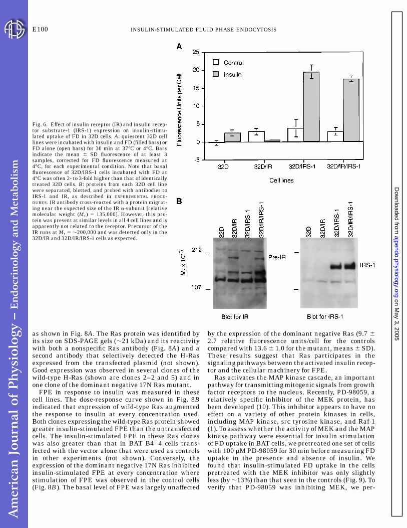
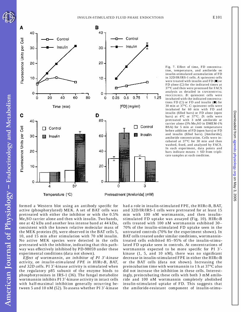
IRS-1 is involved in signaling insulin-stimulatedFPE. Insulin-stimulated mitogenesis is mediatedthough the 180-kDa intracellular protein, IRS-1, that isphosphorylated by the activated insulin receptor (re-viewed in Ref. 36). We evaluated the role of IRS-1 ininsulin-stimulated FPE using the 32D cell line, ahematopoietic precursor line that lacks endogenousIRS-1 and expresses low numbers of insulin receptors(,500/cell). In the parental 32D cell line, the basal levelof FPE was almost undetectable. Insulin treatment ofthese cells increased FPE so that the rate was similarto the basal rate of FPE observed in the lines express-ing either or both exogenous insulin receptor (32D/IR)and IRS-1 (32D/IRS-1, 32D/IR/IRS-1; Fig. 6A). 32D/IRcells responded to insulin with a reproducible decreasein FPE. In contrast, the rate of FPE increased five- tosixfold over basal in response to insulin in 32D/IRS-1cells and in 32D/IR/IRS-1 cells. These two cell linesexpressed approximately equal amounts of the IRS-1protein, as demonstrated by Western blotting (Fig. 6B).We verified that FD behaved as a true fluid phasemarker in these cells using the 32D/IR/IRS-1 line andfound that FD uptake was linear with incubation time,FD concentration, and temperature (Fig. 7, A-C). Fur-thermore, in cells that were pretreated with 3 mMamiloride, insulin-stimulated FD uptake was negli-gible; thus, as in the other insulin-responsive cell types,FD internalization appeared to be mediated thoughFPE (Fig. 7D).
Because we had anticipated that 32D/IRS-1 cells,which express few insulin receptors, would show littleinsulin-stimulated FPE, we verified the identity of allfour cell lines by their antibiotic resistance, by North-ern blotting using a probe for the human insulinreceptor mRNA (the mRNA was only observed in the32D/IR and 32D/IR/IRS-1 lines, as expected; data notshown), and by Western blotting for the insulin recep-tor and IRS-1 (Fig. 6B). We also measured basal andinsulin-stimulated FPE in another 32D/IRS-1 clone(661.5) with essentially the same results as the cloneshown in Fig. 6 (clone Ic). Thus the low numbers ofendogenous insulin receptors present in the 32D/IRS-1cells (,500/cell) were apparently sufficient to mediateinsulin-stimulated FPE in the presence of IRS-1. Weconclude that the presence of IRS-1 was responsibleand necessary for the increased rate of insulin-stimulated FPE observed in 32D/IRS-1 and 32D/IR/IRS-1 cells compared with 32D or 32D/IR cells.
Overexpression of wild-type H-Ras augments insulin-stimulated FPE, whereas expression of a dominantnegative 17N Ras represses it. A BAT cell line (BATB4–4) was stably transfected with wild-type H-Ras orthe dominant negative 17N H-Ras constructs (40).Clones that expressed the exogenous proteins wereselected by Western blot analysis of whole cell lysates,
Fig. 5. Effect of insulin on FD accumulation in cells expressingnormal or mutant insulin receptors. Quiescent cells were treatedwith either 70 nM insulin and FD (hatched bars) or FD alone (openbars) for 60 min, followed by FACS analysis. Each bar represents themean 6 SD fluorescence per cell from 6 samples. Difference betweeninsulin and control was significant for HIRc-B (P , 0.0005),A/K1018-B(P , 0.002), and HIRDCT (P , 0.0005). Difference between theinsulin and control treatments was not significant for Rat-1 fibro-blasts.
E99INSULIN-STIMULATED FLUID PHASE ENDOCYTOSIS
on May 3, 2005
ajpendo.physiology.orgD
ownloaded from
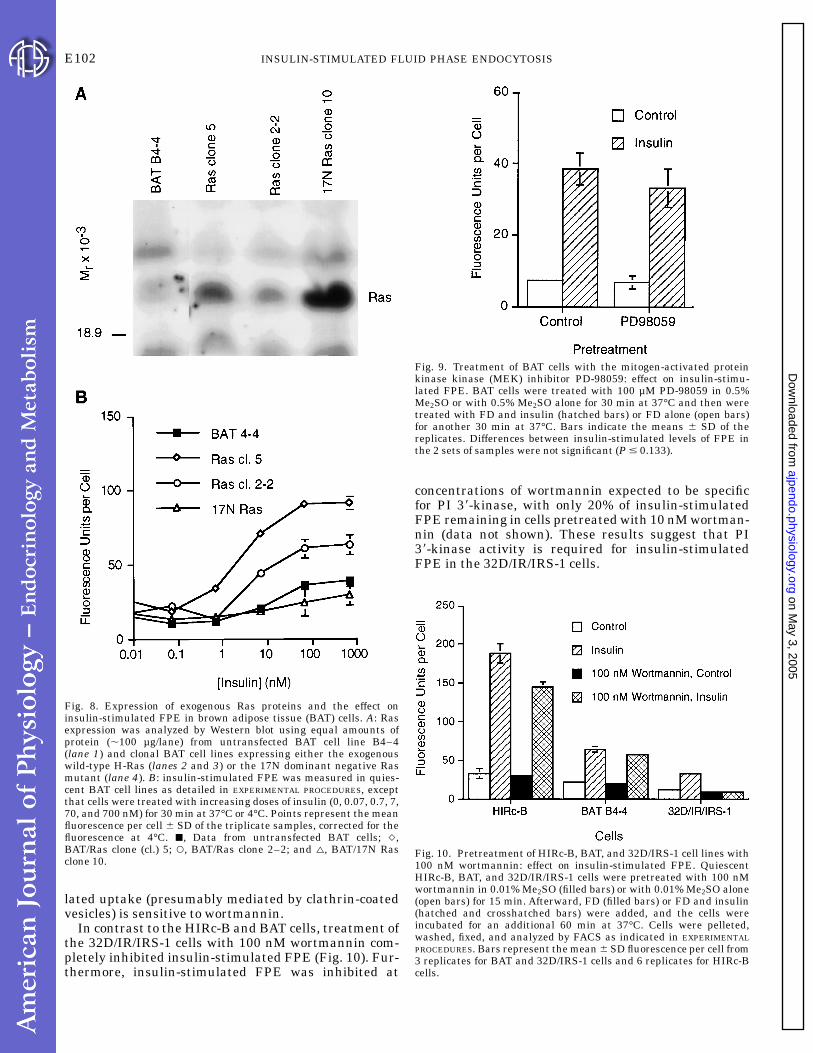
as shown in Fig. 8A. The Ras protein was identified byits size on SDS-PAGE gels (,21 kDa) and its reactivitywith both a nonspecific Ras antibody (Fig. 8A) and asecond antibody that selectively detected the H-Rasexpressed from the transfected plasmid (not shown).Good expression was observed in several clones of thewild-type H-Ras (shown are clones 2–2 and 5) and inone clone of the dominant negative 17N Ras mutant.
FPE in response to insulin was measured in thesecell lines. The dose-response curve shown in Fig. 8Bindicated that expression of wild-type Ras augmentedthe response to insulin at every concentration used.Both clones expressing the wild-type Ras protein showedgreater insulin-stimulated FPE than the untransfectedcells. The insulin-stimulated FPE in these Ras cloneswas also greater than that in BAT B4–4 cells trans-fected with the vector alone that were used as controlsin other experiments (not shown). Conversely, theexpression of the dominant negative 17N Ras inhibitedinsulin-stimulated FPE at every concentration wherestimulation of FPE was observed in the control cells(Fig. 8B). The basal level of FPE was largely unaffected
by the expression of the dominant negative Ras (9.7 62.7 relative fluorescence units/cell for the controlscompared with 13.6 6 1.0 for the mutant, means 6 SD).These results suggest that Ras participates in thesignaling pathways between the activated insulin recep-tor and the cellular machinery for FPE.
Ras activates the MAP kinase cascade, an importantpathway for transmitting mitogenic signals from growthfactor receptors to the nucleus. Recently, PD-98059, arelatively specific inhibitor of the MEK protein, hasbeen developed (10). This inhibitor appears to have noeffect on a variety of other protein kinases in cells,including MAP kinase, src tyrosine kinase, and Raf-1(1). To assess whether the activity of MEK and the MAPkinase pathway were essential for insulin stimulationof FD uptake in BAT cells, we pretreated one set of cellswith 100 µM PD-98059 for 30 min before measuring FDuptake in the presence and absence of insulin. Wefound that insulin-stimulated FD uptake in the cellspretreated with the MEK inhibitor was only slightlyless (by ,13%) than that seen in the controls (Fig. 9). Toverify that PD-98059 was inhibiting MEK, we per-
Fig. 6. Effect of insulin receptor (IR) and insulin recep-tor substrate-1 (IRS-1) expression on insulin-stimu-lated uptake of FD in 32D cells. A: quiescent 32D celllines were incubated with insulin and FD (filled bars) orFD alone (open bars) for 30 min at 37°C or 4°C. Barsindicate the mean 6 SD fluorescence of at least 3samples, corrected for FD fluorescence measured at4°C, for each experimental condition. Note that basalfluorescence of 32D/IRS-1 cells incubated with FD at4°C was often 2- to 3-fold higher than that of identicallytreated 32D cells. B: proteins from each 32D cell linewere separated, blotted, and probed with antibodies toIRS-1 and IR, as described in EXPERIMENTAL PROCE-DURES. IR antibody cross-reacted with a protein migrat-ing near the expected size of the IR a-subunit [relativemolecular weight (Mr) 5 135,000]. However, this pro-tein was present at similar levels in all 4 cell lines and isapparently not related to the receptor. Precursor of theIR runs at Mr 5 ,200,000 and was detected only in the32D/IR and 32D/IR/IRS-1 cells as expected.
E100 INSULIN-STIMULATED FLUID PHASE ENDOCYTOSIS
on May 3, 2005
ajpendo.physiology.orgD
ownloaded from
formed a Western blot using an antibody specific foractive (phosphorylated) MEK. A set of BAT cells waspretreated with either the inhibitor or with the 0.5%Me2SO carrier alone and then with insulin. Two bands,one at 42 kDa and another less intense band at 44 kDa,consistent with the known relative molecular mass ofthe MEK proteins (9), were observed in the BAT cells 5,10, and 15 min after stimulation with 70 nM insulin.No active MEK species were detected in the cellspretreated with the inhibitor, indicating that this path-way was effectively inhibited by PD-98059 under theseexperimental conditions (data not shown).
Effect of wortmannin, an inhibitor of PI 38-kinaseactivity, on insulin-stimulated FPE in HIRc-B, BAT,and 32D cells. PI 38-kinase activity is stimulated whenthe regulatory p85 subunit of the enzyme binds tophosphotyrosines in IRS-1 (36). The fungal metabolitewortmannin inhibits PI 38-kinase activity in intact cellswith half-maximal inhibition generally occurring be-tween 5 and 10 nM (52). To assess whether PI 38-kinase
had a role in insulin-stimulated FPE, the HIRc-B, BAT,and 32D/IR/IRS-1 cells were pretreated for at least 15min with 100 nM wortmannin, and then insulin-stimulated FD uptake was assayed (Fig. 10). HIRc-Bcells treated with 100 nM wortmannin exhibited 30–70% of the insulin-stimulated FD uptake seen in theuntreated controls (70% for the experiment shown). InBAT cells treated under similar conditions, wortmannin-treated cells exhibited 85–95% of the insulin-stimu-lated FD uptake seen in controls. At concentrations ofwortmannin expected to be more specific for PI 38-kinase (1, 5, and 10 nM), there was no significantdecrease in insulin-stimulated FPE in either the HIRc-Bor the BAT cells (data not shown). Increasing thepreincubation time with wortmannin to 1 h at 37°C alsodid not increase the inhibition in these cells. Interest-ingly, preincubating these cells with both 3 mM amilo-ride and 100 nM wortmannin completely abolishedinsulin-stimulated uptake of FD. This suggests thatthe amiloride-resistant component of insulin-stimu-
Fig. 7. Effect of time, FD concentra-tion, temperature, and amiloride oninsulin-stimulated accumulation of FDin 32D/IR/IRS-1 cells. A: quiescent cellswere treated with insulin and FD (j) orFD alone (k) for the indicated times at37°C and then were processed for FACSanalysis as detailed in EXPERIMENTALPROCEDURES. B: quiescent cells wereincubated with the indicated concentra-tions FD (k) or FD and insulin (j) for30 min at 37°C. C: quiescent cells wereincubated for 60 min with FD andinsulin (filled bars) or FD alone (openbars) at 4°C or 37°C. D: cells werepretreated with 3 mM amiloride orcarrier alone (3% Me2SO in DMEM-1%BSA) for 5 min at room temperaturebefore addition of FD (open bars) or FDand insulin (filled bars). [Amiloride],amiloride concentration. Cells were in-cubated at 37°C for 30 min and thenwashed, fixed, and analyzed by FACS.In each experiment, data points andbars indicate means 6 SD from tripli-cate samples at each condition.
E101INSULIN-STIMULATED FLUID PHASE ENDOCYTOSIS
on May 3, 2005
ajpendo.physiology.orgD
ownloaded from
lated uptake (presumably mediated by clathrin-coatedvesicles) is sensitive to wortmannin.
In contrast to the HIRc-B and BAT cells, treatment ofthe 32D/IR/IRS-1 cells with 100 nM wortmannin com-pletely inhibited insulin-stimulated FPE (Fig. 10). Fur-thermore, insulin-stimulated FPE was inhibited at
concentrations of wortmannin expected to be specificfor PI 38-kinase, with only 20% of insulin-stimulatedFPE remaining in cells pretreated with 10 nM wortman-nin (data not shown). These results suggest that PI38-kinase activity is required for insulin-stimulatedFPE in the 32D/IR/IRS-1 cells.
Fig. 8. Expression of exogenous Ras proteins and the effect oninsulin-stimulated FPE in brown adipose tissue (BAT) cells. A: Rasexpression was analyzed by Western blot using equal amounts ofprotein (,100 µg/lane) from untransfected BAT cell line B4–4(lane 1) and clonal BAT cell lines expressing either the exogenouswild-type H-Ras (lanes 2 and 3) or the 17N dominant negative Rasmutant (lane 4). B: insulin-stimulated FPE was measured in quies-cent BAT cell lines as detailed in EXPERIMENTAL PROCEDURES, exceptthat cells were treated with increasing doses of insulin (0, 0.07, 0.7, 7,70, and 700 nM) for 30 min at 37°C or 4°C. Points represent the meanfluorescence per cell 6 SD of the triplicate samples, corrected for thefluorescence at 4°C. j, Data from untransfected BAT cells; q,BAT/Ras clone (cl.) 5; s, BAT/Ras clone 2–2; and n, BAT/17N Rasclone 10.
Fig. 9. Treatment of BAT cells with the mitogen-activated proteinkinase kinase (MEK) inhibitor PD-98059: effect on insulin-stimu-lated FPE. BAT cells were treated with 100 µM PD-98059 in 0.5%Me2SO or with 0.5% Me2SO alone for 30 min at 37°C and then weretreated with FD and insulin (hatched bars) or FD alone (open bars)for another 30 min at 37°C. Bars indicate the means 6 SD of thereplicates. Differences between insulin-stimulated levels of FPE inthe 2 sets of samples were not significant (P # 0.133).
Fig. 10. Pretreatment of HIRc-B, BAT, and 32D/IRS-1 cell lines with100 nM wortmannin: effect on insulin-stimulated FPE. QuiescentHIRc-B, BAT, and 32D/IR/IRS-1 cells were pretreated with 100 nMwortmannin in 0.01% Me2SO (filled bars) or with 0.01% Me2SO alone(open bars) for 15 min. Afterward, FD (filled bars) or FD and insulin(hatched and crosshatched bars) were added, and the cells wereincubated for an additional 60 min at 37°C. Cells were pelleted,washed, fixed, and analyzed by FACS as indicated in EXPERIMENTALPROCEDURES. Bars represent the mean 6 SD fluorescence per cell from3 replicates for BAT and 32D/IRS-1 cells and 6 replicates for HIRc-Bcells.
E102 INSULIN-STIMULATED FLUID PHASE ENDOCYTOSIS
on May 3, 2005
ajpendo.physiology.orgD
ownloaded from

DISCUSSION
Our findings in this study support the idea thatincreased FPE is a general response to insulin in cellsthat express its receptors (33). This response wasobserved in every insulin-sensitive cell line that weexamined and has been observed by others in KB cells(24, 33), 3T3-L1 cells (14), rat H35 hepatoma cells (15),the human monocyte cell line U-937 (27, 38), Xenopusoocytes (34), and in primary cultures of rat adipocytes(13) and hepatocytes (35). However, one apparent excep-tion is the insulinoma cell line bTC6-F7 in whichinsulin reportedly activates its receptor but does notincrease FPE (60). The present study has identified theproximal components of the insulin-stimulated FPEpathway as the insulin receptor, its substrate IRS-1,and Ras. It is conceivable that one of these componentsis defective or absent in the bTC6-F7 cell line, uncou-pling the activation of the receptor from FPE.
Our studies show that the tyrosine kinase activity ofthe insulin receptor is essential for insulin-stimulatedFPE. Although the greatest increase in insulin-stimu-lated FPE was observed in HIRc-B cells, a Rat-1 cellline that overexpresses the normal human insulinreceptor, no increase beyond that of the parental Rat-1cell line was observed in the A/K1018-B line, whichexpresses a kinase-deficient mutant of the humaninsulin receptor. Previous work has shown that thereceptor tyrosine kinase activity is required in thesecells, not only for the mitogenic and metabolic effects ofinsulin (30) but also for the endocytosis of the insulinreceptor and cell ruffling (4, 26, 30). The dependence ofinsulin-stimulated FPE on insulin receptor tyrosinekinase activity has not been examined previously, toour knowledge. Presumably, the tyrosine kinase activ-ity would be essential to stimulation of FPE in all celltypes that exhibit this response.
IRS proteins are phosphorylated by the activatedinsulin receptor and its relatives, the insulin-like growthfactor receptors (36). IRS proteins are essential forinsulin-stimulated mitogenesis (54) and appear to playa role in GLUT-4 glucose transporter recruitment to theplasma membrane (21, 41). Our studies showed thatIRS-1 also plays a role in mediating insulin-stimulatedFPE. The 32D cell line, which lacks IRS proteins,exhibited some insulin-stimulated FPE (Fig. 6). How-ever, the level of insulin-stimulated FPE in 32D/IRS-1cells was increased as much as sixfold over that ofcontrol 32D cells or 32D/IR cells. We also noted thatcells expressing IRS-1 responded to insulin as stronglyas those expressing both the insulin receptor andIRS-1. The small number of endogenous receptors(,500/cell; see Ref. 54) was thus sufficient for genera-tion of the full response when IRS-1 was present. It isalso noteworthy that the FPE response of cells toinsulin is more sensitive than insulin induction of c-fosand egr-1. In HIRc-B cells, we showed that FPE wasalmost maximally stimulated at 0.7 nM insulin, whereasinsulin-induced c-fos and egr-1 gene transcription wasnot observed until 10-fold more insulin was applied(50). This difference in response sensitivity is also
likely to account for the fact that, in 32D cells, othershave reported that overexpression of the insulin recep-tor was necessary to observe insulin-stimulated c-fosand egr-1 expression (16), whereas our present dataindicate that endogenous receptors are sufficient forinsulin-stimulated FPE in these cells (16).
The dependence of insulin-stimulated FPE on IRSproteins suggested that factors that bind to the IRSscaffold might be important mediators of insulin-stimulated FPE. Certain phosphotyrosines within theIRS-1 protein act as docking sites for GRB2/SOS,facilitating the interaction of the SOS guanine nucleo-tide exchange factor with Ras (47, 53). This associationbetween SOS and Ras results in the conversion ofinactive Ras-GDP to active Ras-GTP. Injection of Ras inquiescent fibroblasts has been shown to stimulateruffling and FPE (5). The influence of exogenous wild-type Ras and the dominant negative 17N Ras oninsulin-stimulated FPE was investigated in BAT cells.Stable overexpression of exogenous wild-type Ras pro-teins augmented insulin-stimulated FPE, whereas themutant 17N Ras inhibited it (Fig. 8). These resultsstrongly suggest that insulin-stimulated FPE is medi-ated through Ras (with the caveat that some contribu-tion to this effect could potentially be due to theinteraction of the overexpressed Ras proteins withcomponents of other G protein-regulated processes thatare involved in FPE, i.e., Rab5-regulated endocyticvesicle trafficking and Rac-regulated actin polymeriza-tion).
Ras itself has been shown to represent a point ofbifurcation for signals emanating from activated recep-tor protein tyrosine kinases; signals to the nucleus aretransmitted through the MAP kinase cascade, whereassignals to the membrane (for ruffling) are mediated bythe Rac family of GTP-binding proteins (19, 20). Ourexperiments in the BAT cells using the specific MEKinhibitor PD-98059 indicate that the MAP kinase cas-cade is not involved in signaling insulin-stimulatedFPE, at least under the conditions of these experiments(Fig. 9). Further experiments with dominant negativeRac proteins may show a role for Rac in insulin-stimulated FPE.
PI 38-kinase has been implicated in vesicular traffick-ing due in part to its homology to the yeast protein,VSP34, a component of the lysosomal sorting appara-tus. PI 38-kinase activity is coprecipitated with mostactivated receptor protein tyrosine kinases (44) andmay have a role in the internalization of activatedreceptors. PI 38-kinase is also implicated in insulin-stimulated GLUT-4 translocation and growth factor-induced cell ruffling and FPE (reviewed in Ref. 22).Most of the evidence for PI 38-kinase activity in rufflingand FPE has been obtained using wortmannin, aninhibitor of PI 38-kinase activity (23, 24). We also usedwortmannin to examine the role of PI 38-kinase ininsulin-stimulated FPE in HIRc-B, BAT, and 32D/IR/IRS-1 cells. However, the interpretation of our wortman-nin inhibition experiments was complicated by twofactors, namely, that higher concentrations of wortman-nin have been shown to inhibit enzymes other than PI
E103INSULIN-STIMULATED FLUID PHASE ENDOCYTOSIS
on May 3, 2005
ajpendo.physiology.orgD
ownloaded from

38-kinase, including myosin light chain kinase (37), andthat a wortmannin-sensitive PI 38-kinase activity hasbeen shown to be required for the fusion of membranefolds in macropinocytic vesicles in macrophages (3). Inour experiments, in 32D cells, insulin-stimulated FPEwas sensitive to wortmannin at concentrations ex-pected to be specific for PI-3 kinase (Fig. 10). However,the inference that PI 38-kinase is involved in insulin-stimulated FPE in these cells is invalid if PI 38-kinaseactivity is required for membrane fusion events, as itwas in macrophages (both 32D cells and macrophagesare of hematopoietic lineage).
In contrast to the results in 32D cells, wortmanninhad no significant effect on insulin-stimulated FPE ineither the BAT cells or the HIRc-B cells at nanomolarconcentrations (Fig. 10). Our interpretation of theseresults was that a wortmannin-sensitive PI 38-kinaseactivity was not essential for insulin-stimulated FPE inthese cells. Recently, others have shown that wortman-nin treatment also had minimal effects on either basalor insulin-stimulated FPE in 3T3-L1 fibroblasts, atconcentrations at which it was inhibitory to traffickingof glucose transporters and transferrin receptors (18).However, wortmannin treatment has been shown toinhibit insulin-stimulated FPE and ruffling in CHO-Tcells, underscoring the fact that the role of PI 38-kinasemay be cell-type specific (24, 60).
The physiological role of insulin-stimulated FPE hasbeen the subject of considerable speculation (2, 25, 27,57, 59) but remains unknown. An essential questionthat remains unanswered is the extent to which thisprocess changes in parallel with the other biochemicalpathways that are markedly altered in insulin-resis-tant states, such as type 2 diabetes and obesity. If, forexample, patients developed severe insulin resistancein terms of glucose transport but remained normallysensitive to insulin in terms of FPE, one could imaginethat the increased flux of extracellular nutrients in thecells might have deleterious consequences. An examplemight be the common clinical observation that type 2diabetic patients typically gain weight when placed onthe high doses of insulin often required to achievenormoglycemia in this insulin-resistant state. Studiesof FPE in such insulin-resistant cells will be necessaryto answer these and other intriguing questions aboutthe physiological role of insulin-stimulated FPE and itspossible alterations in pathological states.
We are very grateful to J. Michael Cook and Alan Fisher from theDuke Comprehensive Cancer Center Flow Cytometry Facility forperforming the FACS analyses.
P. J. Blackshear was an Investigator and D. M. Pitterle was anAssociate of the Howard Hughes Medical Institute during the courseof these studies. M. F. White is currently an Investigator of theHoward Hughes Medical Institute.
Address for reprint requests: P. J. Blackshear, A2–05, NationalInstitute of Environmental Health Sciences, Research Triangle Park,NC 27709.
Received 23 February 1998; accepted in final form 4 September 1998.
REFERENCES
1. Alessi, D. R., A. Cuenda, P. Cohen, D. T. Dudley, and A. R.Saltiel. PD 098059 is a specific inhibitor of the activation of
mitogen-activated protein kinase kinase in vitro and in vivo. J.Biol. Chem. 270: 27489–27494, 1995.
2. Al-Habori, M. Mechanism of insulin action, role of ions and thecytoskeleton. Int. J. Biochem. 25: 1087–1099, 1993.
3. Araki, N., M. T. Johnson, and J. A. Swanson. A role forphosphoinositide 3-kinase in the completion of macropinocytosisand phagocytosis by macrophages. J. Cell Biol. 135: 1249–1260,1996.
4. Backer, J. M., S. E. Shoelson, E. Haring, and M. F. White.Insulin receptors internalize by a rapid, saturable pathwayrequiring receptor autophosphorylation and an intact juxtamem-brane region. J. Cell Biol. 115: 1535–1545, 1991.
5. Bar-Sagi, D., and J. R. Feramisco. Induction of membraneruffling and fluid-phase pinocytosis in quiescent fibroblasts byras proteins. Science 233: 1061–1068, 1986.
6. Birnbaum, M. J. The insulin-sensitive glucose transporter. Int.Rev. Cytol. 137: 239–297, 1992.
7. Cooper, A. D. Hepatic uptake of chylomicron remnants. J. LipidRes. 38: 2173–2192, 1997.
8. Cox, A. D., P. A. Solski, J. D. Jordan, and C. J. Der. Analysisof Ras protein expression in mammalian cells. Methods Enzymol.255: 195–220, 1995.
9. Crews, C. M., A. Alessandrini, and R. L. Erikson. Erks: theirfifteen minutes has arrived. Cell Growth Differ. 3: 135–142,1992.
10. Dudley, D. T., L. Pang, S. J. Decker, A. J. Bridges, and A. R.Saltiel. A synthetic inhibitor of the mitogen-activated proteinkinase cascade. Proc. Natl. Acad. Sci. USA 92: 7686–7689, 1995.
11. Ebina, Y., L. Ellis, K. Jarnagin, M. Edery, L. Graf, E.Clauser, J. H. Ou, F. Masiarz, Y. W. Kan, I. D. Goldfine, R. A.Roth, and W. J. Rutter. The human insulin receptor cDNA: thestructural basis for hormone-activated transmembrane signal-ling. Cell 40: 747–758, 1985.
12. Fraser, R., B. R. Dobbs, and G. W. Rogers. Lipoproteins andthe liver sieve: the role of the fenestrated sinusoidal endotheliumin lipoprotein metabolism, atherosclerosis, and cirrhosis. Hepatol-ogy 21: 863–874, 1995.
13. Gibbs, E. M., and G. E. Lienhard. Fluid-phase endocytosis byisolated rat adipocytes. J. Cell. Physiol. 121: 569–575, 1984.
14. Gibbs, E. M., G. E. Lienhard, J. R. Appleman, M. D. Lane,and S. C. Frost. Insulin stimulates fluid-phase endocytosis andexocytosis in 3T3-L1 adipocytes. J. Biol. Chem. 261: 3944–3951,1986.
15. Harada, S., E. G. Loten, R. M. Smith, and L. Jarett.Nonreceptor mediated nuclear accumulation of insulin in H35rat hepatoma cells. J. Cell. Physiol. 153: 607–613, 1992.
16. Harada, S., R. M. Smith, J. A. Smith, M. F. White, and L.Jarett. Insulin-induced egr-1 and c-fos expression in 32D cellsrequires insulin receptor, Shc, and mitogen-activated proteinkinase, but not insulin receptor substrate-1 and phosphatidylino-sitol 3-kinase activation. J. Biol. Chem. 271: 30222–30226, 1996.
17. Janssen, K. Current Protocols in Molecular Biology. New York:Wiley, 1993, vol. I, p. 9.1.3.
18. Jess, T. J., C. M. Belham, F. J. Thomson, P. H. Scott, R. J.Plevin, and G. W. Gould. Phosphatidylinositol 38-kinase, butnot p70 ribosomal S6 kinase, is involved in membrane proteinrecycling: wortmannin inhibits glucose transport and downregu-lates cell-surface transferrin receptor numbers independently ofany effect on fluid-phase endocytosis in fibroblasts. Cell. Signal.8: 297–304, 1996.
19. Joneson, T., M. McDonough, D. Bar-Sagi, and L. Van Aelst.RAC regulation of actin polymerization and proliferation by apathway distinct from Jun kinase. Science 274: 1374–1376,1996.
20. Joneson, T., M. A. White, M. H. Wigler, and D. Bar-Sagi.Stimulation of membrane ruffling and MAP kinase activation bydistinct effectors of RAS. Science 271: 810–812, 1996.
21. Kanai, F., K. Ito, M. Todaka, H. Hayashi, S. Kamohara, K.Ishii, T. Okada, O. Hazeki, M. Ui, and Y. Ebina. Insulin-stimulated GLUT4 translocation is relevant to the phosphoryla-tion of IRS-1 and the activity of PI3-kinase. Biochem. Biophys.Res. Commun. 195: 762–768, 1993.
22. Kapeller, R., and L. C. Cantley. Phosphatidylinositol 3-kinase.Bioessays 16: 565–576, 1994.
E104 INSULIN-STIMULATED FLUID PHASE ENDOCYTOSIS
on May 3, 2005
ajpendo.physiology.orgD
ownloaded from

23. Kotani, K., K. Hara, K. Kotani, K. Yonezawa, and M.Kasuga. Phosphoinositide 3-kinase as an upstream regulator ofthe small GTP-binding protein Rac in the insulin signaling ofmembrane ruffling. Biochem. Biophys. Res. Commun. 208: 985–990, 1995.
24. Kotani, K., K. Yonezawa, K. Hara, H. Ueda, Y. Kitamura, H.Sakaue, A. Ando, A. Chavanieu, B. Calas, F. Grigorescu, M.Nishiyama, M. D. Waterfield, and M. Kasuga. Involvement ofphosphoinositide 3-kinase in insulin- or IGF-1-induced mem-brane ruffling. EMBO J. 13: 2313–2321, 1994.
25. Lewis, W. H. Pinocytosis. Bull. Johns Hopkins Hosp. 49: 17–36,1931.
26. Li, S. L., Y. Miyata, I. Yahara, and Y. Fujita-Yamaguchi.Insulin-induced circular membrane ruffling on rat 1 cells express-ing a high number of human insulin receptors: circular rufflescaused by rapid actin reorganization exhibit high density ofinsulin receptors and phosphotyrosines. Exp. Cell Res. 205:353–360, 1993.
27. Livingston, J. N., B. R. Saran, C. D. Rose, and C. L.Anderson. Rapid effects of insulin on the cycling of the insulinreceptor in a human monocyte cell line (U-937). Diabetes 34:403–408, 1985.
28. Maegawa, H., D. A. McClain, G. Freidenberg, J. M. Olefsky,M. Napier, T. Lipari, T. J. Dull, J. Lee, and A. Ullrich.Properties of a human insulin receptor with a COOH-terminaltruncation. II. Truncated receptors have normal kinase activitybut are defective in signaling metabolic effects. J. Biol. Chem.263: 8912–8917, 1988.
29. Maegawa, H., J. M. Olefsky, S. Thies, D. Boyd, A. Ullrich,and D. A. McClain. Insulin receptors with defective tyrosinekinase inhibit normal receptor function at the level of substratephosphorylation. J. Biol. Chem. 263: 12629–12637, 1988.
30. McClain, D. A., H. Maegawa, J. Lee, T. J. Dull, A. Ulrich,and J. M. Olefsky. A mutant insulin receptor with defectivetyrosine kinase displays no biologic activity and does not undergoendocytosis. J. Biol. Chem. 262: 14663–14671, 1987.
31. McClain, D. A., H. Maegawa, J. Levy, T. Huecksteadt, T. J.Dull, J. Lee, A. Ullrich, and J. M. Olefsky. Properties of ahuman insulin receptor with a COOH-terminal truncation. I.Insulin binding, autophosphorylation, and endocytosis. J. Biol.Chem. 263: 8904–8911, 1988.
32. McClain, D. A., H. Maegawa, R. S. Thies, and J. M. Olefsky.Dissection of the growth versus metabolic effects of insulin andinsulin-like growth factor-I in transfected cells expressing kinase-defective human insulin receptors. J. Biol. Chem. 265: 1678–1682, 1990.
33. Miyata, Y., M. Hoshi, S. Koyasu, T. Kadowaki, M. Kasuga, I.Yahara, E. Nishida, and H. Sakai. Rapid stimulation offluid-phase endocytosis and exocytosis by insulin, insulin-likegrowth factor-I, and epidermal growth factor in KB cells. Exp.Cell Res. 178: 73–83, 1988.
34. Morrill, G. A., A. B. Kostellow, and S. P. Weinstein. Endocyto-sis in the amphibian oocyte. Effect of insulin and progesterone onmembrane and fluid internalization during the meiotic divisions.Biochim. Biophys. Acta 803: 71–77, 1984.
35. Moss, A. L., and W. F. Ward. Multiple pathways for ligandinternalization in rat hepatocytes. I: Effects of anoxia, phenylar-sine oxide and monensin. J. Cell. Physiol. 149: 313–318, 1991.
36. Myers, M. G., Jr., and M. F. White. Insulin signal transductionand the IRS proteins. Annu. Rev. Pharmacol. Toxicol. 36: 615–658, 1996.
37. Nakanishi, S., S. Kakita, I. Takahashi, K. Kawahara, E.Tsukuda, T. Sano, K. Yamada, M. Yoshida, H. Kase, Y.Matsuda, Y. Hashimoto, and Y. Nonomura. Wortmannin, amicrobial product inhibitor of myosin light chain kinase. J. Biol.Chem. 267: 2157–2163, 1992.
38. Oefelein, M. G., G. Arsenis, and J. N. Livingston. Insulin-stimulated fluid-phase pinocytosis and internalization of theinsulin receptor: differences between the U-937 monocyte and ratadipocyte. Metabolism 35: 818–823, 1986.
39. Oliver, J. M., R. D. Berlin, and B. H. Davis. Use of horseradishperoxidase and fluorescent dextrans to study fluid pinocytosis inleukocytes. Methods Enzymol. 108: 336–347, 1984.
40. Quilliam, L. A., K. Kato, K. M. Rabun, M. M. Hisaka, S. Y.Huff, S. Campbell-Burk, and C. J. Der. Identification ofresidues critical for Ras(17N) growth-inhibitory phenotype andfor Ras interaction with guanine nucleotide exchange factors.Mol. Cell. Biol. 14: 1113–1121, 1994.
41. Quon, M. J., A. J. Butte, M. J. Zarnowski, G. Sesti, S. W.Cushman, and S. I. Taylor. Insulin receptor substrate 1mediates the stimulatory effect of insulin on GLUT4 transloca-tion in transfected rat adipose cells. J. Biol. Chem. 269: 27920–27924, 1994.
42. Ridley, A. J. Membrane ruffling and signal transduction. Bioes-says 16: 321–327, 1994.
43. Robinson, M. S., C. Watts, and M. Zerial. Membrane dynam-ics in endocytosis. Cell 84: 13–21, 1996.
44. Ruderman, N. B., R. Kapeller, M. F. White, and L. C.Cantley. Activation of phosphatidylinositol 3-kinase by insulin.Proc. Natl. Acad. Sci. USA 87: 1411–1415, 1990.
45. Saltiel, A. R. Diverse signaling pathways in the cellular actionsof insulin. Am. J. Physiol. 270 (Endocrinol. Metab. 33): E375–E385, 1996.
46. Scherer, P. E., M. P. Lisanti, G. Baldini, M. Sargiacomo,C. C. Mastick, and H. F. Lodish. Induction of caveolin duringadipogenesis and association of GLUT4 with caveolin-richvesicles. J. Cell Biol. 127: 1233–1243, 1994.
47. Schlessinger, J., and D. Bar-Sagi. Activation of Ras and othersignaling pathways by receptor tyrosine kinases. Cold SpringHarb. Symp. Quant. Biol. 59: 173–179, 1994.
48. Storrie, B., and E. A. Madden. Isolation of subcellular organ-elles. Methods Enzymol. 182: 203–225, 1990.
49. Stossel, T. P. On the crawling of animal cells. Science 260:1086–1094, 1993.
50. Stumpo, D. J., and P. J. Blackshear. Cellular expression ofmutant insulin receptors interferes with the rapid transcrip-tional response to both insulin and insulin-like growth factor I. J.Biol. Chem. 266: 455–460, 1991.
51. Thies, R. S., A. Ullrich, and D. A. McClain. Augmentedmitogenesis and impaired metabolic signaling mediated by atruncated insulin receptor. J. Biol. Chem. 264: 12820–12825,1989.
52. Ui, M., T. Okada, K. Hazeki, and O. Hazeki. Wortmannin as aunique probe for an intracellular signalling protein, phosphoino-sitide 3-kinase. Trends Biochem. Sci. 20: 303–307, 1995.
53. Van der Geer, P., T. Hunter, and R. A. Lindberg. Receptorprotein-tyrosine kinases and their signal transduction path-ways. Annu. Rev. Cell Biol. 10: 251–337, 1994.
54. Wang, L. M., M. G. Myers, Jr., X. J. Sun, S. A. Aaronson, M.White, and J. H. Pierce. IRS-1: essential for insulin- andIL-4-stimulated mitogenesis in hematopoietic cells. Science 261:1591–1594, 1993.
55. West, M. A., M. S. Bretscher, and C. Watts. Distinct endocy-totic pathways in epidermal growth factor-stimulated humancarcinoma A431 cells. J. Cell Biol. 109: 2731–2739, 1989 [pub-lished erratum appears in J. Cell Biol. 110: 859, 1990].
56. White, M. F., and C. R. Kahn. The insulin signaling system. J.Biol. Chem. 269: 1–4, 1994.
57. Wiley, H. S., and D. D. Cunningham. Epidermal growth factorstimulates fluid phase endocytosis in human fibroblasts througha signal generated at the cell surface. J. Cell. Biochem. 19:383–394, 1982.
58. Wiley, H. S., and D. N. McKinley. Assay of growth factorstimulation of fluid-phase endocytosis. Methods Enzymol. 146:402–417, 1987.
59. Wiley, H. S., B. J. Walsh, and K. A. Lund. Global modulation ofthe epidermal growth factor receptor is triggered by occupancy ofonly a few receptors. Evidence for a binary regulatory system innormal human fibroblasts. J. Biol. Chem. 264: 18912–18920,1989.
60. Xu, G., J. Howland, and P. L. Rothenberg. Insulin andsecretagogues differentially regulate fluid-phase pinocytosis ininsulin-secreting beta-cells. Biochem. J. 318: 623–629, 1996.
E105INSULIN-STIMULATED FLUID PHASE ENDOCYTOSIS
on May 3, 2005
ajpendo.physiology.orgD
ownloaded from