e myelodysplastic syndromes progression in patients with
TRANSCRIPT

498 haematologica | 2017; 102(3)
Received: July 4, 2016.
Accepted: November 15, 2016.
Pre-published: November 24, 2016.
Correspondence: [email protected]
Ferrata StortiFoundation
EUROPEANHEMATOLOGYASSOCIATION
Haematologica 2017Volume 102(3):498-508
ARTICLE Myelodysplastic Syndromes
doi:10.3324/haematol.2016.152025
Introduction
One salient feature of malignant hematopoiesis is clonal dominance, i.e., the sup-pression of normal hematopoiesis by the neoplastic clone. In myelodysplastic syn-dromes (MDS) associated with deletion of the long arm of chromosome 5 [del(5q)],clonal dominance leads to the expansion of del(5q) hematopoietic stem cells (HSC)at the expense of normal HSC.1 In a recent study we demonstrated that rare HSCcarrying del(5q) are necessary and sufficient to propagate the disease.2 Furthermore,we found that del(5q) HSC are selectively resistant to lenalidomide at the time ofcomplete clinical and cytogenetic remission,3 potentially enabling the continual
AA high proportion of patients with lower-risk del(5q) myelodys-plastic syndromes will respond to treatment with lenalidomide.The median duration of transfusion-independence is 2 years with
some long-lasting responses, but almost 40% of patients progress toacute leukemia by 5 years after starting treatment. The mechanismsunderlying disease progression other than the well-established finding ofsmall TP53-mutated subclones at diagnosis remain unclear. We studied alongitudinal cohort of 35 low- and intermediate-1-risk del(5q) patientstreated with lenalidomide (n=22) or not (n=13) by flow cytometric sur-veillance of hematopoietic stem and progenitor cell subsets, targetedsequencing of mutational patterns, and changes in the bone marrowmicroenvironment. All 13 patients with disease progression were identi-fied by a limited number of mutations in TP53, RUNX1, and TET2,respectively, with PTPN11 and SF3B1 occurring in one patient each.TP53 mutations were found in seven of nine patients who developedacute leukemia, and were documented to be present in the earliest sam-ple (n=1) and acquired during lenalidomide treatment (n=6). By contrast,analysis of the microenvironment, and of hematopoietic stem and pro-genitor cells by flow cytometry was of limited prognostic value. Basedon our data, we advocate conducting a prospective study aimed at inves-tigating, in a larger number of cases of del(5q) myelodysplastic syn-dromes, whether the detection of such mutations before and afterlenalidomide treatment can guide clinical decision-making.
Progression in patients with low- and intermedi-ate-1-risk del(5q) myelodysplastic syndromes is predicted by a limited subset of mutations Christian Scharenberg,1,2 Valentina Giai,1 Andrea Pellagatti,3 Leonie Saft,4Marios Dimitriou,1 Monika Jansson,1 Martin Jädersten,1 Alf Grandien,1Iyadh Douagi,1 Donna S. Neuberg,5 Katarina LeBlanc,1 Jacqueline Boultwood,3Mohsen Karimi,1 Sten Eirik W. Jacobsen,1,6 Petter S. Woll1and Eva Hellström-Lindberg1
1Department of Medicine, Center for Hematology and Regenerative Medicine,Karolinska University Hospital Huddinge, Karolinska Institutet, Stockholm, Sweden;2Department of Medicine, Division of Hematology, Skaraborgs Hospital, Skövde,Sweden; 3Bloodwise Molecular Haematology Unit, Nuffield Division of ClinicalLaboratory Sciences, Radcliffe Department of Medicine, University of Oxford, and NIHRBiomedical Research Centre, Oxford, UK; 4Department of Pathology, KarolinskaUniversity Hospital, Karolinska Institutet, Stockholm, Sweden; 5Department ofBiostatistics and Computational Biology, Dana–Farber Cancer Institute, Boston, MA,USA and 6Department of Cell and Molecular Biology, Karolinska Institutet, Stockholm,Sweden
ABSTRACT
Check the online version for the most updatedinformation on this article, online supplements,and information on authorship & disclosures:www.haematologica.org/content/102/3/498
©2017 Ferrata Storti Foundation
Material published in Haematologica is covered by copyright.All rights are reserved to the Ferrata Storti Foundation. Use ofpublished material is allowed under the following terms andconditions: https://creativecommons.org/licenses/by-nc/4.0/legalcode. Copies of published material are allowed for personal or inter-nal use. Sharing published material for non-commercial pur-poses is subject to the following conditions: https://creativecommons.org/licenses/by-nc/4.0/legalcode,sect. 3. Reproducing and sharing published material for com-mercial purposes is not allowed without permission in writingfrom the publisher.

accrual of mutations and disease progression. At diagno-sis, the majority of MDS patients carry recurrent muta-tions in a number of myeloid candidate genes, several ofwhich are strongly associated with outcome.4-7 Weobserved this pattern also in patients with del(5q) MDS, inwhom 64% had additional mutations detected in the HSCcompartment. However, del(5q) seemed to precede theidentified driver mutations in most cases, arguing thatdeletion of 5q is sufficient for a clonal advantage.2The immunomodulatory drug lenalidomide has a spe-
cific effect in patients with lower-risk del(5q) MDS, abro-gating the need for transfusions in around 50% ofpatients.8,9 The corresponding incidence of complete cyto-genetic remissions varies between 16% and 26%.9 Whilea small subgroup of patients may maintain completeremissions for years even after the withdrawal of lenalido-mide,10 the median response duration is 2 years9 andapproximately 40% of the patients in the MDS 004 studyhad progressed to acute myeloid leukemia at 5 years.11As the molecular mechanisms underlying disease pro-
gression in del(5q) MDS remain to be elucidated, we donot know how to predict disease progression or how tomonitor patients during lenalidomide treatment. We pre-viously reported that small TP53-mutated subclones pre-dict for an unfavorable outcome in del(5q) patients, andthat these subclones expand with disease progression.12However, whether or not other somatic mutations or fac-tors related to the bone marrow microenvironment alsocontribute to disease progression has not been compre-hensively assessed. In this longitudinal study, we show forthe first time that all patients with disease progressionwere identified by a limited subset of mutations. Based onour data, we therefore advocate that mutational profilingshould be used before and during treatment of del(5q)MDS patients in order to guide individual clinical deci-sions.
Methods
Clinical characteristics of the patientsBetween 2004 and 2015 we included consecutive patients at the
Karolinska University Hospital in Stockholm, Sweden based onthe following criteria: MDS with a low- or intermediate-1-riskscore at diagnosis according to the International PrognosticScoring System and standard cytogenetics including del5q31 with-out or with one additional abnormality. All patients were followeduntil April 2016 for survival, disease progression, and treatment. Intotal, 35 patients were analyzed by targeted sequencing for fre-quently mutated genes with known or putative implications in thepathogenesis of myeloid diseases, and material from the vastmajority of patients was available for flow cytometric analysis ofhematopoietic stem- and progenitor cells (HSPC). In six patientswe were able to purify HSPC, by fluorescence activated cell sort-ing (FACS), from two or more consecutive visits for further stud-ies. The above studies of MDS patients, as well as a study ofhealthy individuals were approved by the institutional reviewboard at Karolinska Institute and both patients and healthy indi-viduals provided written informed consent. As a definition forclinical progression, we used the same definition as that used byJadersten et al.12 Besides defining “progression” as the developmentof acute myeloid leukemia (9 cases), four cases were defined ashaving progression based on the acquisition of additional kary-otypic abnormalities (n=3) and an increase of marrow blasts from<5% to 11% (n=1) in combination with worsening cytopenias.
Flow cytometry, cell sorting and analysis of geneexpressionHSC, multipotent progenitors (MPP), lymphoid-primed mul-
tipotent progenitors (LMPP) and three subsets of myeloid pro-genitors, including common myeloid progenitors (CMP), gran-ulocyte–macrophage progenitors (GMP), and megakaryocyte–erythroid progenitors (MEP) were identified using a panel ofantibodies based on the following surface markers:13,14 HSC (lin–
CD34+CD38–CD90+CD45RA–), MPP (lin–CD34+CD38–CD90–
CD45RA–), LMPP (lin–CD34+CD38– CD90–CD45RA+), CMP(lin–CD34+CD38+CD123+CD45RA–), GMP (lin–
CD34+CD38+CD123+CD45RA+); and MEP (lin–
CD34+CD38+CD123–CD45RA–). Cell populations were isolatedfrom CD34-enriched normal and MDS mononuclear cells byFACS on a FACS Aria and used for subsequent analyses. Geneexpression was analyzed by Fluidigm Dynamic Arrays as pre-viously described3 (Online Supplementary Figure S5).
Fluorescence in situ hybridizationFlow-sorted cell populations were spun onto glass slides.
Slides were subsequently treated with pepsin and fixed withformaldehyde/MgCl2. The LSI EGR1/D5S721, D5S23 DualColor Probe (Abbott-Vysis, Downers Grove, IL, USA) was usedto detect deletions of 5q31; LSI EGR1 detects deletions of 5q31,and LSI D5S721, D5S23 detects 5p15.2 and serves as an internalcontrol. Probes were applied as recommended by the manufac-turer. As fluorescence in situ hybridization (FISH) analysis doesnot detect additional cytogenetic changes, standard cytogeneticstudies were performed on mononuclear cells taken at the sametime-points.
Bone marrow morphology and immunohistochemistrySequential bone marrow samples were assessed by routine
morphology and immunohistochemistry at each time-point. Bonemarrow samples were assessed at diagnosis in all patients whilefive were analyzed prior to and at various time-points during treat-ment with lenalidomide (MDS063, 094, 106, 110, and 143). Bonemarrow cellularity and fibrosis were assessed according toEuropean consensus guidelines.15 Immunohistochemistry was per-formed for different markers including p53 DO-1 (Santa Cruz,Biotechnology, Inc., USA),11 CD34, CD68, Nestin, CD271, CD146(Novocastra, UK), using the automated BondTM and Ventana BenchMark XT systems according to the manufacturers’ instructions.Microvascular density was quantified as the number of blood ves-sels per high power field, using regular light microscopy at high(400x) magnification as previously described.16 Blood vessels wereidentified as CD34+ endothelial cells forming a structure with aclearly discernable lumen. The frequency of CD34+ mononuclearcells and the tendency of CD34+ cells to form clusters wereassessed as previously described.17
Mesenchymal stromal cell cultures and RNA isolationMesenchymal stromal cells (MSC) were isolated from six
untreated del(5q) cases and six healthy volunteers using a previ-ously published standard procedure18 and expanded as detailedelsewhere, while uniformly fulfilling the minimal MSC criteria.19
Cell lysates were harvested with lysis buffer (Qiagen, Hilden,Germany), RNA was extracted using a Qiagen RNeasy minikit,and then stored in RNase-free water at -80 °C.
Affymetrix gene expression of mesenchymal stromal cellsGene expression profiling of MSC was performed as was previ-
ously described for CD34+ cells.20,21 Briefly, for each sample 100 ngof total RNA were amplified and labeled with the 3' IVT ExpressKit (Affymetrix, Santa Clara, CA, USA) following the manufactur-
Progression prediction in lower-risk del(5q) MDS
haematologica | 2017; 102(3) 499
er’s recommendations. Biotin-labeled fragmented cRNA washybridized to GeneChip Human Genome U133 Plus2.0 arrays(Affymetrix), covering over 47,000 transcripts. Hybridization wasperformed at 45 °C for 16 h in a Hybridization Oven 640(Affymetrix). Chips were washed and stained in a Fluidics Station450 (Affymetrix) and scanned using a GeneChip Scanner 3000(Affymetrix). Affymetrix CEL files were pre-processed using therobust multiarray average algorithm.22 Data were analyzed usingGeneSpring 12.6 (Agilent Technologies). Quality control resultsobtained for scale factors, background levels, percentages of pres-ent calls, 3’/5’ GAPDH ratio, and intensities of spike hybridizationcontrols were within the acceptable ranges for all samples.23
DNA sequencing and bioinformatics analyses Haloplex target enrichment for Illumina (Agilent) was applied
for mutation screening in panels of either 42 or 74 frequentlymutated genes (Online Supplementary Table S1) according to themanufacturer’s instructions. Of note, the 42-gene panel coversmost genes reported to be recurrently mutated in MDS4-6 and wereincluded in both kits. Briefly, bone marrow mononuclear cellswere separated by density gradient centrifugation usingLymphoprep (Nycomed, Oslo, Norway). Genomic DNA wasextracted using a GeneElute DNA extraction kit (Sigma) and quan-tified by Qubit. All samples were individually barcoded using 96barcoding oligos by Agilent during enrichment and the quality ofindividual libraries was checked by Tape Station D1K assays(Agilent). Sequencing was performed on pooled samples usingeither a HiSeq 2000 (Illumina) sequencer through paired–end, 100bp reads or a MiSeq sequencer through paired-end, 150 bp reads.Illumina Sequencing adapters were removed using Cutadapt(v.0.9.5) and reads were aligned to the hg19 using MosaikAligner(v.2.1.33). Sequence variants were identified using VarScan 2 inmpileup2cns mode. Variants were annotated using ANNOVAR.24
An ECD DNA control included in the Haloplex kit was used to fil-ter out sequencing errors. Variants were selected for further analy-sis if they met the following criteria: (i) minimum coverage of100X, (ii) minimum of 20 variant reads, (iii) having a variant allelefrequency of >0.03 for all genes except for TP53 for which thelimit of detection was set at ≥0.01 based on our previous studiesthat demonstrated these detection levels to be clinically rele-vant,11,12,25 (iv) not present in the 1000 Genome database, (v) notlisted in dbSNP unless listed in the COSMIC 65 database, (vi) trun-cating or damaging based on SIFT if not present in COSMIC 65.Sequencing results for case MDS019 were obtained by exome cap-ture performed using SureSelect Human All Exon 50 Mb (Agilent),with sequencing done on an Illumina Genome Analyzer IIx plat-form. Sequencing results for case MDS110 have already been pub-lished.2
Statistical analysisSurvival and time to progression (defined as blast increase to
>10% or acquisition of a complex karyotype) were updated inApril 2016 and were measured from the time of diagnosis.Continuous variables were compared using the Mann-Whitney U-test or Student t test, as appropriate. Categorical variables wereanalyzed by the Fisher exact test. All statistical calculations wereperformed using Graph Pad Prism 6.0 (GraphPad Software, Inc., LaJolla, CA, USA).
Results
Patients’ outcomeWe analyzed 35 patients with del(5q) MDS, with low-
or intermediate-1-risk disease according to the
International Prognostic Scoring System, at one or moretime-points by targeted sequencing (Online SupplementaryTables S1 and S2). Patients were allocated to receivelenalidomide treatment (n=22) or not (n=13) based onthe severity of their anemia, co-morbidities and availabil-ity of other therapeutic options, such as allogeneic stemcell transplantation. After a median observation period of61 months (range, 0.5-187) from diagnosis, 17 patientsremained alive. The median survival of all patients(median age 82 years, range, 45-96) was 102 months(range, 0.5-187). These patients included six who under-went allogeneic stem cell transplantation at 145, 135,116, 42, 12 and 6 months after diagnosis, with five ofthese six transplanted patients still surviving (median ageat transplantation 63 years; range, 46-73). The mediansurvival for the 29 patients who did not undergo trans-plantation was 70 months. Further demographic andclinical characteristics of the patients are detailed inOnline Supplementary Figure S1.
Disease progression is associated with the emergenceof new mutations In total, 84% of patients had a recurrent mutation in at
least one gene in our panels. The summarized results ofthe targeted sequencing are shown in Online SupplementaryTable S2. We found no differences in other clinical param-eters (e.g., age, blood counts, or additional cytogeneticabnormalities) between patients in whom we detectedrecurrent mutations and those in whom we did not.Considering all time-points for a patient, the most fre-quently mutated genes were TP53 (n=11 patients),DNMT3A (n=8), TET2 (n=7), ASXL1 (n=6) and RUNX1(n=3) (Figure 1B). Interestingly, the mutational landscapeseemed to differ from that of lower-risk MDS in general,as described in earlier reports: while mutations in genesinvolved in splicing were less frequent, the spectrum ofmutations in this pure del(5q) cohort was more similar tothat seen in high-risk MDS patients.4-6,26Diagnostic or pre-treatment samples were available for
14 of 22 patients treated with lenalidomide (the ‘LEN’cohort), and all (13/13) patients who did not receivelenalidomide (the ‘no LEN’ cohort), and there were no sig-nificant differences in the number or type of mutationsbetween these two groups (P>0.99). Only two out of 22patients failed to respond to lenalidomide treatment andalthough both of these patients harbored mutations,meaningful statistical analysis of these two patients wasnot possible. Of the 35 patients, 13 (37%) progressed to high-risk
MDS (refractory anemia with excess blasts-1, n=3 andrefractory anemia with excess blasts-2, n=1) or leukemia(n=9) at a median of 85 months (range, 31-184) after diag-nosis. Of the 27 patients for whom diagnostic or pre-treat-ment samples were available, nine (33%) showed nomutations, while 18 (67%) had one or more mutation(Online Supplementary Table S1). The presence of any recur-rent mutation covered by these MDS panels early in thepatients’ disease-course and prior to treatment did not pre-dict progression (P=0.68). However, when considering the20 patients whose samples were neither from diagnosisnor pre-treatment, an absence of mutations was sugges-tive of freedom from progression (P=0.073). For 16 patients, material was available from more than
one time-point, enabling longitudinal assessment of allelicburden in relation to treatment with lenalidomide or stem
C. Scharenberg et al.
500 haematologica | 2017; 102(3)
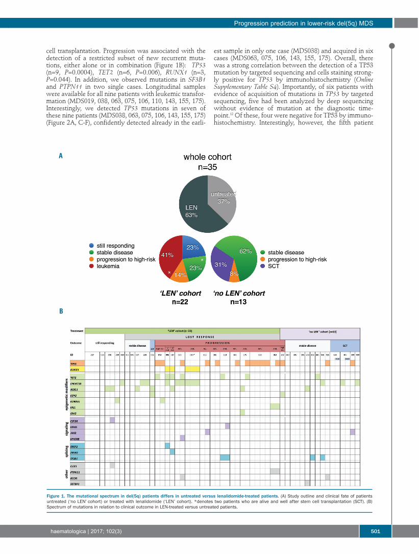
cell transplantation. Progression was associated with thedetection of a restricted subset of new recurrent muta-tions, either alone or in combination (Figure 1B): TP53(n=9, P=0.0004), TET2 (n=6, P=0.006), RUNX1 (n=3,P=0.044). In addition, we observed mutations in SF3B1and PTPN11 in two single cases. Longitudinal sampleswere available for all nine patients with leukemic transfor-mation (MDS019, 038, 063, 075, 106, 110, 143, 155, 175).Interestingly, we detected TP53 mutations in seven ofthese nine patients (MDS038, 063, 075, 106, 143, 155, 175)(Figure 2A, C-F), confidently detected already in the earli-
est sample in only one case (MDS038) and acquired in sixcases (MDS063, 075, 106, 143, 155, 175). Overall, therewas a strong correlation between the detection of a TP53mutation by targeted sequencing and cells staining strong-ly positive for TP53 by immunohistochemistry (OnlineSupplementary Table S4). Importantly, of six patients withevidence of acquisition of mutations in TP53 by targetedsequencing, five had been analyzed by deep sequencingwithout evidence of mutation at the diagnostic time-point.12 Of these, four were negative for TP53 by immuno-histochemistry. Interestingly, however, the fifth patient
Progression prediction in lower-risk del(5q) MDS
haematologica | 2017; 102(3) 501
Figure 1. The mutational spectrum in del(5q) patients differs in untreated versus lenalidomide-treated patients. (A) Study outline and clinical fate of patientsuntreated (‘no LEN’ cohort) or treated with lenalidomide (‘LEN’ cohort). *denotes two patients who are alive and well after stem cell transplantation (SCT). (B)Spectrum of mutations in relation to clinical outcome in LEN-treated versus untreated patients.
A
B

who was negative by deep sequencing did actually showpositive immunohistochemical staining of 4%.In the remaining two patients with leukemic transfor-
mation, we detected mutations in RUNX1 (n=1, MDS019,Figure 2B) and TET2 (n=1, MDS110), present at both time-points at which samples were taken from these patients.Patient MDS110 also acquired a NRAS mutation at thelater time-point. Three patients transformed to higher-riskMDS and all carried mutations in TET2 (MDS094, 096,107). The only patient who progressed to refractory ane-mia with excess blasts-2 in the cohort not treated withlenalidomide showed mutations in TP53 and EZH2. Regardless of whether the three mutations (TP53, TET2
and RUNX1) were present in the initial sample or whetherthey developed subsequently, testing positive for any of
them carried a high probability (13/16, 81%) of progres-sion. Follow-up time after the latest mutation screeningwas similar between patients who progressed (median of18 months, range 1-91 months) and those who did not(median of 27 months, range 0.3-76 months).In 11 out of 13 patients the new mutations were detect-
ed prior to the time of clinical progression and the mediantime from detection of the mutation to clinical evidence ofprogression was 42 months (range, 0-83.9). Thus, we wereable to detect the mutation in the majority of cases wellbefore clinical signs of disease progression (Figure 3).
Surveillance of hematopoietic stem and progenitor cellsubsets under lenalidomide therapy In order to investigate the impact of lenalidomide ther-
C. Scharenberg et al.
502 haematologica | 2017; 102(3)
Figure 2. Longitudinal assessment of mutations during treatment with erythropoietin (shaded in red) and lenalidomide (shaded in gray). (A) Frequency of mutationsin relation to the del(5q) clone in a patient who progressed to high-risk disease. (B) Variant allele frequency in a patient who progressed to leukemia, received inductiontherapy and went into complete remission and was transplanted. *** This patient had trisomy 21, the region in which RUNX1 resides, resulting in a homozygous muta-tion with amplification via trisomy 21. (C-F) Variant allele frequencies in four patients who progressed to leukemia. The size of the del(5q) clone was estimated with flu-orescence in situ hybridization analysis of mononuclear cells. VAF: variant allele frequency; LEN: lenalidomide; MNC: mononuclear cells; HSC: hematopoietic stem cells;HSCT: hematopoietic stem cell transplantation; AML: acute myeloid leukemia; ALL: acute lymphocytic leukemia; CCyR: complete cytogenetic response.
BA
DC
FE

apy on distinct HSPC in del(5q) MDS patients, we per-formed multicolor flow-cytometry. The distribution ofsub-populations within the Lin–CD34+CD38– compart-ments, including HSC, MPP and LMPP,2,13 remained similarin del(5q) MDS patients, both at diagnosis and duringlenalidomide treatment, compared to that in healthy con-trols (Figure 4). However, as previously reported,2,27,28 theGMP frequency within Lin–CD34+CD38+ cells was signifi-cantly suppressed in diagnostic del(5q) MDS with a con-comitant increase in CMP. Upon lenalidomide treatment,the GMP and CMP distribution reverted to frequenciescomparable to those of normal Lin–CD34+CD38+ cells. Serial samples were available for five patients who pro-
gressed to leukemia, including one patient who did notrespond to lenalidomide (MDS063). This allowed us tomonitor kinetic changes in HSPC subsets within the samepatient over time during treatment and disease progres-sion (Figure 4C, Online Supplementary Figure S2 and OnlineSupplementary Table S3). While in each case there was onepredominant HSPC subset that expanded prior to progres-sion, the type of subset varied from patient to patient. Wecombined cell sorting with FISH analysis to assess theclonal size of distinct del(5q) HSPC subsets (Figure 4C andOnline Supplementary Figure S3). Notably, although severalof the patients investigated had a complete clinicalresponse to lenalidomide, in all but one of these patientsthe mononuclear bone marrow cells and to a higherdegree stem- and progenitor compartments contained alarge fraction of 5q-deleted cells, and were thus not in
complete cytogenetic remission. Interestingly, in the onlypatient (MDS106) who initially showed a complete cyto-genetic response based on FISH analysis of mononuclearbone marrow cells, the myeloid progenitor subsets (CMP,GMP and MEP) also showed minimal clonal involvement(Online Supplementary Figure S3B), whereas as much as54% of the Lin–CD34+CD38–CD90+CD45RA– HSC com-partment remained part of the del(5q) clone, supportingprevious studies implicating a selective resistance ofdel(5q) HSC to lenalidomide treatment.
The microenvironment in del(5q) and effects of lenalidomide treatmentTo determine whether the failure to produce mature
progeny is primarily intrinsic due to compromised HSPCor if extrinsic, microenvironmental factors contribute, weinitiated MSC cultures from untreated del(5q) and healthyvolunteers and generated gene expression profiles byAffymetrix microarray. While expression values for a vari-ety of hematopoietic genes were minimal or absent, MSCcultures from both healthy volunteers and untreateddel(5q) MDS patients expressed typical gene signatures forMSC (Online Supplementary Figure S4). However, we foundno genes expressed in a statistically significant differentmanner (P<0.05, Welch t-test and Benjamini–Hochbergmultiple testing correction), even when specifically look-ing for genes previously implicated in HSC-niche interac-tions29 (Figure 5A).We next investigated bone marrow biopsies in healthy
Progression prediction in lower-risk del(5q) MDS
haematologica | 2017; 102(3) 503
Figure 3. Detection of mutations in advance of clinicalsigns of progression. The individual fates of 13patients who progressed to either high-risk MDS (n=4)or leukemia (n=9) are depicted showing the time ofdiagnosis, time-point at which sequencing was per-formed and whether a mutation was detected or not(see legend). SCT: stem cell transplantation.

C. Scharenberg et al.
504 haematologica | 2017; 102(3)
Figure 4. Surveillance of hematopoietic stem and progenitor cell subsets and the phenotypic changes induced by lenalidomide. (A) FACS profiles of bone marrowstem and progenitor cells in a normal age-matched control (top row), and a representative case of del(5q) myelodysplastic syndrome at diagnosis (middle row), anddel(5q) myelodysplastic syndrome treated with lenalidomide. (B) Relative distribution of stem and progenitor cell subsets within lin-CD34+CD38- and lin-CD34+CD38+
compartments in normal controls and diagnostic/untreated del(5q), and lenalidomide-treated del(5q). Indicated P-values are shown when significant by the Mann-Whitney test. (C) Frequency within total bone marrow and ratio of del(5q) versus normal HSC in serial samples of four patients (3 responders and 1 non-responder)during lenalidomide treatment and progression to acute myeloid leukemia. NBM: normal bone marrow; Dx: diagnosis; LEN: lenalidomide; TTP: time to progression(months); MNC: mononuclear cells; TD: transfusion-dependent; CR: complete response; LR: loss of response; PR: partial response).
A
B
C

controls and in del(5q) MDS patients before and duringlenalidomide treatment. Material for longitudinal analysisby immunohistochemistry was available for five patients.These analyses revealed that microvessel density was sig-nificantly higher in del(5q) MDS than in normal controls(microvessel density values of 5.2±3.2 versus 2.4±1.2,respectively; P=0.02) but decreased during the initial phaseof lenalidomide treatment in all five patients analyzed(Figure 5B). Subsequent therapeutic failure was associatedwith an increase in bone marrow cellularity and microves-
sel density in four of the five patients. The number ofCD68+ macrophages was not increased in bone marrowsamples from del(5q) MDS patients as compared to thenumber in controls; however, upon lenalidomide treat-ment a decrease was noted which was paralleled by adecrease in cellularity (Figure 5B). Surrogate markers forMSC (e.g., nestin, CD271, CD146) demonstrated labelingrestricted to perivascular mesenchymal cells includingendothelial cells and adventitial sinusoidal cells.Taken together, these experiments demonstrate that
Progression prediction in lower-risk del(5q) MDS
haematologica | 2017; 102(3) 505
Figure 5. Minor alterations within the microenvironment. (A) Heatmap of 13genes associated with the hematopoietic stem cell-niche interaction. The left sixlanes show the healthy controls (NBM) and the right six the del(5q) cases. (B)Immunohistochemistry for markers associated with niche cells in the bone mar-row microenvironment. Representative images from a normal control (normalBM) compared to one patient (MDS143) before lenalidomide-treatment, duringcomplete cytogenetic response (19 months on lenalidomide, CCyR) and whenthe patient stopped responding to lenalidomide (35 months). LEN: lenalido-mide; CCyR: complete cytogenetic response; resp: response.
A
B

despite affecting microvessel density, lenalidomide did notexhibit its effects primarily via alteration of the cellularcomposition of the microenvironment based on the MSCmarkers tested.
Discussion
In this study we found that all patients with lower-riskMDS and isolated del(5q) who progressed to either higher-risk MDS or transformed to acute leukemia harbored recur-rent mutations in TP53, RUNX1, and TET2 in addition tothe 5q deletion. Not surprisingly, we found that mutationsincreased in individual patients over time. While 62% ofsamples obtained before treatment showed mutations inaddition to del(5q), 84% of samples carried mutations at thelatest time-point analyzed, and several patients showedincreased allele burdens and gains of new mutations duringthe course of disease and treatment. This suggests that clon-al evolution is frequent in patients with lower-risk del(5q)MDS and argues that the del(5q) aberration is associatedwith marked clonal instability. By contrast, Chesnais et al.reported next-generation sequencing data from 94 non-del(5q) lower-risk patients treated with lenalidomide ± ery-thropoietin and found that only about one-third of thesepatients had more than one genetic event, most often con-sisting of a SF3B1 mutation plus one additional mutation.Moreover, response to lenalidomide was associated with adecrease in allelic burdens of the identified mutations, andonly two of 18 patients analyzed at a later time-point hadacquired new recurrent mutations.30 In our cohort, 13 of 35 patients progressed to either
higher-risk MDS (n=4) or leukemia (n=9), 12 of whomwere treated with lenalidomide. Seven of the nine patientswho developed leukemia carried a TP53 mutation. Basedon a median sequencing depth of 370 reads, the mutationwas considered present before treatment in one of thesepatients (MDS038) and to have developed under treat-ment in the other six. The presence of very small TP53mutated subclones prior to treatment cannot be excluded,but five of these six patients had previously been analyzedin a study by Jädersten et al. using deep-sequencing analy-sis (coverage of 1200X) and had been found to benegative,12 and four also proved to be TP53-negative byimmunohistochemistry in the present study. Given thatnormal function of TP53 is a requirement for apoptosis oferythroid cells due to haploinsufficiency of RPS14,31 it ishighly possible that TP53 mutations may be selected outas a consequence of the 5q deletion. We observed that dis-ease progression associated with the acquisition of TP53mutations was relatively common in these lenalidomide-treated del(5q) patients, with some patients even exhibit-ing more than one TP53mutation. The marked clonal het-erogeneity and instability revealed in this study, is likely toplay a role in disease progression of lower-risk del(5q)MDS treated with lenalidomide. While isolated del(5q) inlower-risk MDS has been associated with a relatively lowrisk for leukemic transformation compared to other MDSsubtypes, del(5q) is known to be associated with anadverse prognosis and a high incidence of TP53mutationsin the context of complex karyotypes in newly diagnosedMDS as well as de novo acute myeloid leukemia.32-34Our data show that although TP53 was the most com-
mon molecular event at progression, the emergence ofother mutations could be linked to either loss of treatment
response or to progression. RUNX1 mutations in ourcohort were restricted to patients with disease progressionand found in three of 13 patients. In none of these patientsdid the RUNX1 allele burden suggest the presence of agerm-line mutation. RUNX1 is a well-established markerof poor prognosis in both MDS and acute myeloidleukemia.35-37 Furthermore, we found mutations in TET2 insix of 13 patients with evidence of disease progression.Although three patients had mutations in both TP53 andTET2, our data do not provide evidence that this was nota result of independent mutational processes. We notethat while mutations in TET2 are relatively common inmyeloid neoplasms in general,38,39 their impact in MDS isless clear,40,41 although one study reported that TET2muta-tions were associated with shorter survival in MDSpatients undergoing HSC transplantation.42 Our data ondel(5q) patients are in line with recent findings in myelo-proliferative neoplasms in which TET2 mutations wereassociated with disease progression if they were acquiredin a JAK2-mutated subclone.43Our study of the clonal dynamics of all major HSPC in
vivo shows that clonal advantage is not a feature restrictedonly to MDS stem cells but also extends to the myeloidand erythroid progenitor compartments. Using flowcytometry for surveillance of HSPC subsets in lenalido-mide-treated patients, we found that neither lenalidomidetreatment nor the acquisition of additional mutations ledto any uniform, profound changes in the hematopoietichierarchy unless the patient showed clinical signs of pro-gression. Importantly, among patients who eventuallyprogressed but initially had a complete clinical response,there was no difference between patients who reached acomplete cytogenetic response and those who did not.Although lenalidomide temporarily reduced the size ofthe del(5q) stem and myeloid progenitor cell compart-ments, in no case did we observe complete clearance ofdel(5q) cells, and this was again irrespective of the muta-tional status of the patient. Mutations in either tumor-suppressors or oncogenes
have the potential to modify the competitive nature ofcells, transforming them into either winners or loserswith respect to normal cells.44 The relative cell fitness isdependent upon the cellular context and not simply theresult of altered cell proliferation. In this regard, themicroenvironment is an important regulatory componentwhen cancer cells compete with normal (stem) cells.However, our data do not support that the microenviron-ment in del(5q) MDS exerts a dominant constrainttowards healthy hematopoiesis. Our studies of MSCgrown in vitro confirm previous findings that the stromalcomponent of the marrow microenvironment is notderived from the malignant clone in MDS.45 Microarrayanalysis exhibited an expression footprint consistent withMSC with high expression of MSC markers and absence ofhematopoietic gene signatures. However, we observed onlyminor differences in gene expression between pre-treat-ment del(5q) and healthy MSC. While seemingly at oddswith recent findings in cohorts of multiple subtypes ofMDS,46,47 our studies in a pure del(5q) cohort are in line withearlier studies by other groups who found the stromalabnormalities to be reversible and that MDS stroma is ableto support normal in vitro hematopoiesis.48,49In conclusion, while flow cytometric analysis of HSPC
populations or analysis of the microenvironment had limit-ed predictive value in this cohort of lower-risk del(5q) MDS,
C. Scharenberg et al.
506 haematologica | 2017; 102(3)

all patients who progressed to either higher-risk MDS orleukemia were identified by harboring recurrent mutationsin a limited number of genes, i.e., TP53, RUNX1, and TET2.Based on our data, we advocate conducting a prospectivestudy aimed at investigating, in a larger number of del(5q)MDS cases before and after lenalidomide treatment,whether the detection of such mutations can guide clinicaldecision-making, such as suggesting which patients shouldundergo hematopoietic cell transplantation.
AcknowledgmentsEHL is funded through the Swedish Cancer Society, the
Scientific Research Council and the Cancer Society in Stockholm.CS was supported by a research fund at Skaraborgs Hospital,the Skaraborg Research and Development Council and receiveda PhD fellowship from Karolinska Institutet. AP and JB are sup-ported by Bloodwise (UK). SEWJ is supported by the TobiasFoundation and a grant from the Center for Innovative Medicine(CIMED) at the Karolinska Institute.
Progression prediction in lower-risk del(5q) MDS
haematologica | 2017; 102(3) 507
References
1. Nilsson L, Astrand-Grundström I, ArvidssonI, et al. Isolation and characterization ofhematopoietic progenitor/stem cells in 5q-deleted myelodysplastic syndromes: evi-dence for involvement at the hematopoieticstem cell level. Blood. 2000;96(6):2012–2021.
2. Woll PS, Kjällquist U, Chowdhury O, et al.Myelodysplastic syndromes are propagatedby rare and distinct human cancer stem cellsin vivo. Cancer Cell. 2014;25(6):794–808.
3. Tehranchi R, Woll PS, Anderson K, et al.Persistent malignant stem cells in del(5q)myelodysplasia in remission. N Engl J Med.2010;363(11):1025–1037.
4. Bejar R, Stevenson K, Abdel-Wahab O, et al.Clinical effect of point mutations inmyelodysplastic syndromes. N Engl J Med.2011;364(26):2496–2506.
5. Haferlach T, Nagata Y, Grossmann V, et al.Landscape of genetic lesions in 944 patientswith myelodysplastic syndromes.Leukemia. 2014;28(2):241–247.
6. Papaemmanuil E, Gerstung M, Malcovati L,et al. Clinical and biological implications ofdriver mutations in myelodysplastic syn-dromes. Blood. 2013;122(22):3616–3627.
7. Karimi M, Nilsson C, Dimitriou M, et al.High-throughput mutational screening addsclinically important information inmyelodysplastic syndromes and secondaryor therapy-related acute myeloid leukemia.Haematologica. 2015;100(6):e223–225.
8. List A, Dewald G, Bennett J, et al.Lenalidomide in the myelodysplastic syn-drome with chromosome 5q deletion. NEngl J Med. 2006;355(14):1456–1465.
9. Fenaux P, Giagounidis A, Selleslag D, et al. Arandomized phase 3 study of lenalidomideversus placebo in RBC transfusion-depen-dent patients with low-/intermediate-1-riskmyelodysplastic syndromes with del5q.Blood. 2011;118(14):3765–3776.
10. Giagounidis AAN, Kulasekararaj A,Germing U, et al. Long-term transfusionindependence in del(5q) MDS patients whodiscontinue lenalidomide. Leukemia.2012;26(4):855–858.
11. Saft L, Karimi M, Ghaderi M, et al. p53 pro-tein expression independently predicts out-come in patients with lower-risk myelodys-plastic syndromes with del(5q).Haematologica. 2014;99(6):1049.
12. Jädersten M, Saft L, Smith A, et al. TP53mutations in low-risk myelodysplastic syn-dromes with del(5q) predict disease progres-sion. J Clin Oncol. 2011;29(15):1971–1979.
13. Majeti R, Park CY, Weissman IL.Identification of a hierarchy of multipotenthematopoietic progenitors in human cord
blood. Cell Stem Cell. 2007;1(6):635–645. 14. Goardon N, Marchi E, Atzberger A, et al.
Coexistence of LMPP-like and GMP-likeleukemia stem cells in acute myeloidleukemia. Cancer Cell. 2011;19(1):138–152.
15. Thiele J, Kvasnicka HM, Facchetti F, et al.European consensus on grading bone mar-row fibrosis and assessment of cellularity.Haematologica. 2005;90(8):1128–1132.
16. Lundberg LG, Hellström-Lindberg E, Kanter-Lewensohn L, Lerner R, Palmblad J.Angiogenesis in relation to clinical stage,apoptosis and prognostic score in myelodys-plastic syndromes. Leuk Res. 2006;30(3):247–253.
17. Porta Della MG, Malcovati L, Boveri E, et al.Clinical relevance of bone marrow fibrosisand CD34-positive cell clusters in primarymyelodysplastic syndromes. J Clin Oncol.2009;27(5):754–762.
18. Moll G, Rasmusson-Duprez I, Bahr von L, etal. Are therapeutic human mesenchymalstromal cells compatible with human blood?Stem Cells. 2012;30(7):1565–1574.
19. Krampera M, Galipeau J, Shi Y, et al.Immunological characterization of multipo-tent mesenchymal stromal cells--theInternational Society for Cellular Therapy(ISCT) working proposal. Cytotherapy.2013;15(9):1054–1061.
20. Pellagatti A, Cazzola M, Giagounidis AA, etal. Gene expression profiles of CD34+ cellsin myelodysplastic syndromes: involvementof interferon-stimulated genes and correla-tion to FAB subtype and karyotype. Blood.2006;108(1):337–345.
21. Pellagatti A, Cazzola M, Giagounidis A, etal. Deregulated gene expression pathways inmyelodysplastic syndrome hematopoieticstem cells. Leukemia. 2010;24(4):756–764.
22. Irizarry RA, Hobbs B, Collin F, et al.Exploration, normalization, and summariesof high density oligonucleotide array probelevel data. Biostatistics. 2003;4(2):249–264.
23. Pellagatti A, Benner A, Mills KI, et al.Identification of gene expression-basedprognostic markers in the hematopoieticstem cells of patients with myelodysplasticsyndromes. J Clin Oncol. 2013;31(28): 3557–3564.
24. Koboldt DC, Zhang Q, Larson DE, et al.VarScan 2: somatic mutation and copy num-ber alteration discovery in cancer by exomesequencing. Genome Res. 2012;22(3): 568–576.
25. Jädersten M, Saft L, Pellagatti A, et al. Clonalheterogeneity in the 5q- syndrome: p53expressing progenitors prevail duringlenalidomide treatment and expand at dis-ease progression. Haematologica. 2009;94(12):1762–1766.
26. Yoshida K, Sanada M, Shiraishi Y, et al.
Frequent pathway mutations of splicingmachinery in myelodysplasia. Nature.2011;478(7367):64–69.
27. Will B, Zhou L, Vogler TO, et al. Stem andprogenitor cells in myelodysplastic syn-dromes show aberrant stage-specific expan-sion and harbor genetic and epigenetic alter-ations. Blood. 2012;120(10):2076–2086.
28. Pang WW, Pluvinage JV, Price EA, et al.Hematopoietic stem cell and progenitor cellmechanisms in myelodysplastic syndromes.Proc Natl Acad Sci USA. 2013;110(8): 3011–3016.
29. Méndez-Ferrer S, Michurina TV, Ferraro F, etal. Mesenchymal and haematopoietic stemcells form a unique bone marrow niche.Nature. 2010;466(7308):829–834.
30. Chesnais V, Renneville A, Toma A, et al.Effect of lenalidomide treatment on clonalarchitecture of myelodysplastic syndromeswithout 5q deletion. Blood. 2016;127(6):749–760.
31. Barlow JL, Drynan LF, Hewett DR, et al. Ap53-dependent mechanism underlies macro-cytic anemia in a mouse model of human5q- syndrome. Nat Med. 2010;16 (1):59–66.
32. Zemanova Z, Michalova K, Buryova H, etal. Involvement of deleted chromosome 5in complex chromosomal aberrations innewly diagnosed myelodysplastic syn-dromes (MDS) is correlated with extreme-ly adverse prognosis. Leuk Res. 2014;38(5):537–544.
33. Sebaa A, Ades L, Baran-Marzack F, et al.Incidence of 17p deletions and TP53 muta-tion in myelodysplastic syndrome andacute myeloid leukemia with 5q deletion.Genes Chromosomes Cancer. 2012;51(12):1086–1092.
34. Milosevic JD, Puda A, Malcovati L, et al.Clinical significance of genetic aberrations insecondary acute myeloid leukemia. Am JHematol. 2012;87(11):1010–1016.
35. Chen C-Y, Lin L-I, Tang J-L, et al. RUNX1gene mutation in primary myelodysplasticsyndrome--the mutation can be detectedearly at diagnosis or acquired during diseaseprogression and is associated with poor out-come. Br J Haematol. 2007;139(3):405–414.
36. Steensma DP, Gibbons RJ, Mesa RA, TefferiA, Higgs DR. Somatic point mutations inRUNX1/CBFA2/AML1 are common in high-risk myelodysplastic syndrome, but not inmyelofibrosis with myeloid metaplasia. EurJ Haematol. 2005;74(1):47–53.
37. Tang J-L, Hou H-A, Chen C-Y, et al.AML1/RUNX1 mutations in 470 adultpatients with de novo acute myeloidleukemia: prognostic implication and inter-action with other gene alterations. Blood.2009;114(26):5352–5361.
38. Delhommeau F, Dupont S, Valle Della V, et

al. Mutation in TET2 in myeloid cancers. NEngl J Med. 2009;360(22):2289–2301.
39. Langemeijer SMC, Kuiper RP, Berends M, etal. Acquired mutations in TET2 are commonin myelodysplastic syndromes. Nat Genet.2009;41(7):838–842.
40. Abdel-Wahab O, Mullally A, Hedvat C, etal. Genetic characterization of TET1, TET2,and TET3 alterations in myeloid malignan-cies. Blood. 2009;114(1):144–147.
41. Smith AE, Mohamedali AM, KulasekararajA, et al. Next-generation sequencing of theTET2 gene in 355 MDS and CMMLpatients reveals low-abundance mutantclones with early origins, but indicates nodefinite prognostic value. Blood. 2010;116(19): 3923–3932.
42. Bejar R, Stevenson KE, Caughey B, et al.Somatic mutations predict poor outcomein patients with myelodysplastic syn-
drome after hematopoietic stem-cell trans-plantation. J Clin Oncol. 2014;32(25):2691–2698.
43. Schaub FX, Lehmann T, Looser R, et al.Transition to homozygosity does not appearto provide a clonal advantage to hematopoi-etic progenitors carrying mutations in TET2.Blood. 2011;117(6):2075–2076.
44. Ballesteros-Arias L, Saavedra V, Morata G.Cell competition may function either astumour-suppressing or as tumour-stimulat-ing factor in Drosophila. Oncogene.2014;33(35):4377–4384.
45. Ramakrishnan A, Awaya N, Bryant E,Torok-Storb B. The stromal component ofthe marrow microenvironment is notderived from the malignant clone in MDS.Blood. 2006;108(2):772–773.
46. Medyouf H, Mossner M, Jann J-C, et al.Myelodysplastic cells in patients reprogram
mesenchymal stromal cells to establish atransplantable stem cell niche disease unit.Cell Stem Cell. 2014;14(6):824–837.
47. Geyh S, Oz S, Cadeddu RP, et al. Insufficientstromal support in MDS results from molec-ular and functional deficits of mesenchymalstromal cells. Leukemia. 2013;27(9): 1841–1851.
48. Soenen-Cornu V, Tourino C, Bonnet M-L, etal. Mesenchymal cells generated frompatients with myelodysplastic syndromesare devoid of chromosomal clonal markersand support short- and long-termhematopoiesis in vitro. Oncogene. 2005;24(15):2441–2448.
49. Deeg HJ, Beckham C, Loken MR, et al.Negative regulators of hemopoiesis andstroma function in patients with myelodys-plastic syndrome. Leuk Lymphoma.2000;37(3-4):405–414.
C. Scharenberg et al.
508 haematologica | 2017; 102(3)