e eruca sativa on intestinal barrier and inflammation in a
TRANSCRIPT

nutrients
Article
Effect of Lactobacillus acidophilus Fermented BrothsEnriched with Eruca sativa Seed Extractson Intestinal Barrier and Inflammation in aCo-Culture System of an EnterohemorrhagicEscherichia coli and Human Intestinal Cells
Francesca Bonvicini 1,† , Eleonora Pagnotta 2,† , Angela Punzo 3, Donato Calabria 3,Patrizia Simoni 4, Mara Mirasoli 3, Nadia Passerini 1 , Serena Bertoni 1 , Luisa Ugolini 2 ,Luca Lazzeri 2, Giovanna Angela Gentilomi 1, Cristiana Caliceti 5,6,* and Aldo Roda 3,6
1 Department of Pharmacy and Biotechnology—FABIT, University of Bologna, 40126 Bologna, Italy;[email protected] (F.B.); [email protected] (N.P.); [email protected] (S.B.);[email protected] (G.A.G.)
2 CREA-Council for Agricultural Research and Economics, Research Centre for Cereal and Industrial Crops,via di Corticella 133, 40128 Bologna, Italy; [email protected] (E.P.);[email protected] (L.U.); [email protected] (L.L.)
3 Department of Chemistry “Giacomo Ciamician”, University of Bologna, 40126 Bologna, Italy;[email protected] (A.P.); [email protected] (D.C.); [email protected] (M.M.);[email protected] (A.R.)
4 Department of Medical and Surgical Sciences—DIMEC, University of Bologna, 40126 Bologna, Italy;[email protected]
5 Department of Biomedical and Neuromotor Sciences—DIBINEM, University of Bologna,40126 Bologna, Italy
6 Istituto Nazionale Biosistemi e Biostrutture—INBB, 00136 Rome, Italy* Correspondence: [email protected]† These authors contributed equally to this work.
Received: 16 September 2020; Accepted: 4 October 2020; Published: 7 October 2020
Abstract: Lactic acid bacteria (LAB) “fermentates” confer a beneficial effect on intestinalfunction. However, the ability of new fermentations to improve LAB broth activity in preventingpathogen-induced intestinal inflammation and barrier dysfunction has not yet been studied.The objective of this study was to determine if broths of LAB fermented with Eruca sativa orBarbarea verna seed extracts prevent gut barrier dysfunction and interleukin-8 (CXCL8) release in vitroin human intestinal Caco-2 cells infected with enterohemorrhagic Escherichia coli (EHEC) O157:H7.LAB broths were assayed for their effects on EHEC growth and on Caco-2 viability; thereafter,their biological properties were analysed in a co-culture system consisting of EHEC and Caco-2 cells.Caco-2 cells infected with EHEC significantly increased CXCL8 release, and decreased Trans-EpithelialElectrical Resistance (TEER), a barrier-integrity marker. Notably, when Caco-2 cells were treated withLAB broth enriched with E. sativa seed extract and thereafter infected, both CXCL8 expression andepithelial dysfunction reduced compared to in untreated cells. These results underline the beneficialeffect of broths from LAB fermented with E. sativa seed extracts in gut barrier and inflammation afterEHEC infection and reveal that these LAB broths can be used as functional bioactive compounds toregulate intestinal function.
Keywords: lactic acid bacteria; fermentation; Brassicaceae; glucosinolates; intestinal inflammation;gut barrier; enterohemorrhagic Escherichia coli
Nutrients 2020, 12, 3064; doi:10.3390/nu12103064 www.mdpi.com/journal/nutrients

Nutrients 2020, 12, 3064 2 of 18
1. Introduction
The use of microbial fermenters has been instrumental in making a large range of foods, populararound the world. Numerous studies have evidenced the effect of yogurt consumption on chronicdisease risk biomarkers in adults [1], while relatively few human clinical studies on the effects offermented vegetables on health outcomes have been described in literature [2]. Sauerkraut, kimchi andcortido are all products of fermented Brassicaceae, and their recipes originated in North Europe, SouthKorea and Central America respectively [3].
Lactobacilli fermented cruciferous vegetables are often rich in other functional ingredients suchas garlic, ginger, and red pepper powder which are themselves very rich in vitamins, minerals,and dietary fibres [4,5]. Although fermentation is considered to be a major factor responsible forthe favourable effects of Brassicaceae vegetables in improving overall food quality, few evidencessupporting this concept have yet been reported. Fermentation can be regulated in order to enhance thecontent of vitamin B12 [6], or vitamin C [7], depending on the starting fresh vegetable, the bacterialinoculum, and the presence of other ingredients. More recently Odongo et al. studied the differencesin composition of secondary plant metabolites in raw, fermented, and cooked Brassica carinata leaves,highlighting a sharp decrease in glucosinolate (GSL), a unique class of plant secondary metabolites [8],a structure-dependent degradation of phenolic compounds in fermented products in comparison tofresh leaves, and a slight increase in isothiocyanates, i.e. GSL hydrolysis products, in comparison toboth raw and cooked vegetable materials [9].
The role of non-traditional therapies such us the consumption of fermented dairy ornon-milk-derived foods for the management of intestinal inflammatory diseases is a subject ofcurrent interest [2]. Recent studies in cellular and animal models focus on the effects of probiotics(live bacteria that offer benefits to the host) on various disease states, including inflammatory boweldiseases (IBD) [10].
Lactic acid bacteria (LAB), such as Lactobacillus species, are probiotic microorganisms knownto have protective effects against a variety of pathogenic infections in the gastrointestinal systems ofhumans and animals [11,12]. As an example, Lactobacillus rhamnosus GG (LGG) bacteria are reported toconfer beneficial effects on epithelial intestinal cells, including antagonizing infections and reducingovert proinflammatory responses [13].
There is an increased interest in ways to alter the composition and function of the gut microbiotawithout the introduction of exogenous live microbes [14]. Several studies suggest that the effect causedby different LAB is broth-specific and reveal that broths can be used as functional bioactive compounds,for example in preventing food contamination [15]; however, LAB are widely used as starter culturesfor the production of diverse fermented foods but, to the best of our knowledge, the potential use oftheir fermented broth in the food industry has not been extensively studied.
In vitro studies highlighted the potential influence of well-known polyphenols on the intestinalmicrobiota commonly present in the human gastrointestinal tract, by influencing the adhesionof two representative gut bacteria, Salmonella typhimurium (a Gram-negative pathogen) and LGG(a Gram positive probiotic) [16]. The growth of different lactobacilli has been previously investigatedusing different polyphenol matrices [17]. Recently, the probiotic and immunomodulatory propertiesof different lactobacilli isolated from fermented Brassicaceae such as radish or Chinese cabbage(Brassica rapa L. ssp. Pekinensis), known as kimchi, were examined. Isolated lactobacilli showed abroad-spectrum antimicrobial activity, and a significant adherence to the intestinal epithelial cell lineCaco-2 in comparison with the probiotic LGG [18] and improved clinical signs via immune modulationin a dextran sodium sulphate-induced colitis mouse model [19].
Moreover, the behaviour of three Lactobacillus strains, grown in the presence of previously choppedEruca sativa Miller leaves, was recently investigated [20]. Rocket leaves, as with all Brassicaceae tissues,contain GSLs, which are hydrolysed when they come in contact with the plant or bacterial enzymemyrosinase, leading to the formation of several intermediate products, such as isothiocyanates [8]

Nutrients 2020, 12, 3064 3 of 18
The presence of these molecules in the growth mediums of Lactobacilli affect the antioxidant activityof the medium, the specific antioxidant power of lactobacilli, and their antimicrobial activities [20].
However, to our knowledge, there is no literature reporting the effect of Brassicaceae seed extractson lactobacilli fermented broths’ efficacy in preventing pathogen-induced intestinal barrier dysfunctionand inflammation.
In this study, the ability to improve the epithelial intestinal barrier function, the regulation ofpro- and anti-inflammatory gene expression, and the potential role in altering infectious processesby lactobacilli broths enriched or not with Brassicaceae seed extracts were evaluated in vitro in aCaco-2 cell monolayer system. In particular, E. sativa, synonym of E. vesicaria subsp. sativa (Miller)Thell. which is the only taxon of Eruca vesicaria (L.) Cav. cultivated worldwide, and Barbarea verna(Mill.) Asch seeds were chosen for their glucosinolate (GSL) content and profile. E. sativa andB. verna seeds contain a high amount of 4-methylthiobutyl GSL (glucoerucin) and 2-phenylethylGSL (gluconasturtiin) respectively [21]. Both GSLs form isothiocyanates (ITCs) upon hydrolysiscaused by the plant endogenous myrosinase enzyme, under physiological conditions, upon tissuedisruption. 4-methylthiobutyl GSL is the precursor of 4-methylthiobutyl ITC, or ericin, which isconsidered as a flavoring agent or adjuvant for foods by the Food and Drug Administration (FDA)and the Center for Food Safety and Applied Nutrition (CFSAN). Erucin is also known as an emergingbioactive molecule involved in cancer prevention [22], and as an H2S donor with antihypertensiveand anti-hyperalgesic activities [23]. Moreover, this molecule has well-known in vitro antioxidant andantidiabetic activities [24]. Gluconasturtiin is the precursor of 2-phenylethyl ITC; it is considered aflavoring agent or adjuvant in food as erucin; nevertheless, its effects on health are better known incomparison to those which are emerging for erucin. Indeed 2-phenylethyl ITC has been shown totarget signaling pathways that are involved in the proliferation and survival of several cancer celllines [25,26], and antimicrobial activity against harmful intestinal bacteria such as Clostridium difficile,Clostridium perfringens, enterohemorrhagic Escherichia coli (EHEC) as well as Shiga toxin production,while it showed no effect on beneficial bifidobacteria and lactobacilli growth [27,28]. EHEC strainsof serotypes O157:H7 are foodborne pathogens related to large global outbreaks and responsible forhuman diseases ranging from uncomplicated diarrhea to bloody diarrhea or hemorrhagic colitis (HC)and life-threatening sequelae, such as thrombocytopenic purpura and hemolytic uremic syndrome [29].The mechanisms of the antimicrobial activity of ITCs are still not well understood. The high electrophilicITC group could react with amine, thiol or hydroxyl groups; thus, it may influence activities andfunctions of molecular targets in bacterial cells. Current knowledge suggests that aromatic ITCs seemto be more effective than aliphatic ones against various bacteria, but there are no general rules [30].Nevertheless, most reports are limited to the determination of minimum inhibitory concentration(MIC), which may vary depending on the method used, but showed a dose dependent activity againstpathogen bacteria which could evidence selective inhibitory activity against harmful intestinal bacteriain comparison to beneficial bifidobacteria and lactobacilli [31].
The aim of the present study was to exploit the ability of Lactobacillus acidophilus fermentedbroths enriched with E. sativa or B. verna seed extracts to prevent the intestinal barrier dysfunctionand inflammatory response caused by the human pathogen EHEC. These biological properties wereassayed on the human Caco-2 cell line, a well-recognized in vitro model of intestinal epithelium [32],after having assessed the safety of the broth solutions in terms of viability and the modelling of a “prêta porter” human intestinal infection system.
2. Materials and Methods
2.1. Chemicals
Phosphate-buffered saline (PBS) tabs (a 137 mM NaCl, 2.7 mM KCl and 10 mM phosphate buffersolution, pH 7.4), trypsin-EDTA and 100X antibiotic solution (10,000 U/mL penicillin and 10 mg/mL

Nutrients 2020, 12, 3064 4 of 18
streptomycin), Lipopolysaccharides (LPS), Bovine Serum Albumin (BSA), and Triton X-100 werepurchased from Sigma-Aldrich (St Louis, MO, USA).
Dulbecco’s Modified Eagle Medium (DMEM) high glucose Non-Essential Amino Acids solution(NEAA) and Fetal Bovine Serum (FBS) were purchased from Microgem (Naples, Italy). WST8(2-(2-methoxy-4-nitrophenyl)-3-(4-nitrophenyl)-5-(2,4-disulfophenyl)-2H-tetrazolium, monosodiumsalt) was purchased from Dojindo Molecular Technologies (Japan). RNeasy Mini Kit was from QIAGEN(Hilden, Germany). Primers for RT-PCR were purchased from IDT (Coralville, IA, USA). SuperScript®
III First-Strand Synthesis SuperMix and EXPRESS SYBR® GreenER™ qPCR Super-Mix were purchasedfrom Life Technologies (Carlsbad, CA, USA). All the other chemicals and solvents were of the highestanalytical grade.
Rabbit polyclonal antibody to zona occludens-1 (ZO-1) was purchased from Biorbyt (Cambridge,UK), and goat anti-rabbit IgG DyLight488 conjugate from ImmunoReagents Inc. (Raleigh, NC, USA).Human CXCL8 ELISA kit was purchased from Elabscience (USA), cat.n. E-EL-H6008.
Columbia agar with 5% sheep blood, Mueller-Hinton (MH) broth, and de Man, Rogosa andSharpe (MRS) broth were purchased from Oxoid (Basingstoke, Hampshire, UK).
The standard of allyl glucosinolate was isolated as previously reported [33] at HPLC purity > 99%and stored at −20 C until required. Sulfatase from Helix pomatia (Sigma Aldrich, St. Louis, MO, USA)was purified according to ISO 9167-1:1992/Amd 1:2013 [34] and stored at −20 C until required.
2.2. Plant Materials and Extracts
E. sativa var. NEMAT and B. verna were from the Brassicaceae collection at the Council forAgricultural Research and Economics (CREA-CI) Bologna, Italy [35], and were grown during theseason 2014–2015 and 2016 respectively, adopting a minimum agronomical input approach [36].The cultivation was carried out at the CREA experimental farm located in Budrio (Bologna) in the PoValley area (Emilia Romagna region, 4432′00” N; 1129′33” E, altitude 28 m a.s.l.). After harvesting,E. sativa and B. Verna seeds were threshed and air-dried to reduce the high residual moisture content.Seeds were defatted using a small seed continuous crusher machine (Bracco Company model Elle.Gi type 0.90) with a procedure during which temperature was maintained at a maximum of 70 C.E. sativa and B. Verna defatted seed meals (DSMs) were characterized for residue oil content bystandard automated continuous extraction, following the Twisselmann principle, by using a E-816 ECE(Economic Continuous Extraction) extraction unit (BÜCHI Labortechnik AG, Switzerland), and hexaneas solvent. The extracts were prepared starting from 30 g DSM in 300 mL of 30% ethanol divided in12 Teflon vessels and extraction was performed using the Microwave Extraction System MARS (CEMcorporation), setting at 400 W as maximum power, heating ramp up to 80 C in 3 min, and maintainingat 80 C for further 10 min. Obtained extracts were sonicated for 30 min in a Sonica Sweep System(Soltec) bath at 40 kHz and then centrifuged and filtered, before being collected and stocked in Pyrexbottles at −20 C for 48 h. Finally, they were immediately filtered in 0.45 µm polytetrafluorethylene(PTFE) membranes and concentrated using vacuum rotavapor (40 C) in order to gain a volumereduction of approximately 15 times. GSL concentration of filtered extracts (cellulose acetate 0.45 µmsyringe filter) was determined by HPLC–UV analysis of desulphated-GSL [37]. The desulphated GSLswere detected by monitoring their absorbance at 229 nm and initially identified with respect to theirretention times and UV spectra according to our library [21–38]. The amounts were estimated usingallyl GSL as internal standard; the response factor for desulphated glucoerucin, glucoraphanin GSL,and gluconasturtiin were according to [33].
2.3. Probiotic Bacteria Strain and Culture Conditions
Lactobacillus acidophilus (SD5212) broths were supplied by Incos S.r.l. (Bologna, Italy). Briefly,bacteria were anaerobically grown at 37 C in MRS broth in the presence and the absence of 2 mM totalGSLs of which 96% was represented by glucoerucin and 4% by glucoraphanin, from E. sativa DSMextracts (2 mL aqueous extract in 200 mL MRS broth) or 2 mM gluconasturtiin from B. verna DSM
Nutrients 2020, 12, 3064 5 of 18
extracts (2.9 mL aqueous extract in 200 mL MRS broth). When the cultures reached a concentration of108 CFU/mL, enriched broths were immediately filtered (0.22 µm Sartolab RF/BT, Sartorius Stedim,Firenze, Italy) and stocked in sterile conditions until use. Aliquots of 100 mL of the L. acidophilussuspensions were centrifuged and supernatant, with or without GSL enriched extracts, were used forexperiments. Lactic acid bacteria (LAB) broths obtained by lactobacilli fermentation alone (A1) orin presence of E. sativa (A2) or B. verna (A3) were used for further experiments with dilution range1:2–1:100 (v/v).
2.4. Pathogenic Bacteria Strain and Culture Conditions
The EHEC serotype O157:H7 strain (ATCC 700728), and Staphylococcus aureus (ATCC 25293) wereobtained from the American Type Culture Collection. Strains were routinely cultured in Columbiaagar with 5% sheep blood and before experiment they were grown overnight in MH broth on theshaker (200 rpm) at 37 C in aerobic condition. The optical density (OD) of the suspensions wasmeasured at 600 nm (OD600), and bacterial cultures were diluted before use to obtain the selectedworking concentrations.
2.5. Caco-2 Cell Culture
The human colon adenocarcinoma cell line Caco-2 (ATCC HTB-37) were grown in DMEM highglucose, 10% heat-inactivated FBS, 1% NEAA, penicillin and streptomycin at 37 C in an atmosphere of5% CO2. Prior to experimental trials with EHEC, cell culture medium was changed to an antibiotic-freemedium. Cells were routinely maintained in 25 cm2 tissue-culture treated flaks. For experimentson unpolarized monolayer, cells (passages 30–35) were trypsinised and seeded onto tissue culturetreated plates (6-well, 24-well and 96-well plate depending on assays) and media were changed every2–3 days. For studies on differentiated cells, Caco-2 were seeded onto 24-mm diameter Transwell filterunit with a 0.33 cm2 porous filter membrane (0.2 µm pores) and the media (0.5 mL in both apical andbasolateral compartments) were replaced every other day. Cells were used for experiments following14–16 days of culture, until a TEER (Trans-Epithelial Electrical Resistance) indicative value > 300 Ω× cm2 was achieved.
2.6. Cell Viability Bioassay
The cell viability of the broth solutions towards the human intestinal cells was evaluated by WST8(2-(2-methoxy-4-nitrophenyl)-3-(4-nitrophenyl)-5-(2,4-disulfophenyl)-2H-tetrazolium, monosodiumsalt) that, in the presence of an electron mediator, is reduced, by dehydrogenases in cells (as a vitalitybiomarker), to formazan dye which is soluble in the tissue culture medium. The amount of formazandye generated by dehydrogenases in cells is directly proportional to the number of living cells [39].For experiment, Caco-2 cells were seeded in a transparent 96-well plate at a density of 5 × 10³ cells/well.After 80% confluence had been reached, cells were treated with LAB broths obtained by lactobacillifermentation alone (A1) or in presence of E. sativa (A2) or B. verna (A3) (dilution 1:2; 1:10; 1:100)in complete culture medium for 24 h. As positive control, LPS were used at a concentration of1 µg/mL. The decrease in absorbance between the treatment after 24 h (representing t1) and the control(representing t0) was monitored at 37 C at OD450 using a Varioskan™ Flash Multimode Reader.
2.7. Antimicrobial Activity
The in vitro antimicrobial activity of the LAB broths was evaluated regarding EHEC and S. aureusATCC 25293 by means of a standardized broth microdilution method [40]. Briefly, for antibacterialdeterminations, a suspension at 0.5 McFarland of each reference strain was prepared from an overnightculture, diluted 1:200 in MH broth and incubated with dilutions of the stock solutions in the range1:2–1:100 (v/v). A number of wells were reserved in each microplate for negative (no inoculum added)and positive growth controls. The microplate was incubated at 37 C for 24 h, and subsequently

Nutrients 2020, 12, 3064 6 of 18
the OD630 was measured and used to determine microbial growth percentage values relative to thepositive control.
2.8. Infection of Caco-2 Cells with EHEC
Caco-2 cells were grown on 6-well tissue culture plates (~ 3 × 10⁶/well) with an antibiotic-freemedium for 24 h before bacterial inoculation. Thereafter, monolayers were washed twice with warmPBS and infected with an overnight culture of either EHEC at different multiplicity of infection (MOI1–100) or S. aureus (MOI 100) for 2 h, 4 h and 8 h at 37 C and 5% CO2. At each time point, monolayerswere stained in situ with trypan blue (0.2% in PBS) for the assessment of cell membrane integrity andimaged by using a light microscope.
EHEC infections were also carried out on Caco-2 polarized monolayers grown on the top surfaceof the porous Transwell membrane. For this purpose, Caco-2 cells were seeded at a density of1 × 105 cells/well on the Transwell unit for 14–16 days in DMEM supplemented with 10% (v/v) FBS,1% (v/v) NEAA and 1% (v/v) P/S. Both the apical and basolateral media was changed every otherday. TEER readings were taken at regular intervals during the experimental time course with theElectrical Resistance System, Millicell ERS-2 (Millipore), according to the manufacturer’s instructions.Experiments were carried out after the epithelial monolayer became polarized, i.e., when TEER valuesranged between 300 and 350 Ω × cm2 [41]. For infection (MOI 100), EHEC were added to the apicalchamber after having replaced an antibiotic-free media in both chambers 24 h prior to infection.At different time points after bacterial inoculation, TEER was measured across the Caco-2 monolayerin Transwell inserts. In addition, TEER readings were obtained for Caco-2 monolayers pretreated withthe LAB broths A1, A2 and A3 in order to assess their effects on the integrity of the intestinal barrierafter EHEC infection. For the analysis of the anti-inflammatory properties of the LAB broths, polarizedCaco-2 monolayers were grown for 24 h in antibiotic-free media containing bacterial broths A1, A2and A3 (dilution 1:10), then EHEC infected for 2 h at 100 MOI and CXCL8 expression, as a markerof inflammation, quantitatively evaluated by means of Real Time PCR assays and ELISA (detailedprotocols are described below).
2.9. Immunofluorescence for the Tight Junction-Associated Protein Zona Occludens-1 (ZO-1)
Immediately following TEER determinations, cell monolayers were processed for indirectimmunofluorescence microscopy to detect ZO-1. Monolayers were fixed with 4% paraformaldehydein PBS for 10 min at room temperature, washed with PBS and blocked with PBS-BSA 5% in theapical compartment for 2 h at room temperature. Then, monolayers were incubated with anti-ZO-1antibody diluted 1:100 in PBS-BSA 5% overnight at 4 C. The monolayers were rinsed in PBS beforethe incubation with the DyLight488 conjugated anti-rabbit IgG, diluted 1:200 in PBS-BSA 5% for 1 hat room temperature. After additional washings, the polycarbonate membrane was cut out from theTranswell support, mounted on a glass slide, and observed using a Nikon Eclipse E400 fluorescencemicroscope with DS–Fi1 digital camera.
2.10. RNA Extraction and Quantitative Real Time PCR
Caco-2 cells were incubated with either EHEC at different multiplicity of infection (MOI range of1–100) or S. aureus for 2 h, 4 h and 8 h. Total RNA was extracted using the RNeasy Mini Kit followingthe manufacturer’s instructions [42]. RNA concentration and purity were determined by NanoDrop2000 spectrophotometer (Thermo Fisher Scientific, Waltham, MA, USA). 25 ng of total RNA werereverse transcribed using the SuperScript® III First-Strand Synthesis SuperMix and amplified usingthe EXPRESS SYBR® GreenER™ qPCR SuperMix according to the manufacturer’s protocol at a finalvolume of 20 µL. Real-time PCR reactions were conducted on a RotorGene Q Qiagen Real-Time PCRSystem (QIAGEN GmbH, QIAGEN Strasse 1, D-40724 Hilden), with an initial 5 min incubation at60 C, then 2 min at 95 C, followed by 40 cycles of amplification: 95 C for 15 s and 60 C for 1 min,and examined by Rotor-Gene Real-Time Analysis Software 6.0 (QIAGEN GmbH, QIAGEN Strasse

Nutrients 2020, 12, 3064 7 of 18
1, D-40724 Hilden, Germany). Primer concentration was 500 nM. The following primers were used:CXCL8 forward 5′- CCACCGGAAGGAACCATCTC-3′ reverse 5′- GGCAAAACTGCACCTTCACA-3′;RPL13A forward 5′- CACCCTGGAGGAGAAGAGGA-3′, reverse 5′- CCGTAGCCTCATGAGCTGTT-3′.Changes in gene expression were calculated by the 2−∆∆Ct formula using RPL13A as reference gene.
2.11. ELISA
Supernatants of culture media and Caco-2 cell RNA were collected from the same well.Supernatants were frozen at −20 C for determination of the concentrations of CXCL8 by ELISA(Elabscience, US) according to the manufacturer’s instructions. Plates were read at 450 nm using aVarioskan™ Flash Multimode Reader. The amount of cytokine was quantified within each supernatantin triplicate.
2.12. Statistical Analysis
Results are expressed as mean ± SD of at least three independent experiments. Differencesbetween the means were determined by unpaired student’s t-test or one-way ANOVA followed byBonferroni multiple comparison test using the GraphPad Prism Software, version 6.0 (GraphPadSoftware, Inc., La Jolla, CA, USA).
3. Results and Discussion
3.1. Characterization of the Extracts
E. sativa and B. verna DSMs were characterized in % residual oil, profile and content of GSLs.The oil extraction came to 20.7% and 14.2% residual oil content in E. sativa and B. verna DSMs,respectively. The GSL content accounted for total 131 ± 3 µmoL g−1 glucoerucin, and 5.6 ± 0.8 µmoLg−1 glucoraphanin in E. sativa DSM, while B. verna DSM profile of GSLs was characterized only bygluconasturtiin at a concentration of 123 ± 3 µmoL g−1. Recoveries of total GSLs were > 95% in bothmicrowave extracts as showed in Table 1.
Table 1. Recovery % of total glucosinolates (GSLs) from Eruca sativa and Barbarea verna extracts obtained,starting from 30 g of defatted seed meals, as evaluated by HPLC–UV analysis of desulpho-glucosinolates.Glucosinolate content expressed as mean ± standard deviation of three replicates.
Exctract Final Volume (mL) Total GSLs (µmoL mL−1) Recovery (%)
Eruca sativa 20 201 ± 5 98Barbarea verna 26 142 ± 4 100
3.2. Lactobacillus Acidophilus Broth’s Safety in Human Intestinal Cells
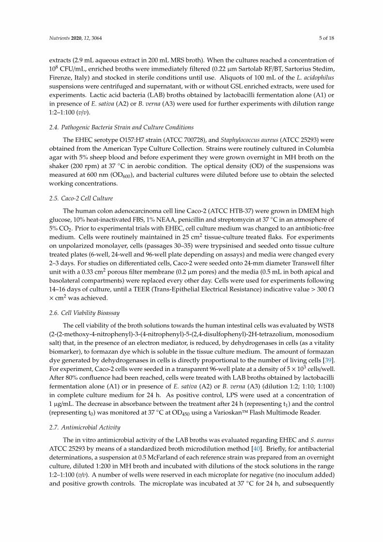
The ability of the LAB broth to directly affect the bacterial growth of two human pathogens (EHECand S. aureus) and to prevent pathogen-induced intestinal barrier dysfunction and inflammation wasevaluated, having assessed their effects on Caco-2 cells. Indeed, bacterial broths obtained by lactobacillifermentation alone (A1), in presence of E. sativa (A2), or B. verna (A3) DSM extracts, were subjected todose-effect safety experiments in Caco-2 cells (dilution range 1:2—1:100, v/v). We chose low dilutionsbecause we assumed that these conditions reflect the LAB “fermentates” quantity in the gut. At dilution1:10 and 1:100, the samples analysed did not show a significant reduction (more than 80%) of cellviability after a 24 h treatment (Figure 1). According to these results we decided to use the 1:10 dilutionfor subsequent experiments.
Nutrients 2020, 12, 3064 8 of 18
Figure 1. Caco-2 cells were treated with A1, A2 and A3 broths (dilution range 1:2–1:100) for 24 h.At dilution 1:2, broths showed a decrease in cell viability of less than 80%, whereas at higher dilutionsthe cell viability is acceptable (more than 80%).
3.3. Lactobacillus Acidophilus Broth Effects on Human Pathogenic Bacteria
The probiotic broths herein obtained were assayed in vitro to measure their inhibitory effect onEHEC and S. aureus growths. S. aureus and E. coli occur as normal flora of the skin and mucousmembranes and in the gastrointestinal tract of humans, respectively; however, virulent strains, resistantto widely used antibiotics, are the most common pathogens causing healthcare-associated infectionsand bacteremia. The EHEC serotype O157:H7, selected in the present study, is a major foodbornepathogen which directly disrupts epithelial cell architecture and intercellular tight junctions, thus beingsuitable as prototype to assess the protective potential of the broth solutions. Data indicated that thebroth solutions did not display inhibitory properties on the microbial growth at tested dilution range(data not shown).
3.4. EHEC Infection of Caco-2 Cells Modelling
Rapid resealing of the epithelial surface barrier following injuries or physiological damage isessential to control inflammation and to restore and maintain intestinal homeostasis.
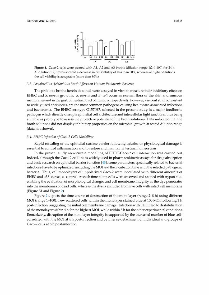
In the present study an accurate modelling of EHEC-Caco-2 cell interaction was carried out.Indeed, although the Caco-2 cell line is widely used in pharmacokinetic assays for drug absorptionand basic research on epithelial barrier function [43], some parameters specifically related to bacterialinfections have to be optimized, including the MOI and the incubation time with the selected pathogenicbacteria. Thus, cell monolayers of unpolarized Caco-2 were inoculated with different amounts ofEHEC and of S. aureus, as control. At each time point, cells were observed and stained with trypan blueenabling the evaluation of morphological changes and cell membrane integrity as the dye penetratesinto the membranes of dead cells, whereas the dye is excluded from live cells with intact cell membrane(Figure S1 and Figure 2).
Figure 2 depicts the time course of destruction of the monolayer (range 2–8 h) using differentMOI (range 1–100). Few scattered cells within the monolayer stained blue at 100 MOI following 2 hpost-infection, suggesting the initial cell membrane damage. Infection with EHEC led to destabilizationof the monolayer within 4 h for the highest MOI, while within 8 h for the other experimental conditions.Remarkably, disruption of the monolayer integrity is supported by the increased number of blue cellscorrelated with the MOI at 4 h post-infection and by intense detachment of individual and groups ofCaco-2 cells at 8 h post-infection.

Nutrients 2020, 12, 3064 9 of 18
Figure 2. Assessment of cell membrane integrity by trypan blue exclusion staining in a time course (2–8 h)of unpolarized Caco-2 cells infection with Enterohemorrhagic Escherichia coli (EHEC) (1–100 multiplicityof infection (MOI)). Dead or damaged cells stain blue. Differences in cell monolayer morphologycan be observed among the mock-infected control and cells at the different experimental conditions(10×magnification).
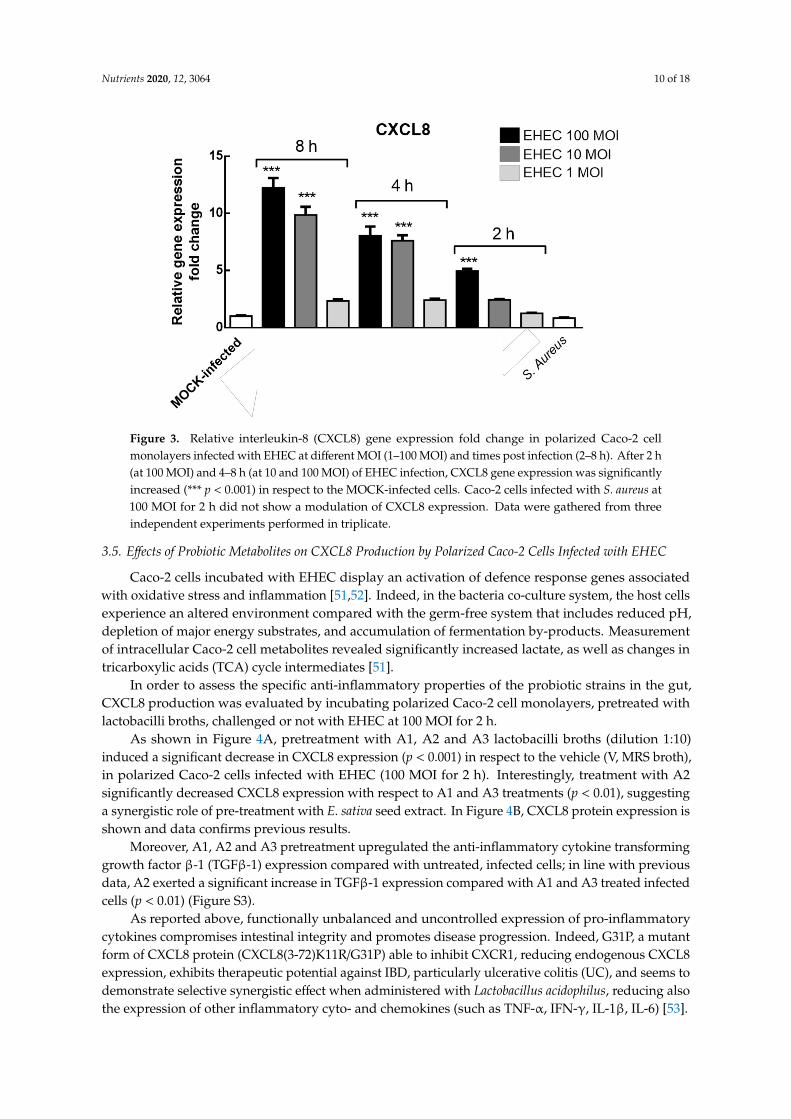
To broadly define the biological parameters in the host pathogen interaction EHEC–Caco-2,a quantitative Real Time PCR assay measuring the CXCL8 expression, as a marker of inflammation,was carried out. CXCL8 is a potent stimulator of neutrophil activation and chemotaxis within theintestinal mucosa and its upregulation is associated with numerous acute and chronic inflammatorydisorders [44]. In line with our data, an increased expression of TNF-α, CXCL1 and CXCL8 mRNAlevels when Caco-2 cells were infected with EHEC has been reported [45]. CXCL8 expression canbe modulated by other pro-inflammatory stimuli such as lipopolysaccharides, IL-1 or TNF-α [46].Patients with diarrhea-associated hemolytic uremic syndrome caused by EHEC O157:H7 have highcirculating levels of CXCL8. So CXCL8 has been identified as a risk factor for the pathogenesis of thedisease and has been associated with high counts of polymorphonuclear leukocytes [47]. EHEC O157bacteria, as well as single bacterial components including purified virulence proteins and O157 LPS,are capable of inducing secretion of CXCL8 from human intestinal epithelial cells [48–50]. Hence,the ability of EHEC to induce proinflammatory CXCL8 response in intestinal epithelial cells, which arethe first cellular targets encountered by the pathogens during infection, is considered a critical stepin the pathogenesis of several intestinal diseases. Herein, a confluent, differentiated and polarizedlayer of enterocyte-like Caco-2 cells was grown on Transwell insert as defined in the supplementarymaterial (Figure S2). Then, polarised Caco-2 cell monolayers were EHEC infected (range 1–100 MOI)at different time points (2–4–8 h).
Figure 3 displays the relative CXCL8 gene expression fold change in our co-culture model system;the highest levels of gene expression were measured in Caco-2 cells incubated with EHEC at 10 and100 MOI for 4–8 h (p < 0.001) and with 100 only MOI for 2 h (p < 0.001), while at 1 MOI no statisticallysignificant difference with the control (mock-infected Caco-2 cells) was detected at each time point.To determine the strain-specificity of EHEC infection in CXCL8 induction, we infected polarized Caco-2cells with S. aureus (100 MOI for 2 h) and any significant effect was detected, suggesting that CXCL8expression is, at least in part, pathogenic strain specific.
Nutrients 2020, 12, 3064 10 of 18
Figure 3. Relative interleukin-8 (CXCL8) gene expression fold change in polarized Caco-2 cellmonolayers infected with EHEC at different MOI (1–100 MOI) and times post infection (2–8 h). After 2 h(at 100 MOI) and 4–8 h (at 10 and 100 MOI) of EHEC infection, CXCL8 gene expression was significantlyincreased (*** p < 0.001) in respect to the MOCK-infected cells. Caco-2 cells infected with S. aureus at100 MOI for 2 h did not show a modulation of CXCL8 expression. Data were gathered from threeindependent experiments performed in triplicate.
3.5. Effects of Probiotic Metabolites on CXCL8 Production by Polarized Caco-2 Cells Infected with EHEC
Caco-2 cells incubated with EHEC display an activation of defence response genes associatedwith oxidative stress and inflammation [51,52]. Indeed, in the bacteria co-culture system, the host cellsexperience an altered environment compared with the germ-free system that includes reduced pH,depletion of major energy substrates, and accumulation of fermentation by-products. Measurementof intracellular Caco-2 cell metabolites revealed significantly increased lactate, as well as changes intricarboxylic acids (TCA) cycle intermediates [51].
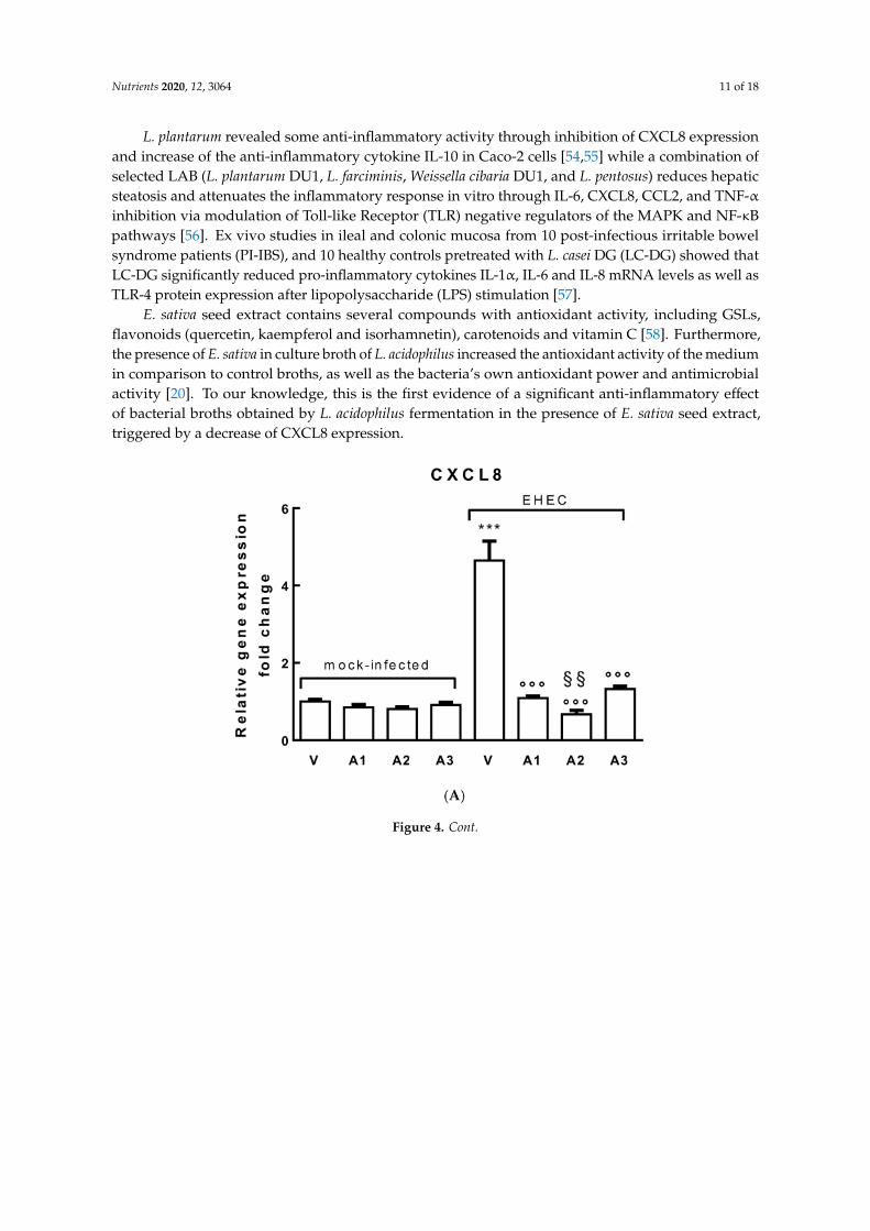
In order to assess the specific anti-inflammatory properties of the probiotic strains in the gut,CXCL8 production was evaluated by incubating polarized Caco-2 cell monolayers, pretreated withlactobacilli broths, challenged or not with EHEC at 100 MOI for 2 h.
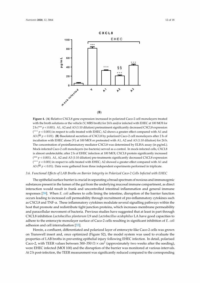
As shown in Figure 4A, pretreatment with A1, A2 and A3 lactobacilli broths (dilution 1:10)induced a significant decrease in CXCL8 expression (p < 0.001) in respect to the vehicle (V, MRS broth),in polarized Caco-2 cells infected with EHEC (100 MOI for 2 h). Interestingly, treatment with A2significantly decreased CXCL8 expression with respect to A1 and A3 treatments (p < 0.01), suggestinga synergistic role of pre-treatment with E. sativa seed extract. In Figure 4B, CXCL8 protein expression isshown and data confirms previous results.
Moreover, A1, A2 and A3 pretreatment upregulated the anti-inflammatory cytokine transforminggrowth factor β-1 (TGFβ-1) expression compared with untreated, infected cells; in line with previousdata, A2 exerted a significant increase in TGFβ-1 expression compared with A1 and A3 treated infectedcells (p < 0.01) (Figure S3).
As reported above, functionally unbalanced and uncontrolled expression of pro-inflammatorycytokines compromises intestinal integrity and promotes disease progression. Indeed, G31P, a mutantform of CXCL8 protein (CXCL8(3-72)K11R/G31P) able to inhibit CXCR1, reducing endogenous CXCL8expression, exhibits therapeutic potential against IBD, particularly ulcerative colitis (UC), and seems todemonstrate selective synergistic effect when administered with Lactobacillus acidophilus, reducing alsothe expression of other inflammatory cyto- and chemokines (such as TNF-α, IFN-γ, IL-1β, IL-6) [53].
Nutrients 2020, 12, 3064 11 of 18
L. plantarum revealed some anti-inflammatory activity through inhibition of CXCL8 expressionand increase of the anti-inflammatory cytokine IL-10 in Caco-2 cells [54,55] while a combination ofselected LAB (L. plantarum DU1, L. farciminis, Weissella cibaria DU1, and L. pentosus) reduces hepaticsteatosis and attenuates the inflammatory response in vitro through IL-6, CXCL8, CCL2, and TNF-αinhibition via modulation of Toll-like Receptor (TLR) negative regulators of the MAPK and NF-κBpathways [56]. Ex vivo studies in ileal and colonic mucosa from 10 post-infectious irritable bowelsyndrome patients (PI-IBS), and 10 healthy controls pretreated with L. casei DG (LC-DG) showed thatLC-DG significantly reduced pro-inflammatory cytokines IL-1α, IL-6 and IL-8 mRNA levels as well asTLR-4 protein expression after lipopolysaccharide (LPS) stimulation [57].
E. sativa seed extract contains several compounds with antioxidant activity, including GSLs,flavonoids (quercetin, kaempferol and isorhamnetin), carotenoids and vitamin C [58]. Furthermore,the presence of E. sativa in culture broth of L. acidophilus increased the antioxidant activity of the mediumin comparison to control broths, as well as the bacteria’s own antioxidant power and antimicrobialactivity [20]. To our knowledge, this is the first evidence of a significant anti-inflammatory effectof bacterial broths obtained by L. acidophilus fermentation in the presence of E. sativa seed extract,triggered by a decrease of CXCL8 expression.
Figure 4. Cont.
Nutrients 2020, 12, 3064 12 of 18
Figure 4. (A) Relative CXCL8 gene expression increased in polarized Caco-2 cell monolayers treatedwith the broth solutions or the vehicle (V, MRS broth) for 24 h and/or infected with EHEC at 100 MOI for2 h (*** p < 0.001). A1, A2 and A3 (1:10 dilution) pretreatment significantly decreased CXCL8 expression( p < 0.001) in respect to cells treated with EHEC; A2 shows a greater effect compared with A1 andA3 (§§ p < 0.01). (B) Basolateral secretion of CXCL8 by polarized Caco-2 cell monolayers after 2 h ofincubation with EHEC alone (V) at 100 MOI or pretreated with A1, A2 and A3 (1:10 dilution) for 24 h.The concentration of proinflammatory mediator CXCL8 was determined by ELISA assay (in pg/mL).Mock-infected Caco-2 cell monolayers (no bacteria) served as a control. In mock-infected cells, CXCL8is almost undetectable; after 2 h of EHEC infection at 100 MOI, CXCL8 protein significantly increased(*** p < 0.001). A1, A2 and A3 (1:10 dilution) pre-treatments significantly decreased CXCL8 expression( p < 0.001) in respect to cells treated with EHEC; A2 showed a greater effect compared with A1 andA3 (§§ p < 0.01). Data were gathered from three independent experiments performed in triplicate.
3.6. Functional Effects of LAB Broths on Barrier Integrity in Polarized Caco-2 Cells Infected with EHEC
The epithelial surface barrier is crucial in separating a broad spectrum of noxious and immunogenicsubstances present in the lumen of the gut from the underlying mucosal immune compartment, as directinteraction would result in frank and uncontrolled intestinal inflammation and general immuneresponses [59]. When E. coli adheres to cells lining the intestine, disruption of the barrier functionoccurs leading to increased cell permeability through recruitment of pro-inflammatory cytokines suchas CXCL8 and TNF-α. These inflammatory cytokines modulate several signalling pathways within thehost that promote and redistribute tight junction proteins, which increases membrane permeabilityand paracellular movement of bacteria. Previous studies have suggested that at least in part throughCXCL8 inhibition Lactobacillus plantarum L9 and Lactobacillus acidophilus LA have good capacities toadhere to the enterocyte monolayer surface of Caco-2 cells resulting in significant inhibition of E. coliadhesion and cell internalisation [55].
Herein, a confluent, differentiated and polarized layer of enterocyte-like Caco-2 cells was grownon Transwell insert and, once optimized (Figure S2), the model system was used to evaluate theproperties of LAB broths in preventing epithelial injury following EHEC infection. In detail, polarisedCaco-2, with TEER values between 300–350 Ω × cm2 (approximately two weeks after the seeding),were EHEC infected (MOI 100) and the disruption of the barrier was monitored at various intervals.At 2 h post-infection, the TEER measurement was significantly reduced compared to the corresponding

Nutrients 2020, 12, 3064 13 of 18
mock-infected sample, which indicated an alteration of membrane integrity, thus this time point wasselected for subsequent experiments (Figure 5).
Figure 5. Trans-Epithelial Electrical Resistance (TEER) values of Caco-2 cell monolayer culturesinoculated with EHEC at 100 MOI over time in comparison to mock-infected control (*** p < 0.001).
In order to monitor the ability of the LAB broths A1, A2 and A3 to prevent barrier dysfunction,the TEER of Caco-2 cells infected with EHEC was measured in their absence and presence (Figure 6).In particular, different experimental conditions were used and data were compared. The differentiatedintestinal cells grown onto Transwell insert were incubated before bacterial inoculation, with tenfolddilution of broth solutions. Samples were added in both medium chambers, and TEER values weremeasured. As readings remained stable over this incubation, the safety of the samples on Caco-2 cellsis assumed, also in accordance with cell viability results. Thereafter, pathogenic bacteria were apicallyadded (MOI 100) and TEER values were assessed after 2 h. As controls, untreated/mock-infectedCaco-2 cells and untreated/infected Caco-2 cells were used in each experimental testing.
Figure 6. TEER values of Caco-2 monolayers at different experimental conditions: before experiments(−24 h), following a 24 h of treatment with A1, A2, A3 (0 h) and at 2 h post infection with EHEC at100 MOI (2 h). TEER values significantly decreased when Caco-2 cells were untreated and pretreatedwith A1 and A3, thereafter EHEC infected (* p < 0.01).
As depicted in Figure 6, TEER values of EHEC-infected Caco-2 cells significantly decreased by 22.5%compared to mock-infected cells (p < 0.001) indicating a membrane-damaging effect. TEER readings ofinfected cells pretreated with LAB broths (1:10 dilution) also decreased, suggesting the disruption ofthe tight junctions of Caco-2. Interestingly, TEER values of Caco-2 cells pretreated with A2 broth andthereafter infected were no significantly different from mock-infected cells when Bonferroni’s MultipleComparison Test was used to compare data; this result suggests that lactobacilli fermented broth

Nutrients 2020, 12, 3064 14 of 18
enriched with E. sativa extract reduced the pathogen-induced changes in barrier integrity. Havingdemonstrated that A2 broth did not exert antimicrobial activity on EHEC, it is possible to speculate thatA2 is an effective in vitro inhibitor of epithelial injury caused by EHEC acting on the intestinal barrier,possibly through its anti-inflammatory activity. Caco-2 monolayers immunostained for tight junctionprotein ZO-1 displayed a uniform intercellular architecture in the control monolayer (mock-infected)while Caco-2 infected with EHEC revealed a more diffuse staining with multiple areas of completebarrier disruption. Treatment with A2 prior to pathogen infection did not remarkably prevent EHECredistribution of ZO-1 staining, confirming its modest positive effect on the intestinal barrier (Figure 7).
Figure 7. Distribution of zona occludens-1 (ZO-1) in Caco-2 monolayers at different experimentalconditions. Mock-infected cells show a well-circumscribed tight junction bands over the entire imagefield. EHEC infected cells (100 MOI for 2 h) display multiple areas of junctional disruption withoutvisually appreciable differences when pretreated with A2 (10×magnification).
A2 is the product of L. acidophilus fermentation of standard MRS broth enriched with E. sativaextract diluted in order to achieve 2 mM glucoerucin. Glucoerucin is the precursor of the ITCerucin of which sulforaphane is an oxidized metabolite [37]. Sulforaphane protects and repairs theinjury to the mucosal epithelium of the colon and cecum in mice with chemical-induced bladdercancer, through the decreasing of inflammation, with a reduction in the release of cytokines (IL-6);and protects immune response, with a reduction of secretory immunoglobulin A [60]. Despite thelimited studies of erucin in comparison to those of sulforaphane, beneficial properties of erucinsimilar to sulforaphane have recently been reported together with its ability to release H2S in vitroand to mediate vasodilatation [37,61]. Natural ITCs, such as erucin, or allyl ITC, highly present inblack mustard (Brassica nigra L.), 4-hydroxybenzyl ITC, highly present in white mustard (Sinapisalba L.), and benzyl ITC, highly present in garden cress (Lepidium sativum L.) have been describedas slow H2S-releasing compounds [62]. H2S is a well-known endogenous gas transmitter that playspivotal roles in the cardiovascular system [61], and in regulating cell growth [23]; moreover, in thegastrointestinal tract H2S has been shown to reduce inflammation and accelerate healing of damagedtissue (such as ulcers), while suppression of H2S production results in impaired healing of tissue injuryand exacerbation of inflammation [63,64].
4. Conclusions
The present study examined the biological properties of three bacterial broths obtained bylactobacilli fermentation alone (A1), in presence of E. sativa (A2), or B. verna (A3). The broth obtainedby L. acidophilus fermentation in the presence of enriched extracts from E. sativa seeds, characterized bya high concentration of glucoerucin, induced a significant decrease in the CXCL8 expression in Caco-2cells following EHEC infection, and reduced epithelial disruption caused by EHEC when intestinalcells were cultured to form a fully differentiate, confluent and tight monolayer.
To our knowledge this is the first study employing complementary in vitro models of gut injuryto study the effects of “functionalised” probiotics in the presence of enteric bacterial pathogens.The findings of the current study demonstrate that pre-treatment with E. sativa seed extract mayincrease the beneficial effect of Lactobacilli in EHEC-injured gut.
Nutrients 2020, 12, 3064 15 of 18
This supplemented fermentation, which successfully enhances the protective effects against gutinflammation and barrier dysfunction induced by pathogens, provides the foundation for the furtherdevelopment and application of this kind of fermentation in food industry. Indeed, lactic acid bacteriaare widely used as starter cultures for the production of diverse fermented foods but, to the best of ourknowledge, the protective role of their fermented broths in gastrointestinal disorders has not beenextensively studied.
Even if these results are very promising, in the near future a pilot trial in human subjects will benecessary to determine whether “super probiotics” administration contributes to the maintenance andprotection of intestinal microflora during enteric infections.
Supplementary Materials: The following are available online at http://www.mdpi.com/2072-6643/12/10/3064/s1,Figure S1: Time course (2–8 h) of destruction of Caco-2 monolayer following EHEC infection at different MOI(100-1); Figure S2: Optimization ofCaco-2 cell culture in a Transwell system device; Figure S3: Relative TGFβ-1gene expression in polarized Caco-2 cell monolayers treated for 24 h with the broth solutions (or the vehicle, V,MRS broth) and infected with EHEC at 100 MOI for 2 h.
Author Contributions: Conceptualization, F.B., E.P., C.C. and G.A.G.; Data curation, F.B., E.P., A.P., D.C., L.U. andC.C.; Formal analysis, F.B., E.P., A.P., N.P., S.B. and C.C.; Funding acquisition, C.C., M.M. and A.R.; Investigation,F.B., E.P., P.S. and C.C.; Methodology, F.B., E.P. and C.C.; Supervision, M.M. and L.L.; Validation, D.C. and L.U.;Writing—original draft, F.B., E.P. and C.C.; Writing—review & editing, M.M., L.L., G.A.G. and A.R. All authorshave read and agreed to the published version of the manuscript.
Funding: This research was funded by PRIN 2015 “Securing and ensuring sustainable use of agriculture waste,co-and by-products: an integrated analytical approach combining mass spectrometry with health effect-basedbiosensing” (Prot. 2015FFY97L_002) and by POR FESR Regione Emilia Romagna 2015 “Alimentazione, sistemaimmunitario, microbioma e tratto gastrointestinale: i nuovi protagonisti del mantenimento della salute e dellaprevenzione delle malattie degenerative. Identificazione e valorizzazione di principi attivi contenuti nei vegetaliper la formulazione di alimenti e integratori utili nelle malattie infiammatorie intestinali, nella malattia metabolica,nell’obesità e nelle sue complicanze” (CUP E88I15000070007).
Acknowledgments: We wish to thank INCOS Cosmeceutica Industriale S.r.l. (Funo di Argelato, Bologna, Italy) forproviding bacterial broths obtained by lactobacilli fermentation grown in presence or absence of 2 mM total GSLs.
Conflicts of Interest: The authors declare no conflict of interest.
References
1. Desroches, S.; Lapointe, A.; Dugrenier, M.; Provencher, V.; Lamarche, B.; Desroches, S. A systematic review ofthe effect of yogurt consumption on chronic diseases risk markers in adults. Eur. J. Nutr. 2016, 56, 1375–1392.[CrossRef]
2. Kok, C.R.; Hutkins, R. Yogurt and other fermented foods as sources of health-promoting bacteria. Nutr. Rev.2018, 76, 4–15. [CrossRef] [PubMed]
3. Chilton, S.N.; Burton, J.P.; Reid, G. Inclusion of fermented foods in food guides around the world. Nutrients2015, 7, 390–404. [CrossRef]
4. Park, K.-Y.; Jeong, J.-K.; Lee, Y.-E.; Daily, J.W. Health benefits of Kimchi (Korean Fermented Vegetables) as aprobiotic food. J. Med. Food 2014, 17, 6–20. [CrossRef]
5. Han, K.; Bose, S.; Wang, J.-H.; Kim, B.-S.; Kim, M.J.; Kim, E.-J.; Kim, H. Contrasting effects of fresh andfermented kimchi consumption on gut microbiota composition and gene expression related to metabolicsyndrome in obese Korean women. Mol. Nutr. Food Res. 2015, 59, 1004–1008. [CrossRef]
6. Ro, S.L.; Burn, M.W.; Sandine, W.E. Vitamin B12 and ascorbic acid in Kimchi inoculated withPropionibacterium freudenreichji ss. shermanii. J. Food Sci. 1979, 44, 873–877. [CrossRef]
7. Du, R.; Song, G.; Zhao, D.; Sun, J.; Ping, W.; Ge, J. Lactobacillus casei starter culture improves vitamin content,increases acidity and decreases nitrite concentration during sauerkraut fermentation. Int. J. Food Sci. Technol.2018, 53, 1925–1931. [CrossRef]
8. Quirante-Moya, S.; García-Ibañez, P.; Quirante-Moya, F.; Villaño, D.; Moreno, D.A. The role of Brassicabioactives on human health: Are we studying it the right way? Molecules 2020, 25, 1591. [CrossRef]
9. Odongo, G.A.; Schlotz, N.; Herz, C.; Hanschen, F.S.; Baldermann, S.; Neugart, S.; Trierweiler, B.; Frommherz, L.;Franz, C.M.A.P.; Ngwene, B.; et al. The role of plant processing for the cancer preventive potential ofEthiopian kale (Brassica carinata). Food Nutr. Res. 2017, 61, 1271527. [CrossRef] [PubMed]
Nutrients 2020, 12, 3064 16 of 18
10. Sanders, M.E.; Guarner, F.; Guerrant, R.; Holt, P.R.; Quigley, E.M.M.; Sartor, R.B.; Sherman, P.M.; Mayer, E.A.An update on the use and investigation of probiotics in health and disease. Gut 2013, 62, 787–796. [CrossRef]
11. Ahrné, N.; Jeppsson, A.; Wold, A.E.; Molin, G. The normal Lactobacillus flora of healthy human rectal and oralmucosa. J. Appl. Microbiol. 1998, 85, 88–94. [CrossRef] [PubMed]
12. Gill, H.S.; Shu, Q.; Lin, H.; Rutherfurd, K.J.; Cross, M.L. Protection against translocating Salmonellatyphimurium infection in mice by feeding the immuno-enhancing probiotic Lactobacillus rhamnosus strainHN001. Med. Microbiol. Immunol. 2001, 190, 97–104. [CrossRef] [PubMed]
13. Donato, K.A.; Gareau, M.G.; Wang, Y.J.J.; Sherman, P.M. Lactobacillus rhamnosus GG attenuates interferon-γand tumour necrosis factor-α-induced barrier dysfunction and pro-inflammatory signalling. Microbiology2010, 156, 3288–3297. [CrossRef] [PubMed]
14. Kellow, N.; Coughlan, M.T.; Reid, C.M. Metabolic benefits of dietary prebiotics in human subjects: A systematicreview of randomised controlled trials. Br. J. Nutr. 2013, 111, 1147–1161. [CrossRef] [PubMed]
15. Gutiérrez, S.; Martínez-Blanco, H.; Rodríguez-Aparicio, L.B.; Ferrero, M.A. Effect of fermented broth fromlactic acid bacteria on pathogenic bacteria proliferation. J. Dairy Sci. 2016, 99, 2654–2665. [CrossRef][PubMed]
16. Parkar, S.G.; Stevenson, D.E.; Skinner, M.A. The potential influence of fruit polyphenols on colonic microfloraand human gut health. Int. J. Food Microbiol. 2008, 124, 295–298. [CrossRef] [PubMed]
17. Rodríguez, H.; Curiel, J.A.; Landete, J.M.; Rivas, B.D.L.; De Felipe, F.L.; Gómez-Cordovés, C.; Mancheño, J.M.;Muñoz, R. Food phenolics and lactic acid bacteria. Int. J. Food Microbiol. 2009, 132, 79–90. [CrossRef]
18. Damodharan, K.; Palaniyandi, S.A.; Yang, S.H.; Suh, J.-W. In vitro probiotic characterization of Lactobacillusstrains from fermented radish and their anti-adherence activity against enteric pathogens. Can. J. Microbiol.2015, 61, 837–850. [CrossRef]
19. Seong, G.-U.; Hwang, I.-W.; Chung, S.-K. Antioxidant capacities and polyphenolics of Chinese cabbage(Brassica rapa L. ssp. Pekinensis) leaves. Food Chem. 2016, 199, 612–618. [CrossRef]
20. Fratianni, F.; Pepe, S.; Cardinale, F.; Granese, T.; Cozzolino, A.; Coppola, R.; Nazzaro, F. Eruca sativa mightinfluence the growth, survival under simulated gastrointestinal conditions and some biological features ofLactobacillus acidophilus, Lactobacillus plantarum and Lactobacillus rhamnosus strains. Int. J. Mol. Sci. 2014, 15,17790–17805. [CrossRef]
21. Franco, P.; Spinozzi, S.; Pagnotta, E.; Lazzeri, L.; Ugolini, L.; Camborata, C.; Roda, A. Development of a liquidchromatography-electrospray ionization-tandem mass spectrometry method for the simultaneous analysisof intact glucosinolates and isothiocyanates in Brassicaceae seeds and functional foods. J. Chromatogr. A2016, 1428, 154–161. [CrossRef] [PubMed]
22. Singh, D.; Arora, S.; Bhatia, A.; Singh, H.; Singh, B.; Arora, S. Molecular targets in cancer preventionby 4-(methylthio) butyl isothiocyanate-A comprehensive review. Life Sci. 2020, 241, 117061. [CrossRef][PubMed]
23. Citi, V.; Piragine, E.; Pagnotta, E.; Ugolini, L.; Mannelli, L.D.C.; Testai, L.; Ghelardini, C.; Lazzeri, L.;Calderone, V.; Martelli, A. Anticancer properties of erucin, an H2S-releasing isothiocyanate, on humanpancreatic adenocarcinoma cells (AsPC-1). Phytotherapy Res. 2019, 33, 845–855. [CrossRef] [PubMed]
24. Hichri, F.; Hichri, A.O.; Maha, M.; Hossan, A.S.M.; Flamini, G.; Ben Jannet, H. Chemical composition,antibacterial, antioxidant and in Vitro antidiabetic activities of essential oils from Eruca vesicaria.Chem. Biodivers. 2019, 16, e1900183. [CrossRef]
25. Gupta, P.; Wright, S.E.; Kim, S.-H.; Srivastava, S.K. Phenethyl isothiocyanate: A comprehensive review ofanti-cancer mechanisms. Biochim. et Biophys. Acta (BBA)-Bioenerg. 2014, 1846, 405–424. [CrossRef]
26. Naidu, S.D.; Suzuki, T.; Yamamoto, M.; Fahey, J.W.; Dinkova-Kostova, A.T. Phenethyl Isothiocyanate, a dualactivator of transcription factors NRF2 and HSF1. Mol. Nutr. Food Res. 2018, 62, 1700908. [CrossRef]
27. Nowicki, D.; Maciag-Dorszynska, M.; Bogucka, K.; Szalewska-Pałasz, A.; Herman-Antosiewicz, A. Variousmodes of action of dietary phytochemicals, sulforaphane and phenethyl isothiocyanate, on pathogenicbacteria. Sci. Rep. 2019, 9, 13677–13712. [CrossRef]
28. Nowicki, D.; Maciag-Dorszynska, M.; Kobiela, W.; Herman-Antosiewicz, A.; Wegrzyn, A.; Szalewska-Pałasz, A.;Wegrzyn, G. Phenethyl isothiocyanate inhibits shiga toxin production in enterohemorrhagic Escherichia coli bystringent response induction. Antimicrob. Agents Chemother. 2014, 58, 2304–2315. [CrossRef]
29. Lim, J.Y.; Yoon, J.; Hovde, C.J. A brief overview of Escherichia coli O157:H7 and its plasmid O157. J. Microbiol.Biotechnol. 2010, 20, 5–14. [CrossRef]

Nutrients 2020, 12, 3064 17 of 18
30. Dufour, V.; Stahl, M.; Baysse, C. The antibacterial properties of isothiocyanates. Microbiology 2015, 161,229–243. [CrossRef]
31. Kim, M.; Lee, H.S. Growth-inhibiting activities of Phenethyl Isothiocyanate and its derivatives againstintestinal bacteria. J. Food Sci. 2009, 74, M467–M471. [CrossRef] [PubMed]
32. Pinto, M.; Robineleon, S.; Appay, M.D.; Kedinger, M.; Haffen, K.; Fogh, J.; Zweibaum, A. Enterocyte-likedifferentiation and polarization of the human colon carcinoma cell line Caco-2 in culture. Biol. Cell. 1983, 4,323–330.
33. Wathelet, J.-P.; Iori, R.; Leoni, O.; Quinsac, A.; Palmieri, S. Guidelines for glucosinolate analysis in greentissues used for biofumigation. Agroindustria 2004, 3, 257–266.
34. ISO 9167:2019 Graines et tourteaux de colza-Dosage des glucosinolates-Méthode par chromatographieliquide à haute performance. Available online: https://www.iso.org/fr/standard/72207.html (accessed on22 January 2019).
35. Lazzeri, L.; Malaguti, L.; Bagatta, M.; D’Avino, L.; Ugolini, L.; De Nicola, G.; Casadei, N.; Cinti, S.; Matteo, R.;Iori, R. Characterization of the main glucosinolate content and fatty acid composition in non-food Brassicaceaeseeds. Acta Hortic. 2013, 331–338. [CrossRef]
36. Matteo, R.; D’Avino, L.; Ramirez-Cando, L.J.; Pagnotta, E.; Angelini, L.G.; Spugnoli, P.; Tavarini, S.; Ugolini, L.;Foschi, L.; Lazzeri, L. Camelina (Camelina sativa L. Crantz) under low-input management systems in northernItaly: Yields, chemical characterization and environmental sustainability. Ital. J. Agron. 2020. [CrossRef]
37. Lucarini, E.; Pagnotta, E.; Micheli, L.; Parisio, C.; Testai, L.; Martelli, A.; Calderone, V.; Matteo, R.; Lazzeri, L.;Mannelli, L.D.C.; et al. Eruca sativa meal against diabetic neuropathic pain: An H2S-mediated effect ofglucoerucin. Molecules 2019, 24, 3006. [CrossRef]
38. Barillari, J.; Gueyrard, D.; Rollin, P.; Iori, R. Barbarea verna as a source of 2-phenylethyl glucosinolate,precursor of cancer chemopreventive phenylethyl isothiocyanate. Fitoterapia 2001, 72, 760–764. [CrossRef]
39. Caliceti, C.; Capriotti, A.L.; Calabria, D.; Bonvicini, F.; Chiozzi, R.Z.; Montone, C.M.; Piovesana, S.;Zangheri, M.; Mirasoli, M.; Simoni, P.; et al. Peptides from cauliflower by-products, obtained by an efficient,ecosustainable, and semi-industrial method, exert protective effects on endothelial function. Oxidative Med.Cell. Longev. 2019, 2019, 1–13. [CrossRef]
40. Bonvicini, F.; Manet, I.; Belluti, F.; Gobbi, S.; Rampa, A.; Gentilomi, G.A.; Bisi, A. Targeting the bacterialmembrane with a new polycyclic privileged structure: A powerful tool to face Staphylococcus aureus infections.ACS Infect. Dis. 2019, 5, 1524–1534. [CrossRef]
41. Srinivasan, B.; Kolli, A.R.; Esch, M.B.; Abaci, H.E.; Shuler, M.L.; Hickman, J.J. TEER Measurement techniquesfor In Vitro barrier model systems. J. Lab. Autom. 2015, 20, 107–126. [CrossRef]
42. Caliceti, C.; Aquila, G.; Pannella, M.; Morelli, M.B.; Fortini, C.; Pinton, P.; Bonora, M.; Hrelia, S.; Pannuti, A.;Miele, L.; et al. 17β-estradiol enhances signalling mediated by VEGF-A-delta-like ligand 4-Notch1 axis inhuman endothelial cells. PLoS ONE 2013, 8, e71440. [CrossRef] [PubMed]
43. Kämpfer, A.A.; Urbán, P.; Gioria, S.; Kanase, N.; Stone, V.; Kinsner-Ovaskainen, A. Development of anin vitro co-culture model to mimic the human intestine in healthy and diseased state. Toxicol. Vitr. 2017, 45,31–43. [CrossRef] [PubMed]
44. Baggiolini, M.; Dewald, B.; Moser, B. Human chemokines: An update. Annu. Rev. Immunol. 1997, 15, 675–705.[CrossRef] [PubMed]
45. Johnson-Henry, K.C.; Pinnell, L.J.; Waskow, A.M.; Irrazabal, T.; Martin, A.; Hausner, M.; Sherman, P.M.Short-chain fructo-oligosaccharide and inulin modulate inflammatory responses and microbial communitiesin Caco2-bbe cells and in a mouse model of intestinal injury. J. Nutr. 2014, 144, 1725–1733. [CrossRef][PubMed]
46. Gross, V.; Andus, T.; Daig, R.; Aschenbrenner, E.; Schölmerich, J.; Falk, W. Regulation of interleukin-8production in a human colon epithelial cell line (HT-29). Gastroenterology 1995, 108, 653–661. [CrossRef]
47. Fitzpatrick, M.M.; Shah, V.; Trompeter, R.S.; Dillon, M.J.; Barratt, T.M. Interleukin-8 andpolymorphoneutrophil leucocyte activation in hemolytic uremic syndrome of childhood. Kidney Int.1992, 42, 951–956. [CrossRef] [PubMed]
48. Berin, M.C.; Darfeuille-Michaud, A.; Egan, L.J.; Miyamoto, Y.; Kagnoff, M.F. Role of EHEC O157:H7 virulencefactors in the activation of intestinal epithelial cell NF-κB and MAP kinase pathways and the upregulatedexpression of interleukin 8. Cell. Microbiol. 2002, 4, 635–648. [CrossRef]
Nutrients 2020, 12, 3064 18 of 18
49. Miyamoto, Y.; Iimura, M.; Kaper, J.B.; Torres, A.G.; Kagnoff, M.F. Role of Shiga toxin versus H7 flagellin inenterohaemorrhagic Escherichia coli signalling of human colon epithelium in vivo. Cell. Microbiol. 2006, 8,869–879. [CrossRef]
50. Thorpe, C.M.; Hurley, B.P.; Lincicome, L.L.; Jacewicz, M.S.; Keusch, G.T.; Acheson, D.W.K. Shiga Toxinsstimulate secretion of Interleukin-8 from intestinal epithelial cells. Infect. Immun. 1999, 67, 5985–5993.[CrossRef]
51. He, X.; Mishchuk, D.O.; Shah, J.; Weimer, B.C.; Slupsky, C.M. Cross-talk between E. coli strains and a humancolorectal adenocarcinoma-derived cell line. Sci. Rep. 2013, 3, 3416. [CrossRef]
52. Pearson, J.S.; Hartland, E.L. The inflammatory response during enterohemorrhagic Escherichia coli infection.Microbiol. Spectr. 2014, 2, 341–358. [CrossRef] [PubMed]
53. Walana, W.; Ye, Y.; Li, M.; Wang, J.; Wang, B.; Cheng, J.-W.; Gordon, J.R.; Li, F. IL-8 antagonist, CXCL8(3-72)K11R/G31P coupled with probiotic exhibit variably enhanced therapeutic potential in amelioratingulcerative colitis. Biomed. Pharmacother. 2018, 103, 253–261. [CrossRef] [PubMed]
54. Chen, S.; Cao, P.; Lang, F.; Wu, Z.; Pan, D.; Zeng, X.; Lian, L. Adhesion-related immunomodulatory activityof the screened Lactobacillus plantarum from Sichuan Pickle. Curr. Microbiol. 2018, 76, 29–36. [CrossRef][PubMed]
55. Wang, B.; Chen, J.; Wang, S.; Zhao, X.; Lu, G.; Tang, X. Lactobacillus plantarum L9 but not Lactobacillusacidophilus LA reduces tumour necrosis factor induced bacterial translocation in Caco-2 cells. Benef. Microbes2017, 8, 497–505. [CrossRef] [PubMed]
56. Kanmani, P.; Kim, H. Protective effects of lactic acid bacteria against TLR4 induced inflammatory response inhepatoma HepG2 cells through modulation of toll-like receptor negative regulators of mitogen-activatedprotein kinase and NF-κB signaling. Front. Immunol. 2018, 9, 1537. [CrossRef]
57. Barillari, J.; Canistro, D.; Paolini, M.; Ferroni, F.; Pedulli, G.F.; Iori, R.; Valgimigli, L. Direct antioxidant activityof purified glucoerucin, the dietary secondary metabolite contained in rocket (Eruca sativa Mill.) seeds andsprouts. J. Agric. Food Chem. 2005, 53, 2475–2482. [CrossRef]
58. Dignass, A.U. Mechanisms and Modulation of Intestinal Epithelial Repair. Inflamm. Bowel Dis. 2001, 7, 68–77.[CrossRef]
59. Béduneau, A.; Tempesta, C.; Fimbel, S.; Pellequer, Y.; Jannin, V.; Demarne, F.; Lamprecht, A. A tunableCaco-2/HT29-MTX co-culture model mimicking variable permeabilities of the human intestine obtained byan original seeding procedure. Eur. J. Pharm. Biopharm. 2014, 87, 290–298. [CrossRef]
60. He, C.; Huang, L.; Lei, P.; Liu, X.; Li, B.; Shan, Y. Sulforaphane normalizes intestinal flora and enhances gutbarrier in mice with BBN-induced bladder cancer. Mol. Nutr. Food Res. 2018, 62, 1800427. [CrossRef]
61. Martelli, A.; Piragine, E.; Citi, V.; Testai, L.; Pagnotta, E.; Ugolini, L.; Lazzeri, L.; Mannelli, L.D.C.; Manzo, O.L.;Bucci, M.; et al. Erucin exhibits vasorelaxing effects and antihypertensive activity by H2S-releasing properties.Br. J. Pharmacol. 2019, 177, 824–835. [CrossRef]
62. Citi, V.; Martelli, A.; Testai, L.; Marino, A.; Breschi, M.; Calderone, V. Hydrogen sulfide releasing capacityof natural isothiocyanates: Is it a reliable explanation for the multiple biological effects of Brassicaceae?Planta Medica 2014, 80, 610–613. [CrossRef] [PubMed]
63. Blackler, R.W.; De Palma, G.; Manko, A.; Da Silva, G.; Flannigan, K.L.; Bercik, P.; Surette, M.G.; Buret, A.G.;Wallace, J.L. Deciphering the pathogenesis of NSAID enteropathy using proton pump inhibitors and ahydrogen sulfide-releasing NSAID. Am. J. Physiol. Liver Physiol. 2015, 308, G994–G1003. [CrossRef] [PubMed]
64. Blackler, R.; Syer, S.; Bolla, M.; Ongini, E.; Wallace, J.L. Gastrointestinal-sparing effects of novel NSAIDs inrats with compromised mucosal defence. PLoS ONE 2012, 7, e35196. [CrossRef] [PubMed]
© 2020 by the authors. Licensee MDPI, Basel, Switzerland. This article is an open accessarticle distributed under the terms and conditions of the Creative Commons Attribution(CC BY) license (http://creativecommons.org/licenses/by/4.0/).