e. coli pulser transformation apparatus operating ... · pdf filetransformation apparatus...
TRANSCRIPT
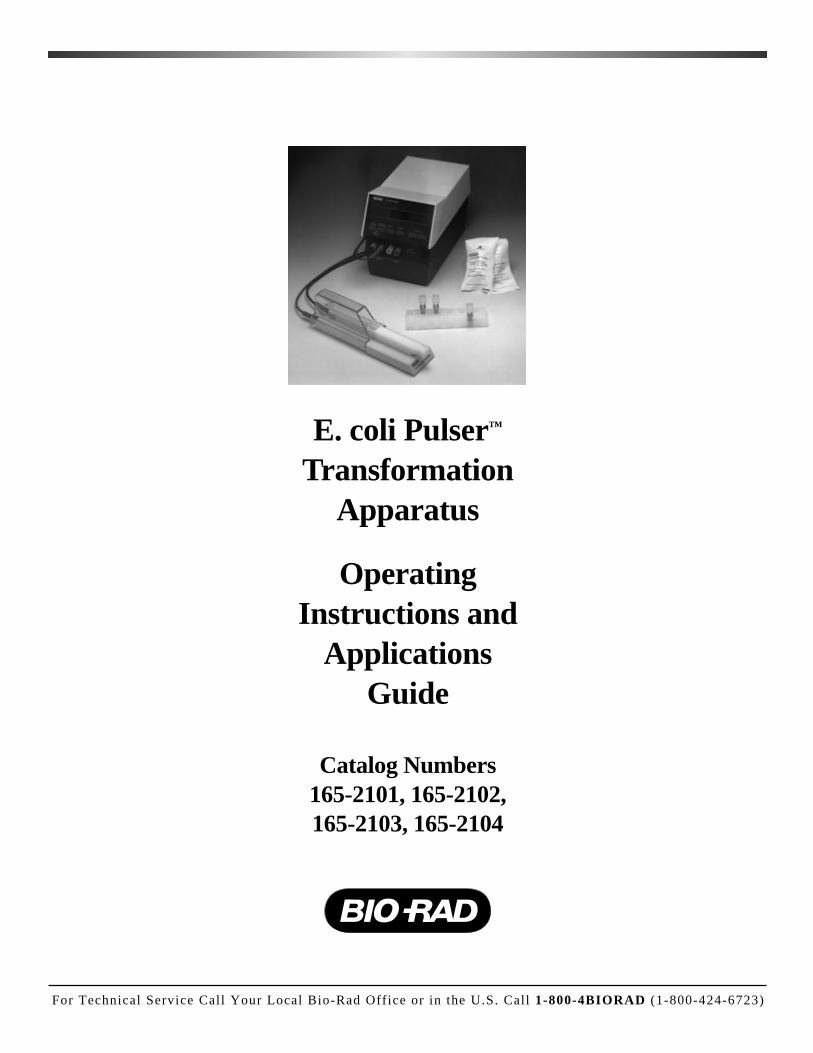
E. coli Pulser™
TransformationApparatus
OperatingInstructions and
ApplicationsGuide
Catalog Numbers165-2101, 165-2102,165-2103, 165-2104
For Technical Service Call Your Local Bio-Rad Off ice or in the U.S. Call 1-800-4BIORAD (1-800-424-6723)

WarrantyBio-Rad Laboratories warrants the E. coli Pulser apparatus against defects in materials and work-
manship for 1 year. If any defects occur in the instrument during this warranty period, Bio-RadLaboratories will at Bio-Rad's option repair or replace the defective parts free. The following defects,however, are specifically excluded:
1. Defects caused by improper operation.
2. Repair or modification done by anyone other than Bio-Rad Laboratories or an authorized agent.
3. Use of fittings or other spare parts supplied by anyone other than Bio-Rad Laboratories.
4. Damage caused by accident or misuse.
5. Damage caused by disaster.
6. Corrosion due to use of improper solvent or sample.
For any inquiry or request for repair service, contact Bio-Rad Laboratories after confirming themodel, serial number, invoice number, and purchase order number of your instrument.
Model
Catalog Number
Date of Delivery
Warranty Period
Serial Number
Invoice Number
Purchase Order Number

Table of ContentsPage
Introduction .................................................................................................................1
Section 1 Safety Precautions.................................................................................11.1 Electrical Hazards ...............................................................................................11.2 Mechanical Hazards............................................................................................11.3 Other Safety Precautions ....................................................................................2
Section 2 E. coli Pulser Apparatus Operating Instructions..............................2
Section 3 Electrical Variables ...............................................................................5
Section 4 Bacterial Electroporation (Electro-transformation).........................64.1 DNA....................................................................................................................64.2 Electroporation Media ........................................................................................74.3 Electroporation Conditions.................................................................................7
Section 5 High Efficiency Electro-transformation of E. coli.............................8
Appendix I E. coli Electro-transformation References .......................................10
Appendix II Field Strength and Cell Survivability Studies of 12 E. coli Strains ................................22
Appendix III Troubleshooting Guide for the E. coli Pulser Apparatus ...............25
Appendix IV Product Information...........................................................................28
IntroductionThe E. coli Pulser apparatus is used for the electroporation of Escherichia coli where
pulses of very high field strength are applied to samples of small volume and high resistance.
The internal circuitry of the E. coli Pulser apparatus greatly reduces the incidence of arc-ing at high voltage, and is protected if a high voltage, high current arc does occur. The unit isdesigned to fire into a high resistance sample.
The internal resistance of the E. coli Pulser apparatus will cause a substantial loss in volt-age when pulses are applied to low resistance samples (less than 1000 Ω). This includes sam-ples in which the growth medium was not completely removed from the E. coli cells, andDNA samples containing salt contributed by residual cesium chloride, or ligation mixtures.The voltage loss is insignificant with high resistance samples (see Section 3 for a detaileddiscussion of this effect). If you have a question or doubt concerning the resistance of yoursample, contact your Technical Representative, or, in the U. S., call Technical Services at1-800-4BIORAD.
Section 1Safety Precautions
READ THIS INFORMATION CAREFULLYBEFORE USING THE E. COLI PULSER APPARATUS.
1.1 Electrical HazardsThe E. coli Pulser apparatus produces voltages up to 2,500 volts and is capable of pass-
ing very high currents. When charged to maximum voltage, the instrument stores about 32joules. A certain degree of respect is required for energy levels of this order. The safety inter-locks of the system prevent accidental charging and discharge (two buttons must be depressedto deliver a pulse), and also prevent operator access to the recessed input jacks, and to therecessed electrode contacts inside the sample chamber. The latter mechanical interlocks shouldnever be circumvented.
There is high voltage present whenever the rectangular red buttons are depressed (charg-ing) and when the capacitor has been partially charged but not fired (for example, when thecharging cycle has been interrupted before the pulse is delivered). In this condition, the chargewill bleed slowly from the capacitor and a shock hazard can exist for several minutes. Duringthis time, do not disconnect or connect the sample chamber.To manually discharge thecapacitor, turn the main power switch of the apparatus off and on twice. This will dischargethe capacitor immediately and should be done whenever there is any doubt about the statusof charge in the capacitor. It is a good idea always to follow this procedure before connect-ing or disconnecting the sample chamber to the unit, to be absolutely sure the capacitor isdischarged.
If the charging cycle is aborted by releasing either of the rectangular red buttons, thecharge/fire cycle can be continued simply by re-pressing the red pulse buttons until the pulseis delivered, or the capacitor can be safely discharged by turning the power switch off andon twice.
1.2 Mechanical HazardsThe circuitry of the E. coli Pulser apparatus greatly reduces the incidence of arcing in the
cuvette when high voltage is delivered into high resistance media. However, arcing can some-
1
times still occur. The sample chamber is effective in containing these small explosions, butnonethelesswe strongly recommend wearing safety glasses when using the instrument.
Do not use the E. coli Pulser apparatus with samples suspended in conductive media(such as saline). Refer to Section 5 for sample preparation.
1.3 Other Safety PrecautionsTurn the unit off when not attended. Avoid spilling any liquids onto the apparatus. Use
only water or alcohol to clean the outside surfaces of the E. coli Pulser apparatus.
This instrument is intended for laboratory use only.
Warning: This equipment generates, uses, and radiates radio frequency energy. If it is notinstalled and used in accordance with the instructions given in this manual, it may causeinterference with radio communications. It has been tested and found to comply with thelimits for Class A computing devices (pursuant to Subpart J of Part 15 of FCC Rules)which provide reasonable protection against such interference, when operated in a com-mercial environment. Operation of this equipment in a residential area is likely to causeinterference. In this case the user will be required, at his/her own expense, to take what-ever measure may be required to correct the interference.
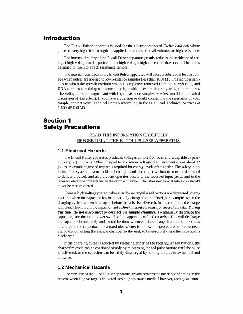
Section 2E. coli Pulser Apparatus Operating Instructions
LED display
Fig 1. The front panel of the E. coli Pulser apparatus. Notice that the lights above both the SETVOLTS (KV) and ACTUAL VOLTS (KV) buttons are illuminated, indicating that the TIME CONSTANT(MSEC) is displayed in the LED>
1. Connect the black power cord to the rear panel of the E. coli Pulser apparatus. Plugthe cord into a wall outlet or power strip.
2
Raise and lower buttonsfor setting voltage
Pulse buttons
2. Connect the leads from the sample chamber to the output jacks marked TO SHOCKINGCHAMBER on the front panel of the E. coli Pulser apparatus. Be sure to observe polar-ity (red to red, black to black).
3. Turn on the apparatus using the power switch on the right-hand panel. The light emit-ting diode (LED) display should illuminate and read “0.00”.
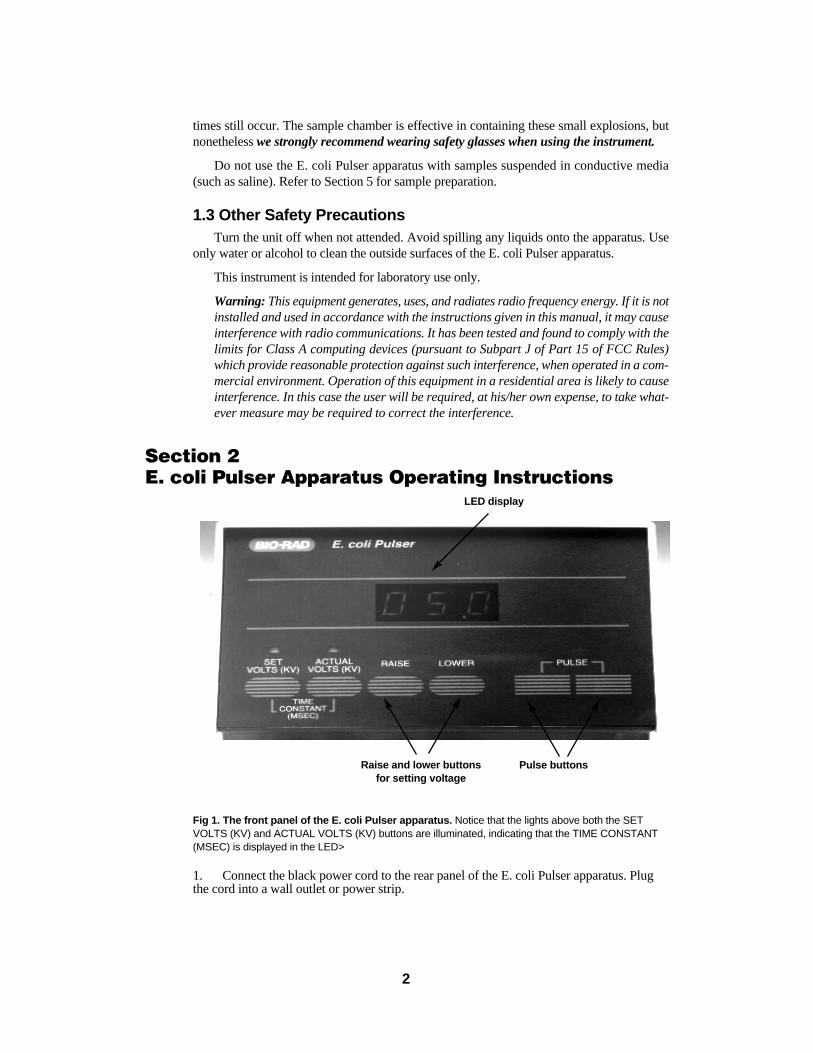
4. The LED above the oval SET VOLTS (KV) button will be illuminated. The LED dis-play is in kilovolts (kV). Use the oval RAISE and LOWER buttons to adjust the volt-age to the desired value in the range of 0.20 - 2.50 kV (Figure 1). If the voltage is set at0.00 kV, ‘no’ will be displayed when the pulse buttons are pressed. To quickly set thevoltage to 1.80 kV (recommended when using 0.1 cm cuvettes, Figure 2), press theRAISE and LOWER buttons simultaneously. “1.80” will be displayed in the LED. Toquickly set the voltage to 2.50 kV (recommended when using the 0.2 cm cuvettes,Figure 2), press the RAISE and LOWER buttons simultaneously twice. “2.50” will bedisplayed. Pressing both buttons a third time will reset the instrument to 0.00 kV.
5. Place the cell suspension in the chilled electroporation cuvette. Use only the lower, nar-row portion of the cuvette between the aluminum plates. Up to 0.4 ml (400 µl) of solu-tion may be placed in the 0.2 cm cuvette, and the 0.1 cm cuvette can hold 0.08 ml (80 µl)during pulse delivery. See Figure 3.
3
Fig. 2. 0.2 cm and 0.1 cm electrode gapcuvettes.
Fig. 3. 400 µl in a 0.2 cm cuvette (themaximum volume for pulse delivery).
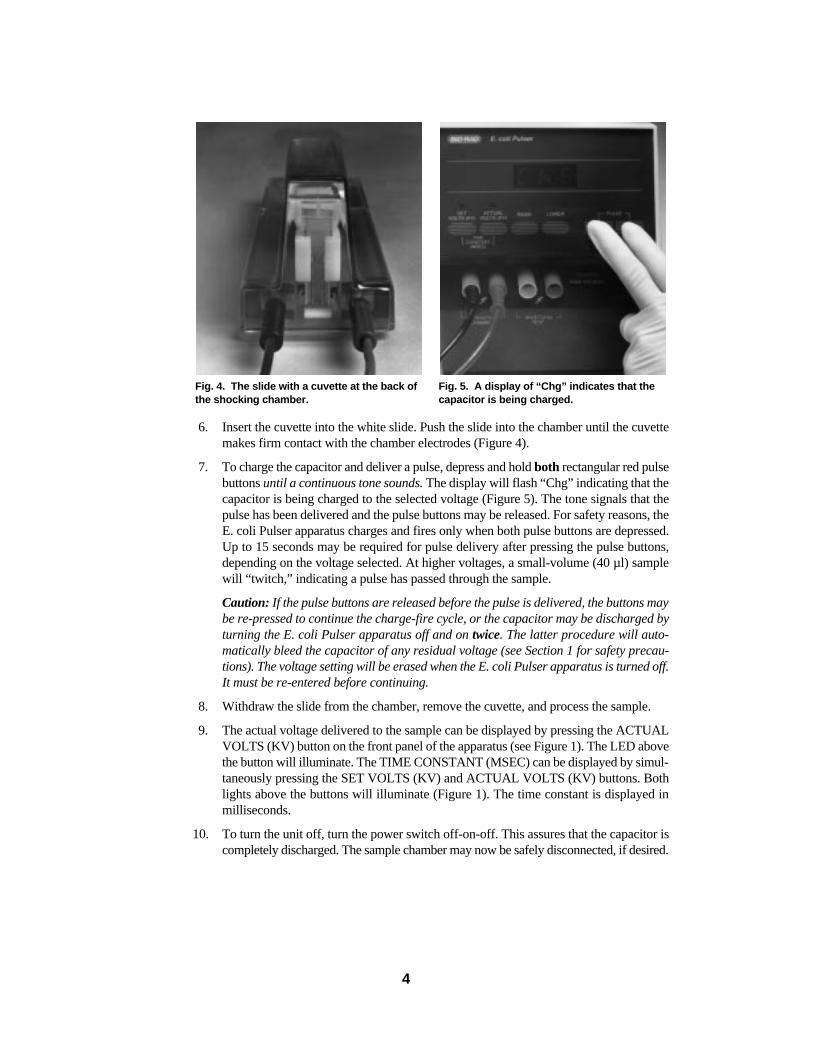
6. Insert the cuvette into the white slide. Push the slide into the chamber until the cuvettemakes firm contact with the chamber electrodes (Figure 4).
7. To charge the capacitor and deliver a pulse, depress and hold both rectangular red pulsebuttons until a continuous tone sounds. The display will flash “Chg” indicating that thecapacitor is being charged to the selected voltage (Figure 5). The tone signals that thepulse has been delivered and the pulse buttons may be released. For safety reasons, theE. coli Pulser apparatus charges and fires only when both pulse buttons are depressed.Up to 15 seconds may be required for pulse delivery after pressing the pulse buttons,depending on the voltage selected. At higher voltages, a small-volume (40 µl) samplewill “twitch,” indicating a pulse has passed through the sample.
Caution: If the pulse buttons are released before the pulse is delivered, the buttons maybe re-pressed to continue the charge-fire cycle, or the capacitor may be discharged byturning the E. coli Pulser apparatus off and on twice. The latter procedure will auto-matically bleed the capacitor of any residual voltage (see Section 1 for safety precau-tions). The voltage setting will be erased when the E. coli Pulser apparatus is turned off.It must be re-entered before continuing.
8. Withdraw the slide from the chamber, remove the cuvette, and process the sample.
9. The actual voltage delivered to the sample can be displayed by pressing the ACTUALVOLTS (KV) button on the front panel of the apparatus (see Figure 1). The LED abovethe button will illuminate. The TIME CONSTANT (MSEC) can be displayed by simul-taneously pressing the SET VOLTS (KV) and ACTUAL VOLTS (KV) buttons. Bothlights above the buttons will illuminate (Figure 1). The time constant is displayed inmilliseconds.
10. To turn the unit off, turn the power switch off-on-off. This assures that the capacitor iscompletely discharged. The sample chamber may now be safely disconnected, if desired.
4
Fig. 4. The slide with a cuvette at the back ofthe shocking chamber.
Fig. 5. A display of “Chg” indicates that thecapacitor is being charged.
11. The E. coli Pulser apparatus is provided with output jacks marked WAVEFORM TESTthat can be used to visualize the exponential waveform generated by the instrument. Aprobe of 500 V maximum voltage and a 50 MHz (or higher) oscilloscope are required.The WAVEFORM TEST jacks provide an output that is approximately 1/10 the volt-age supplied to the shocking chamber. Hence, the need for a high-voltage probe is obvi-ated. Simply attach the oscilloscope through the probe to the rightmost jacks of theE. coli Pulser apparatus. Set the oscilloscope to trigger on a positive waveform, theamplitude for full, and the time base for 1 msec/division. If 2500 V are selected on theE. coli Pulser apparatus, a pulse of approximately 250 V will be produced at the right-most jacks, the shape of which may be observed on the oscilloscope.
Section 3Electrical Variables
The electrical conditions for the electroporation of E. coli have been verified throughyears of research (Böttger, 1988; Dower, W. J., 1990; Dower, et al., 1988; Heery et al., 1989;Jacobs et al., 1990; Kilbane and Bielaga, 1991; Leonardo and Sedivy, 1990; Marcus et al.,1990; Summers and Withers, 1990; Taketo, 1989; Willson and Gough, 1988). The E. coliPulser apparatus is designed to deliver precisely those pulse parameters needed for the high-est transformation efficiencies. The time constant has been set at 5 milliseconds when work-ing with high-resistance samples. The voltage setting depends on the cuvette used (1.80 kVare recommended for 0.1 cm cuvettes, for a field strength of 18.0 kV/cm, and 2.50 kV arerecommended for 0.2 cm cuvettes, for a field strength of 12.5 kV/cm).
The E. coli Pulser apparatus is not meant for use with samples of low resistance (lessthan 1000 Ω; e.g. buffered saline solutions or buffered sucrose solutions). This is because theunit contains a 20 Ω resistor, R20, in series with the sample, and this resistor will decrease thevoltage applied to the sample by [R20/(R20 + Rsample)]. No significant error in voltage occursif the sample resistance is large compared to the 20 Ω resistor. For example, the high resis-tance samples used for electroporation of bacteria are ~2,000 to 5,000 Ω. The error in voltageapplied to these samples would be less than 1%.
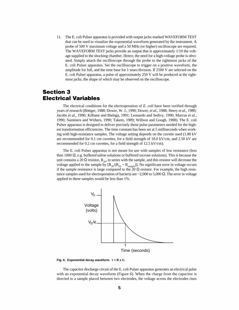
Fig. 6. Exponential decay waveform. ττ = R x C.
The capacitor discharge circuit of the E. coli Pulser apparatus generates an electrical pulsewith an exponential decay waveform (Figure 6). When the charge from the capacitor is directed to a sample placed between two electrodes, the voltage across the electrodes rises
V0
V0/e
Time (seconds)τ
Voltage(volts)
5

rapidly to a peak voltage (also known as the initial voltage, V0), and declines over time asfollows,
Vt = V0 [e -(t/τ)] Equation 1
where τ is the RC time constant, a convenient expression of the pulse length. According toEquation 1, τ is the time over which the voltage declines to 1/e (~37%) of the peak value.The time constant has been fixed at 5 milliseconds, the optimum for the electroporation of E. coli.
The voltage gradient between the electrodes is also known as the electric field (E) and isdescribed by
E = V/d Equation 2
where d is the distance between the electrodes. The strength of the electric field and the sizeof the cells determine the voltage drop across each cell, and it is this voltage drop that seemsto be the important manifestation of the voltage effect in electroporation.
An additional function of the E. coli Pulser apparatus circuit is to place a 20 Ω resistor inseries with the sample. This protects the instrument by limiting the current should an arcoccur. With an E. coli sample prepared as described in Sections 4 and 5, the resistance isabout 5000 Ω; therefore, during normal operation (no arc) the voltage drop across the 20 Ωprotective resistor will be less than 1%. Notice, however, that if this circuit is used with sam-ples of the much lower resistance typical of eukaryotic electroporation (20-200 Ω), the volt-age lost across the protective resistor becomes highly significant. For this reason, the E. coliPulser apparatus is not used with samples of less than 1,000 ΩΩ.
Section 4Bacterial Electroporation (Electro-transformation)
4.1 DNAThe effect of size and topology of the plasmid DNA on electro-transformation of E. coli.
has been examined. Plasmids of 21 kb transform E. coli with the same molar efficiency as plas-mids of 3 to 7 kb (Dower, W. J., 1990). Supercoiled and relaxed circular forms of plasmidsup to at least 20 kb transform with the same efficiency. Linear DNA is about 104-fold lessactive than the corresponding circular plasmid in the recBC+ strain of E. coli that we use.
The concentration of DNA greatly affects the recovery of transformants. With E. coli, thefrequency of transformation (transformants/survivor) is strictly dependent on DNA concen-tration over at least six orders of magnitude (10 pg/ml to 7.5 µg/ml). At the higher DNA con-centrations, up to 80% of the survivors are transformed (Dower et al., 1988). Because thenumber of transformants recovered is the product of the frequency and the number of cellspresent, the efficiency (transformants/µg DNA) increases with cell concentration over the rangeof 109 to at least 3 x 10¹⁰ cells/ml. To obtain high frequencies we use high DNA concentra-tion. To obtain high efficiencies we use high cell concentration (and low DNA concentrationto avoid cotransformations). In each case, a small sample volume (20-50 µl) allows econom-ical use of DNA and cells. (See Dower et al., 1988 for a detailed discussion of these factors).
An important technical consideration in preparing DNA for use in high voltage electro-poration is the ionic strength of the solution. Cesium chloride in a plasmid preparation orresidual ammonium acetate from ethanol precipitation, for example, can cause arcing andshould be reduced to 10 mM or less. DNA dissolved in TE (10 mM Tris-HCl pH 8.0, 1 mMEDTA) is fine as long as it is diluted about 10-fold with the cell suspension. DNA used
6
directly from various enzyme reactions also works, but the final salt concentration in the elec-troporation sample should be kept below ~5 meq for high voltage operation. Transformationwith ligation mixture, for example, is successful when the ionic strength is reduced by dilu-tion (Willson and Gough, 1988), dialysis (Heery and Dunican, 1989; Jacobs et al.,1990), orethanol precipitation (Böttger, 1988; Zabarovsky and Winberg, 1990).
4.2 Electroporation MediaComponents contributing to the conductivity of the electroporation medium include the
solution used to resuspend the cells, the cells themselves, and the DNA solution. For bestresults (highest efficiencies) it is very important to keep the resistance of the medium as highas possible, by making sure that all salts have been removed from the cell and DNA prepa-rations. Glycerol is a convenient electroporation medium, since it is recommended as a cry-oprotectant for storage of E. coli.
4.3 Electroporation ConditionsFor electroporation of E. coli, small volumes are generally best. With the 0.2 cm or 0.1 cm
cuvette, our standard volume is 40 µl (up to 400 µl may be used in the 0.2 cm cuvette; up to80 µl may be used in the 0.1 cm cuvette). This permits economical use of cells and DNA.An additional benefit of the small sample volume is that with a smaller cross section, it pro-vides the higher resistance required for high voltage operation.
With a 40 µl sample, shake the sample to the bottom of the cuvette before pulsing, andrecover the sample by washing with 1 ml of outgrowth medium from a pasteur pipette.
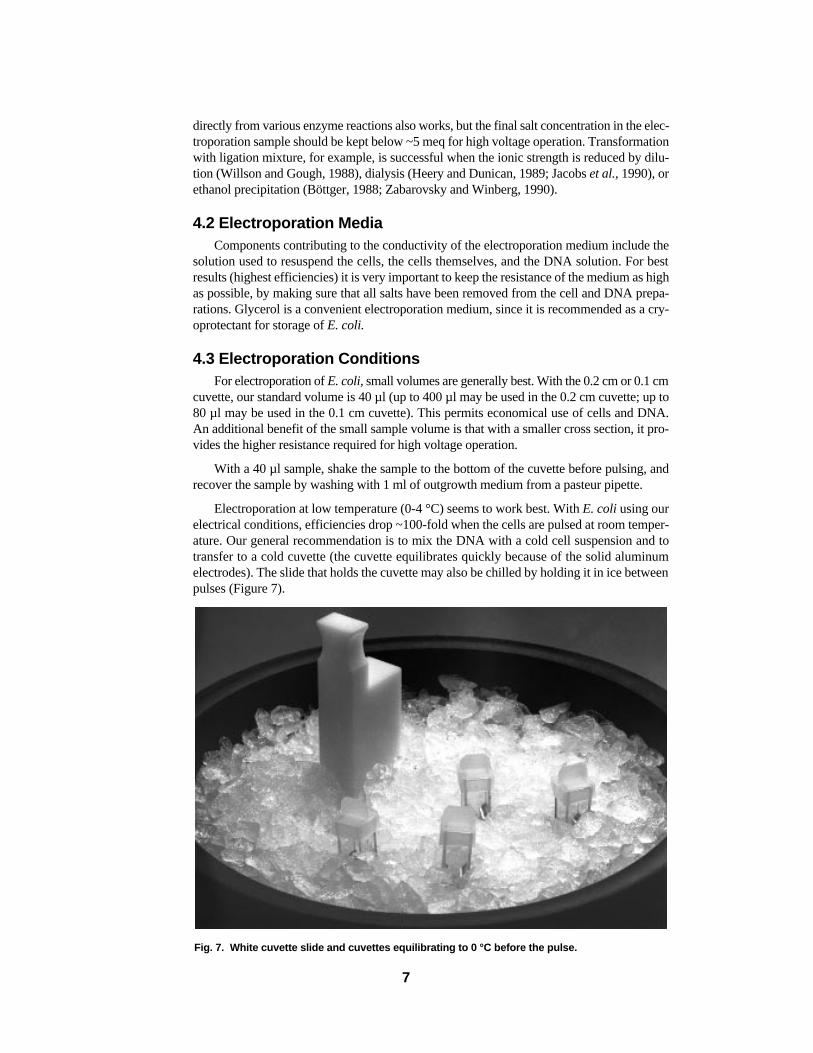
Electroporation at low temperature (0-4 °C) seems to work best. With E. coli using ourelectrical conditions, efficiencies drop ~100-fold when the cells are pulsed at room temper-ature. Our general recommendation is to mix the DNA with a cold cell suspension and totransfer to a cold cuvette (the cuvette equilibrates quickly because of the solid aluminumelectrodes). The slide that holds the cuvette may also be chilled by holding it in ice betweenpulses (Figure 7).
Fig. 7. White cuvette slide and cuvettes equilibrating to 0 °C before the pulse.
7
Incubating E. coli cells with DNA for more than a minute before pulsing does not improvetransformation efficiency (Dower et al., 1988). For this species, binding of the DNA to the cellsis probably not necessary. Therefore, we recommend adding the DNA to the cells in a coldpolypropylene tube, mixing well, transferring to a cold cuvette, and applying the pulse (the nar-row gap of the cuvettes prevents uniform mixing).
The period between applying the pulse and transferring the cells to outgrowth medium iscrucial for recovering E. colitransformants (Dower et al., 1988). Delaying this transfer by even1 minute causes a 3-fold drop in transformation. This decline continues to a 20-fold drop by10 minutes. With E. coli,we transfer the cells to medium as soon as possible after the pulse.
Appendix I lists references of several E. coli strains that have been transformed by elec-troporation. Appendix II provides information on the transformation efficiencies and surviv-abilities of several E. coli strains pulsed at various field strengths with the E. coli Pulserapparatus. We are interested in hearing of additional strains transformed by electroporation andincluding this information in subsequent versions of this manual. Please contact your local Bio-Rad representative, or in the U.S., call our Technical Services at (800) 424-6723 with anycomments or questions.
Section 5High Efficiency Electro-transformation of E. coli
Electroporation provides a method of transforming E. coli to efficiencies greater than arepossible with the best chemical methods. By subjecting mixtures of cells and DNA to expo-nentially decaying fields of very high initial amplitude, we routinely obtain 109 to 1010 trans-formants/µg of DNA with various strains and several plasmids. The survival andtransformation of cells is related to the intensity of the field (field strength = voltage/distancebetween electrodes) and to the length of the pulse (RC time constant).
Protocols for preparing and electro-transforming E. coli to high efficiencies are describedin Table 1.
Table 1. Procedure for High Efficiency Electro-transformation of E. coli
A. Preparation of Cells
1. Inoculate 1 liter of L-brotha with 1/100 volume of a fresh overnight culture.
2. Grow cells at 37 °C with vigorous shaking to an ABS600of approximately 0.5-0.7 (the bestresults are obtained with cells that are harvested at early- to mid-log phase; the appropri-ate cell density therefore depends on the strain and growth conditions).
*3. To harvest, centrifuge cells in cold centrifuge bottles in a cold rotor at 4000 x gmax for 15 minutes.
*4. Remove as much of the supernatant (medium) as possible. It is better to sacrifice the yieldby pouring off a few cells than to leave any supernatant behind.
*5. Gently resuspend the pellets in a total of 1 liter of ice-cold 10% glycerolb taking care notto lyse them. Centrifuge as in step 3.
*6. Resuspend in 0.5 liter of ice-cold 10% glycerol. Centrifuge as in step 3.
*7. Resuspend in ~250 ml of ice-cold 10% glycerol. Centrifuge as in step 3.
* Keep the cells as close to 0 °C as possible (in an ice/water bath) throughout their preparation.
8
8. Resuspend to a final volume of 3 to 4 ml in ice-cold 10% glycerol. The cell concentra-tion should be about 1 - 3 x 10¹⁰ cells/ml.
9. This suspension may be frozen in aliquots on dry ice, and stored at -70 °C. The cells aregood for at least 6 months under these conditions.
B. Electro-transformation and Plating
1. Gently thaw the cells at room temperature and then immediately place them on ice.Remove the sterile cuvettes from their pouches and place them on ice. Place the whitechamber slide on ice (Figure 7).
2. In a cold, 1.5 ml polypropylene tube, mix 40 µl of the cell suspension with 1 to 2 µl ofDNA (DNA should be in a low ionic strength buffer such as TEc). Mix well and let sit onice ~0.5 - 1 minute.
3. Set the E. coli Pulser apparatus to 2.50 kV when using the 0.2 cm cuvettes. Set it to1.80 kV when using the 0.1 cm cuvettes. See Section 2 for operating instructions.
4. Transfer the mixture of cells and DNA to a cold electroporation cuvette, and shake the sus-pension to the bottom. Place the cuvette in a chilled safety chamber slide. Push the slideinto the chamber until the cuvette is seated between the contacts in the base of the cham-ber (Figure 4).
5. Pulse once.
6. Remove the cuvette from the chamber and immediately add 1 ml of SOCd medium to thecuvette and quickly but gently resuspend the cells with a pasteur pipette. (This rapid addi-tion of SOC after the pulse is very important in maximizing the recovery of transfor-mants.)
7. Transfer the cell suspension to a 17 x 100 mm polypropylene tube and incubate at 37 °Cfor 1 hour. (Shaking the tubes at 225 rpm during this incubation may improve the recov-ery of transformants.)
8. Check and record the pulse parameters. The time constant should be close to 5 millisec-onds. The field strength can be calculated as actual volts (kV) / cuvette gap (cm).
9. Plate on selective medium.
a L-Broth: 1% Bacto tryptone, 0.5% Bacto yeast extract, 0.5% NaCl.b 10% Glycerol: Prepare fresh weekly with sterilized water. Do not autoclave or filter-sterilize the glycerol
solution.c DNA containing too much salt will make the sample too conductive and cause arcing at high voltage.
TE: 10 mM Tris-HCl pH 8.0, 1 mM EDTA.d SOC: 2% Bacto tryptone, 0.5% Bacto yeast extract, 10 mM NaCl, 2.5 mM KCl, 10 mM MgCl2, 10 mM MgSO4,
20 mM glucose.
9

Appendix IE. coli Electro-transformation References
The following literature references report the transformation of Escherichia coliby electro-poration. Asterisks denote work done with the Gene Pulser® system.
Escherichia coli References
spheroplasts ..............................................Cymbalyuk et al.(1988)
* (strain not mentioned) ..............................Dahlman and Harlander (1988)Dower et al. (1992)Fikes et al. (1990)Hofmann and Evans (1986)Hosoda et al. (1990)Larimer and Mural (1990)O’Callaghan and Charbit (1990)Sale and Hamilton (1967)Sale and Hamilton (1968)Speyer (1990b)
* wild type ...................................................Fiedler et al. (1989)Wirth et al. (1989)
* strain 1106 ................................................Cocconcelli et al. (1991)
strain 300 V...............................................Hamilton and Sale (1967)
* strain 3132 (wild-type) .............................Fiedler et al. (1989)Wirth et al. (1989)
* strain AB1157...........................................Binotto et al. (1991)Hartke and Schulte-Frohlinde (1991)Sixou et al. (1991)
* strain BB ...................................................Taketo (1989)
* strain BHB2600........................................Gaier et al. (1992)Knauf et al. (1992)
* strain BW 313-1 .......................................Hsieh et al. (1991)
* strain C......................................................Taketo (1989)Taylor and Burke (1990)
* strain CAIQ1..............................................Zink et al. (1991)
* strain C-600 ..............................................Binotto et al. (1991)Kilbane and Bielaga (1991)Marcus et al. (1990)Yuan et al.(1984)
* strain C600Sm ..........................................Metzger et al. (1992)
* strain C600-2Sm.......................................Metzger et al. (1992)
* strain CU9276...........................................Batt et al. (1990)
* strain DB11...............................................Mermelstein et al. (1992)
10
Escherichia coli References
* strain DH1.................................................Craig et al. (1989)Flannagan and Clewell (1991)Fujimoto et al. (1991)Heery et al. (1989)Taketo (1989)
* strain DH10B............................................Gruber (1992)Hanahan et al. (1991)Hsieh et al. (1991)Li et al. (1990)Pacholczyk et al. (1991)Rice et al. (1992)Rodriguez-Palenzuela et al. (1991)Smith et al. (1990)
* strain DH5.................................................Hsieh et al. (1991)Inoue et al. (1990)
* strain DH5α ..............................................Allen and Blaschek (1990)Cirillo et al. (1991)Dower et al. (1988)Fujimoto et al. (1991)Gilchrist and Smit (1991)Kallio et al. (1991)Lyra et al. (1991)Smith et al. (1990)Sung et al. (1990)
strain DH5αF´ ..........................................Marcil and Higgins (1992)
strain DH5αF´IQ ......................................Smith et al. (1990)
* strain DS941 .............................................Summers and Withers (1990)
* strain EM24 ..............................................Thomson and Flint (1989)
strain ER1451 ...........................................Sixou et al. (1991)
* strain GM1829..........................................Joerger and Klaenhammer (1990)
* strain HB101.............................................Calvin (1988)De Rossi et al. (1991)Dower (1987)Fiedler et al. (1989)Fiedler and Wirth (1988)Haider et al. (1991)Haynes and Britz (1990)Keller and Maniatis (1991)Kilbane and Bielaga (1991)Kim and Blaschek (1989)Regué et al. (1992)Smith et al. (1990)Speyer (1990a)Sung et al. (1990)Wirth et al. (1989)
11
Escherichia coli References
* strain JC8679 ............................................Summers and Withers (1990)
* strain JM101 .............................................Halling and Zehr (1990)
* strain JM105 .............................................Fiedler et al. (1989)Fiedler and Wirth (1988)Jacobs et al. (1990)Wirth et al. (1989)
* strain JM107 .............................................Dower (1987)
* strain JM109 .............................................Boivin and Bellemare (1991)Calvin (1988)Gerischer and Dürre (1990)Kovalic et al. (1991)Smith et al. (1990)Taketo (1988)Tobin et al. (1991)Ward and Jarvis (1991)
* strain JM110 .............................................Raya and Klaenhammer (1992)
* strain JPN15.96.........................................Jerse et al. (1990)
* strain JS4...................................................Leonardo and Sedivy (1990)
strain K12..................................................Hülsheger and Niemann (1980)Hülsheger et al. (1981)
* strain K12A...............................................Taketo (1989)
* strain K2-1-4.............................................Schendel et al. (1992)
* strain K802 ...............................................Gilchrist and Smit (1991)
* strain K803 ...............................................Jacobs et al. (1990)Ware et al. (1992)
* strain LE392 .............................................Binotto et al. (1991)Dower (1987)Dower (1990)Dower and Cwirla (1992)Dower et al. (1988)Fujimoto et al. (1991)Taketo (1988)Zabarovsky and Winberg (1990)
* strain M5361.............................................Takahashi and Kobayashi (1990)
* strain MC1061..........................................Calvin (1988)Calvin and Hanawalt (1988)Cwirla et al. (1990)Dower (1990)Dower and Cwirla (1992)Dunican and Shivnan (1989)Hanahan et al. (1991)Rubenstein et al. (1990)Smith et al. (1990)
12

Escherichia coli References
* strain MC1061 (cont.) ..............................Willson and Gough (1988)Zabarovsky and Winberg (1990)
* strain MC1061/P3.....................................Kieffer (1991)Sheen (1989)Ymer (1991)
* strain MG1655..........................................Pfau and Youderian (1990)
* strain MM294 ...........................................Zink et al. (1991)
* strain MV1190..........................................Dower (1987)
strain NCTC 8196 ....................................Hamilton and Sale (1967)
* strain NK5898 ..........................................Mahillon and Kleckner (1992)
* strain NM522............................................Boivin and Bellemare (1991)Willson and Gough (1988)
* strain p678-54...........................................Marcus et al. (1990)
* strain QC774.............................................Van Camp et al. (1990)
* strain SCS-1..............................................King and Goodbourn (1992)Pfau and Youderian (1990)
* strain SDM................................................Batt et al. (1990)
* strain SM10...............................................Rodriguez-Palenzuela et al. (1991)
* strain SR101 .............................................Garrard et al. (1991)
* strain SURE..............................................Clarke et al. (1992)Wang et al. (1992)
* strain TG-1................................................Heery and Dunican (1989)Zabarovsky and Winberg (1990)
* strain UM2................................................Knauf et al. (1992)
* strain WA321............................................Fiedler et al. (1989)Fiedler and Wirth (1988)Wirth et al. (1989)
* strain WM1100.........................................Dower (1990)Elliot et al. (1990)Hsieh et al. (1991)
* strain XL-1 Blue .......................................Barbas et al. (1991)Hermanson et al. (1991)O’Neill and Söll (1990)Ostrander et al. (1992)Patel et al. (1991)Petzel and McKay (1992)Starr and Huse (1990)Swaroop et al. (1991)Taketo (1988)Zabarovsky and Winberg (1990)
* strain XS127 .............................................Böttger (1988)
13

Special Applications References
* Ligation mixtures .............................................Barbas et al. (1991)Böttger (1988)Cirillo et al. (1991)Clarke et al. (1992)Elliot et al. (1990)Flannagan and Clewell (1991)Gaier et al. (1992)Gerischer and Dürre (1990)Gilchrist and Smit (1991)Gruber (1992)Halling and Zehr (1990)Hanahan et al. (1991)Heery and Dunican (1989)Jacobs et al. (1990)Keller and Maniatis (1991)Kieffer (1991), Kieffer (1992)King and Goodbourn (1992)Knauf et al. (1992)Kovalic et al. (1991)O’Neill and Söll (1990)Ostrander et al. (1992)Pacholczyk et al. (1991)Patel et al. (1991)Petzel and McKay (1992)Wang et al. (1992)Willson and Gough (1988)Ymer (1991)Zabarovsky and Winberg (1990)Zink et al. (1991)
* Library construction .........................................Böttger (1988)Elliot et al.(1990)Gerischer and Dürre (1990)Kieffer (1991), Kieffer (1992)Ostrander et al. (1992)Pacholczyk et al. (1991)Wang et al. (1992)
* Single-stranded DNA.......................................Rubenstein et al. (1990)Swaroop et al. (1991)
* M13 DNA.........................................................Hartke and Schulte-Frohlinde (1991)Heery and Dunican (1989)Starr and Huse (1990)
* Other bacteriophage DNA................................Lyra et al. (1991)
* Large DNA fragments......................................Larimer and Mural (1990)Leonardo and Sedivy (1990)
14
Special Applications References
* Influence of DNA size on efficiency ...............Allen and Blaschek (1990)Dower (1990)Leonardo and Sedivy (1990)Marcus et al. (1990)Regué et al. (1992)Smith et al. (1990)
* Influence of DNA conformation
on efficiency..................................................Dower (1990)Hartke and Schulte-Frohlinde (1991)Leonardo and Sedivy (1990)
* RNA..................................................................Taketo (1989)
* Release of cell components..............................Calvin (1988)Calvin and Hanawalt (1988)Hamilton and Sale (1967)Heery et al. (1989)Li et al. (1990)Sixou et al. (1991)
* Direct transfer (donor to recipient) ..................Gilchrist and Smit (1991)Kilbane and Bielaga (1991)Marcil and Higgins (1992)Pfau and Youderian (1990)Summers and Withers (1990)Ward and Jarvis (1991)
* Use of more than one electroporation unit.......Li et al. (1990)
Review Articles
Dower (1990)Dower et al. (1992)Hanahan et al. (1991)Hofmann and Evans (1986)
Key to Electroporation Units UsedGP Gene Pulser apparatusPC Pulse ControllerCE Capacitance ExtenderED Exponential Decay (other than a Gene Pulser apparatus)SW Square WaveOT OtherUK Unknown
* Work done with the Gene Pulser system
15
ReferencesGP Allen, S. P., and Blaschek, H. P., Factors involved in the electroporation-induced
transformation of Clostridium perfringens, FEMS Microbiol. Lett., 70, 217 (1990).
GP + PC Barbas, III, C. F., Kang, A. S., Lerner, R. A., and Benkovic, S. J., Assembly ofcombinatorial antibody libraries on phage surfaces: the gene III site, Proc. Natl.Acad. Sci. USA, 88, 7978 (1991).
GP Batt, C. A., Oren, P., Webb, J., Flicke, P., An improved method for oligonucleotide-mediated site-directed mutagenesis, BioTechniques, 9, 554 (1990).
GP + PC Binotto, J., MacLachlan, P. R., and Sanderson, K. E., Electrotransformation inSalmonella typhimuriumLT2, Can. J. Microbiol., 37, 474 (1991).
GP + PC Boivin, R., and Bellemare, G., A novel approach to the rapid isolation and nucleotidesequencing of genomic clones, Genet. Analysis - Techn. Applic. (GATA), 8, 181(1991).
GP + PC Böttger, E. C., High-efficiency generation of plasmid cDNA libraries using electro-transformation, BioTechniques, 6, 878 (1988).
OT Calvin, N., I. Quantitation and analysis of furocoumarin: DNA adducts II.Introduction and recovery of DNA in bacterial cells by electroporation, Ph.D.Dissertation, Stanford University, Ch. 5, pp. 90 - 120 (1988).
OT Calvin, N. M., and Hanawalt, P. C., High-efficiency transformation of bacterialcells by electroporation, J. Bacteriol.,170, 2796 (1988).
GP + PC Cirillo, J. D., Barletta, R. G., Bloom, B. R., and Jacobs, Jr., W. R., A novel trans-poson trap for mycobacteria: isolation and characterization of IS1096, J. Bacteriol.,173, 7772 (1991).
GP + PC Clarke, B., Stancombe, P., Money, T., Foote, T., and Moore, G., Targeting dele-tion (homoeologous chromosome pairing locus) or addition line single copysequences from cereal genomes, Nucl. Acids Res., 20, 1289 (1992).
GP + PC Cocconcelli, P. S., Gasson, M. J., Morelli, L., and Bottazzi, V., Single-strandedDNA plasmid, vector construction and cloning of Bacillus stearothermophilus α-amilase in Lactobacillus, Res. Microbiol., 142, 643 (1991).
GP + PC + CE Craig, F. F., Coote, J. G., Parton, R., Freer, J. H., and Gilmour, N. J. L., A plasmidwhich can be transferred between Escherichia coliand Pasteurella haemolyticabyelectroporation and conjugation, J. Gen. Microbiol.,135, 2885 (1989).
GP + PC Cwirla, S. E., Peters, E. A., Barrett, R. W., and Dower, W. J., Peptides on phage: avast library of peptides for identifying ligands, Proc. Natl. Acad. Sci. USA, 87, 6378(1990).
ED Cymbalyuk, E. S., Chernomordik, L. V., Broude, N. E., and Chizmadzhev, Y. A.,Electro-stimulated transformation of E. coli cells pretreated by EDTA solution,FEBS Lett., 234, 203 (1988).
ED Dahlman, D., and Harlander, S., Electroporation of intact microbial cells, Abstr.Annual Meeting Amer. Soc. Microbiol.,88, 155 (1988).
GP + PC De Rossi, E., Brigidi, P., Rossi, M., Matteuzzi, D., and Riccardi, G., Characterizationof gram-positive broad host-range plasmids carrying a thermophilic replicon, Res.Microbiol., 142, 389 (1991).
GP (no PC) Dower, B., Electro-transformation of intact bacterial cells, Molecular BiologyReports(Bio-Rad Laboratories), 1, 5 (1987).
GP + PC Dower, W. J., “Electroporation of bacteria: a general approach to genetic transfor-mation,” in Genetic Engineering—Principles and Methods, 1990, vol. 12, pp. 275-296, Plenum Publishing Corp., New York.
GP + PC Dower, W. J., and Cwirla, S. E., Creating vast peptide expression libraries: elec-troporation as a tool to construct plasmid libraries of greater than 109 recombinants,in Guide to Electroporation and Electrofusion(D. C. Chang, B. M. Chassy, J. A.Saunders, and A. E. Sowers, eds.), pp. 291 -301, 1992, Academic Press Inc., SanDiego.
16
GP + PC Dower, W. J., Miller, J. F., and Ragsdale, C. W., High efficiency transformation ofE. coliby high voltage electroporation, Nucl. Acids Res., 16, 6127 (1988).
GP + PC Dunican, L. K., and Shivnan, E., High frequency transformation of whole cells ofamino acid producing coryneform bacteria using high voltage electroporation,Bio/Technol., 7, 1067 (1989).
GP + PC Elliott, J. F., Albrecht, G. R., Gilladoga, A., Handunnetti, S. M., Neequaye, J.,Lallinger, G., Minjas, J. N., and Howard, R. J., Genes for Plasmodium falciparumsurface antigens cloned by expression in COS cells, Proc. Natl. Acad. Sci. USA,87, 6363 (1990).
GP Fiedler, S., Friesenegger, A., and Wirth, R., “Electroporation: a general method fortransformation of gram-negative bacteria,” in Genetic Transformation andExpression, chapter 7, pp. 65 - 69, 1989, L. O. Butler, C. Harwood, and B. E. B.Moseley, eds., Intercept Ltd, Andover, Hants, United Kingdom.
GP (no PC) Fiedler, S., and Wirth, R., Transformation of bacteria with plasmid DNA by elec-troporation, Anal. Biochem., 170, 38 (1988).
GP + PC Fikes, J. D., Becker, D. M., Winston, F., and Guarente, L., Striking conservationof TFIID in Schizosaccharomyces pombe and Saccharomyces cerevisiae, Nature,346, 291 (1990).
GP + PC Flannagan, S. E., and Clewell, D. B., Conjugative transfer of Tn916in Enterococcusfaecalis: transactivation of homologous transposons,J. Bacteriol., 173, 7136 (1991).
ED Fujimoto, S., Hashimoto, H., and Ike, Y., Low cost device for electrotransformationand its application to the highly efficient transformation of Escherichia coliandEnterococcus faecalis, Plasmid,26, 131 (1991).
GP + PC Gaier, W., Vogel, R. F., and Hammes, W. P., Cloning and expression of thelysostaphin gene in Bacillus subtilisand Lactobacillus casei, Lett. Appl. Microbiol.,14, 72 (1992).
GP + PC Garrard, L. J., Yang, M., O’Connell, M. P., Kelley, R. F., and Henner, D. J., FABassembly and enrichment in a monovalent phage display system, Bio/Technol., 9,1373 (1991).
GP + PC Gerischer, U., and Dürre, P., Cloning, sequencing, and molecular analysis of theacetoacetate decarboxylase gene region from Clostridium acetobutylicum,J. Bacteriol., 172, 6907 (1990).
GP + PC + CE Gilchrist, A., and Smit, J., Transformation of freshwater and marine caulobacters byelectroporation, J. Bacteriol., 173, 921 (1991).
ED Gruber, C. E., High-efficiency cDNA cloning: a comparison of electroporation andin vitro packaging, BioTechniques, 12, 804 (1992).
GP + PC Haider, M. Z., Al-Taho, N., Al-Salameen, F., Kadri, M. H., Al-Amad, S., andSpanier, E., Efficient transformation of thermotolerant methanol-utilizing strainsof Methylophilusspp. via electroporation, Acta Biotechnol., 11, 295 (1991).
GP + PC Halling, S. M., and Zehr, E. S., Polymorphism in Brucellaspp. due to highly repeat-ed DNA, J. Bacteriol., 172, 6637 (1990).
SW Hamilton, W. A., and Sale, A. J. H., Effects of high electric fields on microorgan-isms, II. Mechanism of action of the lethal effect, Biochim. Biophys. Acta, 148, 789(1967).
GP + PC, ED Hanahan, D., Jessee, J., and Bloom, F. R., Plasmid transformation of Escherichiacoli and other bacteria, Meth. Enzymol., 204, 63 (1991).
GP + PC Hartke, A., and Schulte-Frohlinde, D., Survival of M13mp18 gapped duplex DNAas a function of gap length, Mutat. Res., 255, 39 (1991).
GP + PC Haynes, J. A., and Britz, M. L., The effect of growth conditions of Corynebacteriumglutamicumon the transformation frequency obtained by electroporation, J. Gen.Microbiol., 136, 255 (1990).
GP + PC Heery, D. M., and Dunican, L. K., Improved efficiency M13 cloning using elec-troporation, Nucl. Acids Res., 17, 8006 (1989).
17
GP + PC Heery, D. M., Powell, R., Gannon, F., and Dunican, L. K., Curing of a plasmidfrom E. coliusing high-voltage electroporation, Nucl. Acids Res., 17, 10131 (1989).
GP + PC Hermanson, G. G., Hoekstra, M. F., McElligott, D. L., and Evans, G. A., Rescue ofend fragments of yeast artificial chromosomes by homologous recombination inyeast, Nucl. Acids Res., 19, 4943 (1991).
UK Hofmann, G. A., and Evans, G. A., Electronic genetic - physical and biologicalaspects of cellular electromanipulation, IEEE Engineering in Medicine and BiologyMagazine, 5, 6 (1986).
UK Hosoda, F., Nishimura, S., Uchida, H., and Ohki, M., An F factor based cloningsystem for large DNA fragments, Nucl. Acids Res., 18, 3863 (1990).
GP + PC Hsieh, C.-L., McCloskey, R. P., Radany, E., and Lieber, M. R., V(D)J recombina-tion: evidence that a replicative mechanism is not required, Molec. Cell. Biol., 11,3972 (1991).
ED Hülsheger, H., and Niemann, E.-G., Lethal effects of high-voltage pulses on E. coliK12, Radiat. Environ. Biophys., 18, 281 (1980).
ED Hülsheger, H., Potel, J., and Niemann, E.-G., Killing of bacteria with electric puls-es of high field strength, Radiat. Environ. Biophys., 20, 53 (1981).
GP + PC Inoue, H., Nojima, H., and Okayama, H., High efficiency transformation ofEscherichia coliwith plasmids, Gene, 96, 23 (1990).
GP + PC Jacobs, M., Wnendt, S., and Stahl, U., High-efficiency electro-transformation ofEscherichia coliwith DNA from ligation mixtures, Nucl. Acids Res., 18, 1653(1990).
GP Jerse, A. E., Yu, J., Tall, B. D., and Kaper, J. B., A genetic locus of enteropathogenicEscherichia colinecessary for the production of attaching and effacing lesions ontissue culture cells, Proc. Natl. Acad. Sci. USA, 87, 7839 (1990).
GP + PC Joerger, M. C., and Klaenhammer, T. R., Cloning, expression, and nucleotidesequence of the Lactobacillus helveticus481 gene encoding the bacteriocin hel-veticin J, J. Bacteriol., 172, 6339 (1990).
GP Kallio, P. T., Fagelson, J. E., Hoch, J. A., and Strauch, M. A., The transition stateregulator Hpr of Bacillus subtilisis a DNA-binding protein, J. Biol. Chem., 266,13411 (1991).
GP + PC Keller, A. D., and Maniatis, T., Selection of sequences recognized by a DNA bind-ing protein using a preparative southwestern blot, Nucl. Acids Res., 19, 4675 (1991).
GP + PC Kieffer, B. L., Optimised cDNA size selection and cloning procedure for the con-struction of representative plasmid cDNA libraries, Gene, 109, 115 (1991).
GP + PC Kieffer, B. L., Optimised cDNA size selection and cloning procedure for the con-struction of representative plasmid cDNA libraries, Gene, 116, 117 (1992).[Correction]
GP + PC Kilbane, II, J. J., and Bielaga, B. A., Instantaneous gene transfer from donor torecipient microorganisms via electroporation, BioTechniques, 10, 354 (1991).
GP (no PC) Kim, A. Y., and Blaschek, H. P., Construction of an Escherichia coli-Clostridiumperfringensshuttle vector and plasmid transformation of Clostridium perfringens,Appl. Environ. Microbiol., 55, 360 (1989).
GP + PC King, P., and Goodbourn, S., A method for sequence-specific deletion mutagenesis,Nucl. Acids Res., 20, 1039 (1992).
GP + PC Knauf, H. J., Vogel, R. F., and Hammes, W. P., Cloning, sequence, and phenotyp-ic expression of katA, which encodes the catalase of Lactobacillus sakeLTH677,Appl. Environ. Microbiol., 58, 832 (1992).
ED Kovalic, D., Kwak, J.-H., and Weisblum, B., General method for direct cloning ofDNA fragments generated by the polymerase chain reaction, Nucl. Acids Res., 19,4560 (1991).
18

GP + PC Larimer, F. W., and Mural, R. J., “Megabase cloning in E. coli,” in Abstracts ofpapers presented at the 1990 meeting on Genome Mapping and Sequencing, ColdSpring Harbor Laboratory, Cold Spring Harbor, New York, p. 99 (1990).
GP + PC Leonardo, E. D., and Sedivy, J. M., A new vector for cloning large eukaryotic DNAsegments in Escherichia coli, Bio/Technol., 8, 841 (1990).
GP + PC, ED Li, S. J., Landers, T. A., and Smith, M. D., Electroporation of plasmids into plasmid-containing Escherichia coli, Focus, 12, 72 (1990).
GP + PC Lyra, C., Savilahti, H., and Bamford, D. H., High-frequency transfer of linear DNAcontaining 5´-covalently linked terminal proteins: electroporation of bacteriophagePRD1 genome into Escherichia coli, Molec. Gen. Genet., 228, 65 (1991).
GP + PC Mahillon, J., and Kleckner, N., New IS10transposition vectors based on a gram-pos-itive replication origin, Gene, 116, 69 (1992).
ED Marcil, R., and Higgins, D. R., Direct transfer of plasmid DNA from yeast to E. coliby electroporation, Nucl. Acids Res., 20, 917 (1992).
GP + PC Marcus, H., Ketley, J. M., Kaper, J. B., and Holmes, R. K., Effects of DNase pro-duction, plasmid size, and restriction barriers on transformation of Vibrio choleraeby electroporation and osmotic shock, FEMS Microbiol. Lett., 68, 149 (1990).
GP + PC Mermelstein, L. D., Welker, N. E., Bennett, G. N., and Papoutsakis, E. T.,Expression of cloned homologous fermentative genes in Clostridium acetobutylicumATCC 824, Bio/Technol., 10, 190 (1992).
GP + PC Metzger, M., Bellemann, P., Schwartz, T., and Geider, K., Site-directed and trans-poson-mediated mutagenesis with pfd-plasmids by electroporation of Erwiniaamylovora and Escherichia colicells,Nucl. Acids Res., 20, 2265 (1992).
GP + PC O’Callaghan, D., and Charbit, A., High efficiency transformation of Salmonellatyphimurium and Salmonella typhi by electroporation, Mol. Gen. Genet., 223, 156(1990).
GP + PC O’Neill, G. P., and Söll, D., Expression of the Synechocystissp. strain PCC 6803tRNAGlu gene provides tRNA for protein and chlorophyll biosynthesis, J. Bacteriol.,172, 6363 (1990).
GP Ostrander, E. A., Jong, P. M., Rine, J., and Duyk, G., Construction of small-insertgenomic DNA libraries highly enriched for microsatellite repeat sequences, Proc.Natl. Acad. Sci. USA, 89, 3419 (1992).
ED Pacholczyk, T., Blakely, R. D., and Amara, S. G., Expression cloning of a cocaine-and antidepressant-sensitive human noradrenaline transporter, Nature, 350, 350(1991).
GP + PC Patel, K., Cox, R., Shipley, J., Kiely, F., Frazer, K., Cox, D. R., Lehrach, H., andSheer, D., A novel and rapid method for isolating sequences adjacent to rare cuttingsites and their use in physical mapping, Nucl. Acids Res., 19, 4371 (1991).
GP + PC Petzel, J. P., and McKay, L. L., Molecular characterization of the integration of thelactose plasmid from Lactococcus lactissubsp. cremorisSK11 into the chromo-some of L. lactissubsp. lactis, Appl. Environ. Microbiol., 58, 125 (1992).
GP + PC Pfau, J., and Youderian, P., Transferring plasmid DNA between different bacterialspecies with electroporation, Nucl. Acids Res., 18, 6165 (1990).
GP + PC Raya, R. R., and Klaenhammer, T. R., High-frequency plasmid transduction byLactobacillus gasseri bacteriophage øadh, Appl. Environ. Microbiol., 58, 187 (1992).
ED Regué, M., Enfedaque, J., Camprubí, S., and Tomás, J. M., The O-antigenlipopolysaccharide is the major barrier to plasmid DNA uptake by Klebsiella pneu-moniaeduring transformation by electroporation and osmotic shock, J. Microbiol.Meth., 15, 129 (1992).
GP + PC Rice, G. C., Goeddel, D. V., Cachianes, G., Woronicz, J., Chen, E. Y., Williams,S. R., and Leung, D. W., Random PCR mutagenesis screening of secreted proteinsby direct expression in mammalian cells, Proc. Natl. Acad. Sci. USA, 89, 5467(1992).
19

20
GP + PC Rodriguez-Palenzuela, P., Burr, T. J., and Collmer, A., Polygalacturonase is a vir-ulence factor in Agrobacterium tumefaciensbiovar 3, J. Bacteriol., 173, 6547 (1991).
GP + PC Rubenstein, J. L. R., Brice, A. E. J., Ciaranello, R. D., Denney, D., Porteus, M. H.,and Usdin, T. B., Subtractive hybridization system using single-stranded phagemidswith directional inserts, Nucl. Acids Res., 18, 4833 (1990).
SW Sale, A. J. H., and Hamilton, W. A., Effects of high electric fields on microorgan-isms, I. Killing of bacteria and yeasts, Biochim. Biophys. Acta, 148, 781 (1967).
UK Sale, A. J. H., and Hamilton, W. A., Effects of high electric fields on micro-organ-isms, III. Lysis of erythrocytes and protoplasts, Biochim. Biophys. Acta, 163, 37(1968).
GP + PC Schendel, F. J., August, P. R., Anderson, C. R., Hanson, R. S., and Flickinger, M.C., Cloning and nucleotide sequence of the gene coding for citrate synthase from athermotolerant Bacillussp., Appl. Environ. Microbiol., 58, 335 (1992).
GP + PC Sheen, J., “High-efficiency transformation by electroporation,” in Current Protocolsin Molecular Biology, supplement 5, pp. 1.8.4 - 1.8.7, 1989, F. M. Ausubel, R.Brent, R. E. Kingston, D. D. Moore, J. G. Seidman, John A. Smith, K. Struhl, eds.,John Wiley & Sons, Inc., New York.
SW Sixou, S., Eynard, N., Escoubas, J. M., Werner, E., and Teissié, J., Optimized con-ditions for electrotransformation of bacteria are related to the extent of electroper-meabilization, Biochim. Biophys. Acta, 1088, 135 (1991).
ED Smith, M., Jessee, J., Landers, T., and Jordan, J., High efficiency bacterial electro-poration: 1 x 10¹⁰ E. coli transformants/µg, Focus, 12, 38 (1990).
ED Speyer, J. F., A simple and effective electroporation apparatus, BioTechniques, 8,28 (1990a).
ED Speyer, J. F., Multi-sample electroporation, BioTechniques, 8, 508 (1990b).
GP + PC Starr, L., and Huse, W. D., Electro-transformation of E. coliwith M13 DNA, Bio-Rad Laboratories Bulletin 1353 (1990).
GP + PC Summers, D. K., and Withers, H. L., Electrotransfer: direct transfer of bacterialplasmid DNA by electroporation, Nucl. Acids Res., 18, 2192 (1990).
GP + PC Sung, L. M., Jackson, M. P., O’Brien, A. D., and Holmes, R. K., Transcription ofthe Shiga-like toxin type II and Shiga-like toxin type II variant operons ofEscherichia coli, J. Bacteriol., 172, 6386 (1990).
GP + PC Swaroop, A., Xu, J., Agarwal, N., and Weissman, S. M., A simple and efficientcDNA library subtraction procedure: isolation of human retina-specific cDNAclones, Nucl. Acids Res., 19, 1954 (1991).
GP + PC Takahashi, N., and Kobayashi, I., Evidence for the double-strand break repair modelof bacteriophage λ recombination, Proc. Natl. Acad. Sci. USA, 87, 2790 (1990).
GP (no PC) Taketo, A., DNA transfection of Escherichia coliby electroporation, Biochim.Biophys. Acta, 949, 318 (1988).
GP (no PC) Taketo, A., RNA transfection of Escherichia coliby electroporation, Biochim.Biophys. Acta, 1007, 127 (1989).
GP + PC Taylor, L. D., and Burke, Jr., W. F., Transformation of an entomopathic strain ofBacillus sphaericusby high voltage electroporation, FEMS Microbiol. Lett., 66,125 (1990).
GP + PC Thomson, A. M., and Flint, H. J., Electroporation induced transformation ofBacteroides ruminicolaand Bacteroides uniformisby plasmid DNA, FEMSMicrobiol. Lett., 61, 101 (1989).
GP + PC Tobin, J. F., Laban, A., and Wirth, D. F., Homologous recombination in Leishmaniaenriettii, Proc. Natl. Acad. Sci. USA, 88, 864 (1991).
GP + PC Van Camp, W., Bowler, C., Villarroel, R., Tsang, E. W. T., Van Montagu, M., andInzé, D., Characterization of iron superoxide dismutase cDNAs from plants obtainedby genetic complementation in Escherichia coli, Proc. Natl. Acad. Sci. USA, 87,9903 (1990).
21
GP + PC Wang, M. L., Leitch, A. R., Schwarzacher, T., Heslop-Harrison, J. S., and Moore,G., Construction of a chromosome-enriched HpaII library from flow-sorted wheatchromosomes, Nucl. Acids Res., 20, 1897 (1992).
GP + PC Ward, L. J. H., and Jarvis, A. W., Rapid electroporation-mediated plasmid transferbetween Lactococcus lactisand Escherichia coliwithout the need for plasmid prepa-ration, Lett. Appl. Microbiol., 13, 278 (1991).
GP Ware, C. E., Bauchop, T., Hudman, J. F., and Gregg, K., Cryptic plasmid pBf1 fromButyrivibrio fibrisolvensAR10: its use as a replicon for recombinant plasmids, Curr.Microbiol., 24, 193 (1992).
GP Willson, T. A., and Gough, N. M., High voltage E. colielectro-transformation withDNA following ligation, Nucl. Acids Res., 16, 11820 (1988).
GP (no PC) Wirth, R., Friesenegger, A., and Fiedler, S., Transformation of various species ofgram-negative bacteria belonging to 11 different genera by electroporation, Molec.Gen. Genet., 216, 175 (1989).
GP + PC Ymer, S., Heat inactivation of DNA ligase prior to electroporation increases trans-formation efficiency, Nucl. Acids Res., 19, 6960 (1991).
SW Yuan, L., Lian-ying, S. and Wong, T., Study on transfer plasmid PBCI by electric-pulse fusion in Butirosin producing strain Bacillus circulans3342 and E. coliC600,Chinese J. Antibiotics, 9, 450 (1984). [Chinese]
GP + PC Zabarovsky, E. R., and Winberg, G., High efficiency electroporation of ligated DNAinto bacteria, Nucl. Acids Res., 18, 5912 (1990).
GP + PC Zink, A., Klein, J. R., and Plapp, R., Transformation of Lactobacillus delbrückiissp. lactisby electroporation and cloning of origins of replication by use of a posi-tive selector, FEMS Microbiol. Lett., 78, 207 (1991).
22
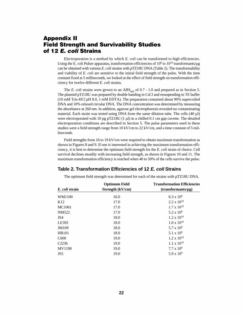
Appendix IIField Strength and Survivability Studies of 12 E. coli Strains
Electroporation is a method by which E. coli can be transformed to high efficiencies.Using the E. coli Pulser apparatus, transformation efficiencies of 109 to 1010 transformants/µgcan be obtained with various E. coli strains with pTZ18U DNA (Table 2). The transformabilityand viability of E. coli are sensitive to the initial field strength of the pulse. With the timeconstant fixed at 5 milliseconds, we looked at the effect of field strength on transformation effi-ciency for twelve different E. colistrains.
The E. coli strains were grown to an ABS600 of 0.7 - 1.0 and prepared as in Section 5.The plasmid pTZ18U was prepared by double banding in CsCl and resuspending in TE buffer(10 mM Tris-HCl pH 8.0, 1 mM EDTA). The preparation contained about 90% supercoiledDNA and 10% relaxed circular DNA. The DNA concentration was determined by measuringthe absorbance at 260 nm. In addition, agarose gel electrophoresis revealed no contaminatingmaterial. Each strain was tested using DNA from the same dilution tube. The cells (40 µl)were electroporated with 10 pg pTZ18U (1 µl) in a chilled 0.1 cm gap cuvette. The detailedelectroporation conditions are described in Section 5. The pulse parameters used in thesestudies were a field strength range from 10 kV/cm to 22 kV/cm, and a time constant of 5 mil-liseconds.
Field strengths from 16 to 19 kV/cm were required to obtain maximum transformation asshown in Figures 8 and 9. If one is interested in achieving the maximum transformation effi-ciency, it is best to determine the optimum field strength for the E. colistrain of choice. Cellsurvival declines steadily with increasing field strength, as shown in Figures 10 and 11. Themaximum transformation efficiency is reached when 40 to 50% of the cells survive the pulse.
Table 2. Transformation Efficiencies of 12 E. coli Strains
The optimum field strength was determined for each of the strains with pTZ18U DNA.
Optimum Field Transformation EfficienciesE. coli strain Strength (kV/cm) (transformants/µg)
WM1100 16.0 6.3 x 10⁹
K12 17.0 2.2 x 10¹⁰
MC1061 17.0 1.7 x 10¹⁰
NM522 17.0 5.2 x 10⁹
JS4 18.0 1.2 x 10¹⁰
LE392 18.0 1.0 x 10¹⁰
JM109 18.0 5.7 x 10⁹
HB101 18.0 5.1 x 10⁹
C600 19.0 1.2 x 10¹⁰
CJ236 19.0 1.1 x 10¹⁰
MV1190 19.0 7.7 x 10⁹
JS5 19.0 5.9 x 10⁹
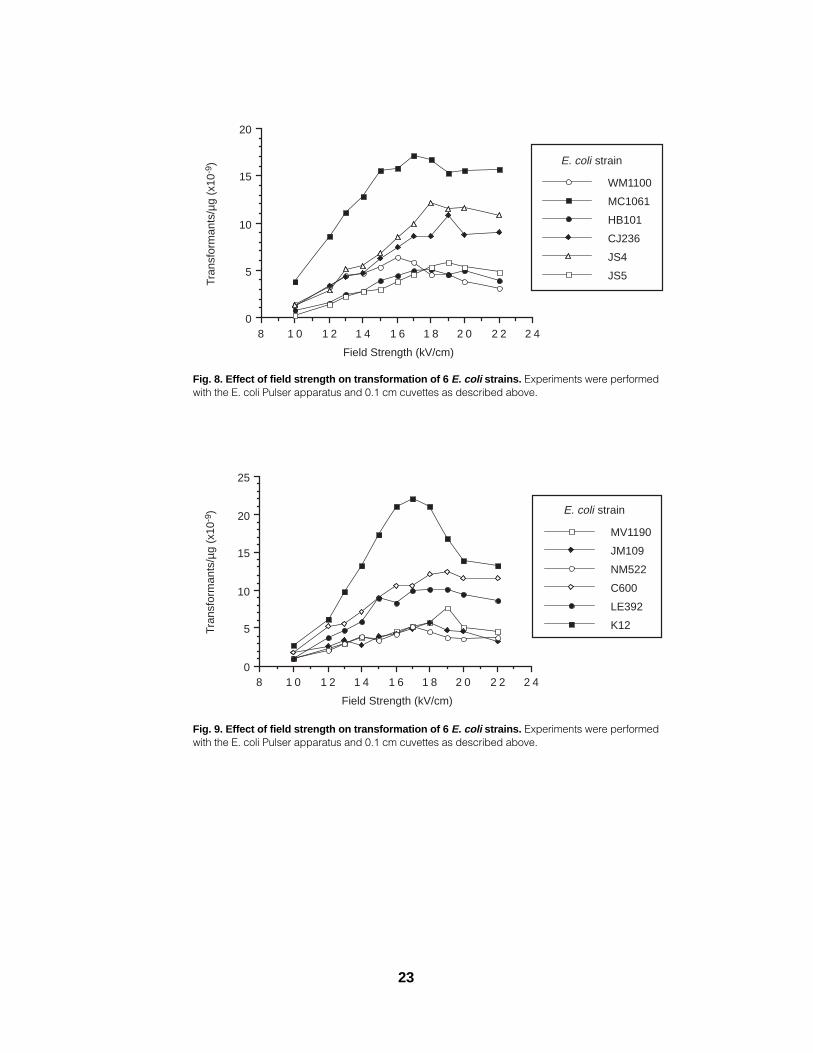
Fig. 8. Effect of field strength on transformation of 6 E. coli strains. Experiments were performedwith the E. coli Pulser apparatus and 0.1 cm cuvettes as described above.
Fig. 9. Effect of field strength on transformation of 6 E. coli strains. Experiments were performedwith the E. coli Pulser apparatus and 0.1 cm cuvettes as described above.
2 42 22 01 81 61 41 21 080
5
10
15
20
25
MV1190
JM109
NM522
C600
LE392
K12
Field Strength (kV/cm)
Tran
sfor
man
ts/µ
g (x
10-9
) E. coli strain
2 42 22 01 81 61 41 21 080
5
10
15
20
WM1100
MC1061
HB101
CJ236
JS4
JS5
Field Strength (kV/cm)
Tran
sfor
man
ts/µ
g (x
10-9
) E. coli strain
23
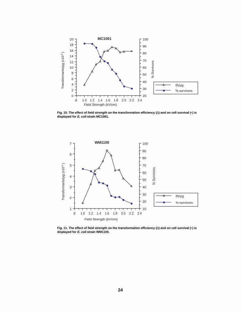
Fig. 10. The effect of field strength on the transformation efficiency ( ∆) and on cell survival ( •) isdisplayed for E. coli strain MC1061.
Fig. 11. The effect of field strength on the transformation efficiency ( ∆) and on cell survival ( •) isdisplayed for E. coli strain WM1100.
2 42 22 01 81 61 41 21 081
2
3
4
5
6
7
10
20
30
40
50
60
70
80
90
100
tfs/µg
% survivors
WM1100
Field Strength (kV/cm)
Tran
sfor
man
ts/µ
g (x
10-9
)
% S
urvi
vors
2 42 22 01 81 61 41 21 080
2
4
6
8
10
12
14
16
18
20
20
30
40
50
60
70
80
90
100
tfs/µg
% survivors
MC1061
Field Strength (kV/cm)
Tran
sfor
man
ts/µ
g (x
10-9
)
% S
urvi
vors
24
Appendix IIITroubleshooting Guide for the E. coli Pulser Apparatus
Operational Problem Likely cause and solution
1. Display does not light Power is not supplied to electronics. Check when unit is turned on. power cord and wall outlet power source.
Check that power switch is on. Check/replace fuse.
2. When the buttons are pressed, No pulse delivery. Pulse buttons are notthe unit does not indicate “Chg”, depressed long enough. Turn power switchor the unit continues to flash “Chg” off and on twice to bleed any residual and the tone does not sound, or charge in the capacitor, or re-press boththe display goes blank. buttons to continue. If problem persists,
contact Bio-Rad.
Only one pulse button is depressed. Both ofthe red pulse buttons must be pressed forpulse delivery until the tone sounds.
ElectricalProblem Likely cause and solution
1. Arcing in the cuvette. Arcing in the cuvette is usually caused by amedium that is too conductive. The limit ofconductivity depends on the voltage, elec-trode gap, and sample volume, but with ourstandard conditions for E. coli, 10 meq or higher will certainly arc.
There are several causes of excessive con-ductivity:1. Washing and resuspending cells in a
buffer too high in ionic strength.2. Insufficient washing of the cells - salts
from the growth medium are not com-pletely removed.
3. Lysed cells in the preparation - cellcontents contribute to conductivity.
4. DNA solution is too high in salt; forexample, CsCl carried over from plas-mid preparation, or residual salts fromethanol precipitation or ligation.
Electroporation with cuvettes above 0 °C.
25

Problem Likely cause and solution
2. Wrong time constant. When using the E. coli Pulser apparatus,the time constant should be close to 5msec. If it is much shorter than the expect-ed value (e.g. 3 msec instead of 5 msec),the sample is too conductive. The probablereasons for this are listed above under “arc-ing”. Correct the problem of high conduc-tivity by additional washing of the cells, orremoval of salts from the DNA prepara-tion.
3. Sample does not “twitch”. This may mean that the pulse is not reach-ing the sample. Check the connectionsbetween the E. coli Pulser apparatus andsample chamber. Check to see that the con-tacts in the base of the sample chamber arenot broken.
4. Instrument displays “no” on front The absence of a twitch does not always panel. mean an error. At voltages below 1.50 kV
the pulse may not be strong enough to cause the sample to twitch. Sometimes the effect is simply difficult to see.
Check connections.
SET VOLTS (KV) not set to 0.20 kV or higher.
BiologicalThe general symptom addressed in this section is transformation efficiencies that are too
low to detect or too low to be useful. The following is a list of the areas of possible problemsand some suggested solutions.
Problem Likely cause and solution
1. The pulse. Is the pulse actually applied to the sample?At high voltage with a small-volume(40 µl) sample this is easy to check. Thesample will “twitch” when pulsed. If youdon’t see a twitch, refer to the electricaltroubleshooting section for information onelectrical problems. Also make sure thatthe cuvette is making contact with the elec-trodes at the back of the sample chamber.Replace electrodes (catalog number 165-2099) if broken or corroded.
Are the amplitude and length of the pulsesufficient? E. coli require pulses of approx-imately 5 milliseconds with field strengthsof 12 to 18 kV/cm. There should (usually)be some cell death with electrical condi-tions producing transformation. Survivalrates of.20 to 80% are to be expected. If no
26
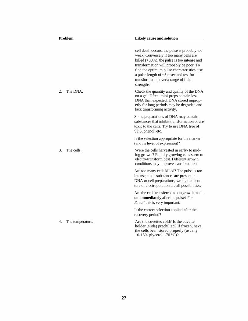
Problem Likely cause and solution
cell death occurs, the pulse is probably tooweak. Conversely if too many cells arekilled (>80%), the pulse is too intense andtransformation will probably be poor. Tofind the optimum pulse characteristics, usea pulse length of ~5 msec and test fortransformation over a range of fieldstrengths.
2. The DNA. Check the quantity and quality of the DNAon a gel. Often, mini-preps contain lessDNA than expected. DNA stored improp-erly for long periods may be degraded andlack transforming activity.
Some preparations of DNA may containsubstances that inhibit transformation or aretoxic to the cells. Try to use DNA free ofSDS, phenol, etc.
Is the selection appropriate for the marker(and its level of expression)?
3. The cells. Were the cells harvested in early- to mid-log growth? Rapidly growing cells seem toelectro-transform best. Different growth conditions may improve transfomation.
Are too many cells killed? The pulse is toointense, toxic substances are present inDNA or cell preparations, wrong tempera-ture of electroporation are all possibilities.
Are the cells transferred to outgrowth medi-um immediately after the pulse? ForE. coli this is very important.
Is the correct selection applied after therecovery period?
4. The temperature. Are the cuvettes cold? Is the cuvette holder (slide) prechilled? If frozen, have the cells been stored properly (usually 10-15% glycerol, -70 °C)?
27

Appendix IVProduct Information
CatalogNumber Product Description
165-2101 E. coli PulserApparatus, 100 V (for use in Japan) includes chamber withpower leads, 6 sterile sample cuvettes (3 0.2 cm gap and 3 0.1 cm gap),cuvette rack
165-2102 E. coli Pulser Apparatus, 120 V (for use in North America and Taiwan)165-2103 E. coli PulserApparatus, 220 V (for use in Europe, Hong Kong, and the
Middle East)165-2104 E. coli Pulser Apparatus, 240 V (for use in Asia, Australia, and the
U.K.)
165-2086 E. coli Pulser/Gene Pulser Cuvettes, 0.2 cm electrode gap, 50, sterile 165-2089 E. coli Pulser/Gene Pulser Cuvettes, 0.1 cm electrode gap, 50, sterile
165-2095 E. coli Pulser/Gene Pulser Cuvette Rack165-2097 E. coli Pulser/Gene Pulser Chamber165-2099 E. coli Pulser/Gene Pulser Chamber Electrode Contacts, 1 pair
170-3105 Electro-Competent E. coli Strain WM1100, includes 0.5 ml E. colicells, 50 µl (10 pg/µl) control plasmid DNA, instructions
170-3106 Electro-Competent E. coli Strain MC1061, 0.5 ml170-3113 Electro-Competent E. coli Strain HB101, 0.5 ml170-3114 Electro-Competent E. coli Strain CJ236, 0.5 ml170-3115 Electro-Competent E. coli Strain MV1190, 0.5 ml170-3116 Electro-Competent E. coli Strain JS4, 0.5 ml170-3117 Electro-Competent E. coli Strain JS5, 0.5 ml
Specifications
Input voltage 100 V RMS, 50/60 Hz120 V RMS, 50/60 Hz220 V RMS, 50/60 Hz240 V RMS, 50/60 Hz
Input current 2 amp RMS
Maximum output 2,500 V peak into ≥3.3 kΩ load limited to 125 amp voltage and current peak max.
Output waveform Decaying exponential waveform with RC time constant of 5 msec, assuming loads of ≥3.3 kΩ
Output voltage adjustment Voltage adjustable in 200-2,500 V range with 10 V resolution, 2 preprogrammed voltage steps
Ambient operating temperature 0 to 35 °C
Dimensions (L x W x H) 29 x 17 x 19 cm
Weight 7.0 kg
28
Life ScienceGroup
2000 Alfred Nobel DriveHercules, California 94547Telephone (510) 741-1000Fax: (510) 741-5800www.bio-rad.com
Australia, Bio-Rad Laboratories Pty. Ltd., Block Y, Unit 1, Regents Park Industrial Estate, 391 Park Road, Regents Park, NSW 2143Phone 02 9914 2800 • Fax 02 9914 2889Austria, Bio-Rad Laboratories Ges.m.b.H., Auhofstraße 78D, A-1130 Wien • Phone (01)-877 89 01 • Fax (01)-876 56 29Belgium, Bio-Rad Laboratories S.A.-N.V., Begoniastraat 5, B-9810 Nazareth • Phone 09-385 55 11 • Free Phone 0800/97032 • Fax 09-385 65 54Brazil, Bio-Rad Laboratories (Brazil), Rua dos Invalidos 212 - 5 andar, Lapa - Rio de Janeiro - RJ, CEP 20331-020 • Phone 55 21 507 6191Canada, Bio-Rad Laboratories (Canada) Ltd., 5671 McAdam Road, Mississauga, Ontario L4Z 1N9 • Phone (905) 712-2771 • Fax (905) 712-2990China, Bio-Rad China (Beijing), Rm 615, Shang Fang Plaza, No. 27, North Third Round Center Road, West District, Beijing 100029 Phone 86-10-8201-1366/68 • Fax 86-10-8201-1367Denmark, Bio-Rad Laboratories, Generatorvej 8 C, 2730 Herlev • Phone 45 44 52-1000 • Fax 45 44 52-1001 Finland, Bio-Rad Laboratories, Pihatörmä 1A, FIN-02240 Espoo • Phone 358 (0)9 804 2200 • Fax 358 (0)9 804 1110France, Bio-Rad Laboratories, 3, Boulevard Raymond Poincaré, 92430 Marnes-la-Coquette • Phone 01 47 95 69 65 • Fax 01 47 41 9133Germany, Bio-Rad Laboratories GmbH, Heidemannstraße 164, D-80939 München, Postfach 45 01 33, D-80901 MünchenPhone 089 318 84-177 • Fax 089 318 84-123Hong Kong, Bio-Rad Pacific Ltd., Unit 1111, 11/F, New Kowloon Plaza, 38 Tai Kok Tsui Road, Tai Kok Tsui, KowloonPhone 852-2789-3300 • Fax 852-2789-1257India, Bio-Rad Laboratories (India) Pvt. Ltd., B&B1, Enkay Towers Vanijyanikunj, Udhyog Vihar Phase V, Gurgaon, Haryana 122016 Phone (91-124)-6398112/113/114 • Fax (91-124)-6398115Israel, Bio-Rad Laboratories, Ltd., 14 Homa Street, P.O. Box 5044, Rishon Le Zion 75150 • Phone 03 951 4124 • Fax 03 951 4129Italy, Bio-Rad Laboratories S.r.l., Via M. Peroglio 23, 00144 Rome • Phone 34 91 590 5200 • Fax 34 91 590 5211Japan, Nippon Bio-Rad Laboratories KK, 7-18 Higashi-Nippori 5-chome, Arakawa-ku Tokyo 116-0014 • Phone 03-5811-6270 • Fax 03-5811-6272Korea, Bio-Rad Korea Ltd., Cambridge Building, 1461-15 Seocho-Dong Seocho-Ku, Seoul 137-070 • Phone 82-2-3473-4460 • Fax 82-2-3472-7003Latin America, Bio-Rad Latin America, 14100 Palmetto Frontage Road, Suite 101, Miami Lakes, Florida USA 33016 • Phone 305-894-5950 • Fax 305-894-5960 Mexico, Bio-Rad Laboratorios Mexico, Adolfo Prieto No. 1653, Col. De Valle, CP. 03100, Mexico D.F. • Phone 52 5 534 2552 to 54 • Fax 52 5 524 5971 The Netherlands, Bio-Rad Laboratories B.V., Fokkerstraat 10, 3905 KV Veenendaal • Phone 0318-540666 • Fax 0318-542216New Zealand, Bio-Rad Laboratories Pty Ltd., PO Box 300-571, Albany, Auckland • Phone 64-9-4152280 • Fax 64-9-443 3097Norway, Bio-Rad Laboratories, Johan Scharffenbergs vei 91, N-0694 Oslo • Phone 47-23-38-41-30 • Fax 47-23-38-41-39Russia, Bio-Rad Laboratorii, ul. Butirskaya 79 "B", office 156 RF-125015 Moscow • Phone 7 095 979 98 00 • Fax 7 095 979 98 56 Singapore, Bio-Rad Laboratories, Singapore, 211 Henderson Rd. #03-02, Henderson Industrial Park, 159552 • Phone 65-2729877 • Fax 65-2734835Spain, Bio-Rad Laboratories, S.A., Lopez de Hoyos, 245-247, 28043 Madrid • Phone 34-91-590-5200 • Fax 34-91-590-5211Sweden, Bio-Rad Laboratories AB, Vintergatan 1, Box 1097, S-172 22 Sundbyberg • Phone 46 (0)8-55 51 27 00 • Fax 46 (0)8-55 51 27 80Switzerland, Bio-Rad Laboratories AG, Nenzlingerweg 2, CH-4153 Reinach • Phone 061-717-9555 • Fax 061-717-9550United Kingdom, Bio-Rad Laboratories Ltd., Bio-Rad House, Maylands Avenue, Hemel Hempstead, Hertfordshire HP2 7TDPhone 0181 328 2000 • Free Phone 0800-181134 • Fax 01442-259118
00-000 0000 Sig 1200Bulletin 0000 US/EG Rev A
Bio-RadLaboratories
M1652101 Rev C