dysphagia begashaw m (md). dysphagia defn difficulty in swallowing classification 1- oropharyngeal...
TRANSCRIPT
DYSPHAGIA
Begashaw M (MD)
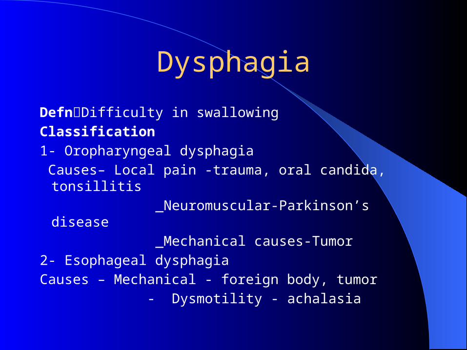
Dysphagia
DefnDifficulty in swallowing
Classification
1- Oropharyngeal dysphagia
Causes– Local pain -trauma, oral candida, tonsillitis
_Neuromuscular-Parkinson’s disease
_Mechanical causes-Tumor
2- Esophageal dysphagia
Causes – Mechanical - foreign body, tumor
- Dysmotility - achalasia
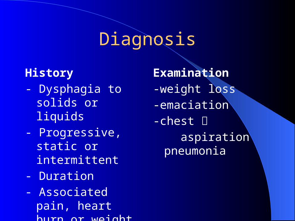
Diagnosis
History
- Dysphagia to solids or liquids
- Progressive, static or intermittent
- Duration
- Associated pain, heart burn or weight loss
Examination
-weight loss
-emaciation
-chest aspiration pneumonia
Investigations
Barium swallow EsophagoscopyEndoscopic ultrasoundManometry
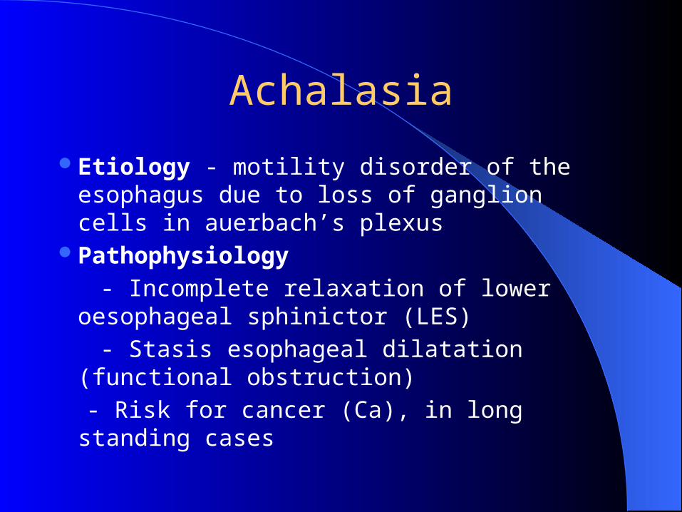
Achalasia
Etiology - motility disorder of the esophagus due to loss of ganglion cells in auerbach’s plexus
Pathophysiology
- Incomplete relaxation of lower oesophageal sphinictor (LES)
- Stasis esophageal dilatation (functional obstruction)
- Risk for cancer (Ca), in long standing cases
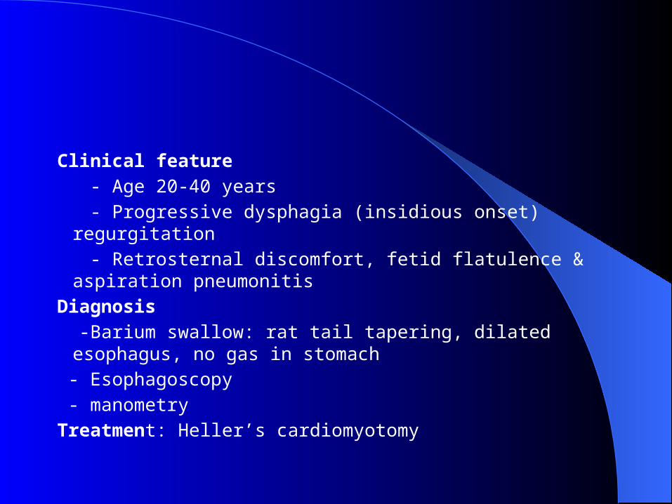
Clinical feature
- Age 20-40 years
- Progressive dysphagia (insidious onset) regurgitation
- Retrosternal discomfort, fetid flatulence & aspiration pneumonitis
Diagnosis
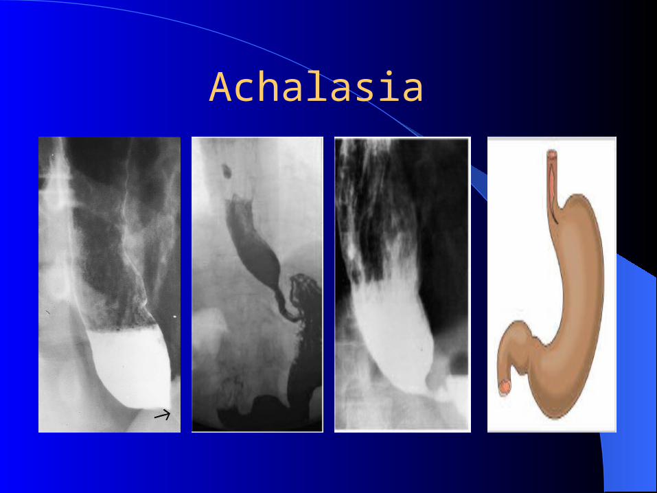
-Barium swallow: rat tail tapering, dilated esophagus, no gas in stomach
- Esophagoscopy
- manometry
Treatment: Heller’s cardiomyotomy
Achalasia
Carcinoma of the esophagus
Epidemiology
> 60 years M > F
5% of all cancers
Predisposing factors
Ingestion of hot meal
Smoking
Alcohol intake
PathologyMicroscopic: squamous cell carcinoma,
Adeno carcinomaMacroscopically: Annular stenosing, ulcer,
fungating, cauli flower like
SpreadDirect, lymphatic and blood stream to liver
and bone
Clinical feature
-Dysphagia, regurgitation, anorexia, weight loss
Diagnosis
- Barium swallow - Irregular, ragged pattern of mucosa with narrow lumen
- Esophagoscopy & biopsy
- Bronchoscopybronchial involvement
- U/S - liver secondaries
- Hgb, plasma proteins, blood chemistry
Treatment
Curative
- surgery
- Radiotherapy
Palliative
- Intubation with specially designed tubes
- Radiotherapy
Foreign bodies
_Coins, pins, dentures..
Diagnosis
- Radiography (neck and chest x-ray)
- Esophagoscopy
Treatment
- Removal by rigid esophagoscope
Oesophagitis
Acute
- burns or scalds
- Infective - candidiasis
- Peptic
Chronic
- reflux due to hiatus hernia or previous surgery
Pathology
- Bleeding granulation tissue replaces epithelium- upward displacement of the cardia
Clinical features
- Pain, heart burn, dysphagia, occult blood, secondary anemia
Diagnosis
- Barium swallow, esophagoscopy
Treatment
- treat the cause, H2 blockers, omeprazole – reflux (peptic)
- surgery for sliding hernia
Caustic strictures
Treatment - acute inflammatory stage
NPO
antibiotics
cortisone- stricture
dilation
esophageal replacement