dual origin of mesenchymal stem cells contributing to
TRANSCRIPT

7/29/2019 Dual Origin of Mesenchymal Stem Cells Contributing To
http://slidepdf.com/reader/full/dual-origin-of-mesenchymal-stem-cells-contributing-to 1/6
Dual origin of mesenchymal stem cells contributing toorgan growth and repairJifan Fenga, Andrea Mantessoa,b, Cosimo De Baric, Akiko Nishiyamad, and Paul T. Sharpea,1
aDepartment of Craniofacial Development and Comprehensive Biomedical Research Centre, Dental Institute, Kings College London, London SE1 9RT, UnitedKingdom; bDepartment of Oral Pathology, Dental Institute, University of Sao Paulo, CEP 05508-900, Sao Paulo, Brazil; cDivision of Applied Medicine, University
of Aberdeen, Aberdeen AB25 22D, United Kingdom; and
d
Department of Physiology and Neurobiology, University of Connecticut, Storrs, CT 06269-4243
Edited by Brigid L. M. Hogan, Duke University Medical Center, Durham, NC, and approved March 18, 2011 (received for review October 15, 2010)
In many adult tissues, mesenchymal stem cells (MSCs) are closely
associated with perivascular niches and coexpress many markersin common with pericytes. The ability of pericytes to act as MSCs,
however, remains controversial. By using genetic lineage tracing,
we show that some pericytes differentiate into specialized tooth
mesenchyme-derived cells—odontoblasts—during tooth growth
and in response to damage in vivo. As the pericyte-derived mes-
enchymal cell contribution to odontoblast differentiation does notaccount for all cell differentiation, we identify an additional source
of cells with MSC-like properties that are stimulated to migrate
toward areas of tissue damage and differentiate into odonto-
blasts. Thus, although pericytes are capable of acting as a source
of MSCs and differentiating into cells of mesenchymal origin, theydo so alongside other MSCs of a nonpericyte origin. This study
identifies a dual origin of MSCs in a single tissue and suggests that
the pericyte contribution to MSC-derived mesenchymal cells in any
given tissue is variable and possibly dependent on the extent of
the vascularity.
dental pulp | tooth development | dental stem cells
The existence of mesenchymal stem cell (MSC) populations inadult tissues and organs that contribute to tissue cell turnover
and also respond to tissue damage is a generally accepted con-cept. Adult stem cell niches potentially offer a source of autol-ogous cells that could be used in a wide variety of stem cell-based
treatments and therapies currently under investigation. How-ever, their precise locations are often unclear and they arepresent in small numbers. The archetypal MSC population re-sides in bone marrow (1–3), but cells with similar molecular,phenotypic, and functional properties, at least in vitro, have beenidentified in a wide range of tissues, including adipose (4), der-mis (5), and teeth (6–9). In addition to their common ability todifferentiate into multiple mesenchymal cell types in vitro, thesecells also share the common property of exhibiting immunesuppressive activity (10, 11). The widespread nature of MSCsin adult tissues has led to suggestions that they have a commonorigin. Paradoxically, however, although they share commonproperties, they do display significant differences, such as theextent to which they form different cell types (12–15). In this
context, recent attention has returned to pericytes/perivascularzones as candidates as a result of their broad distribution in allorgans (16–22). Pericytes are an elusive cell type recognized by their anatomy and position rather than by a precisely definedphenotype. They reside on the abluminal surface of endothelialcells in the microvasculature of every vascularized connective tis-sue and have been postulated as the in situ counterpart of thebone marrow colony-forming units first described in the 1970s (1).
The potential for pericytes to contribute to the formation of tissues in addition to vessels has been established by numerousstudies in several different tissues, including osteoblasts, chondro-blasts, fibroblasts, adipocytes (16, 17), myogenic cells (18), andodontoblasts (19, 20). In addition, perivascular cells from multipleorgans, including skeletal muscle, pancreas, adipose tissue, andplacenta, were recently isolated based on the expression of CD146,
NG2, and PDGF-Rβ, and showed that, after long-term culture,they retained myogenicity and exhibited, at the clonal level, oste-ogenic, chondrogenic, and adipogenic potentials, suggesting thatthe blood vessel walls harbor a reserve of progenitor cells that may be related to MSCs (18, 21, 22).
MSCs are largely defined retrospectively based on in vitroproperties such as the capacity to proliferate extensively, formcolonies, adhere to tissue culture plastic, and differentiate intomature mesenchymal lineages when induced with appropriateculture conditions. When these cells are transplanted into liveanimals, they are also usually subjected to in vitro expansionbefore transplantation. In vitro culture is variable and unable tomimic the stem cell niche, and moreover, somatic cells witha mature phenotype in vivo are able to dedifferentiate (23) ordifferentiate in other cell types in culture (24). Consequently, it isrecognized that some of the MSCs defined operationally fol-lowing in vitro culture may represent experimental artifacts (22).
To date, however, genetic-based linage tracing of pericytedifferentiation has not been reported to definitively show a dif-ferentiated mesenchymal cell that originated from a pericyte. Wechose to address this issue by using a physically and genetically accessible postnatal organ that shows continuous growth andself-repair—the mouse incisor. Rodent incisors continuously sharpen themselves by the shearing action of their tips. As tissueis lost during sharpening, this must be continuously replaced, andstem cells at the cervical end of the tooth have been identified
that provide sources of new specialized cells (25). In common with other teeth, including human teeth, rodent teeth possess alimited repair response to damage of the dentine hard tissue andunderlying specialized mesenchymal-derived dentine-producingcells—odontoblasts—mediated by MSC-like cells residing in thetooth pulp differentiating into new odontoblasts (26). We showthat, during both incisor growth and repair following damage,odontoblasts can be identified that differentiate from pericytes.However, not all odontoblasts are pericyte-derived, and we iden-tify a population of MSC-like cells that exhibit directed cellmigration toward tissue damage and differentiate into odonto-blasts. Although pericytes can differentiate in specialized mes-enchymal cells, they do so alongside other MSC-like cells of nonpericyte origin.
Results and DiscussionLineage Tracing of Pericyte Differentiation During Tooth Growth and
Repair. The NG2 is a proteoglycan that is commonly used as amarker for a gene expressed in pericytes (27). To use Cre-mediated genetic lineage tracing of pericytes, we used two dif-
Author contributions: P.T.S. designed research; J.F. and A.M. performed research; C.D.B.
and A.N. contributed new reagents/analytic tools; A.M. and P.T.S. analyzed data; and P.T.S.
wrote the paper.
The authors declare no conflict of interest.
This article is a PNAS Direct Submission.
1To whom correspondence should be addressed. E-mail: [email protected].
This article contains supporting information online at www.pnas.org/lookup/suppl/doi:10.
1073/pnas.1015449108/-/DCSupplemental.
www.pnas.org/cgi/doi/10.1073/pnas.1015449108 PNAS | April 19, 2011 | vol. 108 | no. 16 | 6503–6508
D E V E L O P M E N T A L
7/29/2019 Dual Origin of Mesenchymal Stem Cells Contributing To
http://slidepdf.com/reader/full/dual-origin-of-mesenchymal-stem-cells-contributing-to 2/6
ferent Cre-expressing mouse lines: a noninductive NG2cre anda tamoxifen-inducible NG2creER crossed with reporter lines(28–31). We first used NG2cre to establish the extent that Cre
was expressed during odontoblast differentiation and then con-firmed that the inducible Cre expression reproduced known sitesof NG2 expression, including pericytes, following tamoxifen ad-ministration (Figs. S1 and S2). We also estimated the frequency of recombination from both Cre drivers by comparing LacZexpression with NG2 protein localization (30, 31) (Fig. S2).
During incisor growth, a small but reproducible number of positive cells were visible in the odontoblast cell layer, with theirprocesses extending into the dentine of NG2creER;Rosa26R(Fig. 1C) and NG2cre;Z/EG mice (Fig. S2). Considering that, inthe 4 d following tamoxifen treatment, only newly formed odon-toblasts could be labeled, whereas in the NG2cre mice, allodontoblasts could have been labeled, we counted the number of LacZ-positive odontoblasts from the first positive cell visible inNG2creER;Rosa26R compared with the equivalent region inNG2cre;Z/EG. In NG2creER mice, 4 d after tamoxifen admin-istration, a maximum of four or fi ve odontoblasts were labeled inany one section, and in NG2cre mice, this figure was double. Thiscorresponds to approximately 3% of odontoblasts labeled inNG2creER and 6% in NG2cre, and thus showed there were twice
as many odontoblasts identifi
able as being derived from pericytesin NG2cre than in NG2creER mice. This figure is comparable with the figure for the frequency of recombination following ta-moxifen (50%), and suggests that, during normal incisor growth,as many as approximately 12% of odontoblasts are formed frompericytes. This pericyte contribution to odontoblast differentia-tion is not observed in noncontinuously growing teeth and is thuslikely to occur as a consequence of the proximity of the largecapillary plexus to the MSC niche in incisors.
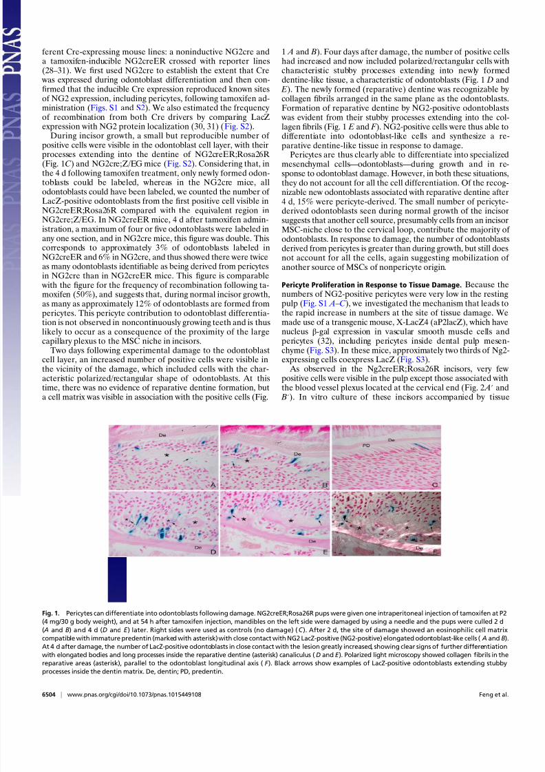
Two days following experimental damage to the odontoblastcell layer, an increased number of positive cells were visible inthe vicinity of the damage, which included cells with the char-acteristic polarized/rectangular shape of odontoblasts. At thistime, there was no evidence of reparative dentine formation, buta cell matrix was visible in association with the positive cells (Fig.
1 A and B). Four days after damage, the number of positive cellshad increased and now included polarized/rectangular cells withcharacteristic stubby processes extending into newly formeddentine-like tissue, a characteristic of odontoblasts (Fig. 1 D and E). The newly formed (reparative) dentine was recognizable by collagen fibrils arranged in the same plane as the odontoblasts.Formation of reparative dentine by NG2-positive odontoblasts
was evident from their stubby processes extending into the col-lagen fibrils (Fig. 1 E and F ). NG2-positive cells were thus able to
differentiate into odontoblast-like cells and synthesize a re-parative dentine-like tissue in response to damage.
Pericytes are thus clearly able to differentiate into specializedmesenchymal cells—odontoblasts—during growth and in re-sponse to odontoblast damage. However, in both these situations,they do not account for all the cell differentiation. Of the recog-nizable new odontoblasts associated with reparative dentine after4 d, 15% were pericyte-derived. The small number of pericyte-derived odontoblasts seen during normal growth of the incisorsuggests that another cell source, presumably cells from an incisorMSC-niche close to the cervical loop, contribute the majority of odontoblasts. In response to damage, the number of odontoblastsderived from pericytes is greater than during growth, but still doesnot account for all the cells, again suggesting mobilization of
another source of MSCs of nonpericyte origin.
Pericyte Proliferation in Response to Tissue Damage. Because thenumbers of NG2-positive pericytes were very low in the restingpulp (Fig. S1 A–C), we investigated the mechanism that leads tothe rapid increase in numbers at the site of tissue damage. Wemade use of a transgenic mouse, X-LacZ4 (aP2lacZ), which havenucleus β-gal expression in vascular smooth muscle cells andpericytes (32), including pericytes inside dental pulp mesen-chyme (Fig. S3). In these mice, approximately two thirds of Ng2-expressing cells coexpress LacZ (Fig. S3).
As observed in the Ng2creER;Rosa26R incisors, very fewpositive cells were visible in the pulp except those associated withthe blood vessel plexus located at the cervical end (Fig. 2 A′ and B′). In vitro culture of these incisors accompanied by tissue
Fig. 1. Pericytes can differentiate into odontoblasts following damage. NG2creER;Rosa26R pups were given one intraperitoneal injection of tamoxifen at P2
(4 mg/30 g body weight), and at 54 h after tamoxifen injection, mandibles on the left side were damaged by using a needle and the pups were culled 2 d
( A and B) and 4 d (D and E ) later. Right sides were used as controls (no damage) (C ). After 2 d, the site of damage showed an eosinophilic cell matrix
compatible with immature predentin (marked with asterisk) with close contact with NG2 LacZ-positive (NG2-positive) elongated odontoblast-like cells ( A and B).
At 4 d after damage, the number of LacZ-positive odontoblasts in close contact with the lesion greatly increased, showing clear signs of further differentiation
with elongated bodies and long processes inside the reparative dentine (asterisk) canaliculus (D and E ). Polarized light microscopy showed collagen fibrils in the
reparative areas (asterisk), parallel to the odontoblast longitudinal axis ( F ). Black arrows show examples of LacZ-positive odontoblasts extending stubby
processes inside the dentin matrix. De, dentin; PD, predentin.
6504 | www.pnas.org/cgi/doi/10.1073/pnas.1015449108 Feng et al.

7/29/2019 Dual Origin of Mesenchymal Stem Cells Contributing To
http://slidepdf.com/reader/full/dual-origin-of-mesenchymal-stem-cells-contributing-to 3/6
damage showed a large increase in pericyte numbers compared with undamaged controls (Fig. 2 A and B). Because these incisors were cultured in vitro, the increase in pericyte numbers could nothave come from any increase in the vasculature that accompaniesan inflammatory response. Phospho-histone H3 (PH3) immu-nostaining showed that LacZ-positive cells close to the site of in
vivo damage were proliferating (Fig. 2C andC′). We thus concludethat damage to the odontoblast cell layer stimulates the prolif-eration of the few isolated pericytes resident in the tooth pulp.
We next investigated the developmental origins of these smallnumbers of isolated pericytes in tooth pulp in X-LacZ4(aP2lacZ) transgenic embryos. Positive cells were first detectedat the late bud stage of tooth development [embryonic day (E)13.5–14], when vessels first appear. Pericytes were seen aroundcapillaries close to the developing teeth and also as a stream withthe appearance of cells migrating from the vessel wall into themesenchymal cells condensing around the epithelial tooth bud(Fig. 2 D). By the cap stage of tooth development (E14.5–15),pericytes could be seen on vessels outside the tooth primordiumand as a few scattered cells in the mesenchymal cells of thedental papilla, the cells that will form the pulp (Fig. 2 E). Peri-cytes thus infiltrate the mesenchymal cell population destined toform the tooth pulp early in development. The approximate
numbers of pericytes seen in sections of the cap stage toothprimordia was consistently greater than the numbers observed insections of adult teeth, suggesting that these pericytes had notincreased significantly in number from those infiltrating themesenchyme during development. To confirm that tooth pulppericytes are slow cycling, nucleoside analogue incorporationassays were performed. Administration of the nucleoside IdU inthe drinking water of adult X-LacZ4 males for 4 wk followed by a chase for 20 d was expected to label only the slowest-cyclingcells. C57BL6 E10.5 or E14.5 first branchial arch primordia weretransplanted under kidney capsules of X-LacZ4 males. After 2 or3 wk, the kidneys were removed, the teeth dissected, and im-munofluorescence for IdU and LacZ staining used to identify correspondingly slow-cycling cells and pericytes. Sections of tooth pulp identified IdU -positive/LacZ -positive cells that wereslow-cycling pericytes that had migrated into the pulp via thehost vasculature (Fig. 2 G– I ).
A small number of pericytes not associated with vessels pop-ulate the tooth mesenchyme during development, where they remain essentially quiescent until damage to the odontoblast celllayer occurs, as a result of extensive caries, for example. Odon-toblast damage stimulates the proliferation of pericytes that arethen able to contribute to the formation of new odontoblasts.
Fig. 2. Pericytes can respond to injury locally, are involved
in tooth development, and are slow-cycling cells. By cul-
turing the X-LacZ4 incisors following damage, and thus
eliminating the circulatory system, an increase in the
number of LacZ-positive pericytes was evident in both pulp
body ( A) and cervical loop parts (B), compared with the
control incisors ( A′ and B′). These cells were characteristi-
cally dispersed throughout the pulp tissue, losing their or-
ganization within the main body plexus in the pulp body ( A
and A′), and were intensely concentrated on the bottom
edge of the cervical loop area (B and B′), where a vessel-rich
area is present. Coexpression of proliferation marker PH3
and LacZ-positive (NG2-positive) pericytes following in vivo
damage (C and C ′
) showed that proliferating pericytes(black arrows) are located on blood vessels extending to-
ward the damaged area (C ′). During tooth development
(bud to bell stages) in X-LacZ4 mice, LacZ-positive cells are
found under the condensing mesenchyme (D) and in the
blood vessels close to the forming tooth ( E ). When colo-
cation of migrating slow-cycling cells and pericytes were
analyzed by transplantation experiments associated with
nucleoside (i.e., IdU) administration, some cells that mi-
grated from the X-LacZ4 host were both LacZ-positive
(pericytes, black stained cells) and IdU positive (slow-cy-
cling, red stained cells) (G and H ). These cells were found in
the blood vessel-rich cervical loop area of incisors (F and G).
Consecutive sections showed that these cells were rare but
clustered (G), suggesting a spatial organization. High
magnification showed colocalization of IdU and LacZ-posi-
tive pericytes under fluorescence (H ) and bright field (I ).
Feng et al. PNAS | April 19, 2011 | vol. 108 | no. 16 | 6505
D E V E L O P M E N T A L

7/29/2019 Dual Origin of Mesenchymal Stem Cells Contributing To
http://slidepdf.com/reader/full/dual-origin-of-mesenchymal-stem-cells-contributing-to 4/6
Mesenchymal Stem Cell Niche Close to Cervical Loop Responds to
Odontoblast Damage. Because, during natural incisor growth andin damage repair, only a small percentage of newly differenti-ating odontoblasts were derived from pericytes, there must beanother source of MSCs in the incisor pulp. The location of theMSC niche in mouse incisors has not been described, but it isassumed to be located close to the cervical end, where odonto-blast differentiation begins. We therefore used 1,1′-dioctadecyl-3,3,3′,3′-tetramethylindocarbocyanine perchlorate (DiI) labeling
of mesenchymal cells in different regions of the incisor pulp toidentify any responses to tooth damage. Labeling of mesenchy-mal cells approximately 400 μm from the cervical opening of theincisor, followed by damage more distally, revealed directedmigration of cells toward the damage after 2 d (Fig. 3 A–C). Inthe absence of damage, no directed cell migration was observedand cells labeled in other areas showed no migration toward thearea of damage (Fig. 3 D– I ). This identifies a discrete population of mesenchymal cells at the cervical end of the incisor that undergodirected cell migration (cell homing) following tooth damage.
If this cell homing is part of the natural repair process, wereasoned that these cells should differentiate into odontoblasts.
As the incisors did not survive in in vitro culture long enough forus to detect new odontoblasts from their morphology, we usedexpression of the dentinsialophosphoprotein ( Dspp) gene todetect early odontoblast differentiation (33, 34). An incisor wasdamaged in three different places in vivo: directly into the areaclose to the cervical loop, midway along its length of the tooth,and close to the distal tip and cultured for 4 d (Fig. 4 A). In situhybridization for Dspp expression showed a massive induction incells at the cervical loop end (Fig. 4 B and B′). Midway along thetooth, a few individual Dspp-expressing cells were visible (Fig.4C), whereas at the tip, there were no positive cells (Fig. 4 D).Thus, odontoblast differentiation is stimulated by damage, andthe extent of differentiation at a given period after the damage
correlates with the distance from the area from which cells mi-grate. The contribution of cells differentiating into new odon-toblasts following damage is thus from at least two differentsources: pericytes resident as quiescent cells in the pulp whoseproliferation is stimulated by the damage, and migration of cellsfrom the MSC niche area of the tooth that normally provide acontinuous cell supply to support tooth growth. These representtwo distinct cell populations that contribute to the formation of most of the odontoblasts in growth and repair.
Concluding Remarks. The extent to which perivascular cells act asMSCs during tissue growth and repair has long been a conten-tious issue. Although a somewhat circular argument, the fact thatmany MSCs in vivo and in vitro express genes also expressed by pericytes is often used to claim a pericyte origin. Similarly, iso-lation of perivascular cells from tissues, based on expression of these markers and retention of MSC-like properties followed by long-term culture, has provided compelling evidence that peri-cytes can act as a generic source of MSCs (18, 35). What thecurrent evidence has lacked is a demonstration of genetically marked pericytes differentiating into mesenchymal cells in vivo.Our results provide this evidence, at least for a tissue duringgrowth and repair, but show that pericytes are not the only celllineage that acts as a source of MSCs.
We suggest that the relative contribution of pericyte-derivedand non–pericyte-derived MSCs to cell differentiation in any given tissue depends on the extent of the vascularity and thekinetics of growth and/or repair. This can explain the conflictingdata on the pericyte contribution to MSC-derived cells in dif-ferent tissues. Thus, in tissues with low vascularity, such as ar-ticular cartilage, the pericyte contribution to MSCs will be lessthan in tissues with more extensive blood supplies.
An obvious question this raises is why are there two mecha-nisms (cell sources) for mesenchymal tissue repair. One hy-
Fig. 3. Directed cell migration toward tissue damage visualized by DiI labeling of incisor mesenchyme pulp cells. Cells labelled in cervical loop area ( A–C ),
damaged pulp cells area ( A–C ), damaged pulp body (D–F ), and control teeth with no damage (G–I ). Arrows indicate position of tooth damage. A lesion
separating the cervical loop and the pulp body was provoked in incisors, and, 48 h later, mesenchymal cells were capable of migrating toward the site of the
damage ( A–C ) whereas the mesenchymal cells from the pulp body showed no migration ( D–F ). In the absence of the lesion, no directed migration of cervical
loop mesenchymal cells was observed (G–I ).
6506 | www.pnas.org/cgi/doi/10.1073/pnas.1015449108 Feng et al.

7/29/2019 Dual Origin of Mesenchymal Stem Cells Contributing To
http://slidepdf.com/reader/full/dual-origin-of-mesenchymal-stem-cells-contributing-to 5/6
pothesis is that this is an evolutionary adaptation to facilitaterapid tissue repair whereby stem cells can quickly accumulate ata site of damage via the inflammatory response (36).
Materials and MethodsMouse Lines. The transgenic X-LacZ4 mouse line contains a single copy of the
transgeneintegratedinto the hostgenome.At latedevelopmentalstagesand
in the adult, LacZ staining marksvascular smooth muscle cells throughout the
vascular bed, with the exception of the major elastic arteries, and in pericytes
(32). The NG2cre mouse line consists of NG2cre BAC transgenic mice ex-
pressing Cre under the control of the NG2 promoter [Cspg4 (ng2) gene] (30).
Inducible NG2CreERBAC transgenic mice were generated by using the same
approach except that NG2cre cDNA was replaced by NG2creER cDNA (28).
NG2cre and NG2creER were mated with Z/EG (29) and Rosa26R reporter mice
(31), respectively, to detect cre-mediated recombination. Postnatal day 2 (P2)
pups were given a single intraperitonial injection (4 mg/30 g body weight)
of tamoxifen (T5648; Sigma) dissolved in corn oil (C-8247; Sigma). fifty-four
hours after the tamoxifen injection, the mandibles at the left side were
damaged using an 18-gauge needle and the pups were culled 2 and 4 d fol-
lowing damage, respectively. Right sides were used as controls. NG2cre;Z/EG
pups ofsimilar age(P1)wereculled forcomparative analysis ofoverall NG2cre
activity during tooth growth. The number of LacZ-positive odontoblasts in
the injured area were counted on a total of 12 sections from three incisors.
LacZ Expression. Samples were fixed in 1% paraformaldehyde (PFA), 0.2%
glutaraldehyde,in PBSsolution fora time dependingon thesizeof thesample
followed by 48 h of staining at 37 °C for detection of β-gal activity using
a solution composed of: 10 mM Tris-HCL pH 7.3, 0.005% Na-dexoxycholate,
0.01% IGEPAL (Sigma), 5 mM K4Fe (CN)6, 5 mM K3Fe(CN)6, 2 mM MgCl2, and
0.8 mg/mL X-Gal. As positive and negative controls, ROSA26 and CD1 mouse
lines were used, respectively.
Recombination Efficiency. The number of LacZ-positive odontoblastsin NG2cre;
Z/EG and NG2creER;Rosa26R incisors were counted in sagittal sections. As
NG2cre is active for a longer period than NG2creER, 4 d after tamoxifen ad-
ministration, we compared the number of LacZ-positive cells per an equivalent
number of odontoblasts (n = 50) in a region proximal to the last forming la-
beled cell. Cells were counted on a total of 14 sections from three incisors.
Response of Pericytes Following Incisor Damage in Culture. Lower incisors at p2
from theX-LacZ4mouselinewereculturedfor 5 d. Theincisors were removed
from the jaws and dissected, and the outer epithelium and developing
enamel were physically removed. The cervical loop and the pulp body wereseparated from each other using a blade to cause pulp and odontoblast
damage. The separated tooth parts were cultured on Millipore 0.10-μm filters
exposed to oxygen. Medium (DMEM 10%, FBS 1%, penicillin/streptomycin
solution) was changed every day. After 5 d, culture samples were photo-
graphed, fixed, stained for detection of β-gal activity, and embedded for
histological analysis.
Immunohistochemistry. Immunohistochemistry was performed on 5- to 7-μm
paraffin sections of samples with or without LacZ staining. Heat-based an-
tigen retrieval was performed before primary antibody incubation. The
primary antibodies anti-NG2 chondroitin sulfate proteoglycan (AB5320;
Millipore), anti–phospho-histone H3 (Ser10) (06-530; Millipore), α-smooth
muscle actin (α-SMA; ab5694; Abcam), and GFP (ab6556, Abcam) were used
at 1:50, 1:100, 1:500 and 1:500 dilution, respectively. Vectastain Elite ABC Kit
(rabbit IgG) (PK-6101; Vector Labs) and DAB Peroxidase Substrate Kit (SK-
Fig. 4. Differentiation into Dspp-positive preodontoblasts
following damage. Following damage to three separate
sites, Dspp expression was visualized by in situ hybridiza-
tion to reveal early odontoblast differentiation. Damage
directly to the cervical loop area showed extensive expres-
sion of Dspp after 2 d (B and B′), whereas only a few
expressing cells were visible in the damaged region midway
along the length of the tooth (C ). At the tip of the tooth,
no Dspp expression was evident (D). Note that evidence of
an inflammatory response can be seen in the tip (G), body
(F ), and cervical loop cells (E ), but presence of osteodentin
is evident in only the cervical loop (E ′).
Feng et al. PNAS | April 19, 2011 | vol. 108 | no. 16 | 6507
D E V E L O P M E N T A L

7/29/2019 Dual Origin of Mesenchymal Stem Cells Contributing To
http://slidepdf.com/reader/full/dual-origin-of-mesenchymal-stem-cells-contributing-to 6/6
4100; Vector Labs) were then used according to manufacturers ’ instructions.
Hematoxylin counterstain (03971; Fluka) was performed only on non–LacZ-
stained samples to avoid masking the LacZ signal.
Colocation of Migrating Pericytes and Slow-Cycling Cells. Seven 6-wk-old X-
LacZ4males received the nucleoside iododeoxyuridine (IdU) in drinkingwater
(1 mg/mL) for 4 wk. After a washout period of 20 d, C57BL6 mandible pri-
mordia at different ages (E10.5–E14.5) were transplanted under kidney
capsules of adult male mice. After 2 or 3 wk (depending on the age of the
transplanted mandible), the tissue was removed from the kidneys, the
mandibles dissected, and IdU immunofluorescence for IdU and LacZ stainingused to identify slow-cycling cells and pericytes, respectively. Both slow-
cycling cells and pericytes would always be originally from the host, and cells
coexpressing both markers would be slow-cycling-migrating pericytes. For
IdU staining, primary antibody was IdU (1:10, ab8152; Abcam) and secondary
antibody was anti–mouse-cy3 (1:300, 87173; Jackson ImmunoResearch Lab-
oratories), tissue was fixed in 4% PFA, decalcified (10% Na citrate, 10%
formic acid, and 1% PFA in PBS solution), and paraffin-embedded. Consec-
utive sections of whole teeth were analyzed.
Migration Capacity Following Incisor Damage in Culture. Lower incisors at p2
from CD1micewerecultured for2 d.The incisors were removed from thejaws
and dissected, and the outer epithelium and developing enamel physically
removed. The cervical loop and the pulp body were separated from each
other by using a blade. Incisor bodies and cervical loops were labeled with
1 mg/mL of CM-DiI (C-7000; Molecular Probes). The separated tooth parts
were cultured on 0.4-μm cell culture membranes (35–3090; Becton Dickinson)
exposed to oxygen. Medium (DMEM 10%, FBS 1%, penicillin/streptomycin
Fungizone solution) was changed every day, and samples were photo-
graphed and after 2 d.
Mobilization Capacity After Injury. Lower incisors of CD1 mice at P5 weredamaged with an 18-gauge needle that was pressed against the tissue,
resulting in a pierced area and/or tissue dislodgement. After 2 d, the animals
were euthanized, and jaws were dissected,fixed, and analyzed histologically.
Sequential slides were submitted to Dspp digoxigenin-labeled in situ hy-
bridization on sections as previously described (37).
ACKNOWLEDGMENTS. We thank Dirk Dietrich (University of Bonn) forNG2creER mice and Moshe Shani (Volcani Center, Israel) for permission touse the X-LacZ4 mice. We thank Alex Huhn for his invaluable technicalassistance. This work was supported by MRC Grant G0600041 (to P.T.S.). C.D.B.is a Medical Research Council Fellow (G108/620).
1. Friedenstein AJ, Chailakhyan RK, Latsinik NV, Panasyuk AF, Keiliss-Borok IV (1974)
Stromal cells responsible for transferring the microenvironment of the hemopoietic
tissues. Cloning in vitro and retransplantation in vivo. Transplantation 17:331–340.
2. Caplan AI (1991) Mesenchymal stem cells. J Orthop Res 9:641–650.
3. Pittenger MF, et al. (1999) Multilineage potential of adult human mesenchymal stem
cells. Science 284:143–147.
4. Zuk PA, et al. (2002) Human adipose tissue is a source of multipotent stem cells. Mol
Biol Cell 13:4279–4295.
5. Toma JG, et al. (2001) Isolation of multipotent adult stem cells from the dermis of
mammalian skin. Nat Cell Biol 3:778–784.
6. Gronthos S, Mankani M, Brahim J, Robey PG, Shi S (2000) Postnatal human dental
pulp stem cells (DPSCs) in vitro and in vivo. Proc Natl Acad Sci USA 97:13625–13630.
7. Gronthos S, et al. (2002) Stem cell properties of human dental pulp stem cells. J Dent
Res 81:531–535.
8. Miura M, et al. (2003) SHED: Stem cells from human exfoliated deciduous teeth. Proc
Natl Acad Sci USA 100:5807–5812.
9. Sonoyama W, et al. (2008) Characterization of the apical papilla and its residing stem
cells from human immature permanent teeth: A pilot study. J Endod 34:166–171.
10. Glennie S, Soeiro I, Dyson PJ, Lam EW, Dazzi F (2005) Bone marrow mesenchymal stem
cells induce division arrest anergy of activated T cells. Blood 105:2821–2827.
11. Spaggiari GM, Capobianco A, Becchetti S, Mingari MC, Moretta L (2006) Mesenchymal
stem cell-natural killer cell interactions: Evidence that activated NK cells are capable
of killing MSCs, whereas MSCs can inhibit IL-2-induced NK-cell proliferation. Blood
107:1484–1490.
12. Kern S, Eichler H, Stoeve J, Klüter H, Bieback K (2006) Comparative analysis of
mesenchymal stem cells from bone marrow, umbilical cord blood, or adipose tissue.
Stem Cells 24:1294–1301.
13. Covas DT, et al. (2008) Multipotent mesenchymal stromal cells obtained from diverse
human tissues share functional properties and gene-expression profile with CD146+
perivascular cells and fibroblasts. Exp Hematol 36:642–654.
14. Huang GT, Gronthos S, Shi S (2009) Mesenchymal stem cells derived from dental
tissues vs. those from other sources: Their biology and role in regenerative medicine.
J Dent Res 88:792–806.
15. Hegyi B, et al. (2010) Identical, similar or different? Learning about immunomodulatory
function of mesenchymal stem cells isolated from various mouse tissues: Bone marrow,
spleen, thymus and aorta wall. Int Immunol 22:551–559.
16. Nehls V, Drenckhahn D (1993) The versatility of microvascular pericytes: From
mesenchyme to smooth muscle? Histochemistry 99:1–12.
17. Lin G, et al. (2008) Defining stem and progenitor cells within adipose tissue. Stem Cells
Dev 17:1053–1063.
18. Crisan M, et al. (2008) A perivascular origin for mesenchymal stem cells in multiple
human organs. Cell Stem Cell 3:301–313.
19. Alliot-Licht B, Hurtrel D, Gregoire M (2001) Characterization of alpha-smooth muscle
actin positive cells in mineralized human dental pulp cultures. Arch Oral Biol 46:
221–228.
20. Shi S, Gronthos S (2003) Perivascular niche of postnatal mesenchymal stem cells inhuman bone marrow and dental pulp. J Bone Miner Res 18:696–704.
21. Satokata I, et al. (2000) Msx2 deficiency in mice causes pleiotropic defects in bone
growth and ectodermal organ formation. Nat Genet 24:391–395.
22. da Silva Meirelles L, Caplan AI, Nardi NB (2008) In search of the in vivo identity of
mesenchymal stem cells. Stem Cells 26:2287–2299.
23. Diaz-Romero J, et al. (2005) Immunophenotypic analysis of human articular chondrocytes:
Changes in surface markers associated with cell expansion in monolayer culture. J Cell
Physiol 202:731–742.
24. Barbero A, Ploegert S, Heberer M, Martin I (2003) Plasticity of clonal populations of
dedifferentiated adult human articular chondrocytes. Arthritis Rheum 48:1315–1325.
25. Harada H, et al. (1999) Localization of putative stem cells in dental epithelium and
their association with Notch and FGF signaling. J Cell Biol 147:105–120.
26. Smith AJ, et al. (1995) Reactionary dentinogenesis. Int J Dev Biol 39:273–280.
27. Ozerdem U, Grako KA, Dahlin-Huppe K, Monosov E, Stallcup WB (2001) NG2
proteoglycan is expressed exclusively by mural cells during vascular morphogenesis.
Dev Dyn 222:218–227.
28. Zhu X, et al. (2011) Age-dependent fate and lineage restriction of single NG2 cells.
Development 138:745–
753.29. Soriano P (1999) Generalized lacZ expression with the ROSA26 Cre reporter strain. Nat
Genet 21:70–71.
30. Zhu X, Bergles DE, Nishiyama A (2008) NG2 cells generate both oligodendrocytes and
gray matter astrocytes. Development 135:145–157.
31. Novak A, Guo CY, Yang WY, Nagy A, Lobe CG (2000) Z/EG, a double reporter mouse
line that expresses enhanced green fluorescent protein upon Cre-mediated excision.
Genesis 28:147–155.
32. Tidhar A, et al. (2001) A novel transgenic marker for migrating limb muscle precursors
and for vascular smooth muscle cells. Dev Dyn 220:60–73.
33. Feng JQ, et al. (1998) Genomic organization, chromosomal mapping, and promoter
analysis of the mouse dentin sialophosphoprotein (Dspp) gene, which codes for both
dentin sialoprotein and dentin phosphoprotein. J Biol Chem 273:9457–9464.
34. Sreenath TL, Cho A, MacDougall M, Kulkarni AB (1999) Spatial and temporal activity
of the dentin sialophosphoprotein gene promoter: Differential regulation in
odontoblasts and ameloblasts. Int J Dev Biol 43:509–516.
35. Dellavalle A, et al. (2007) Pericytes of human skeletal muscle are myogenic precursors
distinct from satellite cells. Nat Cell Biol 9:255–267.
36. Kurth TB, et al. (2011) Functional mesenchymal stem cell niches in the adult knee joint
synovium in vivo. Arthritis Rheum, in press.
37. Nakatomi M, Morita I, Eto K, Ota MS (2006) Sonic hedgehog signaling is important in
tooth root development. J Dent Res 85:427–431.
6508 | www.pnas.org/cgi/doi/10.1073/pnas.1015449108 Feng et al.